journa l homepage: www.e lsev ier .com/ locate /bbr
esearch report
ehavioral profile and dorsal hippocampal cells in carioca high-conditionedreezing rats
isele Pereira Dias a,∗, Mário Cesar do Nascimento Bevilaqua a, Anna Claudia Domingos Silveira a,esus Landeira-Fernandez b,c,1,2, Patrícia Franca Gardino a
Programa de Neurobiologia – Instituto de Biofísica, Universidade Federal do Rio de Janeiro (UFRJ), Laboratório de Neurobiologia da Retina, BrazilLaboratório de Neurociência Comportamental (LANEC), Departamento de Psicologia, Pontifícia Universidade Católica do Rio de Janeiro (PUC-Rio),ua Marquês de São Vicente 225, Edifício Cardeal Leme, sala 201, Gávea, Rio de Janeiro, RJ, CEP: 22453-900, BrazilLaboratório de Psicologia Comparativa, Curso de Psicologia, Universidade Estácio de Sá (UNESA), Rua do Bispo, 83, Rio Comprido, CEP:20261-063, Brazil
r t i c l e i n f o
rticle history:eceived 21 May 2009eceived in revised form 25 June 2009ccepted 28 June 2009
eywords:onditioned fearnxietynimal model
a b s t r a c t
Selection for contextual fear conditioning is an important behavioral paradigm for studying the role ofgenetic variables and their interaction with the surrounding environment in the etiology and developmentof anxiety disorders. Recently, a new line of animals selectively bred for high levels of freezing in responseto contextual cues previously associated with footshock was developed from a Wistar population. Thepurpose of the present study was to evaluate the emotional and cognitive aspects of this new line ofanimals, which has been named Carioca High-Freezing (CHF). For the characterization of anxious behavior,CHF and control animals were tested in the elevated plus-maze (EPM) and the social interaction test. CHFanimals were significantly more anxious than control rats in terms of both the number of entries intoEPM open arms and the percentage of time spent in these arms. The time spent in social interactionbehavior was also significantly decreased. No statistical differences were found in locomotor activity, asmeasured by both the number of entries into the closed arms of the EPM and the number of crossingsinto the social interaction test arena. No differences between CHF and control groups were found inthe depression forced swimming test, suggesting that the anxiety trait selected in the CHF line did not
interact with affective disorders traits such as those for depression. Cognitive aspects of the CHF ratswere evaluated in the object recognition task. Results from this test indicated no difference between thetwo groups. The present study also encompassed histological analysis of the dorsal hippocampus fromCHF and control animals. Results revealed an absence of qualitative and quantitative differences betweenthese two groups of animals in cells located in the dentate gyrus, CA1, and CA3 areas. Therefore, future
rtheus ph
studies are required to fudevelopment of the anxio
. Introduction
Fear and anxiety traits are believed to have been selected inuman evolutionary history for their crucial role in protecting our
Please cite this article in press as: Dias GP, et al. Behavioral profile and dorsBrain Res (2009), doi:10.1016/j.bbr.2009.06.038
unter-gatherer ancestors when facing adverse environments [1].ndeed, appropriate anxious reaction has an adaptive role in dealing
ith threatening situations. However, chronic anxious responses,specially in the absence of the feared stimuli, can characterize
∗ Corresponding author at: Centro de Ciências da Saúde – CCS – Instituto deiofísica Carlos Chagas Filho, Av. Carlos Chagas Filho, 373, Cidade Universitária –EP: 21941-902, Universidade Federal do Rio de Janeiro, Brazil.el.: +55 21 2562 6594; fax: +55 21 2562 6594.
dysfunctional or pathological processes. For this reason, studyingdifferent paradigms of fear and anxiety is crucial for future clinicalinterventions. It is worth noting that stronger acquisition of fearlearning has been observed in anxious patients related to controlsubjects [2]. In this sense, fear learning has been addressed to asan appropriate framework for investigating emotional regulationconcerning fear and anxiety [3].
Freezing response to contextual cues previously associated withfootshock seems to be one of the most reliable animal models ofanxiety disorders [4,5]. Specific and complex circuits in the brainare known to underlie this conditioned response. The hippocam-pus is considered to be one of the major brain structures involved
al hippocampal cells in carioca high-conditioned freezing rats. Behav
in the mediation of learned fear responses, likely via descendingprojections to the amygdala [6], as electrolytic lesions in hippocam-pal regions that connect to the amygdala prevent contextual fearlearning [7,8]. The hippocampus is believed to be responsible forgathering contextual stimuli into representational units and then
ending this information to the amygdala. Efferent projections fromhe central nucleus of the amygdala to the brain stem seem toe responsible for the motor output of the conditioned freezingesponse [9]. In particular, the dorsal portion of the hippocampusas been found to be involved in the modulation of this response10], although the ventral hippocampus is also implicated, withnxiogenic effects when stimulated by serotonergic agonists [11].
Important molecular and pharmacological aspects of mentalisorders are difficult to investigate in humans. Moreover, thetudy of fear and anxiety in animals from normalized populationsight not always mimic the pathophysiology of clinical conditions,
ut natural and adaptive behavioral and physiological reactions torugs and aversive events [12]. For this reason, animals selectivelyred for high emotionality have been considered to be importantools for understanding the neurobiology of anxiety disorders. Dif-erent behavioral paradigms have been employed for this purpose.mong these paradigms are the ambulation and defecation in thepen field, such as in the Maudsley reactive rats [13,14], open armntrance in the elevated plus-maze, as in the high-anxiety relatedehavior rats [12], and active avoidance behavior, as in the high-voidance rats referred to as Roman [15], Syrakuse [16], Koltushi17], and Hatano [18].
Recently, Gomes and Landeira-Fernandez [19] developed twoew lines of Wistar rats, termed Carioca High- and Low-FreezingCHF, CLF), that were selectively bred for high and low levels ofreezing in response to contextual cues previously associated withootshock. After three generations of breeding, CHF rats are con-idered to naturally have a greater propensity for exhibiting higherreezing responses when compared to the low-freezing line. Sincehe characterization of this animal model may be an important toolor investigating the role of genetic variables and their interaction
ith the surrounding environment in the etiology and develop-ent of anxiety disorders, the major objective of this study was
o validate behaviorally the CHF line (Carioca High-Freezing) in annnate animal model of anxiety (i.e., the elevated plus-maze and theocial interaction test). The forced swimming test and the objectecognition task were also employed in order to evaluate whetherraits from other emotional or cognitive systems, such as depressionr memory, were co-selected during the CHF breeding procedure.inally, the present study also investigated whether CHF and con-rol animals presented qualitative and/or quantitative differencesn cells located in the dorsal hippocampus.
. Materials and methods
.1. Animals
Experimental procedures reported herein were performed under the guidelinesor the use of animal experimental research established by the Brazilian Society ofeuroscience and Behavior (SBNeC), in accordance with the National Institute ofealth Guide for the Care and Use of Laboratory Animals (NIH Publications). Animalandling and sacrifice methods were reviewed and approved by the Committee fornimal Care and Use of the CCS/UFRJ (protocol # IBCCF002). Experimental animals
Carioca High-Freezing [CHF], a line selectively bred for high contextual fear con-itioning) were obtained according to procedures described in previous work [19].riefly, albino Wistar rats were selectively bred for differences in defensive freezingehavior in a contextual fear conditioning paradigm. Significant differences in freez-
ng response were acquired after three generations of selective breeding (S3). In theresent work, CHF rats from the fourth generation (S4) were used in the elevatedlus-maze, social interaction, and forced swimming tests, as well as in histologicalxperiments. CHF rats from the sixth generation of selective breeding (S6) were usedn the object recognition test. Control animals corresponded to albino Wistar ratsrom populations that did not undergo any pretreatment or other experimental con-itions. Males from both groups were born in the same animal room and housed incrylic cages (31 cm × 38 cm) in groups of 3–6 in an animal room under a 12 h light-
Please cite this article in press as: Dias GP, et al. Behavioral profile and dorsBrain Res (2009), doi:10.1016/j.bbr.2009.06.038
ark cycle (lights on at 8:00 h), and food and water were provided ad libitum. Foroth groups, 2–3-month-old animals were used. Body weight varied from 250–394 gcontrol) and 254–379 g (CHF). Both the control (CTRL) and the experimental groupsere reared under the same environmental conditions. Experimental animals used
n this work did not undergo a line selecting test (contextual fear conditioning) andid not go through any other stressful events.
PRESSesearch xxx (2009) xxx–xxx
2.2. Behavioral tests
Animals were tested in the elevated plus-maze (EPM) and social interaction testfor anxious behavior screening, in the forced swimming test for depressive behavioridentification, and in the object recognition test for cognitive performance assess-ment. Animals that were tested in the EPM were later perfused for histologicalanalysis; those placed in the social interaction test were also tested in the forcedswimming apparatus, with a minimum of 2 weeks interval between tests; animalsused for object recognition measures did not go through any other tests. All behav-ioral experiments were conducted during the light phase of the dark–light cycle.Animals were brought to the testing room in their home cages and allowed to adaptfor 30 min before testing.
2.2.1. Elevated plus-mazeTwelve control rats and 16 CHF rats were tested in the elevated plus-maze test.
The test was first developed by Handley and Mithani [20] and was validated as amodel for anxious-related behavior by Pellow et al. [21]. The test is based on thenatural conflict faced by rodents when exploring a new environment and the innateaversion of being exposed to open areas [22]. The apparatus consists of two woodenopposed closed arms, two opposed open arms surrounded by an acryclic protectionin order to prevent animals from falling down, and an open square in the center.The maze was elevated 50 cm above the floor. All animals were handled for 2 minon 5 consecutive days prior to the experimental session. The same experimenterwas responsible for both handling and placing the animals into the maze in orderto reduce human contact bias. The apparatus was cleaned with ethanol 98% beforeeach rat was placed within in. The animals were placed into the center of the plus-maze facing one of the closed arms. The experimental session (5 min) was recordedby a camera located 70 cm above the maze, and the following measures were lateranalyzed: the number of entries into closed arms, and both the number of entriesand the percentage of time spent in open arms.
2.2.2. Social interactionThe social interaction protocol was a modified version of that presented by Hen-
niger et al. [22]. The test arena was made of black PVC (54 × 36 × 27 cm) and the floorwas divided into six squares (18 × 18 cm). All animals (n = 12; 6 pairs for each group)were individually familiarized with the apparatus on the 2 days prior to testing for10 min each day. Animals were divided in weight-matched pairs. In the experimentalsession, rats were placed in the center of the arena facing each other. Both mem-bers of a pair belonged to the same line of rats, but were unknown to each other.The testing session was recorded by a Sony Video Hi8 TRV238 camera placed verti-cally over the apparatus. The following parameters were recorded: (1) time spent inactive social interaction (sniffing, following, grooming, kicking, mounting, jumpingon, wrestling and boxing with, crawling under or over the partner); (2) the numberof line crossings of both rats. The arena was cleaned before each trial with ethanol98%.
2.2.3. Forced swimming testThe forced swimming test was first designed by Porsolt, LePichon, and Jalfre [23],
but the protocol used here was an adapted version from Zangen et al. [24], concern-ing tank size, and Chau et al. [25] in relation to experimental procedures. The arenaconsisted of a glass cylindrical tank (42 cm high and 17.5 cm in diameter) that con-tained enough water (25 ◦C) so that the rat could not touch the bottom of the tankwith its hind paws. Rats (n = 9 for each group) were placed in the tank for a 10 minhabituation session on each of the 2 days prior to testing; rats’ performance wasrecorded with a Sony Video Hi8 TRV238 camera, located 90 cm from the apparatus.Before each trial, water from the apparatus was changed. The following swimmingbehaviors were used as measures of coping: diving, vigorous paddling with all fourlegs, circling the tank, and clambering at the tank walls. “Immobility” was scoredas floating and treading water just enough to keep the nose above the water’s sur-face [25,26]. After both habituation and testing sessions, rats were gently dried andreturned to their respective home cages.
2.2.4. Object recognitionThe object recognition test was first established by Ennaceur and Delacour [27]. A
60 cm × 60 cm wooden arena was used for this test and the experimental proceduresperformed were similar to those previously described by De Lima et al. [28]. Animals(n = 12/group) were individually habituated to the apparatus in the 4 consecutivedays prior to testing, for 20 min each day. In the habituation session, there were noobjects in the arena. Twenty-four hours after the last habituation session, animalswere individually placed in the center of the arena for 5 min, where two similarobjects (A1 and A2) were available for free exploration. Typically, objects are madeof plastic, glass, or metal. In this study, soft drink cans and colorful glass cookie jarswere used. Several samples of each object were used in order to avoid olfactory cues.Exploration was defined as sniffing or touching objects with either the nose or the
al hippocampal cells in carioca high-conditioned freezing rats. Behav
forepaws. Twenty-four hours after this first testing session, long-term memory wasevaluated. At this stage, rats were re-exposed to the arena for 5 min in the presenceof a familiar object (A) and a novel one (B). For half of the animals from each group, Awas familiar and B was novel. For the other half, the opposite was presented. The aimof this procedure was to avoid spatial or object preference. This test provides threedifferent measures: index of recognition (TB/(TA + TB) [TA = time spent exploring
he familiar object; TB = time exploring the novel object]; index of exploration 1time spent in both familiar objects exploration), and index of exploration 2 (timepent in both types of object exploration). Exploration indexes validate the index ofecognition as they show that the ability to explore, which is a basic condition forecognizing the object the next day, is unaltered. Both testing session were recordedy a Sony Video Hi8 TRV238 camera, located 130 cm vertically above the apparatus.
.3. Hippocampal histology
Animals were anesthetized (n = 3 for each group) with chloroform perfusedhrough the left ventricle of the heart with 4% paraformaldehyde and 10% saccharosen 0.1 phosphate buffer (pH 7.4), followed by 20% and 30% saccharose in 0.1 phos-hate buffer (pH 7.4). Brains were removed and kept immersed in 30% saccharose in.1 phosphate buffer (pH 7.4) solution for cryoprotection for 1 week. Serial 40 �mrain sections were cut in Leica CM 3050 S cryostat apparatus and thaw-mounted
n poly-l-lysine-treated slides. After 2 days under room temperature, slides wereept at −20 ◦C. Brain sections from interaural 6.2 mm/bregma −2.8 mm to interau-al 4.7 mm/bregma −4.3 mm from each animal were then washed with phosphateuffer saline (PBS) for 5 min and stained with 4′ ,6-diamidino-2-phenylindole (DAPI),fluorescent stain that binds to DNA, for 1.5 min. Slides were washed with PBS onceore for 5 min and mounted in N-propyl galate. Next, staining was observed using a
uorescent microscope (Zeiss Standard 20) and photos from the three hippocampalreas (dentate gyrus, CA1, and CA3) were taken (10× magnification; 3.6 focus) usingdigital camera attached to the microscope. Using Image ProPlus 4.0 software, cellsere counted in eight 100 �m2 fields drawn in each picture (total of 27 pictures of
ach hippocampal region/group). Manipulation of the digital images was restrictedo threshold and brightness adjustments to the entire image.
.4. Statistics
Data were analyzed in the Graph Prism 4.0 program, using t-test for unpairedamples and results are expressed as mean ± S.E.M. Histological analyses arexpressed in mm3, according to estimative calculus based on the thickness of eachrain slice (40 �m).
. Results
.1. Elevated plus-maze
Fig. 1 illustrates performance on the EPM. CHF animals displayedsignificantly lower number of entries into open arms (t(26) = 2.31;< 0.05) and percentage of time spent in these arms (t(26) = 2.16;< 0.05). There were no significant differences in general locomo-
or activity as the number of entries into the closed arms did notiffer significantly between the two groups (t(26) = 1.96; p > 0.05).herefore, the CHF group exhibited a significantly more anxioushenotype, according to two parameters measured in this test, andhese differences cannot be accounted for by variations in locomo-or activity.
.2. Social interaction
Congruent with the results observed in the EPM, CHF ratshowed higher anxiety scores in the social interaction test. Thexperimental group spent significantly less time exhibiting activeocial interactive behavior with an unknown partner from the sameine (t(10) = 4.91; p < 0.05), as can be seen in Fig. 2. Additionally,here were no significant differences in locomotor activity, as mea-ured by the number of line crossings in the arena (t(10) = 1.60;> 0.05).
Please cite this article in press as: Dias GP, et al. Behavioral profile and dorsBrain Res (2009), doi:10.1016/j.bbr.2009.06.038
.3. Forced swimming
Behavior of CHF and control animals in the forced swimming tests illustrated in Fig. 3. As can be seen, the experimental group didot significantly differ from control rats in this depressive behavioraradigm (t(16) = 0.99; p > 0.05), as measured by the time spent inscaping behaviors.
Fig. 1. Baseline scores (mean ± S.E.M.) of CTRL and CHF animals in the elevated plus-maze test (EPM). Top: number of entries into the open arms; middle: percentage oftime spent in the open arms; bottom: number of entries into the closed arms. CTRL,control (n = 12); CHF, carioca high-freezing (n = 16). *P < 0.05.
3.4. Object recognition
Cognitive aspects associated with memory in CHF and controlsanimals were assessed by the object recognition test, as illustratedin Fig. 4. No significant differences were observed in any of the threemeasures taken from this paradigm. Therefore, CHF did not differfrom control group in the recognition index (t(22) = 0.81; p > 0.05),the index of exploration 1 (t(22) = 1.75; p > 0.05), or the index ofexploration 2 (t(22) = 0.34; p > 0.05).
3.5. Histological analysis
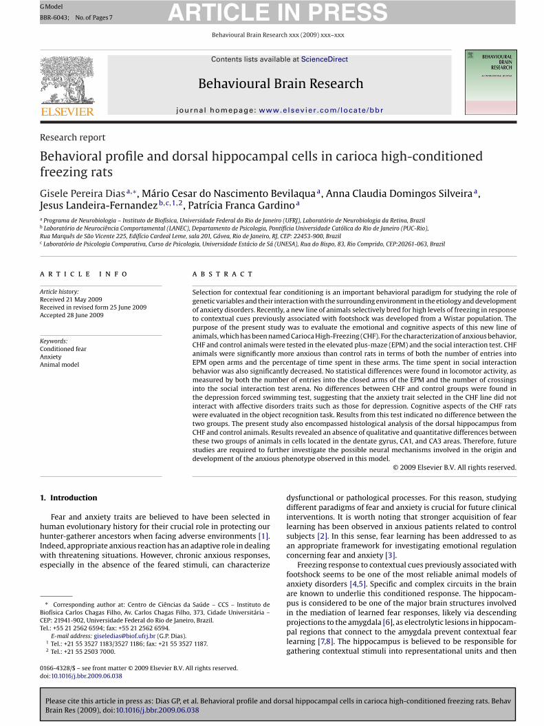
3.5.1. Qualitative analysisQualitative analysis of the three cell layers of the dorsal hip-
pocampus (dentate gyrus, CA1, and CA3) revealed no variationsbetween groups concerning the hippocampal tissue conformation,as seen in Fig. 5.
al hippocampal cells in carioca high-conditioned freezing rats. Behav
3.5.2. Quantitative analysisQuantitative analysis of total dorsal hippocampal cells stained
with DAPI was performed for the same three hippocampal lay-ers: dentate gyrus, CA1, and CA3. No significant differences wereobserved for these measures (dentate gyrus: t(69) = 1.58; p > 0.05;
for anxiety in the elevated plus-maze [29].A commonly stated issue underlined by researchers that work
with behavioral tests of anxiety based on exploratory behaviors isthat factors unrelated to anxiety conditions might alter locomo-
ig. 2. Baseline scores (mean ± S.E.M.) of CTRL and CHF animals in the social inter-ction (top) and locomotor activity (bottom) tests. CTRL, control (n = 6 pairs); CHF,arioca high-freezing (n = 6 pairs). *P < 0.05.
A1: t(68) = 0.14; p > 0.05; CA3: t(68) = 0.38; p > 0.05), as illustratedn Fig. 6.
. Discussion
The ability of an organism to evaluate stimuli and adaptivelyespond to them is one of the most important processes involved inurvival in continuously changing environmental conditions [29].nxiety is a complex trait that has been conserved during evolu-
ion so that animals may assess potentially dangerous situations inrder to enhance the probability of survival [30]. Anxious states inumans are characterized by avoidance behavior and by a tendencyo perceive threatening stimuli [31]. Attentional and mnemonicrocesses are enhanced and ambiguous situations are interpreted
Please cite this article in press as: Dias GP, et al. Behavioral profile and dorsBrain Res (2009), doi:10.1016/j.bbr.2009.06.038
s potentially dangerous [32].The analysis of the neural substrates underlying anxiety and fear
s, in great part, based on the investigation of behavioral inhibitionnduced by natural or learned aversive stimuli in animal models
ig. 3. Baseline scores (mean ± S.E.M.) of CTRL and CHF animals in the forced swim-ing test. CTRL, control (n = 9); CHF, carioca high-freezing (n = 9).
PRESSesearch xxx (2009) xxx–xxx
[7,33]. In this work, we used an animal line of Wistar rats selectivelybred for contextual fear conditioning [19], an animal model foranxiety disorder [4]. The evaluation of anxiety-related behavioraland physiological aspects is crucial for the line to be considered anappropriate model for study of these psychopathologies. Accordingto Landgraf and Wigger [34], an anxious phenotype should presentfeatures related to behaviors and coping strategies characteristic ofthis condition. In this sense, the present work behaviorally validatesthe CHF line as a model for studying anxiety and conditioned fear.
In the elevated plus-maze test, both the total number of entriesand the time spent in open arms were significantly decreased incomparison to the control group. The anxious behavioral patternwas also observed in the social interaction test, where CHF ratsspent significantly less time exhibiting active social interactionbehaviors with their pairs. This observation indicates higher anx-iety, which is in accordance with the performance of rats selected
al hippocampal cells in carioca high-conditioned freezing rats. Behav
Fig. 4. Baseline scores (mean ± S.E.M.) of CTRL and CHF animals in the object recog-nition test. Top: object recognition. Middle: index of exploration 1 (time spent inexploration of both familiar objects). Bottom: index of exploration 2 (time spentin exploration of both types of objects). CTRL, control (n = 12); CHF, carioca high-freezing (n = 12).
G.P. Dias et al. / Behavioural Brain Research xxx (2009) xxx–xxx 5
F yrus,h e gyru
toidarbaasmpsmbeau
scasimtfm6mtca
ig. 5. Qualitative analysis of coronal sections of the dorsal hippocampus (dentate gigh-freezing (n = 3). A = dentate gyrus CTRL; B = CA1 CTRL; C = CA3 CTRL; D = dentat
or activity [35] and compromise data interpretation. The resultsbtained by both anxiety paradigms used here suggest that this
ssue did not introduce bias in this work. Interestingly, the observedifference in the number of entries and the time spent in open arms,s well as the decreased rate of social interactive behavior in CHFats, cannot be attributed to differences in locomotor activity asoth the number of entries in closed arms of the elevated plus-mazend the number of line crossings in the arena of the social inter-ction test did not differ between groups. However, testing largeramples is needed for excluding the apparent trend of less loco-otor activity in CHF rats and a more conclusive interpretation be
erformed. Anyway, performance on the elevated plus-maze andocial interaction test revealed that CHF rats exhibit a significantlyore anxious phenotype when compared to the control group. The
ehavioral difference between CHF and control rats can be consid-red a stable and robust trait in conflict situations that elicit fearnd anxiety, and not only a behavioral profile observed in the testsed for selection [22].
The forced swimming test, used for depressive behaviorcreening, did not show differences between groups. This resultorroborates the consistency of the CHF line as an anxious models traits related to other phenotypes, such as depression, were notelected concomitantly. Landgraf and Wigger [34] underline themportance of this feature and postulated that an animal model
ust be able to capture the specific symptom or mechanism ofhe studied psychopathology, without modeling other processes orunctions. According to Gorman [36], there is a high level of co-
orbidity between anxiety disorders and depression, with an over
Please cite this article in press as: Dias GP, et al. Behavioral profile and dorsBrain Res (2009), doi:10.1016/j.bbr.2009.06.038
0% rate of co-occurrence. This rate of occurrence suggests that co-orbidity is the rule rather than the exception. Therefore, even with
his high probability of co-occurrence of anxious and depressiveharacteristics, the present model was most highly related to thenxious/fearful phenotype.
CA1, and CA3 – 40 �m) of CTRL and CHF animals. CTRL, control (n = 3); CHF, cariocas CHF; E = CA1 CHF; F = CA3 CHF.
Additionally, results from the forced swimming test demon-strated that, although there might be elevated rates of co-morbiditybetween anxiety and depression, the two systems are biologicallydistinct, which is in accordance with the hypothesis that thesestates have different etiologies [37]. The results of the CHF line in theforced swimming test differ from those obtained with some animallines that were selected for innate fear as the latter show depres-sive behaviors in this paradigm [12,29,38]. However, data from thisstudy are in accordance with those presented by Ho et al. [39], inwhich rats selected for anxiety in the elevated plus-maze did notdiffer from the less anxious line in terms of depression, as mea-sured by the forced swimming test. In this sense, it can be statedthat the CHF line can be considered a good predictor of behavioralphenotypes in anxiety models, but cannot be considered a modelfor studying depression.
Since the emotional assessment of a new situation and the uti-lized coping strategies can depend on cognitive functions [30],the hypothesis that non-emotional memory differs between CHFand control rats was also investigated. For this purpose, the objectrecognition test was used. The task of recognizing objects has beenwidely used as a model for investigating the neurobiological mecha-nisms of learning and memory [28]. Interestingly, differences in theindices obtained from this test were not observed between groups,indicating that the memory systems selected during breeding ofthe CHF line were restricted to the emotional memory system.
The hippocampus was chosen as the neural structure responsi-ble for the anxiety differences observed among CHF animals. Thischoice was made considering the fact that this region seems to
al hippocampal cells in carioca high-conditioned freezing rats. Behav
be associated with the etiology of certain types of anxiety dis-order. In fact, several pieces of evidence indicate that patientswith symptoms of post-traumatic stress disorder present smallerhippocampal volume in comparison to control subjects [40]. More-over, experimental research indicates that the dorsal hippocampus
selection response for contextual fear conditioning in a cross between C57BL/6J:
ig. 6. Quantitative analysis of coronal sections of the dorsal hippocampus (dentateyrus, CA1, and CA3 – 40 �m) of CTRL and CHF animals. Top: dentate gyrus. Middle:A1 layer. Bottom: CA3 layer. CTRL, control (n = 3); CHF, carioca high-freezing (n = 3).
odulates several anxiety-like responses. For example, Gonzalezt al. [41] found anxiolytic effects in the social interaction testfter microinjection of benzodiazepinic sites in GABAA into theorsal hippocampus. Additionally, Rezayat et al. [42] showed an
nteraction between GABA and cholecystokinin during modula-ion anxiety in the elevated plus-maze after injections of agonistsnd antagonists of both neurotransmitters into the dorsal hip-ocampus. Nazar et al. [43] also pointed out that GABA anderotonergic systems within the dorsal hippocampus are intimatelynvolved in emotional behaviors. When they microinjected picro-oxin (a non-competitive antagonist of the GABAA receptor) intohis hippocampal portion, the anxiolytic effect caused by serotoninepletion was attenuated. File et al. [38] also showed that the HighPAT Sensitive line (HDS) (an animal model selected for high sen-
itivity to the hypotermic response induced by the serotonergicgonist 8-OH-DPAT) presents reduced scores in the social interac-ion test, accounting for, at least in part, the abnormal functioning
Please cite this article in press as: Dias GP, et al. Behavioral profile and dorsBrain Res (2009), doi:10.1016/j.bbr.2009.06.038
f 5-HT1A receptors in the dorsal hippocampus. Kjelstrup et al. [44],n turn, demonstrated that this hippocampal portion is involved inear conditioning since lesions in this area prevent contextual fearonditioning.
PRESSesearch xxx (2009) xxx–xxx
Qualitative analysis of the dorsal hippocampal tissue, specif-ically the dentate gyrus and CA1 and CA3 areas, revealed thatdevelopment of the hippocampal formation of CHF rats did not dif-fer from that of control animals in relation to the morphologicalorganization of the tissue in these three main cell layers. There-fore, no qualitative damage of the tissue was detected, indicatingthat the tissue was preserved as a whole and that behavioral differ-ences between groups cannot be explained in terms of hippocampalinjury. Cell quantification in the three mentioned areas of the dor-sal hippocampus was not different between groups, which suggeststhat differences in the hippocampal circuitry in CHF animals, ashypothesized, might occur: at the molecular level of this struc-ture, such as differences in the expression of neurotransmittersand/ or receptors and proteins involved in neurotransmission; at themorphological level, concerning dendritic arborization, and at thefunctional level, such as differences in hippocampal neurogenesis.
It is important to mention that the ventral portion of the hip-pocampus was not addressed in the present study. This issue isimportant because there are some reports indicating that thisregion might be involved in anxiety regulation. For example,Kjelstrup et al. [44] reported that lesions within the ventral hip-pocampus alter unconditioned fear responses in the elevatedplus-maze test. Moreover, activation of 5-HT2C receptors withinthe ventral hippocampus induced anxiety responses in the elevatedplus-maze. Therefore, future studies of the ventral hippocampus ofCHF rats might produce additional data in the investigation of themechanisms involved in the anxious trait exhibited by this animalmodel.
In conclusion, these data show that the CHF line representsa robust animal model of anxiety disorder, as differences in theexperimental group were observed in two different anxiety tests.Motor activity did not account for the differences between CHF andcontrol animals. The absence of reliable differences between CHFand control animals in the forced swimming test and object recog-nition task indicated that the breeding procedure that increasedthe occurrence of conditioned freezing to contextual cues did notinterfere with other emotional or memory systems. Possible neuro-physiological differences between CHF and control animals mightbe more specific than the total amount of cells within the dorsal hip-pocampus. Thus, future studies are required to examine the possiblemechanisms involved in the origin and development of the anxiousphenotype observed in this model.
Acknowledgements
This research was supported by Conselho Nacional de Pesquisa(CNPq) grant 52272095-1 to JLF; CNPq/ PRONEX grant E-26/171519/2006 and Fundacão Carlos Chagas Filho de Amparo àPesquisa do Estado do Rio de Janeiro (FAPERJ) grant E-26/171.019/05to GPD, MCNB, ACDLS and PFG. We also thank Vitor de Castro Gomesand Anna Carolina Costa for their help in different stages of thiswork. Additionally, we thank American Journal Experts for theirsupport in document editing.
References
[1] Cosmides L, Tooby J. Evolutionary psychology and the emotions. In: Lewis e M,Haviland-Jones JM, editors. Handbook of Emotions. 2nd ed. NY: Guilford; 2000.p. 91–115.
[2] Lissek S, Powers AS, McClure EB, Phelps EA, Woldehawariat G, Grillon C, PineDS. Classical fear conditioning in the anxiety disorders: a meta-analysis. BehavRes T 2005;43:1391–424.
J. Different patterns of freezing behavior organized in the periaqueductalgray of rats: association with different types of anxiety. Behav Brain Res2008;188:1–13.
[5] Landeira-Fernandez J. Context and Pavlovian conditioning. Braz J Med Biol Res1996;29(2):149–73.
[6] Anagnostaras SG, Craske MG, Fanselow MS. Anxiety: at the intersection of genesand experience. Nat Neurosci 1999;2(9):780–2.
[7] Phillips RG, LeDoux J. Differential contribution of amygdala and hippocampusto cued and contextual fear conditioning. Behav Neurosci 1992;106:274–85.
[8] Maren S, Fanselow MS. Synaptic plasticity in the basolateral amyg-dala induced by hippocampal formation stimulation in vivo. J Neurosci1995;15(11):7548–64.
[9] LeDoux J. Emotion circuits in the brain. Annu Rev Neurosci 2000;23:155–84.10] Kim JJ, Fanselow MS. Modality specific retrograde amnesia of fear following
effects in the rat elevated plus-maze of 5-HT2C agonists into ventral but notdorsal hippocampus. Behav Pharmacol 2004;15:37–43.
12] Liebsch G, Montkowski A, Holsboer F, Landgraf R. Behavioral profiles of twoWistar rat lines selectively bred for high or low anxiety-related behavior. Behav.Brain Res 1998;94:301–10.
13] Broadhurst PL. Determinants of emotionality in the rat: III. Strain differences. JComp Physiol Psychol 1958;51(1):55–9.
14] Hall CS. The inheritance of emotionality. Sigma Xi Quart 1938;26:17–27.15] Bignami G. Selection for high rates and low rates of avoidance conditioning in
the rat. Anim Behav 1965;13(2):221–7.16] Brush FR, Froehlich JC, Sakellaris PC. Genetic selection for avoidance behavior
in the rat. Behav Genet 1979;9(4):309–16.17] Ryzhova LY, Kulagin DA, Lopatina NG. Correlated variability in movement
activity and emotionality in the selection of rats for high and low levels ofconditioned active avoidance reflexes. Genetika 1983;19(1):121–5.
18] Ohta R, Matsumoto A, Hashimoto Y, Nagao T, Mizutani M. Behavioral character-istics of rats selectively bred for high and low avoidance shuttlebox response.Cong Anom 1995;35:223–9.
19] Gomes VC, Landeira-Fernandez J. Amygdaloid lesions produced similar con-textual fear conditioning disruption in the Carioca high- and low-conditionedfreezing rats. Brain Res 2008;1233:137–45.
20] Handley SL, Mithani S. Effects of alpha-adrenoceptor agonists in a maze-exploration model of fear-motivated behavior. Naunyn Schmiedebergs ArchPharmacol 1984;327:1–5.
21] Pellow S, Chopin P, File SE, Briley M. Validation of open:close arm entries inan elevated plus-maze as a measure of anxiety in the rat. J Neurosci Methods1985;14:149–67.
22] Henniger MSH, Ohl F, Hölter SM, Weibenbacher P, Toschi N, Lörscher P, WiggerA, Spanagel R, Landgraf R. Unconditioned anxiety and social behavior in two ratlines selectively bred for high and low anxiety-related behavior. Behav Brain Res2000;111:153–63.
23] Porsolt R, Lepichon M, Jalfre M. Depression: a new animal model sensitive toantidepressant treatments. Nature 1977;266:730–2.
Please cite this article in press as: Dias GP, et al. Behavioral profile and dorsBrain Res (2009), doi:10.1016/j.bbr.2009.06.038
24] Zangen A, Nakash R, Overstreet DH, Yadid G. Association between depres-sive behavior and absence of serotonin-dopamine interaction in the nucleusaccumbens. Psychopharmacology 2001;155:434–9.
25] Chau DT, Rada P, Kosloff RA, Taylor JL. Hoebel BG Nucleus accumbens muscarinicreceptors in the control of behavioral depression: antidepressant-like effects oflocal M1 antagonist in the porsolt swim test. Neuroscience 2001;104(3): 791–8.
[
[
PRESSesearch xxx (2009) xxx–xxx 7
26] Rada P, Moreno SA, Tucci S, Gonzalez LE, Harrisson T, Chau DT, Hoebel BG, Her-nandez L. Glutamate release in the nucleus accumbens is involved in behavioraldepression during the porsolt swim test. Neuroscience 2003;119:557–65.
27] Ennaceur A, Delacour J. A new one-trial test for neurobiological studies ofmemory in rats. 1: Behavioral data. Behav Brain Res 1988;31:47–59.
28] De Lima MNM, Laranja DC, Bromberg E, Roesler R, Schröder N. Pre- or post-training administration of the NMDA receptor blocker MK-801 impairs objectrecognition memory in rats. Behav Brain Res 2005;156:139–43.
29] Landgraf R, Wigger A. High x low anxiety-related behavior rats: an animalmodel of extremes in trait anxiety. Behav Genet 2002;32:301–14.
30] Neumann ID, Wigger A, Liebsch G, Holsboer F, Landgraf R. Increasedbasal activity of the hypothalamo-pituitary-adrenal axis during pregnancyin rats bred for high anxiety-related behaviour. Psychoneuroendocrinology1998;23(5):449–63.
31] Crestani F, Lorenz M, Baer K, Essrich C, Benke D, Laurent JP, Belzung C, Fritschy J,Lüscher B, Mohler H. Decreased GABAA-receptor clustering results in enhancedanxiety and a bias for threat cues. Nature Neurosci 1999;2:833–9.
32] Eysenck MW. In: Gale A, e Eysenck MW, editors. Handbook of Individual Dif-ferences: Biological Perspectives. New York: Wiley; 1991. p. 157–78.
34] Landgraf R, Wigger A. Born to be anxious: neuroendocrine and genetic corre-lates of trait anxiety in HAB rats. Stress 2003;6(2):111–9.
35] Rodgers RJ, Cao BJ, Dalvi A, Holmes A. Animal models of anxiety: an ethologicalperspective. Braz J Med Biol Res 1997;30:289–304.
36] Gorman JM. Comorbid depression and anxiety spectrum disorders. DepressAnxiety 1996;4:160–8.
37] Angst J, Vollrath M, Merikangas K, Ernst C. Comorbidity of anxiety and depres-sion in the Zurich cohort study of young adults. In: Maser JD, Cloninger CR,editors. Comorbidity of mood and anxiety disorders. Washington, D.C.: Amer-ican Psychiatric Press; 1990. p. 123–37.
38] File SE, Ouagazzal AM, Gonzalez LE, Overstreet DH. Chronic fluoxetine in testsof anxiety in rat lines selectively bred for differential 5-HT1A receptor function.Pharmacol Biochem Behav 1999;62(4):695–701.
39] Ho YJ, Eichendorff J, Schwarting RKW. Individual response profiles of maleWistar rats in animal models for anxiety and depression. Behav Brain Res2002;136:1–2.
40] Smith ME. Bilateral hippocampal volume reduction in adults with post-traumatic stress disorder: a meta-analysis of structural MRI studies.Hippocampus 2005;15:798–807.
[41] Gonzalez L, Ouagazzal AM, File SE. Stimulation of benzodiazepine receptorsin the dorsal hippocampus and median raphé reveals differential GABAergiccontrol in two animal test of anxiety. Eur J Neurosci 1998;10:3673–80.
42] Rezayat M, Roohbakhsh A, Zarrindast MR, Massoudi R, Djahanguiri B. Cholecys-tokinin and GABA interaction in the dorsal hippocampus of rats in the elevatedplus-maze test of anxiety. Physiol Behav 2005;84:775–82.
al hippocampal cells in carioca high-conditioned freezing rats. Behav
43] Nazar M, Siemiatkowski M, Czlonkowska A, Sienkiewicz-Jarosk H, Plaznik A.The role of hippocampus and 5-HT/GABA interaction in the central effects ofbenzodiazepine receptor ligands. J Neural Transm 1999;106:369–81.
44] Kjeslstrup KG, Tuvnes FA, Steffenach HA, Edvard RM, Moser I, Moser MB.Reduced fear expression after lesions of the ventral hippocampus. Proc NatlAcad Sci USA 2002;99(16):10825–1083.