40
nz bpac better edicin m e GOUT CAVEDILOL PREXIGE CO-ENZYME Q10

nzbpacbetter edicin m e
GOUTCavedilOl
PrexiGeCO-enzyme Q10

Editorial Team
Tony Fraser
Professor Murray Tilyard
Clinical Advisory Group
Dr Dave Colquhoun
Michelle Cray
Dr Chris Leathart
Dr Lynn McBain
Adam McRae
Dr Peter Moodie
Associate Professor Jim Reid
Dr David Reith
Professor Murray Tilyard
Programme Development Team
Rachael Clarke
Rebecca Didham
Peter Ellison
Sonia Ross
Dr Trevor Walker
Dr Sharyn Willis
Dave Woods
Report Development Team
Justine Broadley
Lana Johnson
Web
Gordon Smith
Design
Sonia Ross
Management and Administration
Kaye Baldwin
Tony Fraser
Kyla Letman
Professor Murray Tilyard
Contact us:
Mail P.O. Box 6032 Dunedin
Email [email protected]
Free-fax 0800 27 22 69
Best Practice Journal (BPJ)
ISSN 1177-5645
BPJ is published and owned by bpacnz
Level 8 10 George Street Dunedin
BPJ, Issue 8, September 2007
Bpacnz is an independent organisation that promotes health care
interventions which meet patients’ needs and are evidence based,
cost effective and suitable for the New Zealand context.
We develop and distribute evidence based resources which describe,
facilitate and help overcome the barriers to best practice.
Bpacnz has four shareholders:
Procare Health, South Link Health, IPAC and The University of Otago
Bpacnz is currently funded through contracts with PHARMAC and DHBNZ.

Gout is a major cause of arthritis in New Zealand and is particularly prevalent in Mäori and Pacific populations. The treatment of gout includes cardiovascular disease risk assessment, management of modifiable risk factors and long-term preventative therapy with allopurinol, aiming to ‘hit the target’ of <0.36 mmol/L serum uric acid levels.
Natural history of gout
Diagnosis & treatment
Allopurinol prescribing guide
Colchicine dosing and adverse effects
CoNTENTS
GouT - HIT THE TARGET9
PHARMACISTS HAvE A kEy RolE IN
THE CARE of PEoPlE WITH GouT15
PREvAlENCE AND IMPACT of GouT16
BPJ I Issue 8 I 3

CoNTENTS
19 A slow death from colchicine
20 Is carvedilol superior to metoprolol in heart failure?
24 lumiracoxib linked to deaths in Australia
Carvedilol may be an option for patients initiating beta-blocker
treatment for heart failure or patients in whom metoprolol is poorly
tolerated. We present the results of the COMET trial and other
research and discuss strategies for initiating carvedilol treatment.
Medsafe have just announced that approval for lumiracoxib
(Prexige) 400 mg tablets has been revoked in New Zealand. This
follows the news that lumiracoxib has been completely withdrawn
in Australia after it emerged that the drug was linked to serious
adverse reactions including liver failure and death.
4 I BPJ I Issue 8

Access best practice online
www.bpac.org.nz
Essentials
6 uPfRoNT Would you like fries with that? The role of
Co-enzyme Q10 supplements in medical
treatment
28 ETC Evidence that counts
32 Bandolier Independent evidence-based thinking about
health care
36 Dear Dave Serotonin toxicity, Concomitant use of the
combined oral contraceptive and antibiotics
38 Correspondence
CME
26 Ten Minute Audit Identifying patients on colchicine
CoNTENTS
BPJ I Issue 8 I 5
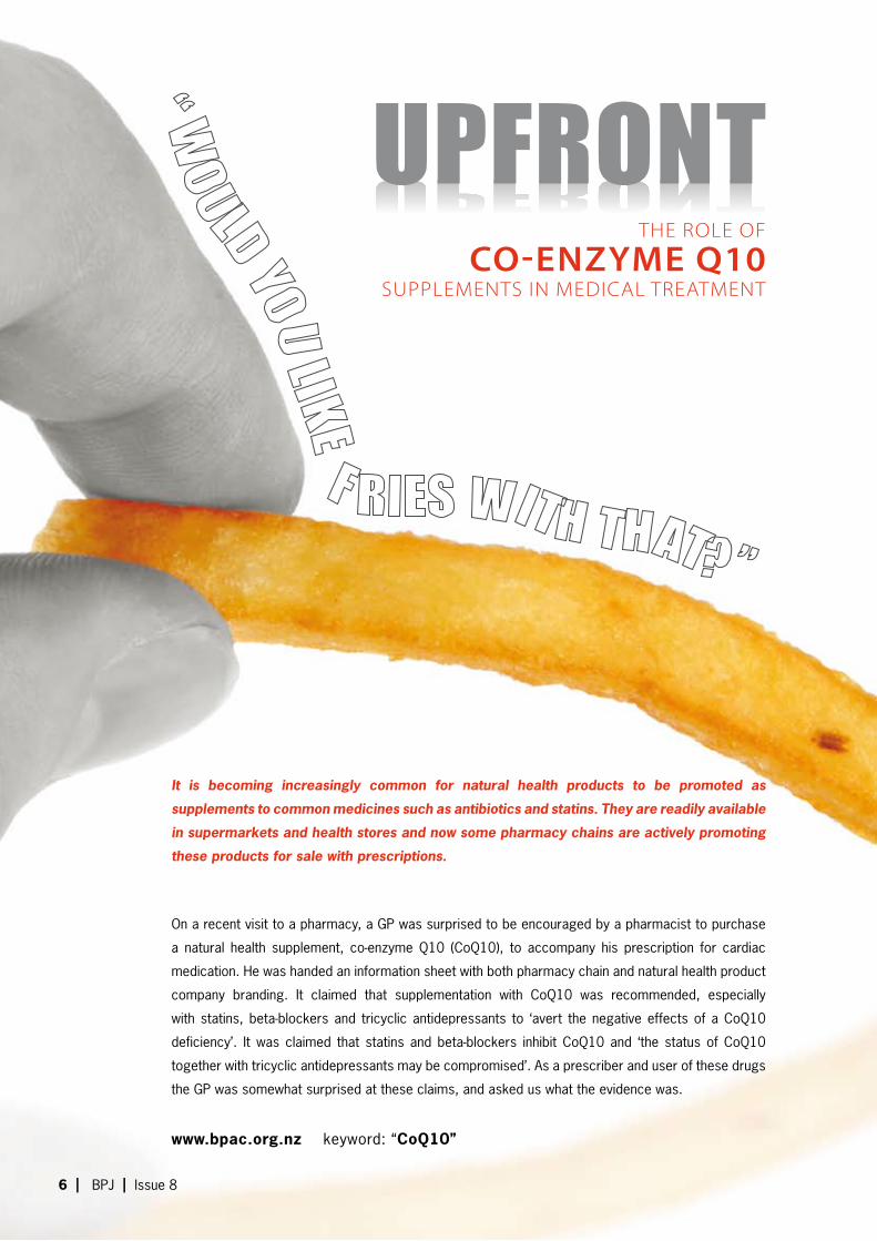
UPFRONTThe role of
co-enzyme Q10 supplemenTs in medicAl TreATmenT
It is becoming increasingly common for natural health products to be promoted as
supplements to common medicines such as antibiotics and statins. They are readily available
in supermarkets and health stores and now some pharmacy chains are actively promoting
these products for sale with prescriptions.
On a recent visit to a pharmacy, a GP was surprised to be encouraged by a pharmacist to purchase
a natural health supplement, co-enzyme Q10 (CoQ10), to accompany his prescription for cardiac
medication. He was handed an information sheet with both pharmacy chain and natural health product
company branding. It claimed that supplementation with CoQ10 was recommended, especially
with statins, beta-blockers and tricyclic antidepressants to ‘avert the negative effects of a CoQ10
deficiency’. It was claimed that statins and beta-blockers inhibit CoQ10 and ‘the status of CoQ10
together with tricyclic antidepressants may be compromised’. As a prescriber and user of these drugs
the GP was somewhat surprised at these claims, and asked us what the evidence was.
6 I BPJ I Issue 8
www.bpac.org.nz keyword: “CoQ10”

Co-enzyme Q10 was discovered in the 1950s and its mechanisms and uses are still being investigated
CoQ10 (also known as ubiquinone) assists in the production of energy
within cells and helps protect internal and external cell membranes
against oxidation. Organs with the greatest energy requirements such
as the heart, lungs and liver have higher concentrations of CoQ10.
Approximately half of the body’s CoQ10 is obtained through dietary fat
ingestion, with the remainder from cellular synthesis.
Supplementation of CoQ10 is used as a treatment for serious
mitochondrial disorders and other metabolic syndromes, when people
are unable to produce enough CoQ10. Current research focuses on
its role in the treatment of neurodegenerative and cardiovascular
disease. CoQ10 is a common ingredient in skin-care products and
CoQ10 supplements are marketed by the cosmetics industry as ‘skin
boosters’.
Routine use of co-enzyme Q10 with statins is not necessary
The rationale for using CoQ10 in association with statin medication
seems to focus on the role it may play in alleviating symptoms of
myopathy – a relatively rare side effect of statin use. Statin treatment
reduces circulating levels of CoQ10.1, 2 However, studies on human
subjects have shown that intramuscular levels of CoQ10 are not
reduced by low-dose statin treatment. Effects may differ with the type
of statin and dose.2 Data on a causal association between low levels
of intramuscular CoQ10 and statin induced myopathy is limited and
contradictory.2
In a recently published systematic review in the Journal of the
American College of Cardiology, Dr Leo Marcoff and Dr Paul Thompson
concluded that there is insufficient evidence at present to prove the
role of CoQ10 deficiency in statin induced myopathy. They state that
routine supplementation of CoQ10 with statin use is neither justified nor
recommended. However they noted that as there are no known risks
associated with CoQ10, it may be trialled for people who develop statin
associated myalgia.2 Other reviews of research and literature have
come to similar conclusions.3, 4
No compelling evidence as yet for using co-enzyme Q10 in cardiovascular disease
In the pharmacy-supplied CoQ10 information
sheet, beta-blockers were highlighted
as medications that would benefit from
concurrent administration of CoQ10
supplements.
There has been some research on using
CoQ10 as a treatment for hypertension.
A recent meta-analysis of clinical trials
concluded that CoQ10 ‘has the potential’
to lower blood pressure in hypertensive
patients.5 In contrast, a study conducted
among healthy individuals found that CoQ10
had only a mild and transient effect on
systolic blood pressure.6 While there is some
emerging evidence of a beneficial effect of
CoQ10 in hypertensive patients, there is
less evidence for its use in cardiovascular
disease as a whole. Large-scale trials are
needed to find any compelling evidence of
clinical effect.
No evidence for supplementing tricyclics with CoQ10
Although the pharmacy information sheet
highlighted tricyclic antidepressants as
benefiting from supplementation of CoQ10,
we could not find any research to support
this.
BPJ I Issue 8 I 7

No clinical evidence of neuroprotection role for CoQ10 in Parkinson’s disease
The mechanisms of Parkinson’s disease are not yet fully known,
but there is emerging evidence that cellular energy depletion and
oxidative stress are contributing factors. CoQ10 is known to be a
potent antioxidant and energy stimulant, therefore its potential role
as a neuroprotectant is being investigated.
A recently published trial testing whether CoQ10 has beneficial
effects on the symptoms in mid-stage Parkinson’s disease, found
that, while it was safe and well-tolerated, there was no difference
between patients receiving CoQ10 and those who did not receive
the supplement.7 Other researchers have found no evidence of a
clinically significant effect of CoQ10 in alleviating symptoms or
halting the progression of Parkinson’s disease, but suggest that
further study is warranted.8,9 One researcher notes that caution
must be applied to the use of CoQ10 without certainty of its efficacy,
especially since it is readily available over-the-counter and may
expose patients to unnecessary risk and significant expense.10
So what does all this mean?
Current evidence on the use of CoQ10 supplements,
alongside medications such as statins, beta-blockers and
tricyclic antidepressants and as a treatment for hypertension
or neurological disorders, shows that while there is no
evidence of harm in taking this supplement clinical benefit
is not proven.
There are good dietary sources of CoQ10 including oily fish, offal
(e.g. liver, kidney), nuts, soy, sesame and some vegetables. In
addition, there are other non-pharmacological strategies for
managing conditions such as hypertension e.g. increased
exercise, weight loss, decreased alcohol consumption and dietary
modifications.11 The use of supplements introduces a relatively
substantial cost, with the recommended dose of 30–90 mg costing
on average 60c − $1.80 a day.
While there is no safety issue preventing the promotion of the
blanket use of CoQ10, we question whether it is ethical to use a
prescription as the basis for promoting a supplement, that has little
evidence of clinical benefit.
References
Littarru G, Langsjoen P. Coenzyme Q10 and 1.
statins: biochemical and clinical implications.
Mitchondrion 2007;7S:S168-S174.
Marcoff L, Thompson P. The role of coenzyme 2.
Q10 in statin-associated myopathy. J Am Coll Cardiol 2007;49(23):2231-7.
Levy H, Kohlhaas H. Considerations 3.
for supplementing with co-enzyme Q10
during statin therapy. Ann Pharmacother 2006;40(2):290-4.
Nawarskas J. HMG-CoA reductase 4.
inhibitors and coenzyme Q10. Cardiol Rev
2005;13(2):76-9.
Rosenfeldt F, Haas S, Krum H, et al. Coenzyme 5.
Q10 in the treatment of hypertension: a
meta-analysis of the clinical trials. J Hum Hypertens 2007;21(4):297-306.
Shah S, Sander S, Cios D, et al. 6.
Electrocardiographic and hemodynamic
effects of coenzyme Q10 in healthy individuals:
a double-blind, randomised controlled trial.
Ann Pharmacother 2007;41(3):420-5.
Storch A, Jost W, Vieregge P, et al. 7.
Randomised, double-blind, placebo-controlled
trial on symptomatic effects of coenzyme
Q10 in Parkinson Disease. Arch Neurol 2007;[Epub ahead of print].
The NINDS NET-PD Investigators. A 8.
randomised clinical trial of coenzyme Q10
and GPI-1485 in early Parkinson disease.
Neurology 2007;68(1):20-8.
Weber C, Ernst M. Antioxidants, supplements 9.
and Parkinson’s Disease. Ann Pharmacother 2006;40(5):935-8.
Galpern W, Cudkowicz M. Coenzyme Q 10.
treatment of neurodegenerative diseases of
aging. Mitchondrion 2007;7S:S146-S153.
Wexler R, Aukerman G. Nonpharmacological 11.
strategies for managing hypertension. Am Fam Physician 2006;73(11):1953-6.
8 I BPJ I Issue 8

TreaTmenTOf GOUT
SUmmary POinTS
Gout is a major cause of arthritis in New Zealand, with high rates of 1.
severe disease in Mäori and Pacific patients
Gout causes significant disability in M2. äori and Pacific men of
working age
All patients with gout should have cardiovascular disease (CVD) risk 3.
assessment, and intensive management of modifiable risk factors
Long-term preventive therapy with allopurinol is critical for 4.
effective gout management:
Prescribe early, before development of tophi -
Monitor serum uric acid levels -
Aim for target serum uric acid <0.36 mmol/L -
Introduce gradually: ‘start low and go slow’ -
Use colchicine prophylaxis -
Minimise diuretic therapy in patients with gout5.
Key adviSer
Dr Nicola Dalbeth, Rheumatologist
and Senior Lecturer, Department of
Medicine, University of Auckland
aCKnOWledGemenTS
We are grateful to Dr Peter Gow and Dr
Doone Winnard for their review of this
article.
www.bpac.org.nz Keyword: “Gout”
BPJ I Issue 8 I 9
TarGeT SerUm UriC aCid <0.36 m
mol/l
WhaT iS GOUT?
Gout is an arthritis caused by the inflammatory response to
intra-articular monosodium urate crystals. Supersaturation
of urate typically occurs in physiological fluids above
concentrations of 0.42 mmol/L. In early disease, gout
presents as recurrent episodes of self-limiting acute
inflammatory attacks (‘flares’) of arthritis. These attacks most
often affect the 1st metatarsophalangeal joint, midfoot and
ankle. In the presence of prolonged hyperuricaemia, some
patients develop recurrent polyarticular attacks, chronic
tophaceous disease, erosive arthritis (images are available
in the online version of this article visit www.bpac.org.nz)
and renal disease (urate nephropathy and uric acid stones).
naTUral hiSTOry Of GOUT
If untreated, the evolution of gout follows four stages:
Asymptomatic hyperuricaemia1. – asymptomatic
hyperuricaemia has traditionally remained untreated
with drugs. Although evidence is building, linking
hyperuricaemia with cardiovascular and renal
disease, treatment remains unproven. Identification of
hyperuricaemia presents an opportunity to suggest diet
and lifestyle changes to patients and also to look for
possible underlying causes for the raised uric acid. Of
those with hyperuricaemia, 20% will go on to develop
acute symptomatic gout.
Acute attacks2. – typically the first attack involves one
joint but it can also be polyarticular. Without specific
treatment, an attack of acute gout is likely to resolve
within 7–10 days. In practice, the severe pain usually
forces patients to seek pharmacological relief.
Intercritical gout3. – the length of time between attacks
can vary widely. Some patients only ever have one
attack, but for the majority, a second attack will occur
within a year. If the urate level remains high (>0.36
mmol/L) despite the patient being symptom free, there
can be ongoing joint inflammation and hence joint
damage and tophi formation.
Chronic tophaceous gout4. – tophi are firm white
translucent nodules in connective tissue arising from
the deposition of urate crystals. They can take at least
10 years after the initial attack to develop. As well as
causing joint destruction, they are disfiguring and also
cause physical hindrance. Tophi can become inflamed
or infected and can exude tophaceous material.
diaGnOSiS Of GOUT
The diagnosis of gout can be made according to the
American College of Rheumatology (ACR)/Wallace
criteria1:
The presence of characteristic urate crystals A.
in the joint fluid,
B. A tophus proved to contain urate crystals
by chemical means or polarized light
microscopy (images are available in the online
version of this article visit www.bpac.org.nz)
OR
C. Six of the following 12 clinical criteria
Maximum inflammation within the first daya.
More than one attack of acute arthritisb.
Monoarticular arthritisc.
Redness observed over jointsd.
First metatarsophalangeal joint pain attacke.
Unilateral metatarsophalangeal joint attackf.
Unilateral tarsal joint attackg.
Suspected tophush.
Hyperuricaemiai.
Asymmetric swelling within a joint on x-rayj.
Subcortical cysts with no erosions on x-rayk.
Negative bacterial culture of joint fluidl.
It is important to note that gout and sepsis can
co-exist. The presence of urate crystals in synovial
fluid does not exclude a diagnosis of sepsis.2
Although hyperuricaemia is a key risk factor
for gout, it is not sufficient to make the
diagnosis of gout; only 20% of patients with
hyperuricaemia will develop gout, and serum
urate concentrations may be normal in patients
during an acute gout flare.3
10 I BPJ I Issue 8
Presenting symptom: Acute gout
Treat acute attack with NSAIDs. -
Use corticosteroids when NSAIDs are contraindicated. -
Evaluate and manage risk factors
(weight, alcohol, diuretics, dietary purines)
TreaTmenT Of GOUT
Treatment of acute gout flares
NSAIDs: - given at regular intervals until the severe pain abates, at
which time the dose may be reduced (e.g. starting with naproxen
500 mg bd or diclofenac 75 mg bd). Always watch for renal
impairment, heart failure and peptic ulceration. If patients are
already taking low dose aspirin for cardiovascular risk reduction
it should be continued.
Oral corticosteroids: - in view of the toxicity of colchicine,
corticosteroids may be preferred to treat acute gout in patients
in whom NSAIDs are contraindicated, provided sepsis has been
excluded. The initial dose is 15–40 mg prednisone daily, gradually
reduced over 10 days. Intra-articular corticosteroids are useful if
monoarthritis is present to reduce risks of systemic therapy.
Colchicine: - can be a useful adjunct to NSAIDs in resistant cases,
particularly when tophi are present, as monotherapy or to prevent
flares when starting allopurinol.
Allopurinol: - If a patient has been taking allopurinol regularly at
the time of developing an acute attack it should be continued at
the same dose.
“Allopurinol should not be started at the time of the
attack”
riSK faCTOrS fOr GOUT
The key risk factors for gout are
Hyperuricaemia -
Male sex -
M - äori and Pacific ethnicity*
Chronic renal impairment -
Hypertension -
Obesity -
Diuretic use** -
Coronary heart disease -
High intake of meat, seafood and alcohol -
(particularly beer)
*Mäori patients with normal uric acid levels have
been shown to have a reduced excretion of urate.
This suggests an underlying renal mechanism.4
**Diuretic therapy is a risk factor for the
development of hyperuricaemia and recurrent
gout attacks. Diuretic therapy should be
minimised and avoided wherever possible.
Adverse effects with Colchicine
Colchicine has a narrow therapeutic margin and
considerable variation in absorption between
individuals. Toxic effects include diarrhoea,
nausea and vomiting, electrolyte imbalance,
alopecia, haematological effects, pancreatitis,
and failure of kidneys, liver or respiratory system.
High doses can be fatal.
Treat resistant cases with - addition of low dose colchicine.
Treat those at risk of NSAID side effects with colchicine -
alone.
BPJ I Issue 8 I 11
Colchicine dosing for acute gout
Due to recent concerns about toxicity,
colchicine is no longer considered first
line treatment for acute gout. In addition
colchicine should be used at a lower dose
than has been recommended in the past.
“…The recommended dose for colchicine
in the treatment of acute gout is 1.0 mg
stat, followed by 0.5 mg six hourly, up to a
maximum dose of 2.5 mg per 24 hours…”
New Zealand Rheumatology Association (NZRA),
endorsed by Medsafe.5
(full statement available at
www.rheumatology.org.nz/colchicine.htm)
After the first 24 hours, the dose should be
reduced to 0.5 mg one or two times daily,
according to renal function. Prescribed in
this way colchicine is safe and effective. The
risk of diarrhoea and other toxic effects is
minimised. Many patients report that one or
two colchicine tablets taken within the first
few hours of the onset of pain can avoid a
major flare.
Adverse effects with Allopurinol
The most common adverse effect
is a rash (1−2%), which may be
more common in patients with
renal impairment.12 Allopurinol
hypersensitivity syndrome (AHS) is
extremely rare but potentially fatal.
It is characterised by fever, rash,
eosinophilia, hepatitis and renal failure.
Adverse effects can occur at any
dose.13
indiCaTiOnS fOr UriC aCid lOWerinG TheraPy6-8
All patients with any one of the following should receive long-term uric
acid lowering therapy:
Recurrent gout attacks (≥2 attacks/year) -
Tophi -
Gouty arthropathy -
Radiographic damage -
Early onset, family history and serum uric acid >0.60 mmol/L -
It should be noted that although effective treatment of gout can lead
to regression of tophi, management is far more difficult once tophi
develop, due to the high total body urate load.
“Early treatment of gout, before onset of tophi
and erosive disease, is recommended”
hiTTinG The TarGeT in GOUT: aim fOr a SerUm UriC aCid COnCenTraTiOn Of <0.36 mmol/l
Several recent studies have emphasised the importance of excellent
long-term control of serum uric acid in order to suppress gout attacks
and achieve regression of tophi. These studies have identified a serum
uric acid level of <0.36 mmol/L as the target required for dissolution
of monosodium urate crystals within the joints and subcutaneous
tissues.9–11 This target has been endorsed in the recent European
League Against Rheumatism (EULAR) guidelines for management of
gout.7
Reduction of the serum uric acid level requires both pharmacological
and non-pharmacological management. Allopurinol is the first choice
urate-lowering drug unless there is a history of allopurinol allergy/
intolerance.
“Patients with gout should be encouraged to think of their
uric acid level in the same way that patients with diabetes
think of their HbA1c”
12 I BPJ I Issue 8
Allopurinol prescribing: a how-to guide
Wait for at least two weeks after an acute gout attack before 1.
starting allopurinol
‘Start low and go slow’2. . Start with allopurinol 100 mg daily,
and increase by 100 mg every two weeks until the serum uric
acid level is <0.36 mmol/L. For most patients with normal
renal function, a dose of 300 mg daily is needed to achieve
this target. Patients with renal impairment may require less
allopurinol to achieve this target. Sudden changes in the
serum uric acid level are likely to precipitate gout attacks.
Gradually increasing the dose of allopurinol is less likely to
trigger a gout attack
Use prophylaxis against acute attacks. Prophylaxis with 3.
colchicine (0.5 mg daily to twice daily) or NSAIDs for the first
three months of starting allopurinol (or until serum uric acid
<0.36 mmol/L) should be prescribed to reduce the risk of
gout attacks.14
Ensure the patient knows that the colchicine is for gout 4.
prevention and the dose should not be altered without medical
advice if an acute episode occurs.
Monitor serum uric acid levels on a monthly basis while 5.
establishing allopurinol. Once serum uric acid is <0.36 mmol/L,
monitor uric acid and renal function on a three-monthly basis.
Allopurinol should be continued as life-long therapy for 6.
management of gout, except in the case of allopurinol
intolerance. Do not stop taking allopurinol during an acute
attack of gout.
Other urate-lowering drugs
The uricosuric agent probenecid is an effective urate-lowering drug
in patients with normal renal function and urate under-excretion.
This agent is particularly useful in combination with allopurinol if
there is persistent hyperuricaemia despite therapeutic doses of
allopurinol, or in allopurinol intolerance.15 A typical dose is 250
mg twice daily for two weeks, then 500 mg twice daily thereafter.
Probenecid is contraindicated in patients with a history of renal
stones. Patients should be advised regarding the importance of
high fluid intake while taking probenecid, around eight glasses of
water per day.
lifeSTyle inTervenTiOnS
Weight management is the key component in dietary
management of gout. A 5% loss in body weight leads
to a 10% reduction in serum uric acid level.16,17 Diets
very low in purines are generally unpalatable and poorly
tolerated over time. Patients are more likely to accept
advice to reduce purine-rich foods than to be told not to
eat them at all (Table 1). Patients should be encouraged
to eat regular meals and to drink plenty of water.
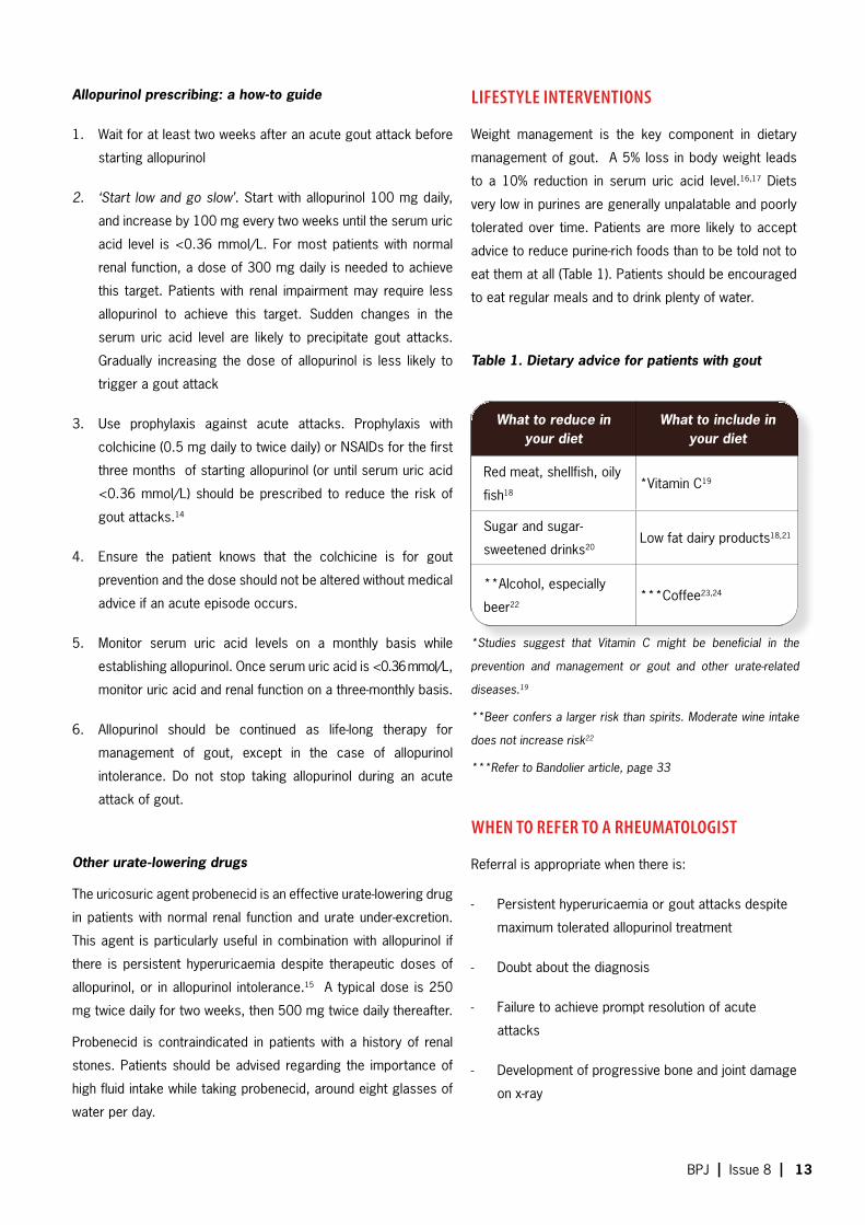
Table 1. Dietary advice for patients with gout
When TO refer TO a rheUmaTOlOGiST
Referral is appropriate when there is:
Persistent hyperuricaemia or gout attacks despite -
maximum tolerated allopurinol treatment
Doubt about the diagnosis -
Failure to achieve prompt resolution of acute -
attacks
Development of progressive bone and joint damage -
on x-ray
What to reduce in your diet
What to include in your diet
Red meat, shellfish, oily
fish18*Vitamin C19
Sugar and sugar-
sweetened drinks20Low fat dairy products18,21
**Alcohol, especially
beer22***Coffee23,24
*Studies suggest that Vitamin C might be beneficial in the
prevention and management or gout and other urate-related
diseases.19
**Beer confers a larger risk than spirits. Moderate wine intake
does not increase risk22
***Refer to Bandolier article, page 33
BPJ I Issue 8 I 13

UraTe
Cam Kyle and Stephen Du Toit
Chemical Pathologists
About one third of body urate comes from the diet, two
thirds from endogenous tissue catabolism. Underexcretion
of urate by the kidneys is the cause of high serum levels
in over 80% of adult patients. Insulin resistance (metabolic
syndrome) is associated with increased urate resorption
and higher serum urate levels.
About 20% of males have a serum urate above 0.42 mmol/L,
but this has been chosen as the upper end of the male
range because at that level urate becomes supersaturated
in body fluids at 37°C, resulting in increased crystal
deposition in tissues. Above this level the 5–year risk of
gout rises fifty-fold from about 0.1% below 0.42 mmol/L
to 5% above 0.54 mmol/L. Above 0.60 mmol/L the 5–year
prevalence of gout is about 30%.
An upper limit of 0.36 mmol/L is used for women because
their levels before menopause average 0.06 mmol/L lower
than men. After menopause, levels in women approach
those in men and the risk of gout increases, being similar
to men over age 60.
Serum urate is the most important predisposing risk factor
for gout, but is not used alone to make the diagnosis. Most
patients with high urate levels do not develop gout and,
conversely, serum urate may be normal, especially during
acute attacks. Visual identification of crystals from joint
fluid or tophi is the gold standard.
For patients with clinical gout on long-term treatment, a
target urate level of 0.36 mmol/L has been recommended
by some international bodies. The long-term risk of gout
recurrence is much lower when levels are maintained
below this threshold and it also favours the slow dissolution
of chronic tophi, being well below the solubility constant of
urate.
D-News, Diagnostic Medlab, August 2007 Available from:
http://snipurl.com/1ptr8
COnSider Cvd riSK and meTabOliC SyndrOme fOr every PaTienT WiTh GOUT
There is increasing recognition that
asymptomatic hyperuricaemia is an
independent risk factor for development
of CVD.25 However, there is no current
evidence that treatment of asymptomatic
hyperuricaemia reduces the risk of
subsequent CVD events.
Re-analysis of the Multiple Risk Factor
Intervention Trial (MRFIT) has addressed the
association of acute myocardial infarction
(MI) in patients with gout. In this study, gout
was associated with increased risk of acute
MI (OR 1.3, p< 0.001), even after adjusting
for BMI and metabolic syndrome.26 In
patients attending gout clinics in Auckland,
59% are at high risk of CVD events (>15%
in the next five years) based on Framingham
risk tables.27
Recent analysis of the National Health and
Nutrition Examination Survey (NHANES
III) showed that gout is associated with
increased risk of metabolic syndrome (OR
3.4, p< 0.001).28 In patients attending gout
clinics in Auckland, 87% have metabolic
syndrome (using the revised Adult Treatment
Panel (ATPIII) definition).27
“All patients with gout should have CVD risk assessment, and intensive management of modifiable risk factors’’
14 I BPJ I Issue 8

If you identify a patient who is regularly
purchasing over-the-counter (OTC)
medications for the treatment of gout,
encourage them to consult their GP to
discuss the use of uric acid lowering
medication, for the prevention of future
attacks.
Pharmacists can make a difference by helping
identify patients at high risk of gout who may
benefit from prescription medication. Gout in New
Zealand is common and increasing, particularly
amongst Mäori and Pacific Islanders. It is often
poorly treated and is a major cause of significant
disability. Early intervention is vital. Educating
patients to accept that OTC pain relievers will
not stop joint damage and that they are only
of limited benefit in an acute attack may help
persuade people to visit their GP. Many patients
are not aware that gout can be prevented through
the use of allopurinol. Those who have had a
second acute attack require GP assessment
and likely use of allopurinol. Good treatment of
gout requires a team approach. Encouraging
people who are in a high risk group to see their
GP will help achieve effective treatment of gout.
These high risk patients may also benefit from
cardiovascular risk factor assessment.
PharmaCiSTShave a Key rOle in
Care Of PeOPleGOUT
TheWiTh
BPJ I Issue 8 I 15

Gout is the most common form of inflammatory arthritis affecting men.29 Gout is
uncommon in pre-menopausal women. Most women with gout are post-menopausal and
taking diuretics.
Gout is on the increase in New Zealand.30 Recent data from primary care in Auckland
shows that gout affects 14.9% Pacific men, 9.3% Mäori men and 4.1% European men
(Richard Hulme, East Tamaki Health Care, 2006). The same data has shown that gout is
more frequently diagnosed than Type II diabetes in Mäori and Pacific Island men.
Gout is now the most frequent cause for new patient referral to the rheumatology
outpatient clinic in South Auckland, and accounts for more than 200 inpatient admissions
to Middlemore Hospital each year.31 Mäori and Pacific patients with severe gout are over-
represented within gout clinics in the Auckland area (Table 1).
Table 1. Percentage of Mäori and Pacific Island people presenting to gout
clinics in Counties Manukau DHB.13
Mäori and Pacific patients attending these rheumatology clinics have higher serum
uric acid levels, more work disability and lower levels of musculoskeletal function than
European patients (N. Dalbeth, unpublished data).
PrevalenCe andimPaCT Of GOUT
% DHB population% presenting to gout
clinics
Mäori 17% 25.6%
Pacific Island 16% 46.0%
maOri and PaCifiC PeOPle Over-rePreSenTed in GOUT CliniCS
16 I BPJ I Issue 8

Why is gout such a problem in Mäori and
Pacific communities?
A study of gout patients in South Auckland has
revealed some key issues (personal communication,
Dr K Lindsay, CMDHB).
There is minimal knowledge about gout and the -
medications used in treatment.
Amongst the Pacific Island community in -
particular, there is a normalisation of gout, a
stoicism and tolerance of the pain.
Often knowledge of gout is based on jokes -
about over-indulgence, old age or unhelpful
myths.
These beliefs contribute to denial and result in -
missed opportunities for early diagnosis.
Families take up the burden of caring for gout -
patients and these patients rarely present to
general practice.
Typically patients will use pain relief but not -
preventative medications, with a resulting
increase in the number of joints involved, the
size of tophi, the frequency of attacks and
number of days off work. Without appropriate
use of allopurinol, their gout is progressive and
becomes chronic.
Further resources
Gow P. Gout. PHARMAC brochure 2002. Available from
http://www.pharmac.govt.nz/pdf/gout.pdf.
Pharmaceutical society of NZ. Gout. Self care pamphlet.
2007. (Available from Pharmacies)
www.rheumatology.org.nz
www.arthritis.org.nz
Genetic research into the causes of gout
Renal excretion of urate is controlled by a number of
organic anion transporters and URAT1, the specific urate
transporter that reabsorbs urate from the proximal renal
tubules into the bloodstream. Genetic variants in URAT1
have been demonstrated to be a primary cause of gout
in overseas populations. Researchers at the University
of Otago, in collaboration with the New Zealand
Rheumatology Research Network and Ngati Porou
Hauora, are testing the URAT1 gene and other urate
transport molecules for genetic variants causative of
gout in patients of Mäori and Pacific ancestry. Patients
with variants in URAT1, that are a primary cause of gout,
may benefit from treatment with uricosuric agents such
as benzbromarone and probenecid which specifically
inhibit the activity of URAT1. (J.Hollis-Moffatt,personal
communication)
BPJ I Issue 8 I 17

referenCeS
Wallace SL, Robinson H, Masi AT, et al. Preliminary criteria for 1.
the classification of the acute arthritis of primary gout. Arthritis
Rheum 1977;20(3):895-900.
Zhang W, Doherty M, Pascual E, et al. EULAR evidence based 2.
recommendations for gout. Part I: Diagnosis. Report of a
task force of the Standing Committee for International Clinical
Studies Including Therapeutics (ESCISIT). Ann Rheum Dis
2006;65(10):1301-11.
Campion EW, Glynn RJ, DeLabry LO. Asymptomatic 3.
hyperuricemia. Risks and consequences in the Normative
Aging Study. Am J Med 1987;82(3):421-6.
Gibson T, Waterworth R, Hatfield P, et al. Hyperuricaemia, gout 4.
and kidney function in New Zealand Maori men. Br J Rheumatol
1984;23(4):276-82.
NZRA consensus statement available online at www.5.
rheumatology.org.nz/colchicine.htm
Mikuls TR, MacLean CH, Olivieri J, et al. Quality of 6.
care indicators for gout management. Arthritis Rheum
2004;50(3):937-43.
Zhang W, Doherty M, Bardin T, et al. EULAR evidence based 7.
recommendations for gout. Part II: Management. Report of a
task force of the EULAR Standing Committee for International
Clinical Studies Including Therapeutics (ESCISIT). Ann Rheum
Dis 2006;65(10):1312-24.
Jones P. The modern management of gout. New Ethicals 8.
Journal 2001;4:29-31.
Perez-Ruiz F, Calabozo M, Pijoan JI, et al. Effect of urate-9.
lowering therapy on the velocity of size reduction of tophi in
chronic gout. Arthritis Rheum 2002;47(4):356-60.
Shoji A, Yamanaka H, Kamatani N. A retrospective study of 10.
the relationship between serum urate level and recurrent
attacks of gouty arthritis: evidence for reduction of recurrent
gouty arthritis with antihyperuricemic therapy. Arthritis Rheum
2004;51(3):321-5.
Li-Yu J, Clayburne G, Sieck M, et al. Treatment of chronic gout. 11.
Can we determine when urate stores are depleted enough to
prevent attacks of gout? J Rheumatol 2001;28(3):577-80.
Medsafe. Apo-allopurinol. Medsafe datasheets 2006.12.
Available from www.medsafe.govt.nz/profs/datasheet/a/
apoallopurinoltab.htm
Dalbeth N, Kumar S, Stamp L, Gow P. Dose adjustment of 13.
allopurinol according to creatinine clearance dose not provide
adequate control of hyperuricaemia in patients with gout. J
Rheumatology 2006;33:1646-50.
Borstad GC, Bryant LR, Abel MP, et al. Colchicine for 14.
prophylaxis of acute flares when initiating allopurinol for
chronic gouty arthritis. J Rheumatol 2004;31(12):2429-32.
Reinders MK, van Roon EN, Houtman PM, et al. Biochemical 15.
effectiveness of allopurinol and allopurinol-probenecid
in previously benzbromarone-treated gout patients. Clin
Rheumatol 2007.
Krejs GJ. Metabolic benefits associated with sibutramine therapy. Int J 16.
Obes Relat Metab Disord 2002;26 Suppl 4:S34-7.
Dessein PH, Shipton EA, Stanwix AE, et al. Beneficial effects of weight 17.
loss associated with moderate calorie/carbohydrate restriction, and
increased proportional intake of protein and unsaturated fat on serum
urate and lipoprotein levels in gout: a pilot study. Ann Rheum Dis
2000;59(7):539-43.
Choi HK, Atkinson K, Karlson EW, et al. Purine-rich foods, dairy 18.
and protein intake, and the risk of gout in men. N Engl J Med
2004;350(11):1093-103.
Huang HY, Appel LJ, Choi MJ, et al. The effects of vitamin C 19.
supplementation on serum concentrations of uric acid: results of a
randomized controlled trial. Arthritis Rheum 2005;52(6):1843-7.
Gao X, Qi L, Qiao N, Choi HK, et al. Intake of added sugar and sugar-20.
sweetened drink and serum uric acid concentration in US men and
women. Hypertension 2007;50(2):306-12.
Choi HK, Liu S, Curhan G. Intake of purine-rich foods, protein, and 21.
dairy products and relationship to serum levels of uric acid: the Third
National Health and Nutrition Examination Survey. Arthritis Rheum
2005;52(1):283-9.
Choi HK, Atkinson K, Karlson EW, et al. Alcohol intake and 22.
risk of incident gout in men: a prospective study. Lancet
2004;363(9417):1277-81.
Choi HK, Willett W, Curhan G. Coffee consumption and risk of incident 23.
gout in men: a prospective study. Arthritis Rheum 2007;56(6):2049-
55.
Choi HK, Curhan G. Coffee, tea, and caffeine consumption and serum 24.
uric acid level: the third national health and nutrition examination
survey. Arthritis Rheum 2007;57(5):816-21.
Baker JF, Krishnan E, Chen L, Schumacher HR. Serum uric acid and 25.
cardiovascular disease: recent developments, and where do they
leave us? Am J Med 2005;118(8):816-26.
Krishnan E, Baker JF, Furst DE, Schumacher HR. Gout and the risk of 26.
acute myocardial infarction. Arthritis Rheum 2006;54(8):2688-96.
Colvine K, Kerr A, McLachlan A, Gow PJ, Kumar S, Ly J, et al. 27.
Cardiovascular Risk Factor Assessment and Management in Gout: An
Analysis Using Guideline Based Electronic Clinical Decision Support.
In: American College of Rheumatology Annual Scientific Meeting.
Washington DC, United States of America; 2006.
Choi HK, Ford ES, Li C, Curhan G. Prevalence of the metabolic 28.
syndrome in patients with gout: the Third National Health and Nutrition
Examination Survey. Arthritis Rheum 2007;57(1):109-15.
Symmons D. Epidemiologic Concepts and Rheumatology. In: Klippel 29.
J, Dieppe P, editors. Rheumatology. 2nd ed. London: Mosby; 1998.
Klemp P, Stansfield SA, Castle B, Robertson MC. Gout is on the 30.
increase in New Zealand. Ann Rheum Dis 1997;56(1):22-6.
Dalbeth N, Gow P. Prevention of colchicine toxicity in patients with 31.
gout. N Z Med J 2007;120(1252):U2503.
18 I BPJ I Issue 8

A slow deAth from COlChiCineContributed by sAfe use of QuAlity mediCines
BPJ I Issue 8 I 19
A patient wakes in the middle of the night with gout related pain. He reaches for his
recently prescribed bottle of colchicine and swallows 30 of the tablets – he wants to
go back to sleep. Three hours later he wakes with vomiting, diarrhoea and stomach
pain. He sees his GP who refers him to hospital where he is admitted. There he suffers
progressive CVS collapse and liver failure and dies three days later. There is nothing
anyone can do once the overdose has occurred. Why did he take 30 tablets despite
the correct directions being on the bottle - English was not his first language, it was the
middle of the night and he was in pain!
How can you stop this happening again?
Only prescribe colchicine for acute gout if the patient has contraindications to -
the first-line treatments, NSAIDs or oral steroids
Forget the directions you were taught at medical school for colchicine (unless -
very recently qualified), these have been superseded
Take colchicine off your favourites list or change the dose instructions to the -
recommendations below
Consider prescribing a maximum of 12 colchicine tablets if the prescription is for -
acute gout
Ensure patients for whom English is a second language understand the directions -
and risks
Children are vulnerable to colchicine poisoning and very small doses can be fatal. -
Please remind people to store out of reach of children and grandchildren1
Do the bpac ‘10 Minute Audit’. See page 26. -
Current dose recommendations for colchicine in acute gout2
Initial dosage 2 tablets (2 x 0.5 mg) followed every six hours by one tablet until -
relief is obtained, up to a maximum of five tablets (2.5 mg) in the first 24 hours
In elderly patients, patients with renal or hepatic impairment, or patients weighing -
less than 50 kg use lower doses
A cumulative oral dose of 6 mg over four days should not be exceeded -
(additional colchicine should not be administered for at least three days after a
course of oral treatment)
Patients should be told to discontinue colchicine immediately if they develop -
abdominal pain, diarrhoea, nausea or vomiting even if the symptoms of the acute
attack have not been relieved
References
Atas B, Çaksen H, Tuncer O, et al. Four children with colchicine poisoning. Hum Exp Toxicol 1. 2004;23:353-356.
Medsafe Pharmacovigilance Team. Colchicine: lower doses for greater safety. Prescriber 2. Update. 2005;26:26-27. Available from: http://snipurl.com/1pzlv
BPJ I Issue 8 I 19

Key POinTS
Carvedilol may be an option if metoprolol -succinate is poorly tolerated.
In patients with heart failure who have not -previously used a beta-blocker, carvedilol may be considered as the first choice agent.
Strategies for initiating carvedilol are -discussed in the following article.
is CarvedilOl superior to meTOPrOlOl in heArt fAilure?
baCKGrOUnd
There has been much debate concerning the relative
effectiveness of different types of beta-blockers,
particularly carvedilol and metoprolol. Several large
clinical trials have been conducted comparing these
drugs.
Carvedilol is a non-selective beta-blocker with α1, β1
and β2 adrenergic receptor blockade properties. It has
shown to be effective in the treatment of hypertension,
coronary heart disease (anti-ischaemic and anti-anginal
properties), chronic heart failure and left ventricular
dysfunction following acute myocardial infarction.1
Metoprolol is a cardioselective beta-blocker, that is
it blocks β1 adrenergic receptors (mainly cardiac in
origin) at lower doses than those needed to block β2
adrenergic receptors (mainly located in the bronchi
and peripheral vessels). There are two chemical
forms of metoprolol. They are different salts of the
same drug; metoprolol succinate (Betaloc CR) and
metoprolol tartrate (Lopressor, Slow Lopressor). In
New Zealand, the succinate is only available as a slow
release preparation designed for once daily dosing.
The tartrate is available as an immediate release
(twice or three times daily dosing) or a once daily slow
release preparation. Metoprolol tartrate is indicated for
the treatment of hypertension, angina, disturbances
of cardiac rhythm, functional heart disorder with
palpitation, hyperthyroidism and migraine prophylaxis.2
In addition, metoprolol succinate is also indicated for
maintenance treatment after myocardial infarction and
for chronic heart failure, as an adjunct to other heart
failure therapy.3 www.bpac.org.nz Keyword:“betablockercarvedilol”
20 I BPJ I Issue 8
COmParinG CarvedilOl and meTOPrOlOl: reSUlTS Of The COmeT Trial
The Carvedilol or Metoprolol European Trial
(COMET) compared overall mortality in patients
with heart failure, randomised to receive either
carvedilol or metoprolol tartrate.4 The doses used
were carvedilol 25 mg twice daily and metoprolol
tartrate 50 mg twice daily. The results of the trial
showed that carvedilol was associated with a
15% relative risk reduction in all cause mortality,
compared to metoprolol tartrate.5 Carvedilol
extended median survival by 1.4 years (95% CI:
0.5–2.3 years) compared with metoprolol and
was associated with significantly lower rates of
death from stroke and new-onset diabetes. There
were no observed differences between carvedilol
and metoprolol tartrate in rate of hospitalisation,
adverse events or drug withdrawal.6
Based on the results of the COMET trial, the
authors concluded that carvedilol, at a dose of
25 mg twice daily, provides superior morbidity
and mortality benefit compared to metoprolol
tartrate at a dose of 50 mg twice daily. However
there is some controversy surrounding the
conclusions drawn from this study, with debate
focusing on whether the doses of the two drugs
were comparable. It has been suggested that
metoprolol tartrate should have been titrated to
a higher dose (up to 200 mg per day). However,
there is no agreement on what the optimal dose
equivalence between the two drugs should be and
in addition it is unproven whether higher doses of
metoprolol tartrate confer lower mortality.6
It is important to note that in the COMET trial,
carvedilol was compared with metoprolol
tartrate. The MERIT-HF trial compared metoprolol
succinate to placebo and it was found that
metoprolol succinate reduced the mortality rate
by 34% in patients with heart failure.7 This is
comparable to carvedilol.6
While carvedilol appears to be preferable to
metoprolol tartrate for patients with heart
failure, there is currently no evidence to
demonstrate that it is superior to higher
doses of metoprolol tartrate (e.g. 200 mg
per day) or metoprolol succinate. Carvedilol
is a more complex, non-selective beta-blocker
and may represent a more comprehensive
antagonism of the characteristics of heart
failure than a cardioselective beta-blocker such
as metoprolol.5 However, these characteristics
also mean that carvedilol is not an appropriate
medication for people with respiratory disease
due to risk of bronchoconstriction (see BPJ
Issue 1 page 38, and BPJ Issue 7 page 48 for
more information).
BPJ I Issue 8 I 21

CarvedilOl may be an OPTiOn if meTOPrOlOl SUCCinaTe iS POOrly TOleraTed.1
There is no advantage in changing to carvedilol for
people who are already taking metoprolol succinate
at effective doses. However, carvedilol may be an
option if metoprolol succinate is poorly tolerated. In
patients with heart failure who have not previously
used a beta-blocker, carvedilol may be considered
as the first choice agent.
If a decision is made to switch from metoprolol
succinate to carvedilol there are some important
considerations:8
Adequate beta-blockade must be maintained to 1.
avoid precipitating ischaemia or arrhythmia.
Initial dosing must be low enough to avoid 2.
hypotension resulting from vasodilation.
A stable heart failure regimen (e.g. ACE 3.
inhibitor, diuretic, etc) must be in place.
The patient must not be acutely 4.
decompensated.
STraTeGieS fOr ChanGinG TO CarvedilOl
Two strategies have been suggested for changing
from metoprolol succinate to carvedilol; either
a non-overlapping protocol where a straight
switch is made, or an over-lapping protocol
where the dose of metoprolol succinate is
gradually reduced whilst simultaneously up-
titrating carvedilol.8 Whichever method is used, co-
existing heart failure medication should be stable
and the patient should be relatively euvolaemic.
An overlapping method may be considered if
the patient is taking high doses of metoprolol. In
this method, the dose of metoprolol is gradually
reduced while the dose of carvedilol is increased.
Most patients seem to tolerate a simple approach
without an overlap period, particularly if they are
taking relatively low doses (i.e. <95 mg daily) of
metoprolol.8 In this method, the metoprolol is
stopped upon initiation of the carvedilol, which is
titrated to the target or maximum tolerated dose
(Table 1).
*At week 6, the dose of carvedilol can be increased to 50 mg twice daily, in patients >85 kg, unless
congestive heart failure (CHF) is severe.
Table 1: Non-overlapping method for switching from metoprolol succinate to carvedilol
Adapted from Abraham et al. 8
Carvedilol (twice daily)
Previous daily metoprolol succinate dose Initiate week 2 week 4 week 6*
≤47.5 mg 6.25 mg 12.5 mg 25 mg 25 mg
>47.5 mg 12.5 mg 25 mg 25 mg 25 mg
22 I BPJ I Issue 8

iniTiaTinG CarvedilOl in PaTienTS WiTh STable ChrOniC hearT failUre1
All other medication (e.g. digoxin, diuretics, -
ACE inhibitors) should be stabilised prior to
starting carvedilol
Carvedilol should be given twice daily -
Recommended starting dose is 3.125 mg, -
twice daily, for two weeks
Increase dose at intervals of at least two -
weeks, to 6.25 mg, 12.5 mg then 25 mg,
twice daily, as tolerated
Maximum dose for patients with severe -
CHF, or weighing less than 85 kg, is
25 mg twice daily. In patients with mild
to moderate CHF and over 85 kg, the
maximum recommended dose is 50 mg
twice daily
Signs of intolerance to carvedilol include -
bradycardia (<50 bpm), systolic BP <80
mmHg or fluid retention
referenCeS
Medsafe. Dilatrend. Medsafe Data Sheets, 2006. Available 1.
from http://snipurl.com/1pvqu
Medsafe. Lopressor. Medsafe Data Sheets, 2006. Available 2.
from http://snipurl.com/1pqqk Accessed July 2007.
Medsafe. Betaloc CR. Medsafe Data Sheets, 2006. Available 3.
from http://snipurl.com/1pqql Accessed July 2007.
Poole-Wilson P, Swedberg K, Cleland J, et al. Comparison of 4.
carvedilol and metoprolol on clinical outcomes in patients
with chronic heart failure in the Carvedilol or Metoprolol
European Trial (COMET): randomised controlled trial. Lancet
2003;362(9377):7-13.
McBride B, White C. Critical differences among beta-5.
adrenoreceptor antagonists in myocardial failure: Debating
the MERIT of COMET. J Clin Pharmacol 2005;45:6-24.
Tang W, Militello M, Francis G. In heart failure, all beta-6.
blockers are not necessarily equal. Cleve Clin J Med
2003;70(12):1081-7.
Hjalmarson A, Goldstein S, Fagerberg B, et al. Effects 7.
of controlled-release metoprolol on total mortality,
hospitalisations, and well-being in patients with heart
failure: the Metoprolol CR/XL Randomised Intervention
Trial in Congestive Heart Failure (MERIT-HF). JAMA
2000;283(10):1295-1302.
Abraham W, Lyengar S. Practical considerations for 8.
switching β-blockers in heart failure patients. Rev Cardiovasc
Med 2004;5(suppl 1):S36-S44.
BPJ I Issue 8 I 23

ARUM LILY
Lumiracoxib (Prexige), a COX-2 inhibitor anti-inflammatory drug, has been withdrawn in Australia due to the emergence of serious adverse reactions, including liver failure (leading to transplant) and death. In New Zealand, Medsafe has just announced that approval for Prexige 400 mg tablets has been revoked (100 mg tablets are still available).
Lumiracoxib (Prexige) is a selective inhibitor of cyclo-oxygenase-2 (COX-2). As
with all COX-2 inhibitors (coxibs), lumiracoxib is not recommended for people at
high risk of heart attack or stroke, for those already taking aspirin, or for routine
pain relief, except where the person is at high risk of developing a serious
gastrointestinal adverse effect from other standard anti-inflammatory drugs.1
Lumiracoxib was deregistered from the Australian market on August 11th,
2007 after the Australian Therapeutic Goods Administration (TGA) received
eight reports of serious liver adverse reactions, including two deaths and
two patients requiring liver transplants. People were advised to stop taking
lumiracoxib immediately and consult their doctor for an assessment of any
clinical or biochemical evidence of liver damage. All doses of Prexige were
withdrawn. Lumiracoxib has been available in Australia since July 2004 but has
only become widely used since being listed on the Pharmaceutical Benefits
Scheme in 2006. All eight cases have occurred since March 2007, with six of
the cases emerging in the last six weeks. While full details are not yet available,
it appears that prolonged use of 200 mg tablets is a risk factor.2
There are limited data available on the hepatic side-effects of lumiracoxib.
However clinical trial data suggested that if a person developed abnormal liver
function while on the drug, their results were likely to normalise when the drug
was ceased. In several of the Australian cases, the patients did not improve
after lumiracoxib was ceased, due to the severity of their hepatic damage.2
Lumiracoxib does not have a significant market share in New Zealand and is
not subsidised by PHARMAC. Until now, it was indicated for the symptomatic
treatment of osteoarthritis, acute pain, primary dysmenorrhoea and acute gout
and was available in 100 mg and 400 mg tablets.3 Medsafe and the Medicines
Adverse Reactions Committee (MARC) reviewed safety data from Australia,
Singapore and the United Kingdom and concluded that the increased risk of
liver damage seen with higher doses of Prexige outweighs any of its potential
benefits.4 Medsafe therefore has revoked consent for the 400 mg Prexige tablet
and it is being recalled. According to Medsafe Interim Manager, Dr Stewart
Jessamine, this recall is likely to affect around 1000 people who take Prexige
400 mg in New Zealand.4
lUmiraCOxib linked to deAths in AustrAliA
Recommendations:
Patients using Prexige 100 mg tablets
for osteoarthritis, should have their
liver function checked and monitored
monthly. GPs should report any
abnormalities found in these tests to
CARM (Centre for Adverse Reactions
Monitoring).
Patients using Prexige 100 mg tablets
for acute pain should be encouraged
to use other suitable analgesics, as it
is no longer approved for this use.
Patients using Prexige 400 mg tablets
should cease use immediately and be
assessed for any signs of adverse
effects.
24 I BPJ I Issue 8
Medsafe also reviewed the safety of the 100 mg daily
dose but concluded that severe liver damage with this
dose is rare.4 Dr Jessamine said that a review of New
Zealand adverse reactions data showed no reports of
liver damage associated with Prexige.5 At this stage,
Prexige 100 mg will still remain on the market, however
its safety will be closely monitored.
Changes to Prexige approval include;
Maximum daily dose now decreased to 100 mg -
Approved indication now limited to osteoarthritis -
Warning statements added to prescriber and -
patient information sheets, advising that patients
should have a liver function test prior to starting
treatment and every month thereafter
While the association between coxibs and adverse
events has been evident for several years, lumiracoxib
is the first of this type of drug to have been withdrawn by
a government agency. Rofecoxib (Vioxx) was voluntarily
withdrawn by its manufacturer in 2004 after it was
found to be associated with an increased risk of heart
attack and stroke. This was followed by the voluntary
withdrawal of valdecoxib (Bextra) in 2005 after reports
of serious skin reactions began to emerge.
An assessment of the clinical pharmacology of
lumiracoxib found that liver function test abnormalities
were more frequent with lumiracoxib (2.57%) than with
comparator NSAIDs (0.63%).6 Information from the
Medsafe drug data sheet indicates that one year trials
with lumiracoxib 200 mg and 400 mg, were associated
with more frequent elevations of ALT/AST (2.6% >3 x
ULN) than lower doses, for shorter time periods. Rare
cases of hepatitis have been reported.3
There is little evidence of clinical reports of hepatic
adverse effects of lumiracoxib in the literature. However
it is known that all NSAIDs (including coxibs) are
associated with an increased risk of hepatotoxicity.
* For more information on cardiovascular risk and
coxibs, see BPJ Issue 1, October 2006.
Medsafe. Minutes of meeting between the MARC chair and Medsafe 1.
re COX-2 inhibitors 2005. Available from http://snipurl.com/1pvqz
Accessed August 2007.
Hammett R. Urgent advice regarding management of patients taking 2.
lumiracoxib (Prexige). Safety Alert: Department of Health and Ageing,
Therapeutic Goods Administration, Australian Government, 2007.
Available from http://www.tga.gov.au/alerts/prexige.htm. Accessed
August 2007.
Medsafe. Prexige. Medicine Data Sheet: Medsafe, 2007. Available from 3.
http://snipurl.com/1pvr1 Accessed August 2007.
Medsafe. Prexige 200 mg and 400 mg tablets to be withdrawn in New 4.
Zealand. Media Releases: Medsafe, 2007. Available from http://snipurl.
com/1pvr2 Accessed August 2007.
Cameron A. Medsafe advice on Prexige in pipeline. Daily News: New 5.
Zealand Doctor Online, 13 August 2007. Available from http://snipurl.
com/1pvr4 Accessed August 2007.
Bannwarth B, Berenbaum F. Clinical pharmacology of lumiracoxib, a 6.
second generation cyclooxygenase 2 selective inhibitor. Expert Opin
Investig Drugs 2005;14(4):521-33.
Tan H, Ong W, Lai S, Chow W. Nimesulide-induced hepatotoxicity and 7.
fatal hepatic failure. Singapore Med J 2007;48(6):582-5.
Yan B, Leung Y, SJ U, Myers R. Rofecoxib-induced hepatotoxicity: 8.
a forgotten complication of the coxibs. Can J Gastroenterol
2006;20(5):351-5.
Sanchez-Matienzo D, Arana A, Castellsague J, Perez-Gutthann S. 9.
Hepatic disorders in patients treated with COX-2 selective inhibitors or
nonselective NSAIDs: a case/noncase analysis of spontaneous reports.
Clin Ther 2006;28(8):1123-32.
Doctors in Singapore recently reported that three
patients presented with acute hepatitis after being
prescribed nimesulide, an NSAID with COX-2 selectivity,
for joint pain. One of these patients subsequently died
from hepatic failure.7 Nimesulide has been associated
with many reports of adverse reactions and has never
been approved for use in New Zealand. There have been
rare reports of hepatic injury attributable to coxibs. One
report describes two cases in which patients developed
severe hepatotoxicity shortly after the initiation of
rofecoxib for arthritic pain. In these cases there was
rapid improvement in liver function once the drug was
discontinued.8 A case analysis of hepatic disorders in
people taking NSAIDs concluded that, the safety profile
of coxibs was no worse than that of traditional NSAIDs.9
BPJ I Issue 8 I 25

Ten minute Audit Identifying your patients on colchicine
There has been recent concern about the toxicity of colchicine which has lead
to a revision of the dosing regimen. This audit is designed to identify patients
who have been prescribed colchicine in the past so that they can be informed of
the changes in dosing. Many patients are used to starting colchicine as soon as
an attack of gout starts. Outdated instructions on packaging may cause these
patients to take doses that are toxic. Please refer to the gout article in this issue
for further information on the safe use of colchicine.
Medsafe1 has issued the following advice:
“Prescribers should be aware that patients might still have supplies of colchicine
at home with previous dosage advice, including instructions to continue dosing
until diarrhoea occurs. Prescribers need to inform patients of the revised
dosage advice for colchicine and stress the importance of not exceeding the
lowered maximum doses. Clear dosage advice (including the maximum daily and
cumulative doses) should be written on the prescription so that this information
can be included on the pharmacy label that is read by the patient. Patients should
be warned of the symptoms of colchicine toxicity, and advised to immediately
discontinue therapy and see their doctor, if symptoms occur.”
26 I BPJ I Issue 8
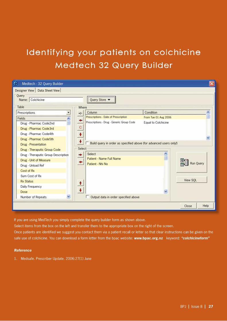
Identifying your patients on colchicineMedtech 32 Query Builder
Medtech - 32 Query Builder
Designer View Data Sheet View
QueryName: Query Store
Prescriptions
Table
Fields
Column Condition
Run Query
View SQL
HelpClose
Output data in order specified above
Build query in order as specified above (for advanced users only!)
Select
Where
Select
Drug - Pharmac Code2nd
Drug - Pharmac Code3rd
Drug - Pharmac Code4th
Drug - Pharmac Code5th
Drug - Presentation
Drug - Theraputic Group Code
Drug - Theraputic Group Description
Drug - Unit of Measure
Drug - Unload Ref
Cost of Rx
Sum Cost of Rx
Rx Status
Daily Frequency
Dose
Number of Repeats
Prescriptions - Date of Prescription
Prescriptions - Drug - Generic Group Code
From Tue 01 Aug 2006
Equal to Colchicine
Patient - Name Full Name
Patient - Nhi No
C
Colchicine
If you are using MedTech you simply complete the query builder form as shown above.
Select items from the box on the left and transfer them to the appropriate box on the right of the screen.
Once patients are identified we suggest you contact them via a patient recall or letter so that clear instructions can be given on the
safe use of colchicine. You can download a form letter from the bpac website: www.bpac.org.nz keyword: “colchicineform”
Reference
Medsafe. Prescriber Update. 2006:27(1) June1.
BPJ I Issue 8 I 27

e t c e v i d e n c e t h a t c o u n t s
Effect of Exercise on HDL
Published in Journal Watch General Medicine June 14, 2007
Available from http://general-medicine.jwatch.org/cgi/
content/full/2007/614/5
Bottom line: In this meta-analysis, aerobic exercise
raised HDL cholesterol levels only modestly, and an
exercise duration of less than 30 minutes per session
failed to raise HDL. However, these results should not
discourage exercise, which is associated with numerous
benefits regardless of effect on lipids.
A meta-analysis finds that duration of aerobic exercise, but
not frequency or intensity, is associated with change in HDL
levels.
We routinely advise patients to increase aerobic exercise as
a means of raising HDL cholesterol levels. These researchers
conducted a meta-analysis to assess the overall effect
of aerobic exercise on HDL levels and to determine which
properties of an exercise program have the greatest effect.
A total of 35 trials, including about 1400 subjects (mean
intervention period, 27 weeks), were included in the analysis.
After exercise training, HDL levels were a mean of 2.53 mg/dL
higher in patients randomized to exercise than in controls — a
significant difference. Duration of exercise was significantly
associated with change in HDL: Increases in HDL were
significant only beyond thresholds of 120 minutes per week
for total duration and 30 minutes for individual sessions. More
frequent exercise sessions (independent of total duration) and
more strenuous exercise were not associated with increased
HDL.
— Jamaluddin Moloo, MD, MPH
Reference
Kodama S et al. Effect of aerobic exercise training on serum levels of high-density lipoprotein cholesterol: A meta-analysis. Arch Intern Med 2007 May 28; 167:999-1008.
Both conventional and atypical antipsychotics increase
risk of femoral fracture in elderly patients
National Electronic Library for Medicines
Bottom Line: The authors conclude that their study found
both atypical and conventional antipsychotic agents to
be associated with an increase in risk of femoral fracture
in elderly, institutionalised patients. These drugs should
therefore be used with caution in elderly patients, especially
those at increased risk of falls.
A case-control study has found that in institutionalised elderly
patients, both conventional and atypical antipsychotic drugs
increase the risk of femoral fracture. Conventional antipsychotics
have been linked to an increase in risk of fracture in the elderly,
via an increase in the risk of falling due to effects on gait and
movement. Some trial evidence suggests a lower risk with the
atypical antipsychotics, and this study aimed to clarify whether this
was so by examining risk of hospitalisation for femoral fracture in
relation to use of drugs in either group.
Study subjects were nursing home residents from six US states.
Cases consisted of 1787 patients with fractured femur, who
were compared with 5606 controls with no fracture living in the
same institution at the same time. After adjusting for potential
confounding factors, the risk of fracture for those taking
atypical antipsychotics was statistically the same as those in the
conventional drugs: relative to non-users, the odds ratios were
1.37 (95% CI 1.11 to 1.69) and 1.35 (95% CI 1.06 to 1.171)
respectively. Numbers were sufficient to calculate risks for three
individual agents - risperidone (OR, 1.42; 95% CI 1.12 to 1.80),
olanzapine (OR, 1.34; 95% CI 0.87 to 2.07), and haloperidol (OR,
1.53; 95% CI 1.18 to 2.26).
References
Atypical antipsychotics raise risk of femoral fracture in nursing home residents. J Clin Psychiatry 2007; 68: 929-934.
28 I BPJ I Issue 8
Rosiglitazone: More Data, Continuing Concern
Published in Journal Watch General Medicine June 12, 2007
Available from http://general-medicine.jwatch.org/cgi/content/
full/2007/612/2
Bottom Line: By itself, this interim analysis doesn’t settle the
question of whether rosiglitazone increases risk for MI or
death. However, the results are not reassuring: The primary
endpoint is in the “wrong direction” for rosiglitazone (although
not statistically significantly so), and the increased risk for
heart failure is striking. This report — superimposed on the
earlier meta-analysis — convinces an editorialist that clinicians
should no longer feel comfortable prescribing rosiglitazone. I
agree.
An editorialist concludes that clinicians should no longer feel
comfortable prescribing the diabetes drug.
In a recent meta-analysis, rosiglitazone was associated with
increased risk for myocardial infarction and possibly cardiovascular
mortality (Journal Watch May 24 2007). The authors noted that the
industry-sponsored RECORD trial, specifically designed to examine
cardiovascular outcomes associated with rosiglitazone, was still in
progress. Because of the controversy sparked by the meta-analysis,
the RECORD investigators conducted this interim analysis (about two
thirds of the way through the trial).
Researchers in Europe and Australasia enrolled 4447 type 2 diabetic
patients taking metformin or sulfonylurea monotherapy. Half the
patients were randomized to receive add-on rosiglitazone; in the control
group, metformin users received add-on sulfonylurea, and sulfonylurea
users received add-on metformin. During an average follow-up of 3.75
years, the primary endpoint (cardiovascular death or cardiovascular
hospitalization) occurred in 217 rosiglitazone patients and 202
controls (hazard ratio, 1.08; P=0.43). For secondary endpoints, the
only statistically significant finding was an increased risk for heart
failure in the rosiglitazone group compared with the control group (38
vs. 17 events; P=0.006). A slight excess of MIs in the rosiglitazone
group was not significant (43 vs. 37 events; P=0.5).
— Allan S. Brett, MD
Reference
Home PD et al. Rosiglitazone evaluated for cardiovascular outcomes — An interim analysis. N Engl J Med 2007 Jun 5; [e-pub ahead of print]. (http://
dx.doi.org/10.1056/NEJMoa073394)
Nathan DM. Rosiglitazone and cardiotoxicity — Weighing the evidence. N Engl J Med 2007 Jun 5; [e-pub ahead of print]. (http://dx.doi.org/10.1056/
NEJMe078117)
Role of statins for the primary prevention of
cardiovascular disease in patients with type 2
diabetes mellitus
National Electronic Library for Medicines
Bottom Line: The authors conclude, “Current
ADA recommendations may be too aggressive as
available evidence suggests that the decision to
initiate pharmacotherapy with a statin in patients
with type 2 diabetes mellitus who do not have pre-
existing CHD should be individualised rather than
based solely on the diagnosis of type 2 diabetes
mellitus.”
The authors of this American article review and evaluate
the major statin trials that included a significant number
of patients with diabetes without pre-existing coronary
heart disease (CHD). They also discuss the role statins
should play in primary prevention. The following primary
prevention trials are discussed in the article:
Antihypertensive and Lipid-Lowering Treatment to -
Prevent Heart Attack Trial (ALLHAT-LLT)
Heart Protection Study (HPS) -
Anglo–Scandinavian Cardiac Outcomes Trial–Blood -
Pressure Lowering Arm (ASCOT-BPLA)
Collaborative atorvastatin diabetes study (CARDS) -
The atorvastatin study for prevention of coronary -
heart disease endpoints in non-insulin-dependent
diabetes mellitus (ASPEN)
Guidelines from the American Diabetes Association
(ADA) recommend statin therapy in the majority of
patients with diabetes. The authors note that the first
4 studies above (which included a significant number of
patients with diabetes and no history of CHD) have had
an impact on treatment guidelines. However, they also
add that these studies had various methodological flaws
and some non-significant results. ASPEN was the most
recent trial published since the ADA guidelines were
issued. This trial found that in patients with diabetes at
lower CHD risk, atorvastatin 10 mg was not superior to
placebo in reducing time to the first major CV event or
procedure.
Reference
Lancet 2007; 370: 292, 293-4, 319-28
BPJ I Issue 8 I 29

e t c e v i d e n c e t h a t c o u n t s
Cannabis use associated with increase in risk of psychotic disorder
National Electronic Library for Medicines
Published evidence is consistent with an increased risk of
psychosis in cannabis users, according to a systematic
review published today. The review, which has inevitably
generated considerable media interest, was funded by
the UK Department of Health.
As cannabis is the most frequently used illegal substance
in many countries, there is considerable concern over
whether it has any long-term adverse effects. Increase in
use at younger ages, while the brain is still developing,
has sharpened this concern. There is strong evidence
that use can provoke transient psychotic and affective
experiences, and this review aimed to determine whether
there was any evidence for any longer term effect. The
authors searched a wide range of sources for published
population-based longitudinal studies or case-control
studies within longitudinal designs that looked at psychotic
or affective mental health outcomes in association with
cannabis use. Study quality was assessed on a range
of factors including methods to address bias and
confounding factors, reverse causation, missing data,
response rates, etc.
The initial literature search yielded 4,804 references of
which 173 were considered potentially relevant on the
basis of title and abstract. Of these, 143 were excluded
after full examination to leave 35 for analysis: 11 for
psychosis (from 7 cohorts) and 24 for affective disorders
(from 15 cohorts). Unadjusted results from all studies on
psychosis showed an increased risk with cannabis use
in all seven cohorts, which remained positive in six after
adjustment for confounding. Pooled estimates showed
an increased risk of psychosis associated with cannabis
use, with an adjusted odds ratio of 1.41 (95% CI 1.20
to 1.65).
Where the data were available, there was evidence for a dose-
response effect with the OR in most frequent users being 2.09
(95% CI 1.54 to 2.84). The evidence for effects on depressive
outcomes was much weaker - effect sizes were small and many
of the included studies were too small.
The authors conclude that the published evidence is consistent
with the view that use of cannabis is associated with an increased
risk of psychosis. They discuss in some depth the steps taken
to try and minimise the weaknesses of the studies included,
the most important being confounding factors (people who use
cannabis are also those at greater risk for other reasons) and
reverse causation (people with early symptoms are more likely to
use cannabis in an attempt to relieve these). While considerable
efforts were made to reduce these, they can never be eliminated
in observational studies. In the studies of affective outcomes
in particular, reverse causation was poorly addressed. These
factors are unlikely to be resolved, as proof would require a
large randomised controlled trial that is not feasible. An estimate
suggests that up to 14% of psychotic outcomes in young adults
in the UK would not occur in the absence of cannabis use,
however this relies on the assumptions that the link is causal
and the pooled estimate is accurate. Incidence figures do not
show parallels between schizophrenia and trends in cannabis
use, however time lags and lack of reliable incidence data may
affect these.
Overall, therefore, they consider that although causality cannot
be proven, and confounding cannot be ruled out, the evidence
for a link between cannabis use and increased risk of psychotic
illness is sufficiently strong to justify public health warnings on
the issue. Although the individual lifetime risk for even regular
users is low (<3%), on a population level the impact would be
significant because of the drug’s widespread use.
30 I BPJ I Issue 8

Cochrane review: saline irrigation
effective in chronic rhinosinusitis
National Electronic Library for Medicines
Nasal irrigation in chronic rhinosinusitis does
appear to be worthwhile, despite the patient
effort involved, according to a Cochrane
review. The authors note that nasal irrigation
as a treatment for chronic nose and sinus
problems originated in some alternative
medical practices and has become more
common as a mainstream therapy. It may
often involve considerable efforts by the
patient, however, and it is not clear how
beneficial it is: this review aimed to determine
whether there was good published evidence
to support the practice. The authors carried
out a comprehensive literature search
for randomised controlled trials in which
saline was evaluated against placebo, no
treatment, against other treatments, or as
an adjunct to other treatments. They also
looked for studies comparing isotonic with
hypertonic saline.
A total of eight eligible trials were located,
comparing saline irrigation with no treatment
(n=3), with placebo (n=1), with an intranasal
steroid spray (n=1), and as an adjunct to
intranasal steroid spray (n=1): there were
two studies comparing different hypertonic
solutions with isotonic saline. Analysis
indicated that saline irrigation provided
effective symptom relief as sole treatment,
and also as an adjunctive therapy. It is
less effective alone than intranasal steroid,
however. There is some evidence that
hypertonic solutions may give better results,
but this is not clear. Overall, the authors
conclude that saline nasal irrigations have
beneficial effects in chronic rhinosinusitis,
both as sole treatment and as an adjunct.
Minor side effects are common, however for
most patients these are outweighed by the
benefits and the procedure is well tolerated.
Inhaled corticosteroids increase risk of pneumonia in elderly patients with
COPD?
National Electronic Library for Medicines
According to the results of this nested case-control study, the use of inhaled
corticosteroids among elderly patients with COPD is associated with an excess risk
of hospitalisation due to pneumonia.
Researchers analysed data from health databases of a health insurance agency
in Quebec (holds information on all 7 million residents), and identified all subjects
aged 66 years or above who were dispensed at least one of the following between
1988 and 2001: any beta-agonist, theophylline, ipratropium bromide, sodium
cromoglycate, nedocromil, or ketotifen. The cohort of patients with COPD was
formed from this source population by identifying all subjects with three or more
prescriptions for these medications in any 1-year period and on at least two different
dates (subjects with a primary or secondary diagnosis of asthma were excluded).
Cases were identified as those who were hospitalised for pneumonia during follow-
up; all cases were age and time matched to four control subjects.
The main findings were as follows:
The cohort included 175,906 patients with COPD of whom 23,942 were -
hospitalised for pneumonia during follow-up, for a rate of 1.9 per 100 per year,
and matched to 95,768 control subjects.
The case subjects had more severe respiratory disease, and more co- -
morbidity.
The current use of inhaled corticosteroids was found to be associated with an -
increased risk of hospitalisation for pneumonia (adjusted rate ratio [ARR] of
1.70; 95% CI 1.63–1.77) and an increased risk of hospitalisation for pneumonia
followed by death within 30 days (ARR 1.53; 95% CI 1.30–1.80).
There was a dose–response relationship, with the rate of pneumonia greatest -
with the highest doses of inhaled corticosteroids, equivalent to fluticasone at
1,000 mcg/day or more (RR, 2.25; 95% CI, 2.07–2.44).
All-cause mortality was similar for patients hospitalised for pneumonia, whether -
or not they had received inhaled corticosteroids in the recent past (7.4 and
8.2%, respectively).
The authors conclude that ‘the use of inhaled corticosteroids is associated with an
excess risk of pneumonia hospitalisation and of pneumonia hospitalisation followed
by death within 30 days, among elderly patients with COPD’. They recommend that
this adverse effect ‘needs to be considered when prescribing these medications to
patients with COPD’.
Reference
Inhaled Corticosteroid Use in Chronic Obstructive Pulmonary Disease and the Risk of
Hospitalization for Pneumonia American Journal of Respiratory and Critical Care Medicine; Vol
176:162-166
BPJ I Issue 8 I 31

Independent evidence-based thinking about health care
Bandolier GastrooesophaGeal reflux and
BMI
There is a general understanding of a relationship between weight and increased prevalence of heartburn, or symptoms of gastrointestinal reflux. Indeed, there is a meta-analysis [1] indicating significant increase for those with a BMI of 25 kg/sq m or more compared with those with lower BMI.
However, this is something of a blunt analysis, and does not tell us much about gradations. For instance, is there a gradual increase in risk, or does the risk increase dramatically at any particular BMI? Is there any evidence of a U-shaped relationship, perhaps with higher rates in underweight people? A new study [2] fills in some of the fine details.
StudyPart of the US Nurses study, this survey involved a questionnaire to a random selection of 12,192 nurses, with questions about frequency, severity, and duration of heartburn or acid regurgitation, using validated definitions of both terms. Severity was defined as mild (can be ignored), moderate (cannot be ignored but does not affect lifestyle), severe (affects lifestyle), and very severe (markedly affects lifestyle). Frequent was an episode occurring at least weekly.
Information was collected regarding height, weight (at various ages), drugs, diet, exercise, tobacco and alcohol use, and concurrent disease. Analysis of results used these data to examine confounding variables. Controls were women without symptoms not taking acid suppressing medicines.
ResultsThe women in the survey had an average age of 66 years, and an average BMI of about 27. Women with symptoms were more likely to have a higher BMI, use medications for asthma or hypertension, or hormone preparations, consumed more calories, and were less active.
Over 10,500 questionnaires were returned, with an 86% response rate. No symptoms of heartburn or acid reflux were reported in 41% (1 in 10 of whom were using proton pump inhibitors), with the remaining 51% reporting symptoms less frequently than monthly, to daily (Figure 1). One woman in five (22%) had symptoms at least weekly. Of those with symptoms, most (95%) were moderate or mild, and only about 5% had symptoms that were severe or very severe, and most (55%) had both heartburn and acid reflux.
Using those women with frequent (at least weekly) symptoms, and women without symptoms as controls, there was increasing reporting of symptoms of heartburn or acid reflux with increasing BMI (Figure 2), even after adjusting for potential confounders. This was the case for mild, moderate and severe or very severe symptoms. With a BMI ≥25, 60% of the increased risk was accounted for by excess weight.
Figure 2: Odds ratio for heartburn or acid reflux at different BMI levels
Among women who had gained weight during the previous 14 years, there was a dose-dependent increase in the risk of symptoms, with about a threefold increase in those whose BMI increased by 3.5 units. Conversely, there was a reduction in almost 40% in the risk of frequent symptoms in women who reduced BMI by 3.5 units or more.
CommentThis nicely captures the relationship between increased risk of heartburn or acid reflux and increased weight. Being or becoming overweight doubles the risk of having these symptoms at least once a week. A back-
Bandolier 160, www.ebandolier.com
Figure 1: Frequency of symptoms
Figure 3: Crude symptom rate and BMI
32 I BPJ I Issue 8
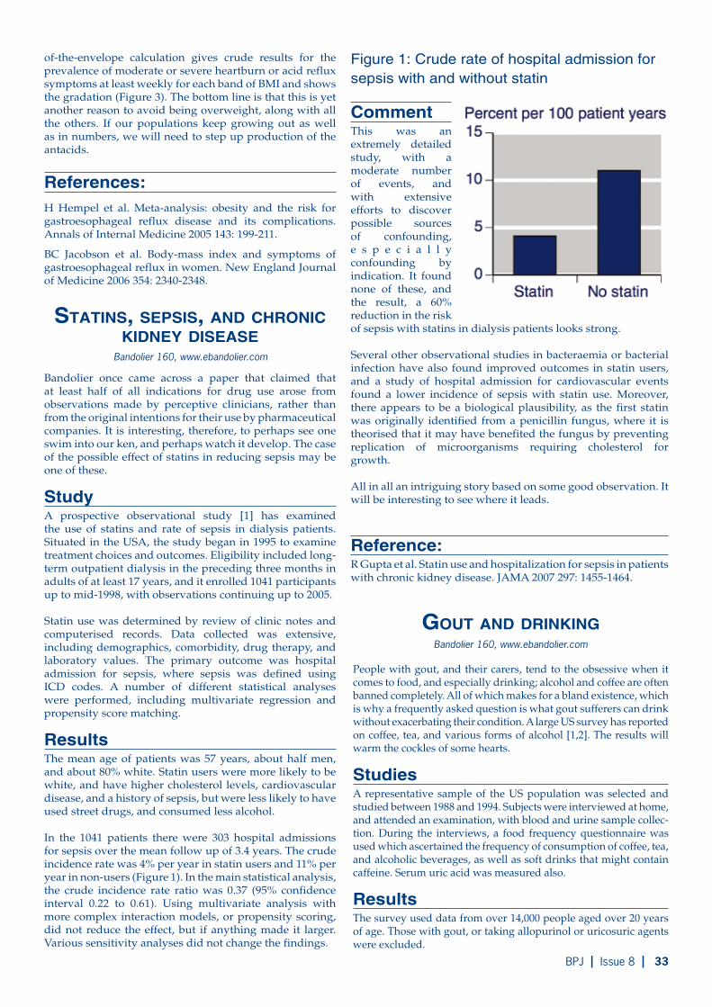
Bandolier 160, www.ebandolier.com
statIns, sepsIs, and chronIc kIdney dIsease
Bandolier once came across a paper that claimed that at least half of all indications for drug use arose from observations made by perceptive clinicians, rather than from the original intentions for their use by pharmaceutical companies. It is interesting, therefore, to perhaps see one swim into our ken, and perhaps watch it develop. The case of the possible effect of statins in reducing sepsis may be one of these.
StudyA prospective observational study [1] has examined the use of statins and rate of sepsis in dialysis patients. Situated in the USA, the study began in 1995 to examine treatment choices and outcomes. Eligibility included long-term outpatient dialysis in the preceding three months in adults of at least 17 years, and it enrolled 1041 participants up to mid-1998, with observations continuing up to 2005.
Statin use was determined by review of clinic notes and computerised records. Data collected was extensive, including demographics, comorbidity, drug therapy, and laboratory values. The primary outcome was hospital admission for sepsis, where sepsis was defined using ICD codes. A number of different statistical analyses were performed, including multivariate regression and propensity score matching.
ResultsThe mean age of patients was 57 years, about half men, and about 80% white. Statin users were more likely to be white, and have higher cholesterol levels, cardiovascular disease, and a history of sepsis, but were less likely to have used street drugs, and consumed less alcohol.
In the 1041 patients there were 303 hospital admissions for sepsis over the mean follow up of 3.4 years. The crude incidence rate was 4% per year in statin users and 11% per year in non-users (Figure 1). In the main statistical analysis, the crude incidence rate ratio was 0.37 (95% confidence interval 0.22 to 0.61). Using multivariate analysis with more complex interaction models, or propensity scoring, did not reduce the effect, but if anything made it larger. Various sensitivity analyses did not change the findings.
Figure 1: Crude rate of hospital admission for sepsis with and without statin
CommentThis was an extremely detailed study, with a moderate number of events, and with extensive efforts to discover possible sources of confounding, e s p e c i a l l y confounding by indication. It found none of these, and the result, a 60% reduction in the risk of sepsis with statins in dialysis patients looks strong.
Several other observational studies in bacteraemia or bacterial infection have also found improved outcomes in statin users, and a study of hospital admission for cardiovascular events found a lower incidence of sepsis with statin use. Moreover, there appears to be a biological plausibility, as the first statin was originally identified from a penicillin fungus, where it is theorised that it may have benefited the fungus by preventing replication of microorganisms requiring cholesterol for growth.
All in all an intriguing story based on some good observation. It will be interesting to see where it leads.
Reference:R Gupta et al. Statin use and hospitalization for sepsis in patients with chronic kidney disease. JAMA 2007 297: 1455-1464.
of-the-envelope calculation gives crude results for the prevalence of moderate or severe heartburn or acid reflux symptoms at least weekly for each band of BMI and shows the gradation (Figure 3). The bottom line is that this is yet another reason to avoid being overweight, along with all the others. If our populations keep growing out as well as in numbers, we will need to step up production of the antacids.
References:H Hempel et al. Meta-analysis: obesity and the risk for gastroesophageal reflux disease and its complications. Annals of Internal Medicine 2005 143: 199-211.
BC Jacobson et al. Body-mass index and symptoms of gastroesophageal reflux in women. New England Journal of Medicine 2006 354: 2340-2348.
Gout and drInkInG
People with gout, and their carers, tend to the obsessive when it comes to food, and especially drinking; alcohol and coffee are often banned completely. All of which makes for a bland existence, which is why a frequently asked question is what gout sufferers can drink without exacerbating their condition. A large US survey has reported on coffee, tea, and various forms of alcohol [1,2]. The results will warm the cockles of some hearts.
StudiesA representative sample of the US population was selected and studied between 1988 and 1994. Subjects were interviewed at home, and attended an examination, with blood and urine sample collec-tion. During the interviews, a food frequency questionnaire was used which ascertained the frequency of consumption of coffee, tea, and alcoholic beverages, as well as soft drinks that might contain caffeine. Serum uric acid was measured also.
ResultsThe survey used data from over 14,000 people aged over 20 years of age. Those with gout, or taking allopurinol or uricosuric agents were excluded.
Bandolier 160, www.ebandolier.com
BPJ I Issue 8 I 33

Coffee, tea, and caffeine
Using a quintile of consumption approach, uric acid levels were identical across quintiles of intake of total caffeine and tea. For coffee (including decaffeinated), drinking more than four cups of coffee a day signifi-cantly lowered serum uric acid levels, by about 8% at maximum (Figure 1). The reduction of uric acid by coffee remained after adjusting for a whole range of variables and dietary factors.
Alcohol
Using the quintile of consumption approach drinking wine did not affect serum uric acid levels at any level of consumption up to one serving per day or more. The consumption of spirits, and especially beer, did increase serum uric acid levels (Figure 2), even after adjusting for a whole range of factors. Beer and spirits drunk daily increased serum uric acid by about 10%; wine did not. The results were similar in men and women, and at lower and higher levels of BMI.
Figure 2: Effect of different daily consumption (quintiles) of different alcoholic beverages on mean serum uric acid levels
Figure 3: Relative risk of incident gout in 12-year follow up of 46,000 men, according to quintiles of daily coffee consumption
Figure 1: Reduction in mean serum uric acid levels according to quintiles of daily intake of coffee
0 <1 1-3 4-5280
290
300
310
320
330
Serum uric acid (µmol/L)
Cups of coffee per day≥
0 <1 1-3 4-50.2
0.4
0.6
0.8
1Relative risk of incident gout
Cups of coffee per day≥
0 0.01-0.09 0.1-0.49 0.5-0.99-10
0
10
20
30
Difference in serum uric acid from no consumption (µmol/L)
Frequency (servings per day)
Beer Spirit Wine
≥
Comment
This constitutes useful additional knowledge about what gout suf-fers might do to avoid increasing their serum uric acid, and perhaps precipitating an attack, or making the pain worse. Drinking beer and spirits are out, but tea and wine have no effect, while coffee ac-tually seems to reduce uric acid levels. We have had some straws in the wind about cof-fee before, but this adds weight.
More weight comes from a large study of coffee consumption and incident gout in men [3], following 46,000 men with no history of gout at baseline for 12 years. There were 750 cases of incident gout, and the risk was lower with higher coffee con-sumption, before and after adjustment for a whole host of different possible confounding factors (Figure 3). So increased coffee drink-ing is linked with both reduced serum uric acid levels and reduced incidence of clinical gout.
We also have information about what we eat and the risk of incident gout [4]. This has been examined in detail on the Bandolier Internet site, but the main results are worth reiterating. Increased consump-tion of meat was associated with increased risk of gout, but only with beef, pork, and lamb. There was less association with seafood, and none with purine rich vegetables. Increased consumption of dairy food reduced the risk of gout. We find the same now for uric acid [5] where high meat and to a small extent seafood consumption is associated with higher uric acid levels, but dairy food with lower uric acid levels. Much food for thought for those with gout and for healthy eating.
References:HK Choi, G Curhan. Coffee, tea, and caffeine consumption and serum 1.
uric acid level: third National Health and Nutrition Examination Survey. Arthritis & Rheumatism 2007 57: 816-821.HK Choi, G Curhan. Beer, liquor, 2. and wine consumption and serum uric acid level: third National Health and Nutrition Examination Survey. Arthritis & Rheumatism 2004 51: 1023-1029.HK Choi et al. Coffee consumption 3. and risk of incident gout in men. Arthritis & Rheumatism 2007 56: 2049-2055.HK Choi et al. Purine-rich foods, 4. dairy and protein intake, and the risk of gout in men. NEJM 2004 350:1093-1103.HK Choi et al. Intake of purine-rich 5. foods, protein, and dairy products and relationship to serum levels of uric acid. Arthritis & Rheumatism 2005 52: 283-289.
34 I BPJ I Issue 8

Bandolier 161, www.ebandolier.com
fracture and qualIty of lIfe In older woMen
Fractures in older people, especially older women, can be problematical. The impact of hip fracture can be devastating. Much treasure is spent on trying to prevent fracture through treatment of osteoporosis, and by trying to reduce loss of bone, especially in postmenopausal women. If we want to know how treatments compete in the clever world of cost effectiveness, then we have to measure the negative impact of fractures, and while much has been done in that area, a new, and very large, study [1] opens another window.
StudyThis was part of a prospective longitudinal study of 200,000 postmenopausal US women aged at least 50 years, without a diagnosis of osteoporosis, no bone density measurement within 12 months, and not taking treatments for osteoporosis. For inclusion they has to have completed two mail or telephone surveys, the first at about 12 months after enrolment, and the second about 36 months after enrolment.
Both surveys elicited information of new fractures, health status using a SF-12 instrument, osteoporosis-related care, and fall history. Analysis of the SF-12 data was according to two composite scores, the physical component score (PCS) and mental component score (MCS). Reported new fractures (hip, spine, wrist, rib) between the first and second surveys formed the cases, with controls being women without fracture.
ResultsThe analysis included 86,128 women (88% white), whose mean age was about 65 years. Just 1.2% had suffered a fracture in the year before the first survey. Fractures between the first and second survey numbered 320 hip, 445 spine, 835 wrist, and 657 rib, 2.6% over the two years. There were 83,871 women without fracture who served as controls.
Women suffering a fracture more frequently had significantly reduced bone mineral density, and were 4-6 times more likely to have suffered a fracture during the 12 months before the first survey. They also had lower quality of life scores at the first survey.
After adjusting quality of life scores for these factors, women suffering a fracture in the two years between the two surveys had significantly reduced PCS scores compared with women without a fracture (Figure 1). Statistically significant reductions were found for hip, spine, wrist and rib fractures for younger postmenopausal women (50-64 years), and for hip, spine and rib fractures in older postmenopausal women (65-99 years).
Women suffering a fracture in the two years between the two surveys had greater reduction in MCS scores than women without a fracture (Figure 2). Statistically significant reductions were found for spine and rib fractures for younger postmenopausal women, and for hip and spine fractures in older postmenopausal women.
CommentWhat makes this study worth thinking about is the combination of its size and detail, and that it provides quality of life results for different fractures in younger and older postmenopausal women. It also did some useful statistical stuff, like taking into account multiple comparisons, so that statistical significance was only reported when the probability value was 0.004, so it does not tell us about associations that crept into conventional levels of significance.
Those in the know about such things may not have been surprised by the findings. For the rest of us, perhaps what stands out is the particular loss of life quality attendant on vertebral fractures. This may reflect the fact that vertebral fractures cannot be healed, and often come with a lot of back pain, and we do know that chronic pain has a large negative impact on quality of life.
It also helps to have some context. The negative impact on PCS scores for hip and spine were at the same level as those for COPD, hip impairment, or rheumatoid or osteoarthritis. Given that we will have more older people with low bone mineral density and at risk of these fractures, this should help in making sense of current and new therapy choices.
Reference:SK Brenneman et al. Impact of recent fracture on health-related quality of life in postmenopausal women. Journal of Bone and Mineral Research 2006 21: 809-816.
Figure 1: Reduction of physical quality of life compared with control
Figure 2: Reduction of mental quality of life compared with control
BPJ I Issue 8 I 35

If you have a clinical question email it to [email protected]
Dear DaveDave and other members of the bpacnz team answer your clinical questions
Does the ‘seven day rule’ still apply with the concomitant use of combined oral contraceptives and all antibiotics?
In June 2006, the American College of Obstetricians and Gynaecologists (ACOG) released a practice bulletin on the use of hormonal contraception in women with coexisting medical conditions. This bulletin has cast some doubt over whether it is valid for GPs to advise their patients taking oral contraceptives that they need to use other methods of contraception for the duration of antibiotic treatment and the following seven days.
“Although there have been many anecdotal reports of
oral contraceptive failure in women taking concomitant
antibiotics, pharmacokinetic evidence of lower serum
steroid levels exists only for rifampicin. Because oral
contraceptive steroid concentrations are strikingly
reduced in women concomitantly taking rifampicin,
such women should not rely on combination oral
contraceptives, progestin-only oral contraceptives or
implants for contraceptive protection”. ACOG1
The fact that rifampicin can cause oral contraceptive
failure is unequivocal due to enzyme induction,
increased oestrogen metabolism and resultant
reduced plasma oestrogen concentrations. Most
other antibiotics have been reported to be associated
with oral contraceptive failure, but as stated in the
ACOG Practice Bulletin, clinical studies have not
demonstrated that antibiotics (other than rifampicin)
decrease serum steroid concentrations.
There is a theoretical basis for an interaction in that
antibiotics reduce gut flora which are responsible for
increasing the reabsorption of oestrogens from the
GI tract. Oestrogens are metabolised in the liver and
conjugated with glucuronide, which is water soluble
and can be excreted in the bile. Under normal gut flora
conditions, bacteria cleave this conjugate and free up
oestrogen, which can then be reabsorbed (enterohepatic
recycling). Although the theory is not backed up by
evidence from clinical studies, an interaction cannot be
completely ruled out as in some women enterohepatic
recycling may be crucial, in maintaining adequate
oestrogen plasma concentrations. Clinical studies
may also not represent the situation in practice where
antibiotics or the underlying illness may cause diarrhoea
or vomiting, which are known to reduce the effectiveness
of oral contraceptives.
Bearing in mind that there is a background failure rate
associated with oral contraception, it is not possible to
prove that an antibiotic given concurrently is causative or
contributory to a case of failure. Although an interaction
and resultant contraceptive failure is probably extremely
unlikely, the possibility cannot be completely excluded.
On moral and ethical grounds most authorities continue
to sanction the cautious approach and continue to
recommend the seven day rule.
Reference
Stockley’s Text Book of Drug Interactions 2007.
ACOG Practice Bulletin. Clinical Management Guidelines for Obstetricians and Gynecologists. Number 73, June 2006.1.
36 I BPJ I Issue 8
Who is Dave?Pharmaceutical Programme Manager Dave Woods is a graduate of Manchester University (B.Sc.
[Hons]) and the University of Otago (MPharm). Dave has extensive experience in hospital pharmacy,
drug information, rational use of drugs and quality assurance. He has published on a range of subjects
and holds editorial positions for several international journals.
If you have a clinical question email it to [email protected]
Dear Dave Serotonin toxicity: “Is combining Reductil with tricyclic antidepressants really such a no-no or are the drug companies just being defensive over the unlikely occurrence of serotonin syndrome?”
Bob Buckham from the Christchurch Drug Information
Centre was asked if he had come across this question in
practice. His advice was as follows:
Sibutramine and amitriptyline work by inhibiting the
reuptake of serotonin and noradrenaline. Concurrent use
of these drugs is actively discouraged because of the
potential for serotonin toxicity (which may range from mild
symptoms, such as diarrhoea or sweating, through to
coma and death). Furthermore, TCAs may cause weight
gain and both agents lower the seizure threshold.
The risk would be (theoretically) greater with tertiary
amine TCAs (compared to secondary amine) as they are
more serotonergic. So it could be argued that nortriptyline
might be ‘safer’ and less likely to cause serotonin toxicity
than amitriptyline or imipramine. The concurrent use of
sibutramine and clomipramine should definitely be avoided
as this TCA has potent serotonerigic properties.
Similarly, it would be a dose-dependent effect, therefore
the relevance of dose needs to be considered. However
overall we try to discourage the combination and suggest
trying orlistat (Xenical) first-line. If that is not an option
then we would need to seriously weigh risk versus
benefit and monitor carefully. We also make the same
recommendation if the patient is taking an SSRI or a
monoamine oxidase inhibitor (MAOI).
Serotonin toxicity
Instead of serotonin ‘syndrome’, we try to refer to it
as serotonin toxicity, as most people usually know
that the ‘syndrome’ is actually only rarely reported
– so they tend to disregard it. Whereas ‘toxicity’
suggests a range of issues from mild symptoms,
like diarrhoea and sweating, many probably wouldn’t
think to associate it with serotonin toxicity, to the
serious signs like tremor, seizures, coma and death
(the ‘syndrome’).
In summary, like other combinations which have
the potential for toxicity (e.g. SSRIs + TCAs) the
combination of sibutramine and a TCA may be
uneventful in many people. The risk of an interaction
is probably lower with low doses of nortriptyline than
with other TCAs at high doses.
Concurrent use is governed by appreciation of risk
versus benefit, recognition of the general advice
against its use and the need to closely monitor
patients if the combination is used.
Further reading about serotonin toxicity
http://snipurl.com/1ps35
BPJ I Issue 8 I 37

IM injections
Dear Editor
Occasionally we need to give IM injections
(other than immunisations) e.g. antibiotics or
anti-emetics, to toddlers and young children.
What is the preferred site and at what 1.
age can the gluteal site be used?
Is the use of lignocaine (plain) ok for 2.
dilution of antibiotics (adult use only) and
if so, how much if say 4 ml total of fluid is
required for the dilution?
Low dose aspirin and the risk of GI complications
Dear Editor
I would be grateful for your advice regarding best
practice for patients on long term aspirin. An increasing
number of middle-aged and elderly patients are on
long term low dose aspirin – and I wondered what the
current advice was as to whether they should also be
on long term PPIs.
If a patient is at a high risk of GI complications or has
a history of dyspepsia, a proton pump inhibitor can be
added to low dose aspirin therapy.
It is well established that low dose aspirin produces significant
inhibition of gastric mucosal prostaglandins, even when taken
as an enteric coated tablet. Therefore, low dose aspirin has
the potential to induce gastric lesions and GI complications
even in normal healthy subjects.
Some factors to consider when assessing GI risk with low
dose aspirin include; a history of peptic ulcer or GI bleeding,
significant co-morbidity and previous NSAID gastropathy.
Peptic ulcer disease is of particular importance because it
leads to recurrent episodes of dyspepsia and is associated
with significant complications of bleeding and perforation.
NICE guidelines recommend that patients taking low-dose
aspirin, who have a history of dyspepsia, would benefit from
concurrent treatment with a proton pump inhibitor (NICE
Clinical Guideline 17).
It is therefore important to assess each patient on an
individual basis – check for red flags (BPJ Issue 4, April
2007), assess risk and take into account any symptoms and
other medications.
It is always worth considering the potential for
adverse effects, particularly in elderly people.
The most common side effects of proton pump
inhibitors include headache, diarrhoea and
skin rashes. Proton pump inhibitors may also
increase the risk of gastrointestinal infections
and pneumonia because of their acid suppressive
effects.
In summary, PPIs can be considered for patients
on long-term low dose aspirin therapy, with a
high risk of GI complications or a history of
dyspepsia.
References
NZGG. Management of Dyspepsia and Heartburn. -June 2004. Available from http://www.nzgg.org.nz
Sweetman SC (Ed), Martindale: The Complete Drug -Reference. London: Pharmaceutical Press. 35th Edition, 2007.
NICE. Secondary prevention in primary and -secondary care for patients following a myocardial infarction. 2007. Available from http://guidance.
nice.org.uk/CG48
38 I BPJ I Issue 8

Send your letters to...Correspondence, PO Box 6032, Dunedin
or email [email protected]
Unfortunately there is no simple answer to this. IM injections
(excluding vaccines) are generally avoided in children. Many
doctors would not use anti-emetics for childen, particulary
not IM. Their use is mainly postoperative, for oncology
or for special situations such as cyclical vomiting. IM
antibiotics would usually only be given in an emergency
situation, for example suspected meningitis en route to
hospital. However, GPs, especially in rural areas, may have
a different situation and environment to deal with. Some
antibiotics, for example benzathine penicillin, can only be
given by IM injection.
Preferred site of IM injection in children
There is little information on recommended sites for IM
injections other than vaccines in children. Manufacturers’
data sheets will often have information on the recommended
site of administration. The Ministry of Health Immunisation
Handbook (2006) states that the recommended sites for
IM vaccines are:
For children under 15 months of age, the vastus lateralis
muscle on the lateral thigh is used
For children over 15 months, both the vastus lateralis and
deltoid sites may be used – the choice will be based on the
vaccinator’s professional judgement
For older children, adolescents and adults, the deltoid
muscle is used
For injections, other than immunisations, there is no clear
guidance and it may be wise to consult MedSafe datasheets.
However there are some general areas of agreement:
The dorsogluteal site (upper outer quadrant): use of
this site is associated with significant risk of damage to
the sciatic nerve and superior gluteal artery. There is often
a deep layer of subcutaneous fat in this region and the
injection may not reach the muscle, resulting in the drug
being deposited in the subcutaneous fat.
This site should not be used in children.
The ventrogluteal site: this is a good site for intramuscular
injections in adults and children over seven months. The
site provides the greatest thickness of gluteal muscle, is
relatively free of major nerves and blood vessels and is
easy to locate. However there is little experience of use
of this site in New Zealand and consequently it is not used
often.
The lateral thigh (vastus lateralis): This site is safer than the
dorsogluteal site and is recommended for intramuscular injection
of adrenalin in anaphylaxis. Patients can be taught to self-inject
in this area.
The deltoid: This site is safe for low volume injections of non-
irritating solutions for older children and adults, provided the
deltoid muscle mass is located with care.
Using lignocaine to dilute antibiotics
Anyone considering using lignocaine for dilution should refer to
the specific datasheet of each medicine to ensure that dilution
with lignocaine is approved and compatible with the injectable
antibiotic. However, some data sheets do not include this
information, stating only that the antibiotic should not be mixed
with other medicines, while acknowledging pain on IM injection.
Note that some antibiotics (e.g. Augmentin) should not be given
by the IM route.
Most injectable drugs that allow the use of a local anaesthetic as
a diluent, will specify the same volume of diluent, be it water for
injection or 1% lignocaine, to reconstitute the powder. However,
great caution must be applied when using lignocaine in an IM
injection, as inadvertent IV administration may result in serious
cardiac adverse effects.
This advice was developed in consultation with Dr David Reith,
Paediatrician, Alan McClintock, Pharmacist and Barbara Warren,
Immunisation Co-ordinator.
References:
Ministry of Health. Immunisation Handbook 2006. Available from
http://snipurl.com/1q26s Accessed August 2007
Murtagh J, Cook I. Ventrogluteal area—a suitable site for
intramuscular vaccination of infants and toddlers. J Vaccine
2006;24(13):2403-8
Greenway K, Using the ventrogluteal site for intramuscular
injection. Nursing Standard 2005;18(25):39-42
ASCIA. Self injecting with EpiPen. Available from http://www.
allergy.org.au/aer/infobulletins/posters/Anaphylaxis_plan_
(gen)_NZ.pdfX

nzbpacbetter edicin m e
bpacnz clinical audits available online
new Zealand Gp’s leading source of rnZCGp endorsed clinical audits..............why?
visit www.bpac.org.nz


