+ All Categories
Home > Documents > Goyal VK et al: Spectrum of Lesions in Urinary Bladder ... of Lesions in Urinary Bladder... ·...

Goyal VK et al: Spectrum of Lesions in Urinary Bladder ... of Lesions in Urinary Bladder... ·...

Date post: 11-Aug-2018
Category:
Upload: tranthuy
View: 232 times
Download: 3 times
Share this document with a friend
5
Int J Dent Med Res | MAR- APR 2015 | VOL 1 | ISSUE 6 42 ORIGINAL RESEARCH Goyal VK et al: Spectrum of Lesions in Urinary Bladder Biopsies Correspondence to: Dr. Dharm Chand Kothari, Room No.- 40, Hostel No.- 4, Vivek Hostel, UG Hostel Medical College Campus, Bikaner PIN- 34001, Rajasthan, India. Spectrum of Lesions in Urinary Bladder Biopsies: Histopathological Study Vaibhav Kumar Goyal 1 , Surendra Prakash Vyas 2 , Dharm Chand Kothari 3 Background: Bladder tumor is the seventh most common tumor worldwide. Urothelial carcinoma is the commonest type accounting for 90% of all primary tumors of the bladder. As per Indian Cancer Registry data, it is the 9 th most common cancer accounting for 3.9% of all cancers. Material method: The study was carried out in the department of Pathology, Sardar Patel medical college Bikaner. Clinicopathological data of all TURBT biopsies collected were analyzed. Results: One hundred TURBT biopsy were studied, and urothelial carcinoma were classified according to WHO /ISUP (2004) classification. The most common age group was 61-70 years (33%) with Male to female ratio was 5.25:1. In carcinoma most common type was high-grade papillary urothelial carcinoma (58%) followed by low-grade papillary urothelial carcinoma (31%) papillary urothelial neoplasm of low malignant potential (4%) moderately differentiated squamous cell carcinoma (2%)and moderately differentiated adenocarcinoma (1%). In cystitis most common type is Chronic non-specific cystitis (3%) followed by eosinophilic cystitis (1%). Conclusion: In bladder most common lesion was of high-grade urothelial carcinomas presented with lamina propria and muscle invasion. Pathological grade and muscle invasion are the important valuable prognostic factors of survival. Awareness is very much needed in the public about haematuria because they neglect it causing in an advanced stage of bladder cancer at the time of presentation. KEYWORDS: Adenocarcinoma, squamous cell carcinoma, Urothelial carcinoma, Urinary Bladder AAA Diseases of the urinary bladder both non-neoplastic and neoplastic are quite common. The non-neoplastic lesions include cystitis, malakoplakia, urachal lesions, and tuberculosis. Urothelial carcinoma is the commonest type accounting for 90% of all primary tumors of the bladder. 1 As per Indian Cancer Registry data, it is the 9 th most common cancer accounting for 3.9% of all cancers. 2 Urothelial bladder tumors are classified in flat and papillary type most tumors are papillary. Carcinoma in situ and few invasive tumors have a flat pattern. 3-6 The papillary equivalent of flat in situ carcinoma is the high- grade noninvasive papillary urothelial carcinoma. 3 Progress has been made in the field of non-invasive imaging and scientists continue to identify and characterize potential markers or surrogate end points for bladder tumor physical examination, cystoscopic evaluation and histopathological analysis of biopsy material are the mainstays of contemporary bladder cancer diagnosis and treatment. The study was carried out in the department of Pathology, Sardar Patel medical college Bikaner including all the patients with urinary bladder lesion diagnosed on biopsy, who attended the hospital. Data were collected in a preset proforma. Clinical and cystoscopic findings with the clinical diagnosis of all cases of urinary bladder lesion sent to the laboratory were noted. The material for the study was comprised of biopsy from Transurethral resection of bladder Tissue (TURBT). Inclusion Criteria All the TURBT biopsies received in the department of Pathology, Sardar Patel medical college Bikaner. Exclusion Criteria Autolysed specimen Inadequate biopsies. Biopsy specimens were processed as per routine histopathological technique. Paraffin section was cut and stained by haematoxylin and eosin. Then bladder lesions were studied according to WHO/ISUP (2004) classification (Table 1). Total of 100 TURBT biopsies were analyzed. A spectrum of different pathological lesions was observed in the study. In our study most common age group was 61-70 years where 33% patients were found followed by 51-60 years (28%), 41-50 years (18%), >70 years (17%) and least common age group was < 40 years (4%). Mean and Median Age of bladder lesion were 60.7911.07 and 61.00 years respectively. Male to female ratio was 5.25:1. How to cite this article: Goyal VK , Vyas SP, Kothari DC Spectrum of Lesions in Urinary Bladder Biopsies: Histopathological Study. Int J Dent Med Res 2015;1(6):42-46. INTRODUCTION 1,3- MD, Department of Pathology, Sardar Patel Medical College, Bikaner. 2- Associate Professor, Department of Pathology, Sardar Patel Medical College, Bikaner. ABSTRACT MATERIALS & METHODS RESULTS
Transcript

Int J Dent Med Res | MAR- APR 2015 | VOL 1 | ISSUE 6 42

ORIGINAL RESEARCH Goyal VK et al: Spectrum of Lesions in Urinary Bladder Biopsies

Correspondence to: Dr. Dharm Chand Kothari, Room No.- 40, Hostel No.- 4, Vivek Hostel, UG Hostel Medical College Campus, Bikaner PIN- 34001, Rajasthan, India.

Spectrum of Lesions in Urinary Bladder

Biopsies: Histopathological Study Vaibhav Kumar Goyal1, Surendra Prakash Vyas2, Dharm Chand Kothari3

Background: Bladder tumor is the seventh most common tumor worldwide. Urothelial carcinoma is the commonest

type accounting for 90% of all primary tumors of the bladder. As per Indian Cancer Registry data, it is the 9th

most

common cancer accounting for 3.9% of all cancers. Material method: The study was carried out in the department of

Pathology, Sardar Patel medical college Bikaner. Clinicopathological data of all TURBT biopsies collected were

analyzed. Results: One hundred TURBT biopsy were studied, and urothelial carcinoma were classified according to

WHO /ISUP (2004) classification. The most common age group was 61-70 years (33%) with Male to female ratio was

5.25:1. In carcinoma most common type was high-grade papillary urothelial carcinoma (58%) followed by low-grade

papillary urothelial carcinoma (31%) papillary urothelial neoplasm of low malignant potential (4%) moderately

differentiated squamous cell carcinoma (2%)and moderately differentiated adenocarcinoma (1%). In cystitis most

common type is Chronic non-specific cystitis (3%) followed by eosinophilic cystitis (1%). Conclusion: In bladder most

common lesion was of high-grade urothelial carcinomas presented with lamina propria and muscle invasion.

Pathological grade and muscle invasion are the important valuable prognostic factors of survival. Awareness is very

much needed in the public about haematuria because they neglect it causing in an advanced stage of bladder cancer at

the time of presentation.

KEYWORDS: Adenocarcinoma, squamous cell carcinoma, Urothelial carcinoma, Urinary Bladder

AAA

Diseases of the urinary bladder both non-neoplastic and

neoplastic are quite common. The non-neoplastic lesions

include cystitis, malakoplakia, urachal lesions, and

tuberculosis. Urothelial carcinoma is the commonest type

accounting for 90% of all primary tumors of the bladder.1

As per Indian Cancer Registry data, it is the 9th

most

common cancer accounting for 3.9% of all cancers.2

Urothelial bladder tumors are classified in flat and

papillary type most tumors are papillary. Carcinoma in

situ and few invasive tumors have a flat pattern.3-6

The

papillary equivalent of flat in situ carcinoma is the high-

grade noninvasive papillary urothelial carcinoma.3

Progress has been made in the field of non-invasive

imaging and scientists continue to identify and

characterize potential markers or surrogate end points for

bladder tumor physical examination, cystoscopic

evaluation and histopathological analysis of biopsy

material are the mainstays of contemporary bladder

cancer diagnosis and treatment.

The study was carried out in the department of Pathology,

Sardar Patel medical college Bikaner including all the

patients with urinary bladder lesion diagnosed on biopsy,

who attended the hospital. Data were collected in a preset

proforma. Clinical and cystoscopic findings with the

clinical diagnosis of all cases of urinary bladder lesion

sent to the laboratory were noted.

The material for the study was comprised of biopsy from

Transurethral resection of bladder Tissue (TURBT).

Inclusion Criteria

All the TURBT biopsies received in the department of

Pathology, Sardar Patel medical college Bikaner.

Exclusion Criteria

Autolysed specimen

Inadequate biopsies.

Biopsy specimens were processed as per routine

histopathological technique. Paraffin section was cut and

stained by haematoxylin and eosin. Then bladder lesions

were studied according to WHO/ISUP (2004)

classification (Table 1).

Total of 100 TURBT biopsies were analyzed. A spectrum

of different pathological lesions was observed in the

study. In our study most common age group was 61-70

years where 33% patients were found followed by 51-60

years (28%), 41-50 years (18%), >70 years (17%) and

least common age group was < 40 years (4%). Mean and

Median Age of bladder lesion were 60.7911.07 and

61.00 years respectively. Male to female ratio was 5.25:1.

How to cite this article: Goyal VK , Vyas SP, Kothari DC Spectrum of Lesions in Urinary Bladder Biopsies: Histopathological Study. Int J Dent Med Res 2015;1(6):42-46.

INTRODUCTION

1,3- MD, Department of Pathology, Sardar Patel Medical College, Bikaner. 2- Associate Professor, Department of Pathology, Sardar Patel Medical College, Bikaner.

ABSTRACT

MATERIALS & METHODS

RESULTS

Int J Dent Med Res | MAR- APR 2015 | VOL 1 | ISSUE 6 43

ORIGINAL RESEARCH Goyal VK et al: Spectrum of Lesions in Urinary Bladder Biopsies

Haematuria was the most common clinical symptoms in

91% cases followed by strangury (48%), burning (39%)

and pain in 38% of cases.

According to cystoscopic findings, 72% patients had

papillary mass, 21% patients had solid mass, 3% patients

each diffuse thickening and ulcer while only one patient

had fungating mass.

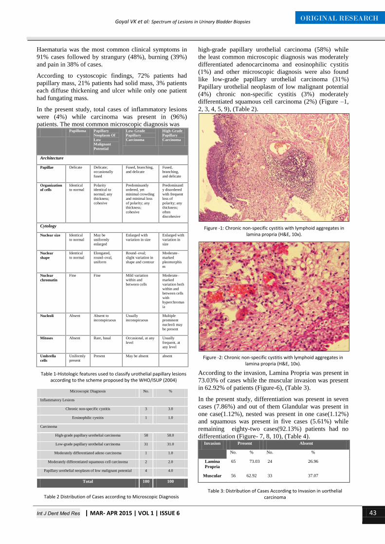
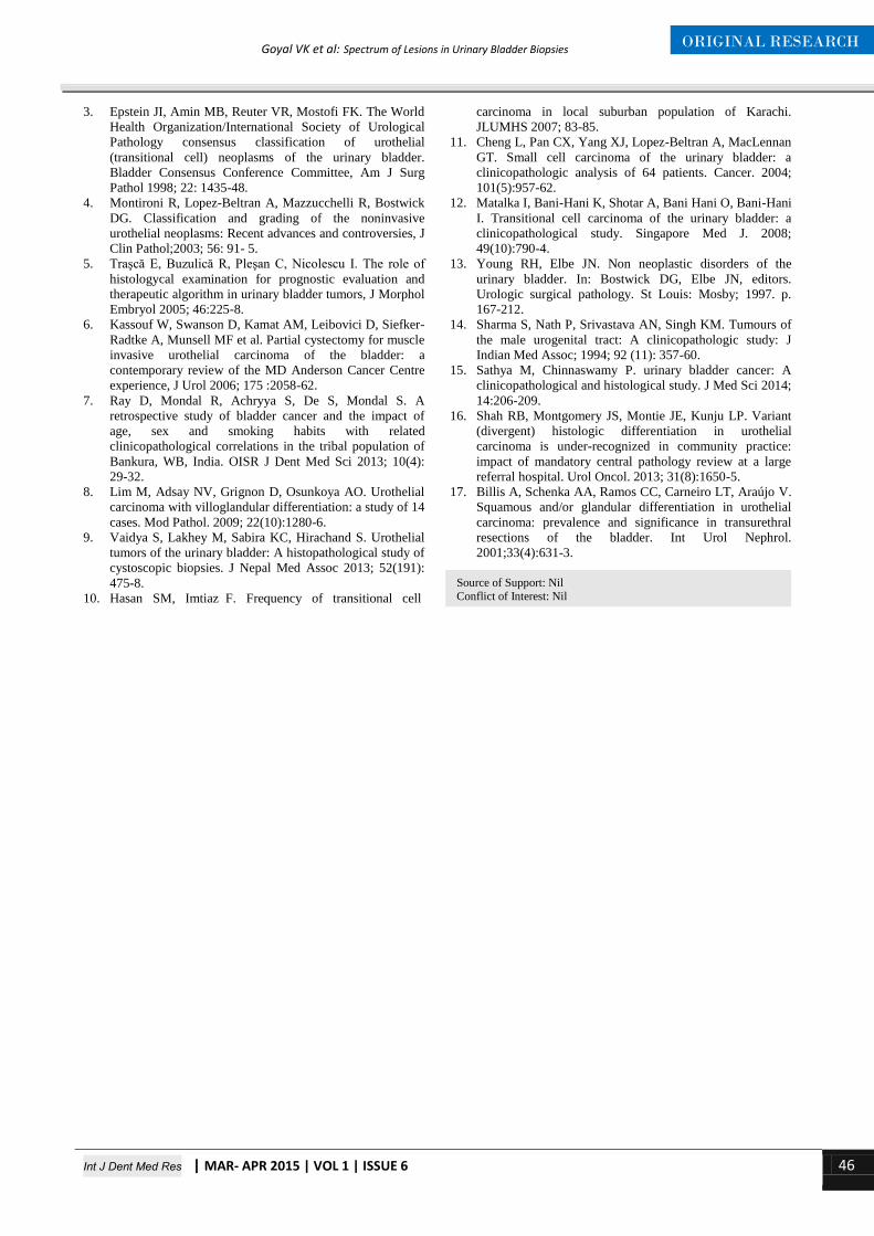
In the present study, total cases of inflammatory lesions

were (4%) while carcinoma was present in (96%)

patients. The most common microscopic diagnosis was Papilloma Papillary

Neoplasm Of

Low

Malignant

Potential

Low-Grade

Papillary

Carcinoma

High-Grade

Papillary

Carcinoma

Architecture

Papillae Delicate Delicate;

occasionally

fused

Fused, branching,

and delicate

Fused,

branching,

and delicate

Organization

of cells

Identical

to normal

Polarity

identical to

normal; any

thickness;

cohesive

Predominantly

ordered, yet

minimal crowding

and minimal loss

of polarity; any

thickness;

cohesive

Predominantl

y disordered

with frequent

loss of

polarity; any

thickness;

often

discohesive

Cytology

Nuclear size Identical

to normal

May be

uniformly

enlarged

Enlarged with

variation in size

Enlarged with

variation in

size

Nuclear

shape

Identical

to normal

Elongated,

round–oval,

uniform

Round–oval;

slight variation in

shape and contour

Moderate–

marked

pleomorphis

m

Nuclear

chromatin

Fine Fine Mild variation

within and

between cells

Moderate–

marked

variation both

within and

between cells

with

hyperchromas

ia

Nucleoli Absent Absent to

inconspicuous

Usually

inconspicuous

Multiple

prominent

nucleoli may

be present

Mitoses Absent Rare, basal Occasional, at any

level

Usually

frequent, at

any level

Umbrella

cells

Uniformly

present

Present May be absent absent

Microscopic Diagnosis No. %

Inflammatory Lesions

Chronic non-specific cystitis 3 3.0

Eosinophilic cystitis 1 1.0

Carcinoma

High-grade papillary urothelial carcinoma 58 58.0

Low-grade papillary urothelial carcinoma 31 31.0

Moderately differentiated adeno carcinoma 1 1.0

Moderately differentiated squamous cell carcinoma 2 2.0

Papillary urothelial neoplasm of low malignant potential 4 4.0

Total 100 100

high-grade papillary urothelial carcinoma (58%) while

the least common microscopic diagnosis was moderately

differentiated adenocarcinoma and eosinophilic cystitis

(1%) and other microscopic diagnosis were also found

like low-grade papillary urothelial carcinoma (31%)

Papillary urothelial neoplasm of low malignant potential

(4%) chronic non-specific cystitis (3%) moderately

differentiated squamous cell carcinoma (2%) (Figure –1,

2, 3, 4, 5, 9), (Table 2).

According to the invasion, Lamina Propria was present in

73.03% of cases while the muscular invasion was present

in 62.92% of patients (Figure-6), (Table 3).

In the present study, differentiation was present in seven

cases (7.86%) and out of them Glandular was present in

one case(1.12%), nested was present in one case(1.12%)

and squamous was present in five cases (5.61%) while

remaining eighty-two cases(92.13%) patients had no

differentiation (Figure- 7, 8, 10), (Table 4). Invasion Present Absent

No. % No. %

Lamina

Propria

65 73.03 24 26.96

Muscular 56 62.92 33 37.07

Table 1-Histologic features used to classify urothelial papillary lesions according to the scheme proposed by the WHO/ISUP (2004)

Table 2 Distribution of Cases according to Microscopic Diagnosis

Figure -1: Chronic non-specific cystitis with lymphoid aggregates in lamina propria (H&E, 10x).

Figure -2: Chronic non-specific cystitis with lymphoid aggregates in lamina propria (H&E, 10x).

Table 3: Distribution of Cases According to Invasion in uorthelial carcinoma

Int J Dent Med Res | MAR- APR 2015 | VOL 1 | ISSUE 6 44

ORIGINAL RESEARCH Goyal VK et al: Spectrum of Lesions in Urinary Bladder Biopsies

Figure 3: Papillary urothelial neoplasm of low malignant potential (H&E, 10x).

Figure 4 - Low grade papillary urothelial carcinoma showing papillae with mild pleomorphism of cells with maintained basal polarity (H&E, 10x).

Figure 5 - high grade papillary urothelial carcinoma with fused papillae with marked pleomorphism and loss of basal polarity (H&E, 4x).

Figure 6- High grade urothelial carcinoma showing muscle invasion (H&E 10X)

Figure 7- showing high grade urothelial carcinoma with squamous differentiation and keratin pearls (H&E, 10x)

Figure 8- high grade urothelial carcinoma with glandular differentiation (H&E, 40x).

Figure 9- Moderately differentiated adenocarcinoma bladder (H&E 10X)

Figure 10- Moderately differentiated squamous cell carcinoma (H&E, 40x).

Int J Dent Med Res | MAR- APR 2015 | VOL 1 | ISSUE 6 45

ORIGINAL RESEARCH Goyal VK et al: Spectrum of Lesions in Urinary Bladder Biopsies

Differentiation No. %

Nil 82 92.13

Glandular 1 1.12

Nested 1 1.12

Squamous 5 5.61

Total 89 100

Diseases of the urinary bladder both non-neoplastic and

neoplastic are quite common. Bladder tumor is the

seventh most common tumor worldwide. Urothelial

carcinoma is the commonest type accounting for 90% of

all primary tumors of the bladder.

Hundred cases of urinary bladder lesions were included

in our study. In our study haematuria was the most

common clinical symptoms (91%), while pain was

present in 38% of cases, burning was present in 39% of

cases and strangury was present in 48% of cases which

was correlated with the study of Ray et al7 (2013) who

find that 91% of urinary bladder patients were presented

with painless haematuria.

In our study we found the male to female ratio was 5.25:1

which was correlated with Lim et al8

and Vaidya et al9

and they found male to female ratio in their study was 5:1

and 4.5:1 respectively.The male to female ratio of our

study was slightly higher than the study of Hasan et al10

(2.58:1), Cheng et al11

(3.3:1.0) and lower than Matalka

et a112

(9:1).

In our study the most common age group was 61-70 years

with 33%cases which was correlated with Vaidya et al9 of

33.73% cases of 61-70 years while Mean age of

presentation was 60.79 years ( range 35-85) which was

correlated with Matalka et al12

studied in which mean age

of the patients was 60.6 years (range 19-91) and median

age of presentation was 61.00 years (range 35-85) which

was correlated with Vaidya et al9 in which median age of

the patients was 65 years (range 16-88) .

We found the urothelial carcinoma was 96.87% out of

total bladder carcinomas cases which were nearly

correlated with the study of Eble and Young13

(80%) and

Sharma et al14

(91.9%)(Table 5).

Small no of cases of chronic non-specific cystitis was due

to unawareness of symptoms by patient and biopsy was

sent in most of the cases only for carcinoma by the

clinician(Table 6).

In our study muscle invasion was seen in 62.92% cases of

urothelial carcinoma which was correlated with Shah et

al16

whose result showed muscle invasion in 69% cases

while lamina propria invasion in our study was seen in

73.03% cases of urothelial carcinoma which was

correlated with Sathya et al15

whose results showed

lamina propria invasion in 87% cases(Table 7).

In our study we found pure urothelial carcinoma was

(92.13%) cases, Squamous differentiation (5.61%),

glandular differentiation (1.12%), nested differentiation

(1.12%) which was nearly correlated with study of Billis

et al17

, which showed (92.72%) were conventional

urothelial carcinomas and 7.27% showed squamous and

glandular differentiation. Microscopic Diagnosis Laishram

et al2

Sathya

et al15 Vaidya

et al9 our

study

PUNLMP 7.69% 10.28% 4.1%

Low grade urothelial

carcinoma.

53.85% 25.0% 29.91% 32.29

%

High grade urothelial

carcinoma

34.61% 62.85% 32.7% 60.41

%.

Moderately

differentiated adeno

carcinoma

- 0.93% 1.04%

Moderately

differentiated

squamous cell

carcinoma

- 2.08%

Microscopic Diagnosis Vaidya et al9 Present study

Chronic Non-Specific Cystitis 14.95% 3.0%

Eosinophilic Cystitis 1.88% 1.0%

Study Grade Present Absent

Present

study

High-grade 48 (82.75%) 10 (17.24%)

Low-grade 8 (25.80%) 23 (74.19%)

Vaidya et

al9 High-grade 76.92% 23.08%

Low-grade 16% 84%

In our study most common bladder lesion was urothelial

carcinoma. Out of total carcinoma cases most common

carcinoma was of high-grade urothelial carcinoma

presented with lamina propria and muscle invasion.

Another bladder tumor was squamous cell carcinoma

followed by adenocarcinoma. Pathological grade and

muscle invasion are the most valuable prognostic

predictors of survival. Awareness is very much needed in

the public about haematuria because they neglect it

causing in an advanced stage of bladder cancer at the

time of presentation.

1. Kumar MU, Yelikar BR. Spectrum of Lesions in

Cystoscopic Bladder Biopsies – A Histopathological

Study. Al Ameen J Med Sci 2012; 5(2):132 – 136.

2. Laishram RS, Kipgen P, Laishram S, Khuraijam S, Sharma

DC. Urothelial Tumors of the Urinary Bladder in Manipur:

A Histopathological Perspective. Asia Pacific J Cancer

Prev, 2012; 13:2477-2479.

Table 4: Distribution of cases according to Differentiation

DISCUSSION

Table 5: Correlation of Distribution of carcinoma Cases according to Microscopic Diagnosis

Table 6: Correlation of Distribution of inflammatory Cases according to Microscopic Diagnosis

Table 7: Correlation of Distribution of Cases of urothelial carcinoma According to muscle Invasion

CONCLUSION

REFERENCES

Int J Dent Med Res | MAR- APR 2015 | VOL 1 | ISSUE 6 46

ORIGINAL RESEARCH Goyal VK et al: Spectrum of Lesions in Urinary Bladder Biopsies

3. Epstein JI, Amin MB, Reuter VR, Mostofi FK. The World

Health Organization/International Society of Urological

Pathology consensus classification of urothelial

(transitional cell) neoplasms of the urinary bladder.

Bladder Consensus Conference Committee, Am J Surg

Pathol 1998; 22: 1435-48.

4. Montironi R, Lopez-Beltran A, Mazzucchelli R, Bostwick

DG. Classification and grading of the noninvasive

urothelial neoplasms: Recent advances and controversies, J

Clin Pathol;2003; 56: 91- 5.

5. Traşcă E, Buzulică R, Pleşan C, Nicolescu I. The role of

histologycal examination for prognostic evaluation and

therapeutic algorithm in urinary bladder tumors, J Morphol

Embryol 2005; 46:225-8.

6. Kassouf W, Swanson D, Kamat AM, Leibovici D, Siefker-

Radtke A, Munsell MF et al. Partial cystectomy for muscle

invasive urothelial carcinoma of the bladder: a

contemporary review of the MD Anderson Cancer Centre

experience, J Urol 2006; 175 :2058-62.

7. Ray D, Mondal R, Achryya S, De S, Mondal S. A

retrospective study of bladder cancer and the impact of

age, sex and smoking habits with related

clinicopathological correlations in the tribal population of

Bankura, WB, India. OISR J Dent Med Sci 2013; 10(4):

29-32.

8. Lim M, Adsay NV, Grignon D, Osunkoya AO. Urothelial

carcinoma with villoglandular differentiation: a study of 14

cases. Mod Pathol. 2009; 22(10):1280-6.

9. Vaidya S, Lakhey M, Sabira KC, Hirachand S. Urothelial

tumors of the urinary bladder: A histopathological study of

cystoscopic biopsies. J Nepal Med Assoc 2013; 52(191):

475-8.

10. Hasan SM, Imtiaz F. Frequency of transitional cell

carcinoma in local suburban population of Karachi.

JLUMHS 2007; 83-85.

11. Cheng L, Pan CX, Yang XJ, Lopez-Beltran A, MacLennan

GT. Small cell carcinoma of the urinary bladder: a

clinicopathologic analysis of 64 patients. Cancer. 2004;

101(5):957-62.

12. Matalka I, Bani-Hani K, Shotar A, Bani Hani O, Bani-Hani

I. Transitional cell carcinoma of the urinary bladder: a

clinicopathological study. Singapore Med J. 2008;

49(10):790-4.

13. Young RH, Elbe JN. Non neoplastic disorders of the

urinary bladder. In: Bostwick DG, Elbe JN, editors.

Urologic surgical pathology. St Louis: Mosby; 1997. p.

167-212.

14. Sharma S, Nath P, Srivastava AN, Singh KM. Tumours of

the male urogenital tract: A clinicopathologic study: J

Indian Med Assoc; 1994; 92 (11): 357-60.

15. Sathya M, Chinnaswamy P. urinary bladder cancer: A

clinicopathological and histological study. J Med Sci 2014;

14:206-209.

16. Shah RB, Montgomery JS, Montie JE, Kunju LP. Variant

(divergent) histologic differentiation in urothelial

carcinoma is under-recognized in community practice:

impact of mandatory central pathology review at a large

referral hospital. Urol Oncol. 2013; 31(8):1650-5.

17. Billis A, Schenka AA, Ramos CC, Carneiro LT, Araújo V.

Squamous and/or glandular differentiation in urothelial

carcinoma: prevalence and significance in transurethral

resections of the bladder. Int Urol Nephrol.

2001;33(4):631-3.

Source of Support: Nil

Conflict of Interest: Nil


Recommended