62
GP Education Haematology laboratory abnormalities- when to refer? Priyanka Mehta Consultant Haematologist UH Bristol NHS Trust

GP Education
Haematology laboratory
abnormalities- when to refer?
Priyanka Mehta
Consultant Haematologist
UH Bristol NHS Trust

Topics for discussion
• Abnormal FBCs
– Lymphocytosis
– high and low platelets
– cytopenias
– raised MCV and its causes
• Blood tests for anaemias
• Polycythaemia – Relevant investigations and treatment
• Raised viscosity & monoclonal proteins – MGUS, what are these and how should we monitor?
– When to refer?
• Familial haemachromatosis, – Symptoms and testing
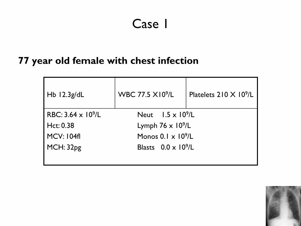
Case 1
77 year old female with chest infection
RBC: 3.64 x 10⁹/L Neut 1.5 x 10⁹/L
Hct: 0.38 Lymph 76 x 109/L
MCV: 104fl Monos 0.1 x 10⁹/L
MCH: 32pg Blasts 0.0 x 10⁹/L
Platelets 210 X 109/L
WBC 77.5 X10⁹/L
Hb 12.3g/dL

Questions
• What is the likely diagnosis?
• How would you confirm this?
• What action would you take?

Blood film

2 diagnoses
• CLL
• Macrocytosis ?cause
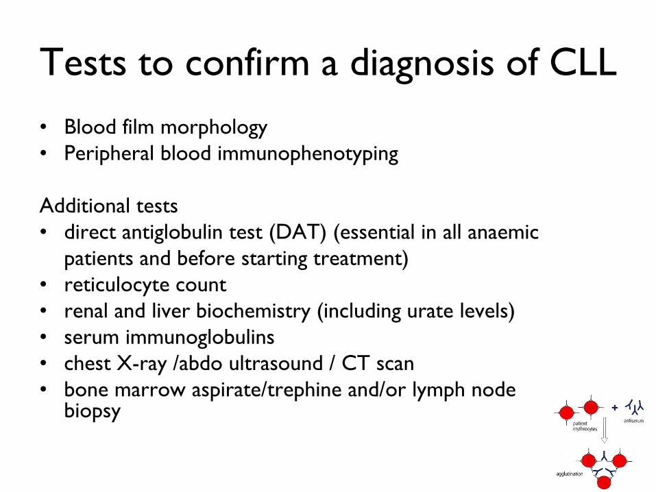
Tests to confirm a diagnosis of CLL
• Blood film morphology
• Peripheral blood immunophenotyping
Additional tests
• direct antiglobulin test (DAT) (essential in all anaemic
patients and before starting treatment)
• reticulocyte count
• renal and liver biochemistry (including urate levels)
• serum immunoglobulins
• chest X-ray /abdo ultrasound / CT scan
• bone marrow aspirate/trephine and/or lymph node biopsy

Chronic Lymphocytic Leukaemia
CLL
Lymphocytosis
BM failure
AIHA/ITP

Prognostic factors in chronic
lymphocytic leukaemia
Factor Low risk High risk
Gender Female Male
Clinical stage Binet A Binet B or C
Rai O,I Rai II, III, IV
Lymphocyte Typical Atypical
morphology
Pattern of marrow Non-diffuse Diffuse
trephine infiltration
Lymphocyte doubling time >12 months <12 months
Serum markers* Normal Raised
CD38 expression <20–30% >20–30%
Genetic abnormalities None del 11q23
del 13q (sole) Loss/mutation of
p53
IgVH gene status Mutated Unmutated

Indications for referral/follow up
• management of CLL requires a collaborative approach
between primary care and haematology
• palliative care team may be valuable in the management of terminal drug resistant patients
Indications for referral to a haematology department include:
• symptomatic disease
• the presence of lymphadenopathy or hepatosplenomegaly
• the investigation of a lymphocytosis, particularly if the lymphocyte count is high or there is anaemia or thrombocytopenia

Macrocytosis CAUSES DIAGNOSTIC TESTS
B12/Folate deficiency B12/Folate
Liver Disease LFTs
Post-splenectomy
Alcohol gGT
Aplastic anaemia FBC, retics
Myeloma Igs/SPEP/urinary BJP
Myelodysplasia blood film
Hypothyroidism TFT
Reticulocytosis DAT, retics, bili, LDH
Pregnancy pregnancy test (!)
Drugs drug history eg hydroxyurea
anti-retroviral agents
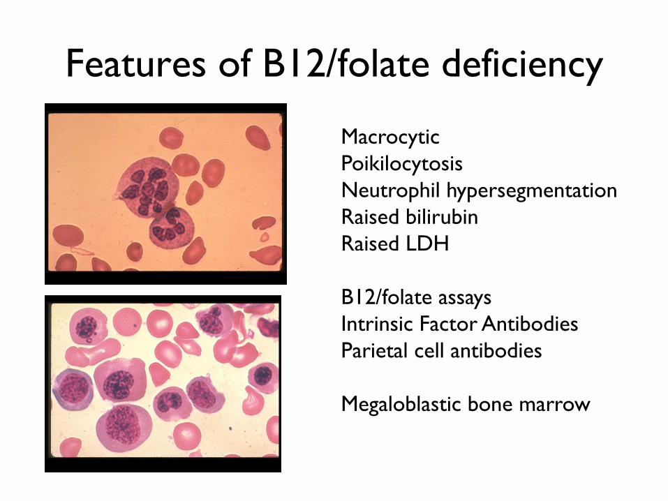
Features of B12/folate deficiency
Macrocytic
Poikilocytosis
Neutrophil hypersegmentation
Raised bilirubin
Raised LDH
B12/folate assays
Intrinsic Factor Antibodies
Parietal cell antibodies
Megaloblastic bone marrow

RBC: 6.65 x 10⁹/L Neut 9.0 x 10⁹/L
Hct: 0.53 Lymph 2.4 x 10⁹/L
MCV: 80.6fl Monos 0.8 x 10⁹/L
MCH: 30 pg Blasts 0.0 x 10⁹/L
Platelets 804
X 109/L
WBC 12.2
X 109/L
Hb 18.2g/dL
Case 2: 55 year old man with hypertension and
recent DVT
What is the likely diagnosis?
What are the risks associated with this?

Symptoms and signs of primary polycythaemia
• facial plethora
• headache
• mental clouding
• pruritis
• hypertension
• splenomegaly
• gout
• occlusive vascular lesions eg stroke, transient ischaemic
attacks, digital ischaemia
• bleeding

Proposed revised WHO criteria for
polycythemia vera
Major criteria
1. Haemoglobin 18.5 g/dL in men, 16.5 g/dL in women or other evidence of increased red cell volume*
2. Presence of JAK2617VF or other functionally similar mutation such as JAK2 exon 12 mutation
Minor criteria
1. Bone marrow biopsy showing hypercellularity for age with trilineage growth proliferation
2. Serum erythropoietin level below the normal reference range
3. Endogenous erythroid colony formation in vitro
Diagnosis requires the presence of both major criteria and 1 minor criterion or the presence of the first major criterion together with 2 minor criteria

Treatment
1. Repeated venesections to maintain a PCV of <
0.45
2. Hydroxyurea if there is also thrombocytosis
3. Low dose aspirin (if there are no bleeding
manifestations)
4. JAK2 inhibitors
5. Radioactive phosphorous
Single dose controls disorder for 12 – 18 months
but associated with increased risk of leukaemia.
Reserved for elderly frail patients. Rarely used

RBC: 3.91 x 10⁹/L Neut 7.0 x 10⁹/L
Hct: 0.32 Lymph 2.2 x 10⁹/L
MCV: 78fl Monos 0.8 x 10⁹/L
MCH: 25pg Blasts 0.0 x 10⁹/L
Platelets 570
X 109/L
WBC 10.0
X 109/L
Hb 9.2g/dL
Case 3: A 69 year old lady with rheumatoid arthritis
What tests are needed?
Why does she have a raised platelet count?

ANAEMIA
Normal ranges for red cell parameters:
Female Male
Hb: g/dL 11.5 – 15.5 13.0 – 17.0
RBC x 1012/L 3.8 – 5.3 4.5 – 6.0
Haematocrit 0.37 – 0.45 0.40 – 0.52
Mean cell volume (fl) 83 – 96 83 - 96
Mean cell 27 – 32 27 - 32
haemoglobin (pg)
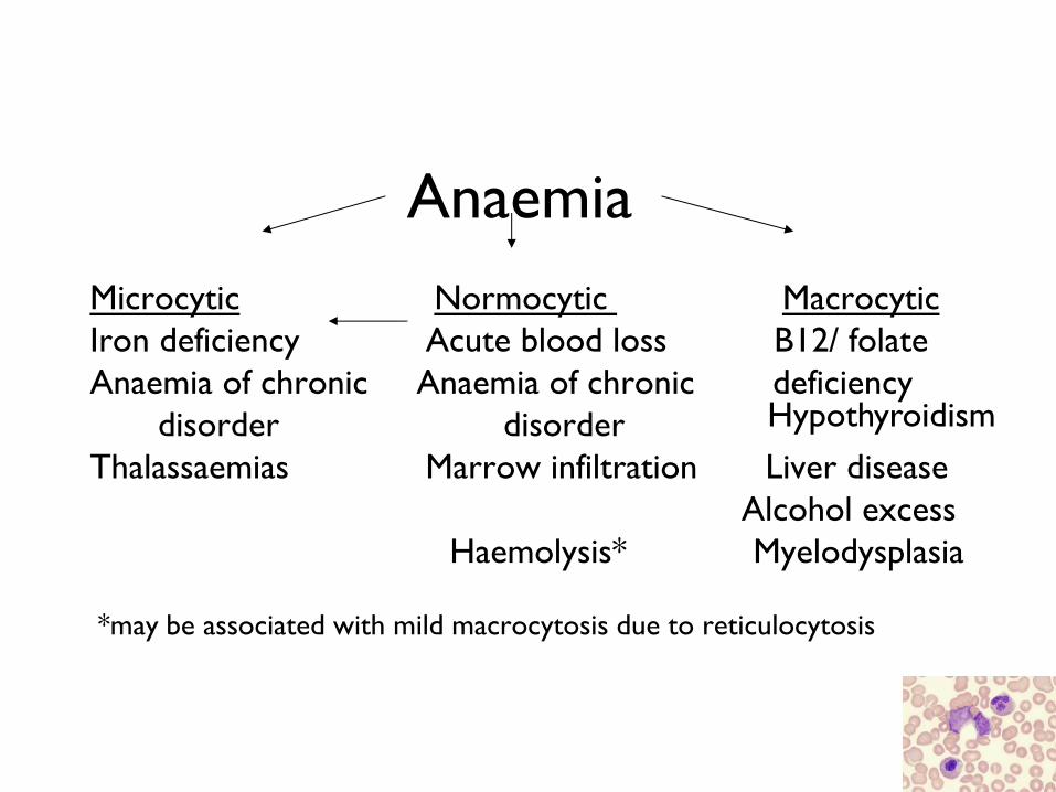
Anaemia
Microcytic Normocytic Macrocytic
Iron deficiency Acute blood loss B12/ folate
Anaemia of chronic Anaemia of chronic deficiency
disorder disorder
Thalassaemias Marrow infiltration Liver disease
Alcohol excess
Haemolysis* Myelodysplasia
*may be associated with mild macrocytosis due to reticulocytosis
Hypothyroidism
Investigation of anaemia: history
• Source of bleeding
• History of chronic illness/renal disease
• Country of origin/family history
• Dietary history
• Drug history
• Surgical history
• Foreign travel
Investigation of anaemia: laboratory
findings (1)
• Red cell indices
– important to distinguish between microcytic,
macrocytic and normocytic and direct subsequent
investigations
• White cell and platelet counts
– helps to distinguish ‘pure’ anaemia from
pancytopenia secondary to a marrow defect
Investigation of anaemia: laboratory
findings (2) • Haematinic assays - ferritin, B12 and folate
• Reticulocyte count (normal 0.5-2%)
– should rise in response to anaemia
• Blood film
– ESSENTIAL!
– Look for abnormal red or white cell morphology, red cell inclusions or dimorphic picture
• Bone marrow examination
– cell development, % cell lines and abnormal cells

Investigation of this patient
• Blood film
• Haematinics
– ferritin, B12 and folate
– Other measures of iron metabolism
• Measure of inflammation eg. CRP

Anaemia of chronic disease (ACD)
• The most frequent anaemia among hospitalized patients
• Caused by – chronic inflammatory disorders eg chronic infections, cancer, autoimmune
diseases
• Causes – diversion of iron traffic and diminished erythropoiesis
– blunted response to erythropoietin, erythrophagocytosis
– bone marrow invasion by tumour cells and pathogens
• Diagnosis of ACD can be assessed by examination of changes in serum iron parameters – low to normal serum iron, TIBC
– normal to increased ferritin, ZPP high in IDA
• Therapy of ACD includes the cure of the underlying the disease. – transfusions for rapid correction of haemoglobin levels
– human recombinant erythropoietin but response rates are sometimes low.
– Iron alone should be avoided

RBC: 5.22 x 10⁹/L Neut 6.3 x 10⁹/L
Hct: 0.44 Lymph 2.2 x 10⁹/L
MCV: 88fl Monos 0.8 x 10⁹/L
MCH: 30pg Blasts 0.0 x 10⁹/L
Platelets 10
X 109/L
WBC 9.3
X 109/L
Hb 15.0g/dL
Case 4: A 26 year old male with spontaneous bruising
Causes of thrombocytopenia
• False thrombocytopenia – Clot in the sample.
– Platelets clumped (citrate effect)
• Congenital thrombocytopenia – Rare inherited disorders (eg May Hegglin anomaly, Bernard Soulier syndrome).
• Defective platelet production – Bone marrow aplasia (failure).
– Metabolic disorders, eg kidney failure, alcohol.
– Abnormal platelet precursors: viral infections, inherited abnormalities.
– Bone marrow infiltration, eg leukaemia, lymphoma.
• Diminished platelet survival – Antibodies in response to drugs, blood transfusion or another disease, eg glandular
fever, malaria, HIV, SLE
– Unknown cause (ITP).
– Clotting disorder (DIC).
– Blood disorder (TTP).
• Loss of platelets from the circulation – Massive blood transfusion or exchange.
– Enlarged spleen
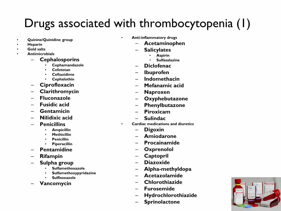
Drugs associated with thrombocytopenia (1) • Quinine/Quinidine group
• Heparin
• Gold salts
• Antimicrobials
– Cephalosporins • Cephamandazole
• Cefotetan
• Ceftazidime
• Cephalothin
– Ciprofloxacin
– Clarithromycin
– Fluconazole
– Fusidic acid
– Gentamicin
– Nilidixic acid
– Penicillins • Ampicillin
• Methicillin
• Penicillin
• Piperacillin
– Pentamidine
– Rifampin
– Sulpha group • Sulfamethoxazole
• Sulfamethoxypyridazine
• Sulfisoxazole
– Vancomycin
• Anti-inflammatory drugs
– Acetaminophen
– Salicylates • Aspirin
• Sulfasalazine
– Diclofenac
– Ibuprofen
– Indomethacin
– Mefanamic acid
– Naproxen
– Oxyphebutazone
– Phenylbutazone
– Piroxicam
– Sulindac • Cardiac medications and diuretics
– Digoxin
– Amiodarone
– Procainamide
– Oxprenolol
– Captopril
– Diazoxide
– Alpha-methyldopa
– Acetazolamide
– Chlorothiazide
– Furosemide
– Hydrochlorothiazide
– Sprinolactone

Drugs associated with thrombocytopenia (2)
• Benzodiazepines – Diazepam
• Anti-epileptic drugs – Carbamazepine
– Phenytoin
– Valproic acid
• H2-antagonists – Cimetidine
– Ranitidine
• Sulfonylurea drugs – Chlorpropamide
– Glibenclamide
• Iodinated contrast agents
• Retinoids – Isotretinoin
– Etretinate
• Anti-histamines – Antazoline
– Chlorpheniramine
• Illicit drugs – Cocaine
– Heroin
• Antidepressants – Amitriptyline
– Desipramine
– Doxepin
– Imipramine
– Mianserine
• Miscellaneous drugs – Tamoxifen
– Actinomycin-D
– Aminoglutethimide
– Danazole
– Desferrioxamine
– Levamizole
– Lidocaine
– Morphine
– Papaverine
– Ticlopidine

Approach to diagnosis
• history of symptoms, signs of bleeding or
bruising, other medical problems, recent
infections and medications.
• Repeat full blood count
• Blood film
• Indications for referral

What is haemochromatosis?
the clinical condition of iron overload
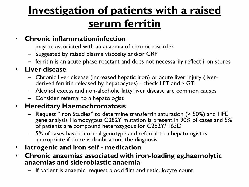
Investigation of patients with a raised
serum ferritin
• Chronic inflammation/infection – may be associated with an anaemia of chronic disorder
– Suggested by raised plasma viscosity and/or CRP
– ferritin is an acute phase reactant and does not necessarily reflect iron stores
• Liver disease – Chronic liver disease (increased hepatic iron) or acute liver injury (liver-
derived ferritin released by hepatocytes) - check LFT and g GT.
– Alcohol excess and non-alcoholic fatty liver disease are common causes
– Consider referral to a hepatologist
• Hereditary Haemochromatosis – Request “Iron Studies” to determine transferrin saturation (> 50%) and HFE
gene analysis Homozygous C282Y mutation is present in 90% of cases and 5% of patients are compound heterozygous for C282Y/H63D
– 5% of cases have a normal genotype and referral to a hepatologist is appropriate if there is doubt about the diagnosis
• Iatrogenic and iron self - medication
• Chronic anaemias associated with iron-loading eg.haemolytic anaemias and sideroblastic anaemia – If patient is anaemic, request blood film and reticulocyte count

Classification of haemochromatosis
• Genetic haemochromatosis – iron accumulation in the body due to the inheritance of mutations in the HFE
gene on both copies of chromosome 6
– leads to excessive absorption of iron from food.
• Juvenile haemochromatosis – an inherited condition in which there is clinical onset in the 2nd or 3rd decade.
– The gene responsible is probably located on chromosome 1
• Secondary iron overload (secondary haemochromatosis, haemosiderosis) – iron overload following chronic blood transfusion for haematological
conditions, including thalassaemia major and aplastic anaemia
– also includes conditions in which enhanced iron absorption is secondary to ineffective erythropoiesis with marrow hyperplasia eg.thalassaemia intermedia
• Neonatal haemochromatosis – condition of acute liver damage with iron accumulation

Genetic haemochromatosis
• In the UK over 90% of patients with genetic
haemochromatosis are homozygous for the C282Y
mutation of the HFE gene and another 4% are
compound heterozygotes (C282Y/H63D).
• There are other rarer forms of inherited
haemochromatosis where patients have ‘classical’
clinical features of haemochromatosis but lack
mutations in the HFE gene
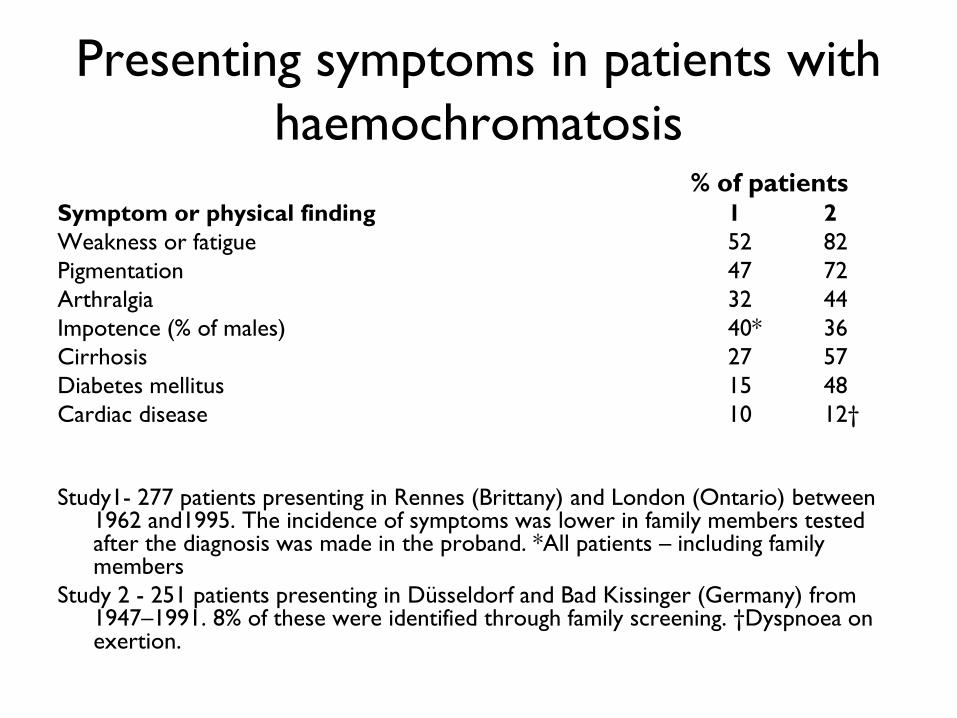
Presenting symptoms in patients with
haemochromatosis % of patients
Symptom or physical finding 1 2
Weakness or fatigue 52 82
Pigmentation 47 72
Arthralgia 32 44
Impotence (% of males) 40* 36
Cirrhosis 27 57
Diabetes mellitus 15 48
Cardiac disease 10 12†
Study1- 277 patients presenting in Rennes (Brittany) and London (Ontario) between 1962 and1995. The incidence of symptoms was lower in family members tested after the diagnosis was made in the proband. *All patients – including family members
Study 2 - 251 patients presenting in Düsseldorf and Bad Kissinger (Germany) from 1947–1991. 8% of these were identified through family screening. †Dyspnoea on exertion.

Diagnosis
• Early diagnosis is not easy
• symptoms with which patients present are
relatively common and non-specific
• Raised ferritin concentrations are common in
hospital patients
• genetic testing offers the best approach to
early detection

Management guidelines
http://www.bcshguidelines.com/documents/haem
ochromotosis_2000.pdf

Monoclonal gammopathy of
undetermined significance (MGUS)
• Definition
the presence of a monoclonal protein in the
serum or urine of an individual with no evidence
of multiple myeloma, AL amyloidosis,
Waldenstrom’s macroglobulinaemia or other
related disorders. (Kyle, Mayo Clinic 1978)

Incidence of M-proteins
• Varies greatly with age.
– 1 - 2% of people in their 6th decade
– 2- 4% in their 7th decade
– 4-5% in their 8th decade
• 694 out of 21,463 in a normal Minnesota population > 50
years. (Kyle et al 2006)
– 14% over the age of 90 Crawford et al, 1987
• Twice as common in black people as white people

Monoclonal gammopathies include:
– Monoclonal gammopathy of undetermined significance (MGUS)
– Multiple myeloma
– Solitary plasmacytoma (skeletal or extra-medullary)
– AL amyloidosis
– Waldenstrom's macroglobulinaemia
– Low grade non-Hodgkin’s lymphoma and other lymphoproliferative disorders
– Other M-protein related disorders

M-proteins may occur in association with:
• Connective tissue disorders
– such as rheumatoid arthritis (RA) systemic lupus
erythematosis, scleroderma, polymyositis and ankylosing
spondylitis.
• Skin disorders
• Infections
– hepatitis C virus (HCV)-related chronic liver disease (may be
accompanied by mixed cryoglobulinaemia)
– HIV
– Helicobacter pylori

Why is guidance on newly diagnosed M-
proteins needed?
• M-proteins are common;
– Overall occur in about 1% of the population – in population-based studies in Europe and North America
– 200 paraproteins a year found in UK DGH serving
300-400k population – St Helier unpublished study

What should happen when
M-proteins are found?
Over investigation
Risk of causing patients unnecessary anxiety,
+
Risk of inappropriate use of resources
Under-investigation
Risk of failing to identify
patients at risk of developing
myeloma (and thus perhaps
missing the opportunity of
avoiding advanced renal and
lytic bone disease), amyloid
etc
v
In the future may be a place for using agents which may delay or prevent
progression
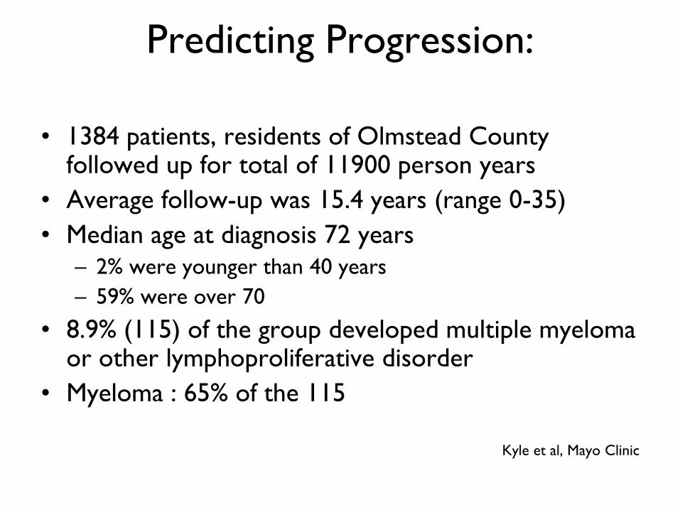
Predicting Progression:
• 1384 patients, residents of Olmstead County
followed up for total of 11900 person years
• Average follow-up was 15.4 years (range 0-35)
• Median age at diagnosis 72 years
– 2% were younger than 40 years
– 59% were over 70
• 8.9% (115) of the group developed multiple myeloma or other lymphoproliferative disorder
• Myeloma : 65% of the 115
Kyle et al, Mayo Clinic

Predicting Progression
• There were only 2 statistically significant risk factors for progression
– The concentration of monoclonal protein
– The type of monoclonal protein
• IgA and IgM gammopathy more likely than IgG to
progress
• IgM rarely becoming myeloma
Predicting Progression
• Not predictive of progression were
– Bence Jones Proteinuria
– Immunosuppression
– Age
– Sex

The risks of progression at
20 years follow-up
M protein level Risk of progression
5 g/l 14%,
10 g/l 16%,
15 g/l 25%,
20 g/l 41%
25 g/l 49%

The cumulative risk of progression
• 10% at 10 years
• 21% at 20 years
• 26% at 25 years
Overall risk : 1% per annum – Risk remained even after 25 years or more
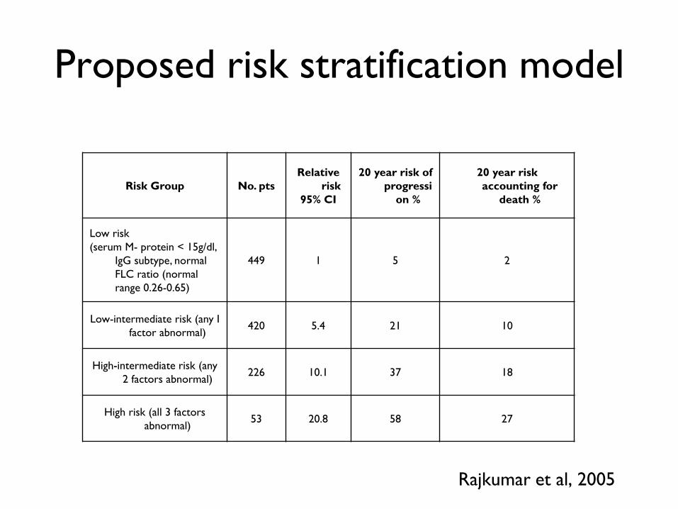
Risk Group No. pts
Relative
risk
95% CI
20 year risk of
progressi
on %
20 year risk
accounting for
death %
Low risk
(serum M- protein < 15g/dl,
IgG subtype, normal
FLC ratio (normal
range 0.26-0.65)
449 1 5 2
Low-intermediate risk (any I
factor abnormal) 420 5.4 21 10
High-intermediate risk (any
2 factors abnormal) 226 10.1 37 18
High risk (all 3 factors
abnormal) 53 20.8 58 27
Proposed risk stratification model
Rajkumar et al, 2005

Recommendations for investigation of M-
proteins in primary care
• The initial evaluation requires the following:
– Detailed history and examination
• Focusing on the possibility that the patient has a plasma cell
or lympho-proliferative malignant disorder.
• Identifying symptoms and signs and test results commonly
associated with myeloma, lymphoma or AL amyloid
– FBC and U and E and calcium
– Definition of the immunoglobulin class of the M-protein
– Serum immunoglobulin levels
– Spot urine for urinary protein excretion and urinary
protein electrophoresis

Indications for referral of a person with
an M-protein to a haematologist • All patients with symptoms or physical signs suggestive of
underlying myeloma, other lympho-proliferative disorder or AL amyloidosis
• M-proteins – IgG M-proteins >15g/l;
– IgA or IgM M-proteins >10g/l
– Any IgD or E paraprotein irrespective of size
• Significant Bence-Jones proteinuria (eg. >500mg/l)
• Unexplained abnormal investigation results even in absence of symptoms – eg anaemia, renal impairment, hypercalcaemia
– lytic lesions,

Monitoring of patients with MGUS
The purpose of monitoring is to try to
identify disease at an early stage when there
is no significant irreversible lytic bone
disease, renal failure, or other disabling
symptoms and at a stage when the patient
is fit enough to benefit from increasingly
effective treatments.
Evidence for the efficacy of monitoring
None!

General principles of monitoring
Clinicians responsible for monitoring patients
should be aware that
– the risk of progression to myeloma or other
lymphoproliferative disease remains lifelong
– that risk never disappears even if the M-protein
remains stable

Monitoring patients with MGUS:
General principles • It is essential that patients should be monitored not
only by laboratory testing but also clinically
– Patients and practitioners should be aware of and report
relevant new symptoms and signs particularly the
development of new bone pain, weight loss, fatigue and
other symptoms which might indicate progression to
myeloma amyloid or other lymphoproliferative disease.
.

Monitoring in the low risk group
• Low risk defined as
– one in which
• IgG M-protein <15g/l
• IgA or IgM M-protein <10g/l
• Non IgD or IgE M-protein
– there are no symptoms, signs or results of initial investigations suggestive of myeloma, other lympho-proliferative disorder or AL amyloidosis

Monitoring in the low risk group
– This group forms the vast majority of M-proteins
detected in routine practice.
– For example, 60 % of M-proteins found in the
laboratory in one District General Hospital were
below 5 g/ l and have a very low risk of
progression
Frequency of follow-up
• It could reasonably be argued that in the people with a very short actuarial life expectancy (perhaps less than 5 years) and very small paraproteins (eg. below 5 g/l) regular follow up is not required once myeloma amyloid and LPD have been excluded.
• However it would not be unreasonable to measure the M-protein occasionally when the patient was having other monitoring blood tests

Blood tests at monitoring visits
• Quantitation of the M-protein and
immunoglobulin levels
• FBC, creatinine, urea and electrolytes,
corrected calcium

Criteria for re-referral
• If symptoms compatible with a diagnosis of myeloma
or lymphoma develop
• If the size of the M-component increases by more
than 25% (a minimum absolute increase of 5g/l)
• If unexplained anaemia, other cytopenias or
abnormal renal function or hypercalcaemia develop
Monitoring in the higher risk group
• Overall this group of patients requires much more frequent follow up, usually under the care of a Consultant Haematologist.
• Anything less that than 4 monthly is likely to prove ineffective.
• Clinicians should be aware of the patterns of progression.

Thank you


![INDEX [] · index 1. department of laboratory medicine • preventive health packages • disease profiles • haematology • biochemistry • serology](https://static.documents.pub/doc/80x56/5c81e9ca09d3f21e6b8b586c/index-index-1-department-of-laboratory-medicine-preventive-health-packages.jpg)


