Grafted Neural Progenitors Integrate and Restore SynapticConnectivity across the Injured Spinal Cord
Joseph F. Bonner,1 Theresa M. Connors,1 William F. Silverman,1,2 David P. Kowalski,1,3 Michel A. Lemay,1
and Itzhak Fischer1
1Department of Neurobiology and Anatomy, Drexel University College of Medicine, Philadelphia, Pennsylvania 19129, 2Department of Morphology,Zlotowski Center for Neuroscience, Ben-Gurion University, Beer Sheva 84105, and Israel, 3School of Biomedical Engineering, Drexel University,Philadelphia, Pennsylvania 19104
Transplantation of neural progenitor cells (NPC) is a promising therapeutic strategy for replacing neurons lost after spinal cord injury,but significant challenges remain regarding neuronal integration and functional connectivity. Here we tested the ability of graft-derivedneurons to reestablish connectivity by forming neuronal relays between injured dorsal column (DC) sensory axons and the denervateddorsal column nuclei (DCN). A mixed population of neuronal and glial restricted precursors (NRP/GRP) derived from the embryonicspinal cord of alkaline phosphatase (AP) transgenic rats were grafted acutely into a DC lesion at C1. One week later, BDNF-expressinglentivirus was injected into the DCN to guide graft axons to the intended target. Six weeks later, we observed anterogradely traced sensoryaxons regenerating into the graft and robust growth of graft-derived AP-positive axons along the neurotrophin gradient into the DCN.Immunoelectron microscopy revealed excitatory synaptic connections between regenerating host axons and graft-derived neurons at C1as well as between graft axons and DCN neurons in the brainstem. Functional analysis by stimulus-evoked c-Fos expression and electro-physiological recording showed that host axons formed active synapses with graft neurons at the injury site with the signal propagatingby graft axons to the DCN. We observed reproducible electrophysiological activity at the DCN with a temporal delay predicted by our relaymodel. These findings provide the first evidence for the ability of NPC to form a neuronal relay by extending active axons across theinjured spinal cord to the intended target establishing a critical step for neural repair with stem cells.
IntroductionSpinal cord injury (SCI) is characterized by cell death and loss ofconnectivity with permanent functional deficits. Repair strategiesare designed to restore neuronal connectivity by promoting re-generation and plasticity or through cell replacement (Eftekhar-pour et al., 2008; Verma et al., 2008). Regeneration of axons in thespinal cord, however, is limited by the intrinsic properties ofadult CNS neurons and by a postinjury environment inhibitoryto axon growth (Cafferty et al., 2008). Consequently, functionalregeneration remains challenging despite progress in the charac-terization of inhibitory molecules and the molecular mechanismsof regeneration (Selzer, 2003). In contrast to regeneration, thereis remarkable endogenous plasticity associated with incompleteSCI. Descending corticospinal neurons can form novel circuitswith intact propriospinal neurons (Bareyre et al., 2004), and thepropriospinal neurons, in turn, serve as a neuronal relay to re-store functional connectivity between injured upper motor neu-
rons and intact lower motor neurons (Courtine et al., 2008). Avariety of strategies to enhance axon growth have been devel-oped, including digestion of the glial scar (Massey et al., 2006),peripheral nerve grafts (Tom and Houle, 2008), neurotrophinadministration (Cao et al., 2005; Chen et al., 2008; Sasaki et al.,2009), as well as combination treatments. For example, com-bined cell transplant, neurotrophin gradient, and conditioninglesion elicits axonal bridging of injured dorsal column sensoryaxons but fails to restore synaptic activity across the lesion (Altoet al., 2009). These studies demonstrate the need to modulateboth extrinsic environment and intrinsic capacity of injured neu-rons to regenerate but underscore the difficulties of regainingfunctional connectivity even when synaptic structure is restored.
We propose an alternative approach to SCI repair that uses amix of neuronal and glial restricted progenitors (NRP and GRP,respectively) derived from the embryonic spinal cord as a sourceof developmentally competent neurons (Fischer et al., 2006) tocreate a novel neuronal relay. We have shown previously thatNRP/GRP grafts generate neurons in the injured spinal cord(Lepore and Fischer, 2005), and these neurons have the intrinsiccapacity to overcome chondroitin sulfate proteoglycans (See etal., 2010). Additionally, we have demonstrated that a gradient ofbrain-derived neurotrophic factor (BDNF) induces guided axonextension from NRP/GRP grafts in the injured spinal cord (Bon-ner et al., 2010), similar to the role of BDNF in neuronal polar-ization (Mai et al., 2009), axon guidance (Yao et al., 2006), andsynaptogenesis (Causing et al., 1997) during development. In the
Received Aug. 6, 2010; revised Jan. 4, 2011; accepted Feb. 2, 2011.This work was funded by National Institutes of Health Grant NS055976, Craig H. Neilsen Foundation, Shriners
Hospital for Children, and the Drexel University College of Medicine Spinal Cord Research Center. We acknowledge C.Tyler-Polsz, M.-P. Cote, and S. Koutzaki for their assistance with experiments and A. C. Lepore, B. Neuhuber, and M. S.Rao for their helpful discussions. We thank A. Blesch for his generous gift of BDNF lentivirus and A. C. Lepore for theGLT-1 antibody.
Correspondence should be addressed to Dr. Itzhak Fischer, Department of Neurobiology and Anatomy, DrexelUniversity College of Medicine, 2900 Queen Lane, Philadelphia, PA 19129. E-mail: [email protected].
The Journal of Neuroscience, March 23, 2011 • 31(12):4675– 4686 • 4675
current report, NRP/GRP expressing thehuman placental alkaline phosphatase(AP) transgenic marker were transplantedinto a C1 dorsal columns injury, and NRPaxons were guided to the dorsal columnnucleus (DCN) with a BDNF gradient.We used immunocytochemistry at lightand electron microscope (EM) levels todemonstrate that NRP-derived neuronsare capable of establishing afferent and ef-ferent synaptic connections with the in-jured host at the site of injury and theDCN and used c-Fos expression and elec-trophysiological analysis to test synapticactivity. Our results demonstrate the abil-ity of NPC to form a neuronal relay acrossthe injured spinal cord and provide theframework for restoring connectivity.
Materials and MethodsAnimal subjects and experimental designAdult (250 –300 g) female Sprague Dawley rats(n � 31) received a dorsal column lesion at C1(Fig. 1) and an acute transplant of �5 � 10 5 NRP/GRP suspended in 2.5�l of the collagen matrix Purecol (Advanced BioMatrix). NRP/GRP wereharvested from animals carrying the human placental AP transgene asverified by PCR. All animals were given daily subcutaneous injections ofcyclosporin A (10 mg/kg; Novartis) beginning 3 d before transplantand/or injury and continuing for the entire study. BDNF– green fluores-cent protein (GFP) or GFP lentiviral vector was injected into the dorsalcolumn nuclei 7 d after injury and cell transplantation. Animals weredivided into groups based on vector type and planned analysis as follows:GFP lentivirus for light microscopy (n � 3), BDNF–GFP lentivirus forlight microscopy (n � 9), BDNF–GFP lentivirus for electron microscopy(n � 6), control animals with no vector for c-Fos expression (n � 3),BDNF–GFP lentivirus for c-Fos expression (n � 2), and BDNF–GFPlentivirus for electrophysiology (n � 3). Control animals with no vectoror transplant for electrophysiology (intact, n � 4; injured control, n � 1).
Harvest and culture of NRP/GRPNRP/GRP were harvested from embryonic day 13.5 spinal cord of Fi-scher 344 rats expressing the AP transgene. Timed-pregnant femaleswere killed at embryonic day 13.5, and the embryos were placed inDMEM/F-12 (Invitrogen). Meninges were removed from the spinalcords after incubation in collagenase I (10 mg/ml)/Dispase II (20 ng/ml)in HBSS for 9 min at room temperature. Cords then were treated withtrypsin (0.5%)/EDTA for 20 min at 37°C. Cells were plated on poly-L-lysine and laminin in NRP complete medium [DMEM/F-12, BSA (1mg/ml), B27, basic fibroblast growth factor (10 �g/ml), penicillin–strep-tomycin (100 IU/ml), N2 (10 �l/ml), and neurotrophin-3 (10 �g/ml)].Medium was changed every 2 d.
Preparation of NRP/GRP for transplantationNRP/GRP were cultured for 5– 6 d before grafting. On the morning oftransplantation, cells were dissociated from the culture dish with 0.05%trypsin–EDTA, and viability was assessed using trypan blue. Cells weresuspended at 200,000 cells/�l in a bovine collagen-based grafting me-dium containing 50% Purecol and 50% NRP Basal Medium, pH 7.5.Cells were �90% viable based on trypan blue staining and were usedwithin 5 h of suspension in grafting medium.
Surgical proceduresSprague Dawley rats were anesthetized by intraperitoneal injection of aXAK mixture of xylazine (10 mg/kg; J. A. Webster), acepromazine mal-eate (0.7 mg/kg; J. A. Webster), and ketamine (95 mg/kg; J. A. Webster).Bupranorphin (J. A. Webster) was used for pain relief every 12 h for 3 dand then as needed for the remainder of the study. All animals were caredfor in accordance with the guidelines established by National Institutes of
Health and the Drexel University College of Medicine Institutional An-imal Care and Use Committee.
Dorsal column injury and cell transplantation. A laminectomy was per-formed at C1 to expose the spinal cord. The dura was cut along therostrocaudal axis above the dorsal column. A 30 gauge needle was used tomake a complete injury in the right dorsal columns, severing the tract,and a small piece of the dorsal columns was excised using light suction.Although the entire right dorsal column was severed, the left dorsal col-umn was only partially damaged. Sutures (9-0) were placed in the duraon both sides of the lesion but not tightened. After achieving hemostasisof the spinal cord lesion, �2–3 �l of cell suspension was injected directlyinto the cavity, and the dura sutures were quickly tightened to maintainthe cell suspension within the lesion site. The muscle was then suturedand the skin stapled.
Vector injection. One week after injury and cell grafting, the animals wereinjected with BDNF–GFP or GFP vector. Animals were placed in a stereo-taxic device with the heads tilted down 45°. A small craniotomy exposed theright DCN, and a 30 gauge needle was used to open the dura. A 10 �lHamilton syringe was fitted with a glass pipette with an external diameterbetween 70 and 100 �l and used to inject 1.25 �l of vector at 2 depths (1.0 and0.5 mm) in the intact parenchyma of the dorsal column nuclei. The incisionwas closed and the animals cared for as stated previously. The pLV vector wasmodified to express GFP or huBDNF–IRES–GFP (Blesch, 2004; Kwon et al.,2007). Vector preparations contained 114 �g/ml p24, 8 � 10e8 TU/ml forBDNF and 150 �g/ml p24, 9 � 10e8 TU/ml for GFP.
Cholera toxin subunit � labeling. Dorsal column axons were antero-gradely labeled with cholera toxin subunit � (CT�) (List Biological Lab-oratories) 3 d before the animals were killed. An incision was madeparallel to the femur to expose the sciatic nerve, and 2 �l of CT� (1% indistilled water) was injected into the intact sciatic nerve using a 10 �lHamilton syringe fitted with a 30 gauge needle. The incision was closedand the procedure was repeated on the contralateral sciatic nerve. Ani-mals were treated after surgery as stated previously.
Fluoro-Gold labeling. Fluoro-Gold (Fluorochrome) was injected bilat-erally into the ventroposterolateral (VPL) thalamus to label DCN neu-rons in a subset of animals (n � 3). Animals were placed in a stereotaxicapparatus, and 0.5 �l of the Fluoro-Gold solution (2% in distilled water)was injected at three sites per hemisphere. Coordinates were as follows:�0.23 mm rostrocaudal, �0.30 mm mediolateral, and 0.55 mm dorso-ventral; �0.30 mm rostrocaudal, �0.32 mm mediolateral, and 0.55 mmdorsoventral; and �0.36 mm rostrocaudal, �0.34 mm mediolateral, and0.55 mm dorsoventral.
Immediate early gene c-Fos expression. The ipsilateral sciatic nerve wasstimulated to induce c-Fos expression in both the graft (monosynaptic)
Figure 1. Experimental design. On day 1, a C1 dorsal columns injury was made and a mixed culture of neuronal and glialrestricted precursors (NRP/GRP) were grafted acutely into the lesion cavity (AP � graft represented in brown). On day 7, BDNF–GFPlentivirus was injected into the DCN to create a gradient (represented in green) of BDNF from the DCN to C1. On day 39 (3 d beforethe animals were killed), CT� was injected into the right and left sciatic nerves to anterogradely label regenerating sensory axons(represented in red). Also on day 39, Fluoro-Gold (FG) was injected into the VPL thalamus to retrogradely label dorsal columnneurons (represented in blue). Synaptic connections between the graft and host at the injury and DCN were analyzed by light andelectron microscopy immunochemistry.
4676 • J. Neurosci., March 23, 2011 • 31(12):4675– 4686 Bonner et al. • Connectivity by Neural Grafts in SCI
and in the DCN (disynaptic). We examined the monosynaptic responsesusing 0.1 ms duration, 2 mA amplitude biphasic pulses applied to thesciatic nerve for 1 h at a 10 Hz frequency and examined the disynapticresponse using a 100 Hz, 300 ms train of 1 mA biphasic pulses (0.1 msduration) applied once per second for 1 h. The latter was used to improvesummation and increasing firing of graft neurons. Animals were killed1 h after termination of the nerve stimulation regimen.
Tissue collectionLight microscopy. Animals used for histology were killed 6 weeks afterinjury/cell transplant with an overdose of euthosol (J. A. Webster) andthen transcardially perfused with 50 –100 ml of ice-cold 0.9% saline and500 ml of ice-cold 4% paraformaldehyde in phosphate buffer. The spinalcord and brainstem were removed and placed in 4% paraformaldehydefor 24 h, followed by cryoprotection in 30% sucrose/0.1 M phosphatebuffer at 4°C for at least 3 d. Spinal cords were embedded in M1 embed-ding matrix (Thermo Fisher Scientific) and cut sagitally in 20 �m sec-tions. Tissue was collected on glass slides coated with poly-L-lysine andgelatin. Slides were stored at 4°C.
Electron microscopy. Animals were killed with an overdose of euthosol(J. A. Webster) and then transcardially perfused with 500 ml of ice-cold 0.2%glutaraldehyde/4% paraformaldehyde in phosphate buffer. The spinal cordand brainstem were removed and postfixed in the same solution for 4 hbefore being transferred to 0.1 M phosphate buffer at 4°C for storage.
Alkaline phosphatase histochemistryAlkaline phosphatase histochemistry was used to visualize transplantsand examine process extension. Serial sections (each �120 �m apart)were washed three times in PBS and then placed in 60°C PBS for 1 h toinactivate endogenous phosphatase activity. Sections were then washedin alkaline phosphatase buffer (in mM: 100 Tris, pH 9.5, 50 MgCl2, and100 NaCl), followed by staining in the dark for 2 h with AP stainingsolution [1.0 mg/ml nitroblue-tetrazolium-chloride (Sigma), 5 mM Le-vamisole (Sigma), and 0.1 mg/ml 5-bromo-4-chlor-indolyl-phosphate(Sigma) in AP buffer]. All steps were at room temperature unless other-wise noted. Slides were coverslipped with Vectashield (Vector Laborato-ries). Slides were viewed using Leica DMBRE microscope runningSlidebook imaging software (3i Inc.).
ImmunohistochemistrySlide-mounted tissue sections were treated for 5 min in 0.2% TritonX-100/PBS, washed three times in PBS for 5 min, and then blocked in10% goat serum/PBS for 1 h at room temperature. Sections were incu-bated with primary antibodies [mouse (Ms) � neuronal-specific nuclearprotein (NeuN), 1:100 (Millipore); rabbit (Rb) � AP, 1:200 (Serotec);goat (Gt) � cholera toxin, 1:2000 (List Biological Laboratories); guineapig (GP) � Synaptophysin1 (Synaptic Systems); Rb � c-Fos, 1:500 (Ab-Cam); Rb � TrkB 1:500] overnight at room temperature in 2% serum/PBS. Sections were then washed three times in PBS to remove unboundprimary antibody. Sections were incubated with secondary antibodies[Gt � Rb FITC, Gt � Ms FITC, Gt � GP rhodamine, donkey (Dk) � Gtrhodamine, and Dk � Rb FITC (Jackson ImmunoResearch)] for 2 h,washed three more times in PBS, and coverslipped with Vectashield(Vector Laboratories). Slides were viewed using a Leica DM5500B mi-croscope with Slidebook (3i Inc.) and DP controller (Olympus) or a CarlZeiss Axioplan microscope with StereoInvestigator software(MicroBrightField).
Preembedded immunoelectron microscopyThe DCN and C1 spinal cord were sectioned sagitally (50 �m) on avibratome in a bath of ice-cold PBS. To examine the synaptic relation-ships formed between AP-expressing (AP �) and host cells, avidin– bio-tin (ABC; Vector Laboratories) immunohistochemistry was performedusing Rb � AP (1:100, AbD Serotec) and biotinylated � Rb (1:200;Vector Laboratories) IgGs visualized with 3,3�-diaminobenzidine tetra-hydrochloride (DAB) (Fast DAB kit; Sigma). The chromagen reaction insome cases was enhanced by addition of nickel ammonium sulfate. Inaddition to the anti-AP IgG, some sections were colabeled with a mixtureof GP � vesicular glutamate transporter 1 (VGlut1) (1:10,000; MilliporeBioscience Research Reagents) and GP � vesicular glutamate transporter
2 (VGlut2) (1:2500; Millipore Bioscience Research Reagents). Incuba-tion with the VGlut1 and VGlut2 antisera was followed by exposure tomouse IgG conjugated to 12 nm gold particles (1:200; Jackson Immu-noResearch). The sections were postfixed in 2% OSO4 in 0.05 M PBS for30 min, rinsed 5� with the same buffer, and then en bloc stained in 5%uranyl acetate in H20 for 10 min. This was followed by five washes with 5M sodium maleate buffer and dehydration in 5 min sequential incuba-tions in an ascending ethanol series to propylene oxide. The tissue wasembedded in Epon–Araldite between SuperFrost glass slides pretreatedwith a liquid mold-release agent. Desired regions from the embeddedspinal cord segments were mounted onto Epon–Araldite beam capsules,and 80 nm sections were cut on a Reichart ultramicrotome and collectedonto 200 copper mesh grids. The tissue was examined and images digi-tally recorded on a Jeol 1200EX Transmission Electron Microscope.
Analysis of NRP/GRP grafts: quantitative analysesGraft volume, axon extension, AP �/NeuN � cells, and AP �/c-Fos � cellswere quantified using StereoInvestigator software, albeit with nonstereo-logical methods. All quantification was performed on every sixth section.Comparisons were made using t tests for volume and AP �/NeuN � cells,ANOVA with Tukey’s post hoc for axon quantification, or � 2 test foraverage c-Fos �/NeuN � cells. Graft area was quantified using the Cava-lieri method of volume estimation with a 100 � 100 �m grid. Coefficientof error for all Cavalieri method analyses (Gunderson m � 1) was�0.025. Axons were quantified on the same grid if they intersected thedorsoventral gridline closest to the region of interest [rostral dorsal col-umns (RDC) or DCN], were at least 100 �m in length, and were notassociated with a cell soma. The RDC region was defined as the area of thedorsal columns �0.5 mm rostral to the lesion site, and the DCN wasdefined by anatomy and NeuN staining. Axons were counted in the RDCand in the approximate center of the most caudal dorsal column nuclei(gracilis is caudal in the medial brainstem and cuneate is caudal in thelateral). AP �/NeuN � cells were counted in three animals per treatmentgroup. Figures are presented as averages � SEM for all analyses exceptAP �/c-Fos �, which is presented as average � SD.
Electrophysiological analysisExtracellular recordings were made in the ipsilateral gracilis nucleus(DCN) in three groups of animals: intact control (control, n � 4), Purcolmatrix only (injury, n � 1), and grafted animals with a neurotrophingradient (gradient, n � 3). A 16-channel electrode (a 4 � 4, 3-mm100-125-177; Neuronexus Technologies) with four sites on each of fourshanks, producing a rectangular recording region with a height of 400�m and width of 500 �m, was inserted into the DCN with the sitescovering the 600 –200 �m depth range. Once the electrode was insertedat a given recording site, a train of 100 single stimulus pulse of 1 mAamplitude (100 �s duration, biphasic) at 1 Hz was applied to the sciaticnerve, and recordings were made on all channels simultaneously using aTDT RZ2 system (Tucker-Davis Technologies). Signals were filtered formultiunit activity (300 – 4000 Hz, second-order Butterworth bandpass)during recording and then time-reverse filtered in MATLAB (Math-Works) to remove frequency-dependent phase delay. To confirm correctplacement of the electrodes, a positive response to stroking or pinching ofthe ipsilateral hindlimb on some of the channels and null response tostroking or pinching of ipsilateral forelimb pinch and contralateralhindlimb on all of the channels sampled was obtained in all controlanimals. We also attempted the same procedure in graft and gradientanimals; positive response to contact of the ipsilateral hindlimb was notalways obtained, but we confirmed that the sites recorded did not re-spond to pinching or stroking of the other limbs. For each channel, the100 responses to individual stimulus pulses were aligned to the peak ofthe stimulus artifacts and averaged to form the peristimulus averageresponse for that channel. Background noise level was calculated as theroot mean square (RMS) value of the signal over a 3 ms prestimuluswindow (4 ms to 1 ms prestimulus). Furthermore, the peristimulus av-erage was divided into 0.5 ms windows, and the RMS value of the signalin each window was calculated to obtain the windowed RMS of theresponse. If the value of that signal after stimulus did not exceed twice thebackground noise level, that channel was determined to be nonrespon-
Bonner et al. • Connectivity by Neural Grafts in SCI J. Neurosci., March 23, 2011 • 31(12):4675– 4686 • 4677
sive and not further analyzed. The latency of the response for each chan-nel was then measured as the time between the onset of stimulus to thepoint at which the magnitudes of both the peristimulus averaged signaland the windowed RMS of the response exceeded twice the backgroundlevel. The process was repeated for every channel individually, and thelatencies for each set of animals were grouped together. Welch’s t test forunpaired data with nonmatching variance was used to assess the statisti-cal significance of the difference in the latency of the DCN responses tostimulation of the sciatic nerve between the groups.
ResultsGraft-derived neurons extend axons to the DCN along aBDNF gradientWe acutely grafted NRP/GRP derived from AP transgenic ratsinto a C1 dorsal columns lesion to test the ability of graft-derivedneurons to integrate and extend axons from the injury site to atarget in the brainstem. One week after the grafting, we injectedGFP lentivirus or BDNF–GFP lentivirus into the DCN (Fig. 1).Six weeks later, AP histology showed that the grafted cells sur-vived and filled the injury site but did not extend long processes inresponse to control GFP lentivirus (Fig. 2A). In contrast, injec-tion of BDNF–GFP lentivirus into the DCN resulted in robustaxon growth by graft neurons from the lesion site into the DCN(Fig. 2B,C). Graft axons primarily grew rostrally along the whitematter tracts, although some axons were also observed extending
caudally from the graft (Fig. 2B). We have reported previouslythat injection of BDNF–GFP lentivirus into the DCN creates agradient of BDNF from the DCN to C1 within 2 weeks of infec-tion as verified by BDNF ELISA and GFP immunohistochemistry(Bonner et al., 2010). In the current report, we observed a similarpattern of GFP expression after injection of BDNF–GFP lentivi-rus into the DCN (Fig. 2D). Dense AP� axons (Fig. 2C) wereobserved growing along the dorsal columns and entering theDCN in the same region that showed GFP expression, indicatinga close relationship between graft axon growth and host cellsinfected by the BDNF–GFP lentivirus. Graft axons were primarilyoriented longitudinally in the dorsal columns, and only few axonswere observed growing into the ventral gray matter.
We quantified the effects of the BDNF gradient on severalproperties of the NRP/GRP graft. The presence of the gradientwas associated with an overall expansion of the area occupied bygraft-derived cells from 0.59 � 0.1 mm 3 in the control group to1.39 � 0.1 mm 3 in the BDNF group (t test, p � 0.05), indicatingan increase in cell number and/or migration. We observed an 3�
Figure 3. BDNF guides AP � axons into the DCN. AP histology demonstrates that NRP/GRP graftsfill the lesion site and extend axons to the DCN in response to the BDNF gradient (A). The boxed region(A) is shown at high magnification with double staining for AP histology (B, D) and immunofluores-cenceoftheneuronalnucleimarker(NeuN).TheimagesofAP(B)andNeuN(C)havebeenmerged (D)to demonstrate that AP � axons extend into the neuron-dense DCN. A subset of animals receivedinjection of Fluoro-Gold into the VPL thalamus to retrogradely label the DCN. The combination (G) ofFluoro-Gold tracing (E; FG) and NeuN labeling (F ) confirms that graft axons reached DCN neurons,which project to the VPL thalamus. Scale bars: A, 400 �m; B–G, 100 �m.
Figure 2. NRP extend axons from an injury at C1 to the DCN along the BDNF gradient. AP �
NRP/GRP were grafted acutely into a dorsal columns injury, and 1 week later GFP lentivirus (A)or BDNF–GFP lentivirus (B) was injected into the DCN. NRP/GRP filled the lesion as shown by thedark reaction product of AP histology (A, B), but in response to the BDNF gradient (D), NRPextended axons from the injury site to the DCN. Examination of consecutive 20 �m sectionsshows that NRP extend axons (C) through areas expressing the GFP reporter of the BDNF–GFPlentivirus (D). GR, Nucleus gracilis; APT, area postrema. Scale bars: A, B, 500 �m; C, D, 100 �m.
4678 • J. Neurosci., March 23, 2011 • 31(12):4675– 4686 Bonner et al. • Connectivity by Neural Grafts in SCI
increase in average number of NeuN�/AP� cells per 20 �m sec-tion from 49.95 � 8.8 in the GFP group to 159.93 � 27.8 in theBDNF group (t test, p � 0.05), suggesting an increase in NRPproliferation and/or neuronal survival. Finally, we quantified the
number of axons extending rostrallythrough the dorsal columns and the num-ber of axons entering the DCN. In theBDNF group, an average of 142.6 axonsextended through the dorsal columns and102.5, representing 71%, reached theDCN. In contrast, only an average of 8.4axons were present in the dorsal columnsand virtually none reaching the DCN(�1) in the GFP group. Both the numberof axons in the dorsal columns and theDCN were greater in the BDNF group ver-sus control, but there was no significantdifference between the number of axonsin the dorsal columns and those thatreached the DCN in the BDNF group(ANOVA, p � 0.05; Tukey’s post hoc,p � 0.05).
Graft axons innervate the DCNWe next combined AP histology andNeuN immunofluorescence on the samesections to assess the relationship betweengraft axons and host neurons in the DCN.This combination exploited the high-resolution and contrast of AP histologyrelative to immunofluorescence to iden-tify graft-derived axons. AP� axons fromgraft neurons (Fig. 3A) grew along the dor-sal columns (Fig. 3B) and were distributedthroughout areas of dense NeuN staining
(Fig. 3C,D) in the DCN. In a subset of animals, we compared retro-grade labeling of DCN neurons, using Fluoro-Gold (Fig. 3E,G) withthe NeuN staining (Fig. 3F,G), and observed a cluster of double-
Figure 4. AP � axons reach the DCN and express presynaptic markers. Images from consecutive 2 �m focal planes show AP � synaptic, bouton-like structures (A, arrows) encompassing a host neuron(AP �) labeled with NeuN (green, A) as well as an AP �axon passing the host neuron (A, arrowheads). AP �axons (red, C, E) express the presynaptic marker synaptophysin (green, B, E; Syn1) in close appositionto host neurons retrogradely labeled with Fluoro-Gold (blue, D, E; FG) from the VPL thalamus. The arrow (E) indicates a Fluoro-Gold-labeled neuron (E, inset) that is closely apposed by an AP � process colabeledwith several puncta of synaptophysin (E). There were similar AP �/Synaptophysin � double-labeled structures that did not appose Fluoro-Gold-labeled neurons (asterisk, D), indicating that Fluoro-Gold onlylabels a fraction of DCN neurons. Scale bars, 10 �m.
Figure 5. AP � axons form synaptic structures in the DCN. Immuno-EM analysis of the DCN revealed AP � presynaptic structures(asterisks, A–D) from graft axons apposed to host (AP �) dendrites. There were instances of both single AP � (A, B, D) and multiplepresynaptic terminals contacting one host dendrite (C). Note that the high density of nickel-enhanced DAB labeling obscures the detail ofpresynaptic vesicles, but they can be seen clustered near the presynaptic membrane (arrowheads), whereas PSDs are present in most AP �
host dendrites (arrows, A, B, D). Scale bar, 500 nm.
Bonner et al. • Connectivity by Neural Grafts in SCI J. Neurosci., March 23, 2011 • 31(12):4675– 4686 • 4679
labeled neurons, which confirmed that graftaxons had indeed reached the DCN.
We further examined the DCN of ani-mals in the BDNF group for evidence ofsynaptic connections between graft axonsand host neurons. Using high-magnifica-tion optical serial Z-sections, we observedindividual cell bodies of DCN neurons sur-rounded by numerous AP� synaptic struc-tures in which bouton-like terminals werejuxtaposed to host cell bodies (Fig. 4A), sug-gesting multiple presynaptic connections.Similarly, we observed punctate synapto-physin expression in AP � axons (Fig.4B,C,E) in apparent contact with retro-gradely Fluoro-Gold-labeled neurons (Fig.4D,E). High magnification revealed anAP� axon with multiple synaptophysin�
puncta apposed to a Fluoro-Gold-labeledneuron (Fig. 4E, inset), again indicating thatgraft axons established synaptic connectionswith host neurons.
Next, we assessed the efferent connec-tivity and synapse formation of graft-derived neurons with targets in the DCNusing the classical structural definition ofa synapse, which requires clustering ofvesicles in the presynaptic element and apostsynaptic density (PSD) (Peters et al.,1970). Immuno-EM analysis revealed fre-quent synaptic contacts between AP� ter-minals and host neurons (AP�) in theBDNF group. Typically, single AP� syn-aptic boutons were seen apposed to hostdendrites (Fig. 5A,B,D), but examples ofmultiple AP� boutons contacting a singlehost dendrite were also observed (Fig.5C). Prominent PSDs were often seen inthe host dendrite (Fig. 5A,B,D, arrows),suggesting these were excitatory axo-dendritic synapses of the type normallyobserved in the DCN. However, we occa-sionally observed AP� synaptic boutonswithout a clear PSD (Fig. 5C). This may bethe result of a PSD that is outside the 80-nm-thick EM section or may indicatesymmetrical, inhibitory synapses or im-mature synapses. Aside from the presenceof a PSD, dendrites were also identified bythe orderly arranged microtubules, paucityof organelles other than mitochondria andlack of neurofilaments (Peters et al., 1970).
Graft axons may also connect withunintended targetsAlthough the majority of graft axons weredirected to the DCN, we also observedsome AP� axons growing into an unin-tended target, the caudal spinal trigeminal nucleus (SN5). How-ever, we have only documented a single case in which themajority of the axons grew through the SN5 and not the DCNtarget (Fig. 6A–C). We carefully examined these errant axons todetermine whether graft neurons could also, in general, connect
with intact nuclei, which are not the designated target. Specifi-cally, we used immuno-EM to evaluate the nature of the connec-tivity between graft axons and host neurons in the SN5. Using adual-label approach to identify graft-derived AP� axons withDAB and glutamatergic synapses expressing VGlut1 and VGlut2
Figure 6. AP � axons may also connect with unintended targets. Although in most cases AP � axons extended only into theDCN, this figure shows an example of growth into the SN5 (A, C). Despite the growth into the SN5, many axons still grew throughthe dorsal columns (A, B, DC) and into the nucleus gracilus (A, B, GR). Immuno-EM of the SN5 demonstrated that graft axons madepresynaptic connections with host neurons (D, F ). The boxed regions in D and F are shown at high magnification (E, G) tounderscore the clustering of synaptic vesicles at the presynaptic membrane and the thick PSDs (arrows, E, G). Double immuno-EM(D, E) was performed by labeling AP � axons with DAB and vesicular glutamate transporter 1 and 2 with 12 nm gold particles. DABlabeling of AP � synaptic boutons is most evident in the mitochondrial membranes (*, D, F ) compared with unlabeled mitochon-dria (#, D, F ). Synaptic vesicles cluster at the presynaptic membrane (D–F ), and some vesicles are labeled with the gold particles(arrowheads, E). Scale bars: A, 500 �m; B, C, 100 �m; D–F, 500 nm; G, 100 nm.
4680 • J. Neurosci., March 23, 2011 • 31(12):4675– 4686 Bonner et al. • Connectivity by Neural Grafts in SCI
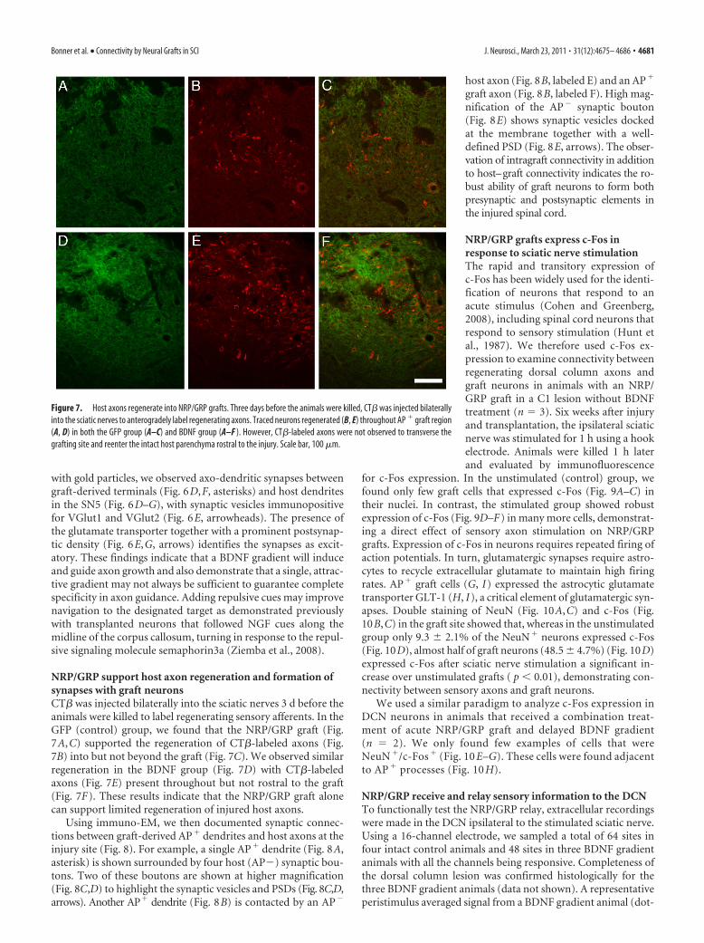
with gold particles, we observed axo-dendritic synapses betweengraft-derived terminals (Fig. 6D,F, asterisks) and host dendritesin the SN5 (Fig. 6D–G), with synaptic vesicles immunopositivefor VGlut1 and VGlut2 (Fig. 6E, arrowheads). The presence ofthe glutamate transporter together with a prominent postsynap-tic density (Fig. 6E,G, arrows) identifies the synapses as excit-atory. These findings indicate that a BDNF gradient will induceand guide axon growth and also demonstrate that a single, attrac-tive gradient may not always be sufficient to guarantee completespecificity in axon guidance. Adding repulsive cues may improvenavigation to the designated target as demonstrated previouslywith transplanted neurons that followed NGF cues along themidline of the corpus callosum, turning in response to the repul-sive signaling molecule semaphorin3a (Ziemba et al., 2008).
NRP/GRP support host axon regeneration and formation ofsynapses with graft neuronsCT� was injected bilaterally into the sciatic nerves 3 d before theanimals were killed to label regenerating sensory afferents. In theGFP (control) group, we found that the NRP/GRP graft (Fig.7A,C) supported the regeneration of CT�-labeled axons (Fig.7B) into but not beyond the graft (Fig. 7C). We observed similarregeneration in the BDNF group (Fig. 7D) with CT�-labeledaxons (Fig. 7E) present throughout but not rostral to the graft(Fig. 7F). These results indicate that the NRP/GRP graft alonecan support limited regeneration of injured host axons.
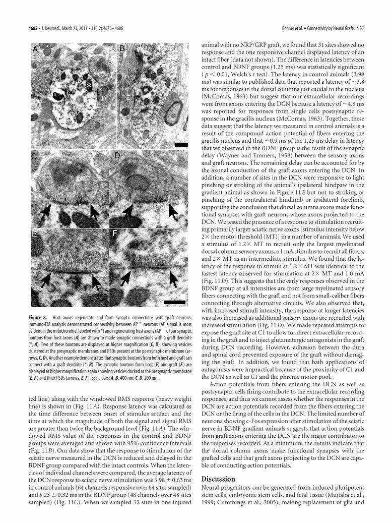
Using immuno-EM, we then documented synaptic connec-tions between graft-derived AP� dendrites and host axons at theinjury site (Fig. 8). For example, a single AP� dendrite (Fig. 8A,asterisk) is shown surrounded by four host (AP�) synaptic bou-tons. Two of these boutons are shown at higher magnification(Fig. 8C,D) to highlight the synaptic vesicles and PSDs (Fig. 8C,D,arrows). Another AP� dendrite (Fig. 8B) is contacted by an AP�
host axon (Fig. 8B, labeled E) and an AP�
graft axon (Fig. 8B, labeled F). High mag-nification of the AP� synaptic bouton(Fig. 8E) shows synaptic vesicles dockedat the membrane together with a well-defined PSD (Fig. 8E, arrows). The obser-vation of intragraft connectivity in additionto host–graft connectivity indicates the ro-bust ability of graft neurons to form bothpresynaptic and postsynaptic elements inthe injured spinal cord.
NRP/GRP grafts express c-Fos inresponse to sciatic nerve stimulationThe rapid and transitory expression ofc-Fos has been widely used for the identi-fication of neurons that respond to anacute stimulus (Cohen and Greenberg,2008), including spinal cord neurons thatrespond to sensory stimulation (Hunt etal., 1987). We therefore used c-Fos ex-pression to examine connectivity betweenregenerating dorsal column axons andgraft neurons in animals with an NRP/GRP graft in a C1 lesion without BDNFtreatment (n � 3). Six weeks after injuryand transplantation, the ipsilateral sciaticnerve was stimulated for 1 h using a hookelectrode. Animals were killed 1 h laterand evaluated by immunofluorescence
for c-Fos expression. In the unstimulated (control) group, wefound only few graft cells that expressed c-Fos (Fig. 9A–C) intheir nuclei. In contrast, the stimulated group showed robustexpression of c-Fos (Fig. 9D–F) in many more cells, demonstrat-ing a direct effect of sensory axon stimulation on NRP/GRPgrafts. Expression of c-Fos in neurons requires repeated firing ofaction potentials. In turn, glutamatergic synapses require astro-cytes to recycle extracellular glutamate to maintain high firingrates. AP� graft cells (G, I) expressed the astrocytic glutamatetransporter GLT-1 (H, I), a critical element of glutamatergic syn-apses. Double staining of NeuN (Fig. 10A,C) and c-Fos (Fig.10B,C) in the graft site showed that, whereas in the unstimulatedgroup only 9.3 � 2.1% of the NeuN� neurons expressed c-Fos(Fig. 10D), almost half of graft neurons (48.5 � 4.7%) (Fig. 10D)expressed c-Fos after sciatic nerve stimulation a significant in-crease over unstimulated grafts ( p � 0.01), demonstrating con-nectivity between sensory axons and graft neurons.
We used a similar paradigm to analyze c-Fos expression inDCN neurons in animals that received a combination treat-ment of acute NRP/GRP graft and delayed BDNF gradient(n � 2). We only found few examples of cells that wereNeuN �/c-Fos � (Fig. 10 E–G). These cells were found adjacentto AP � processes (Fig. 10 H).
NRP/GRP receive and relay sensory information to the DCNTo functionally test the NRP/GRP relay, extracellular recordingswere made in the DCN ipsilateral to the stimulated sciatic nerve.Using a 16-channel electrode, we sampled a total of 64 sites infour intact control animals and 48 sites in three BDNF gradientanimals with all the channels being responsive. Completeness ofthe dorsal column lesion was confirmed histologically for thethree BDNF gradient animals (data not shown). A representativeperistimulus averaged signal from a BDNF gradient animal (dot-
Figure 7. Host axons regenerate into NRP/GRP grafts. Three days before the animals were killed, CT� was injected bilaterallyinto the sciatic nerves to anterogradely label regenerating axons. Traced neurons regenerated (B, E) throughout AP � graft region(A, D) in both the GFP group (A–C) and BDNF group (A–F ). However, CT�-labeled axons were not observed to transverse thegrafting site and reenter the intact host parenchyma rostral to the injury. Scale bar, 100 �m.
Bonner et al. • Connectivity by Neural Grafts in SCI J. Neurosci., March 23, 2011 • 31(12):4675– 4686 • 4681
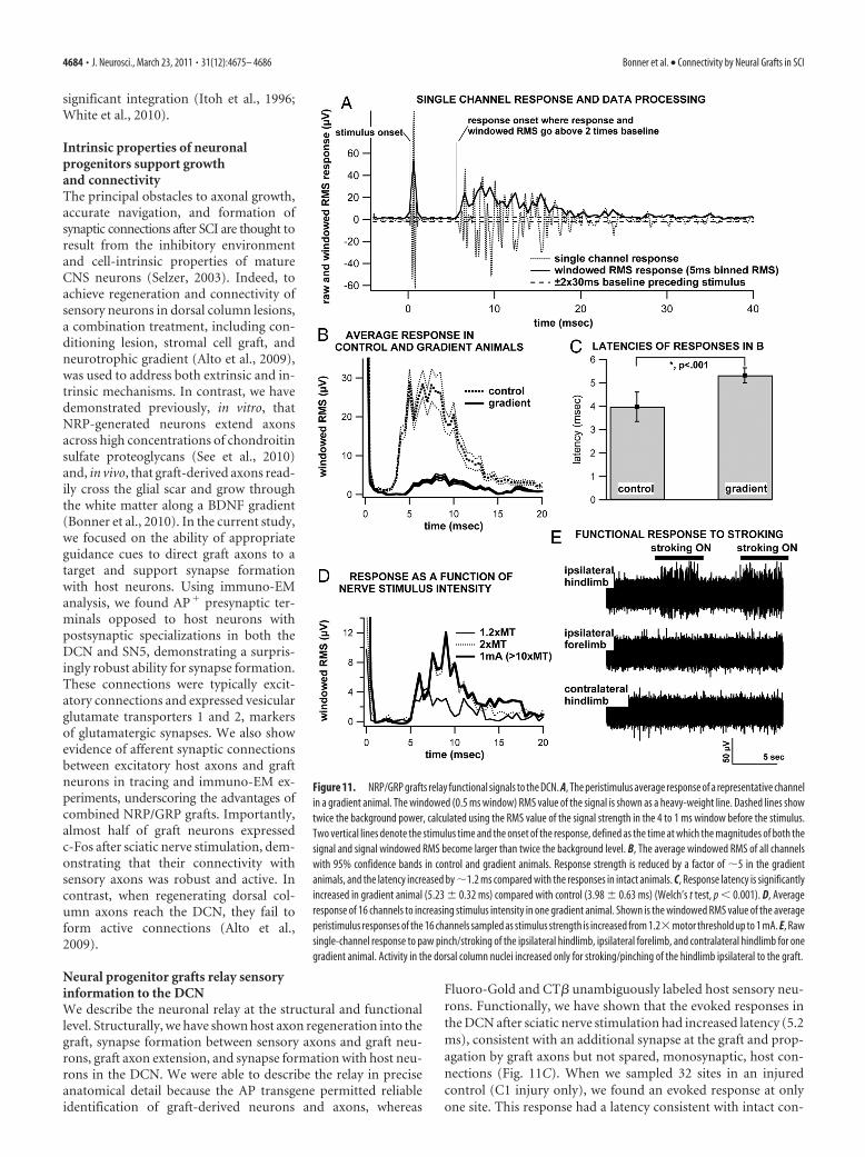
ted line) along with the windowed RMS response (heavy weightline) is shown in (Fig. 11A). Response latency was calculated asthe time difference between onset of stimulus artifact and thetime at which the magnitude of both the signal and signal RMSare greater than twice the background level (Fig. 11A). The win-dowed RMS value of the responses in the control and BDNFgroups were averaged and shown with 95% confidence intervals(Fig. 11B). Our data show that the response to stimulation of thesciatic nerve measured in the DCN is reduced and delayed in theBDNF group compared with the intact controls. When the laten-cies of individual channels were compared, the average latency ofthe DCN response to sciatic nerve stimulation was 3.98 � 0.63 msin control animals (64 channels responsive over 64 sites sampled)and 5.23 � 0.32 ms in the BDNF group (48 channels over 48 sitessampled) (Fig. 11C). When we sampled 32 sites in one injured
animal with no NRP/GRP graft, we found that 31 sites showed noresponse and the one responsive channel displayed latency of anintact fiber (data not shown). The difference in latencies betweencontrol and BDNF groups (1.25 ms) was statistically significant( p � 0.01, Welch’s t test). The latency in control animals (3.98ms) was similar to published data that reported a latency of �3.8ms for responses in the dorsal columns just caudal to the nucleus(McComas, 1963) but suggest that our extracellular recordingswere from axons entering the DCN because a latency of �4.8 mswas reported for responses from single cells postsynaptic re-sponse in the gracilis nucleus (McComas, 1963). Together, thesedata suggest that the latency we measured in control animals is aresult of the compound action potential of fibers entering thegracilis nucleus and that �0.9 ms of the 1.25 ms delay in latencythat we observed in the BDNF group is the result of the synapticdelay (Wayner and Emmers, 1958) between the sensory axonsand graft neurons. The remaining delay can be accounted for bythe axonal conduction of the graft axons entering the DCN. Inaddition, a number of sites in the DCN were responsive to lightpinching or stroking of the animal’s ipsilateral hindpaw in thegradient animal as shown in Figure 11E but not to stroking orpinching of the contralateral hindlimb or ipsilateral forelimb,supporting the conclusion that dorsal columns axons made func-tional synapses with graft neurons whose axons projected to theDCN. We tested the presence of a response to stimulation recruit-ing primarily larger sciatic nerve axons [stimulus intensity below2� the motor threshold (MT)] in a number of animals. We useda stimulus of 1.2� MT to recruit only the largest myelinateddorsal column sensory axons, a 1 mA stimulus to recruit all fibers,and 2� MT as an intermediate stimulus. We found that the la-tency of the response to stimuli at 1.2� MT was identical to thefastest latency observed for stimulation at 2� MT and 1.0 mA(Fig. 11D). This suggests that the early responses observed in theBDNF group at all intensities are from large myelinated sensoryfibers connecting with the graft and not from small-caliber fibersconnecting through alternative circuits. We also observed that,with increased stimuli intensity, the response at longer latencieswas also increased as additional sensory axons are recruited withincreased stimulation (Fig. 11D). We made repeated attempts toexpose the graft site at C1 to allow for direct extracellular record-ing in the graft and to inject glutamatergic antagonists in the graftduring DCN recording. However, adhesion between the duraand spinal cord prevented exposure of the graft without damag-ing the graft. In addition, we found that bath applications ofantagonists were impractical because of the proximity of C1 andthe DCN as well as C1 and the phrenic motor pool.
Action potentials from fibers entering the DCN as well aspostsynaptic cells firing contribute to the extracellular recordingresponses, and thus we cannot assess whether the responses in theDCN are action potentials recorded from the fibers entering theDCN or the firing of the cells in the DCN. The limited number ofneurons showing c-Fos expression after stimulation of the sciaticnerve in BDNF gradient animals suggests that action potentialsfrom graft axons entering the DCN are the major contributor tothe responses recorded. At a minimum, the results indicate thatthe dorsal column axons make functional synapses with thegrafted cells and that graft axons projecting to the DCN are capa-ble of conducting action potentials.
DiscussionNeural progenitors can be generated from induced pluripotentstem cells, embryonic stem cells, and fetal tissue (Mujtaba et al.,1999; Cummings et al., 2005), making replacement of glia and
Figure 8. Host axons regenerate and form synaptic connections with graft neurons.Immuno-EM analysis demonstrated connectivity between AP � neurons (AP signal is mostevident in the mitochondria, labeled with *) and regenerating host axons (AP �). Four synapticboutons from host axons (A) are shown to made synaptic connections with a graft dendrite(*, A). Two of these boutons are displayed at higher magnification (C, D), showing vesiclesclustered at the presynaptic membranes and PSDs present at the postsynaptic membrane (ar-rows, C, D). Another example demonstrates that synaptic boutons from both host and graft canconnect with a graft dendrite (*, B). The synaptic boutons from host (E) and graft (F ) aredisplayed at higher magnification again showing vesicles docked at the presynaptic membrane(E, F ) and thick PSDs (arrows, E, F ). Scale bars: A, B, 400 nm; C, D, 200 nm.
4682 • J. Neurosci., March 23, 2011 • 31(12):4675– 4686 Bonner et al. • Connectivity by Neural Grafts in SCI
neurons a viable therapeutic approach.Although numerous studies explored theuse of glial progenitors (Keirstead et al.,2005; Davies et al., 2006; Bunge, 2008),neuronal precursors received less atten-tion because of challenges of generatingneurons, directing their axons toward in-tended targets within the injured spinal cordand establishing synaptic connectivity. Wehave now shown for the first time that graft-derived neurons, directed by chemotropiccues, are capable of extending axonsthrough the injured spinal cord, formingsynaptic connections with sensory axonsat the graft site and host neurons in thebrainstem and relaying physiological sig-nals across the injury to the DCN. Thesefindings underscore the advantages of us-ing NRP/GRP grafts and set the stage forrestoring functional connectivity withneuronal relays in SCI.
Creating a microenvironmentconducive to neuronal differentiationand synapse formationThe rationale for transplanting neuralstem cells derived from embryonic stemcells or fetal tissue presumes that cells in adevelopmental state will retain intrinsicgrowth properties capable of mediatingneural repair. The mature CNS in generaland SCI microenvironment in particularpresent challenges not only for the lack ofdevelopmental cues but also the presenceof inhibitory cues that promote apoptosis,axon retraction, and scar formation (NandoeTewarie et al., 2009). However, permissiveastrocytes can modify the injury environ-ment (Smith et al., 1986) and promoteformation and maintenance of synapses(Schousboe, 2003). Indeed, our earlierwork demonstrated that transplantationof NRP together with GRP produced neu-rons in the injured spinal cord by creatinga microenvironment that promotes neu-ronal survival and differentiation (Leporeand Fischer, 2005). We now show NRP/GRP grafts also support regeneration ofsensory axons into the graft and promoteformation of functional synapses withNRP-derived neurons. Interestingly, thepresence of neurons has been shown toinduce supportive features of astrocytes,such as the expression of glutamate trans-porter GLT-1 (Yang et al., 2009) and Fig-ure 9, suggesting that mutual interactionsbetween neuronal and glial progenitorsaffects the properties of both phenotypes.Consistent with this interpretation, GRPalone do not support axonal regeneration(Davies et al., 2008), whereas grafts of fetalspinal cord, with similar progenitor pop-ulations to NRP/GRP, have demonstrated
Figure 9. Electricalstimulationofdorsalcolumnaxonsevokesrobustc-Fosexpressioningraftneurons.Thesciaticnervewasstimulatedto determine if graft cells and DCN neurons would express the immediate early gene c-Fos, an indicator of synaptic activity in response tostimulation. Control, unstimulated AP �graft cells (A, C) showed limited c-Fos expression (B, C), whereas stimulated AP �graft cells (D, F )showed robust, nuclear c-Fos staining (E, F ). Additionally, AP � graft cells (G, I ) expressed the astrocytic glutamate transporter GLT-1 (H,I ), a critical requirement for repetitive glutamatergic synaptic transmission and c-Fos expression. Scale bars: A–F, 50 �m; G–I, 10 �m.
Figure 10. Many graft neurons, but few host DCN neurons, express c-Fos after sciatic nerve stimulation. We found manyNeuN � cells in the graft (A, C) also expressed c-Fos (B, C) after sciatic nerve stimulation. Quantification of NeuN/c-Fos doublelabeling (A–C) demonstrated that 48.5% of graft neurons (D) also expressed c-Fos (arrowheads) compared with just 9.3% inunstimulated controls. NeuN �/c-Fos � cells were also present in the graft (arrow, C), indicating that glial cells may also demon-strate an evoked response to sciatic nerve stimulation. A limited number of NeuN � neurons (E, G) in the DCN expressed c-Fos(F; arrowhead, G) and were observed near AP � fibers (H ). Scale bars, 10 �m. Error bars represent the SD.
Bonner et al. • Connectivity by Neural Grafts in SCI J. Neurosci., March 23, 2011 • 31(12):4675– 4686 • 4683
significant integration (Itoh et al., 1996;White et al., 2010).
Intrinsic properties of neuronalprogenitors support growthand connectivityThe principal obstacles to axonal growth,accurate navigation, and formation ofsynaptic connections after SCI are thought toresult from the inhibitory environmentand cell-intrinsic properties of matureCNS neurons (Selzer, 2003). Indeed, toachieve regeneration and connectivity ofsensory neurons in dorsal column lesions,a combination treatment, including con-ditioning lesion, stromal cell graft, andneurotrophic gradient (Alto et al., 2009),was used to address both extrinsic and in-trinsic mechanisms. In contrast, we havedemonstrated previously, in vitro, thatNRP-generated neurons extend axonsacross high concentrations of chondroitinsulfate proteoglycans (See et al., 2010)and, in vivo, that graft-derived axons read-ily cross the glial scar and grow throughthe white matter along a BDNF gradient(Bonner et al., 2010). In the current study,we focused on the ability of appropriateguidance cues to direct graft axons to atarget and support synapse formationwith host neurons. Using immuno-EManalysis, we found AP� presynaptic ter-minals opposed to host neurons withpostsynaptic specializations in both theDCN and SN5, demonstrating a surpris-ingly robust ability for synapse formation.These connections were typically excit-atory connections and expressed vesicularglutamate transporters 1 and 2, markersof glutamatergic synapses. We also showevidence of afferent synaptic connectionsbetween excitatory host axons and graftneurons in tracing and immuno-EM ex-periments, underscoring the advantages ofcombined NRP/GRP grafts. Importantly,almost half of graft neurons expressedc-Fos after sciatic nerve stimulation, dem-onstrating that their connectivity withsensory axons was robust and active. Incontrast, when regenerating dorsal col-umn axons reach the DCN, they fail toform active connections (Alto et al.,2009).
Neural progenitor grafts relay sensoryinformation to the DCNWe describe the neuronal relay at the structural and functionallevel. Structurally, we have shown host axon regeneration into thegraft, synapse formation between sensory axons and graft neu-rons, graft axon extension, and synapse formation with host neu-rons in the DCN. We were able to describe the relay in preciseanatomical detail because the AP transgene permitted reliableidentification of graft-derived neurons and axons, whereas
Fluoro-Gold and CT� unambiguously labeled host sensory neu-rons. Functionally, we have shown that the evoked responses inthe DCN after sciatic nerve stimulation had increased latency (5.2ms), consistent with an additional synapse at the graft and prop-agation by graft axons but not spared, monosynaptic, host con-nections (Fig. 11C). When we sampled 32 sites in an injuredcontrol (C1 injury only), we found an evoked response at onlyone site. This response had a latency consistent with intact con-
Figure 11. NRP/GRP grafts relay functional signals to the DCN. A, The peristimulus average response of a representative channelin a gradient animal. The windowed (0.5 ms window) RMS value of the signal is shown as a heavy-weight line. Dashed lines showtwice the background power, calculated using the RMS value of the signal strength in the 4 to 1 ms window before the stimulus.Two vertical lines denote the stimulus time and the onset of the response, defined as the time at which the magnitudes of both thesignal and signal windowed RMS become larger than twice the background level. B, The average windowed RMS of all channelswith 95% confidence bands in control and gradient animals. Response strength is reduced by a factor of �5 in the gradientanimals, and the latency increased by �1.2 ms compared with the responses in intact animals. C, Response latency is significantlyincreased in gradient animal (5.23 � 0.32 ms) compared with control (3.98 � 0.63 ms) (Welch’s t test, p � 0.001). D, Averageresponse of 16 channels to increasing stimulus intensity in one gradient animal. Shown is the windowed RMS value of the averageperistimulus responses of the 16 channels sampled as stimulus strength is increased from 1.2�motor threshold up to 1 mA. E, Rawsingle-channel response to paw pinch/stroking of the ipsilateral hindlimb, ipsilateral forelimb, and contralateral hindlimb for onegradient animal. Activity in the dorsal column nuclei increased only for stroking/pinching of the hindlimb ipsilateral to the graft.
4684 • J. Neurosci., March 23, 2011 • 31(12):4675– 4686 Bonner et al. • Connectivity by Neural Grafts in SCI
trols (3.9 ms), indicating that it was from a spared fiber. In con-trast, responses in the BDNF group were present at all 48 sitessampled and always demonstrated an increase in latency consis-tent with a relay. To further eliminate the possible contribution ofspared fibers, we validated lesion completeness in all graft ani-mals. To eliminate the possible contribution of alternative hostpathways to the DCN, we recorded evoked potentials at low stim-ulation thresholds (1.2� and 2� MT) that only recruit the largestmyelinated dorsal column sensory axons and found no change inlatency relative to high threshold (1 mA) (Fig. 11D). This findingeliminates the role of small-caliber axons in alternative pathwaysto the DCN. Finally, we show evoked responses to physiologicalstimulation (e.g., stroking) of the ipsilateral but not the contralat-eral hindpaw or ipsilateral forelimb (Fig. 11E), demonstratingthat the response in the DCN was specific to the sensory modalityof dorsal columns axons. Although we could not distinguish thecontributions of action potential of fibers entering the DCN frompostsynaptic cells firing, the limited number of DCN neuronsshowing c-Fos expression after stimulation of the sciatic nervesuggests that action potentials from graft axons entering the DCNare the major contributor to the responses recorded. We there-fore conclude that dorsal column axons made functional syn-apses with the grafted cells and that graft axons projecting to theDCN are conducting the signals but lack the synaptic strength todepolarize host neurons. This study underscores the ability ofembryonic neurons to extend active axons, with proper struc-tural organization that supports conduction of action potentialsto the DCN. In contrast, previous reports that regenerating dorsalcolumn axons fail to produce an electrophysiological response(Alto et al., 2009), indicating that these axons may lack properstructural organization.
Strategies to improve relay functionAnalysis of the components of the relay also suggests strategiesthat could improve functional connectivity by generating post-synaptic responses in target neurons. They include increasing thenumber of graft neurons (and thereby, their axons), more effi-cient guidance of graft axons, better conductance, and improvedsynaptic efficacy through increased spatial or temporal summa-tion. We estimate that our grafts are composed of 10 –30% neu-rons, a number that could be increased by enriching the graft forNRP or including BDNF with the graft to improve NRP survival.During CNS development, combinations of attractive and repul-sive cues guide axons to specific targets. In the current experi-ment, a single attractive cue, BDNF, guided most graft axons intothe DCN, but we did observe axons in other brainstem regions.Thus, a combination of attractive and repulsive cues may im-prove navigation to the designated target, increasing the numberof axons that reach the target as well as the overall fidelity of therelay.
Although action potentials can be transmitted with limitedmyelination, remyelination enhances conduction and improvesfunction after SCI (Sasaki et al., 2007). Because it is unlikely thatgraft axons in our study were extensively myelinated, myelinationof graft axons would improve function of the relay by allowingthe fibers to sustain higher firing rate (Krauthamer and Crosheck,2002), thereby improving the temporal summation of signals tothe DCN. In this respect, the combination of NRP/GRP presentsa promising and possibly essential graft composition for any cell-replacement-based repair strategy. Generating actively myelinat-ing oligodendrocytes presents a considerable challenge becausethe SCI microenvironment promotes astrocyte over oligoden-drocyte differentiation (Hill et al., 2004). Still, specific differenti-
ation protocols can direct GRP to the oligodendrocyte lineage(Rao et al., 1998), and neurotrophic factors can promote myeli-nation (Cao et al., 2010). Finally, denervation can alter the acces-sibility, density, and morphology of postsynaptic sites (Kim et al.,2008; Massey et al., 2008), and therefore, activity-dependenttraining or drug treatments may be required to increase synapticplasticity of target neurons.
In conclusion, we have developed a transplantation strategyusing a mixed population of neuronal and glial progenitors thatexploits their intrinsic developmental properties to form a func-tional relay, as progress toward neuronal replacement therapy.Although this strategy requires additional optimization toachieve behavioral recovery, it demonstrates the therapeuticvalue of neural stem cells for repair of sensory and possibly motorsystems.
ReferencesAlto LT, Havton LA, Conner JM, Hollis Ii ER, Blesch A, Tuszynski MH
(2009) Chemotropic guidance facilitates axonal regeneration and syn-apse formation after spinal cord injury. Nat Neurosci 12:1106 –1113.
Bareyre FM, Kerschensteiner M, Raineteau O, Mettenleiter TC, WeinmannO, Schwab ME (2004) The injured spinal cord spontaneously forms anew intraspinal circuit in adult rats. Nat Neurosci 7:269 –277.
Blesch A (2004) Lentiviral and MLV based retroviral vectors for ex vivo andin vivo gene transfer. Methods 33:164 –172.
Bonner JF, Blesch A, Neuhuber B, Fischer I (2010) Promoting directionalaxon growth from neural progenitors grafted into the injured spinal cord.J Neurosci Res 88:1182–1192.
Bunge MB (2008) Novel combination strategies to repair the injured mam-malian spinal cord. J Spinal Cord Med 31:262–269.
Cafferty WB, McGee AW, Strittmatter SM (2008) Axonal growth therapeu-tics: regeneration or sprouting or plasticity? Trends Neurosci 31:215–220.
Cao Q, Xu XM, Devries WH, Enzmann GU, Ping P, Tsoulfas P, Wood PM,Bunge MB, Whittemore SR (2005) Functional recovery in traumaticspinal cord injury after transplantation of multineurotrophin-expressingglial-restricted precursor cells. J Neurosci 25:6947– 6957.
Cao Q, He Q, Wang Y, Cheng X, Howard RM, Zhang Y, DeVries WH, ShieldsCB, Magnuson DS, Xu XM, Kim DH, Whittemore SR (2010) Trans-plantation of ciliary neurotrophic factor-expressing adult oligodendro-cyte precursor cells promotes remyelination and functional recovery afterspinal cord injury. J Neurosci 30:2989 –3001.
Causing CG, Gloster A, Aloyz R, Bamji SX, Chang E, Fawcett J, Kuchel G,Miller FD (1997) Synaptic innervation density is regulated by neuron-derived BDNF. Neuron 18:257–267.
Chen Q, Smith GM, Shine HD (2008) Immune activation is required forNT-3-induced axonal plasticity in chronic spinal cord injury. Exp Neurol209:497–509.
Cohen S, Greenberg ME (2008) Communication between the synapse andthe nucleus in neuronal development, plasticity, and disease. Annu RevCell Dev Biol 24:183–209.
Courtine G, Song B, Roy RR, Zhong H, Herrmann JE, Ao Y, Qi J, EdgertonVR, Sofroniew MV (2008) Recovery of supraspinal control of steppingvia indirect propriospinal relay connections after spinal cord injury. NatMed 14:69 –74.
Cummings BJ, Uchida N, Tamaki SJ, Salazar DL, Hooshmand M, SummersR, Gage FH, Anderson AJ (2005) Human neural stem cells differentiateand promote locomotor recovery in spinal cord-injured mice. Proc NatlAcad Sci U S A 102:14069 –14074.
Davies JE, Huang C, Proschel C, Noble M, Mayer-Proschel M, Davies SJ(2006) Astrocytes derived from glial-restricted precursors promote spi-nal cord repair. J Biol 5:7.
Davies JE, Proschel C, Zhang N, Noble M, Mayer-Proschel M, Davies SJ(2008) Transplanted astrocytes derived from BMP- or CNTF-treatedglial-restricted precursors have opposite effects on recovery and allodyniaafter spinal cord injury. J Biol 7:24.
Eftekharpour E, Karimi-Abdolrezaee S, Fehlings MG (2008) Current statusof experimental cell replacement approaches to spinal cord injury. Neu-rosurg Focus 24:E19.
Fischer I, Lepore A, Han S, Tessler A (2006) Cellular replacement in spinalcord injury. In: Textbook of neural rehabilitation and repair (Selzer M,
Bonner et al. • Connectivity by Neural Grafts in SCI J. Neurosci., March 23, 2011 • 31(12):4675– 4686 • 4685
Hill CE, Proschel C, Noble M, Mayer-Proschel M, Gensel JC, Beattie MS,Bresnahan JC (2004) Acute transplantation of glial-restricted precursorcells into spinal cord contusion injuries: survival, differentiation, andeffects on lesion environment and axonal regeneration. Exp Neurol190:289 –310.
Hunt SP, Pini A, Evan G (1987) Induction of c-fos-like protein in spinalcord neurons following sensory stimulation. Nature 328:632– 634.
Itoh Y, Waldeck RF, Tessler A, Pinter MJ (1996) Regenerated dorsal rootfibers form functional synapses in embryonic spinal cord transplants.J Neurophysiol 76:1236 –1245.
Keirstead HS, Nistor G, Bernal G, Totoiu M, Cloutier F, Sharp K, Steward O(2005) Human embryonic stem cell-derived oligodendrocyte progenitorcell transplants remyelinate and restore locomotion after spinal cord in-jury. J Neurosci 25:4694 – 4705.
Kim BG, Dai HN, McAtee M, Bregman BS (2008) Modulation of dendriticspine remodeling in the motor cortex following spinal cord injury: effectsof environmental enrichment and combinatorial treatment with trans-plants and neurotrophin-3. J Comp Neurol 508:473– 486.
Krauthamer V, Crosheck T (2002) Effects of high-rate electrical stimulationupon firing in modelled and real neurons. Med Biol Eng Comput40:360 –366.
Kwon BK, Liu J, Lam C, Plunet W, Oschipok LW, Hauswirth W, Di Polo A,Blesch A, Tetzlaff W (2007) Brain-derived neurotrophic factor genetransfer with adeno-associated viral and lentiviral vectors prevents rubro-spinal neuronal atrophy and stimulates regeneration-associated gene ex-pression after acute cervical spinal cord injury. Spine 32:1164 –1173.
Lepore AC, Fischer I (2005) Lineage-restricted neural precursors survive,migrate, and differentiate following transplantation into the injured adultspinal cord. Exp Neurol 194:230 –242.
Mai J, Fok L, Gao H, Zhang X, Poo MM (2009) Axon initiation and growthcone turning on bound protein gradients. J Neurosci 29:7450 –7458.
Massey JM, Hubscher CH, Wagoner MR, Decker JA, Amps J, Silver J, OniferSM (2006) Chondroitinase ABC digestion of the perineuronal net pro-motes functional collateral sprouting in the cuneate nucleus after cervicalspinal cord injury. J Neurosci 26:4406 – 4414.
Massey JM, Amps J, Viapiano MS, Matthews RT, Wagoner MR, WhitakerCM, Alilain W, Yonkof AL, Khalyfa A, Cooper NG, Silver J, Onifer SM(2008) Increased chondroitin sulfate proteoglycan expression in de-nervated brainstem targets following spinal cord injury creates a bar-rier to axonal regeneration overcome by chondroitinase ABC andneurotrophin-3. Exp Neurol 209:426 – 445.
McComas AJ (1963) Responses of the rat dorsal column system to mechan-ical stimulation of the hind paw. J Physiol 166:435– 448.
Mujtaba T, Piper DR, Kalyani A, Groves AK, Lucero MT, Rao MS (1999)
Lineage-restricted neural precursors can be isolated from both the mouseneural tube and cultured ES cells. Dev Biol 214:113–127.
Nandoe Tewarie RS, Hurtado A, Bartels RH, Grotenhuis A, Oudega M(2009) Stem cell-based therapies for spinal cord injury. J Spinal CordMed 32:105–114.
Peters A, Palay SL, Webster HF (1970) The fine structure of the nervoussystem, Ed 1. New York: Harper and Row.
Rao MS, Noble M, Mayer-Proschel M (1998) A tripotential glial precursorcell is present in the developing spinal cord. Proc Natl Acad Sci U S A95:3996 – 4001.
Sasaki M, Li B, Lankford KL, Radtke C, Kocsis JD (2007) Remyelination ofthe injured spinal cord. Prog Brain Res 161:419 – 433.
Sasaki M, Radtke C, Tan AM, Zhao P, Hamada H, Houkin K, Honmou O,Kocsis JD (2009) BDNF-hypersecreting human mesenchymal stem cellspromote functional recovery, axonal sprouting, and protection of corti-cospinal neurons after spinal cord injury. J Neurosci 29:14932–14941.
Schousboe A (2003) Role of astrocytes in the maintenance and modulationof glutamatergic and GABAergic neurotransmission. Neurochem Res28:347–352.
See J, Bonner J, Neuhuber B, Fischer I (2010) Neurite outgrowth of neuralprogenitors in presence of inhibitory proteoglycans. J Neurotrauma27:951–957.
Selzer ME (2003) Promotion of axonal regeneration in the injured CNS.Lancet Neurol 2:157–166.
Smith GM, Miller RH, Silver J (1986) Changing role of forebrain astrocytesduring development, regenerative failure, and induced regeneration upontransplantation. J Comp Neurol 251:23– 43.
Tom VJ, Houle JD (2008) Intraspinal microinjection of chondroitinaseABC following injury promotes axonal regeneration out of a peripheralnerve graft bridge. Exp Neurol 211:315–319.
Wayner MJ Jr, Emmers R (1958) Spinal synaptic delay in young and agedrats. Am J Physiol 194:403– 405.
White TE, Lane MA, Sandhu MS, O’Steen BE, Fuller DD, Reier PJ (2010)Neuronal progenitor transplantation and respiratory outcomes followingupper cervical spinal cord injury in adult rats. Exp Neurol 225:231–236.
Yang Y, Gozen O, Watkins A, Lorenzini I, Lepore A, Gao Y, Vidensky S,Brennan J, Poulsen D, Won Park J, Li Jeon N, Robinson MB, Rothstein JD(2009) Presynaptic regulation of astroglial excitatory neurotransmittertransporter GLT1. Neuron 61:880 – 894.
Yao J, Sasaki Y, Wen Z, Bassell GJ, Zheng JQ (2006) An essential role forbeta-actin mRNA localization and translation in Ca 2�-dependent growthcone guidance. Nat Neurosci 9:1265–1273.
Ziemba KS, Chaudhry N, Rabchevsky AG, Jin Y, Smith GM (2008) Target-ing axon growth from neuronal transplants along preformed guidancepathways in the adult CNS. J Neurosci 28:340 –348.
4686 • J. Neurosci., March 23, 2011 • 31(12):4675– 4686 Bonner et al. • Connectivity by Neural Grafts in SCI