18
Bio Practicals Nagaraj Balasubramanian Gram Staining of Bacteria Thursday 29 August 13
Bio Practicals
Nagaraj Balasubramanian
Gram Staining of Bacteria
Thursday 29 August 13

1884
• Statue of Liberty presented to US in Paris
• First England-Australia Test series played
• The first edition of the Oxford English Dictionary was published
• Robert Koch and Friedrich Loeffler formulate Koch's postulates on the causal relationship between microbes and diseases.
• First president of India Rajendra Prasad was born
Thursday 29 August 13

Gram staining developed by Hans Christian Gram, a
Danish doctor working in Berlin
While examining lung tissue from patients who had died of pneumonia, he discovered that certain stains were preferentially taken up and retained by bacterial cells.
Gram devised his technique to enable bacteria to be seen more readily in stained sections of lung tissue.
He published his method in 1884, and included in his short report the observation that the Typhus bacillus did not retain the stain.
Thursday 29 August 13

Gram staining is a bacteriological laboratory technique used to differentiate bacterial species into
two large groups (Gram-positive and Gram-negative)
based on the physical properties of their cell walls.
2013
Thursday 29 August 13

Cell envelope seen in bacteria.
The cell envelope may be defined as the cell membrane and cell wall plus an outer membrane if one is present.
The cell wall is the tough, usually flexible but sometimes fairly rigid layer that surrounds cells. It is located outside the cell membrane and provides these cells with structural support and protection.
Thursday 29 August 13

Cell wall in Bacteria.
The cell wall consists of the peptidoglycan layer and attached structures.
Most bacterial cell envelopes fall into two major categories a) Gram positive and b) Gram negative.
This is based on Gram staining characteristics that reflect major structural differences between the two groups.
Thursday 29 August 13

The peptidoglycan is a single bag-shaped, highly cross-linked macromolecule that surrounds the bacterial cell membrane and provides rigidity.
It is huge (billions in molecular weight; compare proteins which are thousands in molecular weight).
Peptidoglycan consists of a glycan (polysaccharide) backbone consisting of N-acetyl muramic acid and N-acetyl glucosamine with peptide side chains containing D- and L- amino acids and in some instances diaminopimelic acid.
The side chains are cross-linked by peptide bridges. These peptide bridges vary in structure among bacterial species
Peptidoglycan Cell wall in Bacteria.
Thursday 29 August 13

Peptidoglycan Cell wall in Bacteria.
Thursday 29 August 13

Gram Positive Cell wall in Bacteria.
Thursday 29 August 13

Gram Negative Cell wall in Bacteria.
Thursday 29 August 13
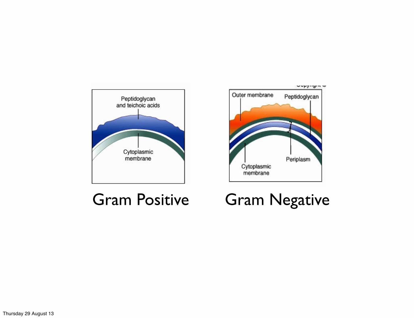
Gram Positive Gram Negative
Thursday 29 August 13

Gram Staining kit has...
Thursday 29 August 13

‣ Addition of iodine (mordant) forms crystal violet iodine complex within the cell wall. ‣ The complex formed is larger than crystal violet so it cannot be easily washed out from the intact peptidoglycan layer.
Primary stain, crystal violet stains all the cells purple.
‣ Application of alcohol (decolorizer) decolorizes the stain.‣ Since gram negative organism have thin peptidoglycan layer and have additional lipopolysaccharide layer which gets dissolved due to the addition of alcohol, so gram negative organism fails to retain the complex and gets decolorized
‣To observe the decolorized cells secondary stains like Basic fuchsin or Safranin is added which stains the gram negative organisms pink
Thursday 29 August 13

Gram Positive vs Gram Negative Bacteria.
Thursday 29 August 13

Thursday 29 August 13
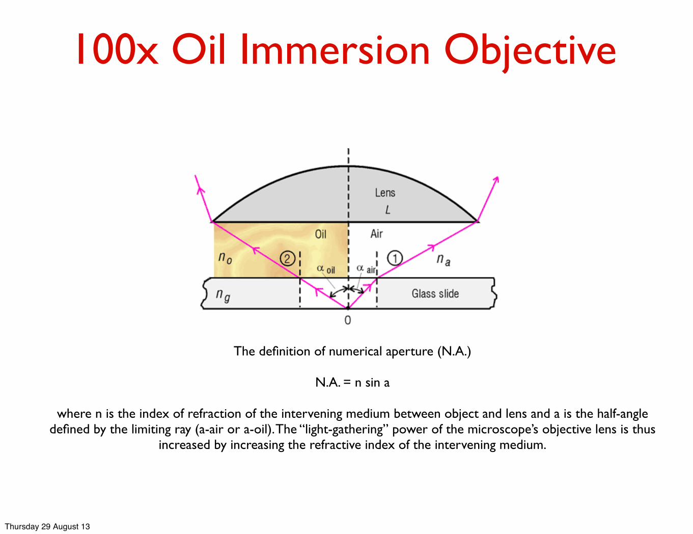
100x Oil Immersion Objective
The definition of numerical aperture (N.A.)
N.A. = n sin a
where n is the index of refraction of the intervening medium between object and lens and a is the half-angle defined by the limiting ray (a-air or a-oil). The “light-gathering” power of the microscope’s objective lens is thus
increased by increasing the refractive index of the intervening medium.
Thursday 29 August 13
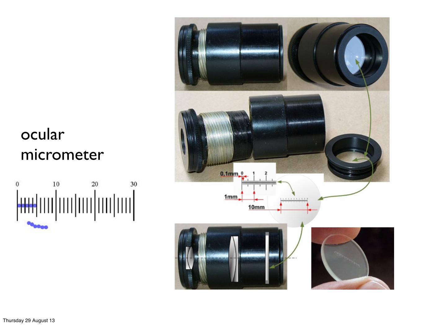
ocular micrometer
Thursday 29 August 13

ocular micrometer
Thursday 29 August 13


