BIOCHEMICAL AND BIOPHYSICAL RESEARCH COMMUNICATIONS
Pages 1161-1168
GRANULINS, A NOVEL CLASS OF PEPTIDE FROM LEUKOCYTES
Andrew Bateman*, Daniel Belcourt*, Hugh Bennett*, Claude Lazure**, and Samuel Solomon*
*Endocrine Laboratory, Royal Victoria Hospital/McGill University, Departments of Medicine, Obstetrics, and Gynecology, Montreal, Quebec, H3A 1A1, Canada
**Institut de recherches cliniques de Montr6al, Montreal, Quebec, Canada
Received November i, 1990
SUMMARY. We report the isolation and characterization of a novel class of leukocyte peptides with possible cytokine-like activities which we call granulins. They are cystine-rich with molecular weights of approximately 6Kda, except for granulin D, which appears to be a dimer. We present the sequence of one member of this family, a 56 residue peptide, granulin A, and amino-terminal sequences for three other granulins from human peripheral leukocytes. A fifth related peptide was isolated and partially sequenced from rat bone marrow, suggesting that at least some of the granulin in peripheral leukocytes is preformed in the marrow. Rat granulin, and human granulin A, are closely related, showing that the granulin structures are highly conserved between species. ® 1990 Academic Press, Inc.
It has recently become clear that leukocytes are peptiderglc cells. Neutrophil granules
contain large amounts of basic, cystine-rich peptides of 29 to 34 amino acids, that have been
variously called defensins (1), corticostatins (2), myeloid-related sequences (3), and cryptidins (4).
Some of these peptides are antimicrobial agents at micromolar concentrations (5), and it was
initially thought that their only biological activity was in non-oxidative, non-enzymatic, destruction
of phagocytosed microorganisms. More recently, however, we have shown that corticostatins have
potential regulatory functions, including the ability to inhibit the action of the hormone
adrenocorticotropin on glucocorticoid secretion (2,6,7) and to stimulate nifedipine-sensitive L-type
Ca 2+ channels in villus enterocytes (8). It has also been reported that a human defensin is a
monocyte chemotactic agent (9). Other granulocyte-associated peptides have also been shown to
have regulatory activities. For example, hemoregulatory peptide 1 is a granulocyte-associated thiol
containing pentapeptide, with potent inhibitory actions on myelopoiesis (10). Several groups have
reported the existence of immunomodulatory or cytokine-like activities associated with neutrophll
extracts or supernatants (11,12,13,14). These activities include mast cell degranulation, chemotaxis,
and the inhibition of myelopoetic-colony formation. Despite these reports, and the evidence for
regulatory actions associated with known granulocyte peptides, few systematic attempts to
Vol. 173, No. 3, 1990 BIOCHEMICAL AND BIOPHYSICAL RESEARCH COMMUNICATIONS
characterize the regulatory molecules of the granulocyte seem to have been made. Granulocyte-
enriched extracts contain several cystine-rich components at levels approximately three orders of
magnitude lower than the defensin/corticostatins. From their compositional analysis and
chromatographic behaviour these peptides appeared unrelated to any known hormone, including
the defensin/corticostatins. In view of the potential role of granulocyte-derived peptides both as
immunoregulatory molecules, and in host resistance, it was clearly important to characterize these
peptides. Here we report the isolation and characterization a family of novel leukocyte-associated
peptides that are cystine-rich, of approximately 6 Kda, and that may be c~okines.
MATERIALS AND METHODS
Tissue Sources. Blood was taken from healthy volunteers, prepared and fractionated using Ficoll-Hypaque
(Pharmacia, Upsalla, Sweden) as previously described (6). Differential counts were obtained from the Hematology Department, Royal Victoria Hospital. For structural studies the first wash peritoneal exudate from patients with peritonitis was used as a source of leukocytes. Typically, this comprises from 75 to 95% neutrophils. Crude granule preparations were made by lysing the cells in Hank's buffered saline solution using a Cole Palmer Ultrasonic Homogeniser 4710 Series. Preparations were inspected visually under a microscope to ensure complete cell lysis. The cellular debris was pelleted by centrifugation at 500 x g for 10 mins, and the supernatant inspected to ensure the complete removal of broken cells. The supernatant was then pelleted by centrifugation for 20 minutes at 5000 x g, and the crude granule pellet washed twice in HBSS. The two human granule peptides, HP-1 and HP-4, and the granule enzyme lysozyme, were used as granule markers, and the cytoplasmic peptide thymosin-g-4 was used as a marker for cytoplasmic contamination.
Extraction and Purification. Whole cell preparations or crude granule fractions were extracted by sonication using an
acidic high-salt extraction medium as described elsewhere (15). The extract was then centrifuged at 2000 x g for 15 mins, and the pellets re-extracted. Pooled supernatants were then adsorbed on SepPak C18 cartridges (Waters Associates, Milford, Mass) and eluted in 5 to 10 m180% acetonitrile in 0.1% TFA and the eluate lyophilized. The SepPak eluate was fractionated by reversed phase HPLC using a Waters C18 I~Bondapak column (7.8mm x 30cm) eluted over a three hour period using a gradient of 0 to 80% acetonitrile in 0.1% TFA throughout at an elution rate of 1.5ml rain -1. Aliquots of the eluted fractons were then screened by amino acid analysis.The fractions of interest were further purified by size-exclusion HPLC using two 1-125 ProteinPak columns (Waters) connected in series, eluted isocratically in 40% acetonitrile in 0.1% TFA at i ml min -1 (16). Partially purified peptides were then purified to homogeneity using a second C-18 i~Bondapak HPLC column (3.9mm x 30cm), with a gradient of 10 to 40% acetonitrile in 0.1% TFA throughout at 1.5 ml min "1 for 90 mins. The rat peptide was purified from the aspirated femural bone marrow of 50 Sprague- Dawley rats (Charles Rivers, St Constant, Quebec), and extracted directly as outlined above. SepPak eluates were fractionated on a C18 IxBondapak column using a gradient of 4 to 48% acetonitrile in
1 0.1% TFA throughout over 1 hour at 1.5 ml min". Fractions were screened by amino acid analysis, and granulin-like material further purified using the same column with a gradient of 20 to 40% acetonitrile in 0.13% HFBA throughout over one hour (15). Final purification was by size-exclusion HPLC as described above. Amino Acid Analysis and Microsequencing.
For amino acid analysis aliquots of the peptide were lyophilized in borosilicate glass tubes and hydrolyzed in an evacuated reacti-vial for 16 hours at 105°C with 6N HC1. Amino acid analysis was performed using a model 6300A Analyser (Beckman Instruments, Palo Alto, CA). For microsequence analysis purified peptides were reduced with 10 mM dithiothreitol, or 2- mercaptoethanol, in 8M guanidine-HC1, 1 mM EDTA, 0.25M Tris, pH 8.5 for 1.5 hours at 37°C and then pyridylethylated with 2 Ixl 4-vinylpyridine (Aldrich Chemicals) under the same conditions.
1162
Vol. 173, No. 3, 1990 BIOCHEMICAL AND BIOPHYSICAL RESEARCH COMMUNICATIONS
The S-pyridylethylated peptides were then purified using a gradient of 5% to 60% acetonitrile containing 0.1% TFA throughout over 60 minutes with an initial 40 minute isocratic stage at 5% aeetonitrlle to elute polymeric vinylpyridine side products. The derivatized peptides were then submitted directly to sequence analysis or further processed by enzymatic digestion. The amino acid sequence determinations were carried out with an Applied Biosystem gas-phase sequenator (model 470A) as described in (17) but using a sequence program adapted from Speicher (18). The resulting phenylthiohydantoin (PTH)-amino acids were analysed by reverse-phase HPLC on the on line PTH-analyser (Applied Biosystem model 120A) and/or a stand alone Varian HPLC unit as described previously (17). The PTH-yields for each standard were normalized according to a PTH-NorLeucine internal standard while the initial and repetitive yields were obtained by linear regression from the yields of selected stable PTH-derivatives. Sequence analysis of rat granulin and its fragments was undertaken at the McGill Peptide and Protein Sequencing facility located in the laboratory of Dr Michel van der Rest at the Shriner's Hospital for Crippled Children in Montreal.
Enzymatic Digestion. S-pyridylethylated peptideswere digested using trypsin (TPCK-treated, Sigma), chymotrypsin
(Sigma), and S. aureus V8 protease (Sigma), at enzyme to substrate ratios of approximately 1 to 50 by weight. Digestions were performed at 37°C in 1001~1 50mM ammonium bicarbonate buffer, pH 8.3 for 3 hours, and terminated by the addition of lml of 0.1% TFA. The proteolytic fragments were then fractionated by rp-HPLC on a C-18 i~Bondapak column using a gradient of 0 to 40% acetonitrile in 0.1% TFA throughout over 60 mins at 1.5ml mln "1. Fractions were collected, aliquots removed for amino acid analysis, and then stored frozen at -80°C.
RESULTS
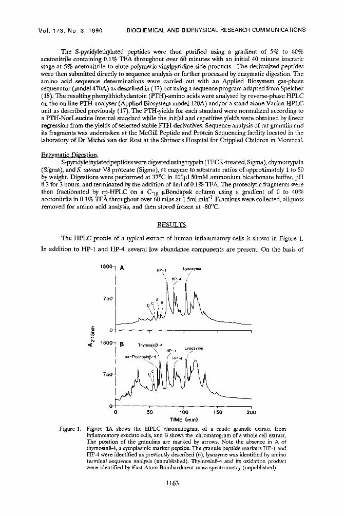
The HPLC profile of a typical extract of human inflammatory cells is shown in Figure 1.
In addition to HP-1 and HP-4, several low abundance components are present. On the basis of
Figure 1.
1 5 0 0 -
750 -
E 0 IO
04 <~ 1 5 0 0 -
7 5 0 -
A HP- 1 Lysozyme
7,/ O C / B
B ThymosinB-4 \ ' HP-1 Lysozyme
ox-Thymosin~-4~ //HP_4 /
0 5'0 ~ ' ' 0 100 150 2 0 0
T I M E (rain)
Figure 1A shows the HPLC chromatogram of a crude granule extract from inflammatory exudate cells, and B shows the chromatogram of a whole cell extract. The position of the granulins are marked by arrows. Note the absence in A of thymosinB-4, a cytoplasmic marker peptide. The granule peptide markers HP-1 and HP-4 were identified as previously described (6), lysozyme was identified by amino terminal sequence analysis (unpublished). ThymosinB-4 and its oxidation product were identified by Fast Atom Bombardment mass spectrometry (unpublished).
1163
Vol. 173, No. 3, 1990 BIOCHEMICAL AND BIOPHYSICAL RESEARCH COMMUNICATIONS
Figure 2.
~t3
¢,1 ,¢{
1.0.
0.5"
0
1.0-
0.5-
0
1.0-
o.5'
t f ~
A
i i i r i J i i
Thymosin-~-4
i i i [ i i i i
C o C
_Z ' '0 ' 2'0 ' ' ' 4'0 I 30
TIME (min)
Size-exclusion purification of granulin A, (panel A), granulin B, (panel B), and granulins C and D, (panel C). Size markers were substance P, CLIP, and ACTH1_39. Apparent m.wts for native granulin A, 2700; granulin B,3200; granulin C, 1700; and granulin D, 3900.
amino acid composition analysis of these components we identified three low abundance
components that had unusually high levels of cystine. These are labelled A, B, and C/D, and were
present in both whole cell extracts (Fig 1B) and crude granule preparations (Fig 1A). Each of these
extracts was then further purified using size-exclusion HPLC, revealing that the component C/D
contained two peptides, one of which, D, eluted as a larger molecule than the other three. Each
peptide was further purified on rp-HPLC. Their amino acid compositions are given in Table 1. The
purified peptides were S-pyridylethylated, and amino terminal sequence analyses were performed,
revealing that the four peptides were distinct but related molecules with no homology to any known
protein. Because these peptides were associated with the granule fraction, we call them granulins
A, B, C, and D. Granulin D was run on reducing and non-reducing SDS-PAGE, and ran as a
smaller molecule after reduction, indicating that it is probably a dimer. Only one peptide was
recovered after S-pyridylethylation of granulin D suggesting that it is a homodimer. However, until
a full sequence is determined we cannot exclude the possibility that it is a heterodimer of closely
related subunits. The rat defensin/corticostatins elute in the same region of the chromatogram as
1164
Vol. 173, No. 3, 1990 BIOCHEMICAL AND BIOPHYSICAL RESEARCH COMMUNICATIONS
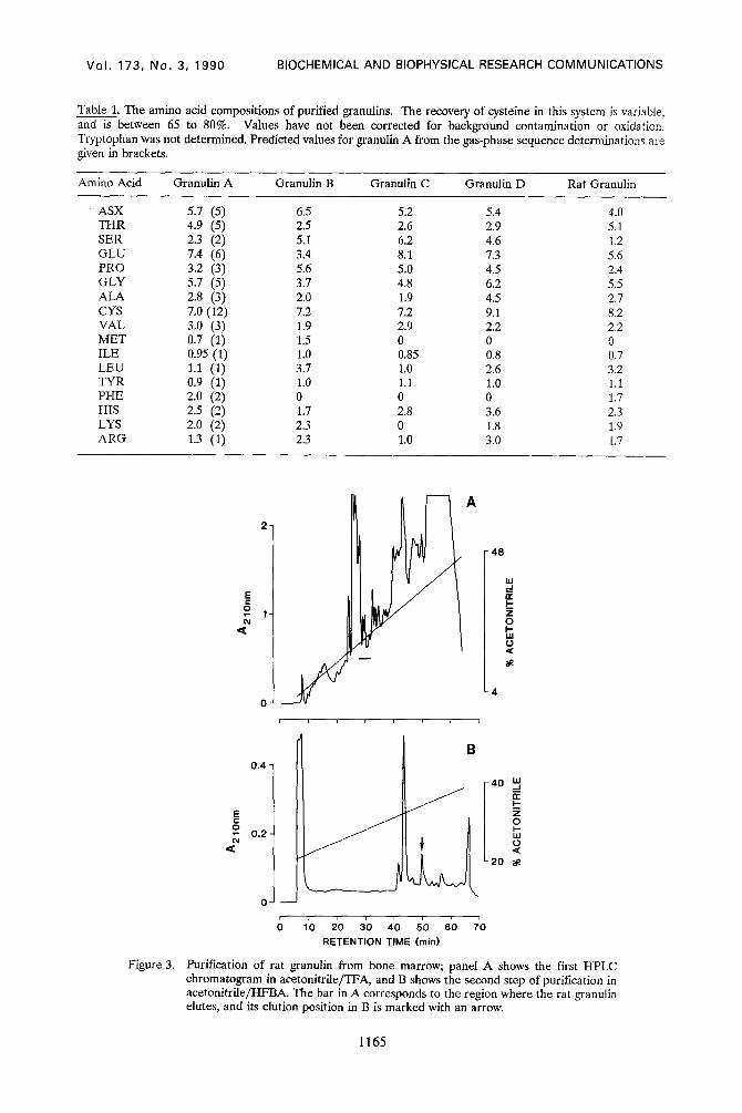
Table 1. The amino acid compositions of purified granulins. The recovery of cysteine in this system is variable, and is between 65 to 80%. Values have not been corrected for background contamination or oxidation. Tryptophan was not determined. Predicted values for granulin A from the gas-phase sequence determinations are given in brackets.
Amino Acid Granulin A Granulin B Granulin C Granulin D Rat Granulin
Purification of rat granulin from bone marrow; panel A shows the first HPLC chromatogram in acetonitrile/TFA, and B shows the second step of purification in acetonitrile/HFBA. The bar in A corresponds to the region where the rat granulin elutes, and its elution position in B is marked with an arrow.
1165
Vol. 173, No. 3, 1990 BIOCHEMICAL AND BIOPHYSICAL RESEARCH COMMUNICATIONS
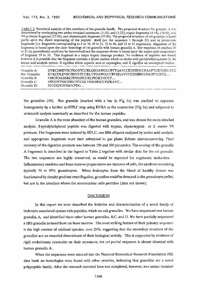
TABLE 2. Structural analysis of five members of the granulin family. The proposed structure for granulin A was determined by overlapping two amino terminal sequences, (1-11), and (1-23); tryptic fragments (4-18), (19-52), and V8 protease fragment (37-56), and ehymotryptic fragment (47-56). The proposed structure of rat granulin is based partly upon the direct sequencing of the peptide itself (i.e. the sequence 1 through 21) and its proteolytic fragments (i.e. fragments corresponding to 4 to 18, 19 to 31, 32 to 46, and 32 to 51 sequences). Alignment of the fragments is based upon the clear homology of rat granulin with human granulin A. The sequence of residues 26 to 31 (in parenthesis) could not be determined and the sequence shown is based upon the amino acid composition of fragment 19 to 31. This fragment is a major tryptic cleavage product. No evidence of arginine was found, however it is possible that the fragment contains a lysine residue which co-elutes with pyridylethyl-cysteine in the amino acid analysis system. B signifies either aspartic acid or asparagine, and X signifies an unassigned residue.
Granulin A: Rat Granulin: Granulin B: Granulin C: Granulin D:
the granulins (19). Rat granulin (marked with a bar in Fig 3A) was purified to apparent
homogeneity by a further rp-HPLC step using HFBA as the counterion (Fig 313) and subjected to
structural analysis essentially as described for the human peptides.
Granulin A is the most abundant of the human granulins, and was chosen for more detailed
analysis. S-pyridylethylated peptide was digested with trypsin, chymotrypsin or S. a u r e u s V8
protease. The fragments were isolated by HPLC, one fifth aliquots analysed by amino acid analysis,
and appropriate fragments were then submitted to gas phase Edman microsequencing. Final
recovery of the digestion products was between 150 and 300 picomoles. The overlap of the granulin
A fragments is described in the legend to Table 2 together with similar data for the rat granulin.
The two sequences are highly conserved, as would be expected for regulatory molecules.
Inflammatory exudates and bone marrow preparations are mixtures of cells, the exudates containing
typically 70 to 95% granulocytes. When leukocytes from the blood of healthy donors was
fractionated by density gradient centrifugation, granulins could be detected in the granulocyte pellet,
but not in the interface where the mononuclear cells partition (data not shown).
DISCUSSION
In this report we have described the isolation and characterization of a novel family of
leukocyte associated cystine-rich peptides, which we call granulins. We have sequenced one human
granulin, A, and identified three other human granulins, B,C, and D. We have partially sequenced
a fifth granulin isolated from rat bone marrow. The most striking feature of their primary sequence
is the high content of oxidised cysteine, over 20%, suggesting that the secondary structure of the
granulins are an essential determinant of their biological activity. This is supported by evidence of
rigid evolutionary constraint on their structures; the rat partial sequence is almost identical with
human granulin A .
When the sequences were entered into the National Biomedical Research Foundation PIP,
data bank no homologies were found with other proteins, indicating that granulins are a novel
polypeptide family. After the research reported here was completed, however, two amino terminal
1166
Vol. 173, No. 3, 1990 BIOCHEMICAL AND BIOPHYSICAL RESEARCH COMMUNICATIONS
sequences were published, epithelin 1 and 2, isolated from the rat kidney, which are homologous
with the granulins. The kidney peptides are putative cytokines that have growth inhibitory and
stimulatory properties on some epithelial cells in vitro (20). Rat granulin and the reported amino
terminal sequence of epithelin 1 differ at only one residue; epithelin lacks the amino terminal
glutamyl residue of rat granulin A. At present it is not certain if both peptides are the product of
the same, or different genes. It is also too early to determine whether the epithelins are intrinsic
to renal cells, or if they are derived from blood borne cells trapped in the kidney. It is clear,
however, that the two peptides from the kidney are members of a larger family, the granulins, and
that a major source of the granulins, and probably also the epithelins, is from circulating leukocytes.
Rat granulin was isolated from bone marrow, indicating that granulins are of myeloid origin.
Whether granulins are also synthesized in circulating leukocytes remains to be determined.
Granulins were extracted from granuloeyte rich preparations, and are recoverable from the
granulocyte pellet after Ficoll-Hypaque density gradient centrifugation (data not shown). This
suggests that their cellular origin may be the neutrophll, however we cannot exclude the possibilty
that other granulocytes such as eosinophils, or contaminating monocytes, contribute to the granulin
content of these extracts. Extracts of human platelets contained no detectable granulins (data not
shown). It is possible that each granulin belongs to a distinct cell type, or that sub-classes of the
same cell-type contain different granulins. These are issues best answered using immunolocalization
procedures. The granulins co-purify in a crude granule extract. Granulocytes have several different
granule subclasses, including a true secretory compartment (21) that can be activated independently
of phagocytosis. The availability of suitable immunoassay techniques will allow us to unambiguously
locate the granulins to a cell type, and a subcellular compartment.
In conclusion, the granulins are a novel family of cystine-rich immunoinflammatory peptides.
Their presence in circulating leukocytes and inflammatory exudates, and their structural similarity
with the rat epithelins (20), suggests possible roles in inflammation, wound repair, and tissue
remodeling.
ACKNOWLEDGMENTS. The authors wish to acknowledge the expert technical assistance of Nathalie Croteau and Nicola Franco. Part of this study was supported by a grant (PG2) from the Medical Research Council of Canada, and operating grants MT-1658 and MT-6733 from the Medical Research Council of Canada, and operating grant HDO-4365 of the National Institute of Child Health and Human Development. HB was supported by a Scholarship, and for part of this work, AB was supported by a Fellowship, both from the Fond de la Recherche en Sant6 du Quebec. C.L. is a Chercheur-boursier from the Fond de la Recherche en Sant6 du Quebec.
1.
2.
3.
REFERENCES
Selsted M.E., Brown D.M., Delange R.J., and Lehrer R.I. (1983) J. Biol Chem. 258, 14485- 14489 Zhu Q., Hu J., Mulay S., Esch F., Shimasaki S., and Solomon S. (1988) Proc. Natl. Acad. Sci. (USA) 85, 592-596 Mars W.M., van Tuinen P., Drabkin H., White J., and Saunders G. (1988) Blood 71, 1713- 1719
1167
Vol. 173, No. 3, 1990 BIOCHEMICAL AND BIOPHYSICAL RESEARCH COMMUNICATIONS
4.
5.
6.
7. 8.
9. 10.
11. 12.
13. 14. 15. 16. 17.
18.
19.
20.
21.
Ouellette A.J., Greco R.M., James M., Frederick, D., Naftilan J., and Fallon J.T. (1989) J. Cell Biol., 108, 1687-1695. Ganz T., Selsted M.E., Szklarek, D., Harwig S.S.L., Daher K., and Lehrer R.I. (1985) J. Clin. Invest. 76, 1427-1435 Singh A., Bateman A., Zhu Q., Shimasaki S., Esch F., and Solomon S. (1988) Biochem. Biophys. Res. Comm. 155, 524-529. Zhu Q., Bateman A., Singh A., Solomon S. (1989) Endocrine Research 15, 129-149 MacLeod R.J., Hamilton J.R., Bateman A., Belcourt D., Hu J., Bennett H.P.J., and SolomOn S. (1991) Proc Natl Acad Sci (USA), In Press. Territo M.C., Ganz T., Selsted M.E., and Lehrer R.I., (1989) J. Clin. Invest. 84, 2017-2020 Laerum O.D., and Paukovits W.R., (1988) in The Inhibitors of Hematopoiesis, eds Najman A, Guignon M., Gorin N-C., and Mary J-Y., (John Libbey Eurotext, London, Paris) 21-30 White M.V., and Kaliner M.A. (1987) 139, 1624-1630 Doherty D.E., Downey G.P., Worthern G.S., Haslett C., and Henson P.M. (1988) Lab. Invest 59, 200-213 WiUemze R., Walker R.I., Herion J.C., and Palmer J.G. (1978), Blood 51, 21-28 Benestad H.B., Hersleth I.B. (1984), Blut 48, 201-211 Bennett H.P.J., Browne C.A., Solomon S. (1981)Biochem. 20, 4530-4538 Bennett H.P.J., Browne C.A., and Solomon S. (1983) Anal Biochem. 128 121-129 Lazure C., Saayman H.S., Naud6 R.J., Oelofsen W., and Chr6tien M., (1989) Int. J. Peptide Res. 33, 46-58. Speicher D.W. (1989) in Techniques in Protein Chemistry (Hugli T., ed), Academic Press, San Diego, pp 24-35. Belcourt D.,Bateman A.,Singh A., Lazure C., Bennett H.P.J., and Solomon S. (1990) 72nd Annual Meeting of the Endocrine Society, Atlanta, Georgia pp276 (abs.). Shoyab M., McDonald V.L., Byles C., Todaro G., and Plowman G.D. (1990) Proc. Natl Acad. Sci. (USA) 87,7912-7916. Dewald B., Bretz U., and Baggiolini M., J. Clin. Invest. (1982) 36, 518-525.