Grayscale Medical Image Segmentation Method Based on 2D&3D Object Detection with Deep Learning Yunfei Ge Tongji University Qing Zhang Tongji University Yuantao Sun ( [email protected]) Tongji University Yidong Shen The First people’s Hospital of Yancheng Xijiong Wang Shanghai Bojin Electric Instrument & Device Co., Ltd Research Article Keywords: Grayscale medical image, Image segmentation, Deep learning, Object detection, Point cloud Posted Date: October 29th, 2021 DOI: https://doi.org/10.21203/rs.3.rs-1018292/v1 License: This work is licensed under a Creative Commons Attribution 4.0 International License. Read Full License
Transcript
Grayscale Medical Image Segmentation MethodBased on 2D&3D Object Detection with DeepLearningYunfei Ge
1 School of Mechanical Engineering, Tongji University, Shanghai, China 5
2 Department of Orthopaedics, The First people’s Hospital of Yancheng, Yancheng, China 6
3 Shanghai Bojin Electric Instrument & Device Co., Ltd, Shanghai, China 7
Abstract 8
Background: Grayscale medical image segmentation is the key step in clinical computer-aided diagnosis. Model-9 driven and data-driven image segmentation methods are widely used for their less computational complexity and more 10 accurate feature extraction. However, model-driven methods like thresholding usually suffer from wrong 11 segmentation and noises regions because different grayscale images have distinct intensity distribution property thus 12 pre-processing is always demanded. While data-driven methods with deep learning like encoder-decoder networks 13 always are always accompanied by complex architectures which require amounts of training data. 14
Methods: Combining thresholding method and deep learning, this paper presents a novel method by using 2D&3D 15 object detection technologies. First, interest regions contain segmented object are determined with fine-tuning 2D 16 object detection network. Then, pixels in cropped images are turned as point cloud according to their positions and 17 grayscale values. Finally, 3D object detection network is applied to obtain bounding boxes with target points and 18 boxes’ bottoms and tops represent thresholding values for segmentation. After projecting to 2D images, these target 19 points could composite the segmented object. 20
Results: Three groups of grayscale medical images are used to evaluate the proposed image segmentation method. 21 We obtain the IoU (DSC) scores of 0.92 (0.96), 0.88 (0.94) and 0.94 (0.94) for segmentation accuracy on different 22 datasets respectively. Also, compared with five state of the arts and clinically performed well models, our method 23 achieves higher scores and better performance. 24
Conclusions: The prominent segmentation results demonstrate that the built method based on 2D&3D object detection 25 with deep learning is workable and promising for segmentation task of grayscale medical images. 26 27 Keywords: Grayscale medical image, Image segmentation, Deep learning, Object detection, Point cloud. 28 29 *Corresponding Author Yuantao Sun, E-mail: [email protected] 30
1 Background 31
Medical imaging plays the key role in diagnosis or disease treatment by revealing internal 32
structures with technologies mainly of computer tomography (CT), magnetic resonance imaging 33
(MRI), ultrasound, and especially X ray radiography [1]. Due to different absorption capability of 34
various organs or tissues for radiations, waves, and etc., pixels belong to various object in grayscale 35
medical images have diverse grayscale values usually from 0-255 [2] and meanwhile values of 36
pixels of the same object always gather within a range. 37
2
Medical image segmentation has been widely applied to make images clearer with anatomical or 38
pathological structures changes [3], such as bone segmentation [4], lung segmentation [5,6], heart 39
fat segmentation [7] and liver or liver-tumor segmentation [8,9], etc. They could be considered to 40
divide origin images into several sub regions for picking up some crucial objects and extracting 41
interesting features which improve the computer aided diagnostic efficiency. There has raised 42
enormous approaches and they could be classified into two categories: model-driven techniques 43
and data-driven techniques. [5,10] 44
Many model-driven methods for medical image segmentation, including thresholding, clustering, 45
and region growing, were presented in particular before the widespread application of deep 46
learning. [10] Thresholding was one of the most common used method in practice due to its 47
efficiency. [11] The basic working of thresholding was to determine specific threshold values and 48
each pixel in the image could be classified as the foreground or background depending on the 49
comparison between their intensity values and threshold values. [12-14] Traditional thresholding 50
methods always relied on single models for universal segmentation tasks which could lead to 51
incorrect results. Also, segmentation objects often occupied only parts of whole images and pixels 52
of different objects may share same intensity values, so noises could appear if image segmentation 53
was applied overall. 54
With the era of big data coming, emerging data-driven technologies with deep learning have 55
remarkably demonstrated in variety medical image segmentation task. Supervised learning 56
methods and especially some CNN (Convolutional neural network) based encoder-decoder 57
structures such as FCN (Fully convolutional networks) [15], U-Net [16], DeepLab [17] has 58
practically proved [5]. Compared with traditional methods, deep learning could help analyze 59
medical images more effectively and extract more detailed features. 60
3
Although these end-to-end structures was pragmatic for medical images semantic segmentation, 61
the segmentation accuracy always relied on a large amount of training dataset. But medical image 62
annotation could be time-consuming and quite expensive, thus transfer learning was used to solve 63
the problem of limited labeled data and pre-trained networks on natural images as ImageNet [18] 64
were often adopted for image segmentation. [19,20] However, considering these datasets were 65
mainly designed to train models for object detection or classification, they may be more suitable 66
to pretrain networks for object detection. This inspired us to segment images with object detection. 67
We find that grayscale images could be segmented according to the comparison of thresholding 68
values with values of pixels in images and these pixels could be turned into 3D point cloud 69
according to their positions and grayscale values. Thus, by applying 3D object detection in the 70
point cloud, we could achieve groups of points within 3D bounding boxes. The top and the bottom 71
of boxes represent the thresholding values for segmentation and after mapping these points into 72
Through the setting of 3D IoU threshold for NMS and ranking with classification scores, it remains 220
only one box for each candidate class which could be considered as the proposal bounding box. 221
222
Fig.9 IoU computation for 3D. The intersection volume is highlighted in gray. 223
2.2.4 Refinement of proposal bounding box 224
Even though high classification scores of the proposal bounding boxes, the location and scale 225
errors between them and ground truth exist. We train and implement a class-specific bounding box 226
linear regression model to reduce errors and improve detection performance. 227
On the assumption that we achieve one proposal bounding box 𝑃𝑖 and its nearby ground-truth box 228 𝐺𝑖 as shown in Fig.10, where 𝑃𝑖 = (𝑃𝑙ℎ𝑖 , 𝑃𝑙𝑤𝑖 ) specifies height 𝑙ℎ of the center of proposal 229
bounding box together with its width 𝑙𝑤 . Meanwhile, the ground-truth bounding box 𝐺𝑖 is 230
specified in the same way: 𝐺𝑖 = (𝐺𝑙ℎ𝑖 , 𝐺𝑙𝑤𝑖 ). The goal of the bounding box regressor is to learn a 231
transformation which could map each proposal bounding box 𝑃 to the ground-truth box 𝐺. 232
233
Fig.10 Refinement of proposal bounding box. 234
14
The transformation could be parameterized in terms of two functions 𝑑𝑙ℎ(𝑃) and 𝑑𝑙𝑤(𝑃). The first 235
function specifies the translation of bounding box 𝑃’s center which is scale-invariant, while the 236
second specifies the log-space translation of its width. By applying the transformation as following 237
equations, an input proposal bounding box 𝑃 could be transformed into a predicted ground-truth 238
Inspired by the 2D object detection, the bounding box regression of our method is performed on 242
global features which is max pooled from PointNet model. Above two functions 𝑑𝑙ℎ(𝑃) and 243 𝑑𝑙𝑤(𝑃) could be modeled as linear functions of the global features of proposal bounding box 𝑃, 244
denoted as 𝑓𝑚𝑝(𝑃). Therefore, we have 𝑑∗(𝑃) = T∗ × 𝑓𝑚𝑝(𝑃), where ∗ represents 𝑙ℎ or 𝑙𝑤, and T∗ 245
is a vector composed of learnable model parameters. 246
The transformation targets 𝑡∗ between proposal bounding box 𝑃 and the real ground-truth box 𝐺 247
could be defined as: 248
𝑡𝑙ℎ = 𝐺𝑙ℎ − 𝑃𝑙ℎ𝑃𝑙𝑤 (4) 249
𝑡𝑙𝑤 = log (𝐺𝑙𝑤𝑃𝑙𝑤 ) (5) 250
Thus, after setting the loss function and by optimizing the regularized least squares objective as 251
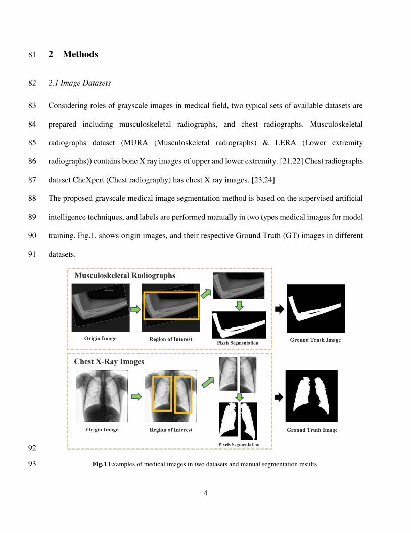
following, we could learn T∗ and achieve the transformation to refine the proposal bounding box. 252
𝑳𝒐𝒔𝒔 = ∑ (𝑡∗𝑖 − T̂∗ × 𝑓𝑚𝑝(𝑃𝑖))2𝑁𝑖 (6) 253
T∗ = argminT̂∗𝑳𝒐𝒔𝒔 + 𝜆‖T̂∗‖2 (7) 254
15
2.2.5 Training strategy 255
The proposed grayscale medical image segmentation method follows a three-stage training 256
strategy. First, we obtain interest regions from raw grayscale images with fine-tuning YOLOv3 257
model. Second, by training the pixel-features point cloud classification model based on PointNet, 258
proposal 3D bounding boxes could be achieved from the point cloud representations of pixels in 259
interest regions. By training the linear regressor, proposal bounding boxes are refined with location 260
and scale transformation. Three independent modules including regions extractor, point cloud 261
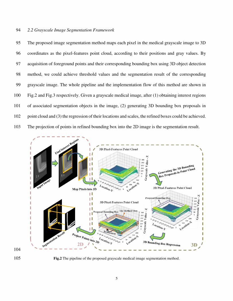
classifier and bounding box regressor in three stages compose our method. 262
2.3 Performance assessment 263
In this study, we evaluate the segmentation performance by following four metrics: Dice similarity 264
coefficient (DSC) scores [6], intersection over union (IoU), False negative (FN) and False positive 265
(FP) [7]. Ranges of DSC and IoU are between 0 and 1, higher values of them and lower values of 266
FN and FP indicate the higher accuracy. The calculation formula of DSC is defined as: 267
DSC = 2|𝑇 ∩ 𝐺||𝑇| + |𝐺| (8) 268
where 𝑇 is the detected region and 𝐺 is the ground truth region. 269
3 Results 270
We conduct experiments by the proposed grayscale image segmentation method on above 271
mentioned datasets including musculoskeletal radiographs dataset and chest radiographs dataset. 272
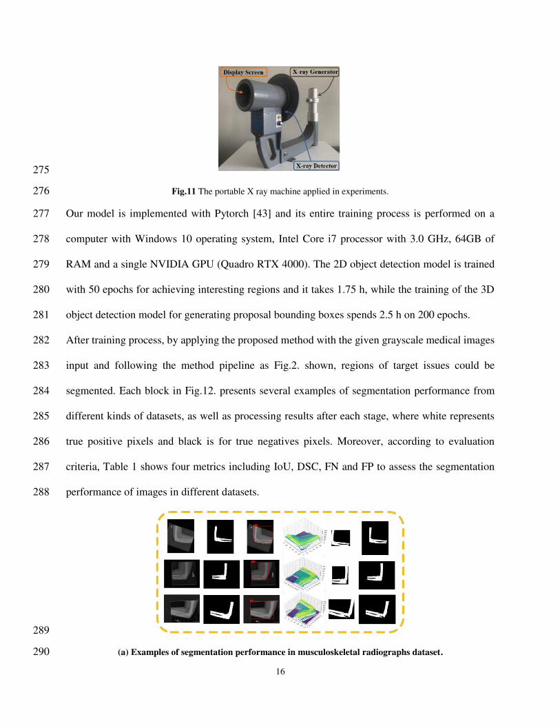
Moreover, our prepared phalanx and forearm X ray images obtained with the portable X ray 273
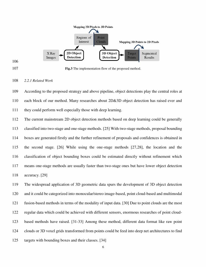
machine as Fig.11. shown are also adopted for model training and validation. 274
16
275
Fig.11 The portable X ray machine applied in experiments. 276
Our model is implemented with Pytorch [43] and its entire training process is performed on a 277
computer with Windows 10 operating system, Intel Core i7 processor with 3.0 GHz, 64GB of 278
RAM and a single NVIDIA GPU (Quadro RTX 4000). The 2D object detection model is trained 279
with 50 epochs for achieving interesting regions and it takes 1.75 h, while the training of the 3D 280
object detection model for generating proposal bounding boxes spends 2.5 h on 200 epochs. 281
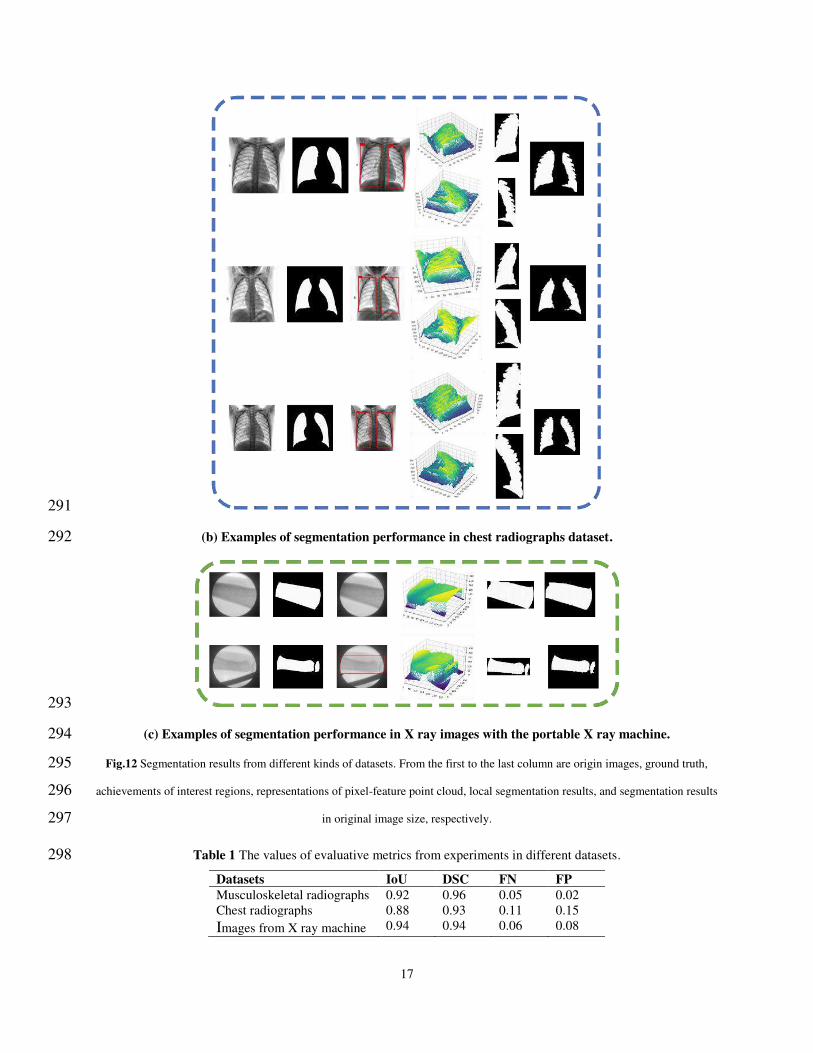
After training process, by applying the proposed method with the given grayscale medical images 282
input and following the method pipeline as Fig.2. shown, regions of target issues could be 283
segmented. Each block in Fig.12. presents several examples of segmentation performance from 284
different kinds of datasets, as well as processing results after each stage, where white represents 285
true positive pixels and black is for true negatives pixels. Moreover, according to evaluation 286
criteria, Table 1 shows four metrics including IoU, DSC, FN and FP to assess the segmentation 287
performance of images in different datasets. 288
289
(a) Examples of segmentation performance in musculoskeletal radiographs dataset. 290
17
291
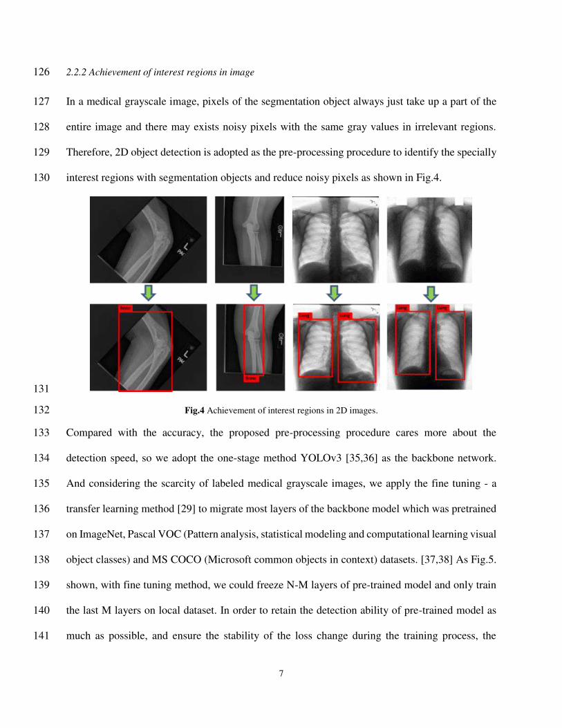
(b) Examples of segmentation performance in chest radiographs dataset. 292
293
(c) Examples of segmentation performance in X ray images with the portable X ray machine. 294
Fig.12 Segmentation results from different kinds of datasets. From the first to the last column are origin images, ground truth, 295
achievements of interest regions, representations of pixel-feature point cloud, local segmentation results, and segmentation results 296
in original image size, respectively. 297
Table 1 The values of evaluative metrics from experiments in different datasets. 298
Datasets IoU DSC FN FP
Musculoskeletal radiographs 0.92 0.96 0.05 0.02 Chest radiographs 0.88 0.93 0.11 0.15 Images from X ray machine 0.94 0.94 0.06 0.08
18
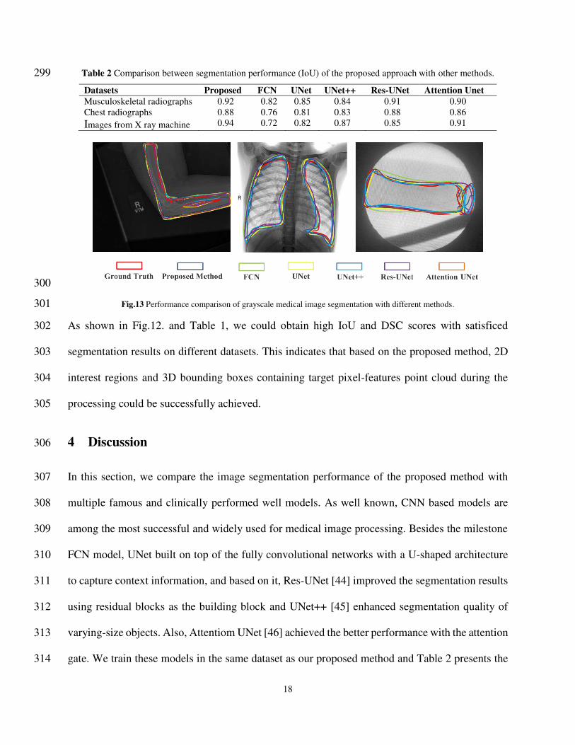
Table 2 Comparison between segmentation performance (IoU) of the proposed approach with other methods. 299
We declare that all of us obey the principles of the Declaration of Helsinki. In other words, all 357
experiments and methods in this paper are in accordance with these principles. The study was 358
approved by the Ethics Committee of the First people’s Hospital of Yancheng. 359
Consent to participate 360
The fully anonymized phalanx and forearm X ray images were received by authors on 2 April, 361
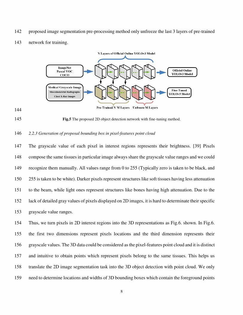
2021 and the requirement for informed consent was waived for this study because of the 362
anonymous nature of the data. 363
21
Consent for publication 364
Not applicable for this paper 365
Availability of data and materials 366
Musculoskeletal radiographs and chest radiographs which support our research are available from 367
Stanford ML Group. But restrictions apply to the availability of these data, which were used under 368
license for the current study, and so are not publicly available. Data are however available from 369
the authors upon reasonable and with permission of Stanford ML Group. While phalanx and 370
forearm X ray images are available only upon request by emailing authors due to the ethical 371
restrictions on sharing these data which could contain potentially sensitive information of patients. 372
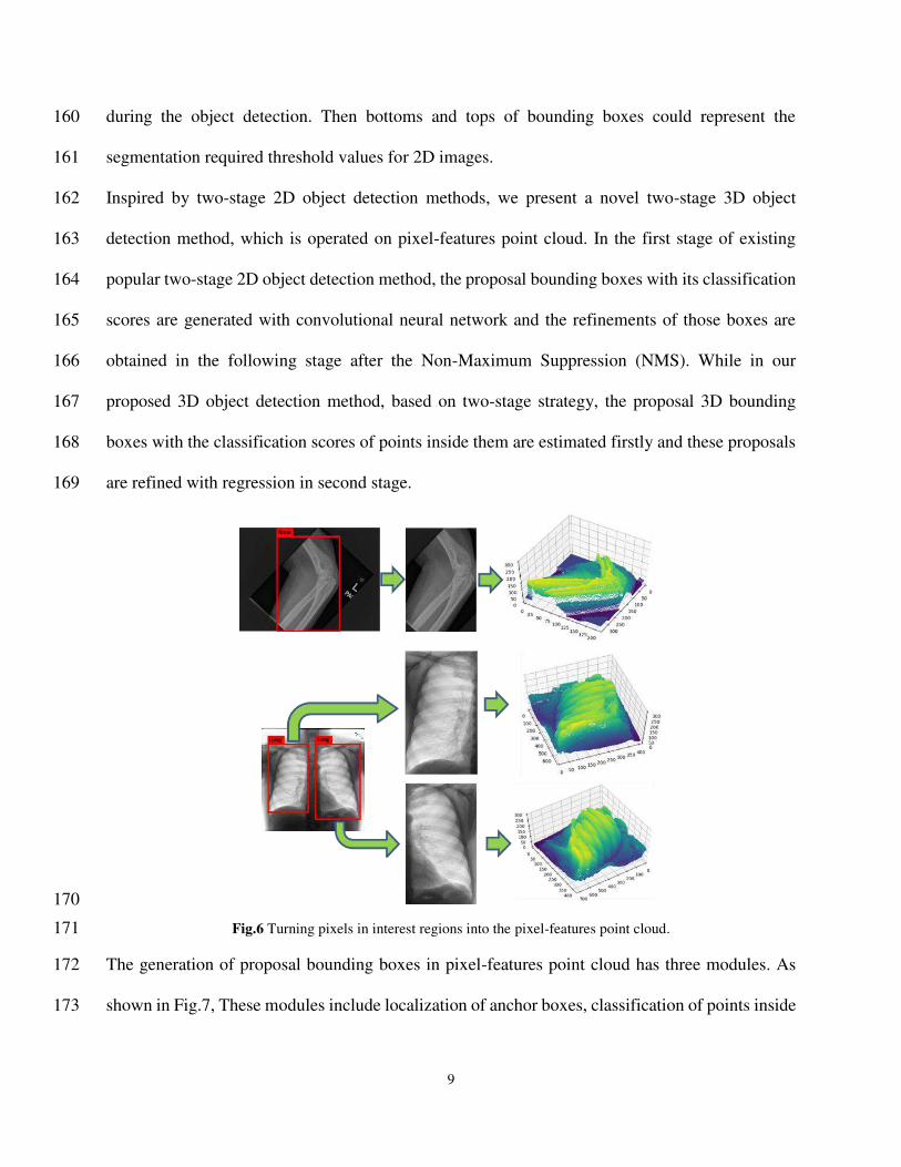
Competing interests 373
All authors declare that they have no interest conflicts or competing interests. 374
Founding 375
This work was supported by the project of Tongji University Sheng Feiyun College Student 376
Science and Technology Innovation Practice Found. 377
Authors' contributions 378
Qing Zhang conceived the research. Yunfei Ge and Yidong Shen analyzed the clinical and imaging 379
data. Yuantao Sun, Yunfei Ge, and Xijiong Wang designed the study. Yunfei Ge and Yidong Shen 380
performed the experiments and collected the results. Yunfei Ge and Yuantao Sun drafted the 381
manuscript. Qing Zhang reviewed the final manuscript. All authors read and approved the final 382
manuscript. 383
Acknowledgements 384
Not applicable. 385
References 386
1. Justine Wallyn, Anton Nicolas, Akram Salman, et al. Biomedical imaging: principles, technologies, 387 clinical aspects, contrast agents, limitations and future trends in nanomedicines. Pharmaceutical 388 Research. 2019; 36(6):78-108. 389
2. Yeo W K, Yap D F W, et al. Grayscale medical image compression using feedforward neural networks. 390 2011 IEEE International Conference on Computer Applications and Industrial Electronics (ICCAIE). 391 2011; 633-638. 392
3. Lei Tao, et al. Medical Image Segmentation Using Deep Learning: A Survey. arXiv. 2020; 13120. 393 4. Rathnayaka K, Sahama T, Schuetz MA, et al. Effects of CT image segmentation methods on the 394
accuracy of long bone 3D reconstructions. Medical Engineering & Physic. 2011; 33(2): 226-233. 395
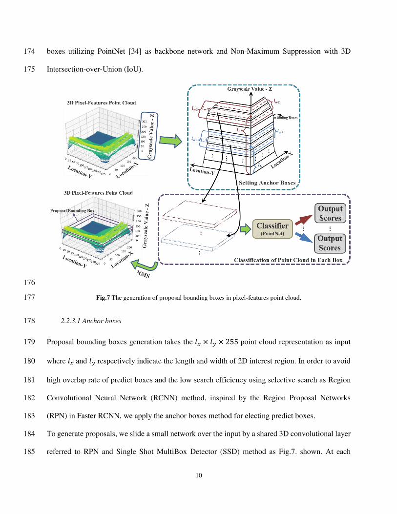
22
5. Shuo Wang, Zhou Mu, Liu Zaiyi, et al. Central focused convolutional neural networks: Developing a 396 data-driven model for lung nodule segmentation. Medical Image Analysis. 2017; 40: 172-183. 397
6. Han Liu, Wang Lei, Nan Yandong, et al. SDFN: Segmentation-based deep fusion network for thoracic 398 disease classification in chest X ray images. Computerized Medical Imaging and Graphics. 2019; 75: 399 66-73. 400
7. de Albuquerque VHC, Rodrigues D A, Ivo RF, et al. Fast fully automatic heart fat segmentation in 401 computed tomography datasets. Computerized Medical Imaging and Graphics. 2020; 80: 101674. 402
8. Li Wen, et al. Automatic segmentation of liver tumor in CT images with deep convolutional neural 403 networks. Journal of Computer and Communications. 2015; 3(11): 146. 404
9. Vivanti R, Ephrat A, Joskowicz L, et al. Automatic liver tumor segmentation in follow-up CT studies 405 using convolutional neural networks. Proc. Patch-Based Methods in Medical Image Processing 406 Workshop. 2015; 2: 2. 407
10. Saleha Masood, Sharif Muhammad, Masood Afifa, et al. A survey on medical image segmentation. 408 Current Medical Imaging. 2015; 11(1): 3-14. 409
11. Khandare ST, Isalkar A D. A survey paper on image segmentation with thresholding. International 410 Journal of Computer Science and Mobile Computing. 2014; 3(1): 441-446. 411
12. Sezgin M, Sankur B. Survey over image thresholding techniques and quantitative performance 412 evaluation. Journal of Electronic Imaging. 2004; 13(1): 146-165. 413
13. Maolood I Y, Al-Salhi Y E A, Lu S. Thresholding for medical image segmentation for cancer using 414 fuzzy entropy with level set algorithm. Open Medicine. 2018; 13(1): 374-383. 415
14. Duo Hao, Li Qiuming, Li Chengwei. Histogram-based image segmentation using variational mode 416 decomposition and correlation coefficients. Signal, Image and Video Processing. 2017; 11(8): 1411-417 1418. 418
15. Long J, Shelhamer E, Darrell T. Fully convolutional networks for semantic segmentation. Proceedings 419 of the IEEE Conference on Computer Vision and Pattern Recognition (CVPR). 2015; 3431-3440. 420
16. Ronneberger O, Fischer P, Brox T. U-net: Convolutional networks for biomedical image segmentation. 421 International Conference on Medical Image Computing and Computer-Assisted Intervention 422 (MICCAI). 2015; 234-241. 423
17. Chen LC, Papandreou G, Kokkinos I, et al. Deeplab: Semantic image segmentation with deep 424 convolutional nets, atrous convolution, and fully connected crfs. IEEE Transactions on Pattern 425 Analysis and Machine Intelligence. 2017; 40(4): 834-848. 426
18. Deng J, Dong W, Socher R, et al. Imagenet: A large-scale hierarchical image database. Proceedings of 427 the IEEE Conference on Computer Vision and Pattern Recognition. 2009; 248-255. 428
19. Kalinin A A, Iglovikov V I, Rakhlin A, et al. Medical image segmentation using deep neural networks 429 with pre-trained encoders. Deep Learning Applications. 2020; 39-52. 430
20. Pierre-Henri Conze, Brochard Sylvain, Burdin Val-E-Rie, et al. Healthy versus pathological learning 431 transferability in shoulder muscle MRI segmentation using deep convolutional encoder-decoders. 432 Computerized Medical Imaging and Graphics. 2020; 83: 101733. 433
21. Pranav Rajpurkar, Irvin Jeremy, Bagul Aarti, et al. Mura: Large dataset for abnormality detection in 434 musculoskeletal radiographs. arXiv. 2017; 1712.06957. 435
22. LERA - lower extremity radiographs. https://aimi.stanford.edu/lera-lower-extremity-radiographs-2. 436 23. Irvin J, Rajpurkar P, Ko M, et al. Chexpert: A large chest radiograph dataset with uncertainty labels 437
and expert comparison. Proceedings of the AAAI Conference on Artificial Intelligence. 2019; 33(01): 438 590-597. 439
24. Joseph-Paul Cohen, Morrison Paul, Dao Lan, et al. Covid-19 image data collection: Prospective 440 predictions are the future. arXiv. 2020; 2006.11988. 441
25. Jiao L, Zhang F, Liu F, et al. A survey of deep learning-based object detection. IEEE Access. 2019; 442 7:128837-128868. 443
26. Ross Girshick, Donahue Jeff, Darrell Trevor, et al. Rich feature hierarchies for accurate object detection 444 and semantic segmentation. Proceedings of the IEEE Conference on Computer Vision and Pattern 445 Recognition. 2014; 580-587. 446
27. Joseph Redmon, Divvala Santosh, Girshick Ross, et al. You only look once: Unified, real-time object 447 detection. Proceedings of the IEEE Conference on Computer Vision and Pattern Recognition. 2016; 448 779-788. 449
28. Wei Liu, Anguelov Dragomir, Erhan Dumitru, et al. SSD: Single shot multibox detector. European 450 Conference on Computer Vision. 2016; 21-37. 451
29. Shin HC, Roth H R, Gao M, et al. Deep convolutional neural networks for computer-aided detection: 452 CNN architectures, dataset characteristics and transfer learning. IEEE Transactions on Medical 453 Imaging. 2016; 35(5): 1285-1298. 454
30. Qian R, Lai X, Li X. 3D Object Detection for Autonomous Driving: A Survey. arXiv. 2021; 455 2106.10823. 456
31. Zhou Y, Tuzel O. Voxelnet: End-to-end learning for point cloud based 3d object detection. Proceedings 457 of the IEEE conference on computer vision and pattern recognition. 2018: 4490-4499. 458
32. Chen Y, Liu S, Shen X, et al. Fast point r-cnn. Proceedings of the IEEE/CVF International Conference 459 on Computer Vision. 2019: 9775-9784. 460
33. Shi S, Wang X, Li H P. 3d object proposal generation and detection from point cloud. Proceedings of 461 the IEEE Conference on Computer Vision and Pattern Recognition, Long Beach, CA, USA. 2019: 16-462 20. 463
34. Qi CR, Su H, Mo K, et al. Pointnet: Deep learning on point sets for 3d classification and segmentation. 464 Proceedings of the IEEE Conference on Computer Vision and Pattern Recognition. 2017; 652-660. 465
35. Redmon J, Farhadi A. Yolov3: An incremental improvement. arXiv. 2018; 1804.02767. 466 36. Rasmus Rothe, Guillaumin Matthieu, Van Gool Luc. Non-maximum suppression for object detection 467
by passing messages between windows. Asian Conference on Computer Vision. 2014; 290-306. 468 37. Everingham M, Van Gool L, Williams C K, et al. The pascal visual object classes (voc) challenge: A 469
Retrospective. International Journal of Computer Vision. 2014; 111: 98-136. 470 38. Lin T Y, Maire M, Belongie S, et al. Microsoft COCO: Common Objects in Context. European 471
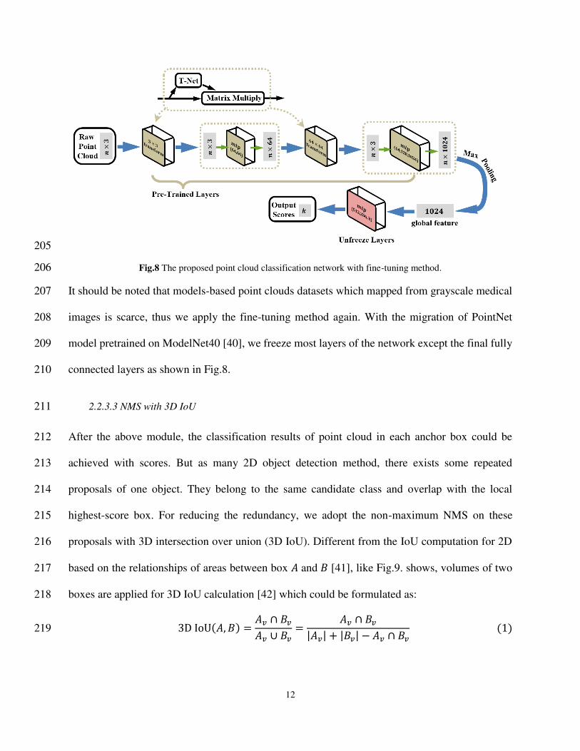
Conference on Computer Vision. 2014; 740-755. 472 39. Tan L, Jiang J. Digital signal processing: fundamentals and applications. Academic Press; 2019. 473 40. Zhirong Wu, Song Shuran, Khosla Aditya, et al. 3d shapenets: A deep representation for volumetric 474
shapes. Proceedings of the IEEE Conference on Computer Vision and Pattern Recognition. 2015; 1912-475 1920. 476
41. Hamid Rezatofighi, Tsoi Nathan, Gwak JunYoung, et al. Generalized intersection over union: A metric 477 and a loss for bounding box regression. Proceedings of the IEEE Conference on Computer Vision and 478 Pattern Recognition. 2019; 658-666. 479
42. Zhou D, Fang J, Song X, et al. Iou loss for 2d/3d object detection. International Conference on 3D 480 Vision (3DV). 2019; 85-94. 481
43. Adam Paszke, Gross Sam, Massa Francisco, et al. Pytorch: An imperative style, high-performance deep 482 learning library. Advances in Neural Information Processing Systems. 2019; 32: 8026-8037 483
44. X. Xiao, S. Lian, Z. Luo and S. Li. Weighted Res-UNet for High-Quality Retina Vessel Segmentation. 484 2018 9th International Conference on Information Technology in Medicine and Education (ITME). 485 2018; 327-331. 486
45. Z. Zhou, M. M. R. Siddiquee, N. Tajbakhsh and J. Liang. UNet++: Redesigning Skip Connections to 487 Exploit Multiscale Features in Image Segmentation. IEEE Transactions on Medical Imaging. 2020; 488 39(6): 1856-1867. 489
46. Ozan Oktay, Jo Schlemper, et al. Attention U-Net: Learning Where to Look for the Pancreas. arXiv. 490 2018; 1804.03999. 491
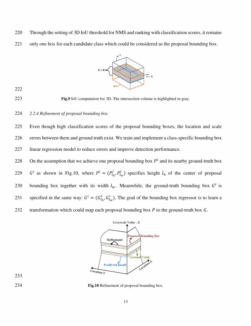


![[POSTER] 2D-3D Co-segmentation for AR-based Remote …bnuernberger/2d-3d-co... · 2015-10-11 · [POSTER] 2D-3D Co-segmentation for AR-based Remote Collaboration Kuo-Chin Lien Benjamin](https://static.documents.pub/doc/80x56/5e509f35437c7308227e885f/poster-2d-3d-co-segmentation-for-ar-based-remote-bnuernberger2d-3d-co-2015-10-11.jpg)

![Review Images – an array of colors Color – RGBA Loading, modifying, updating pixels pixels[] as a 2D array Simple filters – tinting, grayscale, negative,](https://static.documents.pub/doc/80x56/56649c885503460f949405a2/review-images-an-array-of-colors-color-rgba-loading-modifying-updating.jpg)


