KATHLEEN LAMBERT* AND S. J.PIRTMicrobiology Department, Queen Elizabeth College(University of London), Campden Hill, London, W. 8, England
SUMMARY
A calf serum ultrafiltrate fraction permitted growth for at least 3-5 generations, includingone subculture, of MRC-5 cells in defined medium in the absence of whole serum. The activematerial has a molecular weight of 10000 Daltons or less. This suggests that there may be norequirement for a large macromolecular component of serum. The ultrafiltrate was assayed bymaximum cell yield from a serum-limited inoculum in a denned medium containing non-limiting amounts of vitamins, amino acids, glucose, a 68-component supplement, iron andmethylcellulose. The levels of vitamins, amino acids and glucose were based on quantitativemeasurements of uptake and the levels of the other components by minimum amount requiredfor maximum yield in defined medium without ultrafiltrate or serum. With excess ultrafiltratemaximum cell yield was limited by the defined part of the medium, probably the supplement.The cell doubling time in defined medium with ultrafiltrate fractions was 70 h compared with27 h in the medium with serum. Excess ultrafiltrate did not inhibit gTowth. The loweredgrowth rate is attributed to a nutritional deficiency in the supplement.
INTRODUCTION
Replacement of serum by defined media for cell growth is essential to obtain fullreproducibility of the cell's environment, reduce costs and avoid toxins or infectionsfrom serum. Identification of nutrients specific to serum must be preceded by defini-tion of the knovm nutrient requirements, to ensure that these do not limit growth whenserum is reduced. We have previously reported (Lambert & Pirt, 1975) the quanti-tative requirements of MRC-5 human diploid cells for amino acids, B group vitamins,glucose, inositol and choline. More recently (Lambert & Pirt, 1977) we determinedthe requirements for methylcellulose, iron and a chemically defined supplement. Thiswork resulted in a defined medium allowing significant growth (1-5 generations) ofMRC-5 c e u s without added serum. However, growth then became limited by un-known serum growth factors. We have found maximum cell yield (cells/ml medium)produced by test fractions when they are growth limiting to be the most appropriate(Pirt & Lambert, 1977) means of assaying the growth factor. McKeehan & Ham(1977), using a different assay method (clonal growth of WI-38 human diploid cells)
• Present address: Department of Experimental Pathology, Charing Cross Hospital MedicalSchool, Fulham Palace Road, London, W. 6, England.
25 CEL 35
382 K. Lambert and S. J. Pirt
found that despite comprehensive optimization of the defined nutrients and cultureconditions in their system (McKeehan, McKeehan, Hammond & Ham, 1977), onlylimited division of WI-38 cells occurred in the absence of serum protein. Unknownserum factors then limited growth.
In recent attempts to purify the growth factor, we found that growth factor activitywas gradually lost on prolonged dialysis of serum but that activity could be recovered,particularly in fraction IV, in fractions of serum prepared by the cold ethanol methodof Cohn et al. (1946). The diffusible nature of the growth factor and its associationwith the a-globulins seemed consistent with the properties of somatomedin (VanWyk, 1975) or NSILA (Rinderknecht & Humbel, 1976). We have applied the NSILApurification method to calf serum to separate molecules of more than 10 000 Daltonsfrom those of less than 10000 Daltons and tested the fractions on MRC-5 cells. Herewe report (a) growth factor activity in the < 10 000 Daltons fraction in the absence ofserum, (b) significant growth (3-5 generations including one subculture) of MRC-5cells in serum ultrafiltrate fraction in the absence of serum, and (c) limitation of growthby the defined part of our medium in the presence of excess ultrafiltrate.
MATERIALS AND METHODS
Details of the cell line, stock culture, preparation of phosphate-buffered saline (PBS) andmeasurement of growth rate have been given elsewhere (Lambert & Pirt, 1975).
Culture media
The composition of the improved experimental culture medium, MEME, is shown in Table 1.The medium is prepared and stored as a series of sterile stock solutions as described for MEM Aand MEMB (Lambert & Pirt, 1975) or MEMC and MEMD (Lambert & Pirt, 1977). Themodifications from MEMD are the increase in inositol concentration from 10 to 2-0 mg/1.and the inclusion of selenium as a 5 mg/1. ( x 1000) stock solution of sodium selenite sterilizedby membrane filtration and stored at 4 °C.
Preparation of inocula
Three-day-old stock cultures, with the serum content of the medium reduced to 5 % (v/v)to cause limitation of the inoculum by serum factors, were used as inoculum. Monolayers werewashed twice with PBS and the cells removed by treatment with 05 % trypsin (1/250 Difco)in citrate buffer, pH 7-6. After resuspension in PBS containing 10 % serum, cells were separatedby centrifugation at isog for 5 min, washed in PBS, and resuspended in the appropriateexperimental medium. This procedure was found to eliminate detectable protein carry-overin the medium (Lambert & Pirt, 1977). Cells were separated by aspiration with a 5-ml pipette,counted and inoculated to give an initial population density attached to the culture surface of5-7 x io4 cells/ml (1-1-4 x IC>4 cells/cm1).
To prepare inocula from cells grown in serum-free medium, trypsin treatment was avoidedand the cells were removed from the monolayer as follows. Monolayers were washed twice withPBS; 1 -o ml of the fresh experimental medium was added and the cells were scraped from thesurface into the medium using a Pasteur pipette covered with silicone-rubber tubing. Growthmedium was added and the cells separated by aspiration. Cells were then pooled, counted andinoculated into fresh vessels to give an initial population density attached to the culture surfaceof ca. 3 x io4 cells/ml (06 x io* cells/cm1).
Human diploid cell defined medium 383
Cultural procedures for MRC-5 cells
Stock cultures were grown in sealed 75-0x1* (250-ml) Nunc plastic flasks containing 15 mlof medium. For experimental cultures 5-ml volumes of medium in 25-cm1 (30-ml) Nuncplastic flasks were used. To obtain a complete growth curve and estimate of maximum cellyield (maximum cell population in numbers minus initial population attached) 1 1 or moresuch replicates were used.
The air above the medium was replaced by 5 % CO2 in air and all cultures were incubatedat 37 °C
* These amounts are in addition to those in the medium supplement.f One half added after 72 h of incubation.% As described in Lambert & Pirt (1975, 1977).
Cell counts
Cell suspensions prepared from monolayers by trypsinization were diluted in PBS and cellcounts made in a Fuchs-Rosenthal counting chamber using trypan blue exclusion to distinguishviable cells. Cells stained with trypan blue were not counted. For cells grown without serum,cell attachment was less firm and 3-s treatment at 37 °C (as opposed to 30 s for cells grownwith serum) was sufficient to detach the monolayer.
Determination of protein
The protein content of assay media and fractions was determined by the method of Lowry,Rosebrough, Farr & Randall (1951).
Elution of protein from columns was followed by absorbance at 280 nm (Pye UnicamSP1800 spectrophotometer).
25-2
384 K. Lambert and S. J. Pirt
Fractionation of serum
Calf serum (Gibco Biocult L262501) was used.Acid treatment. 25-ml aliquots of serum were made 05 M with acetic acid by dropwise addi-
tion of glacial acetic acid over 5 min with constant stirring, then stirred for 1 h at room tempera-ture (18—22 °C). The acidified serum was then filtered through a o-45-/tm Millipore filter beforegel filtration or ultrafiltration.
G-75 Sephadex chromatography. 10 ml of acid-treated serum were applied on a PharmaciaK50/100 column of Sephadex G-75 (bed volume 1600 ml), equilibrated in 0-5 M acetic acidat room temperature. Columns were poured at room temperature at a flow rate of 250 ml/hand run at 200 ml/h. Elution of protein was followed by ZsVjo absorbance and the columns werecalibrated using Blue Dextran 2000 (Pharmacia Fine Chemicals) and bovine insulin (SigmaChemical Co.). One fraction (> 10000 Daltons) comprising the eluate from 27-55 % of thetotal bed volume and a second (< 10000 Daltons) comprising 56—98 % of the total bed volumewere collected and lyophilized. The dried samples were redissolved in glass-distilled water andlyophilized. This procedure was repeated once more prior to the growth factor assay to removeacetic acid.
Ultrafiltration. 15-ml aliquots of acid-treated serum were ultrafiltered through immersiblemolecular separator membranes (Millipore) with a nominal cut off limit of 10000 Daltons.Ultrafiltration was continued until a volume of 20 ml of non-filterable material remained onthe protein side of the membrane, a process taking 26-28 h at room temperature. The volumesof ultrafiltrate (< 10000 Daltons) and non-filterable protein (> 10000 Daltons) were recordedand the fractions were suspended in distilled water and lyophilized as for the Sephadex G-75fractions.
Desalting chromatography of ultrafiltrate. Recovery of ultrafutrate was estimated from therecovery of £280 and Ea0 absorbance from the columns. The flow rate for all separations was20 ml/h. The void volume of columns was determined using Blue Dextran 2000 and the saltsfraction detected from conductivity measurements using an electrolytic conductivity measuringbridge (Electronic Switchgear (London) Ltd). For fractions A, B and C, ultrafilrrate from105 ml acidified serum was resuspended in io-o ml distilled water, filtered through a 04.5-fimmembrane filter and applied to a Pharmacia K16/40 column containing Sephadex G-25, bedvolume 68 ml, equilibrated in distilled water at room temperature. The .E^-absorbing eluatenot containing salts was lyophilized as 2 fractions, A and C. The central portion of the eluatecontaining salts, was lyophilized, resuspended in io-o ml distilled water and loaded onto aPharmacacia K26/40 column containing Biogel P-2, bed volume 106 ml, equilibrated in dis-tilled water at room temperature. The i?,s0-absorbing eluate not containing salts was lyophil-ized as fraction B.
For fractions D, E and F, ultrafiltrate from 190 ml acidified serum was resuspended in6'O ml distilled water, filtered through a 0-45-/^1 membrane filter and applied to a PharmaciaK26/40 column containing Biogel P-2, bed volume 178 ml, equilibrated in distilled water atroom temperature. The .Eaflo-ab8orbing eluate not containing salts was lyophilized as 2 fractionsD and F. The central portion of the eluate, containing salts, was lyophilized, resuspended in75 ml distilled water and rechromatographed on the Sephadex G-25 column, (bed volume68 ml). The .E,30-absorbing eluate not containing salts was lyophilized as fraction E.
Preparations of fractions for growth factor assay
Lyophilized fractions from chromatography were resuspended in PBS. Ultrafiltrate andnon-filterable protein from ultrafilrrate were resuspended in distilled water to allow for thetonicity of the serum salts. (Isotonic distribution on each side of the membrane at the end ofultrafiltration assumed.) All test fractions were adjusted to pH 7-2 and membrane-filtered beforestorage at 4 °C. Amounts of fractions to be added to test media were calculated as volumes oforiginal serum per unit volume taking into account dilutions and mechanical losses throughoutthe fractionation procedure.
Human diploid cell defined medium 3 85
Growth factor assay of fractions and serum
All assays included a 5 % serum control and a blank containing basal medium with no serumor fraction. Maximum cell yield from a serum-limited inoculum was determined for each testsample and the blank value subtracted. The unit of growth factor activity was the amountwhich gave a cell yield of io6 cells. The units attributable to each fraction were comparedto serum within each assay and thus related to a single eternal standard serum, which had agrowth factor activity of 37 units/ml.
RESULTS
Growth factor activity of Sephadex G-75 and ultrafiltration fractions
The 2 fractions of acid-treated serum from Sephadex G-75 a nd ultrafiltration wereassayed for growth factor activity at a concentration equivalent to 5 % original serum.The > 10000 Daltons fraction from each fractionation promoted cell attachment and
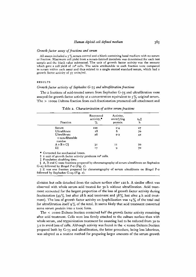
Table 2. Characterization of active serum fractions
Fraction
SerumUltrafiltrateUltrafUtrate
+ non-filterableresidue
A + B + C§E||
Recoveredactivity, •
/o
IOO
1828
3117
Activity,unitsf/mg
protein
0-46O-2
I I
9
h
277042
7070
• Corrected for mechanical losses.f 1 unit of growth factor activity produces io5 cells.% Population doubling time.§ A, B and C were fractions prepared by chromatography of serum ultrafiltrate on Sephadex
G-25 followed by Biogel P-2 (Fig. 1).|| E was one fraction prepared by chromatography of serum ultrafiltrate on Biogel P-2
followed by Sephadex G-25 (Fig. 2).
division but cells detached from the culture surface after 120 h. A similar effect wasobserved with whole serum acid treated for 30 h without ultrafiltration. Acid treat-ment accounted for the largest proportion of the loss of growth factor activity duringfractionation (49% lost after 28 h acid treatment and 38% lost after 4 h acid treat-ment). The loss of growth factor activity on lyophilization was 14% of the total andfor ultrafiltration itself 9 % of the total. It seems likely that acid treatment convertedsome serum protein into a toxic form.
The < 10000 Daltons fraction contained half the growth factor activity remainingafter acid treatment. Cells were less firmly attached to the culture surface than withwhole serum, and trypsinization treatment for counting had to be reduced from 30 to3 s to avoid loss of cells. Although activity was found in the < 10000 Daltons fractionprepared both by G-75 a nd ultrafiltration, the latter procedure, being less laborious,was adopted as a routine method for preparing larger amounts of the serum growth
386 K. Lambert and S. J. Pirt
factor. Table 2 compares growth factor activity of ultrafiltrate with or without non-filterable residue to that of untreated serum, tested at 5% (v/v) medium concentra-tion; 18% of the growth factor activity of serum was recovered in the ultrafiltrate,with a 15-fold purification based on protein content. The growth rate with ultra-filtrate was slower {ta 70 h) than that with serum (ta 27 h). The non-filterable residuehad a lower specific activity than serum but addition of the non-filterable residue tothe ultrafiltrate increased the growth rate. Thus extraction of growth factor activity inour fractionation was not complete. This could mean either that our method of
100% bed volume
200
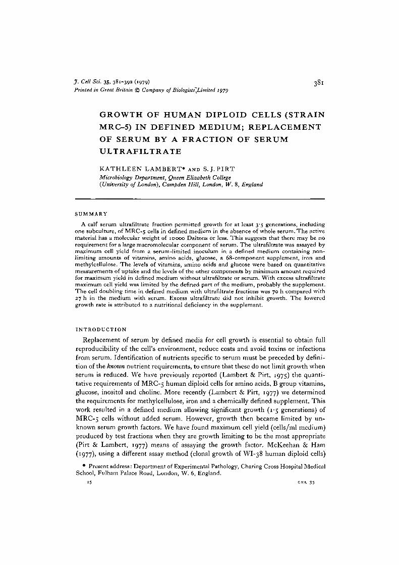
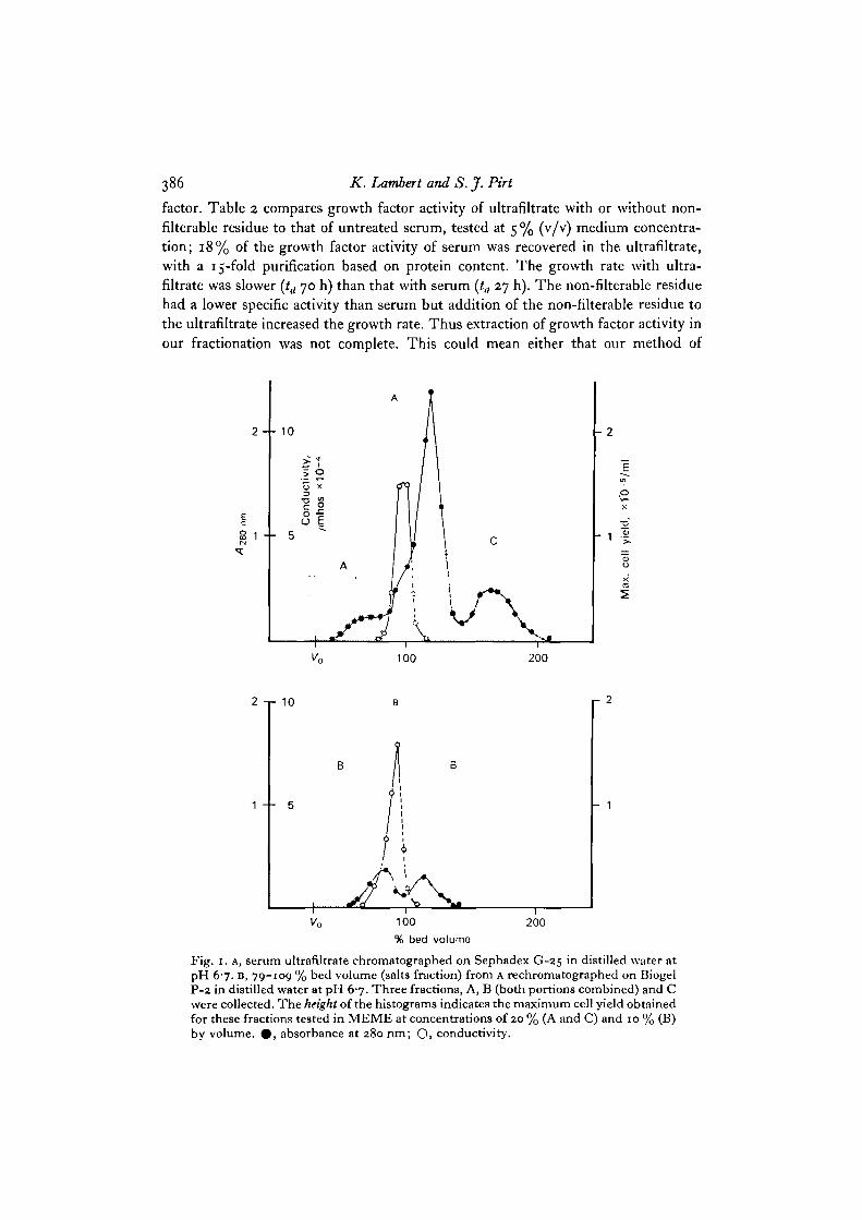
Fig. 1. A, serum ultrafiltrate chromatographed on Sephadex G-25 in distilled water atpH 67. B, 79-109 % bed volume (salts fraction) from A rechromatographed on BiogelP-2 in distilled water at pH 6-7. Three fractions, A, B (both portions combined) and Cwere collected. The height of the histograms indicates the maximum cell yield obtainedfor these fractions tested in MEME at concentrations of 20 % (A and C) and 10 % (B)by volume. 0 , absorbance at 280 nm; Q, conductivity.
Human diploid cell defined medium 387
fractionation caused loss of the growth factor or that more than one growth factorwas supplied by the serum.
The maximum cell yield obtained with ultrafiltrate at 5 % (v/v) serum equivalentwas o-4 + o-ixioB cells/ml, compared to I-8XIO6 cells/ml with untreated serum.When the ultrafiltrate was tested at 10-20% (v/v) serum equivalent, no increase incell yield was obtained. This was not due to breakdown of ultrafiltrate in the medium:adding the ultrafiltrate in 2 parts, one initially and one after 72 h, did not produce a
1 -1-10
0 5 - - 5
f •p \\ f •
r 20
-1 0
100% bed volume
200
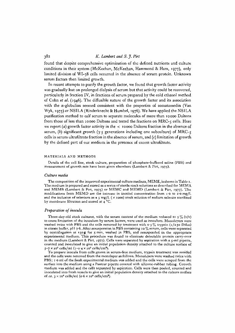
Fig. 2. A, serum ultrafiltrate chromatographed on.Biogel P-2 in distilled wateratH 6-7.D, 65-88 % bed volume (salts fraction) from A rechromatographed on Sephadex G-25in distilled water at pH 6-7. Three fractions, D, E (both portions combined) and Fwere collected. The height of the histograms indicates the maximum cell yield obtainedfor these fractions tested in MEME at concentrations of 20 or 40 % (D and F) and 15or 30 % (E) by volume. The maximum cell yield for E at 5 % by volume was 0-3 xio6/ml. • , absorbance at 280 ran; O, conductivity.
388 K. Lambert and S. J. Pirt
larger cell yield than that obtained when all the ultrafiltrate was included at the start.The simplest explanation is that at ultrafiltrate concentrations greater than 5 % (v/v)serum equivalent, cell yield was limited by a nutrient other than ultrafiltrate. Thepresence of an inhibitor seemed unlikely since increase in the concentration of ultra-filtrate had no effect on growth rate (Pirt, 1975). Before testing the ultrafiltrateat higher concentrations, however, the possibility of suboptimal tonicity due tounsuspected salts in the ultrafiltrate was excluded by desalting the ultrafiltrate.
Characterization of growth factor activity of desalted serum ultrafiltrate
The ultrafiltrate was desalted by chromatography on Sephadex G-25 followed byBiogel P-2 (Fig. 1) or Biogel P-2 followed by Sephadex G-25 (Fig- 2)- A surprisingresult was that material absorbing at 280 nm (E2eo absorbance followed a very similar
1-0 -
0 5 -
(10 20
Fraction concentration, %30
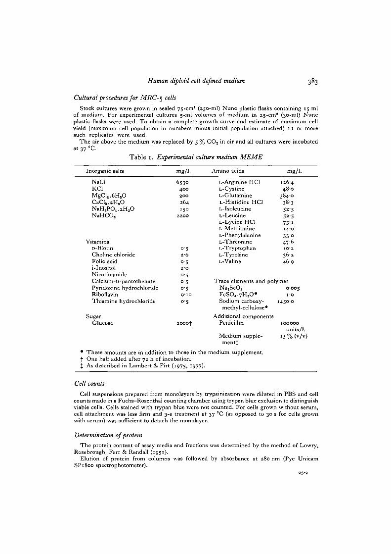
Fig. 3. Effect of serum ultrafiltrate fraction concentration (% by volume) on maximumcell yield of MRC-5 cells grown in denned medium MEME. x, fraction E; O,fractions A + B + C.
pattern) was eluted well beyond the bed volume of the column, that is retarded bysome factor other than molecular size. (The fractionation ranges for Biogel P-2 andSephadex G-25 a r e 100-1800 and 1000-5000 Daltons respectively.) In both prepara-tions (Figs. 1, 2), 5 % of the ultrafiltrate applied to the column could not be separatedfrom the salts and was not tested. The rest of the column filtrate was in each casecombined into 3 fractions, A, B, C (Fig. 1) or D, E, F (Fig. 2). These fractions wereall found to have some growth factor activity (height of histograms in Figs. 1 and 2)when tested at 5-40% (v/v) serum equivalent. The growth factor activity was more
Human diploid cell defined medium 389
concentrated in the fractions immediately preceding and following the salts. Fraction Ehad 17% of the activity of serum (Table 2) with a 22-5-fold purification. For pooledfractions A + B + C (Table 2) 31% of the activity of serum was obtained with a27%5-fold purification. The purification factor for ultrafiltrate was 15-fold only,suggesting that desalting removed an inhibitor or a toxic substance. Growth rate in
50-i
20 -
1 0 -
3 5
60 180Incubation time, h
300
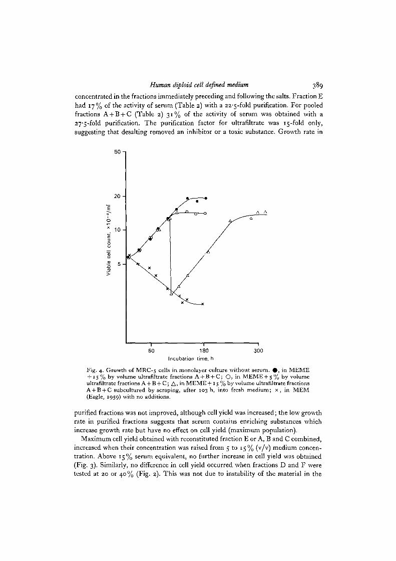
Fig. 4. Growth of MRC-5 cells in monolayer culture without serum. # , in MEME+ 15 % by volume ultrafiltrate fractions A + B + C; O, in MEME + S % by volumeultrafiltrate fractions A + B + C; A, in MEME + 15 % by volume ultrafiltrate fractionsA + B + C subcultured by scraping, after 103 h, into fresh medium; x, in MEM(Eagle, 1959) with no additions.
purified fractions was not improved, although cell yield was increased; the low growthrate in purified fractions suggests that serum contains enriching substances whichincrease growth rate but have no effect on cell yield (maximum population).
Maximum cell yield obtained with reconstituted fraction E or A, B and C combined,increased when their concentration was raised from 5 to 15% (v/v) medium concen-tration. Above 15% serum equivalent, no further increase in cell yield was obtained(Fig. 3). Similarly, no difference in cell yield occurred when fractions D and F weretested at 20 or 40% (Fig. 2). This was not due to instability of the material in the
390 K. Lambert and S. jf. Pirt
medium since supplementation with further fraction, 72 h after inoculation, into thesame medium did not affect cell yield. Hence it is concluded that some factor otherthan ultrafiltrate became growth limiting. To test whether there was a requirementfor unknown serum factors at this point, cells at their maximum yield in definedmedium with 15% combined fractions A + B + C were subcultured by scraping fromthe culture surface into fresh defined medium. Further division, to a total of 3-5doublings occurred (Fig. 4). A similar result was obtained when cells were subculturedby scraping from defined medium with 20% fraction D. This growth on subculturewe interpret to mean that the material limiting growth immediately before subculturewas not a serum growth factor but rather the defined part of our medium, probablythe supplement, since limitation of the maximum population by the Eagle's mediumamino acids, vitamins and glucose could be eliminated from knowledge of the quanti-tative requirements (Lambert & Pirt, 1975).
The population doubling time for MRC-5 cells in defined medium with ultra-filtrate growth factor was limited to 70 h compared to 27 h with serum (Table 2).The increase in ultrafiltrate fraction from 5 to 15 %, which affected cell yield (Fig. 4)had no effect on growth rate. The fact that increase in the amount of ultrafiltrate inthe medium failed to increase the growth rate is attributed to a second nutritionaldeficiency in the medium which affects growth rate but not cell yield.
DISCUSSION
Few workers seem to have appreciated the necessity for defining quantitativerequirements for known nutrients before attempting to reduce or replace serum ingrowth media for normal human diploid cells. McKeehan & Ham (1977) and Mc-Keehan et al. (1977) have, however, demonstrated how systematic qualitative andquantitative modification of the medium and culture conditions can reduce the amountof dialysed foetal bovine serum protein required for clonal growth of VVI-38 cells to25 fig/m\ (0-05 % (v/v) whole serum). Unfortunately McKeehan & Ham do not givetheir results in terms of cell yield per unit weight of nutrient utilized, a limitation on theapplication of their results to other systems. Factors limiting growth of WI-38colonies may not be identical to those limiting division in the more crowded conditionsof routine cell culture where nutrient depletion and accumulation of cell-derivedproducts become important. Results from our growth factor assay system (cell yieldproduced from a relatively large serum-limited inoculum) have the advantage ofdirect application to routine cell culture of human diploid cells.
Our results show that calf serum ultrafiltrate contains an unknown factor, ofmolecular weight 10000 Daltons or less, limiting cell yield of MRC-5 cel's- Furtherpurification of the ultrafiltrate yielded no discrete active fraction, suggesting that thegrowth factor, rather than a single peptide hormone, could be a mixture of factors.These could include characterized peptide growth factors such as NSILA, somato-medins, EGF or FGF (Rechler & Nissley, 1977), although to date none of these hasbeen shown to completely replace serum for growth of serum-limited human fibro-blasts (Gospodarowicz & Moran, 1976; Shields, 1977). Cell yield could also be limited
Human diploid cell defined medium 391
by lack of a trace element. Our medium contains 11 trace elements but lacks vanadiumand molybdenum which McKeehan et al. (1977) found to be most effective for clonalgrowth at 5 x io~9 and 7 x io~9 M, respectively. However, they also found considerablebackground growth in the absence of added amounts of these salts: it thus seemslikely that we have non-limiting levels of these components derived as contaminantsfrom other medium constituents.
Specific growth rate of MRC-5 cells in defined medium with high or low levels ofultrafiltrate was much lower than in defined medium with serum, suggesting a secondnutritional deficiency (in addition to the ultrafiltrate factor) in our medium. Thespecific growth rate limitation is reminiscent of that obtained with iron deficiency inthe defined medium for LS cells (Birch & Pirt, 1970) and this, together with associa-tion of the growth factor activity with the salts fraction of desalted ultrafiltrate suggeststhat the second factor may be iron, or a deficiency in the medium supplement.Chelation of iron (1 mol. of EDTA per mol. of Fe) in our medium may have beenineffective.
The 3-5 MRC-5 cell doublings obtained with ultrafiltrate in the absence of wholeserum indicate that there may be no requirement for a macromolecular componentof serum or that the requirement does not become apparent until after 3-5 generations.The only serum factor specifically shown to stimulate growth rate of human diploidcells (Houck & Cheng, 1973) may be a macromolecule operating as a carrier for asmaller molecule with a much greater activity. Extension of our assay system beyond3-5 generations for MRC-5 cells requires the solution of a technical problem in themaking of cell suspensions without serum to inhibit trypsin; scraping cells from theculture surface by physical means is an inefficient method of obtaining a rapid viablesuspension. Extension to a multiple subculture assay is important, as exemplified bythe insulin requirement of HeLa cells, which only became apparent after 13-5 genera-tions in defined medium (Blaker, Birch & Pirt, 1971).
In conclusion, our progress towards growth of human diploid cells without serumis summarized in Fig. 4, which compares growth of MRC-5 ceHs m MEM (Eagle,1959) without serum, to growth in our defined medium with the active fraction ofserum ultrafiltrate. Any further development of the defined medium for MRC-5 c e " s
should aim at identifying both the factor limiting maximum cell population and thatlimiting the growth rate and preferably extend the maximum yield assay to severalsubcultures.
We gratefully acknowledge a grant in aid from the Medical Research Council. We thankMr J. P. Jacobs of the N.I.M.R. Laboratories, Hampstead, for the supply of MRC-5 cells,Dr C. F. Thurston for advice on protein fractionation and Mr G. Wilkie for technical assistance.
REFERENCESBIRCH, J. R. & PIRT, S. J. (1970). Improvements in a chemically denned medium for the growth
of mouse cells (strain LS) in suspension. J. Cell Sci. 7, 661-760.BLAKER, G. J., BIRCH, J. R. & PIRT, S. J. (1971). The glucose, insulin and glutamine require-
ments of suspension cultures of HeLa cells in a defined culture medium. J. Cell Sci. 9,529-537-
392 K. Lambert and S. J. Pirt
COHN, E. J., STRONG, L. E., HUGHES, W. L., MULFORD, D. J., ASHWORTH, J. N., MELIN, M.& TAYLOR, H. J. (1956). Preparation and properties of serum and plasma proteins.IV. A system for the separation into fractions of the protein and lipoprotein components ofbiological tissues and fluids. J. Am. chem. Soc. 68, 459—475.
EAGLE, H. (1959). Amino acid metabolism in mammalian cells in culture. Science, N.Y. 130,432-437-
GOSPODAROWICZ, D. & MORAN, J. S. (1976). Growth factors in mammalian cell cultures. A. Rev.Biochem. 45, 531-558.
HOUCK, J. C. & CHENG, R. F. (1973). Isolation, purification and chemical characterization ofthe serum mitogen for diploid human fibroblasts. J. cell. Physiol. 81, 257-270.
LAMBERT, K. & PIRT, S. J. (1975). The quantitative requirements of human diploid cells (strainMRC-5) for amino acids, vitamins and serum. J. Cell Set. 17, 397-411.
LAMBERT, K. & PIRT, S. J. (1977). The nutrient requirements of MRC-5 human diploid cells.Devi biol. Standard. 37, 67-70.
LOWRY, O. H., ROSEBROUGH, N. J., FARR, A. L. & RANDALL, R. J. (1951). Protein measure-ment with the folin phenol reagent. J. biol. Chem. 193, 265-275.
MCKEEHAN, W. L. & HAM, R. G. (1977). Methods for reducing the serum requirement forgrowth in vitro of nontransformed diploid fibroblasts. Devi biol. Standard. 37, 97-108.
MCKEEHAN, W. L., MCKEEHAN, K. A., HAMMOND, S. L. & HAM, R. G. (1977). Improvedmedium for clonal growth of human diploid fibroblasts at low concentrations of serumprotein. In Vitro 13, 399-416.
PIRT, S. J. (1975). Principles of Microbe and Cell Cultivation. Oxford: Blackwell.PIRT, S. J. & LAMBERT, K. (1977). Towards a chemically defined medium for the growth of
normal human diploid cells. Devi biol. Standard. 37, 63-66.RECHLER, M. M. & NISSLEY, S. P. (1977). Somatomedins and related growth factors. Nature,
Lond. 270, 665-666.RINDERKNECHT, E. & HUMBEL, R. E. (1976). Polypeptides with nonsuppressible insulin-like
and cell-growth promoting activities in human serum: isolation, chemical characterization,and some biological properties of forms I and II. Proc. natn. Acad. Sci. U.S.A. 73, 2365-2369.
SHIELDS, R. (1977). Growth hormones and serum factors. Nature, Lond. 267, 308-310.VAN WYK, J. J. (1975). Postsession discussion of papers. In Advances in Metabolic Disorders,
vol. 8 (ed. R. Luft & K. Hall), p. 393. New York & London: Academic Press.


![79: ' # '6& *#7 & 8 · 2018. 4. 3. · the ratio-optimized strain was further improved by making it diploid [19]. The optimized diploid showed an ability to produce ethanol di rectly](https://static.documents.pub/doc/80x56/60ffec9e0e522f10ec7aea83/79-6-7-8-2018-4-3-the-ratio-optimized-strain-was-further.jpg)


