GUIDELINE FOR ACUTE MYELOID LEUKAEMIA IN CHILDREN AND YOUNG ADULTS February 2016 Childhood Leukaemia Clinicians Network (CLCN) CCLG and CLCN do not sponsor or indemnify the treatment detailed herein. These treatment recommendations are provided to inform and for use at the sole discretion of treating clinicians who retain professional responsibility for their actions and treatment decisions. Treatment recommendations are based on current best practice and not what is necessarily proposed for any forthcoming clinical trial.
Transcript
GUIDELINE FOR ACUTE MYELOID LEUKAEMIA IN CHILDREN AND YOUNG ADULTS February 2016 Childhood Leukaemia Clinicians Network (CLCN)
CCLG and CLCN do not sponsor or indemnify the treatment detailed herein. These treatment recommendations are provided to inform and for use at the sole discretion of treating clinicians who retain professional responsibility for their actions and treatment decisions. Treatment recommendations are based on current best practice and not what is necessarily proposed for any forthcoming clinical trial.
2
RISK GROUP ALLOCATION
3
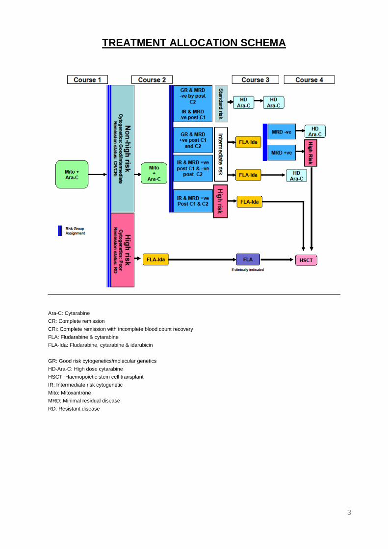
TREATMENT ALLOCATION SCHEMA
Ara-C: Cytarabine
CR: Complete remission
CRi: Complete remission with incomplete blood count recovery
FLA: Fludarabine & cytarabine
FLA-Ida: Fludarabine, cytarabine & idarubicin
GR: Good risk cytogenetics/molecular genetics
HD-Ara-C: High dose cytarabine
HSCT: Haemopoietic stem cell transplant
IR: Intermediate risk cytogenetic
Mito: Mitoxantrone
MRD: Minimal residual disease
RD: Resistant disease
4
CONTENTS
1. Introduction Page 5
2. Recommendations Page 5 – 12
2.1 Induction Anthracycline (Courses 1 and 2)
2.2 Consolidation Chemotherapy (Courses 3 and 4)
2.3 Stem Cell Transplantation
2.4 Risk Group Stratification o Risk Group Allocation (Figure1) o Risk Group Treatment (Table 1 )
2.5 Non-CNS Extramedullary Disease
3. Summary of Treatment Page 13-14
3.1 SR Patients
3.2 IR Patients
3.3 HR Patients
4. Treatment Allocation Schema (Figure2) Page 14
5. Investigations at Presentation Page 15-17
6. Treatment Page 17-28
6.1 Pre Treatment Supportive Care
6.2 CNS Directed Therapy
6.3 Induction Therapy
6.4 Consolidation Therapy
6.5 Patients with Non CNS Extramedullary AML
6.6 Supportive Treatment
6.7 Dose Modifications for Toxicity
7. References Page 28-31 8. Appendix 1 Page 31-33
5
1. INTRODUCTION
This is a treatment guideline for children with newly diagnosed Acute Myeloid Leukaemia (AML), High Risk Myelodysplastic Syndrome (MDS) (>10% blasts in the bone marrow ) or isolated myeloid sarcoma (MS), who are not eligible for MyeChild 01 or who do not consent to be treated on MyeChild 01. Children with Acute Promyelocytic Leukaemia (APL) or Myeloid Leukaemia of Down Syndrome (ML DS) are excluded and should be treated according to their respective guideline. MyeChild 01 will recruit patients up to their 18th birthday and this guideline may be used for the same age range. The guideline is based on the MyeChild 01 protocol which is, in turn, based on experience from previous UK, French and international trials [1-11]. The evidence for the risk stratification and treatment are given in Appendix A.
2. RECOMMENDATIONS
Treatment will be risk based. Patients will be stratified into standard (SR), intermediate (IR) and high risk (HR) based on cytogenetic/molecular characteristics at presentation and response to treatment assessed morphology and by minimal residual disease (MRD) measurement after each course of treatment. The risk group stratification is described in section 2.4.
2.1. Induction Anthracycline (Courses 1 and 2): The induction anthracycline for all patients in course 1 and for non HR patients in course 2 will be mitoxantrone, which will be combined with cytarabine (MA). The cumulative anthracycline dose in course 1 and 2 will be 420mg/m2 daunorubicin equivalence which represents an increase in anthracycline dose compared to that previously delivered in the UK. Increasing the anthracycline dose in induction has been reported to improve outcome in adult AML and it seems reasonable to assume that this benefit of anthracycline intensification can be extrapolated to children, although this has not been proven. AML 15 reported FLAG-Ida (fludarabine, cytarabine, idarubicin and G-CSF) to result in a greater reduction in RR than ADE (daunorubicin, cytarabine and etoposide): the 8 year OS/ RR for children (112 randomised) for FLAG-Ida vs. ADE was 71% v 67% (2p=0.8) and 28% vs. 38% (2p=0.4) respectively [11]. The reduction in the RR in favour of FLAG-Ida was highly significant (p< 0.001) when the trial as a whole (n=3251) was considered [11]. Whilst FLAG-Ida may have a superior anti-leukaemic effect, it is associated with greater toxicity and for that reason will be reserved for HR patients. Whilst all patients will receive mitoxantrone and cytarabine (MA) in course 1, patients identified as HR after
6
course1 will receive FLA-Ida in course 2. FLA-Ida is being used rather than FLAG-Ida because of a lack of proven benefit for the addition of G-CSF.
2.2 Consolidation Chemotherapy (Courses 3 and 4):
Standard risk patients, those with the lowest risk of relapse, will receive two courses of high dose cytarabine (HD Ara-C 18g/m2) in consolidation to limit further anthracycline exposure. Their cumulative daunorubicin equivalence anthracycline dose in courses 1 and 2 will be 420mg/m2. Intermediate risk patients will have their consolidation therapy intensified to reduce their leukaemic burden, achieve MRD negativity and avoid HSCT. They will receive FLA-Ida as course 3. If MRD negativity is achieved, they will receive HD Ara C as course 4. High risk children will receive HSCT as consolidation therapy.
2.3. Stem Cell Transplantation: Transplantation is recommended for patients with HR disease. There is growing evidence that MRD may be used to risk stratify and guide the need for HSCT [7, 12-13]. The suitability for HSCT of any patient with AML treated on this guideline should be discussed at the National Paediatric HSCT MDT.
2.4. Risk Group Stratification:
Cytogenetic/molecular characteristics at presentation and serial assessment of response will identify patients considered to be at high risk of relapse, who may benefit from HSCT and those at intermediate risk of relapse who may benefit from treatment intensification. Patients will be stratified into standard (SR), intermediate (IR) and high risk (HR). MRD should be assessed by flow cytometry or transcript levels (molecular monitoring) for informative patients measured after course 1 and 2 of treatment and course 3 for those patients whose treatment may be further stratified by the level of MRD post course 3. There is more evidence to date for the use of flow MRD and therefore this should be used to direct treatment [14]. However, it is anticipated that between 10-15% of children will either not have a marker, or not have a marker of sufficient sensitivity, for flow monitoring. These patients should be monitored by a molecular marker, if an informative molecular marker is present. Patients with neither a flow nor a molecular marker should have their treatment assigned by cytogenetic/molecular genetic risk group. The MRD discriminatory level for flow monitoring will be 0.1% and for molecular monitoring a transcript level reduction of 3 logs. Any testing for MRD which is used to guide treatment should be performed in an accredited laboratory. Laboratories providing MRD monitoring in MyeChild 01 are listed in section 7.
7
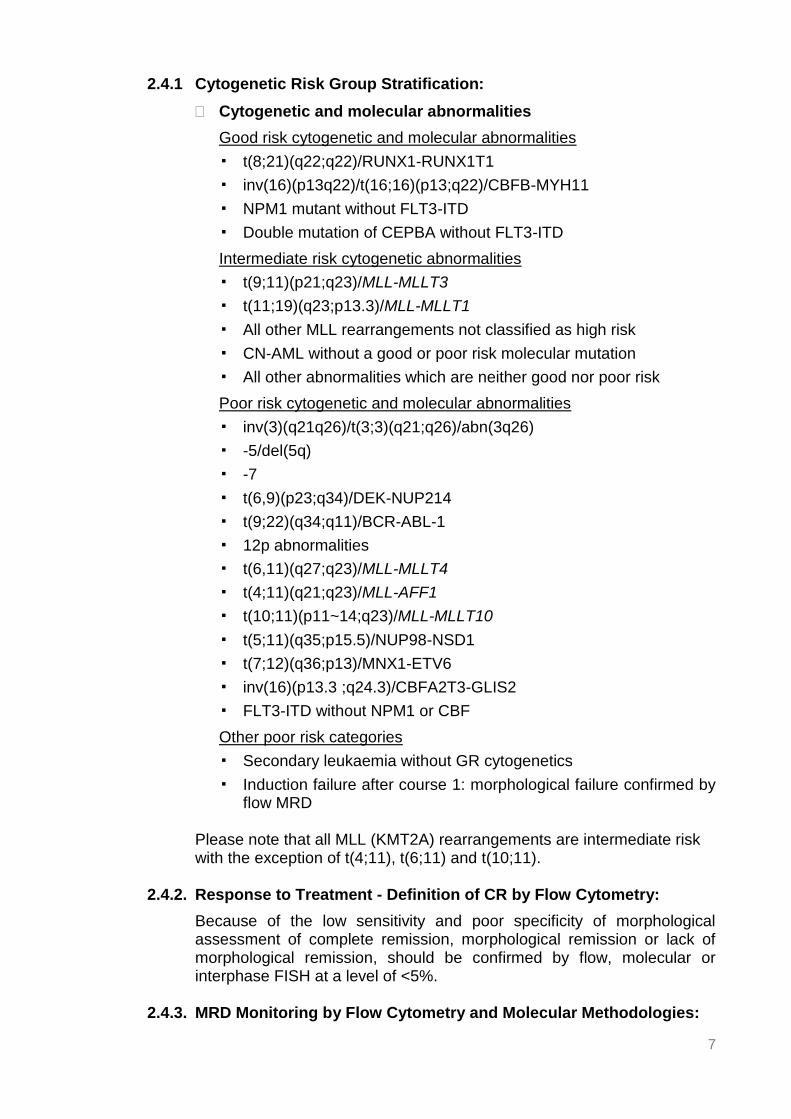
2.4.1 Cytogenetic Risk Group Stratification:
Cytogenetic and molecular abnormalities
Good risk cytogenetic and molecular abnormalities
▪ t(8;21)(q22;q22)/RUNX1-RUNX1T1
▪ inv(16)(p13q22)/t(16;16)(p13;q22)/CBFB-MYH11
▪ NPM1 mutant without FLT3-ITD
▪ Double mutation of CEPBA without FLT3-ITD
Intermediate risk cytogenetic abnormalities
▪ t(9;11)(p21;q23)/MLL-MLLT3
▪ t(11;19)(q23;p13.3)/MLL-MLLT1
▪ All other MLL rearrangements not classified as high risk
▪ CN-AML without a good or poor risk molecular mutation
▪ All other abnormalities which are neither good nor poor risk
Poor risk cytogenetic and molecular abnormalities
▪ inv(3)(q21q26)/t(3;3)(q21;q26)/abn(3q26)
▪ -5/del(5q)
▪ -7
▪ t(6,9)(p23;q34)/DEK-NUP214
▪ t(9;22)(q34;q11)/BCR-ABL-1
▪ 12p abnormalities
▪ t(6,11)(q27;q23)/MLL-MLLT4
▪ t(4;11)(q21;q23)/MLL-AFF1
▪ t(10;11)(p11~14;q23)/MLL-MLLT10
▪ t(5;11)(q35;p15.5)/NUP98-NSD1
▪ t(7;12)(q36;p13)/MNX1-ETV6
▪ inv(16)(p13.3 ;q24.3)/CBFA2T3-GLIS2
▪ FLT3-ITD without NPM1 or CBF
Other poor risk categories
▪ Secondary leukaemia without GR cytogenetics
▪ Induction failure after course 1: morphological failure confirmed by flow MRD
Please note that all MLL (KMT2A) rearrangements are intermediate risk with the exception of t(4;11), t(6;11) and t(10;11).
2.4.2. Response to Treatment - Definition of CR by Flow Cytometry:
Because of the low sensitivity and poor specificity of morphological assessment of complete remission, morphological remission or lack of morphological remission, should be confirmed by flow, molecular or interphase FISH at a level of <5%.
2.4.3. MRD Monitoring by Flow Cytometry and Molecular Methodologies:
8
Multiparameter flow cytometry detecting leukaemia associated aberrant immunophenotypes (LAIP) and real-time quantitative PCR (RT-qPCR) in patients with a leukaemia-specific molecular marker have shown themselves in a number of studies to be independently predictive of outcome [10, 14-16].
The ability of multidimensional flow (MDF) to define absolute risk remains limited, with nearly a quarter of patients without measurable RD at a level of 0.1% at EOI1 relapsing and a similar sized cohort of patients with documented RD at a level of 0.1% or greater remaining relapse free long term. MDF may be most informative in patients with intermediate risk cytogenetics [10]. Molecular monitoring of transcript levels is more sensitive than LAIP monitoring for some leukaemia subtypes.
Over 90% of patients will have a LAIP for monitoring and approximately 60% an informative molecular marker:
t(8;21)(q22;q22)/RUNX1-RUNX1T1
inv(16)(p13q22)/CBFB-MYH11
11q23/MLL fusions
t(6;9)(p23;q34)/DEK-NUP214
t(5;11)(q35;p15.5)/NUP98-NSD1
inv(16)(p13.3q24.3)/CBFA2T3-GLIS2
other rare fusions
NPM1 mutations
2.4.4 Risk Groups:
Patients will initially be stratified into high risk (HR) and non HR based on cytogenetic/molecular characteristics at presentation and response to Course 1. Non high risk patients will be subsequently reclassified as standard, intermediate or high risk based on MRD response to further treatment.
Standard and intermediate risk patients will receive 4 courses of chemotherapy
High risk patients will be eligible for HSCT and will receive two or three courses of chemotherapy prior to HSCT. A course of treatment may be given to bridge the time to transplant.
9
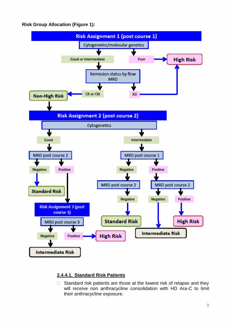
Risk Group Allocation (Figure 1):
2.4.4.1. Standard Risk Patients
Standard risk patients are those at the lowest risk of relapse and they will receive non anthracycline consolidation with HD Ara-C to limit their anthracycline exposure.
10
Standard risk patients are those patients with either good risk (GR) cytogenetics/molecular genetics and a MRD level of <0.1% by flow and/or decrease in transcript levels >= 3 logs after course 2, or those patients with intermediate risk cytogenetics/molecular genetics and a MRD level of <0.1% by flow after course 1 and course 2.
2.4.4.2 Intermediate Risk Patients
Intermediate risk patients are those at intermediate risk of relapse. They will have their treatment intensified to reduce their leukaemic burden, achieve MRD negativity and avoid HSCT, if possible. Treatment intensification will be with fludarabine, cytarabine and idarubicin (FLA-Ida) as course 3. FLA-Ida will increase the cumulative anthracycline dose by 120 mg/m2 to 540 mg/m2 based on a 5:1 conversion factor. This cumulative dose is similar to that delivered to high risk patients receiving liposomal daunorubicin on BFM AML 2004 [17] and slow responding patients on LAME 89/91 [2] who received mitoxantrone. Both these studies reported low rates of cardiotoxicity. There is little evidence base for the conversion factor of 5:1 for mitoxantrone. Mitoxantrone may be less cardiotoxic than daunorubicin at equivalent doses. Cardiotoxicity should be carefully monitored.
Intermediate risk patients are those with intermediate risk cytogenetics/molecular genetics and a MRD level of >0.1% by flow after course 1, which falls to <0.1% after course 2. They will have their 3rd course intensified with FLA-Ida, will receive HD Ara-C as course 4, and will not proceed to HSCT.
or
Intermediate risk patients are those with good risk cytogenetics/molecular genetics and a MRD level of >0.1% by flow after course 2, which falls to <0.1% after intensification of therapy with FLA-Ida in course 3. They will receive HD Ara-C as course 4, and will not proceed to HSCT.
2.4.4.3. High Risk Patients
High risk patients are those at high risk of relapse and they will receive treatment intensification with fludarabine, cytarabine and idarubicin (FLA-Ida) prior to HSCT with the aim of reducing their leukaemic burden.
All patients with poor risk (PR) cytogenetics/molecular genetics are defined as high risk by cytogenetics alone, irrespective of their response to course 1. MRD status in patients with poor risk cytogenetics should not influence the decision to proceed to HSCT based on preliminary data from adult studies and AAML 03P1 [10] which suggests limited discriminatory value for flow MRD in patients with poor risk cytogenetics.
Patients with intermediate risk cytogenetics/molecular genetics who fail to achieve confirmed CR or CR with incomplete blood count recovery (CRi) after course 1. CR will be defined as a MRD flow level <5%. Patients considered not to be in morphological CR/CRi after
11
course 1 but with a MRD flow level <5%, will not be classified as high risk, because of the recognised poor sensitivity and specificity of morphological assessment post course 1. In the absence of a flow marker for assessment of CR/CRi, an informative molecular marker will be used, and in the absence of an informative molecular marker, fluorescence in situ hybridisation (FISH) assessment.
Patients with intermediate risk cytogenetics/molecular genetics and a MRD level of >0.1% by flow after course 1 and 2. The decision to intensify treatment and proceed to HSCT for patients with intermediate risk cytogenetics and MRD positivity after course 2 is based on the observation from COG AAML03P1 that patients with no RD at the end of treatment, but with previously documented RD, remain at high risk of relapse and poor outcome, suggesting that intervention beyond clearance of RD is required for improved outcome [10].
Patients with good risk cytogenetics/molecular genetics and a MRD level of >0.1% by flow and/or a decrease in transcript levels <3logs or with rising transcript levels after course 3, despite treatment intensification, are at higher risk of relapse. The criteria for classifying patients with good risk cytogenetics as high risk are more stringent to limit HSCT where possible in these patients because of the lack of evidence for benefit. Stable or falling transcript levels may be an indication for further chemotherapy rather than HSCT. The lack of statistical significance for MRD observed in patients with good risk cytogenetics in the AAML03P1 study favours a monitoring/chemotherapy approach rather than early intervention with HSCT. These patients should be discussed.
2.5. Non-CNS Extramedullary Disease: Extramedullary (EM) disease can occur at a number of sites - skin, soft tissues and gingival infiltrates. EM is classified into leukaemia cutis (LC) and myeloid sarcoma (MS). MS occurs with an incidence of 3-9% and is most commonly seen in the CBF leukaemias, whilst LC has an incidence of 1-3% and is usually associated with MLL rearranged AML. LC and MS may occur with or without bone marrow involvement and their impact on outcome is poorly understood. Both should be treated systemically as de novo AML [18-20].
Available evidence suggests that EM disease should be risk stratified by the same criteria as AML without EM. Every effort should be made to biopsy to confirm the diagnosis and to carry out cytogenetics/FISH/molecular diagnostics to risk stratify.
Treatment by Risk Group (Table 1)
Cytogenetic risk group
CR status Post course 1 (% blasts by FLOW)
MRD status Post course 1
MRD status Post course 2
MRD status Post course 3
Risk Group Treatment
Good <5% <0.1% or>0.1% <0.1% Standard MA→MA→HD Ara-C→HD Ara-C
Good <5% <0.1% or>0.1% >0.1% <0.1% Intermediate MA→MA→FLA-Ida→HD Ara-C
Good <5% <0.1% or>0.1% >0.1% >0.1% High MA→MA→FLA-Ida→consider HSCT
Intermediate <5% <0.1% <0.1% Standard MA→MA→HD Ara-C→HD Ara-C
Intermediate <5% >0.1% >0.1% High MA→MA→FLA-Ida→ HSCT
Intermediate >5%* >0.1% High MA→Fla-Ida→HSCT
Poor+ High MA→Fla-Ida→HSCT
*Refractory disease – patients with refractory disease irrespective of cytogenetic risk group may require further discussion
+Patients with poor risk cytogenetics are considered high risk irrespective of flow MRD results
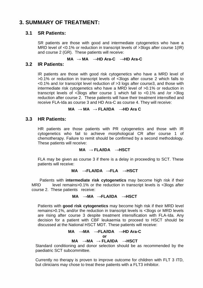
3. SUMMARY OF TREATMENT:
3.1 SR Patients: SR patients are those with good and intermediate cytogenetics who have a MRD level of <0.1% or reduction in transcript levels of >3logs after course 1(IR) and course 2 (GR). These patients will receive:
MA → MA →HD Ara-C →HD Ara-C
3.2 IR Patients: IR patients are those with good risk cytogenetics who have a MRD level of >0.1% or reduction in transcript levels of <3logs after course 2 which falls to <0.1% and /or transcript level reduction of >3 logs after course3, and those with intermediate risk cytogenetics who have a MRD level of >0.1% or reduction in transcript levels of <3logs after course 1 which fall to <0.1% and /or >3log reduction after course 2. These patients will have their treatment intensified and receive FLA-Ida as course 3 and HD Ara-C as course 4. They will receive:
MA → MA → FLA/IDA →HD Ara C
3.3 HR Patients: HR patients are those patients with PR cytogenetics and those with IR cytogenetics who fail to achieve morphological CR after course 1 of chemotherapy. Failure to remit should be confirmed by a second methodology. These patients will receive:
MA → FLA/IDA →HSCT
FLA may be given as course 3 if there is a delay in proceeding to SCT. These patients will receive:
MA →FLA/IDA →FLA →HSCT
Patients with intermediate risk cytogenetics may become high risk if their MRD level remains>0.1% or the reduction in transcript levels is <3logs after course 2. These patients receive:
MA →MA →FLA/IDA →HSCT
Patients with good risk cytogenetics may become high risk if their MRD level remains>0.1%, and/or the reduction in transcript levels is <3logs or MRD levels are rising after course 3 despite treatment intensification with FLA-Ida. Any decision for a patient with CBF leukaemia to proceed to HSCT should be discussed at the National HSCT MDT. These patients will receive:
MA →MA →FLA/IDA →HD Ara-C or
MA →MA → FLA/IDA →HSCT Standard conditioning and donor selection should be as recommended by the paediatric SCT subcommittee.
Currently no therapy is proven to improve outcome for children with FLT 3 ITD, but clinicians may chose to treat these patients with a FLT3 inhibitor.
14
4. TREATMENT ALLOCATION SCHEMA:
Ara-C: Cytarabine
CR: Complete remission
CRi: Complete remission with incomplete blood count recovery
FLA: Fludarabine & cytarabine
FLA-Ida: Fludarabine, cytarabine & idarubicin
GR: Good risk cytogenetics/molecular genetics
HD-Ara-C: High dose cytarabine
HSCT: Haemopoietic stem cell transplant
IR: Intermediate risk cytogenetic
Mito: Mitoxantrone
MRD: Minimal residual disease
RD: Resistant disease
15
5. INVESTIGATIONS AT PRESENTATION:
Investigations at presentation should be performed according to local practice and may include the following:
Full medical history and physical examination
Assessment of performance status (Karnofsky score for patients aged >16 years or Lansky score for patients aged ≤16 years)
Height, weight and body surface area (BSA)
Full blood count to include haemoglobin (Hb), white blood cells (WBC) with differential count, neutrophil count, blast count and platelet count
Coagulation screen
Biochemistry to include urate
Liver function to include bilirubin, alkaline phosphatase (Alk Phos), ALT or AST
All patients who might be considered for HSCT should be tissue typed
Bone marrow aspiration for local morphology, immunophenotype, and cytogenetics. A trephine biopsy is not essential, but should be carried out if the bone marrow aspirate yields a dry tap
EDTA bone marrow sample to be sent to flow cytometry laboratory to define LAIP-details given below
EDTA bone marrow and peripheral blood samples to be sent to molecular laboratory for genetic screening – details given below
Lumbar puncture for cell count and cytospin (intrathecal chemotherapy should be administered at the same time provided the diagnosis of AML has been confirmed )
Echocardiogram
Pregnancy test for all female patients of child bearing age. This must be performed within 2 weeks prior to starting treatment.
Where local laboratories are unable to identify rare cryptic abnormalities, either in house or by established local referral pathways, advice on reference laboratories may be available from:
Christine J Harrison Leukaemia Research Cytogenetics Group, Northern Institute for Cancer Research, Newcastle University, Level 5, Sir James Spence Institute, Royal Victoria Infirmary, Newcastle upon Tyne NE1 4LP
Any testing for MRD which is used to guide treatment should be performed in an accredited laboratory. Laboratories providing MRD monitoring in MyeChild 01 are listed below.
16
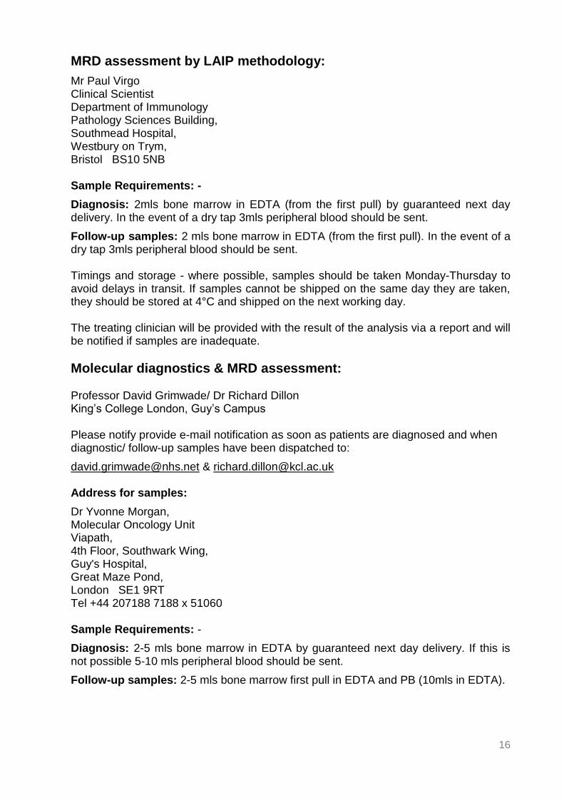
MRD assessment by LAIP methodology:
Mr Paul Virgo Clinical Scientist Department of Immunology Pathology Sciences Building, Southmead Hospital, Westbury on Trym, Bristol BS10 5NB
Sample Requirements: -
Diagnosis: 2mls bone marrow in EDTA (from the first pull) by guaranteed next day delivery. In the event of a dry tap 3mls peripheral blood should be sent.
Follow-up samples: 2 mls bone marrow in EDTA (from the first pull). In the event of a dry tap 3mls peripheral blood should be sent. Timings and storage - where possible, samples should be taken Monday-Thursday to avoid delays in transit. If samples cannot be shipped on the same day they are taken, they should be stored at 4°C and shipped on the next working day. The treating clinician will be provided with the result of the analysis via a report and will be notified if samples are inadequate.
Molecular diagnostics & MRD assessment: Professor David Grimwade/ Dr Richard Dillon King’s College London, Guy’s Campus Please notify provide e-mail notification as soon as patients are diagnosed and when diagnostic/ follow-up samples have been dispatched to:
Dr Yvonne Morgan, Molecular Oncology Unit Viapath, 4th Floor, Southwark Wing, Guy's Hospital, Great Maze Pond, London SE1 9RT Tel +44 207188 7188 x 51060 Sample Requirements: -
Diagnosis: 2-5 mls bone marrow in EDTA by guaranteed next day delivery. If this is not possible 5-10 mls peripheral blood should be sent.
Follow-up samples: 2-5 mls bone marrow first pull in EDTA and PB (10mls in EDTA).
Timing and storage - please schedule samples to be taken in the first half of a week and sent by guaranteed next day delivery to Yvonne Morgan at Guy’s –see address above .At diagnosis, where a patient presents later in the week or before a bank holiday, please take the diagnostic samples at the next available opportunity, even if this is after the patient has started treatment, so that the sample can be posted on the same day as collection. Samples should be sent at ambient temperature and not stored in a fridge before dispatch. The treating clinician will be provided with the result of the analysis via a report and will be notified if samples are inadequate. Patients treated on MyeChild 01 will have their level of MRD measured after each course of chemotherapy. Patients treated on this guideline should have their MRD assessed after course 1 and 2 of treatment and course 3 for those patients whose treatment may be further stratified by the level of MRD post course 3. This is required to guide treatment and may also allow trial entry at R 4 (HSCT randomisation).
6. TREATMENT
6.1. Pre Treatment Supportive Care: 6.1.1. Tumour Lysis:
All patients should be adequately hydrated with 2.5-3L/m2/day of hydration fluid at diagnosis. Potassium should not be added to hydration fluid during induction. Allopurinol should be started prior to induction therapy and continued for at least 5 days. In patients considered to be high risk for tumour lysis syndrome (e.g. white cell count >100 x 109/L, or renal impairment) rasburicase should be considered in place of allopurinol.
6.1.2 Management of Hyperleukocytosis:
Patients with high count leukaemia (hyperleukocytosis) are at risk of death or serious complications due to leukostasis/hyperviscosity syndrome, coagulopathy, or tumour lysis syndrome. A high count leukaemia is generally defined as a WCC>100 x 109/L except in monocytic AML (FAB type M5) when a WCC of >50 x 109/L may be problematic because the cells are large, tend to aggregate, and cause coagulopathy more readily.
Packed Red Cells, because of their high haematocrit (~70%), may exacerbate leukostasis. Generally red cell transfusion should be avoided or be very limited until the white cell count has been reduced to safe levels.
Start rasburicase (if indicated check G6PD before starting), hyperhydration and biochemistry monitoring as per local supportive care protocols.
Maintain the platelet count above 50 x109/L. In the presence of active bleeding or coagulopathy maintain platelets above 100x109/L
Correct any coagulopathy and keep the fibrinogen>1g/L
18
Commence chemotherapy urgently
The use of leukophoresis/exchange transfusion will be at the discretion of the treating physician. There is lack of evidence of benefit from systematic review.
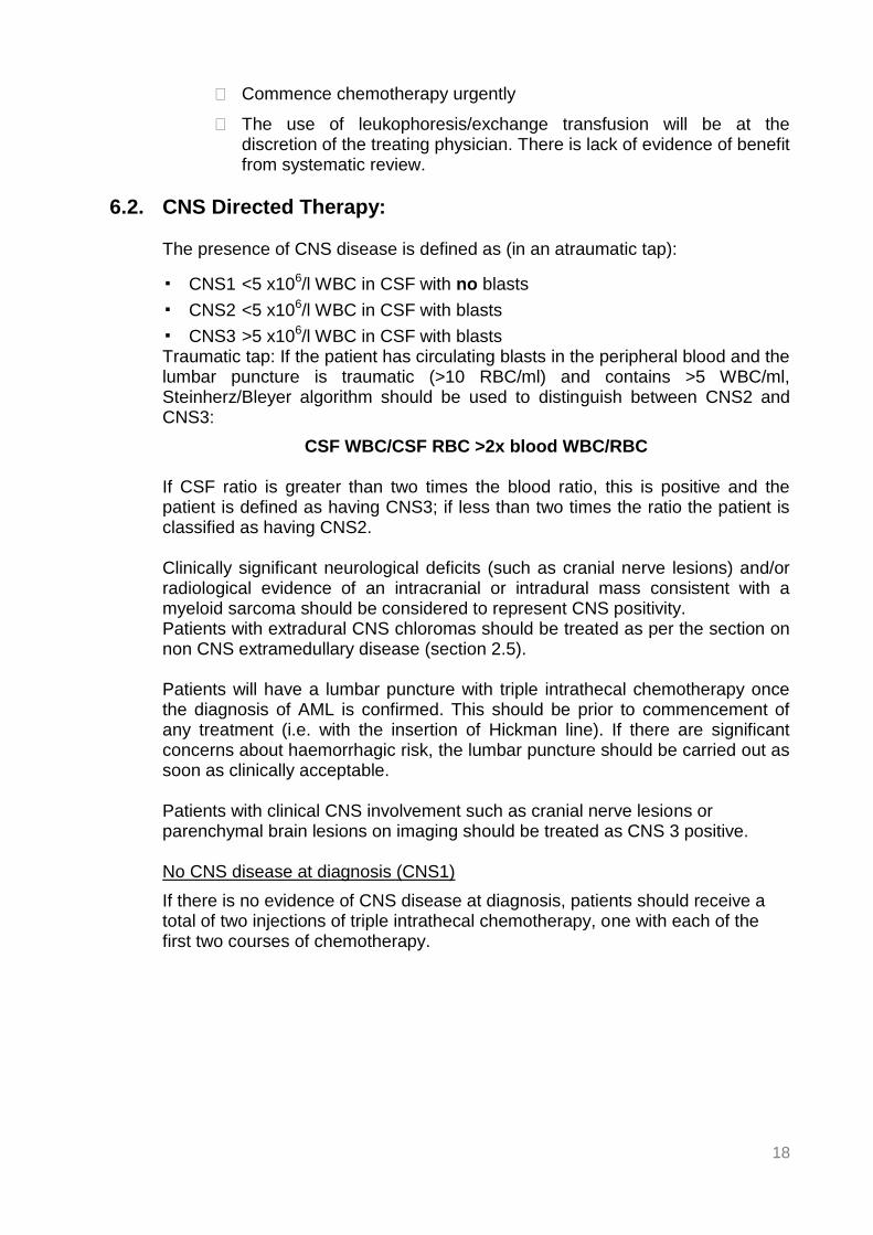
6.2. CNS Directed Therapy:
The presence of CNS disease is defined as (in an atraumatic tap):
▪ CNS1 <5 x10⁶/l WBC in CSF with no blasts
▪ CNS2 <5 x10⁶/l WBC in CSF with blasts
▪ CNS3 >5 x10⁶/l WBC in CSF with blasts Traumatic tap: If the patient has circulating blasts in the peripheral blood and the lumbar puncture is traumatic (>10 RBC/ml) and contains >5 WBC/ml, Steinherz/Bleyer algorithm should be used to distinguish between CNS2 and CNS3:
CSF WBC/CSF RBC >2x blood WBC/RBC If CSF ratio is greater than two times the blood ratio, this is positive and the patient is defined as having CNS3; if less than two times the ratio the patient is classified as having CNS2.
Clinically significant neurological deficits (such as cranial nerve lesions) and/or radiological evidence of an intracranial or intradural mass consistent with a myeloid sarcoma should be considered to represent CNS positivity. Patients with extradural CNS chloromas should be treated as per the section on non CNS extramedullary disease (section 2.5). Patients will have a lumbar puncture with triple intrathecal chemotherapy once the diagnosis of AML is confirmed. This should be prior to commencement of any treatment (i.e. with the insertion of Hickman line). If there are significant concerns about haemorrhagic risk, the lumbar puncture should be carried out as soon as clinically acceptable. Patients with clinical CNS involvement such as cranial nerve lesions or parenchymal brain lesions on imaging should be treated as CNS 3 positive.
No CNS disease at diagnosis (CNS1)
If there is no evidence of CNS disease at diagnosis, patients should receive a total of two injections of triple intrathecal chemotherapy, one with each of the first two courses of chemotherapy.
19
Age Methotrexate Cytarabine Hydrocortisone
<1 5mg 15mg 5mg
1 7.5mg 20mg 7.5mg
2 10mg 25mg 10mg
>3 or over 12.5mg 30mg 12.5mg
CNS disease at diagnosis (CNS3)
If CNS disease is present at diagnosis, patients should receive two injections of triple intrathecal chemotherapy (doses as above) each week until the CNS is clear plus a further two injections of triple intrathecal chemotherapy. A minimum of six triple intrathecal injections should be given in a period of three weeks from diagnosis. This intensive phase is followed by triple intrathecal chemotherapy (as above) with each cycle of chemotherapy.
If CNS disease clears after the intensive phase then cranial irradiation will not be used as standard treatment. CNS2 disease or traumatic tap at diagnosis
Due to concerns about clear definition/characterisation of this group and evidence of some increase in relapse rate, these patients should receive 2 injections of triple intrathecal chemotherapy per week until CNS is cleared plus a further 2 injections of triple intrathecal chemotherapy . They should also receive a further triple intrathecal chemotherapy at the start of cycle 2.
6.3. Induction Therapy:
Please note that Ara-C, cytosine arabinoside, cytarabine and cytosine refer to the same cytotoxic compound. Drugs should be prepared in the standard manner employed in individual centres.
6.3.1. Course 1 (for all patients): mitoxantrone & cytarabine
▪ Mitoxantrone 12mg/m2 daily by intravenous infusion over 1 hour on days 1, 2, 3 and 4 (total 4 doses).
▪ Cytarabine 100mg/m2 12 hourly by intravenous bolus on days 1-10 inclusive (total 20 doses).
Day 1 Day 2
Day 3
Day 4
Day 5
Day 6
Day 7
Day 8
Day 9
Day 10
Mitoxantrone • • • •
Cytarabine •• •• •• •• •• •• •• •• •• ••
20
Infants
Infants less than 12 months or weighing ≤10 kg or less or with a BSA <0.5m2 should have all drug doses calculated as mg/kg:
▪ Mitoxantrone: 0.4 mg/kg/dose
▪ Cytarabine: 3.3 mg/kg/dose
Patients without CNS disease should receive age appropriate triple intrathecal chemotherapy at the start of the block of chemotherapy. Patients with CNS disease should follow the instructions given in section 8.2
A bone marrow aspirate should be carried out to establish remission status on count recovery from course 1 and no later than day 35.
Patients will move to the next block of chemotherapy on count recovery (neutrophil count >1.0x10⁹/l and platelet count > 80 x 10 9/l) and when clinically well.
6.3.2. Course 2 (for non HR patients): mitoxantrone & cytarabine
▪ Mitoxantrone 12mg/m2 daily by intravenous infusion over 1 hour on days 1, 2 and 3 (total 3 doses).
▪ Cytarabine 100mg/m2 12 hourly by intravenous bolus on days 1-8 inclusive (total 16 doses).
Day 1 Day 2
Day 3
Day 4
Day 5
Day 6
Day 7
Day 8
Day 9
Day 10
Mitoxantrone • • •
Cytarabine •• •• •• •• •• •• •• ••
Infants
Infants less than 12 months or weighing ≤10 kg or less or with a BSA <0.5m2 should have all drug doses calculated as mg/kg:
▪ Mitoxantrone: 0.4 mg/kg/dose
▪ Cytarabine: 3.3 mg/kg/dose Patients without CNS disease should receive age appropriate triple intrathecal chemotherapy at the start of the block of chemotherapy. Patients with CNS disease should follow the instructions given in section 8. 2.
Patients will move to the next block of chemotherapy on count recovery (neutrophil count >1.0x10⁹/l and platelet count> 80 x 109/l) and when clinically well.
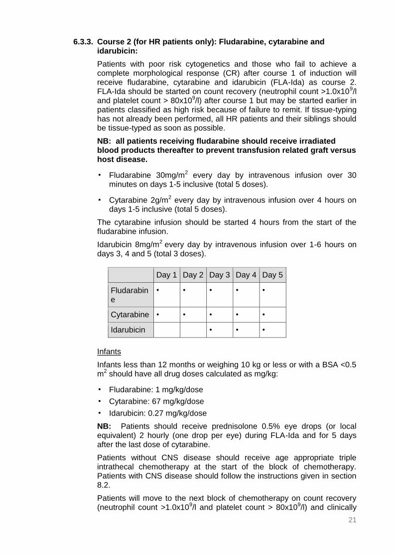
Patients with poor risk cytogenetics and those who fail to achieve a complete morphological response (CR) after course 1 of induction will receive fludarabine, cytarabine and idarubicin (FLA-Ida) as course 2. FLA-Ida should be started on count recovery (neutrophil count >1.0x10⁹/l and platelet count > 80x109/l) after course 1 but may be started earlier in patients classified as high risk because of failure to remit. If tissue-typing has not already been performed, all HR patients and their siblings should be tissue-typed as soon as possible.
NB: all patients receiving fludarabine should receive irradiated blood products thereafter to prevent transfusion related graft versus host disease.
▪ Fludarabine 30mg/m2 every day by intravenous infusion over 30 minutes on days 1-5 inclusive (total 5 doses).
▪ Cytarabine 2g/m2 every day by intravenous infusion over 4 hours on days 1-5 inclusive (total 5 doses).
The cytarabine infusion should be started 4 hours from the start of the fludarabine infusion.
Idarubicin 8mg/m2 every day by intravenous infusion over 1-6 hours on days 3, 4 and 5 (total 3 doses).
Day 1 Day 2 Day 3 Day 4 Day 5
Fludarabine
• • • • •
Cytarabine • • • • •
Idarubicin • • •
Infants
Infants less than 12 months or weighing 10 kg or less or with a BSA <0.5 m2 should have all drug doses calculated as mg/kg:
▪ Fludarabine: 1 mg/kg/dose
▪ Cytarabine: 67 mg/kg/dose
▪ Idarubicin: 0.27 mg/kg/dose
NB: Patients should receive prednisolone 0.5% eye drops (or local equivalent) 2 hourly (one drop per eye) during FLA-Ida and for 5 days after the last dose of cytarabine.
Patients without CNS disease should receive age appropriate triple intrathecal chemotherapy at the start of the block of chemotherapy. Patients with CNS disease should follow the instructions given in section 8.2.
Patients will move to the next block of chemotherapy on count recovery (neutrophil count >1.0x10⁹/l and platelet count > 80x109/l) and clinically
22
well. Patients can proceed directly to HSCT or have a third course of chemotherapy, with FLA if indicated.
6.4 Consolidation Therapy: 6.4.1. Standard Risk only: Courses 3 and 4: HD Ara-C:
All children other than those who are high risk will receive two courses of HD-Ara-C as consolidation chemotherapy.
Course 3 (consolidation) should start on count recovery from course 2 (neutrophil count >1.0x10⁹/l and platelet count > 80x109/l) and the patient is clinically well.
Course 3: HD Ara-C
▪ Cytarabine3g/m212 hourly by intravenous infusion over 4 hours on days 1, 3 and 5 (total 6 doses).
Day 1 Day 2 Day 3 Day 4
Day 5
HD Ara-C •• •• ••
Infants
Infants less than 12 months or weighing 10 kg or less or those with a BSA <0.5 m2 should have all drug doses calculated as mg/kg:
▪ Cytarabine: 100 mg/kg/dose
NB: Patients should receive prednisolone 0.5% eye drops (or local equivalent) 2 hourly (one drop per eye) during HD Ara-C and for 5 days after the last dose of cytarabine.
Course 4 consolidation should start on count recovery (neutrophil count >1.0x10⁹/l and platelet count > 80x109/l) from course 3 and when the patient is clinically well. Course 4: HD Ara-C
▪ Cytarabine 3g/m2 12 hourly by intravenous infusion over 4 hours on days 1, 3 and 5 (total 6 doses).
Day 1 Day 2 Day 3 Day 4
Day 5
HD Ara-C •• •• • •
Infants
Infants less than 12 months or weighing 10 kg or less or those with a BSA <0.5 m2 should have all drug doses calculated as mg/kg:
▪ Cytarabine: 100 mg/kg/dose
23
NB: Patients should receive prednisolone 0.5% eye drops (or local equivalent) 2 hourly (one drop per eye) during HD Ara-C and for 5 days after the last dose of cytarabine.
6.4.2 Intermediate Risk only: Courses 3 and 4: FLA-Ida and HD Ara-C:
Course 3: FLA-Ida
Intermediate risk patients will receive fludarabine, cytarabine and idarubicin (FLA-Ida) as course 3. FLA-Ida should be started on count recovery (neutrophil count >1.0x10⁹/l and platelet count > 80x109/l) after course 2.
NB: all patients receiving fludarabine should receive irradiated blood products thereafter to prevent transfusion related graft versus host disease.
▪ Fludarabine 30mg/m2 every day by intravenous infusion over 30 minutes on days 1-5 inclusive (total 5 doses).
▪ Cytarabine 2g/m2 every day by intravenous infusion over 4 hours on days 1-5 inclusive (total 5 doses).
The cytarabine infusion should be started 4 hours from the start of the fludarabine infusion.
▪ Idarubicin 8mg/m2 every day by intravenous infusion over 1-6 hours on days 3, 4 and 5 (total 3 doses).
Day 1 Day 2 Day 3
Day 4
Day 5
Fludarabine
• • • • •
Cytarabine • • • • •
Idarubicin • • •
Infants
Infants less than 12 months or weighing 10 kg or less or with a BSA <0.5 m2 should have all drug doses calculated as mg/kg:
▪ Fludarabine: 1 mg/kg/dose
▪ Cytarabine: 67 mg/kg/dose
▪ Idarubicin: 0.27 mg/kg/dose
NB: Patients should receive prednisolone 0.5% eye drops (or local equivalent) 2 hourly (one drop per eye) during FLA-Ida and for 5 days after the last dose of cytarabine.
24
Patients without CNS disease do not require intrathecal chemotherapy with course 3 and 4 as long as triple intrathecal chemotherapy has been previously given with course 1 and 2. Patients with CNS disease should follow the instructions given in section 8.2.
Patients will move to the next block of chemotherapy on count recovery (neutrophil count >1.0x10⁹/l and platelet count > 80x109/l) and clinically well.
Course 4: HD Ara-C
▪ Cytarabine 3g/m2 12 hourly by intravenous infusion over 4 hours on days 1, 3 and 5 (total 6 doses).
Day 1 Day 2 Day 3
Day 4
Day 5
HD Ara-C •• •• • •
Infants
Infants less than 12 months or weighing 10 kg or less or those with a BSA <0.5 m2 should have all drug doses calculated as mg/kg:
▪ Cytarabine: 100 mg/kg/dose
NB: Patients should receive prednisolone 0.5% eye drops (or local equivalent) 2 hourly (one drop per eye) during HD Ara-C and for 5 days after the last dose of cytarabine.
Patients without CNS disease do not require intrathecal chemotherapy with course 3 and 4 as long as triple intrathecal chemotherapy has been previously given with course1 and 2. Patients with CNS disease should follow the instructions given in section 8.2.
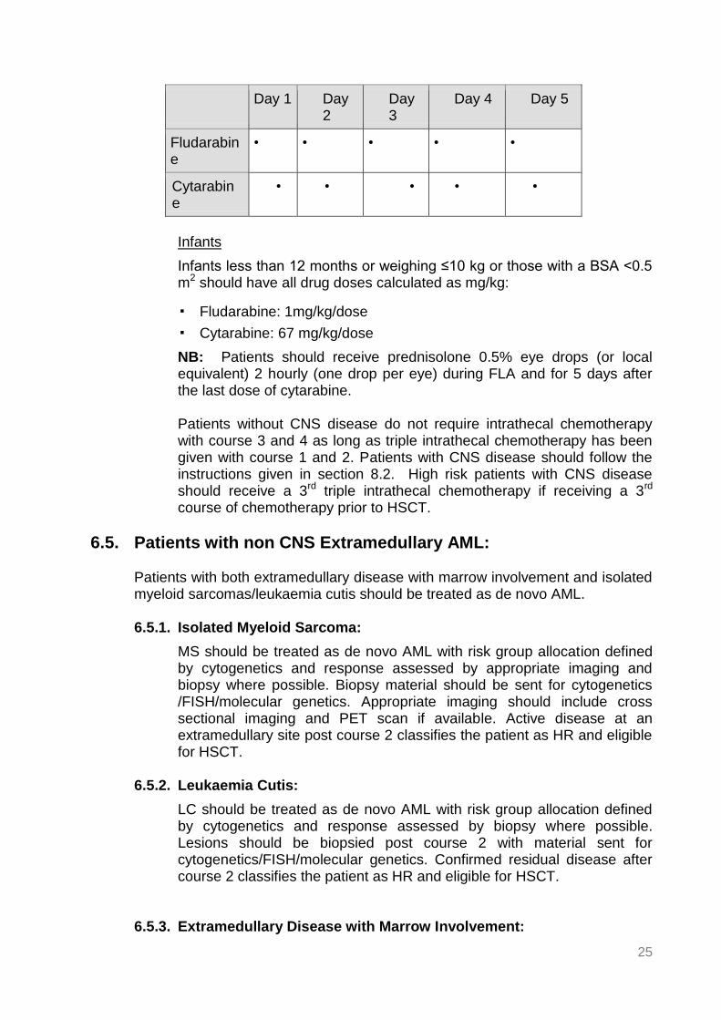
6.4.3. High Risk only: Courses 3: FLA
Course 3: FLA
Patients identified as HR after course 1 and who received FLA-Ida as course 2 may receive a course of fludarabine and cytarabine (FLA) as course 3 if a third course of chemotherapy is indicated as a bridge to HSCT .
FLA should start on count recovery (neutrophil count >1.0x10⁹/l and platelet count > 80x109/l) after course 2 and the patient is clinically well.
NB: All patients receiving fludarabine should receive irradiated blood products thereafter to prevent transfusion related graft versus host disease.
▪ Fludarabine 30mg/m2 every day by intravenous infusion over 30 minutes on days 1-5 inclusive (total 5 doses).
▪ Cytarabine 2g/m2 every day by intravenous infusion over 4 hours on days 1-5 inclusive (total 5 doses). The cytarabine infusion should be started 4 hours from the start of the fludarabine infusion.
25
Day 1 Day 2
Day 3
Day 4 Day 5
Fludarabine
• • • • •
Cytarabine
• • • • •
Infants
Infants less than 12 months or weighing ≤10 kg or those with a BSA <0.5 m2 should have all drug doses calculated as mg/kg:
▪ Fludarabine: 1mg/kg/dose
▪ Cytarabine: 67 mg/kg/dose
NB: Patients should receive prednisolone 0.5% eye drops (or local equivalent) 2 hourly (one drop per eye) during FLA and for 5 days after the last dose of cytarabine.
Patients without CNS disease do not require intrathecal chemotherapy with course 3 and 4 as long as triple intrathecal chemotherapy has been given with course 1 and 2. Patients with CNS disease should follow the instructions given in section 8.2. High risk patients with CNS disease should receive a 3rd triple intrathecal chemotherapy if receiving a 3rd course of chemotherapy prior to HSCT.
6.5. Patients with non CNS Extramedullary AML:
Patients with both extramedullary disease with marrow involvement and isolated myeloid sarcomas/leukaemia cutis should be treated as de novo AML.
6.5.1. Isolated Myeloid Sarcoma:
MS should be treated as de novo AML with risk group allocation defined by cytogenetics and response assessed by appropriate imaging and biopsy where possible. Biopsy material should be sent for cytogenetics /FISH/molecular genetics. Appropriate imaging should include cross sectional imaging and PET scan if available. Active disease at an extramedullary site post course 2 classifies the patient as HR and eligible for HSCT.
6.5.2. Leukaemia Cutis:
LC should be treated as de novo AML with risk group allocation defined by cytogenetics and response assessed by biopsy where possible. Lesions should be biopsied post course 2 with material sent for cytogenetics/FISH/molecular genetics. Confirmed residual disease after course 2 classifies the patient as HR and eligible for HSCT.
6.5.3. Extramedullary Disease with Marrow Involvement:
26
The bone marrow response should be assessed after course 1. Clinically residual extramedullary disease after course 2 should have appropriate imaging with cross sectional imaging and PET scan if available and be biopsied, if possible, with material sent for cytogenetics/FISH/molecular genetics. Residual active disease at an extramedullary site post course 2 classifies the patient as HR and eligible for HSCT.
6.6. Supportive Treatment:
AML therapy is intensive and associated with significant morbidity. Centres should consult their local supportive care protocols for further guidance. It is advisable to keep children in hospital during the induction period, when severely neutropenic.
6.6.1. Tumour Lysis:
All patients should be adequately hydrated with 2.5-3 L/m2/day of hydration fluid at diagnosis. Potassium should not be added to hydration fluid during induction. Allopurinol should be started prior to induction therapy and continued for at least 5 days. In patients considered to be high risk for tumour lysis syndrome (e.g. white cell count > 100 x 109/L, or renal impairment) rasburicase should be considered in place of Allopurinol.
PCP prophylaxis is recommended following fludarabine containing regimens and post stem cell transplant. Cotrimoxazole is considered the first line agent. Dosing regimens should be according to local practice.
6.6.3. Antimicrobials:
Antimicrobial prophylaxis, where considered appropriate, will be at the discretion of the treating physician. Antibiotic treatment of febrile neutropenia should be based on local supportive care guidance in conjunction with NICE Guideline CG151 [21]. The use of G-CSF should be based on local supportive care guidance.
6.6.4. Antifungal Prophylaxis:
Patients should either be nursed in laminar flow or receive anti-fungal prophylaxis. For recommended agents and dosing please see ECIL 4 Guidelines on prevention and treatment of invasive fungal diseases in paediatric patients with cancer or allogeneic haematopoietic stem cell transplantation [22].
6.6.5. Cardiotoxicity Monitoring:
It is recommended that an echocardiogram is carried out prior to course 1 and before each course containing anthracycline. Thereafter cardiac monitoring should follow local practice.
6.6.6. Additional Supportive Therapy:
Additional supportive therapy will be provided according to local practice.
27
6.7. Dose Modifications for Toxicity
Dose modifications for obesity are not recommended due to the risk of underdosing. The exception is dose reductions for toxicity as detailed below for all patients. Cardiotoxicity monitoring guidelines should be rigidly adhered to in obese patients.
Course1: Abnormal hepatic or renal function at diagnosis may be secondary to leukaemic infiltration or an unrelated disorder such as Gilbert’s disease. Therefore patients with abnormal hepatic or renal function at diagnosis require thorough investigation to identify the cause before consideration is given to reducing doses of anthracyclines in course 1.
All other courses: -
Toxicity Mitoxantrone
Idarubicin
Fludarabine High dose Cytarabine
(>2g/m2/course)
Absolute LVFS <28% or Ejection Fraction <45%
Further advice from AML WG
Further advice from AML WG
Bilirubin ≥2.5 x ULN Treat with caution or 50% dose
Treat with caution or 50% dose
Ocular irritation Increase frequency of steroid eye drops or as local practice.
Cerebellar toxicity Omit
Renal Function
Creatinine clearance
30-70ml/min/1.73m2
75% dose
Renal Function
Creatinine clearance <30ml/min/1.73m
2
50% dose Discuss Discuss
Calculate creatinine clearance from serum creatinine according to the BNFc formula, or that in use nationally. Child > 1 year
1. Stevens, R.F., et al., Marked improvements in outcome with chemotherapy alone in paediatric acute myeloid leukemia: results of the United Kingdom Medical Research Council's 10th AML trial. MRC Childhood Leukaemia Working Party. Br J Haematol, 1998. 101(1): p. 130-40.
2. Perel, Y., et al., Impact of Addition of Maintenance Therapy to Intensive Induction and Consolidation Chemotherapy for Childhood Acute Myeloblastic Leukemia: Results of a Prospective Randomized Trial, LAME 89/91. Journal of Clinical Oncology, 2002. 20(12): p. 2774-2782.
3. Perel, Y., et al., Treatment of childhood acute myeloblastic leukemia: dose intensification improves outcome and maintenance therapy is of no benefit--multicenter studies of the French LAME (Leucemie Aigue Myeloblastique Enfant) Cooperative Group. Leukemia, 2005. 19(12): p. 2082-9.
4. Ho, P.A., et al., Prevalence and prognostic implications of CEBPA mutations in pediatric acute myeloid leukemia (AML): a report from the Children's Oncology Group. Blood, 2009. 113(26): p. 6558-66.
5. Harrison, C.J., et al., Cytogenetics of childhood acute myeloid leukemia: United Kingdom Medical Research Council Treatment trials AML 10 and 12. J Clin Oncol, 2010. 28(16): p. 2674-81.
6. von Neuhoff, C., et al., Prognostic impact of specific chromosomal aberrations in a large group of pediatric patients with acute myeloid leukemia treated uniformly according to trial AML-BFM 98. J Clin Oncol, 2010. 28(16): p. 2682-9.
7. Rubnitz, J.E., et al., Minimal residual disease-directed therapy for childhood acute myeloid leukaemia: results of the AML02 multicentre trial. The Lancet Oncology, 2010. 11(6): p. 543-552.
8. Burnett, A.K., et al., Identification of patients with acute myeloblastic leukemia who benefit from the addition of gemtuzumab ozogamicin: results of the MRC AML15 trial. J Clin Oncol. 2011 Feb 1;29(4):369-77.
9. Gibson, B.E., et al., Results of a randomized trial in children with Acute Myeloid Leukaemia: medical research council AML12 trial. Br J Haematol, 2011. 155(3): p. 366-76.
10. Loken, M.R., et al., Residual disease detected by multidimensional flow cytometry signifies high relapse risk in patients with de novo acute myeloid leukemia: a report from Children's Oncology Group. Blood, 2012. 120(8): p. 1581-8.
11. Burnett, A.K., et al., Optimization of chemotherapy for younger patients with acute myeloid leukemia: results of the medical research council AML15 trial. J Clin Oncol. 2013 Sep 20;31(27):3360-8
12. Cilloni, D, et al., Real-time quantitative polymerase chain reaction detection of minimal residual disease by standardized WT1 assay to enhance risk stratification in acute myeloid leukemia: a European LeukemiaNet study. J Clin Oncol. 2009 Nov 1;27(31):5195-201.
13. Zhu, H.H., et al., MRD-directed risk stratification treatment may improve outcomes of t(8;21) AML in the first complete remission: results from the AML05 multicenter trial. Blood. 2013 May 16;121(20):4056-62.
29
14. Grimwade, D., Freeman SD. Defining minimal residual disease in acute myeloid leukemia: which platforms are ready for "prime time”? Blood. 2014 Nov 27;124(23):3345-55.
15. Sievers, E.L., et al., Immunophenotypic evidence of leukemia after induction therapy predicts relapse: results from a prospective Children's Cancer Group study of 252 patients with acute myeloid leukemia. Blood. 2003 May 1;101(9):3398-406.
16. Terwijn, M., et al., High prognostic impact of flow cytometric minimal residual disease detection in acute myeloid leukemia: data from the HOVON/SAKK AML 42A study. J Clin Oncol. 2013 Nov 1;31(31):3889-97.
17. Creutzig, U., et al., Randomized trial comparing liposomal daunorubicin with idarubicin as induction for pediatric acute myeloid leukemia: results from Study AML-BFM 2004. Blood. 2013 Jul 4;122(1):37-43.
18. Pui, M.H., B.D. Fletcher, and J.W. Langston, Granulocytic sarcoma in childhood leukemia: imaging features. Radiology, 1994. 190(3): p. 698-702.
19. Agis, H., et al., A comparative study on demographic, hematological, and cytogenetic findings and prognosis in acute myeloid leukemia with and without leukemia cutis. Ann Hematol, 2002. 81(2): p. 90-5.
20. Reinhardt, D. and Creutzig, U., Isolated myelosarcoma in children--update and review. Leuk Lymphoma, 2002. 43(3): p. 565-74.
21. Neutropenic sepsis: prevention and management in people with cancer. NICE guidelines [CG151] Published date: September 2012. http://www.nice.org.uk/guidance/cg151/evidence/full-guideline-188303581
22. Groll, A.H., et al.,Fourth European Conference on Infections in Leukaemia (ECIL-4): guidelines for diagnosis, prevention, and treatment of invasive fungal diseases in paediatric patients with cancer or allogeneic haemopoietic stem-cell transplantation. Lancet Oncol. 2014 Jul;15(8):e327-40.
23. Balgobind, B.V., et al., Novel prognostic subgroups in childhood 11q23/MLL-rearranged acute myeloid leukemia: results of an international retrospective study. Blood, 2009. 114(12): p. 2489-96.
24. Hollink, I.H., et al., NUP98/NSD1 characterizes a novel poor prognostic group in acute myeloid leukemia with a distinct HOX gene expression pattern. Blood, 2011. 118(13): p. 3645-56.
25. Shiba, N., et al., NUP98-NSD1 gene fusion and its related gene expression signature are strongly associated with a poor prognosis in pediatric acute myeloid leukemia. Genes, Chromosomes and Cancer, 2013. 52(7): p. 683-693.
26. Tosi S, H.J., et al, t(7;12)(q36;p13), a new recurrent translocation involving ETV6 in infant leukemia. Genes, Chromosomes and Cancer, 2000. 29(4): p. 8. 7.
27. Beverloo, H.B., et al., Fusion of the Homeobox Gene HLXB9 and the ETV6 Gene in Infant Acute Myeloid Leukemias with the t(7;12)(q36;p13). Cancer Research, 2001. 61(14): p. 5374-5377.
28. Gruber, T.A., et al., An Inv(16)(p13.3q24.3)-Encoded CBFA2T3-GLIS2 Fusion Protein Defines an Aggressive Subtype of Pediatric Acute Megakaryoblastic Leukemia. Cancer Cell, 2012. 22(5): p. 683-697.
30
29. Masetti, R., et al., CBFA2T3-GLIS2 fusion transcript is a novel common feature in pediatric, cytogenetically normal AML, not restricted to FAB M7 subtype. Blood, 2013. 121(17): p. 3469-72.
30. Akiki, S., et al., NUP98-NSD1 fusion in association with FLT3-ITD mutation identifies a prognostically relevant subgroup of pediatric acute myeloid leukemia patients suitable for monitoring by real time quantitative PCR. Genes, Chromosomes and Cancer, 2013. 52(11): p. 1053-1064.
31. Brown, P., et al., The incidence and clinical significance of nucleophosmin mutations in childhood AML. Vol. 110. 2007. 979-985.
32. Meshinchi, S., et al., Prevalence and prognostic significance of Flt3 internal tandem duplication in pediatric acute myeloid leukemia. Blood, 2001. 97(1): p. 89-94.
33. Creutzig, U., et al. Comparison of chemotherapy alone with allogeneic bone marrow transplantation in first full remission in children with acute myeloid leukemia in the AML-BFM-83 and AML-BFM-87 studies--matched pair analysis. Klin Padiatr. 1992 Jul-Aug;204(4):246-52.
31
8. APPENDIX 1
Rationale for Guideline Treatment
Cytogenetic Risk Score:
The cytogenetic risk group stratification has been adopted from MyeChild 01 and acknowledges a number of key publications in the field, including the combined analysis of the cytogenetic data from children (n=729) treated on MRC AML 10 and12 [5]. The results from the MRC AML 10 and 12 trials suggested the overall outcome for all patients with 11q23 (MLL or KMT2A) abnormalities was intermediate with no difference observed for those with t (9; 11) (p21-22; q23). However data from an international retrospective study of 756 childhood AML patients with MLL rearrangements reported a poor outcome for patients with the translocations, t(4;11) (q21;q23), t(6;11) (q27;q23), t(10;11) (p12;q23) and t(10;11) (p11.2;q23) [23]. A favourable outcome was reported for patients with t(1;11) (q21;q23), but numbers were small and UK trials were unable to confirm this finding: only two paediatric cases were identified with one dying at 5 years and the other in long term remission. Therefore all patients with MLL (KMT2A) rearrangements other than those previously specified as PR are classified as IR.
The MRC AML 10 and 12 trial data confirmed the adverse outcome of monosomy 7, abnormalities of 5q, t (6;9)(p23;q34)/DEK-NUP214 and t(9;22)(q34;q11) in children. In contrast to adults, abnormalities of 3q and complex karyotypes, in the absence of other PR features, were not associated with a significantly adverse outcome in children. The variable outcome for 3q abnormalities may be explained by age related differences in the incidence of 3q abnormalities with a poorer prognosis being specifically dependent on the EV11 expression status and/or the presence of the inv(3)(q21q26/t(3,3)(q21;q26). The presence of 12p abnormalities predicted a poor outcome; a finding confirmed by the BFM [6]. More recently rare cryptic chromosomal abnormalities have been described, which confer a poor outcome. These include: t(5;11)(q35;p15.5)/NUP98-NSD1 [24-25]; t(7;12 )(q36;p13)/MNX1-ETV6 [26-27; and inv(16)(p13.3q24.3)/CBFA2T3-GLIS2 [28-29].
NUP98-NSD1 mutations tend to be associated with FLT 3 ITD, to occur in older patients and result in refractoriness to current chemotherapy, but can potentially be salvaged by allo-HSCT [30]. MNX1-ETV6 is usually cryptic, mainly but not exclusively seen in infants, and is often accompanied by a deletion of the long arm of chromosome 7 [27].
CBFA2T3-GLIS2 was originally associated with the AML M7 French American British (FAB) type, but it has recently been observed in all FAB types [29]. Mutations in CEBPA (4.5%) and NPM1 (8%) of paediatric AML respectively are associated with a normal karyotype and a favourable outcome [4]; the latter only in the absence of FLT3 ITD [31].FLT3 ITD increases in incidence with age and is associated with a poor outcome in the absence of good risk cytogenetic features[32]. Response to Treatment – Definition of CR by Flow Cytometry:
Two large paediatric studies in AML have demonstrated morphological assessment of response to the first course of induction chemotherapy to be of low sensitivity and poor specificity [7,10]. Both have shown multiparameter/multidimensional flow cytometry (MDF) using aberrant expression of surface antigens on leukaemic blasts to be more predictive of outcome.
32
COG reported 24% of 188 patients in complete morphological remission at the end of their first course of induction therapy (EOI1) to have MDF detectable disease at a level of 0.1% or greater. Patients in CR with residual disease (RD) by MDF had a RR of 60% ± 16% at 3 years compared with that of 29% ± 8% in patients without RD (p<0.001) and an OS of 56%± 16% vs. 80% ± 8% (p=0.002) respectively. Nineteen percent of 180 patients in morphological CR after course 2 (EOI2) had evidence of RD by MDF at a level of >0.1% or greater and a CIR (cumulative incidence of relapse) at 3 years from EOI2 of 67%± 18% compared to 30%± 8% in those without RD (p<0.001). MDF was available for 27 of 42 patients who failed to achieve morphological CR (>5% blasts). All 7 (26%) patients who were RD negative are long term survivors, whilst 20 (74%) who were RD positive had a 3 year OS of 35% ± 21% (p=0.005). In a multivariate analysis, including cytogenetic and molecular risk factors, RD by MDF was an independent predictor of relapse (p<0.001), confirming the superior predictive value for outcome of MDF over morphology. Because of the low sensitivity and poor specificity of morphological assessment of complete remission, morphological remission or lack of morphological remission, should be confirmed by flow, molecular or interphase FISH. Anthracyclines:
The induction anthracycline for all patients in course 1 and non HR patients in course 2 will be mitoxantrone, which will be combined with cytarabine (MA). This is based on data from MRC AML 12, LAME 89/91 and ELAM02 [2-3,9]. MRC AML12 identified a benefit in DFS (p=0.03) and RR (p=0.05), a trend in improvement in EFS p=0.08, although no difference in OS (p=0.2) for mitoxantrone (MAE) compared to daunorubicin (ADE). Whilst AML 12 delivered mitoxantrone (12mg/m2/d x 3d) with cytarabine and etoposide in induction courses 1 and 2, the French group have used much higher doses of mitoxantrone with acceptable toxicity. They have given mitoxantrone 12mg/m2/d x 5d (300 mg/m2daunorubicin equivalence based on 5:1 conversion factor) with cytarabine 200mg/m2/d x 7d in course 1 since 1989.
In LAME 89/91 the cumulative anthracycline dose was 460mg/m2 (daunorubicin equivalence), rising to 580mg/m2 in patients with >20% blasts in the bone marrow at day 20. Despite this high anthracycline exposure, both LAME 89/91 and ELAM 02 reported a low induction death rate from toxicity (LAME 2.3%), reflecting acceptable acute toxicity for this dose of mitoxantrone in induction. The 5 year OS for ELAM 02 was 71% with EFS of 57% (personal communication and ASH 2014). 4/441 (0.9%) patients in ELAM 02 had grade 3/4 early cardiotoxicity and for both LAME 89/91 and ELAM02 the cumulative incidence of late clinical cardiotoxicity at 10years is 3%. Long term follow up data from the MRC AML12 trial suggests that mitoxantrone may be associated with less late cardiotoxicity than daunorubicin (personal communication). Mitoxantrone may be associated with more prolonged myelosuppression than Daunorubicin.
Consolidation Therapy in Standard Risk Patients:
Their cumulative daunorubicin equivalence anthracycline dose in courses 1 and 2 for non-HR patients will be 420mg/m2 to limit anthracycline exposure in this group of patients with the lowest risk of relapse. At 5 years AML15 reported no difference in OS between HD Ara-C/HD Ara-C and MACE/MidaC (0-15yrs: 78% v 73%; 2p 0.7; based on 65 v 64 children) in patients with GR and IR cytogenetics [11].
Children with core binding factor (CBF) leukaemias and those with IR cytogenetics had an OS of 96% vs. 93% (2p= 0.7) and 75% vs. 64% (2p=0.4) respectively from 2nd randomisation for HD Ara-C vs. MACE/MidAC. The number of children with PR
33
cytogenetics was too small to be evaluable. However, adult data suggests that HD Ara-C may be inferior in patients with PR cytogenetics. There were only 8% children in this group and less than half of these entered the consolidation randomisation, so no conclusion can be made, other than from extrapolation of adult data. Haemopoietic Stem Cell Transplantation:
High risk patients are eligible for HSCT. AML 10 reported a significant reduction in RR for MSD HSCT which did not translate into a significant advantage in OS because of a high procedure related mortality (PRM) (by donor vs. no donor analysis: RR allo-HSCT vs. no allo-HSCT: 30% vs. 45%, p=.0.02; OS at 10 yrs 68% vs. 59%, p=0.3; PRM 15% for those who received a HSCT, p = 0.001) [15]. The BFM reported a similar lack of benefit for allo–HSCT in CR1 [33] and these data influenced the UK approach to HSCT in CR1 for the next decade and longer. The PRM for both related and unrelated allo-HSCT is now low (5- 10-%), allowing the allo-HSCT associated reduction in RR the potential to improve EFS in patients at risk of relapse.
In ELAM 02 all cytogenetic GR and IR patients with a matched family donor (MFD), with the exception of those children with t(8;21),were candidates for HSCT after 1 or 2 courses of consolidation [3], whilst children with PR cytogenetics were eligible for HSCT with an unrelated HLA -identical donor. 119 children underwent allogeneic HSCT in CR1. The OS for transplanted patients was 76.2% and DFS 69.8%.