76
1 Funded by: Guidelines for the Training of Veterinary Professionals on Camel Health and Diseases

1Funded by:
Guidelines for the Training
of Veterinary Professionalson Camel Health and Diseases

Guidelines for the Training
of Veterinary Professionalson Camel Health and Diseases
Funded by:


Table of Contents
Acknowledgment 5
Introduction 6
HOW TO USE THE MODULES 8
MODULE 1: LEAD SYMPTOM: WEIGHT LOSS 9
MODULE 2: LEAD SYMPTOM: POX LIKE SKIN LESIONS 11
MODULE 3: LEAD SYMPTOM: RESPIRATORY DISTRESS 13
MODULE 4: LEAD SYMPTOM: SICK CALF 15
MODULE 5: LEAD SYMPTOM: CENTRAL NERVOUS PROBLEMS 17
MODULE 6: LEAD SYMPTOM: ACUTE DEATH IN ADULT CAMELS 19
MODULE 7: LEAD SYMPTOM: SKIN PROBLEM 21
MODULE 8: LEAD SYMPTOM: ABORTION 23
MODULE 9: EXAMINATION OF THE CAMEL 25

Acknowledgments:We would like to thank Biovison Foundation for funding the development, testing
and publishing of the Camel Training Manual. We would like to acknowledge
Dr. Ilona Gluecks and Dr. Mario Younan for conceptualizing and developing the
training manual. We would also like to thank the following who participated in the
initial testing and reviewing of the training manual; Mr. Stanely Kirimi,
Mr. Gilford M. Boore, Dr. Mwongela D. T, Mr. Stanely Kinyua, Mr. Virano Mathiu,
Mr. Albert M. Kingia, Dr. Josephat Muema, Ms. Nancy Kamau. Mr. Mohamed Nur
Abdikadir, Dr. Kimathi G. M., Dr. Diana Onyango and Dr. Douglas Machuchu.
Copyright © Veterinaries Sans Fron�eres (VSF Suisse)Owashika road off Isaac Gathanju road, Lavington, PO Box 25656-00603 Nairobi
First e-published 2014 by VSF-Suisse
ISBN 9966-956-85-9 Rights reserved, and sharedThis publica�on may be printed, reproduced, stored in a retrieval system or transmi�ed, in any form or by any means, electronic, mechanical, photocopying, recording or otherwise, but only when retained in this full form with logos and names of authors, editors, designers and credited organiza�ons included as is. Requests for permission to print and distribute hard copy edi�ons can be made to VSF-S, Nairobi, This permission must be granted in wri�ng by the authors and publisher prior to prin�ng and the requester who shall be fully liable for the full cost of prin�ng and distribu�on will also have the possibility of including the organiza�onal logo/s credited with funding of prin�ng.
Designed by Jacaranda Africa—Home of the Young African ExpressDesign & layout by Grace King’oriPhotos by Maurizio Dioli
PO Box 1202Nairobi 00606, Kenya Tel: +254 (0) 20-3260-4433 or 0722-66 77 47Email: [email protected] Website: www.jacaranda-africa.com

7
Over 70% of Kenya’s land mass consists of arid and semi-arid lands
(ASALs). Extensive livestock grazing, in a nomadic pastoral production
system, is seen as a suitable means of utilizing these agro-ecological
areas.
Camels are well adapted to the harsh conditions of the ASALs and have
been kept for centuries by various pastoral communities in the Greater
Horn of Africa. The dromedary camel constitutes an important part of
their livelihood, it is essential to their subsistence economy. Many argue
that dromedaries are the most important livestock species in terms of
food security. First and foremost, the camel contributes to the pastoral
livelihood via its milk and meat for household consumption. Milk is the
most important product of the camel, and contributes between 50 –
60 % of the nutrient intake of some of the pastoralist communities of
sub Saharan Africa, especially during the dry season. In addition sales
of milk, meat, hides and live animals contribute to household income.
The camel represents a saving mechanism and contributes draught
power mainly for transport but recently also for land preparation. Last
but not least the camel plays an important role for the socio-cultural
interaction of the community. The camel is used for payment of dowry,
settlement of fines in tribal feuds and recreational activities. Especially
the Somali, who represent the largest and oldest camel keeping tribe,
see camels as a banking system or security against drought, disease,
and other natural disasters that affect smaller stock more seriously.
In the past decades the interest in the camel sector has continuously
risen. Various stakeholders, including the Government, Universities,
Scientists, Development Actors and the Private Sector are currently
trying to support camel keeping communities in improving
health, husbandry, production, products and marketing – even
the introduction of camels into traditionally non-camel keeping
communities (for example the Maasai and Samburu tribes in Kenya)
becomes a common programmatic approach.
Availability of and access to information, knowledge and educational
material on camel health and diseases is very limited. Only recently the
camel has been included in the curriculum of veterinary studies i.e. in
Kenya.
The aim of these training modules including the training guidelines is to
improve the availability and accessibility of teaching material on camel
health and disease topics, especially for those veterinary professionals
already working with camel keeping communities. It is anticipated to
enhance the knowledge of these professionals on the most common
camel diseases, their epidemiology, diagnosis and treatment covering
the theory as one aspect but focusing more on the practical side, to
help veterinary professionals in their diagnosis when confronted with a
sick camel in the field.
Introduc�on

8
The target audience for this training are veterinary professionals
who have a basic knowledge of camels. However the groups can
be split into two:
GROUP A, representing veterinary professionals with NO field
experience of camels and their diseases and
GROUP B, representing veterinary professionals with field
experience of camels and their diseases.
It is advisable to know the level of experience of the trainees
beforehand, in order to plan the training accordingly.
GROUP A will need two extra days in order to cover the theory
of the diseases in a more classic classroom style prior to starting
with the more participatory training sessions.
Trainees of GROUP B can move straight to the participatory
training sessions.
How to use the Modules
Suggested time table:
Day 1 Disease groups 1 – 4 Group A only
Day 2 Disease groups 5 – 8 Group A only
Day 1 Module 1
Session 1 A – C
Module 2
Session 2 A – C
Group A and B
Day 2 Module 3
Session 3 A – B
Module 4
Group A and B
Day 3 Module 5
Session 5 A – B
Module 6
Session 6 A – B
Group A and B
Day 4 Module 7
Session 7 A – B
Module 8
Session 8 A – B
Group A and B
Day 5 Module 9
Session 9 A – F
Group A and B
9
AIM OF THIS MODULE:
The aim of this first module is
1) to enable the trainee to differentiate between acute and
chronic Trypanosomosis and the infection with gastrointestinal
helminths when confronted with a sick camel showing the
lead symptom of weight loss in the field;
2) to enhance the knowledge of the trainee on the two covered
diseases especially on their epidemiology, diagnosis and
treatment focusing on field conditions.
Handouts to be used:
Group work:
The trainees are split into three groups and Module 1 Handout 1
Each of the handouts describes a case of a camel presented
by a herdsman with the lead symptom of weight loss. It
further includes some pre-empted questions and answers and
examination results.
The task of the group is to
1. define a clinical diagnosis;
2. support the clinical diagnosis with main observations;
3. describe what additional examinations or diagnostic tests they
would carry out and how;
4. define the recommended therapy;
5. name three most important differential diagnoses and their
major differences to support the clinical diagnosis;
6. give a practical advice to the herder on how to control the
disease.
TIME FRAME:
Each group has one hour to discuss their specific case.
Each group should select a presenter to present their specific
the plenary followed by discussions.
The aim of this session is to discuss the three covered diseases in
plenary to
support the diagnosis
MODULE 1: Lead Symptom: Weight Loss
Diseases Covered:• Trypanosoma Evansi – Acute • Trypanosoma Evansi – Chronic• Gastroinstes�nal Helminths

10
laboratory analysis to support the diagnosis (comparing the
ideal scenario as taught with practical field conditions)
herder
At the end of the session each trainee should receive
TIME FRAME:
One hour
Summary of each disease in plenary, developing “disease
summary cards”.
For each disease so called “disease summary cards” will be
developed together with the facilitator including:
At the end of the session Module 1 Handout 2 will be given to
each participant.
TIME FRAME:
One hour

11
AIM OF THIS MODULE:
The aim of this module 2 is 1) to enable the trainee to differentiate between camel pox and
Orf when confronted with a sick camel showing the lead
symptom of weight loss in the field;
2) to enhance the knowledge of the trainee on the two covered
diseases especially on their epidemiology, diagnosis and
treatment focusing on field conditions.
Handouts to be used:
Handouts to be used:
Group work:
The trainees are split into three groups and Module 2 Handout 1
Each of the handouts describes a case of a camel presented by
a herdsman with the lead symptom of pox like skin lesions. It
further includes some pre-empted questions and answers and
examination results.
The task of the group is to
1. define a clinical diagnosis;
2. support the clinical diagnosis with main observations;
3. describe what additional examinations or diagnostic tests they
would carry out and how;
4. define the recommended therapy;
5. name three most important differential diagnoses and their
major differences to support the clinical diagnosis;
6. give a practical advice to the herder on how to control the
disease.
TIME FRAME:
Each group has one hour to discuss their specific case.
Each group should select a presenter to present their specific
the plenary followed by discussions.
MODULE 2: Lead Symptom: Skin Lesions
Diseases Covered:• Camel Pox • ORF (Contagious Ecthyma)

12
The aim of this session is to discuss the three covered diseases in plenary to
diagnosis
support the diagnosis (comparing the ideal scenario as taught with practical
field conditions)
Summary
TIME FRAME:
One hour
Summary of each disease in plenary, developing “disease summary cards”.
For each disease so called “disease summary cards” will be developed together
with the facilitator including:
At the end of the session Module 2 Handout 2 will be given to each participant.
TIME FRAME:
One hour

13
AIM OF THIS MODULE:
The aim of module 3 is
1) to discuss and share knowledge and experience of the trainee
on the lead symptom: respiratory distress and finally be able
to differentiate between nasal bot fly, tuberculosis, acute and
chronic pneumonia, influenza-like viral infections and bacterial
infections;
2) to enhance the knowledge of the trainee on the covered
diseases especially on their epidemiology, diagnosis and
treatment focusing on field conditions.
Handouts to be used:
Plenary discussion
The facilitator will ask the participants to name known diseases
with the lead symptom of respiratory in plenary. All named
Note: Try to narrow the named diseases down as much as
possible (the mentioned diseases in the module will act as a guide
and should be covered).
TIME FRAME:
30 minutes
The participants are then divided into two groups. Each
group will discuss half of the named diseases written on the
1. Describe the main symptoms of each disease;
2. Describe the condition of the camel and the progression of
the disease;
3. Describe treatment and control measures.
TIME FRAME:
One hour
Each disease will then be presented by various participants from
the groups to the plenary.
TIME FRAME:
30 minutes
MODULE 3: Lead Symptom: Respiratory Distress
Diseases Covered:• Nasal Bot Fly• Tuberculosis• Pneumonia (Acute,
Chronic) due to - Viral Infec�ons
(Influenza like) - Bacterial Infec�ons

14
Divide the trainees into four groups. The work task of this session
is to develop “Handouts” by each of the group covering one
disease. The task of each group is as follows:
1. Describe how a herdsman would present each disease
2. What relevant questions would you ask and what answers
would you expect?
3. What examination would you undertake?
4. What would support your diagnosis to exclude possible
differential diagnosis?
carried out – how feasible is it?
6. What are the treatment and control measures?
7. Are there any important extension messages for the
herdsman?
Handouts of Module 1 and 2 can be used as guidelines. At the end
of this session a similar handout should have been produced for
each disease by the four groups.
TIME FRAME:
One hour
Presentation of the “Handouts” by each group to the plenary
and discussions. It is anticipated that after each presentation
a discussion with the other trainees will take place in order to
include other ideas and questions and share experience and
knowledge.
TIME FRAME:
One hour
At the end of the session Module 3 Handout 1 and 2 will be
distributed.
The information of both Handouts should be used by facilitator
as a guide to facilitate the presentation and discussions in

15
AIM OF THIS MODULE:
The aim of module 4 is
a) to discuss and share knowledge and experience of the
trainee on the lead symptom: sick calf and finally be able to
differentiate between tick paralysis, diarrhoea in suckling
camels calves, lack of colostrums & meconium retention,
peri-arthricular abscesses & navel ill;
b) to enhance the knowledge of the trainee on the covered
diseases especially on their epidemiology, diagnosis and
treatment focusing on field conditions.
Handouts to be used:
Plenary discussion
The facilitator will ask the participants to name known diseases
with the lead symptom of sick calf in plenary. All named
Note: Try to narrow the named diseases down as much as
possible (the mentioned diseases in the module will act as a
guide and should be covered).
TIME FRAME:
30 minutes
The participants are then divided into two (or more if necessary)
groups. Each group will discuss half of the named diseases
1. Describe the main symptoms of each disease;
2. Describe the condition of the camel and the progression of
the disease;
3. Describe treatment and control measures.
TIME FRAME:
One hour
Each disease will then be presented by various participants
from the groups to the plenary.
TIME FRAME:
30 minutes
MODULE 4: Lead Symptom: Sick Calf
Diseases Covered:• Tick Paralysis• Diarrhoea in
Suckling Camel Calves Lack of Colostrum & Meconium Reten�on
• Peri-Arthricular • Abscesses & Navel Ill

16
Divide the trainees into four groups. The work task of this session
is to develop “Handouts” by each of the group covering one
disease. The task of each group is as follows:
1. Describe how a herdsman would present each disease
2. What relevant questions would you ask and what answers
would you expect?
3. What examination would you undertake?
4. What would support your diagnosis to exclude possible
differential diagnosis?
carried out – how feasible is it?
6. What are the treatment and control measures?
7. Are there any important extension messages for the
herdsman?
Handouts of Module 1 and 2 can be used as guidelines. At the end
of this session a similar handout should have been produced for
each disease by the four groups.
TIME FRAME:
One hour
Presentation of the “Handouts” by each group to the plenary
and discussions. It is anticipated that after each presentation
a discussion with the other trainees will take place in order to
include other ideas and questions and share experience and
knowledge.

17
AIM OF THIS MODULE:
The aim of module 5 is
a) to enhance the trainees capacity to develop the anamnesis
of cases with the lead symptom: central nervous symptoms
and finally be able to differentiate between rabies, viral and
bacterial meningitis, Capparis tomentosa poisoning and central
nervous from of Trypanosomosis;
b) to enhance the knowledge of the trainee on the covered
diseases especially on their epidemiology, diagnosis and
treatment focusing on field conditions.
Handouts to be used:
Participants will be split into four groups and each group receives
Task:
Preparation phase: Handouts should be read by all participants.
case for one selected disease to another group. The case
should be developed beforehand in such a way that possible
questions can be answered, and condition of camels can be
described when “examined”.
diagnose the presented case by another group. The questions
should be developed before, taking into consideration
important differences in the diagnosis. The group will not
know, what disease they will have to investigate.
The following table is an example of how the diseases and the
roles can be distributed to the groups:
MODULE 5: Lead Symptom: Central Nervous Problems
Diseases Covered:• Rabies• Viral and Bacterial • Meningi�s• Capparis Tomentosa Poisoning• Central Nervous Form of Trypanosomosis
Group A Group B Group C Group D
Presents as
herdsman the
following disease
Rabies Viral and
Bacterial
meningitis
Capparis tomentosa poisoning
Central nervous
form of
Trypanosomosis
Investigates as
veterinarian the
following diseases
Viral and
bacterial
meningitis
Rabies Central nervous
form of
Trypanosomosis
Capparis tomentosa
poisoning
Note: Each group will only be given the name of the disease they
should develop as herdsman.
TIME FRAME:
1.5 hours

18
Task:
Role play
Each group selects one person to play the role of the herdsman
and one person to play the role of the veterinarian.
veterinarian while Group C and D watch.
veterinarian while Group C and D watch.
veterinarian while Group A and B watch.
veterinarian while Group A and B watch.
Each case is to be presented in form of a role play until the
diagnosis has been done.
After the presentation discussions can be held on positive and
negative things the audience observed (e.g. type of questions,
behaviour, things that were forgotten, things that were done
very well etc). Again emphasis should be put on the diagnosis
and potential differential diagnosis of each disease and how the
diagnosis can be supported!

19
AIM OF THIS MODULE:
The aim of module 6 is
a) to enhance the trainees capacity to develop the anamnesis
of cases with the lead symptom: acute deaths in adult camels
and finally be able to differentiate between Anthrax, Acute
Trypanosomosis, “Haemorrhagic Septicaemia”, Camel Sudden
Death Syndrome;
b) to enhance the knowledge of the trainee on the covered
diseases especially on their epidemiology, diagnosis and
treatment focusing on field conditions.
Handouts to be used:
Participants will be split into four groups already formed in
Task:
Preparation phase: Handouts should be read by all participants.
case for one selected disease to another group. The case
should be developed beforehand in such a way that possible
questions can be answered, and condition of camels can be
described when “examined”.
diagnose the presented case by another group. The questions
should be developed before, taking into consideration
important differences in the diagnosis. The group will not
know, what disease they will have to investigate.
The following table is an example on how the diseases and the
roles can be distributed to the groups:
MODULE 6: Lead Symptom: Acute death in adult camels
Diseases Covered:• Anthrax• Acute Trypanosomosis• Haemorrhagic Sep�caemia• Camel Sudden Death Syndrome• Snakebites
Group D Group B Group A Group C
Presents as
herdsman the
following disease
Anthrax Acute
Trypanosomosis
“Haemorrhagic
Septicaemia”
Camel Sudden
Death Syndrome
Investigates as
veterinarian the
following diseases
Acute
Trypanosomosis
Anthrax Camel Sudden
Death Syndrome
“Haemorrhagic
Septicaemia”
TIME FRAME:
1.5 hours
Note: Each group will only be given the name of the disease they
should develop as herdsman. Groups should be switched so that
a group will present to a different group in this session.

20
Task:
Role play
Each group selects one new person to play the role of
the herdsman and one new person to play the role of the
veterinarian.
veterinarian while Group C and A watch.
veterinarian while Group C and A watch.
veterinarian while Group D and B watch.
veterinarian while Group D and B watch.
Each case is to be presented in form of a role play until the
diagnosis has been done.
After the presentation discussions can be held on positive and
negative things the audience observed (e.g. type of questions,
behaviour, things forgotten, things done very well etc). Again
emphasis should be put on the diagnosis and potential differential
diagnosis of each disease and how the diagnosis can be
supported!

21
AIM OF THIS MODULE:
The aim of module 7 is
a) to enhance the trainees capacity to develop the anamnesis of
cases with the lead symptom: skin diseases and finally be able
to differentiate between Ringworm, Mange, Contagious skin
Necrosis, Lymphnode abscess;
b) to enhance the knowledge of the trainee on the covered
diseases especially on their epidemiology, diagnosis and
treatment focusing on field conditions.
Handouts to be used:
Participants will be split into four groups already formed in
Task:
Preparation phase: Handouts should be read by all participants.
Tasks are the same as in session 5 & 7. Lessons learnt from the
previous day should be briefly discussed in plenary with all
participants in order to improve this session.
case for one selected disease to another group. The case
should be developed beforehand in such a way that possible
questions can be answered, and condition of camels can be
described when “examined”.
diagnose the presented case by another group. The questions
should be developed before, taking into consideration
important differences in the diagnosis. The group will not
know, what disease they will have to investigate.
The following table is an example on how the diseases and the
roles can be distributed to the groups:
MODULE 7: Lead Symptom: Skin problem
Diseases Covered:• Ringworm• Mange• Contagious Skin
Necrosis• Lymphnode Abscesses
Group B Group C Group D Group A
Presents as
herdsman
the following
disease
Ringworm Mange Contagious
skin necrosis
Lymphnode
abscess
Investigates as
veterinarian
the following
diseases
Mange Ringworm Lymphnode
abscess
Contagious
skin necrosis

22
Note: Each group will only be given the name of the disease they
should develop as herdsman. Groups should be switched so that
a group will present to a different group in this session.
TIME FRAME:
1.5 hours
Task:
Role play
Each group selects one new person to play the role of
the herdsman and one new person to play the role of the
veterinarian.
veterinarian while Group D and A watch.
veterinarian while Group D and A watch.
veterinarian while Group C and B watch.
veterinarian while Group C and B watch.
Each case is to be presented in form of a role play until the
diagnosis has been done.
After the presentation discussions can be held on positive and
negative things the audience observed (e.g. type of questions,
behaviour, things forgotten, things done very well etc). Again
emphasis should be put on the diagnosis and potential differential
diagnosis of each disease and how the diagnosis can be
supported!

23
AIM OF THIS MODULE:
The aim of module 8 is
a) to enhance the trainees capacity to develop the anamnesis
of cases with the lead symptom: abortion and finally be
able to differentiate between Brucellosis, Rift Valley fever,
Trypanosomosis and other abortion causes (e.g Camel pox,
Q-fever, Chlamydophila, Salmonella);
b) to enhance the knowledge of the trainee on the covered
diseases especially on their epidemiology, diagnosis and
treatment focusing on field conditions.
Handouts to be used:
Participants will be split into four groups already formed in
Task:
Preparation phase: Handouts should be read by all participants.
Tasks are the same as in session 5, 6 & 7. Lessons learnt from
the previous day should be briefly discussed in plenary with all
participants in order to improve this session.
case for one selected disease to another group. The case
should be developed beforehand in such a way that possible
questions can be answered, and condition of camels can be
described when “examined”.
diagnose the presented case by another group. The questions
should be developed before, taking into consideration
important differences in the diagnosis. The group will not
know, what disease they will have to investigate.
The following table is an example on how the diseases and the
roles can be distributed to the groups:
Module 8: Lead Symptom: abor�on
Diseases Covered:• Brucellosis• Ri� Valley fever• Abor�on due to
Trypanosomosis• Other Abor�on
causes (Camel Pox, Q-Fever,
• Chlamydophila, Salmonella)
Group C Group D Group A Group B
Presents as
herdsman
the following
disease
Brucellosis Rift Valley
fever
Trypanosomosis Other causes
Investigates as
veterinarian
the following
diseases
Rift Valley
fever
Brucellosis Other causes Trypanosomosis

24
Note: Each group will only be given the name of the disease they
should develop as herdsman. Groups should be switched so that
a group will present to a different group in this session.
TIME FRAME:
1.5 hours
Task:
Role play
Each group selects one new person to play the role of
the herdsman and one new person to play the role of the
veterinarian.
veterinarian while Group A and B watch.
veterinarian while Group A and B watch.
veterinarian while Group C and D watch.
veterinarian while Group C and D watch.
Each case is to be presented in form of a role play until the
diagnosis has been done.
After the presentation discussions can be held on positive and
negative things the audience observed (e.g. type of questions,
behaviour, things forgotten, things done very well etc). Again
emphasis should be put on the diagnosis and potential differential
diagnosis of each disease and how the diagnosis can be
supported!

25
AIM OF THIS MODULE:
The aim of module 9 is
a) to enhance the trainees capacity on doing practical clinical
examination of the camel taking into consideration signs of
diseases and discussing differential diagnosis
Handouts to be used:
This session is ideally done in the field using at least 2 or 3
camels.
The first examination of the camel is done step by step with the
whole group.
TIME FRAME:
1.5 hours
Depending on the number of camels available the group is split
accordingly.
Task for each group is to examine their allocated camel carefully,
noting down any possible clinical signs and differential diagnosis.
Each group will need a trainer to guide them through the
examination and act as a back-up for questions.
Module 9: Examina�on of the Camel
26
MODULE 1
HANDOUT 1 & 2
LEAD SYMPTOM: WEIGHT LOSS
DISEASES COVERED:TRYPANOSOMA EVANSI – ACUTE
TRYPANOSOMA EVANSI – CHRONICGASTROINSTESTINAL HELMINTHS

27
Module 1: Weight loss and poor condi�on in a lacta�ng (or adult) camel
HANDOUT 1/HERD 1
Herd 1
Q When was the problem observed for the first �me?
A Three weeks agoQ Are other camels in your herd sick?
A NoQ How is the camel feeding
A Feeding, but rests when other camels are feedingQ What do you think about the condi�on of your camel?
A Hump is shrinkingQ Is the camel s�ll giving milk?
A Very li�le, has almost stopped giving milkE Examina�on of the skin
F Skin is dullQ Did you observe any swelling of the skin?
A Yes, but only in the morning
E Examina�on of the skin IIF No oedema
Q Did you observe any other signs?A Lacrima�on
E Examina�on of the eyeF Lacrima�on, pale or pink mucosa
E Examina�on of the subcutaneous lymphnodes
F Not enlargedQ Are the signs of the disease becoming worse?
A Yes
Q = Ques�on; A = Answer; E = Examina�on; F= Findings;
Task:• What is your clinical diagnosis? • What main observa�ons support your clinical diagnosis?• What addi�onal examina�ons or diagnos�c tests would you carry out?• What therapy would you recommend?• Name three most important differen�al diagnoses and the major differences to rule them out in this case!• What advice would you give the herder to control the disease?
Photo: Dioli
Herdsman: “My camel is in poor condi�on and is losing weight.”

28
Herd 2Q When was the problem observed for the first �me?
A Three months agoQ Are other camels in your herd sick?
A NoQ How is the camel feeding
A Not feeding at all, is very sleepyQ What do you think about the condi�on of your camel?
A Hump is gone, abdomen retracted, muscle atrophyQ Is the camel s�ll giving milk?
A Has stopped giving milkE Examina�on of the skin
F Very rough skin, tail hair coming off easilyQ Did you observe any swelling of the skin?
A YesE Examina�on of the skin II
F Palpable oedema on the lower neck, abdomen and/or limbsQ Did you observe any other signs?
A Lacrima�on of both eyes, breath and urine smell musty, like slightly ro�en fruitE Examina�on of the eye
F Lacrima�on on both sides, very pale mucosaE Examina�on of the subcutaneous lymphnodes
F EnlargedQ Are the signs of the disease becoming worse?
A It is already very bad since a long �me Q = Ques�on; A = Answer; E = Examina�on; F= Findings;
Task:• What is your clinical diagnosis?• What main observa�ons support your clinical diagnosis?• What addi�onal examina�ons or diagnos�c tests would you carry out?• What therapy would you recommend?• Name three most important differen�al diagnoses and the major differences to rule them out in this case!• What advice would you give the herder to control the disease?
Photo: Dioli
Herdsman: “My camel is in poor condi�on and is losing weight.”
Module 1: Weight loss and poor condi�on in a lacta�ng (or adult) camel
HANDOUT 1/HERD 2
29
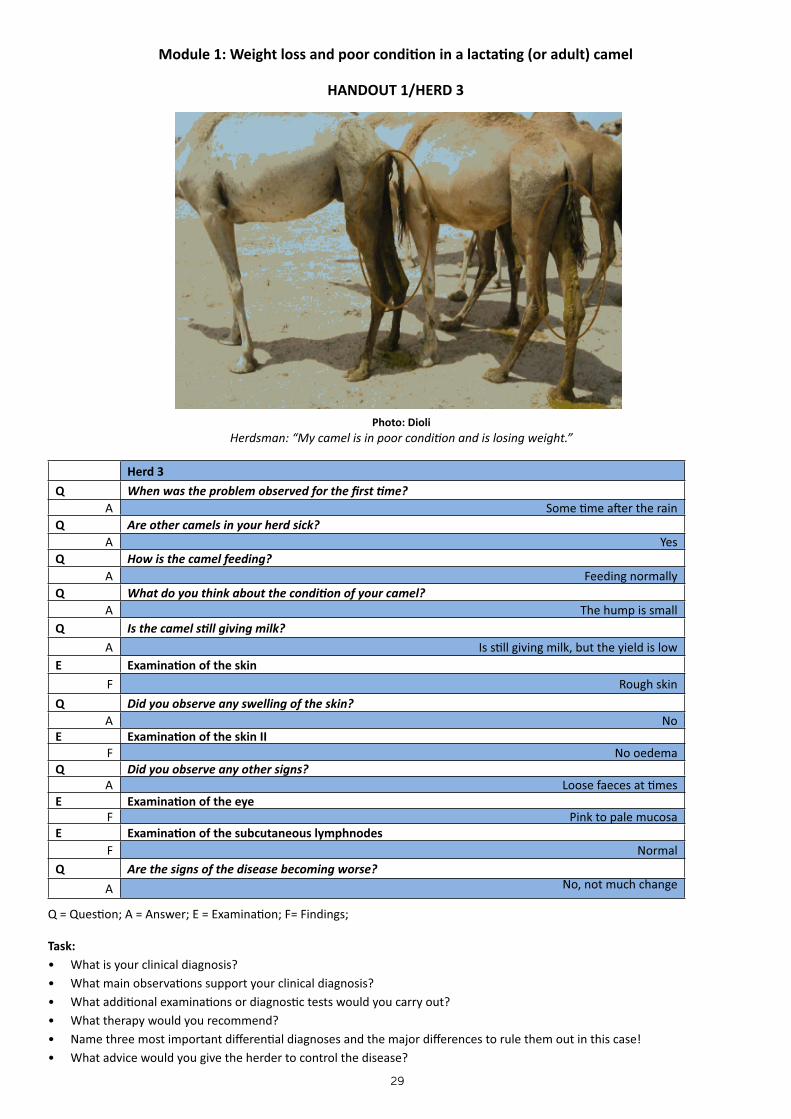
Module 1: Weight loss and poor condi�on in a lacta�ng (or adult) camel
HANDOUT 1/HERD 3
Herd 3
Q When was the problem observed for the first �me?A Some �me a�er the rain
Q Are other camels in your herd sick?A Yes
Q How is the camel feeding?A Feeding normally
Q What do you think about the condi�on of your camel?A The hump is small
Q Is the camel s�ll giving milk?A Is s�ll giving milk, but the yield is low
E Examina�on of the skinF Rough skin
Q Did you observe any swelling of the skin?A No
E Examina�on of the skin IIF No oedema
Q Did you observe any other signs?A Loose faeces at �mes
E Examina�on of the eyeF Pink to pale mucosa
E Examina�on of the subcutaneous lymphnodesF Normal
Q Are the signs of the disease becoming worse?
A No, not much change Q = Ques�on; A = Answer; E = Examina�on; F= Findings;
Task:• What is your clinical diagnosis?• What main observa�ons support your clinical diagnosis?• What addi�onal examina�ons or diagnos�c tests would you carry out?• What therapy would you recommend?• Name three most important differen�al diagnoses and the major differences to rule them out in this case!• What advice would you give the herder to control the disease?
Photo: DioliHerdsman: “My camel is in poor condi�on and is losing weight.”

30
Module 1: Weight loss and poor condi�on in a lacta�ng (or adult) camel
Herdsman: “My camel is in poor condi�on and is losing weight.”Herd 1 Herd 2 Herd 3
Q When was the problem observed for the first �me?
AThree weeks ago
Three months ago Some �me a�er the rain
Q Are other camels in your herd sick?
A No No Yes
Q How is the camel feeding
AFeeding, but rests when other camels are
feedingNot feeding at all, is very sleepy
Feeding normally
Q What do you think about the condi�on of your camel?
AHump is shrinking Hump is gone, abdomen retracted, muscle
atrophyHump is small
Q Is the camel s�ll giving milk?
A Very li�le, has almost stopped giving milk Has stopped giving milkIs s�ll giving milk, but the
yield is low
E Examina�on of the skin
F Skin is dull Very rough skin, tail hair coming off easily Rough skin
Q Did you observe any swelling of the skin?
A Yes, but only in the morning Yes No
E Examina�on of the skin II
F No oedemaPalpable oedema on the lower neck, abdomen
and/or limbsNo oedema
Q Did you observe any other signs?
A Lacrima�onLacrima�on of both eyes, breath and urine
smell musty, like slightly ro�en fruitNo
E Examina�on of the eye
F Lacrima�on, pale or pink mucosa Lacrima�on on both sides, very pale mucosa Pink to pale mucosa
E Examina�on of the subcutaneous lymphnodes
F Not enlarged Enlarged Not enlarged
Q Are the signs of the disease becoming worse?
A Yes It is already very bad since a long �me No, not much change
D What is your clinical diagnosis
D Recent T.evansi infec�on Chronic T.evanis infec�on Gastro-intes�nal parasites
D What main observa�ons support your clinical diagnosis?
D Drowsiness LethargyNormal sensorium and
appe�te
D Addi�onal examina�on / diagnos�c tests
DBlood sample (haematocrit / PCV,
microscopic detec�on of the parasiteBlood sample (haematocrit / PCV, microscopic
detec�on of the parasiteFaecal sample (EPG)
DD Are you sure this animal does not suffer from malnutri�on / mineral deficiency?
DDYes, nutri�onal deficiency would affect more
than one animal in the herdYes, nutri�onal deficiency would affect more
than one animal in the herdNo
DD Any other disease to be considered?
DD Chronic was�ng disease, e.g. Tuberculosis can look like chronic infec�on with parasites
T What therapy do you recommend?
T Trypanocide Trypanocide Anthelminthic
Q = Ques�on; A = Answer; E = Examina�on; F= Findings; D=Diagnosis; DD= Differen�al Diagnosis; T= Treatment

31
Brief on diseases listed under Module 1
Trypanosomosis / Surra In the Greater Horn of Africa this is an endemic and very common disease of camels leading to major economical losses. The subacute and chronic forms typical for Trypanosoma evansi infec�on of camels (Arabic “Surra”) are characterized by sleepiness. Surra is predominantly a disease of adult camels and very uncommon in animals below one year of age.
Epidemiology & Clinical Symptoms:T. evansi is transmi�ed mechanically by blood-sucking flies (Tabanids), including the camel fly (a large shiny reddish coloured fly that sucks blood specifically from camels, Hippobosca camelina).
Transmission occurs over short distance, mostly within the herd or between herds that are close together (e.g. during watering).
Transmission rates and disease incidence are higher during rainy season, when insect vectors are more abundant.
Early symptoms in female camels are sudden drop in milk produc�on and abor�on.
Pale mucous membranes and frequent lacrima�on (both eyes) are typical.
The picture emerging over months is that of a chronic ‘was�ng disease’ characterized by progressive weight loss and deteriora�ng general condi�on.
Camels appear “sleepy” - they sit down and rest while other camels are feeding.
Dull coat, the long hairs at the tail coming off easily.
Immune-suppression, camels become suscep�ble to many other diseases (e.g, pneumonia).
Urine and breath develop a characteris�c smell (signs of ketosis).
In majority of cases death occurs a�er long illness; spontaneous self-cure occurs, but is very rare.
At post-mortem there are no specific lesions, camels are anaemic and have a lot of fluid in the abdomen and also in the thorax; the post-mortem picture is influenced by secondary infec�ons.
In clinically healthy pregnant camels abor�on is very common soon a�er infec�on; see Module ‘Abor�on’.
There is an acute central nervous condi�on in camels caused by T. evansi, see Module ‘Central Nervous Disease’.
Diagnosis:Trypanosoma evansi can be seen in the buffy-coat of centrifuged EDTA blood samples. Trypanosoma evansi infected camels generally show a very low parasitaemia. For this reason a nega�ve blood test does not rule out the presence of T. evansi infec�on. An�gen EIA for T. evansi if available offers a more sensi�ve diagnosis. A low haematocrit (<17%) is an indica�on for T. evansi infec�on. – In the field treatment decisions are mostly based on tenta�ve clinical diagnosis.
Differen�al Diagnosis:Heavy worm infesta�on.
Internal abscesses and tuberculosis.
Malnutri�on.

32
Treatment:Trypanocides are rela�vely toxic. Do not inject dehydrated camels with trypanocides, they can collapse and die. Water and rest such animals before injec�ng the drug.
A combina�on of quinapyramine- salt and pro-salt (Triquin®) is available in a vial containing 2.5 g pale yellow/whi�sh 1. powder that has to be dissolved in sterile water (provided with the drug, otherwise use boiled water that has cooled down). The prepara�on is administered through subcutaneous injec�on at a dose of 0.03 ml per kg live body weight. Treat sick camels as early as possible for high success rates. Chronic cases do not respond well to treatment. Also Triquin does not enter the brain and cannot cure CNS disease caused by T. evansi. The drug is highly irrita�ng and should not be used other than subcutaneously. It is very important to observe clean injec�on prac�ces by using new disposable needles to avoid contamina�on that can lead to abscesses. A total dose of 20 ml should not be exceeded for one camel. The drug may also be used for chemoprophylaxis and protects camels against T. evansi infec�ons for 6 to 12 weeks.
Isomethamidium chloride (Samorin®, Trypamidium®) - This drug is applied as a 1% solu�on, i.e. contents of the 2. 1 g sachet dissolve in 100 ml sterile water (or boiled water that has cooled). Administer the solu�on intravenously or by deep intramuscular injec�on at 0.5 mg/kg live body weight (equal to 1 ml/20 kg). The drug is irrita�ng and toxic and should preferably be used intravenously. Most camels with Triquin® resistant T. evansi infec�on when treated with Isomethamidium do not respond to treatment and may suffer from toxic effects.
Melarsamine hydrochloride (Cymelarsan®) - is available as a ready-made 0.5% solu�on and administered at 0.25 mg/kg3. live body weight by deep intramuscular injec�on into the neck muscles. The drug has only short ac�vity and cannot be used for prophylaxis; only for cura�ve treatment (drug of 2nd choice in cases where resistance of parasites to other Trypanocides is suspected). Cymelarsan crosses the blood-brain-barrier and is the only Trypanocide that can be used to treat acute CNS disease in camels.
Cau�on:Do not use products that contain diminazene aceturate (Berenil®, Veriben®, Diminasan®, Dimaze®, Diminatryp®) because this drug is very toxic for dromedary camels and can cause mortality!
Preven�on:Reduce risk of exposure to bi�ng flies by avoiding highly infested areas where possible. Use pour-on insec�cides when flies are troublesome. In areas known to have high seasonal incidence of Trypanosomosis, administer Quinapyramine (Triquin®) prepara�ons for protec�ve cover (Chemoprophylaxis) following the onset of the rainy season. Chemoprophylaxis is especially important for pregnant camels.
Triquin® Dosage Table
Body wt (kg) Volume (ml) Body wt (kg) Volume (ml) Body wt (kg) Volume (ml)
10 0.3 150 4.5 400 12.0
20 0.6 200 6.0 450 13.5
40 1.2 250 7.5 500 15.0
60 1.8 300 9.0 550 16.5
100 3.0 350 10.5 600 18.0

33
Isomethamidium (1% solu�on) Dosage Table
Body wt (kg) Volume (ml) Body wt (kg) Volume (ml) Body wt (kg) Volume (ml)
100 5.0 300 15.0 500 25.0
150 7.5 350 17.5 550 27.5
200 10.0 400 20.0 600 30.0
250 12.5 450 22.5
Carry out correct weight es�mate and dose carefully – Trypanocide drugs are more toxic than other veterinary drugs that are used in camels.
Gastrointes�nal Helminths For prac�cal purposes two groups, blood-sucking and non-blood-sucking roundworms or helminths can be differen�ated. Both groups include stomach worms, which live in the abomasums, are difficult to see with the eye, but cause more damage than the intes�nal worms. The largest stomach worm in camels is Haemonchus longis�pes, which is a blood-sucker and causes chronic anaemia in camels.
While causing similar clinical symptoms in the animal, helminth species do vary in pathogenicity. When present in large enough numbers they cause general weakness and reduced produc�vity. With moderate infesta�on levels, the animals are alert and feed well but lose condi�on progressively (subclinical infec�ons). Clinical manifesta�ons of helminth infec�ons are especially severe in growing and young camels, in female camels under lacta�on stress and in animals on very poor pastures.
Epidemiology & Clinical Symptoms of Helminth Infesta�on:• Non-bloodsucking helminths cause poor absorp�on of nutrients which manifests itself as diarrhoea, rough hair coat,
bloated stomach, chronic weight loss and stunted growth in young camels; helminths in the abomasum (Trichostrongylus and Ostertagia) are very common and can cause severe problems.
• Bloodsucking helminths (esp. Haemonchus) are a�ached to the wall of the stomach and cause mainly anaemia; diarrhoea can also occur. Haemonchus infesta�on causes severe anaemia, loss in body condi�on and can kill young animals; other symptoms of heavy Haemonchus infesta�on are diarrhoea, weight loss and oedema on the lower limbs.
• Tapeworms (Moniezia) are the biggest gastrointes�nal worms and are easily detectable in the faeces; they can cause obstruc�on and colic in young camel calves.
• Camels become mainly infected when it is wet, but high worm burdens can be carried into dry season and nega�vely affect absorp�on of nutrients and performance under dry condi�ons.
• High worm burdens in camels mostly occur about 3-4 weeks a�er the start of the rains.
• At post-mortem the worms are present in the abomasum and in the intes�nes (e.g. Trichuris), but can be overlooked (esp. Trichostrongylus) if the post-mortem is performed in the field and without applying correct (and �me-consuming!) parasitological post-mortem techniques.
Differen�al Diagnosis:Trypanosomosis (anaemia, oedema, weight loss).Other causes of diarrhoea (viruses, bacteria, coccidia).Chronic was�ng condi�ons (chronic malnutri�on; internal abscesses; tuberculosis).
Treatment:Anthelminthic treatment should target the animals most at risk (young growing camels, lacta�ng females). – Due to underes�ma�on of body weight and under-dosing resistance to anthelmin�cs is becoming more and more common. Certain species of helminths (Haemonchus contortus) are shared between camels and goats/sheep and resistance to anthelmin�cs can

34
occur. It is very important to correctly es�mate the body weight before dosing. – Due to underdosing and/or use of sub-standard drugs previous deworming of camels by the owner may not have been effec�ve!Most dewormers used for ca�le can be used at the same dose rate in camels. Levamisole is not recommended for camels because it shows late and inconsistent ac�on.
Albendazole is widely used in camels at a recommended dosage of 7.5 mg/kg; some dosage examples:
As drench: - 12 ml drench (10%) for a calf of 120 kg body weight.- 25 ml drench (10%) for a young adult of 250 kg body weight.- 50 ml drench (10%) for an adult camel of 500 kg body weight (difficult to administer the total volume).
As bolus:- One 600 mg Bolus for a calf of 120 kg body weight.- Half a 2500 mg Bolus for a young adult of 250 kg body weight.- One 2500 mg Bolus for an adult camel of 500 kg body weight.- One 3000 mg Bolus for heavy adult camel of 600 kg.
Preven�on:Treat groups most at risk early to avoid build up of a too high worm burden; deworm groups of high risk animals when the first animal in the group starts showing clinical signs of helminth infesta�on. There is no need to deworm the whole herd - trea�ng the en�re herd can promote resistance to anthelmin�cs.
Avoidance of prolonged grazing of camels on the same pasture, especially when overstocked with sheep and goats, is the most important prophylaxis.

35
Module 2: Pox - like skin lesions
HANDOUT 1/HERD 1:
Herd 1Q Are all camels in your herd affected?
A Only suckling calves are sickQ Are your camels s�ll feeding normally?
A Yes most of the calves are s�ll suckling their damsQ Have your heard of other herds in this area with similar problems?
A No sure about thisQ When did the problem start?
A About one to two weeks agoE Examina�on of the skin
F Pustules around the mouthE Examina�on of the head and inside the mouth
F A foul breath can be no�ceQ Are the dams or lacta�ng females s�ll giving milk?
A Yes they s�ll give milkQ Have any your camels died since the disease outbreak?
A NoQ Have you observed any other signs or symptoms in the camels?
A No Q = Ques�on; A = Answer; E = Examina�on; F= Finding;
Task:• What is your clinical diagnosis?• What main observa�ons support your clinical diagnosis?• What addi�onal examina�ons or diagnos�c tests would you carry out?• What therapy would you recommend?• Name three most important differen�al diagnoses and the major differences to rule them out in this case!• What advice would you give the herder to control the disease?
Photo: Gluecks
Herdsman: “My camels are not feeding well and have pustules on their skin.”

36
Module 2: Pox - like skin lesions
HANDOUT 1/HERD 1:
Photo: Gluecks
Herdsman: “My camels are not feeding well and have pustules on their skin.”
Herd 2Q Are all camels in your herd affected?
A Mainly young and weaned camels are affectedQ Are your camels s�ll feeding normally?
A No, some of them really have difficul�es in feeding normally
Q Have your heard of other herds in this area with similar problems?A Not sure
Q When did the problem start?A It started one to two weeks ago
E Examina�on of the skinF Pustules around the mouth
E Examina�on of the head and inside the mouth
FThere is a foul breath and the head is swollen including the lips. In some cases the swelling even reaches
the eyes so that the camel cannot open its eyes
Q Are the dams or lacta�ng females s�ll giving milk?A Yes they s�ll give milk.
Q Have any of your camels died since the disease outbreak?A No
Q Have you observed any other signs or symptoms in the camels?A No
Q = Ques�on; A = Answer; E = Examina�on; F= Findings;
Task:• What is your clinical diagnosis• What main observa�ons support your clinical diagnosis?• What addi�onal examina�ons or diagnos�c tests would you carry out?• What therapy would you recommend?• Name three most important differen�al diagnoses and the major differences to rule them out in this case!• What advice would you give the herder to control the disease?

37
Module 2: Pox-like skin lesions
HANDOUT 1/HERD 3:
Herd 3Q Are all camels in your herd affected?
A All the young camels are sick and many of the old onesQ Are your camels s�ll feeding normally?
A No, some have stopped feedingQ Have your heard of other herds in this area with similar problems?
A Yes, my neighbour has the same problemQ When did the problem start?
A It started one to two weeks ago.E Examina�on of the skin
FPustules around head, teats and under the tail. Some camels have lesions all over their body with pus which
a�racts the fliesE Examina�on of the head and inside the mouth
F Pustules can be found, but are all over the bodyQ Are the dams or lacta�ng females s�ll giving milk?
A Most of the females have stopped giving milk
Q Have any of your camels died since the disease outbreak?
A Yes, one of my male adult camels died. But this was before I saw any lesions on the other camels.
Q Have you observed any other signs or symptoms in the camels?
AYes, two of my female camels aborted, some of the weaners have difficul�es with breathing, and one camel has a
problem with the udder (mas��s)
Q = Ques�on; A = Answer; E = Examina�on; F= Finding;
Task:• What is your clinical diagnosis?• What main observa�ons support your clinical diagnosis?• What addi�onal examina�ons or diagnos�c tests would you carry out?• What therapy would you recommend?• Name three most important differen�al diagnoses and the major differences to rule them out in this case!• What advice would you give the herder to control the disease?
Photos: Dioli
Herdsman: “My camels are not feeding well and have pustules on their skin.”

38
Module 2: Pox - like skin lesions
Herdsman: “My camels are not feeding well and have pustules on their skin.”
Herd 1 Herd 2 Herd 3
Q Are all camels in your herd affected?
A Only suckling calves are sickMainly young and weaned camels are
affectedAll the young camels are sick and many
of the old ones
Q
Are your camels s�ll feeding normally?
AYes most of the calves are s�ll
suckling their damsNo, some of them really have difficul�es in feeding normally
No, some have stopped feeding
Q
Have your heard of other herds in this area with similar problems?
A Not sure about this Not sure about thisYes, my neighbour has the same
problem
Q
When did the problem start?
A About one to two weeks ago About one to two weeks ago About one to two weeks ago
E
Examina�on of the skin
F Pustules around the mouth Pustules around the mouth
Pustules around head, teats and under the tail. Some camels have lesions all
over their body with pus which a�racts the flies
E
Examina�on of the head and inside the mouth
F A foul breath can be no�ced
There is a foul breath and the head is swollen including the lips. In some cases the swelling even reaches the
eyes so that the camel cannot open its eyes
Pustules can be found, but are all over the body
Q Are the dams or lacta�ng females s�ll giving milk?
A Yes they s�ll give milk Ques�on not applicableMost of the females have stopped
giving milk
Q
Have any of your camels died since the disease outbreak?
A No NoYes, one of my male adult camels died. But this was before I saw any lesions on
the other camels
Q
Have you observed any other signs or symptoms in the camels?
A No No
Yes, two of my female camels aborted, some of the weaners have difficul�es with breathing, and one camel has a
problem with the udder (mas��s)
Q = Ques�on; A = Answer; E = Examina�on; F= Findings; D= Diagnosis; DD= Differen�al Diagnosis; T= Treatment; C= Control

39
D What is your clinical diagnosis
D Most likely Orf
Most likely Orf Camel Pox
D What main observa�ons support your clinical diagnosis?
DOnly calves and weaners affected,
but no other herds.Only calves and weaners affected, but
no other herds.
All age groups affected; addi�onal symptoms like abor�on, respiratory
distress and sudden death; other herds also affected
D Addi�onal examina�on / diagnos�c tests
D
Skin lesion can be examined with electron microscope if available
DD Any other disease to be considered?
DDOther skin condi�ons, however they usually do not cause fever.
Anthrax can cause the swelling of the head, but no skin lesions found.
T What therapy do you recommend?
TKeep lesion so� with Vaseline or milking fat, use an�sep�c mouth wash, in severe cases inject with penicillin &
streptomycin and use oodine or gen�an violet spray for the skin lesions. Treatment is only symptoma�c.
C What control measures do you recommend?
CIdeally camels should get infected at an early age. The progression of
the disease will then be milder.
Ideally camels should get infected at an early age. The progression of the
disease will then be milder.
A vaccine against camel pox does exist (Ducapox®) however it is not available
in East Africa.
In general infected animals, or an infected herd should be kept separated from other herds. Rest and good nutri�on should be ensured as well as no addi�onal stress factors (e.g. trekking long distances, limited water and food etc.)
Q = Ques�on; A = Answer; E = Examina�on; F= Findings; D= Diagnosis; DD= Differen�al Diagnosis; T= Treatment; C= Control
40
MODULE 2
HANDOUT 2
LEAD SYMPTOM: POX-LIKE SKIN LESIONS
DISEASES COVERED:POXORF

41
Brief on Diseases Listed under Module 2
Camel Pox Camel Pox is endemic in camel - keeping regions and are caused by Orthopoxvirus cameli, which is specific to and affects only camels. Short distance transmission between and within camel herds is mainly through inhaling, but the virus also enters the body through skin injuries and/or through insect bites. – Camel Pox severely disrupts reproduc�on and milk produc�on in the herd!
Epidemiology & Clinical Symptoms:• Due to the lifelong immunity in all recovered animals, successive regional Camel Pox outbreaks are separated by several
Pox-free years. During epizoo�cs the spread of Pox in a wider region is slow and mainly through contact. The disease is
par�cularly dangerous in isolated camel herds that have not been part of the regular Pox infec�on cycles (adults, sub-
adults and young are all fully suscep�ble!)
• During rainy season wet and dirty condi�ons aggravate Pox disease due to more frequent transmission of bacteria
(abundance of bi�ng flies) causing severe secondary infec�ons. Pox lesions start as small red patches; they swell and
become liquid - filled pustules with a depressed centre (= the Pox), these then rupture and turn into blisters (it is at this
ruptured stage that most lesions are seen on the skin).
• Acute swelling of the head can occur before any Pox lesions appear on the skin; this causes breathing problems.
• Mild form: Pox found only around nose, mouth, eyes and under the tail, recovery without any problems.
• Severe form: High fever, the animal is very dull and almost completely off feed, swollen lymph nodes; generalized
Pox lesions appear all over the skin surface on all parts of the body; secondary infec�on of the Pox lesions by bacteria
(transmi�ed by flies!) result in camels developing mul�ple purulent skin lesions, which can lead to extreme weakness
and some�mes death; healing can take 4-6 weeks.
• Especially in young camels, Pox lesions also develop inside the respiratory tract and can lead to secondary bacterial
pneumonia and death if not treated early with an�bio�cs; in severe outbreaks, 3 out of 10 infected young camels can
die from secondary infec�ons.
• Pox lesions on the teats make milking difficult which regularly leads to mas��s; almost all pregnant females abort.
• Peracute form (only seen in adults): Severe swelling of the head and throat, leading to rapid death (anaphylac�c shock
with oedema, asphyxia and death occur within less than 24 hours), no skin lesions.
Differen�al Diagnosis:Orf - mild Camel Pox and Orf look exactly the same!
Mange, Ringworm and Contagious Skin Necrosis can also result in purulent skin lesions and produce confluent scabs, but no Pox lesions.
Treatment:There is no specific treatment for Pox virus; in severe cases especially when young camels start showing signs of bacterial pneumonia or when skin lesions become purulent Penicillin-Streptomycin (daily injec�on for 5 days) or Oxytetracycline 20% (long-ac�ng, repeat injec�on on day 4) should be used for symptoma�c treatment. If available, also inject vitamin A (normally available only as vitamin ADE combina�on), which helps in recovery. Petroleum jelly should be applied to the lesions especially around the mouth the keep them so� and prevent them from cracking and prevent further secondary bacterial infec�on.
Preven�on:Vaccina�on is possible. There is a commercial vaccine, which is available in the UAE but not in East Africa (Ducapox®, two successive vaccina�ons confer lifelong immunity). When a camel herd is going through a Pox outbreak, ensure rest and good nutri�on and avoid all stress. During outbreaks avoid all contact with other camel herds.

42
Orf (Contagious Ecthyma) Orf is endemic in all camel keeping regions. It is caused by Parapoxvirus ovis and is a typical and very common disease of suckling camel calves. Orf can occasionally also occur in adults. It is more severe in older animals than in young calves.
Epidemiology & Clinical Symptoms of Orf:• Some Parapoxvirus ovis strains are shared between lambs, kids and camel calves and outbreaks in all three species can occur
simultaneously.• Virus transmission is sustained via healthy carrier camels present in the herd. Orf outbreaks occur mostly in rela�on to
calving cycles and affect especially suckling camel calves, elder calves and weaners below three years of age. It can occur in adult camels as well.
• The general picture is characterized by Pox-like swellings on the head, around the mouth, lips and nose. – Generalised Orf can occur in weak animals.
• Before skin lesions appear there can be acute severe swelling of the head, especially of the lips, causing breathing problems and completely preven�ng the animal from suckling or feeding.
• From the start the calves lacrimate and are very dull.• The swellings and nodules turn into blisters that look exactly like Pox lesions.• The Pox-like lesions then form scabs that can become confluent on the head.• The same lesions as on the skin also appear inside the mouth and nose.• In severe cases Pox-like lesions can spread to the alimentary tract (seen in the oesophagus and stomach at post-mortem).• In rela�on to secondary bacterial infec�ons other symptoms include s�nking breath and swollen lymph nodes.• Throughout the clinical illness calves have serious difficul�es in suckling and feeding.• In mild Orf, only a few blisters/scabs form around the mouth and heal quickly. • Adult camels show swollen, oedematous heads, especially around lips and eyes together with the Pox-like lesions. They have difficul�es in breathing and feeding and lose condi�on.
Differen�al Diagnosis:• Camel Pox, although Orf and Pox present a somewhat different epidemiological pa�ern the two diseases cannot be
differen�ated on clinical grounds.• Contagious Skin Necrosis, Ringworm and Mange all do not cause Pox-like lesions, but can form confluent skin scabs.
Treatment:Severe cases should be treated with penicillin-streptomycin daily un�l recovery (at least for 5 consecu�ve days). Good nursing and an�sep�c mouth washes (e.g. iodine) are very important. Petroleum jelly should be applied on the affected areas to so�en the skin. Sick calves must be assisted to suckle if necessary, including bo�le feeding in extreme cases. Injec�ng vitamin ADE helps with the recovery.
Preven�on:Prac�cally every camel will become infected by Orf once in its’ life�me. There is no specific vaccine for camels. Camel calves and weaners in good condi�on recover fast. Good management of lacta�ng mothers, allowing calves to suckle enough milk and �mely deworming of elder calves and weaners are important.

43
MODULE 3
HANDOUT 2
LEAD SYMPTOM: RESPIRATORY DISTRESS
DISEASES COVERED:NASAL BOT FLYTUBERCULOSIS
PNEUMONIA (ACUTE, CHRONIC)VIRAL INFECTIONS (INFLUENZA LIKE)
BACTERIAL INFECTIONS

44
Brief on Diseases Listed under Module 3
Respiratory DiseaseKnowledge on respiratory diseases of camels is limited to outbreak reports and remains sketchy to date. Similari�es with other livestock species do exist and camels share respiratory pathogens with ruminants and with equines. One has to be careful not to go too far in assuming that all details of respiratory diseases established in other livestock species also apply to camels in the same way.
Respiratory pathogens described in camels include:• Parainfluenza virus 3: serological evidence in Africa and Middle East and suspected involvement in respiratory camel disease outbreaks in Ethiopia and in Somalia.• PPR virus: serological evidence, implicated in respiratory disease outbreaks in Ethiopia and in Sudan.• Adenovirus: only serological evidence• Bacterial pathogens described in camels include: beta-haemoly�c Streptococci, Staphylococcus aureus, Klebsiella spp., Pasteurella spp., Mannheimia spp., Mycobacterium spp., Burkholderia pseudomallei.
(Lungworms of camels only play a role in cold parts of Asia.)
The following sub-division of respiratory disease syndromes is based on prac�cal clinical considera�ons.
1. Infec�on of the Upper Respiratory Tract
Clinical & Epidemiological Features:• Spread within the herd is quick and a large number of animals is affected within a short period (1-2weeks), some outbreaks
probably caused by Parainfluenza virus 3.• Main symptoms are sneezing, serous secre�on from the nose and conjunc�vi�s, also mild cough.• Animals develop fever and may be off feed for a short �me, most will resume feeding while s�ll recovering.• In the absence of clima�c (wet & chilly) and/or nutri�onal stress (emaciated animals coming out of a drought) recovery is
mostly swi� and complete in less than two weeks (per individual case).• Secre�on from the nose may turn purulent, coughing become more severe and breathing painful due to secondary bacterial
infec�ons; bacteria involved include saprophy�c Streptococci that are very common in the nasopharynx of healthy camels; animals that develop secondary infec�ons remain dull and feed only very li�le or are completely off-feed.
Treatment:It is important to rest camel herds during outbreaks of respiratory disease, driving them at most only over short distances. There is no specific treatment for viruses affec�ng the respiratory tract. In cases with prolonged recovery and signs of secondary bacterial infec�on an�bio�c therapy is indicated. For Kenya, based on an�bio�c sensi�vity tes�ng of isolates from the respiratory tract of camels and on empirical evidence from treatment of respiratory infec�ons in camels, Penicillin-streptomycin (treatment for at least 5 days) appears to be the drug of choice. Other drugs that have been successfully used for trea�ng respiratory disease in camels are amoxycillin (long-ac�ng) and 20% oxytetracycline (Long-Ac�ng).
Camel Nasal Bo�ly (Cephalopina ��llator)Maggots of this fly commonly inhabit the nose and nasal sinuses of camels. Heavy infesta�on can lead to conges�on of the nasal cavity, blockage of the sinuses and severe rhini�s. Sneezing and secre�on of thick pus are seen especially in the morning but do not affect feeding behaviour and condi�on of the camel in a significant way. In severe cases parenteral treatment with ivermec�n is efficient, but expensive.
45
2. Acute Pneumonia
Clinical & Epidemiological Features:• Cases seen mostly in immuno-incompetent (calves) or immuno-compromised (T. evansi, PPR) individuals o�en as a sequel
to acute respiratory infec�on; infec�on progresses from upper to lower respiratory tract: bronchi�s → bronchopneumonia; manifested as painful coughing, increasing dullness and accelerated/laboured breathing.
• Affects all age groups but more common in calves and weaners.• Poor physical condi�on especially when combined with clima�c stress (sudden drop in temperatures, wet and chilly
condi�ons at onset of rains), Pox, also PPR, chronic Trypanosoma evansi infec�on and high worm burden all predispose camels to bacterial pneumonia.
Treatment:Pneumonia can progress fast to reach a fatal state. Affected camels should be treated early with:- Penicillin-streptomycin (once daily for 3 to 5 days)or- Amoxycillin long-ac�ng (every second day)or- 20% Oxytetracycline long-ac�ng (repeat on fourth day).
There are no pneumonia vaccines for camels. Avoidance of the predisposing factors listed is important.
Aspira�on PneumoniaAccidental introduc�on of fluid into the lungs (incorrect drenching with dewormer or rehydra�on fluid) can cause sudden severe inflamma�on of the lung. O�en very difficult to treat.
3. Chronic Pneumonia
Clinical & epidemiological features:• Individual cases mostly seen in old animals.• Characterized by painful chronic cough and progressive loss of condi�on.• Specific causes include Tuberculosis, which is not common in camels kept under extensive management but can become a
problem where animals are kept under crowded condi�ons; Tuberculosis is a dangerous zoonosis.• Corynebacteria have occasionally been found in lesions of the respiratory tract of camels.• Burkholderia pseudomallei (“Melioidosis”) causes chronic Pneumonia, which presents itself as a was�ng disease in camels;
the pathogen occurs in the tropics & subtropics but has so far not been reported from Africa; Melioidosis is a dangerous zoonosis with extremely high fatality rates in humans!
• Chronic Pneumonia may occasionally present as a long-standing respiratory infec�on in younger animals as a result of untreated / inefficiently treated acute Pneumonia.
Treatment:Chronic lung disease is resistant to an�bio�c treatment. Transient improvements seen during treatment are followed by relapse a�er the end of the an�biosis. Both, Tuberculosis and Melioidosis cannot be treated and are very difficult if not impossible to confirm in the live animal. Old camels with chronic lung disease should be killed (or slaughtered) under controlled condi�ons (presence of a veterinarian, taking precau�ons to avoid exposure of humans). If suspicious lung lesions are seen the carcass must be immediately destroyed. Samples should be taken to a laboratory for confirma�on.

46
MODULE 4HANDOUT 2
LEAD SYMPTOM: SICK CALF
DISEASES COVERED:TICK PARALYSIS
DIARRHOEA IN SUCKLING CAMEL CALVESLACK OF COLOSTRUM & MECONIUM RETENTION
PERI-ARTHRICULAR ABSCESSES & NAVEL ILL
47
Brief on Diseases Listed under Module 4Tick ParalysisEpidemiology:The disease occurs in suckling calves. First symptoms are seen about one week a�er the �cks (nymphs) have a�ached. Especially Hyalomma nymphs (“white �cks” a�ached mainly to the skin under the long hair in front of the hump) inject a salivary toxin into the blood while feeding. Older camels are normally immune against �ck toxin.
Clinical Symptoms:The toxin causes hind leg paresis, fast breathing, swea�ng, ataxia and rapid death. Death can occur within a few hours(!) - otherwise within 1-2 days.
Treatment:Manual removal of the �cks is followed by fast improvement of the clinical condi�on and full recovery. Time is of the essence! Do not treat sick calves with acaricide, rather start removing �cks immediately. Suppor�ve therapy (Cor�sone, Vitamin B) can help.
Preven�on:Apart from the risk of �ck paralysis, heavy infesta�on of calves with �cks causes general weakness, anaemia, mul�ple skin abscesses and stunted growth. Advise owner on need to reduce �ck burden in the herd. Spray at least the calves against �cks (Knapsack).
Diarrhoea in Suckling CalvesDiarrhoeaDiarrhoea is the main cause of pre-weaning losses in camel calves and affects especially animals aged from birth up to 12 weeks of age. It has been studied in different camel keeping regions and some of the intes�nal pathogens common in domes�c ruminants have also been iden�fied in camel calves. Salmonella spp. have been found in all camel popula�ons studied and are very significant pathogens, causing diarrhoea and Sep�caemia in camel calves. In addi�on Isospora orlovi, a pathogen of carnivores, birds, pigs and humans, also plays an important role in diarrhoea of suckling camel calves.The intes�nal pathogens described in camel calves include:- Salmonella- E. coli (incl. Capsular type K99)- Klebsiella pneumoniae- (Clostridia - sporadic cases in weaners)- Rota and Corona virus- Coccidia – play only a minor role in suckling calves, but can cause diarrhoea in older calves, weaners and adults- Isospora
Clinical & Epidemiological Features:• Diarrhoea occurs most frequently in camel calves 1 - 10 weeks old.• Ini�al symptoms include frequent passage of loose faeces (watery, bloody, pasty, with pieces of mucosa, some�mes also
smelly), followed by very li�le if any passage of faeces and constant pressing; soiled hind legs.• Dehydra�on symptoms, which rapidly intensify over �me, are: complete inappetence, dullness, weakness, eyes sink
deep into the socket, cold nose, cold skin surface, raised skin fold does not slide back, inability to stand → death due to dehydra�on.
• Dehydrated calves that cannot stand up any more are about to die.
Treatment:• Pastoralists normally withhold or severely limit milk intake of diarrheic calves which aggravates the calf’s condi�on severely.• The most important treatment measure regardless of the cause of diarrhoea is replacement of lost fluid and electrolytes
(oral rehydra�on, see below); oral rehydra�on treatment is only successful if the diarrheic calf can s�ll stand and suckle!

48
• If available, repeated i.v. or s.c. administra�on of electrolyte-glucose solu�ons can be a�empted in severely dehydrated recumbent calves, but most will s�ll die.
• An�bio�c therapy targe�ng the infec�ous agent is indicated for bacterial Sep�caemia (esp. for Salmonella) but has li�le effect on intes�nal infec�ons, which are mostly self limi�ng; an�bio�c sensi�vity tests are useful in providing guidance on the best choice of drugs, esp. for Salmonella.
• Sulphonamide-TMP combina�ons, o�en used orally for diarrhoea treatment, are contraindicated in dehydrated calves.• Coccidial infec�ons are self-limi�ng, an�-coccidial drugs have not been tested specifically for camels; most have no effect on;
one coccidiosta�c (Salinomycin) is extremely toxic for camels.
Where ready-made rehydra�on salt mixes are not available the following rehydra�on formula can be used:• Mix 5 tablespoons of sugar and 1 tablespoon of salt with 2 litres of clean water (boil water and let it cool down before
mixing); instead of 5 tablespoons sugar one can also use 5 tablespoons of honey.• In addi�on, pulverised charcoal (to absorb and remove toxins from the intes�ne) can be added to the rehydra�on fluid: 2
handfuls of charcoal powder per litre, passed through a sieve before giving it to the calf.
Oral rehydra�on protocol• A 30 kg calf needs minimum 3 litres of oral rehydra�on fluid per day (minimum 1 litre for 10 kg body weight per day).
Rehydra�on fluid must be given orally in small por�ons at the rate of max. 0.5 litre at a �me.• Rehydra�on fluid should be given for 5 days.• Milk may be withheld for the first 24 hours but not for longer than 36 hours; from the second day on small amounts of milk
should be fed.• Keeping calves in the shade slows down dehydra�on.
Preven�on:• Diarrhoea treatment is a race against �me, to prevent camel calves from entering into severe dehydra�on camel owners
must be educated to start oral rehydra�on treatment as soon as diarrhoea symptoms have been observed.• Camel calves are born into an environment contaminated with faecal organisms and coccidia / Isospora; the immune status
of the calf and the level of contamina�on with pathogens decide on the outcome of inevitable oral infec�ons in suckling calves.
• Ensure early suckling a�er birth for op�mum transfer of maternal colostral an�bodies.• Minimise environmental exposure of the newborn (frequent reloca�on of the camel enclosure, clean calving area, separate
fresh enclosure for the dam and her newborn).• Oral applica�on of crushed raw eggs: chicken eggs contain acid-fast Immunoglobulin–Y; chicken sharing the same
environment with camels will have an�bodies to faecal organisms present in camel enclosures, which can confer some protec�on.
Lack of Colostrum & Meconium reten�onAs in ruminants, camel calves are not immune-competent at birth and depend on transfer of colostral an�bodies to acquire passive immunity as a protec�on during their first weeks of life.
Clinical & Epidemiological Features:• Some pastoralist communi�es believe that excess colostrum causes diarrhoea; in consequence they restrict or delay
colostrum intake by the newborn calf; this causes low absorp�on of maternal colostral an�bodies or even complete failure of an�body transfer between the dam and the newborn.
• Low immunoglobulin levels in newborn camel calves predispose them to fatal infec�ons with opportunis�c pathogens present in the environment.
• Colostrum also acts as a mild laxa�ve and s�mulates the passage of the first faeces (meconium), hence lack or absence of colostrum can lead to meconium reten�on.

49
Preven�on / Treatment:• Educate camel owners on the absolute need to let newborn calves suckle as early as possible a�er birth for op�mum
transfer of maternal colostral an�bodies• Colostral an�bodies are s�ll absorbed by the newborn on day two postpartum, albeit at lower efficiency; giving colostrum
late is be�er than giving no colostrum at all.• Colostrum from other camels that have calved at roughly the same �me can be used for the newborn in cases where its’ own
mother gives no milk, dies or rejects the calf.• Rectal applica�on of liquid paraffin can assist with passage of the first faeces.
Peri-arthricular Abscesses & Navel illClinical & Epidemiological Features:• Suckling camel calves can develop small external abscesses around the joints where the skin is rubbing on the soil when they
are lying down because they have not yet developed hard skin pads like adult camels.• These abscesses around the joints can become chronic and can spread to the joint capsule, the inside of the joint, tendons
and muscles - resul�ng in inability of the calf to stand and suckle.• Peria-arthricular abscesses appear at three to eight weeks of age and resolve in most cases; some progress and increase in
severity and are s�ll present in camel calves up to nine months old.• If untreated the condi�on can cause severe stun�ng and some�mes death.• Infec�ons of the umbilical cord leading to omphalogen ascending infec�ons (omphalophlebi�s, ‘navel ill’) are by far less
common in newborn camel calves as compared to newborns of other livestock species.
Preven�on / Treatment:• In cases where the process is not self limi�ng and where the abscess capsule is so�, make an incision with a clean scalpel at
a low point to allow the pus to drain out.• Flush with hydrogen peroxide (3%), iodine or gen�an violet and repeat flushing for several days.• In severe cases, especially in calves with mul�ple abscesses around the joints, treat with penicillin-streptomycin (daily
injec�on for 5 days).• For navel ill the prophylaxis and treatment are the same as in other livestock.
50
MODULE 5
HANDOUT 1LEAD SYMPTOM: CENTRAL NERVOUS PROBLEMS
DISEASES COVERED:RABIES
VIRAL AND BACTERIAL MENINGITISCAPPARIS TOMENTOSA POISONING
CENTRAL NERVOUS FORM OF TRYPANOSOMOSIS

51
Brief on Diseases Listed under Module 5Central Nervous Problems
Knowledge on CNS disease of camels is very limited. It can be expected that some CNS infec�ons known from other livestock also affect camels. - Taking as complete a case history as possible is extremely important for CNS disease. Clinical symptoms vary as the disease progresses and only very few clinical signs may be observed during the visit.
RabiesEpidemiology:Rabies virus can infect all warm blooded vertebrates and is transmi�ed via infec�ve saliva. Camels contract Rabies virus when they are bi�en by rabid stray dogs or rabid predators (e.g. hyena); they can also become infected through bites from a rabid camel within the herd.
Clinical Symptoms & Diagnosis:‘Silent Fury’ is uncommon in camels who mostly exhibit the ‘Raging Fury’. The symptoms listed below are not all seen in individual cases.A�er an incuba�on period from 3 weeks to 6 months (can be longer!) the signs seen in ‘Raging Fury’ are:• Ini�al restlessness, stops feeding and drinking (lasts 1-2 days)• Excita�ve stage - bi�ng, snapping, itching, self-mu�la�on, hypersexual behaviour in bulls (this stage lasts 1 -3 days)• Paraly�c stage – hypersaliva�on, tremor, almost constant a�empts to yawn, lateral recumbency with flailing legs (lasts up to
two days)• Death within 7 (possibly up to 10) days
In cases of ‘Silent Fury’ the only clinical signs are weakness, trembling, sternal recumbency and death. Rabies cases cannot be differen�ated from meningi�s on clinical grounds and may go unno�ced. This puts the owner, the public and the examining vet at an extreme risk unless the head/brain is taken to a laboratory a�er post-mortem to be examined for Rabies (iden�fica�on of Negri bodies in the Ammon’s horn).Note: in camels, Negri bodies may some�mes only be present in the medulla oblongata→ always include the neck/spinal cord samples for laboratory inves�ga�on!!!Abnormal objects found in the Rumen at post-mortem can be an indica�on of Rabies.
Treatment:There is no treatment and no spontaneous recovery. A rabid camel must be killed (not slaughtered)! without exposing humans to infec�on risk and without destroying the brain/skull. Repeatedly vaccinate all in contact livestock and humans against Rabies (follow vaccine manufacturers instruc�ons for post-exposure vaccina�on)! Rabies virus is excreted by the infected animal several days before first clinical symptoms appear. Also vaccinate livestock and humans who were in contact with the rabid camel during the week before it fell sick.
Preven�on:Vaccina�on. Unless followed up by a booster injec�on, one single vaccina�on only protects for under one year.
Viral and Bacterial Meningi�sEpidemiology and Clinical Symptoms:Listeria have been isolated in New World camelids fed on silage but not from camels. No specific viral or bacterial meningi�s pathogens have been confirmed in camels. Clinical cases of meningi�s are seen sporadically in camels in the field. Depending on the part of the brain affected such cases can present as unilateral or par�al paralysis and normally result in recumbency.
It is of imminent importance to conclusively differen�ate and confirm any form of CNS disease from Rabies! The clinical picture of ‘Silent Fury’ and other bacterial/viral meningi�s can look exactly the same.

52
Treatment A�empt:High doses of penicillin-streptomycin or other an�bio�cs can be injected daily for 5 days.
Hyda�d CystsCysts of the Echinococcus granulosus tapeworm of dogs and other carnivores are found regularly in the organs of camels (esp. in the liver and lung), including in the brain. In rela�on to the localisa�on of the cyst in the brain a commonly seen symptom is circling movement (“turning sickness”). Bacterial abscesses, including Tuberculosis, localised in the brain may cause a similar clinical picture.There is no efficient treatment for Hyda�d cysts. Tapeworm cysts found at slaughter or during post-mortem are a major public health hazard and must be burned or buried to disrupt the tapeworm transmission cycle.
Taenia mul�ceps cysts (Coenurus cerebralis) can occasionally be found in the brains of camels causing dizziness and staggering.
Capparis Tomentosa PoisoningEpidemiology:Under very dry condi�ons livestock may have to be grazed in areas normally not browsed by camels. The most common toxic plant found in such areas of East Africa is the Capparis tomentosa tree, which contains poison in its’ fruits, flowers and leaves. Apart from severe drought situa�ons, older camels normally avoid this tree, but young camels may ingest the fruits when they are in season.
Clinical Symptoms:These include sideways bending of the neck, difficulty in walking, paralysis and emacia�on in prolonged cases.
Note:Many other plant poisonings are known locally by experienced camel pastoralists.
Treatment:Stop camels from feeding on the tree. Various tradi�onal treatments are applied by camel pastoralists. There is no clear informa�on on the efficacy of general an�- poisoning treatment.
Preven�on:Advise camel owners to avoid grazing in areas where the tree is known to occur. In case such areas have to be used for grazing during dry season or during drought the herdsmen must prevent camels from feeding on this tree. If herdsmen are negligent or inexperienced whole groups of camels, especially subadults and weaners, can die from plant poisoning.
Wry NeckThis is a clinically similar syndrome of unclear ae�ology, which is commonly found in camels in East Africa. It is not related to feeding on any specific plant and causes only isolated cases. Most likely it is related to physical trauma. The typical clinical picture includes:• Twisted neck, o�en double bent (S-shape)• Only severely with feeding in severely affected cases• Camels may recover spontaneously, even overnight!Tradi�onally it is treated by branding of the neck. Unconfirmed reports indicate that vitamin B injec�on or bakers yeast given as an oral drench have a beneficial effect.
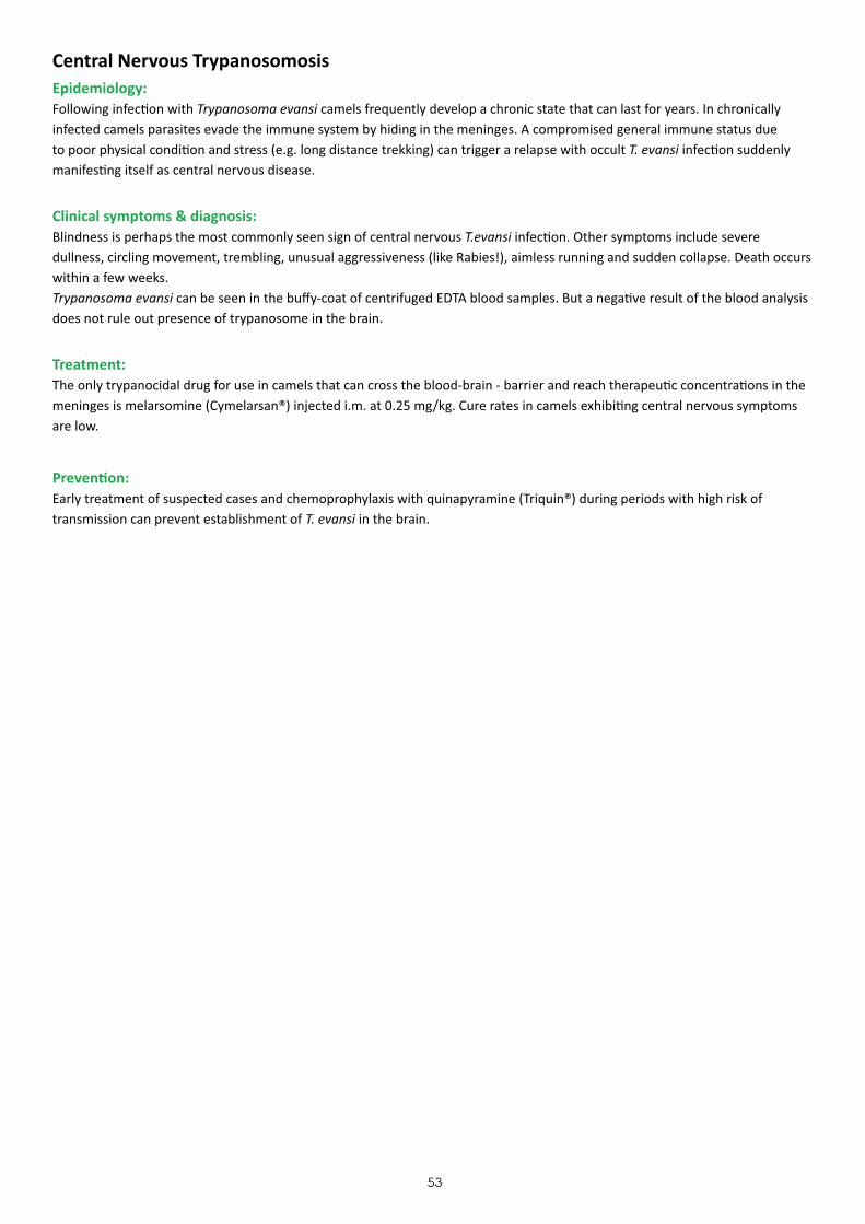
53
Central Nervous TrypanosomosisEpidemiology:Following infec�on with Trypanosoma evansi camels frequently develop a chronic state that can last for years. In chronically infected camels parasites evade the immune system by hiding in the meninges. A compromised general immune status due to poor physical condi�on and stress (e.g. long distance trekking) can trigger a relapse with occult T. evansi infec�on suddenly manifes�ng itself as central nervous disease.
Clinical symptoms & diagnosis:Blindness is perhaps the most commonly seen sign of central nervous T.evansi infec�on. Other symptoms include severe dullness, circling movement, trembling, unusual aggressiveness (like Rabies!), aimless running and sudden collapse. Death occurs within a few weeks.Trypanosoma evansi can be seen in the buffy-coat of centrifuged EDTA blood samples. But a nega�ve result of the blood analysis does not rule out presence of trypanosome in the brain.
Treatment:The only trypanocidal drug for use in camels that can cross the blood-brain - barrier and reach therapeu�c concentra�ons in the meninges is melarsomine (Cymelarsan®) injected i.m. at 0.25 mg/kg. Cure rates in camels exhibi�ng central nervous symptoms are low.
Preven�on:Early treatment of suspected cases and chemoprophylaxis with quinapyramine (Triquin®) during periods with high risk of transmission can prevent establishment of T. evansi in the brain.
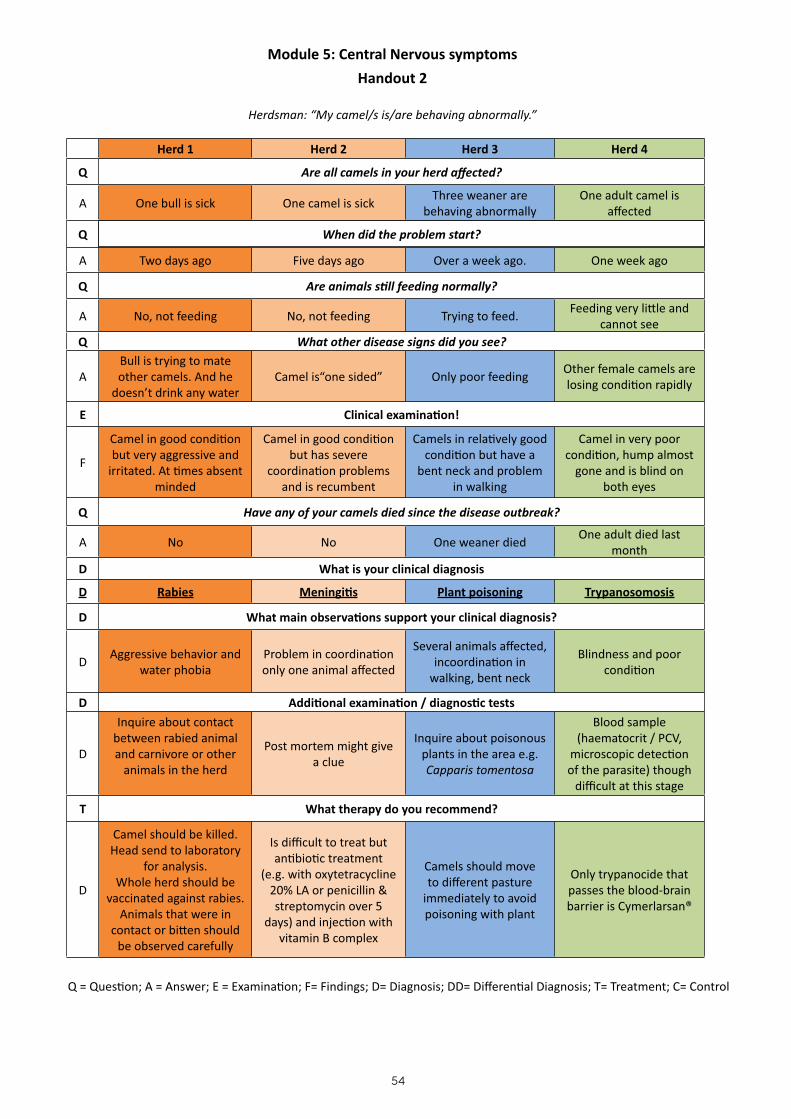
54
Module 5: Central Nervous symptoms
Handout 2
Herdsman: “My camel/s is/are behaving abnormally.”
Herd 1 Herd 2 Herd 3 Herd 4
Q Are all camels in your herd affected?
A One bull is sick One camel is sickThree weaner are
behaving abnormallyOne adult camel is
affected
Q When did the problem start?
A Two days ago Five days ago Over a week ago. One week ago
Q Are animals s�ll feeding normally?
A No, not feeding No, not feeding Trying to feed.Feeding very li�le and
cannot seeQ What other disease signs did you see?
ABull is trying to mate other camels. And he
doesn’t drink any waterCamel is“one sided” Only poor feeding
Other female camels are losing condi�on rapidly
E Clinical examina�on!
F
Camel in good condi�on but very aggressive and
irritated. At �mes absent minded
Camel in good condi�on but has severe
coordina�on problems and is recumbent
Camels in rela�vely good condi�on but have a
bent neck and problem in walking
Camel in very poor condi�on, hump almost
gone and is blind on both eyes
Q Have any of your camels died since the disease outbreak?
A No No One weaner diedOne adult died last
monthD What is your clinical diagnosis
D Rabies Meningi�s Plant poisoning Trypanosomosis
D What main observa�ons support your clinical diagnosis?
DAggressive behavior and
water phobiaProblem in coordina�on only one animal affected
Several animals affected, incoordina�on in
walking, bent neck
Blindness and poor condi�on
D Addi�onal examina�on / diagnos�c tests
D
Inquire about contact between rabied animal and carnivore or other
animals in the herd
Post mortem might give a clue
Inquire about poisonous plants in the area e.g. Capparis tomentosa
Blood sample (haematocrit / PCV,
microscopic detec�on of the parasite) though
difficult at this stage
T What therapy do you recommend?
D
Camel should be killed. Head send to laboratory
for analysis.Whole herd should be
vaccinated against rabies.Animals that were in
contact or bi�en should be observed carefully
Is difficult to treat but an�bio�c treatment
(e.g. with oxytetracycline 20% LA or penicillin & streptomycin over 5
days) and injec�on with vitamin B complex
Camels should move to different pasture
immediately to avoid poisoning with plant
Only trypanocide that passes the blood-brain barrier is Cymerlarsan®
Q = Ques�on; A = Answer; E = Examina�on; F= Findings; D= Diagnosis; DD= Differen�al Diagnosis; T= Treatment; C= Control

55
C What control measures do you recommend?
C
Vaccina�on of the whole herd in areas where
rabies is a big problem.
Avoid known areas with poisonous plants
Treat whole herd with quinapyramide (e.g.
Triquin®) at least twice a year to prevent
infec�on.Use pour-on or spray
with acaricides
Photo: Diolo Photo 1: Camel with terminal signs of rabies (yawning and paralysis).
Photo: Diolo Photo 2: Camel with Capparis tomentosa poisoning, difficul�es in ge�ng up, s�ff walking, s�ffly joints
swollen and emacia�on
Q = Ques�on; A = Answer; E = Examina�on; F= Findings; D= Diagnosis; DD= Differen�al Diagnosis; T= Treatment; C= Control
56
MODULE 6
HANDOUT 1
LEAD SYMPTOM: ACUTE DEATH IN ADULT CAMELS
DISEASES COVERED:ANTHRAX
ACUTE TRYPANOSOMOSISHAEMORRHAGIC SEPTICAEMIA
CAMEL SUDDEN DEATH SYNDROMESNAKEBITES

57
Dead Adult CamelApart from Anthrax, the veterinary knowledge on confirmed causes of acute deaths in adult camels is virtually non-existent. This puts the veterinarian in a rather difficult situa�on when faced with disease outbreaks in the field involving deaths of valuable adult camels. Taking no ac�on at all is not an op�on. Pragma�c decisions have succeeded in curbing outbreaks of unclear ae�ology in the past.
AnthraxEpidemiology:Bacillus anthracis is the cause of a soil-born infec�on that leads to peracute and acute disease in camels. Typically infec�ons occur when camels are grazed in seasonally dry flood plains or when they have to feed on very short grass stubbles during drought. The infec�on is transmi�ed via spores that are released from cadavers of animals that died of Anthrax. Anthrax spores are extremely resistant and can survive for decades in the environment. Anthrax contaminated high risk pastures are o�en well known to pastoralists. Following unusual rain and heavy flooding new pastures can become contaminated when Anthrax cadavers or spores are carried by the water into hitherto clean areas.Anthrax is a very dangerous zoonosis. Humans become infected when they slaughter or dissect Anthrax infected camels. Anthrax spores survive cooking; humans regularly die a�er ea�ng Anthrax contaminated meat.
Clinical Symptoms:In the peracute form sudden death is o�en the only symptom seen in camels.The acute form can show any of the following signs:• Fast, laboured breathing• Trembling• Colic & bloat• Painful swelling on throat & on base of the neck, swelling of other body parts• Most camels die within 2 to 4 days• Tar - like un-clo�ed blood visible on body orifices
Diagnosis:Blood smears and a ligated ear are the easiest and safest to collect and to transport to the laboratory. – Anthrax spores only form a�er the inside of the dead carcass comes into contact with oxygen. To protect yourself and others against Anthrax infec�on and to prevent pasture contamina�on don’t perform post mortem when suspec�ng Anthrax!
Treatment:High doses of penicillin if given immediately a�er the first clinical signs can be successful. Normally such treatment comes too late in the field but can be used to protect clinically healthy camels at risk.Very early intravenous treatment of Anthrax infected humans with high doses of penicillin is be life saving in many cases.
Preven�on:The best prophylaxis is to vaccinate camels annually against Anthrax before moving them into or through Anthrax risk areas. If camels are not vaccinated provide an�bio�c coverage for the whole herd with Penicillin G and move camels out of the Anthrax infected pasture immediately a�er the first case has occurred.- To prevent contamina�on of the pastures and water with Anthrax spores burying the dead camel deep underground or burning it completely is the safest disposal method. O�en this is not feasible in remote loca�ons. Anthrax cadavers, if not cut or ripped open, decontaminate themselves rapidly during decomposi�on. Guard dead camel against dogs and other scavengers for at least two days.Meat from dead camels must not be consumed by humans, not even if well cooked.
Acute TrypanosomosisEpidemiology:Occasionally camels are kept near or even inside Tse-Tse fly infested zones (e.g. entering riverine Tse-Tse fly belt for watering). Tse-Tse - transmi�ed Trypanosoma brucei, congolense, vivax and simiae have all been shown to cause acute deaths in camels. In East Africa certain strains of T. vivax cause an acute haemorrhagic syndrome—Transloca�on of Tse-Tse - transmi�ed trypanosomes via migra�ng infected ca�le and short distance mechanical transmission can occasionally occur outside Tse-Tse infested areas.

58
Clinical Symptoms:Acute febrile disease leading to death within few days. Some�mes haemorrhages in the skin and visible mucous membranes.
Diagnosis:Internal bleeding with petechiae of serosa membranes is typically present at post mortem. Collect blood smear and EDTA blood for confirma�on of the parasite.
Treatment:Treat with trypanocide licensed for use in camels (see Module 1)..
Preven�on:Move animals through Tse-Tse fly infested zones during the night when Tse-Tse flies are inac�ve. Apply chemoprophylaxis (trypanocide) and topical repellent or insec�cide.
Haemorrhagic Sep�caemia (HS)Epidemiology and Clinical Symptoms:In camels the term ‘Haemorrhagic Sep�caemia’ is used for at least two different clinical syndromes.
Swollen GlandsThis syndrome is endemic to certain areas. Typical signs are swollen lymph nodes, especially those of head and neck. Camels are extremely dull, reluctant to move, develop breathing problems and may die a�er 3-5 days. Recovery is very slow. This form occurs regularly in certain areas and herds and affects mainly weaners and sub-adults.
Acute Camel HSThis syndrome resembles clinical HS in ca�le. Sporadic outbreaks occur suddenly in herds that may not have experienced the disease for many years. Reportedly the disease-free intervals can last as long as 7 to 10 years. From the onset of clinical symptoms there are acute and severe breathing problems; camels lie down with their necks stretched out gasping for air. They are unable to stand up and die within less than 24 hours. Some�mes the intes�ne is also affected, resul�ng in bloody or dark faeces. Epistaxis has also been reported. Some outbreaks appear to be triggered by sudden clima�c changes (abrupt transi�on to chilly and wet weather). The disease affects mainly adults in good body condi�on. At post-mortem there are massive haemorrhages in the lungs and/or intes�nes.For both syndromes there are indica�ons that direct contact, esp. sharing of watering points, play a role in transmission.
Treatment:Iden�fica�on of the infec�ous agent is s�ll lacking for both syndromes. Swollen Glands and HS do not respond well to an�bio�c treatment. But immediate an�bio�c coverage for clinically healthy camels at risk has repeatedly curbed HS outbreaks in camels.
Preven�on:Avoid contact with HS affected herds during outbreaks. So far there is no evidence that HS vaccine for ca�le also protects camels.
“Camel Sudden Death” SyndromeEpidemiology:The least understood of all acute deaths in adult camels. Between end 2005 and 2007 a protracted outbreak of what was then named ‘Camel Sudden Death’ moved slowly through the lowlands of Northeast, East and South Ethiopia, the whole of Somalia and North Kenya. The disease syndrome was clearly new to the whole region and has not reoccurred since—Sporadic deaths occurred in affected herds over a very limited period of �me and then stopped.

59
Clinical symptoms:Sudden collapse of animals in best condi�ons, affec�ng the heaviest animals in the herd include: prime breeding bulls, heavily pregnant and lacta�ng females. No clinical symptoms in thin and young camels. ‘Sudden Death’ camels exhibited very sudden difficulty in breathing, collapse, peracute oedema with white froth in the nostrils and death within 1-4 hours a�er the collapse. There were no warning symptoms before and in contact camels remained perfectly healthy. There also were no obvious or consistent post-mortem changes, but sub-epicardial bleeding was observed in several cases.
Treatment:None.
Preven�on:The cardiovascular system seemed to be involved with heavier and metabolically stressed camels being most at risk. The following recommenda�ons were applied during the outbreak• Avoid contact with affected herds.• Avoid stressing pregnant and lacta�ng animals on long treks.• Avoid sudden intake of large amounts of salt and/or water.
Snakebites do occasionally kill adult camels. Look for bite-marks. Some�mes the herdsman may have seen a snake close to his camels.

60
Module 6: Death in Adult Camels
Handout 2
Herdsman: “My camel died suddenly.”
Herd 1 Herd 2 Herd 3 Herd 4
Q How long was your camel sick before it died?
A Two days About a week Just one day Just a few hours
Q Have any other camels died in your herd recently?
A No NoYes, two died in the last
three daysNo
Q Have your camels been browsing in this area for a long �me?
AWe moved here because
of droughtAbout one month now About six weeks now Yes
Q Have you been with your camels here before?
A Yes No, it is our first �me. Yes Yes
Q Have you ever seen such a case before?
A Yes NoYes but a very long �me
agoNo
E Clinical examina�on / observa�on!
F
Camel in good body condi�on.
Unclo�ed blood oozing from nostrils
Camel in good body condi�on.
Bleeding in eyes, visible in the mucosa
Camel in good body condi�on.
Pasty dark faeces but small amount
Camel in good body condi�on.
Foamy froth in nostrils
Q Have you observed anything else in the camels or in the area before the camel died?
A Camel had a swollen neckThere are Tse tse flies in
this areaIt rained heavily about a week ago and it was cold
No
D What is your clinical diagnosis
D Could be AnthraxCould be acute form of
Trypanosomosis
Could be Haemorrhagic Disease Syndrome or
other?
Could be Camel Sudden Death or other?
D What main observa�ons support your clinical diagnosis?
DUn-clo�ed blood and men�oning of swollen
neck
Bleeding in eye/mucosa but difficult
Several animals affected. Rain
No clinical signs beforehand
D Addi�onal examina�on / diagnos�c tests
DBlood smear. DO NOT OPEN THE CARCASS!!! Blood smear Blood smear
Post mortem examina�on only
showing lung oedema and haemorrhages in
endocard
T What therapy/control measures do you recommend?
DNone. Only safe disposal
of the carcass
Inject whole herd with quinapyramide (e.g.
Triquin®) and move from the area
Treat affected animals with oxytetracycline
20% LANot known
Q = Ques�on; A = Answer; E = Examina�on; F= Findings; D= Diagnosis; DD= Differen�al Diagnosis; T= Treatment; C= Control

61
DD What else could cause sudden death in an adult camel?
DD Snake bites can also cause sudden death in adult camels
Photo: DioliPhoto 1: Ca�le that died of Anthrax, classic un-clo�ed blood oozing from nose
Q = Ques�on; A = Answer; E = Examina�on; F= Findings; D= Diagnosis; DD= Differen�al Diagnosis; T= Treatment; C= Control

62
Photo: VSF Germany Photo 6: Camel with nasal froth just before collapsing
and dying
Photo: VSF Suisse Photo 7: Bloody froth found in a camel that died from
Camel Sudden Death Syndrome
Photo: DioliPhoto 3: Tarry faeces found in camels with
Haemorrhagic Disease Syndrome
Photo: Dioli Photo 3: Tarry faeces found in camels with
Haemorrhagic Disease Syndrome
Photo: Dioli Photo 2: Camel dying of Haemorrhagic Disease Syndrome
Photo: Dioli Photo 2: Camel dying of Haemorrhagic Disease Syndrome

63
MODULE 7
HANDOUT 1
LEAD SYMPTOM: SKIN PROBLEM
DISEASES COVERED:RINGWORM
MANGECONTAGIOUS SKIN NECROSIS
LYMPHNODE ABSCESSES
64
Brief on Diseases Listed under Module 5 Skin Problems
Camel skin is thinner than the skin of bovines and prone to micro-trauma and infec�ons. The sensi�vity of the camel skin and the importance of skin disease as a debilita�ng factor in camels compares in some respect to equines. With camels s�ll playing a significant role as baggage animals, healthy skin is a necessity for their use in transport.
RingwormThis fungal skin disease presents as typical circular, expanding and later confluent bold patches. Chalky crusts can be seen on older lesions. There is no itchiness. It is seen mostly in suckling calves and also in weaners. In 90% of cases Trichophyton verrucosum can be isolated as the causa�ve agent. Other species of Trichophyton and Microsporum also occur in camels. Unless calves are in extremely poor condi�on the infec�on is self limi�ng and there is no need for an�-fugal treatment. Low vitamin-A content of camel milk seems to be a predisposing factor. Wet and unhygienic condi�ons can aggravate the clinical course of this normally harmless disease.
MangeEpidemiology:Camel mange is specific to camels and is caused by Sarcoptes scabei var. cameli. Very excep�onally, transmission of camel mange to humans has been reported. The disease is spread via close contact and also through contaminated objects, including trees on which camels like to rub or via contaminated night enclosures. The mite lives in the skin but can survive for some �me in dead skin scabs shed by affected camels, especially under wet condi�ons with li�le sunshine. Mange is more severe in the rainy season.
Clinical Symptoms:The earliest and most persistent clinical symptom is severe itching, especially in the morning. The almost permanent itching is not seen in other skin diseases and results in shorter feeding hours and gradual loss of condi�on in affected camels. Some animals may show almost normal skin, but there is always severe itching, which is much more intensive than normally observed grooming behaviour. Boldness begins mostly on the head (dorsum of the nose) and spreads from there to other parts of the body. A�er several months forma�on of skin scabs and thickening of the skin follow (hyperceratosis: skin develops folds and looks like elephant skin). The mange has now entered its’ chronic stage and is very difficult to treat.
In East African camels mange can be present in an extremely mild form over prolonged periods, the only clinical signs being boldness and itching.Sporadic cases of typical “mangy” looking camels with hyperceratosis in otherwise mange-free herds must not necessarily be infected by mange mites but may be suffering from auto-immune problems and/or chronic deficiency.
Treatment:A single treatment does not eradicate disease from the herd because it only kills adult mites, while mite eggs in the skin hatch within a week. Therefore the treatment of choice is a double s.c. injec�on of 1% ivermec�n. Two doses must be given 8 days apart (note that this treatment interval is specific for camels and is shorter than the interval recommended by the manufacturer for ca�le). Some�mes three treatments at 8 days’ interval may be necessary (very costly!). Ivermec�n does not penetrate into dead skin scabs. In between the two ivermec�n injec�ons, it is important to first wash all camels with a detergent and in par�cular rub off all dead skin scabs (with a brush); then an acaricide must be applied on the skin, best done by spraying manually with a knapsack. A�er washing and acaricide treatment, move camels to a clean fresh enclosure/environment. In cases where ivermec�n is too costly, the following treatment protocol is also efficient if carried out me�culously:
1. Washing with detergent (+ brushing of dead skin); 2. Spraying twice (be�er three �mes!) with acaricide at eight days’ interval and moving camels to new clean boma a�er the
first spraying. Spraying must be done thoroughly such that the whole skin surface is reached by the acaricide, including the head and the inside of the ears.

65
Note that any calves born in between two treatments must also be injected twice and treated with acaricide, the same as for adult camels. One single untreated calf can act as a source of slow re-infec�on for the whole herd; despite spending a lot of money on treatments, there will be a relapse with the mange spreading again throughout the herd within 4—8 weeks.
Note: Because of its’ very long drug withdrawal period, ivermec�n should not be used in lacta�ng camels. If lacta�ng camels are to be treated against mange, washing and 3 x acaricide spraying should be applied (observe milk withdrawal period for acaricide).
Preven�on:Once in the herd, mange is very difficult and very costly to eradicate. A major problem is sharing of grazing and water points with camel herds of uncertain mange status and also introduc�on of mange by clean looking but infected camels from other herds. When introducing new camels into a clean herd, inject them twice with ivermec�n at 8 days’ interval, before allowing them to mix with the clean camels.
Contagious Skin NecrosisEpidemiology:This skin disease of camels was ini�ally thought to be a specific infec�on. But there is strong evidence that the underlying cause is rather a prolonged mineral deficiency. Skin lesions become rapidly super-infected by ubiquitous pathogens (esp. Streptococci and Staphylococci).
Clinical Symptoms:Contagious Skin Necrosis begins as a painful swelling of circumscribed areas of skin, followed by localised alopecia and hardening of the skin star�ng from the centre of the affected area. Subsequently dry scabs evolve, star�ng again from the centre of the lesion. A�er 7 -14 days a sharp demarca�on between dead and living skin begins. If pressure is applied on the centre of the lesion a clear discharge appears along its margins, which rapidly becomes purulent. The central por�on then separates and sloughs away. The subcu�s is not affected. Lesions can be located anywhere on the body, but are more common on the neck, shoulders, flanks and hind quarters. The condi�on can persist for months. A�er healing, a star - shaped scar remains.
Treatment:Feed ad lib quality salt/minerals containing ample NaCl to camels and/or move camels to salty pastures/salty springs. Salt from natural salt pans can be short in NaCl. Clean affected skin area and apply iodine. In severe cases, penicillin-streptomycin can be injected daily for 5 days.
Preven�on:Ensure that camels have regular access to quality mineral salt containing NaCl, salty pasture and/or salty water sources. Move herd to new pasture when first cases occur.Dermatophilus congolensis is some�mes confounded with Contagious Skin Necrosis in the literature. In the East African region Dermatophilosis (also called “cutaneous streptothricosis”) of camels is a seasonal skin problem that occurs in rela�on to humid warm condi�ons. It presents as an exsuda�ve, pustular (ini�al small nodules) derma��s characterised by forma�on of crusty scabs that occur especially in skin areas covered by long hair. Dermatophilosis normally resolves a�er rains subside. Dermatophilus can also be isolated from the skin of the hind quarters of female camels kept wet by urina�ng.
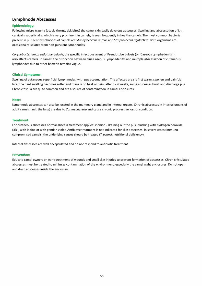
66
Lymphnode AbscessesEpidemiology:Following micro-trauma (acacia thorns, �ck bites) the camel skin easily develops abscesses. Swelling and abscessa�on of Ln. cervicalis superficialis, which is very prominent in camels, is seen frequently in healthy camels. The most common bacteria present in purulent lymphnodes of camels are Staphylococcus aureus and Streptococcus agalac�ae. Both organisms are occasionally isolated from non-purulent lymphnodes.
Corynebacterium pseudotuberculosis, the specific infec�ous agent of Pseudotuberculosis (or ‘Caseous Lymphadeni�s’) also affects camels. In camels the dis�nc�on between true Caseous Lymphadeni�s and mul�ple abscessa�on of cutaneous lymphnodes due to other bacteria remains vague.
Clinical Symptoms:Swelling of cutaneous superficial lymph nodes, with pus accumula�on. The affected area is first warm, swollen and painful; later the hard swelling becomes so�er and there is no heat or pain; a�er 3 - 4 weeks, some abscesses burst and discharge pus. Chronic fistula are quite common and are a source of contamina�on in camel enclosures.
Note:Lymphnode abscesses can also be located in the mammary gland and in internal organs. Chronic abscesses in internal organs of adult camels (incl. the lung) are due to Corynebacteria and cause chronic progressive loss of condi�on.
Treatment:For cutaneous abscesses normal abscess treatment applies: incision - draining out the pus - flushing with hydrogen peroxide (3%), with iodine or with gen�an violet. An�bio�c treatment is not indicated for skin abscesses. In severe cases (immuno-compromised camels) the underlying causes should be treated (T. evansi, nutri�onal deficiency).
Internal abscesses are well encapsulated and do not respond to an�bio�c treatment.
Preven�on:Educate camel owners on early treatment of wounds and small skin injuries to prevent forma�on of abscesses. Chronic fistulated abscesses must be treated to minimize contamina�on of the environment, especially the camel night enclosures. Do not open and drain abscesses inside the enclosure.

67
Module 7: Skin problems
Handout 2
Herdsman: “My camel/s has/have skin problems.”
Herd 1 Herd 2 Herd 3 Herd 4 Herd 5
Q Are all camels in your herd affected?
AOnly suckling calves are sick
Almost the whole herd is sick
Only a few camels are affected
Some younger adults are affected
Some adults are sick
Q When did the problem start?
AMore than three
weeks agoAbout two weeks
agoMore than six
monthsAbout a month ago
Not sure, but cases are
increasing
Q Are the animals s�ll feeding/suckling?
AYes, calves s�ll
sucklingYes herd is feeding
normallyFeeding normally,
but losing condi�onFeeding normally Feeding normally
E Clinical examina�on / observa�on!
FPatches without
hair visible. Calves in good condi�on
Dull coats. Animals scratching
frequently
Dull coat, animals in poor condi�on
Camels in good condi�on, some
abnormal areas visible
Swollen Lymphnodes
visible
E Clinical examina�on of the skin!
F
Confluent bold patches. Dry,
chalky scabs found on some
Skin rough, frequent scratching
visible, skin irritated
Some large bold areas, skin wrinkled and hyperceratosis
Sharp demarcated lesions. Some with irregular edges and
pus, some with scabs only. Skin rough
Lymphnodes enlarged
Q Have any of your camels died?
AOne calf died from
diarrhoeaNo No No No
D What is your clinical diagnosis
D Ringworm Acute Mange Chronic MangeContagious Skin
Necrosis
Lymphnode abscess/
Lymphadeni�s
D What main observa�ons support your clinical diagnosis?
D
Only calves affected, and
confluent bold patches
Most of the herd affected. Frequent
scratching and rough skin
Poor condi�on of affected ones.
Typical clinical signs (Hyperceratosis,
wrinkly skin, bold areas)
Only few camels affected. Typical
clinical signs (demarcated lesions,
with pus at �mes)
Typical clinical signs
D Addi�onal examina�on / diagnos�c tests
D No
Skin scrapings for microscopic examina�on of mange mites
Skin scrapings for microscopic examina�on of mange mites
Bacteriological swab can be taken.
Iden�fica�on of bacteria and
an�bio�c sensi�vity tes�ng
Bacteriological swab can be taken.
Iden�fica�on of bacteria
and an�bio�c sensi�vity tes�ng
Q = Ques�on; A = Answer; E = Examina�on; F= Findings; D= Diagnosis; DD= Differen�al Diagnosis; T= Treatment; C= Control

68
T What therapy do you recommend?
D
If calves are in poor condi�on,
suppor�ve treatment with
vitamin ADE
Ivermec�n 1% injec�on, second
a�er 8 days
Chronic mange difficult to treat.
Ivermec�n 1% can be tried. Culling is
be�er as chronically infected camel can
infect other animals
Supplement with quality salt. Spray
infected areas with blue spray
Abscess treatment.
C What control measures do you recommend?
C
Ensure newly bought camels are clean. Treat with
ivermec�n 2% twice
Cull chronically infected animal. Most to different
pasture
Regular supplementa�on
with salt
Photo: Dioli Photo 1: Classic itching and scratching in herds
with mange
Photo: Dioli Photo 2: Camel calf with generalized acute mange
Q = Ques�on; A = Answer; E = Examina�on; F= Findings; D= Diagnosis; DD= Differen�al Diagnosis; T= Treatment; C= Control

69
Photo: Dioli Photo 3&4: Camel with chronic mange, hyperceratosis and wrinkled skin visible
Photo: Dioli Photo 5: Camel with ringworm
Photo: Dioli Photo 6: Contagious skin necrosis on the neck
of an adult camel

70
MODULE 8
HANDOUT 1LEAD SYMPTOM: ABORTION
DISEASES COVERED:BRUCELLOSIS
RIFT VALLEY FEVERABORTION DUE TO TRYPANOSOMOSIS
OTHER ABORTION CAUSES (CAMEL POX, Q-FEVER, CHLAMYDOPHILA, SALMONELLA)
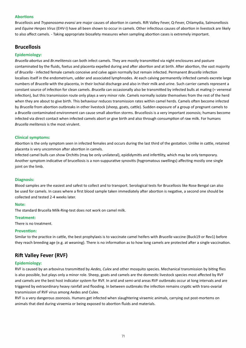
71
Abor�onsBrucellosis and Trypanosoma evansi are major causes of abor�on in camels. Ri� Valley Fever, Q-Fever, Chlamydia, Salmonellosis and Equine Herpes Virus (EHV-I) have all been shown to occur in camels. Other infec�ous causes of abor�on in livestock are likely to also affect camels. - Taking appropriate biosafety measures when sampling abor�on cases is extremely important.
BrucellosisEpidemiology:Brucella abortus and Br.melitensis can both infect camels. They are mostly transmi�ed via night enclosures and pasture contaminated by the fluids, foetus and placenta expelled during and a�er abor�on and at birth. A�er abor�on, the vast majority of Brucella - infected female camels conceive and calve again normally but remain infected. Permanent Brucella infec�on localises itself in the endometrium, udder and associated lymphnodes. At each calving permanently infected camels excrete large numbers of Brucella with the placenta, in their lochial discharge and also in their milk and urine. Such carrier camels represent a constant source of infec�on for clean camels. Brucella can occasionally also be transmi�ed by infected bulls at ma�ng (= venereal infec�on), but this transmission route only plays a very minor role. Camels normally isolate themselves from the rest of the herd when they are about to give birth. This behaviour reduces transmission rates within camel herds. Camels o�en become infected by Brucella from abor�on outbreaks in other livestock (sheep, goats, ca�le). Sudden exposure of a group of pregnant camels to a Brucella contaminated environment can cause small abor�on storms. Brucellosis is a very important zoonosis; humans become infected via direct contact when infected camels abort or give birth and also through consump�on of raw milk. For humans Brucella melitensis is the most virulent.
Clinical symptoms:Abor�on is the only symptom seen in infected females and occurs during the last third of the gesta�on. Unlike in ca�le, retained placenta is very uncommon a�er abor�on in camels.Infected camel bulls can show Orchi�s (may be only unilateral), epididymi�s and infer�lity, which may be only temporary. Another symptom indica�ve of brucellosis is a non-suppura�ve synovi�s (hygromatous swellings) affec�ng mostly one single joint on the limb.
Diagnosis:Blood samples are the easiest and safest to collect and to transport. Serological tests for Brucellosis like Rose Bengal can also be used for camels. In cases where a first blood sample taken immediately a�er abor�on is nega�ve, a second one should be collected and tested 2-4 weeks later.
Note:The standard Brucella Milk-Ring-test does not work on camel milk.
Treatment:There is no treatment.
Preven�on:Similar to the prac�ce in ca�le, the best prophylaxis is to vaccinate camel heifers with Brucella vaccine (Buck19 or Rev1) before they reach breeding age (e.g. at weaning). There is no informa�on as to how long camels are protected a�er a single vaccina�on.
Ri� Valley Fever (RVF)Epidemiology:RVF is caused by an arbovirus transmi�ed by Aedes, Culex and other mosquito species. Mechanical transmission by bi�ng flies is also possible, but plays only a minor role. Sheep, goats and camels are the domes�c livestock species most affected by RVF and camels are the best host indicator system for RVF. In arid and semi-arid areas RVF outbreaks occur at long intervals and are triggered by extraordinary heavy rainfall and flooding. In between outbreaks the infec�on remains cryp�c with trans-ovarial transmission of RVF virus among Aedes and Culex.RVF is a very dangerous zoonosis. Humans get infected when slaughtering viraemic animals, carrying out post-mortems on animals that died during viraemia or being exposed to abor�on fluids and materials.

72
Clinical Symptoms:RVF abor�on rates in camels can reach 100%! Abor�ons occur at any stage of pregnancy. Apart from mass abor�ons there is increased neonatal mortality. In older camel calves cases of severe febrile disease with jaundice can occur. Apart from abor�ons adult camels do not show sigs of disease.In humans RVF mostly produces an influenza like disease, but fatal haemorrhagic cases do occur.
Diagnosis:The combina�on of abnormally intense rainfall/flooding, an extraordinary high number of pregnant camels abor�ng within a very short period and mass abor�ons also occurring in sheep and goats kept in the same area should be regarded as highly suspicious of RVF. Serum samples from animals with and without abor�ons plus EDTA blood samples should be submi�ed to the laboratory when suspec�ng RVF do not carry out any field post-mortems!
Treatment:There is no treatment.
Preven�on:Vaccina�on with the new Clone13 RVF vaccine, which unlike older vaccines does not cause abor�ons in pregnant sheep. Clone13 is yet to be tested in pregnant camels.Topical applica�on of insec�cides during RVF outbreaks reduces the risk of infec�ons considerably.
WarningRVF is an extremely dangerous zoonosis - biosafety rules when dealing with RVF infected livestock:• Avoid direct contact with body fluids of sick or dead animals.• Do not slaughter or dissect animals during RVF outbreaks.• Wear gloves and mask (or any other device to avoid direct contact e.g. plas�c bags) when handling sick or dead animals,
par�cularly when assis�ng at birth, burying placenta or fetus.• Wash your hands with disinfectant or soap immediately a�er contact with any animal body fluid.• Animal products are infec�ous and must be well cooked before consump�on (esp. milk).• During RVF outbreaks people should protect themselves against mosquito bites by using covering clothes, repellents, bed
nets and insec�cides.• People with fever for more than 48 hours must urgently seek medical advice.
Trypanosoma evansiEpidemiology and Clinical Symptoms:Trypanosoma evansi is transmi�ed mechanically by blood-sucking flies. Transmission rates are higher during rainy season and in areas with more insect vectors. Most transmissions are short distance (within the herd, at watering points). Abor�ons occur within 5–60 days a�er infec�on at any state of pregnancy. Abor�ng animals are otherwise healthy. Camels infected chronically with T. evansi pose a serious infec�on risk to pregnant females of the same herd. (See also Module 1)
Treatment:There is no treatment.
Preven�on:Chemoprophylaxis with trypanocides during periods with increased transmission risk (rainy season) and when moving animals during high risk zones.

73
Other causes of abor�on
Epidemiology:Other causes of abor�ons in camels include Camel Pox, Q-Fever, Chlamydophila and Salmonella. Apart from Camel Pox (see Module 2), there is no informa�on about the camel specific epidemiology of these pathogens. Infec�on occurs via contact with abor�on materials and contaminated environments. Salmonella are very common in camel herds and an�bodies to Coxiella burne�i (Q-Fever) and to Chlamydophila abortus (former Chlamydia psi�aci) have been found in camels. Ticks play a role in maintenance of Q-Fever infec�on.
Clinical Symptoms:Chlamydophila abortus causes abor�on towards end of the pregnancy.Coxiella burne�i causes sporadic abor�ons.Camel Pox outbreaks frequently cause abor�ons.Salmonella dublin can cause sporadic abor�ons.
Treatment:None.
Preven�on:The only vaccine validated for camels is Camel Pox vaccine. Vaccines against Chlamydia, Coxiella and Salmonella are available for other livestock species.

74
Herd 1 Herd 2 Herd 3
Q How many camels have aborted?
A
One first calving heifer and two femalesAbout six camels. It might be seven but I
did not see the foetus of the last oneTwo camels
Q How many adult female camels do you have and how many were pregnant before the abor�on started?
A I have about 60 female camels and 12 were pregnant
Q When did the abor�on begin and did you have abor�ons before?
AAbout two days ago and there was no
abor�on before that
It started about a week ago is s�ll ongoing. Before I did not have a problem
with abor�on
One aborted yesterday another one two months ago
Q At what stage of pregnancy did your camel abort?
A About 6 to 7 months Not sure, they were all at different stagesOne was about to give birth,
one was three months
E Clinical examina�on / observa�on
FNo retained placenta
Camels in good condi�on
Q Have you observed anything else abnormal?
A No No, but today 2 more camels aborted No
D What is your clinical diagnosis
D Could be Brucellosis or other
Could be Ri� Valley feverCould be caused by
Trypanosomosis
D What main observa�ons support your clinical diagnosis?
D
Difficult
Massive abor�on at various stages of pregnancy. Confirm if very wet condi�ons
(during heavy rains)
Confirm if camels are regular treated with quinapyramide
D Addi�onal examina�on / diagnos�c tests
DSubmit blood sample of affected animals and others to laboratory.
If possible submit also foetus or placenta to laboratory – NOTE: Follow biosafety measures (RVF and Brucellosis are zoonosis!!!) transport chilled and in �ghtly closed containers
C What therapy/control measures do you recommend?
C
If possible affected animals should be separated from herd.Calving should take place in a different enclosure.
Be careful when handling dead foetus and placenta and ensure proper disposal (bury or burn).Remind the herdsman that Brucellosis and RVF are zoonosis and can affect humans!
Module 8: Abor�on
Herdsman: “My camel/s has/have aborted.”
Q = Ques�on; A = Answer; E = Examina�on; F= Findings; D= Diagnosis; DD= Differen�al Diagnosis; T= Treatment; C= Control
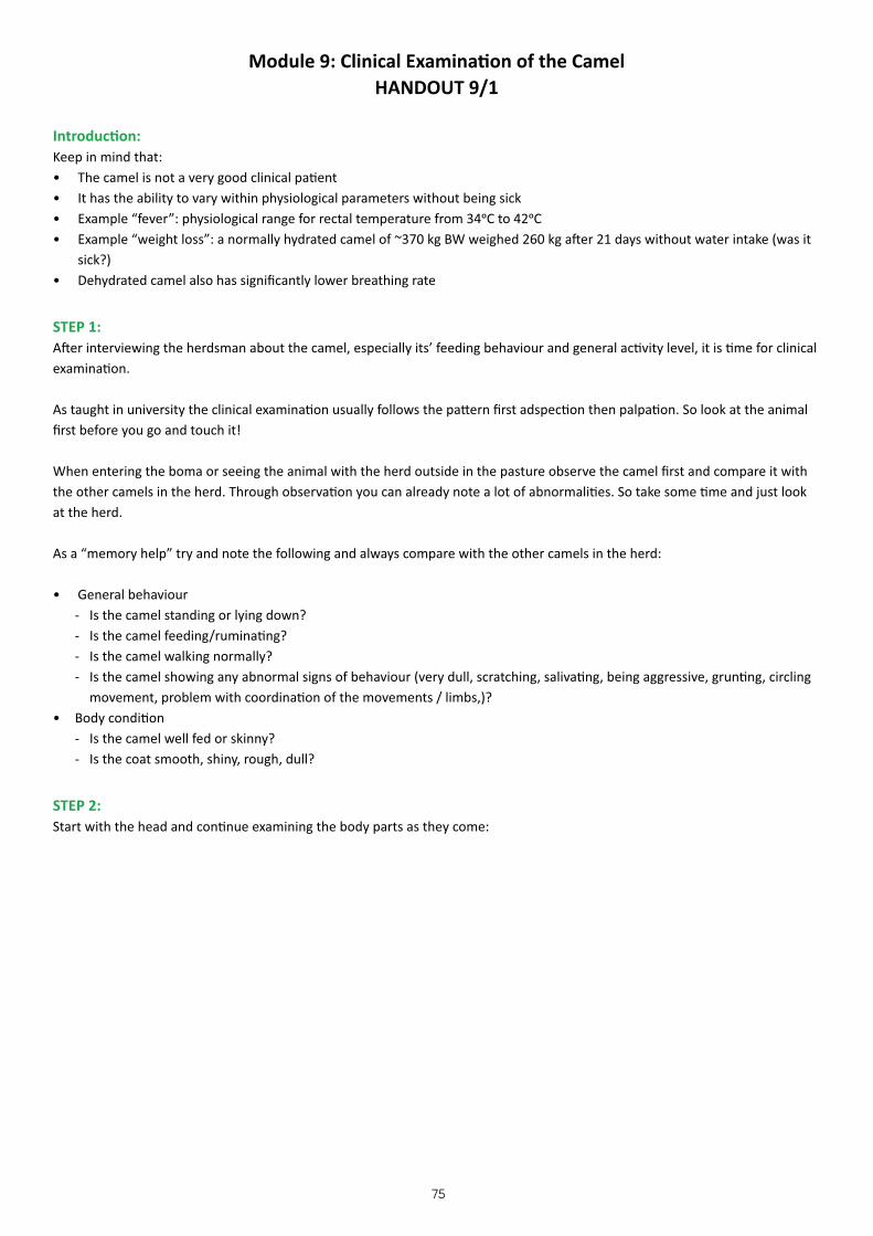
75
Module 9: Clinical Examina�on of the CamelHANDOUT 9/1
Introduc�on:Keep in mind that:• The camel is not a very good clinical pa�ent• It has the ability to vary within physiological parameters without being sick• Example “fever”: physiological range for rectal temperature from 34C to 42C• Example “weight loss”: a normally hydrated camel of ~370 kg BW weighed 260 kg a�er 21 days without water intake (was it
sick?)• Dehydrated camel also has significantly lower breathing rate
STEP 1:A�er interviewing the herdsman about the camel, especially its’ feeding behaviour and general ac�vity level, it is �me for clinical examina�on.
As taught in university the clinical examina�on usually follows the pa�ern first adspec�on then palpa�on. So look at the animal first before you go and touch it!
When entering the boma or seeing the animal with the herd outside in the pasture observe the camel first and compare it with the other camels in the herd. Through observa�on you can already note a lot of abnormali�es. So take some �me and just look at the herd.
As a “memory help” try and note the following and always compare with the other camels in the herd:
• General behaviour - Is the camel standing or lying down? - Is the camel feeding/rumina�ng? - Is the camel walking normally? - Is the camel showing any abnormal signs of behaviour (very dull, scratching, saliva�ng, being aggressive, grun�ng, circling
movement, problem with coordina�on of the movements / limbs,)?• Body condi�on - Is the camel well fed or skinny? - Is the coat smooth, shiny, rough, dull?
STEP 2:Start with the head and con�nue examining the body parts as they come:

76


