46
0 State of Kuwait Ministry of Health Guidelines of Microbiological Environmental Sampling in Healthcare Settings 2011
0
State of Kuwait
Ministry of Health
Guidelines of Microbiological Environmental
Sampling in Healthcare Settings
2011
1
Contents 1 INTRODUCTION
2 INDICATIONS
3 PRE SAMPLING INSPECTION
4 PROGRAM OPERATION PROCESSES
5 TYPES OF SAMPLING
5.1 AIR SAMPLING
5.1.1 Non Cultural Sampling
5.1.2 Indoor
5.1.3 Outdoor
5.1.2. Cultural Sampling
5.1.3. Equipment
5.2 SURFACE SAMPLING
5.3 WATER SAMPLING
5.3.1 Sampling methods for bacteriological testing
5.3.2 Sampling Procedure
5.3.2.1 Visual inspection
5.3.2.2 Sampling and control
5.3.3 Preserving and Storing your Sample:
5.3.4 Sampling containers and its preparation
5.3.5. Types of water sampling
5.3.5.1 Sampling of Drinking Water
5.3.5.1.1 Location of sampling points
5.3.5.1.2 Sampling frequency
5.3.5.2 Renal unit water
5.3.5.3 Drinking, tap and hydrotherapy water
5.3.5.4 Dental unit water
5.3.5.5 Pharmacy water
6 ISOLATION AND IDENTIFICATION OF MICROORGANISMS
7 INTERPRETATION AND STANDARD OF RESULT
7.1 Air sampling
2
7.2 Surface sampling
7.3 Water sampling
8 REFRENCES
9 APPENDICES
- Appendix A. Preparation of area prior to sampling
- Appendix B. Environmental checklist for monitoring cleaning prior to sampling
- Appendix C. Environmental sampling request form
- Appendix D. Environmental sampling result form
- Appendix E. Water sampling request and result form
- Appendix F. Figure (1) The suggested sampling technique for the flat surfaces
- Appendix G. Table (7) Gram-negative bacteria associated with water and moist
environment
- Appendix H. Table (8) Airborne fungi associated with the transmission of infection, and
their implicated environments at Kuwait health care settings.
- Appendix I. Water sampling from three basic source types
- Appendix J. Thio-Bag EPA Approved for Potable Water Collection
3
Tables
• Table (1) Sampling time adjustment according to environmental site
and dust particles
• Table (2) Environmental monitoring limits for controlled areas and
devices in operation
• Table (3) Testing requirements and interpretation of results air
quality (as determined by active air sampling)
• Table (4) Testing requirements and interpretation of results for renal
dialysis fluid Testing
• Table (5) requirements and interpretation of results for hydrotherapy
water samples
• Table (6) Testing requirements and interpretation of results for
pharmacy samples
• Table (7) Gram-negative bacteria associated with water and moist
environment
• Table (8) Airborne fungi associated with the transmission of
infection, and their implicated environments at Kuwait health care
settings.
4
This guidelines was the result of the combined effort of :
Infection Control Directorate, Medical Laboratory Administration, Occupational Health Administration
and Public Health Administration.
Faculty of Medicine- Kuwait University Infection control Directorate
Dr. Noura Al-Sweih
Consultant- Clinical Microbiologist
(Committee chairperson)
Dr. Wafaa Jamal
Consultant-Clinical Microbiologist
(committee member)
Dr. Kholoud Al-Fadhalah
Head, Infection Control Department
(Committee coordinator)
Dr.Nawar alnassrallah
Head of health promotion programs
(committee member)
Public Health Directorate Occupational Health Directorate
Dr.Salah Elfashni
Public health specialist- Environmental health
unit
(committee member)
Ms. Laila Ashkanani
Head, Aerobiology Department
(committee member)
Ms. Suad Marafie
Head of Medical Laboratory Technicians
(committee member)
5
1. INTRODUCTION Microbiologic sampling of air, water, and inanimate surfaces (i.e., environmental
sampling) is an expensive and time-consuming process that is complicated by many variables
in protocol, analysis, and interpretation. In this guideline, we advocate the targeted instead of
routine environmental culturing because rates of healthcare–associated infection had not been
associated with levels of general microbial contamination of air or environmental surfaces, and
because meaningful standards for permissible levels of microbial contamination of
environmental surfaces or air did not exist. However, targeted microbiologic sampling for
defined purposes and not routine sampling should be conducted in accordance with defined
protocols that include:
a) A written, defined, multidisciplinary protocol for sample collection and culturing;
b) Analysis and interpretation of results using scientifically determined or anticipatory baseline
values for comparison; and
c) Expected actions based on the results obtained.
Infection control, in conjunction with laboratories ( Aerobiology, Public Health and Hospitals),
should assess the health-care facility’s capability to conduct sampling and determine when
expert consultation and/or services are needed.
Effective sampling of surfaces depends on the selection of appropriate sampling and assay
techniques. If sampling is conducted as part of an epidemiologic investigation of a disease
outbreak, identification of isolates to species level is mandatory, and characterization beyond
the species level is preferred.
2. INDICATIONS OF ENVIRONMENTAL SAMPLING Microbiologic sampling of air, water, and inanimate surfaces (i.e., environmental sampling)
is indicated for only the following situations:
1. To support an investigation of an outbreak of disease or infections when environmental
reservoirs or fomites are implicated epidemiologically in disease transmission.
• It is important that such culturing be supported by epidemiologic data.
• Environmental sampling, as with all laboratories testing, should not be conducted if
there is no plan for interpreting and acting on the results obtained.
6
• Linking microorganisms from environmental samples with clinical isolates by
molecular epidemiology is crucial whenever it is possible to do so.
2. Limit microbiologic sampling for quality assurance purposes:
• Biologic monitoring of sterilization processes;
• Microbiologic air sampling after construction of critical care areas : operation theatre
(OT), intensive care unit (ICU) and protective environment room (PE) for allogeneic
HSCT recipients;
• Periodic culturing of water and dialysate in hemodialysis units (at least once a month
and during outbreaks);
• Periodic culturing and during outbreaks of drinking water, tap water, dental unit,
pharmacy, swimming and hydrotherapy pool water;
• Certain equipment in health-care settings (e.g., biological safety cabinets) may also
be monitored with air flow and particulate sampling as part of adherence to a
certification program.
• For air handling unit, it is inappropriate to carry out microbiological investigations
on cooling coils as they do not contribute significantly to the microbial quality of air
delivered by the system. It is also inappropriate to carry out microbiological
investigations on humidifiers since they should generate humidity via steam, in
which case they pose a negligible risk. Humidifiers that aerosolise recirculated water
(spinning disk humidifiers) pose too high a hazard and should not be used. The air
handling unit should be properly constructed, finished and functioning. The
humidifier and cooling coil in air handling units should be disinfected at least six-
monthly. It is considered that physical cleaning, rather than disinfection with
microbiological monitoring, is more appropriate.
3. For research purposes.
• Well-designed and controlled experimental methods and approaches can provide new
information about the spread of health-care–associated diseases.
4. To monitor a potentially hazardous environmental condition, confirm the presence of a hazardous
chemical or biological agent, and validate the successful abatement of the hazard.
7
• This type of sampling can be used to detect bioaerosols released from the operation of
health-care equipment (e.g., an ultrasonic cleaner) and detect the release of an agent of
bioterrorism in an indoor environmental setting.
3. PRE SAMPLING INSPECTION Several preliminary concerns must be addressed when designing a microbiologic environmental
sampling strategy, which should meet with the four Circumstances of environmental sampling
(mentioned earlier). Pre sampling inspection should be performed prior to sampling to ensure
that area is well prepared for sampling. This is usually performed by the head nurse of the
sampling area in cooperation with hotel services department. It should be supervised by
Infection control personnel.
A. The inspector should evaluate and assure that these criteria’s are met prior to each
sampling event, to identify the conditions that may affect or interfere with the proposed
testing:
• All new or reestablishment work is completed.
• The ventilation system had been running continuously for at least 24 hours following the
completion of structural work, and cleaning.
• All engineering commissioning procedures has been completed.
• All fixed and portable equipments placed in their places.
• Floors, all surfaces and equipment are finished from cleaning prior the investigation by
24 hours. A Checklist for Monitoring Cleaning Prior to Sampling (appendix B) should be
used by the head nurse of the sampling area to ensure proper cleaning.
• Ducting and air diffusion sites (inlet, outlet) are cleaned prior to sampling.
• Minimizing indoor traffic at all times when possible.
• The sampling personal should be completely familiar with the sampling protocol and
type of the particular method to be used in different situations.
B. In case of microbiological out breaks by one or more microorganisms. The following should
be considered:
• Avoid excessive cleaning.
• The decision to sample should be based on the extent and location of any suspected
8
contamination and the potential for the contaminant to migrate and the activities for
which the facility is used.
• The decision to collect environmental samples should be made by the infection control
committee with liaising with Aerobiology laboratory.
• All personnel who enter the contaminated area must follow the safety and infection
control plan developed for that particular site.
• Shutting down the ventilation system serving the contaminated area may be necessary
only in special cases to avoid dispersing certain contaminate (i.e. Mucor species, Bacillus
anthracis etc).
4. OPERATION PROCESSES
When environmental sampling (air or surface) is needed, the corresponding Infection control
department should inform the Air Microbiology Department verbally and using the request form
(appendix C) to collect the samples and analysis it. Result will be send back to Infection control
department in another result form (appendix D).
For water sampling on a routine basis, a defined annual schedule of all areas required (drinking
water points, hydrotherapy, dental units, renal units) according to the considered frequency for
each area should be prepared by the corresponding preventive medicine department. For water
sampling as a part of outbreak investigation, the corresponding preventive medicine department
is called by Infection control department to collect the samples, fill the request form and send it
to Public Health Laboratory for analysis. All samples should be accompanied by an appropriate
collection form (appendix E). Result will be send back to preventive medicine department which
should act upon.
5. TYPES OF SAMPLING The sampling method and number of samples collected will be influenced by the potential
contamination circumstances. The first priority should be to collect samples from locations near
the suspected release sources.
9
5.1. AIR SAMPLING
Sampling protocols should include:
1. Indoor (problem and non problem areas).
2. An outdoor reference samples.
5.1.1. NON CULTURAL SAMPLING: (Air borne and non viable particulates)
5.1.1.1. Indoor Air Sampling
• Air quality methodologies imply testing in representative areas to determine airborne
particulates influences and access general indoor air quality parameters. Testing is conducted
to access airborne particulates using laser particle counter or optical counters which quantifies
particulates by size (0.3 to 10.0 microns).
• Results are comparatively evaluated to determine particulates concentration by size, the
efficiencies of the air filtering systems and to identify areas with elevated particulate concerns
(e.g. fungal influences are usually suspected when predominantly high concentration are
noted between 1.0 - 10 microns.
• Other type of devices such as Allergen co – D is designed for a rapid collection and analysis
for air borne particulates including bioaerosols (mold spore, pollen insect parts, skin cell
fragments) or particulates such as (cellulose, fiber glass etc) and opaque particles such as (fly
ash, oil droplets' etc). This device works using the inertial impaction principles similar to
other spore trap devices. These air sampling cassettes designed for evaluating total airborne
fungi and non viable particulates. Depending on site conditions, samples are collected at 15
litters/minutes over 5 minutes or 10 minutes sampling periods. Samples are analyzed using
direct microscopy examination.
5.1.1.2. Out Door Air Sampling
A comparison of microbial species and densities in outdoor air versus indoor air has been used
to help pin point fungal spore bursts. Fungal spore densities in outdoor air vary and the degree of
variation based on many factors (season, climate changes, temperature, relative humidity). Seven
10
day volumetric spore trap “Burkard” is used as a reference device of choice and as a monitoring
device for outdoor air borne contaminates method. (Fungal spore).
5.1.2. Cultural Sampling
The objective of this method is to access the quantities as well as differentiation of viable (alive
or / and capable of germination) fungi and bacteria, that can grow on surface of microbiologic
media plates.
5.1.3. Equipment and Supplies
Impactors: Commercially available volumetric impactors are available and capable of viable air
sampling. The properties and specification of the impactors (particle size cut point, operating
flow rate, type of collection media i.e. Agar, etc) are considered.
A volumetric air sampler, Anderson N6 is considered in the following air sampling procedures.
5.1.4. Sampling strategy
Air sampling can provide information that should inform infection control professionals that the
air quality is good enough for safe patient care, because the control measures are properly
applied.
These factors should be considered before starting the air sampling:
1. Vacuum pump calibrated to sample 28.3L/M (Anderson two stages samplers N6).
2. The air sampler should be checked and cleaned by 70% alcohol before use following the
manufacturer’s instruction.
3. The area being sampled should have been left vacant to avoid false-positive results due
to recent area usage.
4. The doors must be kept closed prior to and during the air sampling period.
5. Staff should wear a proper disposable coveralls if available or sterile gowns and surgical
mask. Shoe cover is acceptable. Hand should be washed and sterile non powdered gloves
worn just before entering the room.
6. Using aseptic technique in handling all the sterile equipment should be followed (Agar
sampler, swabs etc).
11
7. Establish laboratory time lines for sample collection, processing method and provision of
results.
8. Ensure that adequate supplies are available.
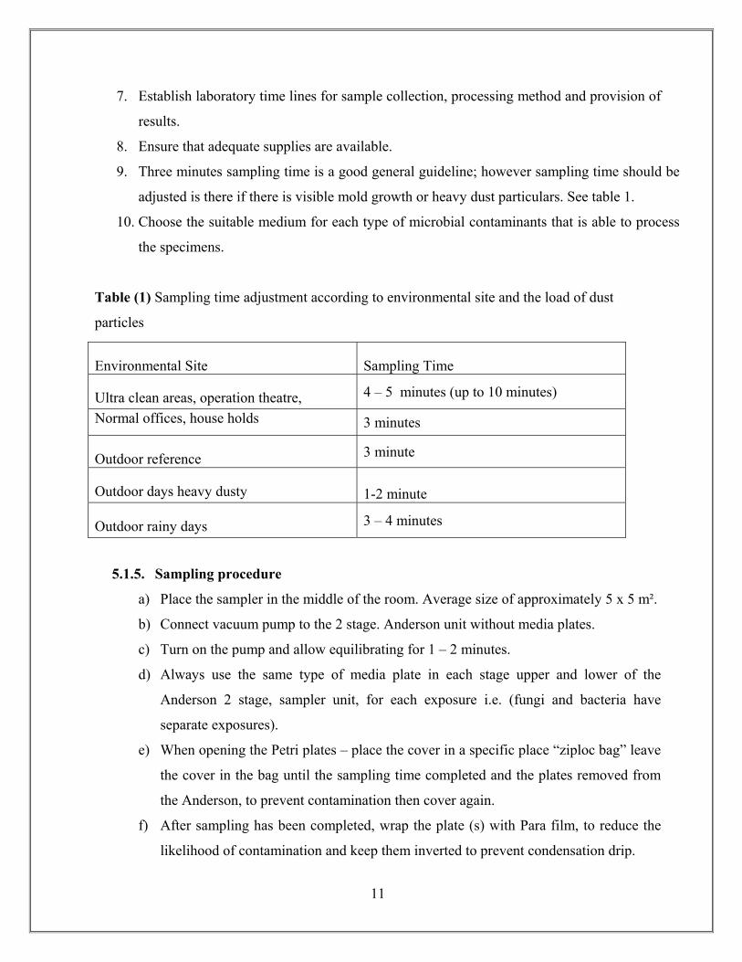
9. Three minutes sampling time is a good general guideline; however sampling time should be
adjusted is there if there is visible mold growth or heavy dust particulars. See table 1.
10. Choose the suitable medium for each type of microbial contaminants that is able to process
the specimens.
Table (1) Sampling time adjustment according to environmental site and the load of dust
particles
5.1.5. Sampling procedure
a) Place the sampler in the middle of the room. Average size of approximately 5 x 5 m².
b) Connect vacuum pump to the 2 stage. Anderson unit without media plates.
c) Turn on the pump and allow equilibrating for 1 – 2 minutes.
d) Always use the same type of media plate in each stage upper and lower of the
Anderson 2 stage, sampler unit, for each exposure i.e. (fungi and bacteria have
separate exposures).
e) When opening the Petri plates – place the cover in a specific place “ziploc bag” leave
the cover in the bag until the sampling time completed and the plates removed from
the Anderson, to prevent contamination then cover again.
f) After sampling has been completed, wrap the plate (s) with Para film, to reduce the
likelihood of contamination and keep them inverted to prevent condensation drip.
Environmental Site Sampling Time
Ultra clean areas, operation theatre, 4 – 5 minutes (up to 10 minutes)
Normal offices, house holds 3 minutes
Outdoor reference 3 minute
Outdoor days heavy dusty 1-2 minute
Outdoor rainy days 3 – 4 minutes
12
g) With a permanent marker, label each Petri dish from the bottom with following
information’s :
- specific location (room number)
- date
- facilities (IV lab, operation theatre etc)
- duration of sampling (in minutes)
- label upper plate and lower plate in the 2 stage Anderson Sampler (Respirable
and Non respirable)
h) Plates should be kept cool after collection and during transportation to laboratory to
prevent growth of contaminates.
i) Samples “Agar plates” are then incubated as soon as possible at 37ºC for 24 – 48
hours, for the environmental bacteria. And at 27ºC for 5 – 14 days for maximum
recovery of fungi.
Note: Preliminary culture results are rarely available until after 24 hours incubation.
j) After incubation, the total number and the type of colonies in each plate are counted,
and analyzed for their species.
k) The mean number of viable bacteria or fungi by cubic meter (m²) of air can be
calculated by knowing the air sample flow rate (28.3 L/m³) and the sampling time,
and the results will be written as colony forming unit per cubic meter of air (CFU/
m³).
l) Documentation of all the results should be done and sent to the infection control
department as soon as possible.
5.2. SURFACE SAMPLING “ SWAB SAMPLES”
- Surface sampling is a non destructive methods which allows the determination of possible
microbial contamination of suspected sites or and un transportable materials,
- Several methods can be applied for collecting environmental surface samples (i.e. tape – lift
method and swab method)
- Swab method has the advantage of enabling a sample to be taken in a hard to reach area(s)
without a removal of any material and it is a neutral, stable way to transport the sample.
13
-Swab sampling is performed using pre sterile swabs in various transport media that are
commercially available.
5.2.1. Pre Sample collection
These factors should be considered before starting the environmental surface sampling:
• Background information (sampling objective (a) outbreak (b) renovation).
• Determine the target microorganism (s) in case of outbreaks.
• Location of surfaces to be sampled.
• Method of sample collection and the appropriate equipment or supplies for each task.
• Weather the sampling will be qualitative or/and quantitative.
• An estimate of maximum allowable microbial numbers or types on the sampled surfaces.
(Clinical control points).
• If disinfectant residuals are expected on surfaces being samples, specific neutralizer,
chemical should be used in both.
• Non sterile equipment used for sampling should be sanitized before sample collection
(e.g. cooler, sample boxes, ice packs, sampling machine).
• Arrange all the supplies and equipments needed.
- Sterile non powdered vinyl gloves (should be replaced as often as necessary).
- A sterile non cotton or cotton swabs with transport media or dry swabs for
moistened areas.
- Sterile sponges for large surfaces.
- Neutralizing buffer, sterile diluents, sterile water, sterile saline solution, and
sterile peptone water to be used for moistening the swabs or sponges.
- Alcohol.
- The appropriate medium for each study case.
5.2.2 Sampling Procedure
1. Wear the appropriate clothing.
2. Wear the non powdered sterile gloves.
14
3. Remove a sterile, cotton swab from the package.
4. Moisten the swab with (sterile water, peptone water, or transport media).
5. Wipe the surface to be sampled by touching the cotton tip to the surface and rotating and
rubbing back and forth for the deep irregular areas.
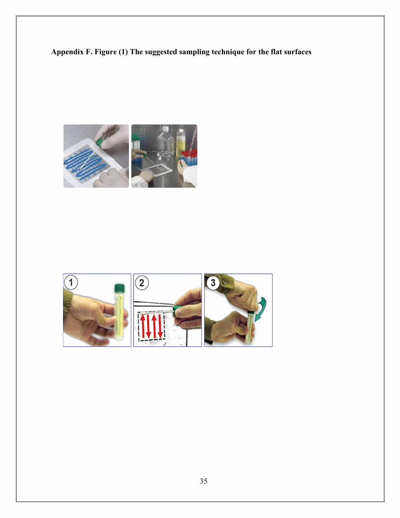
6. For the flat surfaces, the suggested sampling technique is to make a vertical S – strokes
to cover an area of 10 x 10 cm². Figure (1)
7. Place the swab in its tube taking care not to touch the tip or the shaft of the swab.
8. Label tube containing the swab with date, specific location, and facility).
9. Transport the samples to laboratory for analysis as soon as possible, and samples should
be kept in a refrigeration temperature (4ºC). Do not freeze.
10. The appropriate methodology for testing micro organism should be followed to detect the
organism(s) of concern (fungi / bacterial).
11. Documentation of the results sends to the health care setting personally.
5.3. WATER SAMPLING
5.3.1 Sampling methods for bacteriological testing All samples should be accompanied by an appropriate collection form. When water samples are
collected for analysis, care should be taken to ensure that there is no external contamination of
the samples. Unless valid samples are collected, the results of the subsequent analysis may be
misleading.
5.3.2 Sampling Procedure 5.3.2.1 Visual inspection Visual inspection of the site should cover the following aspects:
1) Potential pollution sources (industrial, agricultural, domestic, animals)
2) Condition of water (clear, cloudy, color, smell, foam)
3) Map of Site (electronic or sketched, photograph if possible)
4) Brief written description of site.
15
5.3.2.2 Sampling and control
1. Sample all sites in triplicate.
2. Collect all samples in 100mL or 1L Whirlpak bags.
3. All samples must be clearly labelled by site, number of sample, and status as field blank (FB) or
lab blank (LB). Use an indelible marker to label all bags before trying to take sample (wet plastic
does not take ink well). In addition to the sample IDs, all samples should be dated.
4. If sampling a body of running water, point the mouth of the bag upstream and your hands
downstream to avoid contamination.
5. If sampling from a water faucet, run the faucet for 1 minute before obtaining a sample.
6. Rinse the bag twice with the sample water prior to filling and closing.
7. Fill bag as full as possible. Half-filling the bottle leaves more room for oxygen which will
promote degradation of your sample.
8. Collect data such as temperature and pH which affect the solubility of many ions.This is
particularly important if an investigation is being carried out to determine the source of
Legionella. Hot water should reach 50°C within 1 minute at outlets, whilst cold water should be
20°C or below after running the water for two minutes.
5.3.3 Preserving and Storing your Sample:
Storage before filtration should be at 4 °C for not more than 24 hours.
5.3.4 Sampling containers and its preparation
- Several types of bottle may be used for sampling, but glass bottles are best.
- These should have securely fitting stoppers or caps with nontoxic liners, and both bottles and
stoppers should be sterilized.
- Each cap should have a metal sleeve clear of the screw thread to ensure that the risk of
contaminating the water sample is minimized.
- Cotton wool plugs and paper caps should be avoided as they tend to fall off during and after
sampling and increase the risk of contamination.
- The bottles should hold at least 200 ml of water.
- Whenever chlorine is used for disinfection, chlorine residual may be present in the water after sampling
and will continue to act on any bacteria in the sample; the results of the microbiological analysis may
16
therefore not be indicative of the true bacteriological content of the water. To overcome this difficulty, it
is common procedure to add sodium thiosulfate to the sample, which immediately inactivates any
residual chlorine but does not affect the microorganisms that may be present.
- The sodium thiosulfate should be added to the sample bottles before they are sterilized. For 200-
ml samples, four or five drops of aqueous sodium thiosulfate solution (100 g/liter) should be
added to each clean sample bottle.
- The stopper is loosely inserted into the bottle, and a brown paper or aluminum foil cover is tied
to the neck of the bottle to prevent dust from entering.
5.3.5. Types of water sampling 5.3.5.1 Sampling of Drinking Water 5.3.5.1.1 Location of sampling points In selecting sampling points, each locality should be considered individually; however, the
following general criteria are usually applicable:
• Sampling points should be selected such that the samples taken are representative of the
water source, treatment plant, storage facilities, distribution network, points at which
water is delivered to the consumer, and points of use..
• These points should include those that yield samples representative of the conditions at
the most unfavorable sources or places in the supply system, particularly points of
possible contamination such as unprotected sources, loops, reservoirs, low-pressure
zones, ends of the system, etc.
• Sampling points should be uniformly distributed throughout a piped distribution system,
taking population distribution into account; the number of sampling points should be
proportional to the number of links or branches.
• Sampling points should be located in such a way that water can be sampled from reserve
tanks and reservoirs, etc.
• In systems with more than one water source, the locations of the sampling points should
take account of the number of inhabitants served by each source.
• There should be at least one sampling point directly after the clean-water outlet from
each treatment plant.
17
5.3.5.1.2 Sampling frequency
The recommended minimum frequencies for these measurements for piped drinking water in the
distribution system are once monthly.
The same frequency of sampling can be applied to piped tap water in healthcare facilities.
5.3.5.2 Renal unit water
Bacteriologic assays of renal unit water should be performed at least once a month and during
outbreaks by using standard quantitative methods. In conjunction with microbiologic testing,
Endotoxin testing should be performed. Renal unit water include the water used to prepare
dialysis fluid (Ultra-pure) and water used to rinse and reprocess dialyzers.
If the sample is to be collected from a tap used solely for sampling ensure that this has been
appropriately sanitized.
a. Aseptically open a labeled sterile water bottle (usually 500 ml bottle containing
neutralizer) and fill almost to the brim with water; replace the lid.
Note: If only small volumes of liquid are available for sampling, a smaller sterile
plastic container can be used, as neutralizer is not essential for this sample type.
b. Store the water between 2 and 8 C and return to the lab for examination ideally on
the same day but always within 24 hours of collection.
5.3.5.3 Swimming and hydrotherapy pool water
The recommended minimum frequencies for these critical measurements minimum sample
numbers for and hydrotherapy is weekly.
5.3.5.4 Dental unit water
Interest in the contamination of dental unit water lines (DUWLs) is growing amongst dental
researchers. The recommended frequency of sampling is once monthly.
5.3.5.5 Pharmacy water
The recommended minimum frequency for the pharmacy where water can be used for
preparation of medications is once weekly.
18
6. ISOLATION AND IDENTIFICATION OF MICROORGANISMS
MEDIA REQUAIRED:
There are a number of different media types available depending on the sampling objective,
unless the microorganism of specific type are suspected, the sampling medium should support
growth of the widest variety of common fungi and bacteria, selective media to be used for
certain type of contaminations.
• PDA (potato dextrose agar) for general mold identification.
• SDA (sabroud dextrose agar) also general mold identification and yeast.
• BA – (blood agar – tryptsoy agar + 5% sheep blood) for general bacterial culture.
• NA - (nutrient agar) used for sampling and cultivation of general bacteria.
• MH (Muller Hinton agar) for susceptibility testing.
• MAC (macConkey agar) selective medium for Gram-negative bacteria.
D -MICROSCOPIC EXAMINATION FOR ALL GROWN COLONIES, FUNGI OR BACTERIA:
Gross appearance
Morphological appearances by microscope
Identification as will be described below.
I. Gram-positive bacteria
1. Staphylococcus aureus
A. Media used for isolation of Staphylococcus aureus:
Blood agar and Mannitol salt agar incubated in air at 37°C for 24hour. Staphylococcus
aureus appear as yellow colonies on Mannitol salt agar and white creamy color on blood
agar.
B. Identification:
i. Gram-stain: Gram-positive cocci in clusters
19
ii. Catalase test: All Staphylococci are Catalase positive.
• A loopful of growth is transferred from an agar plate culture to a glass of
microscope slide. Evolution of bubbles on addition of a drop of 3% H2O2
signifies a positive result. S. aureus is used as a positive control and
Streptococci as a negative control.
iii. Coagulase: S. aureus is Coagulase positive
• Slide Coagulase: place one drop of normal saline solution on a clean
microscope slide and emulsify in it 1-2 colonies of the tested organism.
Add one drop of undiluted rabbit plasma warmed to room temperature and
stir the adhering traces of plasma into the drop of bacterial suspension on
the slide with a sterile loop. A positive reaction appears as coarse
clumping visible to the naked eye within 5-10 seconds. While a negative
reaction does not show any coarse clumping. With each patch put a
known positive control (S. aureus) and negative control (S. epidermidis).
• Tube Coagulase: prepare a 1:10 dilution of rabbit plasma in normal saline
solution and place a 0.5ml of the diluted plasma in small tubes. Inoculate
into the diluted plasma with 1-2 colonies of the strain under test. With
each patch put a known positive control (S. aureus) and negative control
(S. epidermidis). Then incubate the tubes at 37°C in air and examine after
4 hours incubation. Check for a clot or a stiff gel formation. A clot
formation indicate a positive reaction (S. aureus). If a clot is not present,
keep the tubes overnight at room temperature and check for the presence
of a clot next day. A clot formation indicates a positive reaction (i.e. S.
aureus) and absence of the clot indicate a negative reaction (i.e. S.
epidermidis).
If Coagulase test is not available , then use DNase test.
• Deoxyribonuclease test (DNase): heavily spot inoculation several
colonies of the test organism on DNase test medium containing the
20
metachromatic dye toluidine blue O. After 24 hr incubation at air at 35°C,
the medium under and around the inoculums turns from azure blue to
pink, indicating the hydrolysis of the DNA. The content of the toluidine
blue in the medium should not exceed 0.005% because the blue color
imparted to the agar by higher concentrations may mask detection of
DNase activity.
iv. Susceptibility testing to Methicillin: by disk diffusion method and using oxacillin
1µg disk on Mueller-Hinton agar following CLSI methodology. A suspension of
the bacterium is prepared in Normal saline (0.9%) to a turbidity equivalent to 0.5
McFarland standards and then streaked on Mueller-Hinton agar. Then oxacillin
disk is applied on the inoculated plate. Another plate is inoculated with a control
S. aureus strain (S. aureus ATCC 25923) and oxacillin disc. Both plates will be
incubated overnight in air at 35°C. The interpretation will be read according the
zone diameter as follows: ≥13 mm sensitive, 11-12 mm intermediate, ≤10 mm
resistant.
2. Coagulase negative staphylococci
Catalase positive
Coagulase negative
DNase negative
Susceptibility testing: as section iv
3. Micrococcus spp.:
A. Media used for isolation: blood agar
B. Identification:
• Pigment production (white to yellow pigmentation)
• Colony morphology
• Gram stain: Gram positive cocci in clusters, pairs, tetrads or cubes
• Catalase test : positive
• Oxidase test: positive
21
4. Other Gram-positive cocci: e.g. Kocuria and Kytococcus spp.
A. Media used for isolation: blood agar
B. Identification
• Colony morphology
• Gram stain: Gram-positive cooci in tetrads
• Pigment production: Kocuria spp. produce pale crème to orange, yellow,
or pink to red pigmentation. Kytococcus spp. produce creamy white to
yellow pigment.
5. Gram positive bacilli e.g. Bacillus spp.
A. Media used for isolation: blood agar
B. Identification:
• Colony morphology: irregular, raised, rhrizoid, dry, and rootlike
outgrowth from the colony margin.
• Gram stain: Gram positive-bacilli with spore formation
II. Gram-negative bacilli
e.g. Pseudomonas aeruginosa, Acinetobacter baumanni, Enterobacteriaceae e.g. E
coli, Klebsiella pneumoniae, Serratia spp.
A. Media used to isolate Gram negative bacilli: MacConkey
B. Identification: via API 20E (for Enterobacteriaceae) and API 20NE for non-
fermenters. Methodology: according to the manufacturer’s instructions.
III. Legionella pneumophila
A minimum of 200 ml of water should be collected (after a swab sample) and 100ml is
filter concentrated using a 47-mm, 0.2-µ-pore size polycarbonate filter. A reducing agent
(i.e., sodium thiosulfate [Na2S2O3]) needs to be added to neutralize residual chlorine or
other halogen in the collected sample.
A. Media used for isolation :
22
Buffered Charcoal-Yeast Extract agar (BCYE) buffered with ACES [N-(2-acetamido)-2-
aminoethane sulfonic acid] (10 mg/ml), and supplemented with L-cysteine (400mg/ml),
ferric pyrophosphate (250 µg/ml), α-ketoglutaric acid (1 mg/ml) and activated charcoal
(pH 6.9). It should also include antifungal agent cycloheximide. The plates will be
incubated in 5-10%CO2 incubator at 35-37°C and examined every 2 days for 1 week.
B. Identification:
i. Gram stain: thin Gram-negative bacilli
ii. Colony morphology: colonies are variable in size, glistening, convex, circular
colonies
iii. L-cysteine requirement: when subcultured under unsupplemented 5% sheep
blood agar or L-cysteine deficient (BCYE) media or MacConkey agar, Legionella
pneumophila will not grow in these media.
iv. Direct fluorescent antibody testing.
NB. In case of outbreak investigation, send all isolates to Microbiology Laboratory of the
involved hospital.
7. Interpretation and standard of results
7.1 Air Sampling
Hospital environments that require sampling include: OT, ICU and PE. Acceptable bacterial and
fungal counts are listed in the following table (2) and (3).
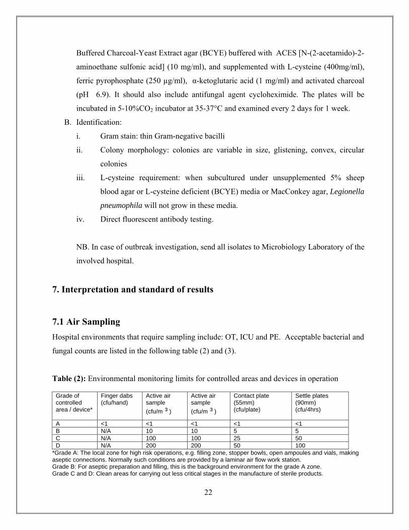
Table (2): Environmental monitoring limits for controlled areas and devices in operation
*Grade A: The local zone for high risk operations, e.g. filling zone, stopper bowls, open ampoules and vials, making aseptic connections. Normally such conditions are provided by a laminar air flow work station. Grade B: For aseptic preparation and filling, this is the background environment for the grade A zone. Grade C and D: Clean areas for carrying out less critical stages in the manufacture of sterile products.
Settle plates (90mm) (cfu/4hrs)
Contact plate (55mm) (cfu/plate)
Active air sample (cfu/m ³ )
Active air sample (cfu/m ³ )
Finger dabs (cfu/hand)
Grade of controlled area / device*
<1 <1 <1 <1 <1 A 5 5 10 10 N/A B 50 25 100 100 N/A C 100 50 200 200 N/A D
23
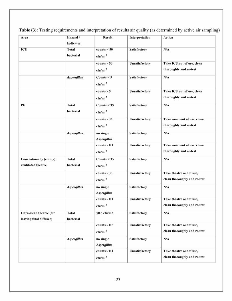
Table (3): Testing requirements and interpretation of results air quality (as determined by active air sampling) Area Hazard /
Indicator Result Interpretation Action
ICU Total
bacterial
counts < 50
cfu/m ³
Satisfactory N/A
counts > 50
cfu/m ³
Unsatisfactory Take ICU out of use, clean
thoroughly and re-test
Aspergillus
Counts < 5
cfu/m ³
Satisfactory N/A
counts > 5
cfu/m ³
Unsatisfactory Take ICU out of use, clean
thoroughly and re-test
PE Total
bacterial
Counts < 35
cfu/m ³
Satisfactory N/A
counts > 35
cfu/m ³
Unsatisfactory Take room out of use, clean
thoroughly and re-test
Aspergillus
no single
Aspergillus Satisfactory N/A
counts > 0.1
cfu/m ³
Unsatisfactory Take room out of use, clean
thoroughly and re-test
Conventionally (empty)
ventilated theatre
Total
bacterial
Counts < 35
cfu/m ³
Satisfactory N/A
counts > 35
cfu/m ³
Unsatisfactory Take theatre out of use,
clean thoroughly and re-test
Aspergillus no single
Aspergillus Satisfactory N/A
counts > 0.1
cfu/m ³
Unsatisfactory Take theatre out of use,
clean thoroughly and re-test
Ultra-clean theatre (air
leaving final diffuser)
Total
bacterial
≤0.5 cfu/m3
Satisfactory N/A
counts > 0.5
cfu/m ³
Unsatisfactory Take theatre out of use,
clean thoroughly and re-test
Aspergillus no single
Aspergillus Satisfactory N/A
counts > 0.1
cfu/m ³
Unsatisfactory Take theatre out of use,
clean thoroughly and re-test
24
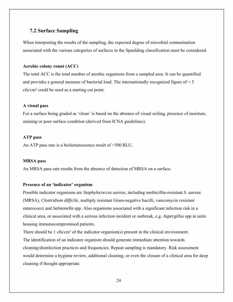
7.2 Surface Sampling
When interpreting the results of the sampling, the expected degree of microbial contamination
associated with the various categories of surfaces in the Spaulding classification must be considered.
Aerobic colony count (ACC)
The total ACC is the total number of aerobic organisms from a sampled area. It can be quantified
and provides a general measure of bacterial load. The internationally recognized figure of < 5
cfu/cm² could be used as a starting cut point.
A visual pass
For a surface being graded as ‘clean’ is based on the absence of visual soiling, presence of moisture,
staining or poor surface condition (derived from ICNA guidelines).
ATP pass
An ATP pass rate is a bioluminescence result of <500 RLU.
MRSA pass
An MRSA pass rate results from the absence of detection of MRSA on a surface.
Presence of an ‘indicator’ organism
Possible indicator organisms are Staphylococcus aureus, including methicillin-resistant S. aureus
(MRSA), Clostridium difficile, multiply resistant Gram-negative bacilli, vancomycin resistant
enterococci and Salmonella spp. Also organisms associated with a significant infection risk in a
clinical area, or associated with a serious infection incident or outbreak, e.g. Aspergillus spp in units
housing immunocompromised patients.
There should be 1 cfu/cm² of the indicator organism(s) present in the clinical environment.
The identification of an indicator organism should generate immediate attention towards
cleaning/disinfection practices and frequencies. Repeat sampling is mandatory. Risk assessment
would determine a hygiene review, additional cleaning, or even the closure of a clinical area for deep
cleaning if thought appropriate.
25
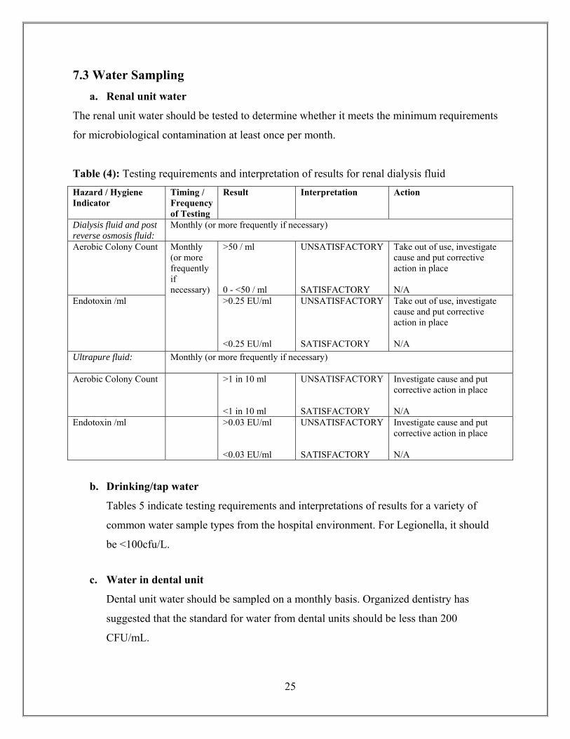
7.3 Water Sampling a. Renal unit water
The renal unit water should be tested to determine whether it meets the minimum requirements
for microbiological contamination at least once per month.
Table (4): Testing requirements and interpretation of results for renal dialysis fluid Hazard / Hygiene Indicator
Timing / Frequency of Testing
Result Interpretation Action
Dialysis fluid and post reverse osmosis fluid:
Monthly (or more frequently if necessary)
Aerobic Colony Count Monthly (or more frequently if necessary)
>50 / ml 0 - <50 / ml
UNSATISFACTORY SATISFACTORY
Take out of use, investigate cause and put corrective action in place N/A
Endotoxin /ml >0.25 EU/ml <0.25 EU/ml
UNSATISFACTORY SATISFACTORY
Take out of use, investigate cause and put corrective action in place N/A
Ultrapure fluid: Monthly (or more frequently if necessary)
Aerobic Colony Count >1 in 10 ml <1 in 10 ml
UNSATISFACTORY SATISFACTORY
Investigate cause and put corrective action in place N/A
Endotoxin /ml >0.03 EU/ml <0.03 EU/ml
UNSATISFACTORY SATISFACTORY
Investigate cause and put corrective action in place N/A
b. Drinking/tap water
Tables 5 indicate testing requirements and interpretations of results for a variety of
common water sample types from the hospital environment. For Legionella, it should
be <100cfu/L.
c. Water in dental unit
Dental unit water should be sampled on a monthly basis. Organized dentistry has
suggested that the standard for water from dental units should be less than 200
CFU/mL.
26
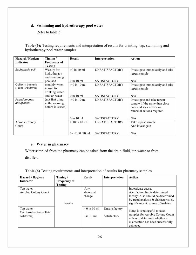
d. Swimming and hydrotherapy pool water
Refer to table 5
Table (5): Testing requirements and interpretation of results for drinking, tap, swimming and hydrotherapy pool water samples
Hazard / Hygiene Indicator
Timing / Frequency of Testing
Result Interpretation Action
Escherichia coli Weekly for hydrotherapy and swimming pool and monthly when in use for drinking water, and tap water (not first thing in the morning before it is used)
>0 in 10 ml 0 in 10 ml
UNSATISFACTORY SATISFACTORY
Investigate immediately and take repeat sample N/A
Coliform bacteria (Total Coliforms)
> 0 in 10 ml 0 in 10 ml
UNSATISFACTORY SATISFACTORY
Investigate immediately and take repeat sample N/A
Pseudomonas aeruginosa
> 0 in 10 ml 0 in 10 ml
UNSATISFACTORY SATISFACTORY
Investigate and take repeat sample. If the same then close pool and seek advice on remedial actions required N/A
Aerobic Colony Count
> 100 / 10 ml 0 - <100 /10 ml
UNSATISFACTORY SATISFACTORY
Take repeat sample And investigate N/A
e. Water in pharmacy
Water sampled from the pharmacy can be taken from the drain fluid, tap water or from
distiller.
Table (6) Testing requirements and interpretation of results for pharmacy samples Hazard / Hygiene Indicator
Timing / Frequency of Testing
Result
Interpretation
Action
Tap water – Aerobic Colony Count
weekly
Any abnormal change
Investigate cause. Alert/action limits determined locally. Also should be determined by trend analysis & characteristics, significance & source of isolates. Note: it is not useful to take samples for Aerobic Colony Count unless to determine whether a disinfection has been successfully achieved
Tap water- Coliform bacteria (Total coliforms)
> 0 in 10 ml 0 in 10 ml
Unsatisfactory Satisfactory
27
References:
• Guidelines for Environmental Infection Control in Health-Care Facilities
Recommendations of CDC and the Healthcare Infection Control Practices Advisory
Committee (HICPAC) Prepared by: Lynne Sehulster, Ph.D.1, Raymond Y.W. Chinn,
M.D.21Division of Healthcare Quality Promotion, National Center for Infectious Diseases
HICPAC member, Sharp Memorial Hospital, San Diego, California 30333, 2003.
• DRAFT Guidelines for the Collection and Interpretation of Results from Microbiological
Examination of Food, Water and Environmental Samples from the Hospital Environment
Caroline Willis, David Lamph, Kathy Nye, Elizabeth Youngs, Heather Aird, Andrew Fox
and Susanne Surman-Lee Health Protection Agency Food Water and Environmental
Microbiology Network .June 2010.
• Microbiological Air Sampling In Operating Theatres, Operational Circular OP 2023/06,
Dr John de Campo, Acting Director General, Department of Health, 20-Jan-2006.
• Colquhoun J, Partridge L. Computational Fluid Dynamics Applications in Hospital
Ventilation Design. The Australian Hospital Engineer 2003; 26(1):35-40.
• Sampling and characterization of bioaerosols. Paul A. Jensen, Ph.D., PE, CIH and Millie
P. Schafer, Ph.D., NIOSH/DPSE NIOSH. Manual of Analytical Methods. Jan 98.
Revision of Jensen PA, Lighthart B, Mohr AJ, Shaffer BT [1994]. Instrumentation used
with microbial bioaerosol. In: Lighthart B, Mohr AJ, eds. Atmospheric microbial aerosols:
theory and applications. New York, NY: Chapman & Hall, pp. 226-284.
• Performance standards for antimicrobial susceptibility testing; nineteenth informational
supplement. January 2009, MI00-S19, Vol. 29, No 3. Clinical and Laboratory Standards
Institute.
28
• Senior BW. Examination of water, milk, food and air. In: Collee GJ, Duguid JP, Fraser
AG, Marmion BP. Mackie and McCartney: Practical Medical Microbiology. Churchill
Livingstone 13th ed. Edinburgh UK. 1989.
• Stout JE, Rihs JD, Yu VL. Legionella. In: Manual of Clinical Microbiology by Murray
PR, Baron EJ, Jorgensen JH, Pfaller MA, Yolken RH. 8th edition, Volume No. 1, ASM
press, Washington DC, USA, 2003.
• Barbaree JM, Gorman GW, Martin WT, Fields BS, Morrill WE. Protocol for sampling
environmental sites for Legionellae. Applied and environmental Microbiology 1987; 53:
1454- 1458.
• Koneman EW, Allen SD, Janda WM, Schreckenberger PC, Winn Jr WC. Color Atlas and
textbook of diagnostic microbiology. Fifth edition, Lippincott Williams and Wilkins,
Philadelphia, USA, 1997.
• Microbiological Air Sampling of Operating Theatres. The Newcastle upon Tyne Hospitals
NHS Foundation Trust.: July 2009 Reviewed: July 2010
• Hoffman PN, Williams J, Stacey A et al. Microbial commissioning and monitoring of
operating theatre suites. A report of a working party of the Hospital Infection Society.
Journal of Hospital Infection 2002;52:1-28.
• NHS Estates. Health Technical Memorandum 2025: Ventilation in Healthcare Premises.
1994; HMSO: London.
• Shearer, BG. Biofilm and the dental office. J Am Dent Assoc 1996; 127:181-9.
29
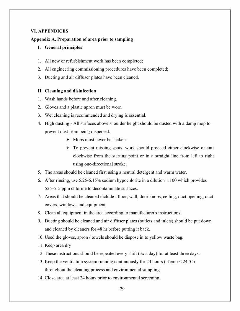
VI. APPENDICES
Appendix A. Preparation of area prior to sampling
I. General principles
1. All new or refurbishment work has been completed;
2. All engineering commissioning procedures have been completed;
3. Ducting and air diffuser plates have been cleaned.
II. Cleaning and disinfection
1. Wash hands before and after cleaning.
2. Gloves and a plastic apron must be worn
3. Wet cleaning is recommended and drying is essential.
4. High dusting:- All surfaces above shoulder height should be dusted with a damp mop to
prevent dust from being dispersed.
Mops must never be shaken.
To prevent missing spots, work should proceed either clockwise or anti
clockwise from the starting point or in a straight line from left to right
using one-directional stroke.
5. The areas should be cleaned first using a neutral detergent and warm water.
6. After rinsing, use 5.25-6.15% sodium hypochlorite in a dilution 1:100 which provides
525-615 ppm chlorine to decontaminate surfaces.
7. Areas that should be cleaned include : floor, wall, door knobs, ceiling, duct opening, duct
covers, windows and equipment.
8. Clean all equipment in the area according to manufacturer's instructions.
9. Ducting should be cleaned and air diffuser plates (outlets and inlets) should be put down
and cleaned by cleaners for 48 hr before putting it back.
10. Used the gloves, apron / towels should be dispose in to yellow waste bag.
11. Keep area dry
12. These instructions should be repeated every shift (3x a day) for at least three days.
13. Keep the ventilation system running continuously for 24 hours ( Temp < 24 ºC)
throughout the cleaning process and environmental sampling.
14. Close area at least 24 hours prior to environmental screening.
30
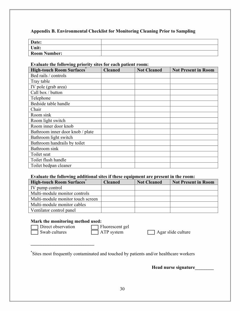
Appendix B. Environmental Checklist for Monitoring Cleaning Prior to Sampling
Date:Unit:Room Number:
Evaluate the following priority sites for each patient room: High-touch Room Surfaces* Cleaned Not Cleaned Not Present in RoomBed rails / controlsTray tableIV pole (grab area)Call box / buttonTelephoneBedside table handleChairRoom sink Room light switchRoom inner door knobBathroom inner door knob / plateBathroom light switchBathroom handrails by toiletBathroom sink Toilet seatToilet flush handleToilet bedpan cleaner
Evaluate the following additional sites if these equipment are present in the room: High-touch Room Surfaces* Cleaned Not Cleaned Not Present in RoomIV pump controlMulti-module monitor controlsMulti-module monitor touch screenMulti-module monitor cablesVentilator control panel
Mark the monitoring method used: Direct observation Fluorescent gel Swab cultures ATP system Agar slide culture
___________________________
*Sites most frequently contaminated and touched by patients and/or healthcare workers
Head nurse signature________
31
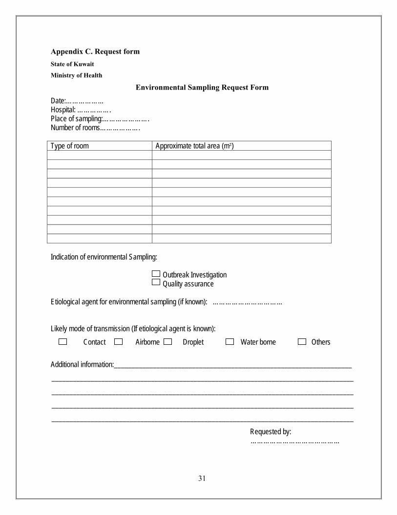
Appendix C. Request form State of Kuwait
Ministry of Health
Environmental Sampling Request Form
Date:……………… Hospital: ……………. Place of sampling:…………………. Number of rooms……………….
Type of room Approximate total area (m²)
Indication of environmental Sampling:
Outbreak Investigation Quality assurance
Etiological agent for environmental sampling (if known): ……………………………
Likely mode of transmission (If etiological agent is known): Contact Airborne Droplet Water borne Others
Additional information:___________________________________________________________________
____________________________________________________________________________________________________________________________________________________________________________________________________________________________________________________________________________________________________________________________________________________
Requested by: ……………………………………
32
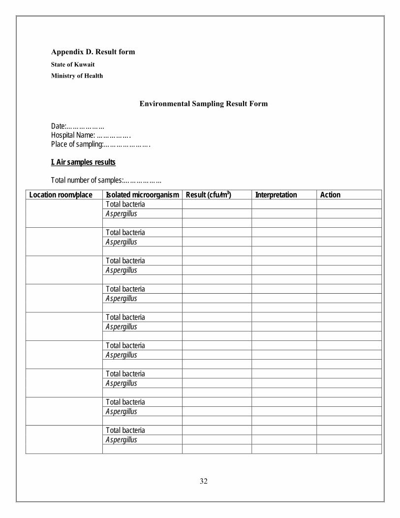
Appendix D. Result form State of Kuwait
Ministry of Health
Environmental Sampling Result Form
Date:……………… Hospital Name: ……………. Place of sampling:…………………. I. Air samples results Total number of samples:………………
Location room/place Isolated microorganism Result (cfu/m³) Interpretation Action
Total bacteria Aspergillus
Total bacteria Aspergillus
Total bacteria Aspergillus
Total bacteria Aspergillus
Total bacteria Aspergillus
Total bacteria Aspergillus
Total bacteria Aspergillus
Total bacteria Aspergillus
Total bacteria Aspergillus
33
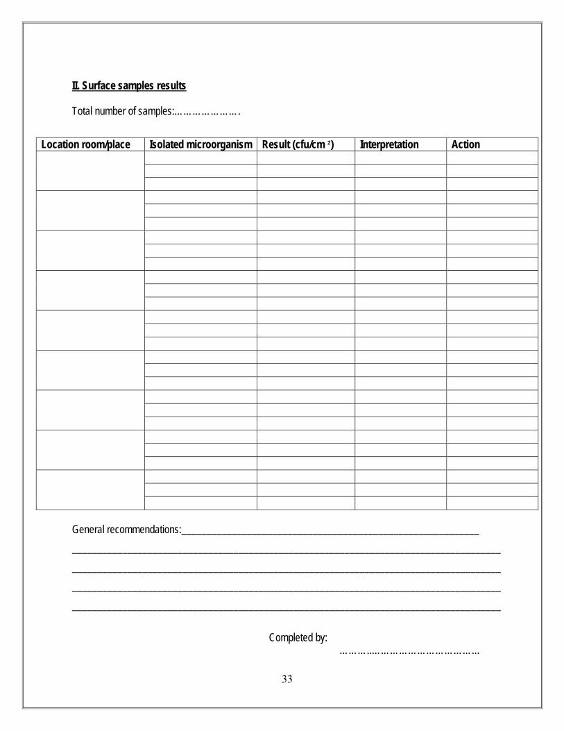
II. Surface samples results Total number of samples:………………….
General recommendations:___________________________________________________________ ____________________________________________________________________________________________________________________________________________________________________________________________________________________________________________________________________________________________________________________________________________________
Completed by: ………..………………………………
Location room/place Isolated microorganism Result (cfu/cm ²) Interpretation Action
34
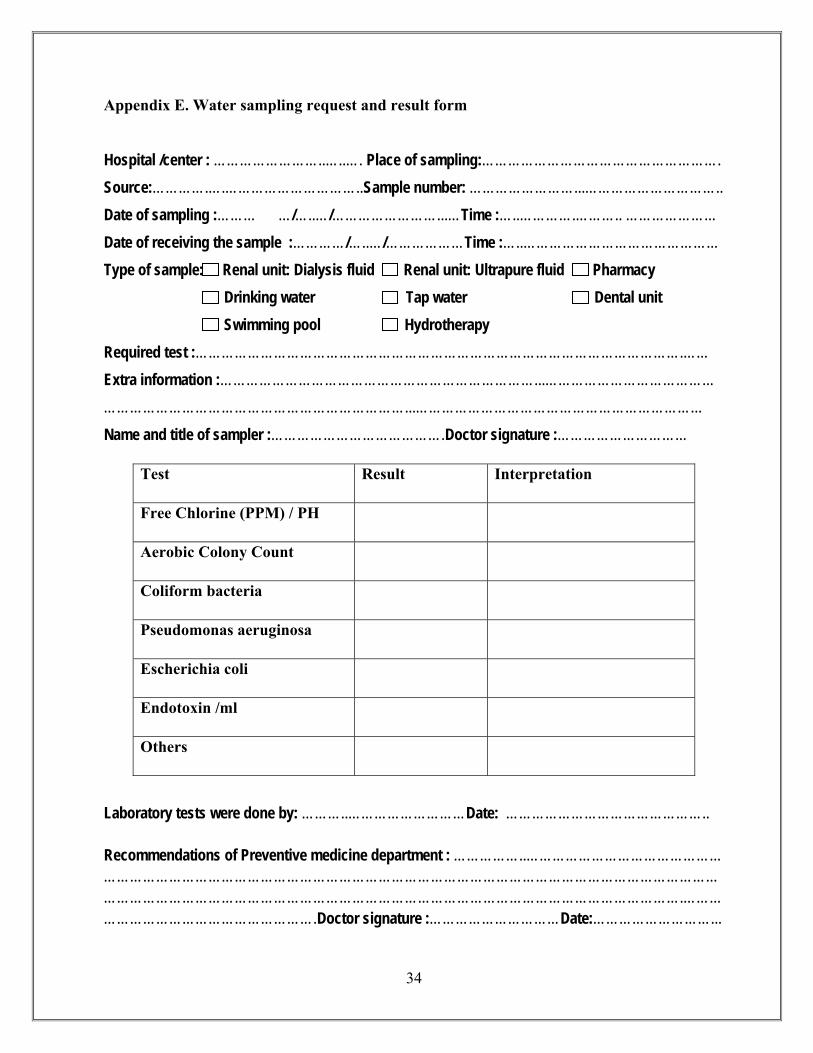
Appendix E. Water sampling request and result form Hospital /center : ……………………..…..…. Place of sampling:………………………………………………. Source:………….….…………………………..Sample number: ……………………...………………………….. Date of sampling :……… …/…..…/……………………...…Time :…..………….……….. ………………… Date of receiving the sample :…………/…..…/………………Time :…..……………………………………… Type of sample: : Renal unit: Dialysis fluid Renal unit: Ultrapure fluid Pharmacy Drinking water Tap water Dental unit Swimming pool Hydrotherapy Required test :………………………………………………………………………………………………….…… Extra information :………………………………………………………………...………………………………… ……………………………………………………………...………………………………………………………… Name and title of sampler :………………………………….Doctor signature :…………………………
Laboratory tests were done by: ………..………………………Date: ……………………………………….. Recommendations of Preventive medicine department : ……………..……………………………………… ………………………………………………………………………………………………………………………………………………………………………………………………………………………………………………….……… ………………………………………….Doctor signature :…………………………Date:………………………...
Test Result Interpretation
Free Chlorine (PPM) / PH
Aerobic Colony Count
Coliform bacteria
Pseudomonas aeruginosa
Escherichia coli
Endotoxin /ml
Others
35
Appendix F. Figure (1) The suggested sampling technique for the flat surfaces
36
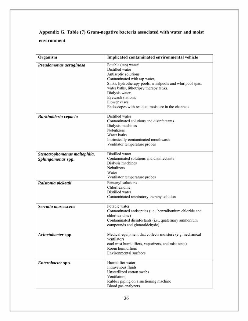
Appendix G. Table (7) Gram-negative bacteria associated with water and moist
environment
Organism Implicated contaminated environmental vehicle
Pseudomonas aeruginosa Potable (tap) water\ Distilled water Antiseptic solutions Contaminated with tap water, Sinks, hydrotherapy pools, whirlpools and whirlpool spas, water baths, lithotripsy therapy tanks, Dialysis water, Eyewash stations, Flower vases, Endoscopes with residual moisture in the channels
Burkholderia cepacia
Distilled water Contaminated solutions and disinfectants Dialysis machines Nebulizers Water baths Intrinsically-contaminated mouthwash Ventilator temperature probes
Stenotrophomonas maltophlia, Sphingomonas spp.
Distilled water Contaminated solutions and disinfectants Dialysis machines Nebulizers Water Ventilator temperature probes
Ralstonia pickettii
Fentanyl solutions Chlorhexidine Distilled water Contaminated respiratory therapy solution
Serratia marcescens
Potable water Contaminated antiseptics (i.e., benzalkonium chloride and chlorhexidine) Contaminated disinfectants (i.e., quaternary ammonium compounds and glutaraldehyde)
Acinetobacter spp.
Medical equipment that collects moisture (e.g.mechanical ventilators cool mist humidifiers, vaporizers, and mist tents) Room humidifiers Environmental surfaces
Enterobacter spp.
Humidifier water Intravenous fluids Unsterilized cotton swabs Ventilators Rubber piping on a suctioning machine Blood gas analyzers
37
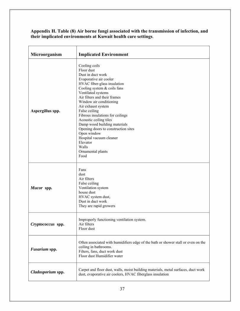
Appendix H. Table (8) Air borne fungi associated with the transmission of infection, and their implicated environments at Kuwait health care settings.
Microorganism Implicated Environment
Aspergillus spp.
Cooling coils Floor dust Dust in duct work Evaporative air cooler HVAC fiber-glass insulation Cooling system & coils fans Ventilated systems Air filters and their frames Window air conditioning Air exhaust system False ceiling Fibrous insulations for ceilings Acoustic ceiling tiles Damp wood building materials Opening doors to construction sites Open window Hospital vacuum cleaner Elevator Walls Ornamental plants Food
Mucor spp.
Fans dust Air filters False ceiling Ventilation system house dust HVAC system dust, Dust in duct work They are rapid growers
Cryptococcus spp.
Improperly functioning ventilation system. Air filters Floor dust
Fusarium spp.
Often associated with humidifiers edge of the bath or shower stall or even on the ceiling in bathrooms. Filters, fans, duct work dust Floor dust Humidifier water
Cladosporium spp. Carpet and floor dust, walls, moist building materials, metal surfaces, duct work dust, evaporative air coolers, HVAC fiberglass insulation
38
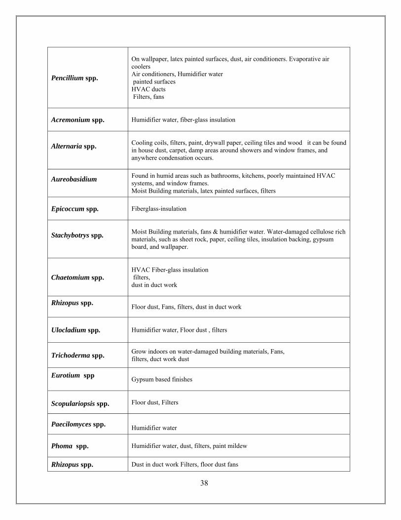
Pencillium spp.
On wallpaper, latex painted surfaces, dust, air conditioners. Evaporative air coolers Air conditioners, Humidifier water painted surfaces HVAC ducts Filters, fans
Acremonium spp. Humidifier water, fiber-glass insulation
Alternaria spp. Cooling coils, filters, paint, drywall paper, ceiling tiles and wood it can be found in house dust, carpet, damp areas around showers and window frames, and anywhere condensation occurs.
Aureobasidium Found in humid areas such as bathrooms, kitchens, poorly maintained HVAC systems, and window frames. Moist Building materials, latex painted surfaces, filters
Epicoccum spp. Fiberglass-insulation
Stachybotrys spp. Moist Building materials, fans & humidifier water. Water-damaged cellulose rich materials, such as sheet rock, paper, ceiling tiles, insulation backing, gypsum board, and wallpaper.
Chaetomium spp.
HVAC Fiber-glass insulation filters, dust in duct work
Rhizopus spp. Floor dust, Fans, filters, dust in duct work
Ulocladium spp.
Humidifier water, Floor dust , filters
Trichoderma spp.
Grow indoors on water-damaged building materials, Fans, filters, duct work dust
Eurotium spp
Gypsum based finishes
Scopulariopsis spp.
Floor dust, Filters
Paecilomyces spp. Humidifier water
Phoma spp. Humidifier water, dust, filters, paint mildew
Rhizopus spp. Dust in duct work Filters, floor dust fans
39
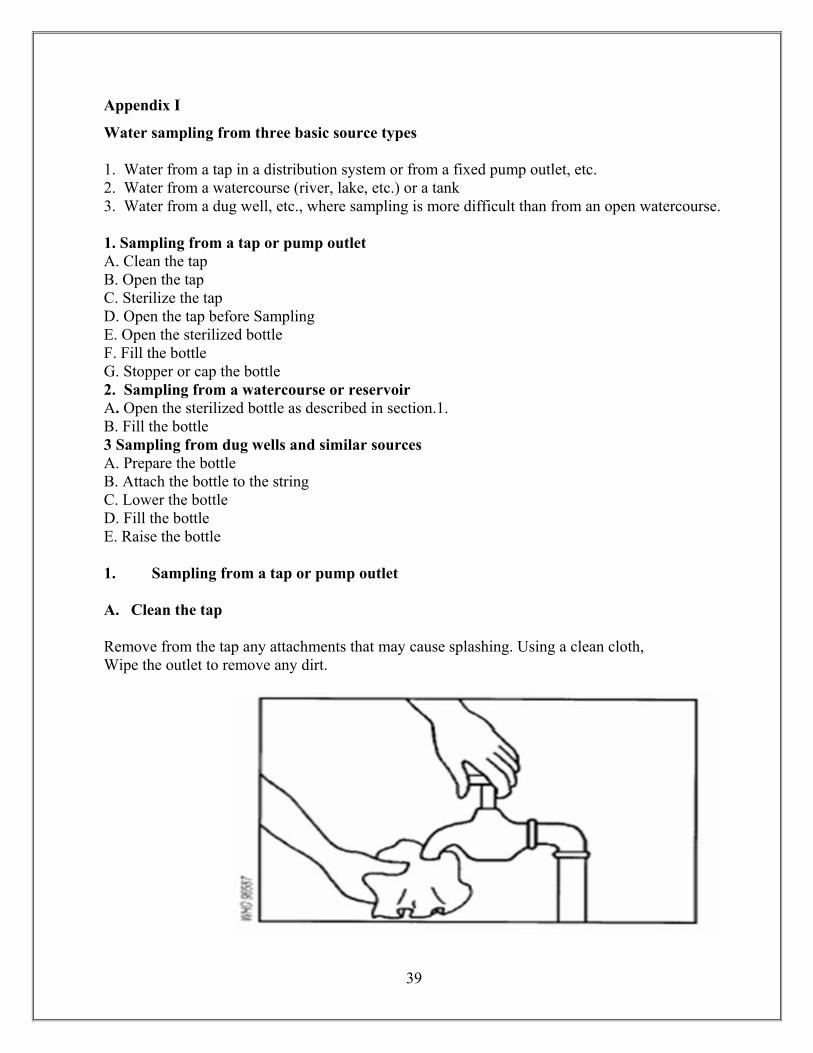
Appendix I Water sampling from three basic source types 1. Water from a tap in a distribution system or from a fixed pump outlet, etc. 2. Water from a watercourse (river, lake, etc.) or a tank 3. Water from a dug well, etc., where sampling is more difficult than from an open watercourse. 1. Sampling from a tap or pump outlet A. Clean the tap B. Open the tap C. Sterilize the tap D. Open the tap before Sampling E. Open the sterilized bottle F. Fill the bottle G. Stopper or cap the bottle 2. Sampling from a watercourse or reservoir A. Open the sterilized bottle as described in section.1. B. Fill the bottle 3 Sampling from dug wells and similar sources A. Prepare the bottle B. Attach the bottle to the string C. Lower the bottle D. Fill the bottle E. Raise the bottle 1. Sampling from a tap or pump outlet
A. Clean the tap Remove from the tap any attachments that may cause splashing. Using a clean cloth, Wipe the outlet to remove any dirt.
40
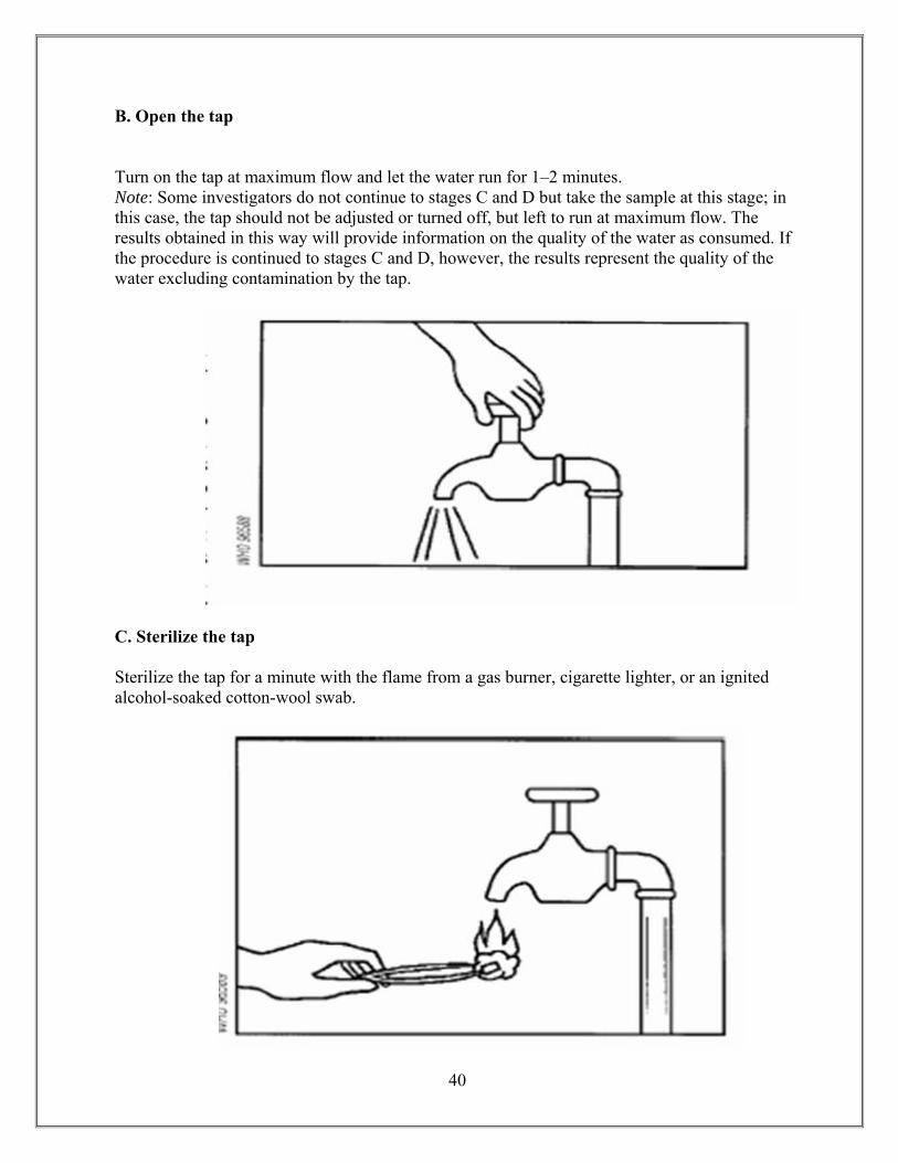
B. Open the tap
Turn on the tap at maximum flow and let the water run for 1–2 minutes. Note: Some investigators do not continue to stages C and D but take the sample at this stage; in this case, the tap should not be adjusted or turned off, but left to run at maximum flow. The results obtained in this way will provide information on the quality of the water as consumed. If the procedure is continued to stages C and D, however, the results represent the quality of the water excluding contamination by the tap.
C. Sterilize the tap Sterilize the tap for a minute with the flame from a gas burner, cigarette lighter, or an ignited alcohol-soaked cotton-wool swab.
41
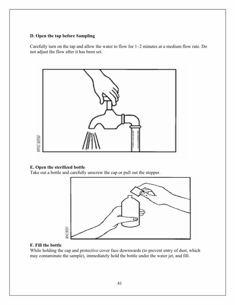
D. Open the tap before Sampling Carefully turn on the tap and allow the water to flow for 1–2 minutes at a medium flow rate. Do not adjust the flow after it has been set.
E. Open the sterilized bottle Take out a bottle and carefully unscrew the cap or pull out the stopper.
F. Fill the bottle While holding the cap and protective cover face downwards (to prevent entry of dust, which may contaminate the sample), immediately hold the bottle under the water jet, and fill.
42
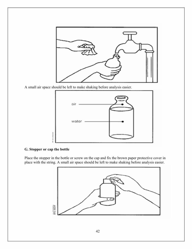
A small air space should be left to make shaking before analysis easier.
G. Stopper or cap the bottle Place the stopper in the bottle or screw on the cap and fix the brown paper protective cover in place with the string. A small air space should be left to make shaking before analysis easier.
43
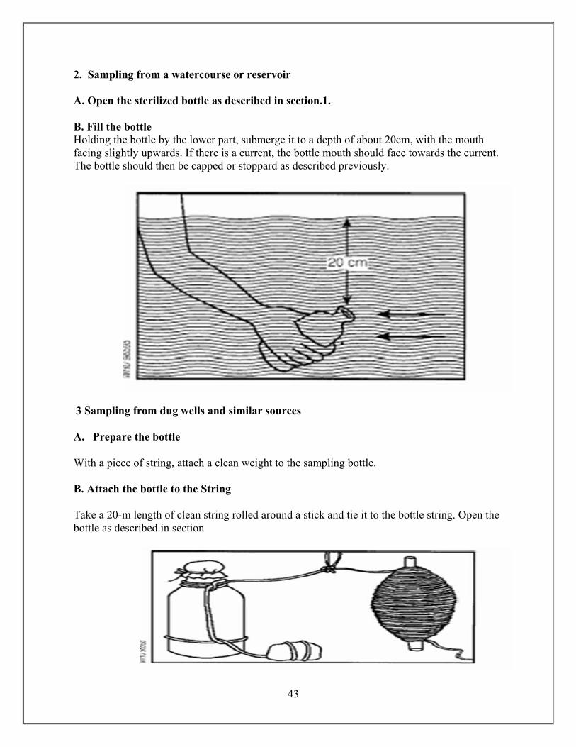
2. Sampling from a watercourse or reservoir A. Open the sterilized bottle as described in section.1. B. Fill the bottle Holding the bottle by the lower part, submerge it to a depth of about 20cm, with the mouth facing slightly upwards. If there is a current, the bottle mouth should face towards the current. The bottle should then be capped or stoppard as described previously.
3 Sampling from dug wells and similar sources A. Prepare the bottle
With a piece of string, attach a clean weight to the sampling bottle. B. Attach the bottle to the String Take a 20-m length of clean string rolled around a stick and tie it to the bottle string. Open the bottle as described in section
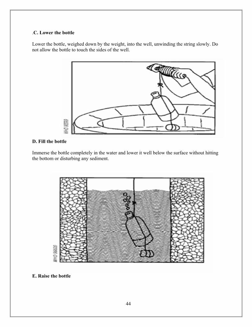
44
.C. Lower the bottle Lower the bottle, weighed down by the weight, into the well, unwinding the string slowly. Do not allow the bottle to touch the sides of the well.
D. Fill the bottle
Immerse the bottle completely in the water and lower it well below the surface without hitting the bottom or disturbing any sediment.
E. Raise the bottle
45
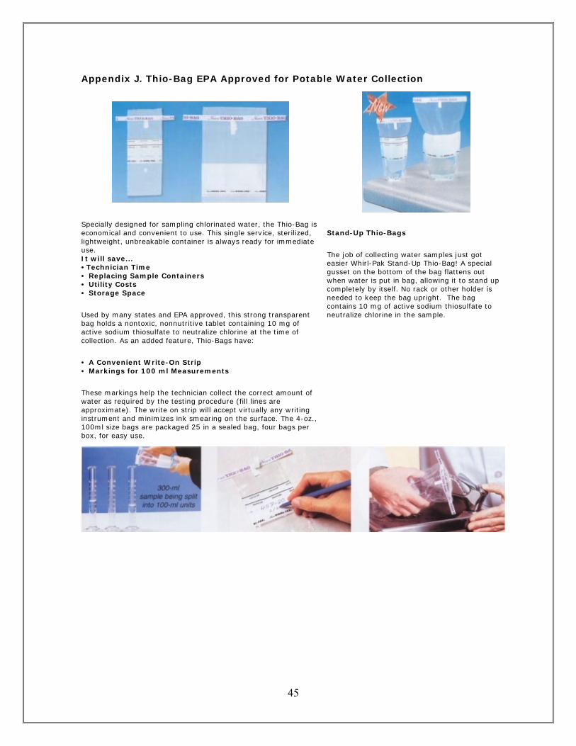
Appendix J. Thio-Bag EPA Approved for Potable Water Collection
Specially designed for sampling chlorinated water, the Thio-Bag is economical and convenient to use. This single service, sterilized, lightweight, unbreakable container is always ready for immediate use. It will save... •Technician Time • Replacing Sample Containers • Utility Costs • Storage Space
Used by many states and EPA approved, this strong transparent bag holds a nontoxic, nonnutritive tablet containing 10 mg of active sodium thiosulfate to neutralize chlorine at the time of collection. As an added feature, Thio-Bags have:
• A Convenient Write-On Strip • Markings for 100 ml Measurements
These markings help the technician collect the correct amount of water as required by the testing procedure (fill lines are approximate). The write on strip will accept virtually any writing instrument and minimizes ink smearing on the surface. The 4-oz., 100ml size bags are packaged 25 in a sealed bag, four bags per box, for easy use.
Stand-Up Thio-Bags
The job of collecting water samples just got easier Whirl-Pak Stand-Up Thio-Bag! A special gusset on the bottom of the bag flattens out when water is put in bag, allowing it to stand up completely by itself. No rack or other holder is needed to keep the bag upright. The bag contains 10 mg of active sodium thiosulfate to neutralize chlorine in the sample.


