126
Guidelines For Professional Ultrasound Practice Society and College of Radiographers and British Medical Ultrasound Society December 2015 Revision1, December 2016
Guidelines For
Professional
Ultrasound
Practice
Society and College of Radiographers and British Medical Ultrasound Society
December 2015
Revision1, December 2016
SCoR/BMUS Guidelines for Professional Ultrasound Practice. Revision 1. December 2016.
1
SOCIETY AND COLLEGE OF RADIOGRAPHERS AND BRITISH MEDICAL
ULTRASOUND SOCIETY
GUIDELINES FOR PROFESSIONAL ULTRASOUND PRACTICE
DECEMBER 2015
Revision 1, December 2016.
LIST OF CONTENTS
Acknowledgements 3
Foreword 5
Revision 1, December 2016 6
Introduction 7
Section 1
General information
1.1 Explanation of the professional title ‘sonographer’ 9
1.2 Registration for sonographers 10
1.3 Professional Indemnity Insurance 11
1.4 Profession vs tool 11
1.5 Safety of medical ultrasound 12
1.6 Medico-legal issues 13
1.7 Transducer cleaning and disinfection 14
1.8 Screening examinations using ultrasound 14
1.9 Ergonomic practice including managing the high BMI patient 16
1.10 Intimate examinations and chaperones 18
1.11 Examination times 19
1.12 Communication, ID and consent 20
1.13 Clinical governance 22
1.14 E-Learning for Healthcare (E-LfH) 23
1.15 Imaging Services Accreditation Scheme (ISAS) 24
1.16 Ultrasound equipment and quality assurance testing 25
1.17 Raising concerns; safeguarding; statutory requirements for reporting female genital mutilation;
Duty of Candour. 25
1.18 Continuing professional development (CPD) 27
1.19 Codes of professional conduct for sonographers 28
1.20 Independent practice 30
Section 2
The ultrasound examination
2.1 Overview of ultrasound examination procedures 32
2.2 Obstetric, vascular and echocardiography examinations 32
2.3 NICE and other guidelines 33
SCoR/BMUS Guidelines for Professional Ultrasound Practice. Revision 1. December 2016.
2
2.4 Vetting of ultrasound requests 33
2.5 Justification of ultrasound requests 34
2.6 Recommendations for the production of an ultrasound report 36
2.7 Gynaecological ultrasound examinations 42
2.8 Abdominal ultrasound examinations 49
Examination specific guidelines and common scenarios
2.8.1 General principles 49
2.8.2 Ultrasound examinations of the liver 52
2.8.3 Imaging of the gallbladder and biliary tree 53
2.8.4 Transabdominal ultrasound of the pancreas 55
2.8.5 Ultrasound of the spleen 56
2.8.6 Ultrasound of the bowel 58
2.9 Imaging of the uro-genital system including testes and scrotum 62
2.10 Ultrasound of the adult head and neck 65
2.11 Paediatric ultrasound examinations
2.11.1 Paediatric and neonatal liver and biliary system (including pancreas and spleen) 69
2.11.2 Paediatric urinary system 73
2.11.3 Paediatric gastro-intestinal tract 74
2.11.4 Neonatal hip 75
2.11.5 Neonatal intracranial ultrasound 76
2.12 Musculoskeletal ultrasound examinations 77
Examination specific guidelines and common scenarios
2.12.1 Shoulder 77
2.12.2 Elbow 83
2.12.3 Wrist and hand 87
2.12.4 Hip 90
2.12.5 Knee 93
2.12.6 Foot and ankle 96
2.12.7 Rheumatology ultrasound examinations 100
2.13 Elastography 110
2.14 Contrast Enhanced Ultrasoun Examinations (CEUS) 113
2.15 Interventional and extended scope examinations (sonographers) 115
2.16 Patiend Group Directions (PGD) 115
2.17 Acquisition, archiving and use of ultrasound data 117
2.18 Audit and learning from discrepancy 119
2.19 Recording of images by patients during ultrasound examinations 124
2.20 ‘Have you Paused and Checked’ prompt cards and posters 125
SCoR/BMUS Guidelines for Professional Ultrasound Practice. Revision 1. December 2016.
3
ACKNOWLEDGEMENTS
The Society and College of Radiographers and the British Medical Ultrasound Society would like to acknowledge
the help and assistance provided by the following:
For the Society and College of Radiographers (SCoR)
Mr Nigel Thomson, Professional Officer (Ultrasound), Society and College of Radiographers
Ms Wendy Williams, Superintendent Sonographer, University Hospital Llandough, Cardiff.
Member, Ultrasound Advisory Group, Society and College of Radiographers.
All past and current members of its Ultrasound Advisory Group who have contributed to or commented on
previously published documents available via www.sor.org
For the British Medical Ultrasound Society (BMUS) Professional Standards Group
Dr Oliver Byass, Consultant Radiologist, Hull and East Yorkshire Hospitals NHS Trust
Mr Peter Cantin, Consultant Sonographer, Plymouth Hospitals Trust
Dr Rhodri Evans, Consultant Radiologist/ Assoc. Professor, College of Medicine, Swansea University
Honorary Treasurer BMUS
Miss Pat Farrant, Sonographer, Kings College Hospital, London
Mrs Alison Hall, Consultant Sonographer, Keele University
Mr Gerry Johnson, Consultant Sonographer, Tameside Hospitals NHS Trust
Mrs Pamela Parker, Ultrasound Specialty Manager, Hull and East Yorkshire Hospitals NHS Trust.
Professional Development Officer, BMUS.
Dr Peter Rodgers, Consultant Radiologist, University Hospitals of Leicester
Mrs Jane Smith, Consultant Sonographer, United Leeds Hospital Trust
Mrs Jean Wilson, Programme Director, Diagnostic Imaging, University of Leeds
Due recognition is given to the editors and contributors to previous editions of United Kingdom Association of
Sonographers (UKAS) Guidelines whose earlier work forms the foundation of this new document. UKAS merged
with the Society and College of Radiographers in January 2009
The following SCoR members provided feedback on final drafts:
Alexandra Drought, Gill Harrison, Sally Hill, Steve Savage, Margaret Taylor.

SCoR/BMUS Guidelines for Professional Ultrasound Practice. Revision 1. December 2016.
4
The previous 2008 UKAS ‘Guidelines for Professional Working Standards: Ultrasound Practice’ has been archived
but is available on-line at http://www.sor.org/learning/document-library/guidelines-professional-working-
standards-ultrasound-practice
Thanks is also given to J M Bridson and G Johnson of University of Liverpool, and Dr P Rowlands of The Royal
Liverpool and Broadgreen University Hospitals NHS Trust, Liverpool for their previous work on the document
"Clinical Standards and framework for the assessment of initial and ongoing competence of ultrasound
practitioners " (BMUS 2011) which forms the foundation for section 2.6 : Recommendations for the production of
an ultrasound report.
SCoR/BMUS Guidelines for Professional Ultrasound Practice. Revision 1. December 2016.
5
FOREWORD TO THE DECEMBER 2015 EDITION
It is my pleasure to introduce the updated ‘Guidelines for Professional Ultrasound Practice’, in the past
affectionately known to sonographers as the ‘UKAS Guidelines’. The United Kingdom Association of Sonographers
was set up to support sonographers, provide advice and practice guidance and ultimately get sonography
recognised as a profession in its own right. To this day the latter still remains a challenge! However, since the last
edition of the Guidelines was produced in 2008, UKAS has merged with SCOR, so, while UKAS no longer exists, its
legacy lives on in this revised document. It is a testament to the quality of the original Guidelines that some
sections are relatively unchanged. The advice is as equally sound and relevant today as it was then.
Guidelines, however, need to keep in step with evolving technology, changes in practice and professional
progression. For this reason it was decided to produce the revised version as a web-based document that can be
regularly updated, amended and expanded as and when required.
These revised Guidelines have been produced in collaboration with the British Medical Ultrasound Society. It has
been both informative and enjoyable working with them and hopefully it is just the first of many similar future
ventures.
As with all previous editions, these Guidelines are not designed to be prescriptive but to inform good practice.
May they continue to be used in departments across the United Kingdom for years to come.
Wendy Williams
Member, Ultrasound Advisory Group, Society and College of Radiographers.
Former UKAS committee member
December 2015
REVISION 1, DECEMBER 2016
It was intended that this document would be reviewed and updated at yearly intervals (ref: Introduction, page 7).
This first revision is dated December 2016 and includes the changes indicated below. All pages are clearly marked
in the header as ‘Revision 1’. It has been given a pink cover.
The December 2015 edition will be archived but is available for future reference if needed.
Summary of main changes
Re-ordered into two main sections to simplify.
Additional acknowledgements to recognise past contributions to related documents.
All web links confirmed as completing and updated as necessary.
Significant changes to information in the following sections:
1.2 Link to HCPC statement on professional regulation
1. 7 NHS Scotland ultrasound probe cleaning recommendations. Link added
(Post Revision 1 publication- link to Wales Guidelines added January 2017)
1.8 Duty of Candour in the national screening programmes (link also added to section 1.17)
1.10 Medical Defence Union advice on protecting yourself against a sexual assault allegation. Link added
1.12 General Medical Council advice on consent. Link added
1.12 Clinical Imaging Board advice on patient identification. Link added. This advice is endorsed by BMUS
1.14 e-Learning for Healthcare information amended and updated
1.16 Updated section on ultrasound equipment quality assurance testing
1.17 HCPC guidance on raising concerns. Link added
1.18 Introduction of CPD audit to the Public Voluntary Register of Sonographers. Link added
1.20 Independent sector providers. NHS Litigation Authority (NHSLA). Link added
Registration of independent clinics in Scotland from April 2017. Link added
SCoR/BMUS Guidelines for Professional Ultrasound Practice. Revision 1. December 2016.
6
2.5 Justification- rationale remains within this document but specific examination examples now via
log-on at www.bmus.org
2.8.3 Updated guidance on biliary tree imaging
2.8.4 Updated guidance on imaging of the pancreas
2.8.5 New section on ultrasound of the spleen
2.8.6 New section on ultrasound of the bowel
2.12.7 New section on rheumatology ultrasound examinations
2.13 New section on elastography
2.20 ‘Have you paused and checked’ posters and prompt cards. New section.
PUBLICATION HISTORY- SUMMARY
SCoR/BMUS Guidelines for Professional Ultrasound Practice. December 2015 Purple cover
Revision 1 December 2016 Pink cover
SCoR/BMUS Guidelines for Professional Ultrasound Practice. Revision 1. December 2016.
7
INTRODUCTION TO DECEMBER 2015 EDITION
This document is as a result of collaboration between the Society and College of Radiographers (SCoR) and the
British Medical Ultrasound Society (BMUS). It follows five previous documents published by the United Kingdom
Association of Sonographers (UKAS) which merged with the Society and College of Radiographers (SCoR) in
January 2009. For the record these documents were:
i) Guidelines for Professional Working Practice, published in December 1993
ii) Guidelines for Professional Working Practice - Reporting, published in April 1995
iii) Guidelines for Professional Working Standards, published in August 1996
iv) Guidelines for Professional Working Standards- Ultrasound, published in October 2001
v) Guidelines for Professional Working Standards-Ultrasound Practice, published in October 2008
It has been designed as a web-based document and will only be available on-line for easier updating and to allow
for active hyperlinks to other guidance documents and organisations to be provided. As this is a new format and
there is much new and updated content, the document will initially only be available to SCoR and BMUS members
via their log-on codes. Comments and feedback are welcome and can be directed to:
https://www.sor.org/contact-us or https://www.bmus.org/contact-us/
Some links within the Guidelines are to members only SCoR or BMUS content and may require additional log-on.
Clear indication is given alongside the hyperlink when this applies. It is proposed that log-on requirements will be
reviewed in due course once feedback and comments have been taken into account. (Note: after publication of
Revision 1 in December 2016 the document will become open-access on both the SCoR and BMUS websites).
The document has been written to complement the 2014 Royal College of Radiologists (RCR) and Society and
College of Radiographers joint document entitled ‘Standards for the Provision of an Ultrasound Service’.
https://www.rcr.ac.uk/publication/standards-provision-ultrasound-service
It provides guidance on topics that were not included in the joint RCR/SCoR Standards document and provides
further detailed advice on some areas of practice that were.
There can be overlap between the terms ‘Standards’, ‘ Guidelines’ and ‘Protocols’ and this can cause confusion.
For the purposes of this document, the definitions used are the same as those in the above 2014 Royal College of
Radiologists (RCR) and Society and College of Radiographers (SCoR) document.
Standard: ‘A required or agreed level of quality or attainment. A standard is a way of ensuring optimum levels of
care or service delivery. Standards promote the likelihood of an ultrasound examination being delivered safely and
effectively, are clear about what needs to be done to comply, are informed by an evidence base and are effectively
measureable’.
Guideline: ‘A general rule, principle or piece of advice. Guidelines provide recommendations on how ultrasound
examinations should be performed and are based on best available evidence. They help ultrasound practitioners in
their work but they do not replace their knowledge and skills’.
Protocol: An agreement, preferably based on research, between practitioners to ensure the delivery of high quality
standardised ultrasound examinations.
The title of this 2015 edition ‘Guidelines for Professional Ultrasound Practice’ reflects the above definitions.
These Guidelines, which are not prescriptive, are made available to be used as recommendations for good
practice. Since the first publication of the UKAS ‘Guidelines for Professional Working Practice' in 1993, service
provision, technology and patient expectations in medical ultrasound have been transformed. The examination-
specific section, including guidelines and common clinical scenarios (ref: section 2) has been compiled by the
British Medical Ultrasound Society Professional Standards team and is presented as examples of best practice.
SCoR/BMUS Guidelines for Professional Ultrasound Practice. Revision 1. December 2016.
8
They have been included so that departments can use them as a basis to generate their own departmental
examination protocols when there are no nationally agreed ones available. There are also sections giving general
guidance and advice, including reporting and audit. Hyperlinks have been extensively used to give access to the
many relevant documents already published on a wide range of topics by organisations other than the SCoR and
BMUS. These guidelines do not and cannot cover all elements of an ultrasound examination and, in addition,
ultrasound practitioners are advised to access standard texts, documents and research in order to fully inform
local departmental protocols and procedures.
There are no guidelines included for obstetric ultrasound in this edition. Practitioners are referred to publications
from the national fetal anomaly screening programmes, the Royal College of Obstetricians and Gynaecologists
(especially their Greentop Guidelines), the Fetal Medicine Foundation, Association of Early Pregnancy Units,
British Society of Gynaecological Imaging and the International Society of Ultrasound in Obstetrics and
Gynaecology.
The term patient has been used throughout the document in preference to other terms such as client or service
user.
Several professional titles are used by those who practice ultrasound and this can lead to considerable confusion.
The term ultrasound practitioner is used throughout this document when appropriate to do so. This is consistent
with use of this term within the 2014 RCR/SCoR document referred to above. The definition of ultrasound
practitioner within the Glossary section of the above document is:
‘A healthcare professional who holds recognised qualifications in medical ultrasound and is able to competently
perform ultrasound examinations falling within their personal scope of practice. The professional background of
ultrasound practitioners can be very varied and will include radiologists, radiographers, sonographers, midwives,
physiotherapists, obstetricians and clinical scientists’.
A definition of ‘sonographer’ that is used in connection with the Public Voluntary Register of Sonographers
(PVRS) which is administered by the SCoR can be found in Section 1. This definition makes a distinction between
those ultrasound practitioners who are registered with the General Medical Council (GMC) and those who are
not. These Guidelines will be of relevance to all, hence the use of the term ‘ultrasound practitioner’ whenever
possible.
Occasionally the term ‘operator’ is used. This term is defined within the Glossary of the 2014 RCR/SCoR Standards
for the Provision of an Ultrasound Service document as:
‘A generic term used for someone who uses ultrasound equipment. It does not imply that they hold recognised
ultrasound qualifications as would an ultrasound practitioner’.
It is the nature of any document whether published in a traditional format or on-line that it can very quickly
become out of date. It is the intention of BMUS and the SCoR that this document will be regularly updated but it
is the responsibility of the ultrasound practitioner to ensure that they research and apply the most up to date
evidence in association with the contents of this document. At the time of publication (Revision 1, December
2016), all hyperlinks have been checked and are complete. Please report any broken links to the following
contact addresses: https://www.sor.org/contact-us or https://www.bmus.org/contact-us/ Comments and
feedback are also very welcome and will guide us in the further development of these Guidelines.
The Society and College of Radiographers and the British Medical Ultrasound Society would like to thank all who
have contributed to this new on-line edition of what was previously the UKAS Guidelines. Please see
acknowledgements section.
We would also like to again take this opportunity thank all the contributors and editors of previous editions of the
Guidelines who have provided us with such a firm foundation on which to build.
SCoR/BMUS Guidelines for Professional Ultrasound Practice. Revision 1. December 2016.
9
SECTION 1
GENERAL INFORMATION
1.1 EXPLANATION OF THE PROFESSIONAL TITLE ‘SONOGRAPHER’
Although it is the intention within this document to use wherever possible the term ‘ultrasound practitioner (ref:
Introduction), a full explanation of the term ‘sonographer’ will be helpful for context and important in terms of
professional recognition and recommended qualifications.
Sonographers are qualified healthcare professionals who undertake, report and take responsibility for the
conduct of diagnostic, screening and interventional ultrasound examinations. Their individual scope of practice
can be wide and varied. Sonographers also perform advanced diagnostic and therapeutic ultrasound procedures
such as biopsies and joint injections. Sonographers are either not medically qualified or they hold medical
qualifications but are not registered as a doctor with a licence to practice with the General Medical Council
(GMC).
The following definition of ‘sonographer’ is used in connection with the Public Voluntary Register of
Sonographers:
‘A healthcare professional who undertakes and reports diagnostic, screening or interventional ultrasound
examinations. They will hold qualifications equivalent to a Postgraduate Certificate or Diploma in Medical
Ultrasound that has been accredited by the Consortium for the Accreditation of Sonographic Education (CASE).
They are either not medically qualified or hold medical qualifications but are not statutorily registered with the
General Medical Council.’
Ref: https://www.sor.org/practice/ultrasound/register-sonographers Scroll down for ‘Policy and Processes’ PDF.
The minimum qualifications a sonographer would be expected to hold to practice in the UK is a postgraduate
certificate in medical ultrasound that has been accredited by the Consortium for the Accreditation of Sonographic
Education (CASE) or equivalent. Individuals without a recognised qualification, including student sonographers
should always be supervised by qualified staff.
The CASE website and a list of accredited courses can be found at http://www.case-uk.org/
The British Society of Echocardiography (BSE) and Society for Vascular Technology of Great Britain and Ireland
(SVT) also accredit individual ultrasound practitioners working within their respective specialties.
http://www.bsecho.org/home/
http://www.svtgbi.org.uk/
The Society and College of Radiographers (SCoR) can provide accreditation of advanced and consultant practice
for its sonographer members http://www.sor.org/career-progression (SCoR member log-in required). A
sonographer should:
i) recognise and work within their personal scope of practice, seeking advice as necessary;
ii) ensure that a locally agreed and written scheme of work is in place;
iii) work with reference to national and local practice and guideline recommendations;
iv) ensure they hold appropriate professional indemnity insurance or obtain this by virtue of their
employment (ref: section 1.3).
See also section 1.19 on codes of professional conduct for sonographers.

SCoR/BMUS Guidelines for Professional Ultrasound Practice. Revision 1. December 2016.
10
The general standards of education and training for ultrasound practitioners are set out on page 12 of the 2014
Royal College of Radiologists/Society and College of Radiographers document ‘Standards for the Provision of an
Ultrasound Service’: https://www.rcr.ac.uk/publication/standards-provision-ultrasound-service
1.2 REGISTRATION FOR SONOGRAPHERS
This section uses the professional term ‘sonographer’ instead of the generic ‘ultrasound practitioner’ (ref:
Introduction) and refers to the long running campaign to have ‘sonography’ recognised as a profession and for
the professional title of ‘sonographer’ to be legally protected.
The registration situation for sonographers is complex. 1
The majority of sonographers are statutorily registered but this will depend on their professional background and
is not achievable for all. Statutory registration will most likely be as a radiographer or clinical scientist with the
Health and Care Professions Council (HCPC) or as a midwife or nurse with the Nursing and Midwifery Council
(NMC) and not as a sonographer, which is not a protected title. Whether statutorily registered or not,
sonographers are encouraged to apply to register with the Public Voluntary Register of Sonographers (PVRS)
which is administered by the Society and College of Radiographers:
http://www.sor.org/practice/ultrasound/register-sonographers
For some sonographers, this will be the only register available to them. For those sonographers who are already
statutorily registered, applying to register with the PVRS will help to protect the public and support the case for
statutory regulation. This was recommended by the then Health Professions Council (HPC) to the Secretary of
State for Health in 2009 but has not progressed. Government policy since 2011 has been not to bring further
aspirant groups into statutory registration unless there is a clear evidence of clinical risk that requires this. 2
Link to HCPC advice on aspirant groups for registration, including their May 2016 policy statement on extending
professional regulation:
http://www.hcpc-uk.co.uk/aboutregistration/aspirantgroups/ and http://www.hcpc-
uk.co.uk/assets/documents/10005047Policystatementonextensionofprofessionalregulation.pdf
NHS employers have advice on sonographer registration available at:
http://www.nhsemployers.org/your-workforce/retain-and-improve/standards-and-assurance/professional-
regulation/medical-radiography-and-ultrasound-workforce
The Society and College of Radiographers has advice on ultrasound training, employment, registration and
professional indemnity insurance at:
https://www.sor.org/learning/document-library/ultrasound-training-employment-registration-and-professional-
indemnity-insurance-0
The British Medical Ultrasound Society has information available at:
https://www.bmus.org/careers-training/ and https://www.bmus.org/careers-training/training/
For some sonographers working in areas of practice coming within the remit of the Academy for Healthcare
Science (AHCS), statutory registration may be available either by following approved education and training
routes as a clinical scientist or by being able to demonstrate ‘equivalence’. Statutory registration, if it is
obtainable, will be with the HCPC as a clinical scientist. The AHCS also administers a voluntary register that is
accredited by the Professional Standards Authority.
http://www.ahcs.ac.uk/
https://www.professionalstandards.org.uk/accredited-registers
The Registration Council for Clinical Physiologists runs a voluntary register that is relevant for professionals
specialising in echocardiography: https://www.rccp.co.uk/
SCoR/BMUS Guidelines for Professional Ultrasound Practice. Revision 1. December 2016.
11
References
1) Thomson N, Paterson A. Sonographer registration in the UK- a review of the current situation. Ultrasound Feb
2014. 22(1):52-56
2) Enabling excellence. Autonomy and accountability for healthcare workers, social care workers and social care
workers. London: HMSO. Feb 2011
1.3 PROFESSIONAL INDEMNITY
The UK government introduced legislation in 2014 which requires ultrasound practitioners who are statutorily
registered with the Health and Care Professions Council (HCPC) (eg as a radiographer, physiotherapist or clinical
scientist), Nursing and Midwifery Council (NMC) (eg as a nurse or midwife), or other statutory regulator, to have a
professional indemnity arrangement as a condition of their statutory registration. The majority of statutorily
registered ultrasound practitioners will already meet this requirement and will not need to take any further
action. They will either work in an employed environment where their employer will indemnify them, and / or if
they undertake self-employed work, they will have already made their own professional indemnity arrangements.
However, some statutorily registered ultrasound practitioners may need to take steps to make sure that they
have appropriate professional indemnity arrangements in place.
Registrants and applicants for statutory registration will be asked to confirm that they meet, or will meet, this
requirement by completing a professional declaration when renewing or registering for the first time. The HCPC
have published guidance on the requirements along with an accompanying flow diagram which can be
downloaded from:
Professional indemnity and your registration - Guidance
Meeting the professional indemnity requirements as a condition of HCPC registration
NMC guidance: http://www.nmc-uk.org/Registration/Professional-indemnity-arrangements/
GMC guidance: http://www.gmc-uk.org/doctors/information_for_doctors/insurance_and_indemnity.asp
In addition to working in an employed environment, professional indemnity insurance can be obtained through
membership of trade unions and professional bodies or by purchasing from medical defence unions or
commercial insurers.
Ultrasound practitioners who are self employed or who work in a part employed/ part self-employed
environment are particularly advised to read the guidance published by their statutory regulator.
There is no professional indemnity insurance associated with voluntary registration on the Public Voluntary
Register of Sonographers.
If an ultrasound practitioner is not statutorily registered, it is clearly good practice to ensure that they have
appropriate professional indemnity arrangements in place both to protect the public and themselves.
1.4 PROFESSION vs TOOL
There are many healthcare professionals working within the UK who use ultrasound as a ‘tool’ to assist with their
overall treatment or evaluation of patients. There is published advice on education and training available to
those who use ultrasound in this way but whose main work and role is not that of an ultrasound practitioner. For
those who use the professional title of ‘sonographer’, ultrasound is their daily work and their primary profession.
When used as a ‘tool’, ultrasound aids and assists a healthcare practitioner with their wider examination and
treatment, but in overall terms, ultrasound is only a small part of their work. It is important for safe and effective
service delivery that all ultrasound examinations are undertaken by appropriately trained and competent
personnel and that there is associated audit and continuing professional development in the use of ultrasound.
i) CASE focused courses via http://www.case-uk.org/ and
http://www.case-uk.org/resources/Directory+of+Courses+2015+-+2016+v2.pdf
SCoR/BMUS Guidelines for Professional Ultrasound Practice. Revision 1. December 2016.
12
ii) Royal College of Radiologists –‘Ultrasound training recommendations for medical and surgical specialities’
http://www.rcr.ac.uk/publications.aspx?PageID=310&PublicationID=385
iii) Royal College of Radiologists- ‘Focused ultrasound training standards’
http://www.rcr.ac.uk/publications.aspx?PageID=310&PublicationID=386
1.5 SAFETY OF MEDICAL ULTRASOUND
‘Ultrasound is now accepted as being of considerable diagnostic value. There is no evidence that diagnostic
ultrasound has produced any harm to patients in the four decades that it has been in use. However, the acoustic
output of modern equipment is generally much greater than that of the early equipment and, in view of the
continuing progress in equipment design and applications, outputs may be expected to continue to be subject to
change. Also, investigations into the possibility of subtle or transient effects are still at an early stage.
Consequently diagnostic ultrasound can only be considered safe if used prudently’. 1
A broad range of ultrasound exposure is used in the different diagnostic modalities currently available. Doppler
imaging and measurement techniques may require higher exposures than those used in B- and M-modes, with
pulsed Doppler techniques having the potential for the highest levels.
Recommendations related to ultrasound safety assume that the equipment being used is designed to
international or national safety requirements and that it is operated by competent and trained personnel.
It is the responsibility of the operator or ultrasound practitioner to be aware of, and apply, the current safety
standards and regulations and to undertake a risk/benefit assessment for each examination.
Key principles for the safe use of ultrasound: 2
i) Medical ultrasound imaging should only be used for medical diagnosis.
ii) Ultrasound equipment should only be used by people who are fully trained in its safe and proper operation.
This requires:
• an appreciation of the potential thermal and mechanical bio-effects of ultrasound;
• a full awareness of equipment settings;
• an understanding of the effects of machine settings on power levels.
iii) Examination times should be kept as short as is necessary to produce a useful diagnostic result.
iv) Output levels should be kept as low as is reasonably achievable while producing a useful diagnostic result.
v) The operator should aim to stay within the BMUS recommended scan times (especially for obstetric
examinations).
vi) Scans in pregnancy should not be carried out for the sole purpose of producing souvenir videos or
photographs.
The British Medical Ultrasound Society has UK leading advice on ultrasound safety that all ultrasound
practitioners should be familiar with at https://www.bmus.org/policies-statements-guidelines/safety-
statements/
Also available via this link are Guidelines for the management of safety when using volunteers and patients for
practical training in ultrasound scanning.
SCoR/BMUS Guidelines for Professional Ultrasound Practice. Revision 1. December 2016.
13
https://www.bmus.org/static/uploads/resources/MANAGEMENT_OF_SAFETY_WHEN_USING_VOLUNTEERS__PAT
IENTS_FOR_PRACTICAL_TRAINING_YtWarot.pdf
BMUS Guidelines for live demonstrations of patient scans to an audience can be found at:
https://www.bmus.org/static/uploads/resources/GUIDELINES_FOR_LIVE_DEMONSTRATIONS_OF_PATIENT_SCAN
S_TO_AN_AUDIENCE.pdf
BMUS have a sample consent form for ultrasound scanning for the purposes of teaching and/or demonstration at:
https://www.bmus.org/static/uploads/resources/Consent_Form_for_Ultrasound_Scanning_for_the_Purposes_of
_Teaching.pdf
References:
1) British Medical Ultrasound Society, Statement on the safe use and potential hazards of diagnostic ultrasound.
2) British Medical Ultrasound Society, Guidelines for the safe use of diagnostic ultrasound equipment.
1.6 MEDICO-LEGAL ISSUES
The place of work should have a written set of protocols that accurately describes the range of ultrasound
examinations undertaken. Their content should address the ultrasound examinations, their reporting and the
appropriate referral pathways for patients with normal and abnormal ultrasound findings. The details in the
protocols should be such that a new staff member, having read them, could carry out and report these
examinations and appropriately refer the patient after the examination to the expected standard. Protocols
should be updated regularly and their review date should be included in their content. Superseded protocols
should be kept on file permanently.
Records are currently required by law to be kept for a number of years as specified by Department of Health
advice (ref: section 2.11).
The following guidance should be considered:
• ultrasound practitioners should be aware that they are legally accountable for their professional
actions, including the reporting of ultrasound examinations, in all circumstances.
• the report is a public document and part of the patient’s medical record, together with any images,
and/or video recordings which may accompany it.
• the patient consents to an ultrasound examination that he or she has the right to expect will be
delivered and reported by a competent ultrasound practitioner.
• a competent ultrasound practitioner is one who works to the standards defined by the guidelines of
his or her place of work, the code of conduct of his or her professional body, the guidelines of that
and other relevant bodies and of the regulatory body where appropriate.
• the standard of care provided by a competent ultrasound practitioner is that which the majority of
similar individuals would provide and/or which a significant body of similar individuals would provide
in similar and contemporaneous circumstances.
• images that accompany an ultrasound examination carried out by a competent ultrasound
practitioner evidence the assumption that the necessary standard of care has been delivered (ref:
section 2.15).
• all images must be capable of being attributed to the correct examination and should include the
patient identifier(s), examination date and time.
• nationally published requirements for the storage of images must be followed. Examples would be
the image storage requirements of the abdominal aortic aneurysm and fetal anomaly screening
programmes and those published by the Department of Health (ref: section 2.1).
SCoR/BMUS Guidelines for Professional Ultrasound Practice. Revision 1. December 2016.
14
See also Duty of Candour, section 1.17.4
1.7 TRANSDUCER CLEANING AND DISINFECTION
In March 2016, NHS Scotland published ‘Guidance for the decontamination of semi-critical ultrasound probes,
semi-invasive and non-invasive ultrasound probes’ http://www.hps.scot.nhs.uk/documents/hai/infection-
control/guidelines/NHSScotland-Guidance-for-Decontamination-of-Semi-Critical-Ultrasound-Probes.pdf
The following published information (2014) applies to Wales. Welsh Health Technical Memorandum 01-06.
There is a section within on cleaning and disinfecting transvaginal and transrectal probes
http://www.wales.nhs.uk/sites3/Documents/254/WHTM%2001%2D06%20Part%20C.pdf (link added January
2017).
The SCoR published general advice and an overview in September 2014. This includes reference to two Medicines
and Healthcare Products Regulatory Authority (MHRA) alerts. This document is available as a PDF from
https://www.sor.org/practice/ultrasound/professional-issues (SCoR log-in required), the relevant PDF can be
found at the bottom of the SCoR web page.
The hyperlinks to the two MHRA alerts are:
https://www.gov.uk/drug-device-alerts/medical-device-alert-reusable-transoesophageal-echocardiography-
transvaginal-and-transrectal-ultrasound-probes-transducers-failure-to-appropriately-decontaminate
https://assets.digital.cabinet-office.gov.uk/media/5485ac42ed915d4c100002a7/con065543.pdf
BMUS advice on transducer cleaning and disinfection is available at https://www.bmus.org/policies-statements-
guidelines/clinical-protocols/
1.8 SCREENING EXAMINATIONS USING ULTRASOUND
Since the 2008 edition of the UKAS Guidelines there have been major developments as far as screening and
ultrasound are concerned.
The United Kingdom National Screening Committee advises ministers in all four countries and resides within
Public Health England, an executive agency of the Department of Health.
Before any pathology or condition is accepted for national screening there is a full evaluation against the NSC
published criteria.
The NSC website can be found at: http://www.screening.nhs.uk/uknsc
Details of the evidence review process: https://www.gov.uk/guidance/evidence-and-recommendations-nhs-
population-screening#evidence-review-proces
It should be noted that there may be variations in the screening programmes that operate across the four
countries of the UK and ultrasound practitioners should contact the relevant organisations for current advice.
Scotland: http://www.nsd.scot.nhs.uk/services/screening/
Wales: http://www.screeningforlife.wales.nhs.uk/home
Northern Ireland: http://www.publichealth.hscni.net/directorate-public-health/service-development-and-
screening/screening
In England there have been recent (2015) major changes to the screening programme websites. Information for
the public has been moved to NHS Choices. Information for professionals is now hosted on the .gov.uk website
and e-learning for professionals and linked to the programmes is hosted by Health Education England.
SCoR/BMUS Guidelines for Professional Ultrasound Practice. Revision 1. December 2016.
15
The four national screening programmes that are of particular relevance to ultrasound practitioners are:
i) Antenatal screening
In England the Fetal Anomaly Screening Programme (FASP) is responsible for the two ultrasound scans that are
offered to every pregnant woman. There are equivalent organisations to FASP in the devolved countries although
the 11+2
w to 14
+1 w scan is not offered as a screening scan in Northern Ireland.
The two ultrasound scans for which FASP (England) is responsible are the 11+2
to 14+1
w scan that includes the
combined test for Trisomies 21, 13 and 18 and the 18w to 20+6
w fetal anomaly scan.
FASP has published comprehensive information for professionals that is available at:
https://www.gov.uk/topic/population-screening-programmes/fetal-anomaly
Equivalent organisations to FASP in Scotland and Wales:
Scotland: National Services Division: http://www.pnsd.scot.nhs.uk/
Wales: Antenatal Screening Wales http://www.antenatalscreening.wales.nhs.uk/public/home
Independent providers offering screening ultrasound scans to NHS patients during pregnancy must work within
the published screening programme standards for the country in question.
ii) NHS Abdominal Aortic Aneurysm (AAA) Screening Programme
The Abdominal Aortic Aneurysm screening programme has now successfully completed its roll out across the UK.
Advice for professionals is available at:
https://www.gov.uk/topic/population-screening-programmes/abdominal-aortic-aneurysm
Information on AAA screening in the devolved countries can be found via:
Scotland: http://www.isdscotland.org/Health-Topics/Public-Health/AAA-Screening/
Wales: http://www.aaascreening.wales.nhs.uk/
Northern Ireland: http://www.publichealth.hscni.net/directorate-public-health/service-development-and-
screening/abdominal-aortic-aneurysm-aaa-screening
iii) NHS Breast Screening Programme
http://www.cancerscreening.nhs.uk/breastscreen/
Although ultrasound is not part of the initial screening examination, specialists in breast ultrasound will use
ultrasound techniques for further evaluation and biopsy.
iv) NHS Neonatal and Infant Physical Examination (NIPE) screening programme
This national screening programme is responsible for issuing guidance and standards regarding the physical
examination of the newborn in England. There is no equivalent screening programme in the devolved countries.
Guidance on when ultrasound examinations of the neonatal hip should be performed can be found at:
https://www.gov.uk/government/uploads/system/uploads/attachment_data/file/572685/NIPE_programme_han
dbook_2016_to_2017_November_2016.pdf (page 17)
The overall ‘Standards’ document for the NIPE programme can be found at:
https://www.gov.uk/government/publications/newborn-and-infant-physical-examination-screening-standards
SCoR/BMUS Guidelines for Professional Ultrasound Practice. Revision 1. December 2016.
16
It is important to note that the neonatal hip ultrasound examination itself is a post- screening examination and is
outside the remit of NIPE.
Public Heath England advice on private screening for different conditions and diseases.
Information outlining the advantages and disadvantages of screening outside the national programmes can be
found via the following web link. There is information and leaflets available for healthcare professionals and links
to leaflets written for patients.
https://www.gov.uk/guidance/private-screening-for-health-conditions-nhs-recommendations
Duty of candour guidance in the screening programmes
Published in October 2016 (See also section 1.17)
https://phescreening.blog.gov.uk/2016/10/05/new-duty-of-candour-guidance-helps-ensure-were-open-and-
honest-in-screening/
1.9 ERGONOMIC PRACTICE INCLUDING MANAGING THE HIGH BMI PATIENT
Prevention and management of Work Related Musculo-Skeletal Disorders
Work related musculo-skeletal disorders (WRMSD) are known to be associated with ultrasound practice. There
are several causative factors including high workloads, increasing body mass index of patients, poor equipment
and room design and poor posture when scanning. It is important that ultrasound practitioners take care of
themselves and their working environment whilst scanning.
Employers have a legal duty of care to their employees and should be guided in ways to avoid potential work
related injuries ie by supplying equipment fit for purpose and being realistic about time management.
Departmental guidelines should include strategies to minimise the risk of WRMSD, including appropriate
management of workload (ref: section 1.11).
Many advice and guidance documents have been published to which ultrasound practitioners are referred:
Health and Safety Executive
Risk management of musculoskeletal disorders in sonography work (2012):
http://www.hse.gov.uk/healthservices/management-of-musculoskeletal-disorders-in-sonography-work.pdf
Society and College of Radiographers
Work related musculo-skeletal disorders (sonographers) (2014, Revision August 2016):
https://www.sor.org/learning/document-library/work-related-musculo-skeletal-disorders-sonographers
The causes of musculoskeletal injury amongst sonographers in the UK (2002):
https://www.sor.org/learning/document-
library?sort_by=field_date_published_value&title=causes+&taxonomy_topics_tid=All&field_archive_value=0
Prevention of work related musculoskeletal disorders (2007):
https://www.sor.org/learning/document-
library?sort_by=field_date_published_value&title=SOR+prevention+of+work+related+musculoskeletal+disorders
&taxonomy_topics_tid=All&field_archive_value=0
Royal College of Radiologists and Society and College of Radiographers
Standards for the provision of an ultrasound service (2014):
https://www.rcr.ac.uk/publication/standards-provision-ultrasound-service (section 2)
SCoR/BMUS Guidelines for Professional Ultrasound Practice. Revision 1. December 2016.
17
Managing the high BMI patient
The following is an extract from the 2014 SCoR document ‘Work related musculo-skeletal disorders’:1
Factors to consider when scanning patients with a high BMI:
The following points are all particularly relevant when scanning high body mass index (BMI)/bariatric patients and
are in addition to general good practice methods of reducing the incidence of WRMSDs.
All Trusts and Health Boards should have policies relating to care and manual handling associated with high
BMI/bariatric patients which should also be available and consulted.
• use ‘high BMI’ presets on the machine as a starting point to manipulating the image. Manufacturers can set
these up to your requirements at the time of installation and will optimise features such as transducer frequency
and harmonics.
• Wherever possible the sonographer workforce should be rotated to ensure that it is not the same sonographer
group exposed to risk. This is of course will depend on the skill mix of the local sonographer workforce.
• Do not extend the examination time beyond what is normally allowed if there is unlikely to be any gain. It may
be that a second appointment is necessary in some cases. FASP provide guidance with respect to
repeat examinations on those women attending for the 18 – 20+6
weeks fetal anomaly scan and where the image
quality is compromised by such as by an increased BMI. There is also ‘twice on the couch only’ advice for the 11+2
w to 14+1
w scan which forms part of the combined test. The FASP Programme Handbook can be found at:
https://www.gov.uk/government/publications/fetal-anomaly-screening-programme-handbook (See sections 5.2
page 12 and 5.6.2 page 16).
• Avoid pressing unnecessarily hard and for too long. This may increase the risk of WRMSD and it can be
uncomfortable for the patient. Firm pressure may be contra-indicated for some types of pathology or clinical
situations.
• Use a helper to support tissue/fatty aprons and generally assist with the examination.
• Consider the patient’s feelings.
• Use good quality equipment with good harmonics.
• Do not exceed the couch weight limit which should be clearly posted.
• Use available manual handling aids when necessary; scan in-patients in their beds rather than transferring to an
examination couch.
• Report pain/injury to occupational health/line manager as a record and so that current practise can be
reviewed.
• BMI should be recorded on request forms if above 30.
• If image quality is compromised, state how the examination has been affected in the report.
• Record BMI on report.
• Keep current practice for high BMI patients under review.
• Consider wording of information leaflets about limitations of scanning at time of booking
SCoR/BMUS Guidelines for Professional Ultrasound Practice. Revision 1. December 2016.
18
References
1) Work related musculo-skeletal disorders. 2014 London: SCoR
https://www.sor.org/learning/document-library/work-related-musculo-skeletal-disorders-sonographers
1.10 INTIMATE EXAMINATIONS AND CHAPERONES
The definition of an intimate examination may differ between individual patients for ethnic, religious or cultural
reasons. In addition, some patients may have a clear preference for a health carer of specific gender due to their
ethnic, religious or cultural background, because of previous experiences or in view of their age. Where possible,
such individual needs and preferences should be taken into consideration.
When conducting an intimate examination, the ultrasound practitioner should:
• act with propriety and in a courteous and professional manner;
• communicate sensitively and politely using professional terminology,
• use a chaperone when appropriate;
• respect the patient’s rights to dignity and privacy;
• comply with departmental schemes of work and protocols.
Patients should not be asked to remove clothing unnecessarily. When required, private, warm, comfortable and
secure facilities for dressing and undressing should be provided. Care should be taken to ensure privacy in
waiting areas used by patients not fully dressed in their own clothes. During the ultrasound examination, only
those body parts under examination should be exposed.
Care must be taken to maintain confidentiality when non-health care personnel are nearby.
Patients should be given the opportunity to have a chaperone, irrespective of the ultrasound practitioner’s
gender and the examination being undertaken. The ultrasound practitioner should give equal consideration to
their own need for a chaperone, again, irrespective of the examination being undertaken or the gender of the
patient.
A record should be made in patients’ records when chaperones are offered and used, and when they are
declined. The record should include the name and designation of the chaperone. Chaperones should normally be
members of the clinical team who are sufficiently familiar with the ultrasound examination being carried out to
be able to reliably judge whether the ultrasound practitioner’s actions are professionally appropriate and
justifiable.
Patients’ privacy and dignity should be maintained throughout the examination which should be conducted
without interruption. Only personnel essential for carrying out the examination should be in the room.
It is good practice (as for any examination) to ensure that, when possible, hand washing and equipment cleaning
are carried out in full view of the patient at the beginning and end of the examination to reassure him or her that
effective infection control procedures are being applied.
Advice on students/trainees and intimate examinations is given in the 2011 SCoR document and in the 2015 RCR
guidance (hyperlinks below).
There are several organisations that have produced advice on the conduct of intimate examinations and also on
the use and role of chaperones.
Society and College of Radiographers (2016) Intimate Examinations and Chaperone Policy
https://www.sor.org/learning/document-library/intimate-examinations-and-chaperone-policy-0
General Medical Council (2013) Intimate examinations and chaperones
http://www.gmc-uk.org/guidance/ethical_guidance/21168.asp
SCoR/BMUS Guidelines for Professional Ultrasound Practice. Revision 1. December 2016.
19
Royal College of Radiologists (2015) Intimate examinations and the use of chaperones
https://www.rcr.ac.uk/sites/default/files/bfcr154_intimateexams.pdf
In 2015, the Medical Defence Union published advice on protecting yourself from a sexual assault allegation
https://www.themdu.com/guidance-and-advice/latest-updates-and-advice/protecting-yourself-from-a-sexual-
assault-allegation
1.11 EXAMINATION TIMES
The time allowed for an ultrasound examination should take into account the fact that the actual transducer time
is only a component of the overall examination.
Time needs to be allowed for room preparation, assessing the ultrasound request, introductions, explanations,
obtaining consent and assisting the patient when necessary on to and off the examination couch. Post-
procedure time is required to discuss the findings with the patient, write the report, archive the images and
attend to the after-care of the patient, including making arrangements for further appointments and/or further
investigations. Equipment will also need cleaning and disinfecting as required post examination.
An ultrasound practitioner has a professional responsibility to ensure that the time allocated for an examination is
sufficient to enable it to be carried out competently. It is critical to patient management that no ultrasound
examination is compromised by departmental and or government targets.
The allocated appointment time will vary depending on the type and complexity of the ultrasound examination.
It may also be influenced by the expertise of the ultrasound practitioner and training commitments within the
department. In addition, the duration of the examination will be influenced by the scan findings and/or the
physical condition of the patient. The quality of equipment and general support available to the sonographer are
also relevant.
Examination times should be determined with reference to national standards such as those published by the
Fetal Anomaly Screening Programme, by organisations such as NICE and by proper evaluation of the local working
arrangements and resources that will be different for each service provider. Examination times will need to take
into account whether there are trainees present and their stage of training if teaching is to be effective.
Although obtstrics is not included in the scope of this document the following recommended times are included
in this setion for completeness. NHS England has published service specifications (2016-2017) for commissioners
relating to fetal anomaly screening. The relevant service specifications are Nos. 16 (combined test) and 17 (18w
to 20w 6d fetal anomaly scan).
https://www.england.nhs.uk/commissioning/wp-content/uploads/sites/12/2016/04/serv-spec-16-apr16.pdf
Extract from page 12 of service specification No. 16:
To complete the ultrasound component of this screening strategy, the scan appointment should allocate time to
incorporate pre-scan counselling, the ultrasound examination, post-scan counselling and reporting. The time
allocation for appointments to meet these requirements is a minimum of twenty (20) minutes.
https://www.england.nhs.uk/commissioning/wp-content/uploads/sites/12/2016/04/serv-spec-17-apr16.pdf
Extract from page 13 of service specification No: 17:
The ultrasound scan’s appointment should incorporate pre -scan counselling, the ultrasound examination, post-
scan counselling and reporting. The time allocation for appointments to meet these requirements for a singleton
pregnancy is a minimum of thirty (30) minutes and for a multiple pregnancy is forty five (45) minutes.
The National Institute for Health and Care Excellence (NICE) has published recommendations for multiple
pregnancy (growth) ultrasound examinations. ‘Multiple pregnancy: The management of twin and triplet
pregnancies in the antenatal period‘(2011). CG129. These can be found at:
SCoR/BMUS Guidelines for Professional Ultrasound Practice. Revision 1. December 2016.
20
http://www.nice.org.uk/guidance/cg129/chapter/1-Guidance
Section 1.3.3.4
Thirty minutes is recommended.
The Society and College of Radiographers has published guidance on examination times at:
http://www.sor.org/learning/document-library/ultrasound-examination-times-and-appointments-0 In the
absence of any local evidence-based determination of examination times or national standards, the SCoR advises
that 20 minutes should be the minimum for a general abdominal ultrasound examination. The complete
document should be read for full context.
Individual departments can determine examination times taking into account local circumstances. A tool to help
evaluate these is the former ‘NHS Improvement -Examination Times Assessment Tool’. This is still available via
http://webarchive.nationalarchives.gov.uk/20130221101407/http:/www.improvement.nhs.uk Full details of
how to access are in Appendix 3 of the SCoR examination times document.
Many request forms are very non-specific in terms of the patient’s symptoms and due allowance may need to be
made for this in schedule planning if it is decided to proceed (Ref: sections 2.4, 2.5). For example, it may be
necessary to perform both transabdominal and transvaginal scans to fully evaluate the female abdomen and
pelvis with ultrasound.
1.12 PATIENT IDENTIFICATION, COMMUNICATION AND CONSENT
While undertaking any ultrasound examination and working in accordance with locally agreed practice,
ultrasound practitioners should:
• correctly identify the patient . Clinical Imaging Board advice that is endorsed by BMUS is available at:
https://www.sor.org/sites/default/files/document-
versions/cib_medical_ultrasound_examinations_document.pdf
• obtain sufficient verbal and/or written information from the referring clinician to undertake correctly the
examination requested (ref: sections 2.4,2.5);
• be mindful of the need to use interpreters as and when necessary to communicate adequately with the
patient;
• greet the patient using his or her full name and status;
• be able to discuss the relative risks and benefits of the examination with the patient;
• explain the scanning procedure appropriately to the patient;
• obtain informed consent from the patient or their representative being mindful of his/her capacity to
understand;
• be aware of the individual patient’s special needs including chaperoning and privacy during the
examination (ref: section 1.10 );
• be professional and understanding throughout the examination; manage the interaction between the
patient and any accompanying adults and children in a way that enables the examination to be carried
out to a competent standard;
• explain and discuss the findings with the patient within local guidelines;
• interpret and communicate the findings appropriately and in a timely fashion to the referring clinician;
• ensure appropriate arrangements have been made for further care before the conclusion of the
examination as necessary.
SCoR/BMUS Guidelines for Professional Ultrasound Practice. Revision 1. December 2016.
21
Valid informed consent must be obtained before commencing any ultrasound examination or procedure.
Ultrasound practitioners who do not respect the right of a patient to determine what happens to their own body
in this way may be liable to legal or disciplinary action.
The consent process is a continuum beginning with the referring health care professional who requests the
ultrasound examination and ending with the ultrasound practitioner who carries it out. It is the responsibility of
the referring professional to provide sufficient information to the patient to enable the latter to consent to the
ultrasound examination being requested. It is the responsibility of the ultrasound practitioner to ensure that the
patient understands the scope of the ultrasound examination prior to giving his or her consent.
Verbal informed consent must be obtained for all examinations. Additional informed verbal consent should be
obtained where a student ultrasound practitioner undertakes part or all of the ultrasound examination under
supervision.
Verbal informed consent for those examinations of an intimate nature should be recorded in the ultrasound
report. Some categories of ultrasound examination (interventional ultrasound, guided procedures eg biopsy) will
require written consent.
Literature which explains the scope of the examination clearly and accurately should be made available to
patients prior to the ultrasound examination. NHS Choices carries information on a wide range of topics.
An example for ultrasound examinations is:
http://www.nhs.uk/conditions/Ultrasound-scan/Pages/Introduction.aspx
The national screening programmes have explanatory literature available for patients obtainable via the NHS
Choices website and have published consent standards which are usually now included with the various NHS
England service specifications.
NHS Choices provides information on abdominal aortic aneurysm screening (for example) at:
http://www.nhs.uk/conditions/abdominal-aortic-aneurysm-screening/pages/introduction.aspx
There is much information that has been published on informed consent. The following are all relevant:
Royal College of Radiologists
Standards for patient consent particular to radiology (2012):
https://www.rcr.ac.uk/standards-patient-consent-particular-radiology-second-edition
General Medical Council
Consent: Patients and Doctors making decisions together
http://www.gmc-uk.org/guidance/ethical_guidance/consent_guidance_index.asp
Society and College of Radiographers
Consent to Imaging and Radiotherapy Treatment Examinations (2007) (SCoR log-on required and link may need to
be copied into browser).
https://www.sor.org/system/files/document-library/members/sor_consent_document.pdf
Patient identification, confidentiality and consent, further guidance (2009):
https://www.sor.org/learning/document-library/patient-identification-confidentiality-and-consent-further-
guidance
Consent and adults of impaired capacity (2010):
https://www.sor.org/learning/document-library/consent-and-adults-impaired-capacity
SCoR/BMUS Guidelines for Professional Ultrasound Practice. Revision 1. December 2016.
22
British Medical Ultrasound Society
Statement on patient information and informed consent:
https://www.bmus.org/static/uploads/resources/STATEMENT_ON_PATIENT_INFORMATION_AND_INFORMED_C
ONSENT_gQkvKTu.pdf
1.13 CLINICAL GOVERNANCE
Clinical governance is defined in the 1998 consultation document ‘A First Class Service in the New NHS’1 and also
in 1998 by Scally and Donaldson 2
in the British Medical Journal as:
“A framework through which NHS organisations are accountable for continuously improving the quality of their
services and safeguarding high standards of care by creating an environment in which excellence in clinical care
will flourish."
As clinical governance is based on professional values and concern for others, the ultrasound practitioner is
actively involved in this process of accountability as part of his or her daily activities. By safeguarding high
standards of care and seeking to continuously improve its quality, it ensures that health care provision is patient-
centred which is central to the concept.
The main components of a clinical governance framework can be summarised as follows: 3
i) Risk management
ii) Clinical audit
iii) Education, training and Continuous Professional Development
iv) Patient and carer experience and involvement
v) Staffing and staff management
An example of published Trust information on clinical governance can be found at:
http://www.uhb.nhs.uk/clinical-governance-components.htm
Department of Health advice can be found at:
https://www.gov.uk/government/uploads/system/uploads/attachment_data/file/213304/Final-NQB-report-v4-
160113.pdf
For the ultrasound practitioner, clinical governance involves:
i) clinical effectiveness: taking part in personal, departmental and wider audit programmes to evaluate
clinical practice and service to patients. This will include audit of ultrasound examinations and
reports: participation in multi-disciplinary team meetings and radiology discrepancy meetings would
be further examples;
ii) patient identification, communication and consent: (ref: section 1.12);
iii) patient safety: including avoiding physical injury and following published ultrasound safety guidelines
(ref: sections 1.5, 1.9);
iv) risk management: ultrasound practitioners have a duty to participate in education and training
offered by employers on subjects such as back care, health and safety and infection control;
v) education, training and Continuous Professional Development (ref: section 1.18)
vi) team working: RCR/SCoR document ‘Team Working in Clinical Imaging’ (2012);
https://www.rcr.ac.uk/team-working-clinical-imaging
vii) patient, public and carer involvement;
viii) being accountable for one’s own actions;
SCoR/BMUS Guidelines for Professional Ultrasound Practice. Revision 1. December 2016.
23
ix) the implementation of national clinical guidance which reflects the best standards of care. Examples
would include implementing NICE Guidelines and national screening programme guidance and
requirements;
x) incident reporting and raising concerns. This is of particular importance following the publication of
the Francis Report in 20134
(ref: section 1.17).
In 2008 the National Ultrasound Steering Group published a document entitled ‘Ultrasound Clinical Governance’.
The National Ultrasound Steering Group was a short-term sub-group of the National Imaging Board.
https://www.bmus.org/static/uploads/resources/ClinicalGovernanceInUltrasound-061108.pdf
The following ‘four layer’ advice from the General Medical Council for those who work in an employed
environment as part of wider clinical teams 5
is also relevant to the topic of clinical governance and is included for
consideration:
‘The first layer (of patient protection) is the individual practitioner and their commitment to a common set of
ethics, values and principles which puts patients first. Next is team-based regulation which reflects the importance
of acting if a colleague’s conduct or performance is putting patients at risk. After that comes workplace regulation
which reflects the responsibilities of NHS and other healthcare providers and finally, the regulator, through work
on standards, education and fitness to practise’.
Web links
Standards for the provision of an ultrasound service (2014):
https://www.rcr.ac.uk/system/files/publication/field_publication_files/BFCR%2814%2917_Standards_ultrasound.
Ultrasound Clinical Governance in Wales:
http://www.wales.nhs.uk/sitesplus/documents/1064/Ultrasound%20and%20Clinical%20Governance.doc
Royal College of Radiologists Standards for learning from discrepancies meetings (2014):
https://www.rcr.ac.uk/publication/standards-learning-discrepancies-meetings
Royal College of Radiologists Cancer multi-disciplinary team meetings. Standards for clinical radiologists (Second
edition) (2014):
https://www.rcr.ac.uk/sites/default/files/docs/radiology/pdf/BFCR%2814%2915_MDTMs.pdf
References
1) Department of Health (1998) A First Class Service: Quality in the New NHS. London. DH.
2) Scally G, Donaldson L. Clinical governance and the drive for quality improvement in the new NHS in England.
British Medical Journal 1998. 317: 61
3) University Hospital Birmingham Foundation Trust
4) Report of the Mid Staffordshire NHS Trust Public Enquiry. Chair: Robert Francis QC. (2013)
https://www.gov.uk/government/publications/report-of-the-mid-staffordshire-nhs-foundation-trust-public-
inquiry
5) General Medical Council. Report and accounts 2004.
1.14 E-LEARNING FOR HEALTHCARE (e-LfH)
e-LfH is an award winning Health Education England programme in partnership with the NHS and professional
bodies providing high quality content free of charge for the training of the NHS workforce across the UK.
http://www.e-lfh.org.uk/home/
Formed following a ground-breaking project in Radiology, e-LfH is now in the process of delivering over 300 e-
learning projects in partnership with medical Royal Colleges and other healthcare organisations. The on-line
training sessions enhance traditional learning, support existing teaching methods and provide a valuable
reference point. They are designed and built to be engaging and interactive, using quality images, video, audio
SCoR/BMUS Guidelines for Professional Ultrasound Practice. Revision 1. December 2016.
24
and animation to help trainees learn and retain knowledge. Content is presented using various templates such as
‘real-life’ scenarios, case studies and ‘knowledge bites’. The full e-LfH syllabus which was current at the
preparation of this Revision 1 (October 2016) can be viewed via the embedded link below.
There are seven modules on ultrasound; each module contains a number of sessions as indicated below:
· 15 Gynaecological Ultrasound
· 16 Abdominal Ultrasound
· 17 Men's Health Ultrasound
· 18 Vascular Ultrasound
· 19 Musculoskeletal Ultrasound
· 20 Head and Neck Ultrasound
· 21 Obstetric Ultrasound
The sessions within the ‘image interpretation’ programme have been written by expert ultrasound practitioners
to match the format used by e-LfH. This is a very valuable learning resource and can contribute to an ultrasound
practitioner’s continuing professional development (Ref: section 1.18)
All of these sessions can be accessed from the programme page:
http://portal.e-lfh.org.uk/Component/Details/391087
There is also an introductory session: e-IRI_14_06 - Technology: Ultrasound
http://portal.e-lfh.org.uk/Component/Details/386697
A sample demonstration session is available at: http://www.e-lfh.org.uk/demo/
In order to access these free -learning sessions, is it necessary to first register with the programme at
http://portal.e-lfh.org.uk/Register This web page gives information on who is eligible to register and alternative
access routes for those who are not.
1.15 IMAGING SERVICES ACCREDITATION SCHEME (ISAS)
All providers of ultrasound services are encouraged to have their services independently assessed by the United
Kingdom Accreditation Service (UKAS) against the Imaging Services Accreditation Standard (ISAS), jointly
established by the Royal College of Radiologists and the College of Radiographers. ISAS accreditation provides
strong and independent confirmation that high quality services are being delivered. Details on ISAS and how to
apply can be obtained from https://www.rcr.ac.uk/clinical-radiology/service-delivery/imaging-services-
accreditation-scheme-isas and http://www.sor.org/about-isas
ISAS is now recognised by the Care Quality Commission and has been approved for use within CQC hospital
inspection methodology: http://www.isas-uk.org/CQC_Recognition.shtml
UKAS are always recruiting new assessors from all areas of imaging. If anyone is interested in applying to become
an assessor please contact http://www.isas-uk.org/jobs.shtml
image_interpretation_sessions_syllabus.pdf
SCoR/BMUS Guidelines for Professional Ultrasound Practice. Revision 1. December 2016.
25
1.16 ULTRASOUND EQUIPMENT AND QUALITY ASSURANCE TESTING
An ultrasound practitioner is expected to:
i) Have detailed knowledge of ultrasound equipment in order to ensure that it is appropriate for
purpose;
ii) Manipulate the equipment correctly so that patient diagnosis and management are not compromised;
iii) Take care when using the equipment in order to avoid damage;
iv) Ensure that regular planned preventative maintenance is carried out by qualified personnel;
v) Ensure that an agreed quality assurance programme is in place that incorporates the regular inspection of
ultrasound machines and ancillary equipment
The stated aims of quality assurance procedures applied to ultrasound equipment are to ensure consistent and
acceptable levels of performance of the imaging system and image recording facilities and to ensure the safety of
the patient. The foundation of a good quality assurance programme is regular visual inspection of the
equipment and the reverberation pattern of each probe by the users, since the majority of faults may be
detected in this way. Common faults are damage to probes, which may present an electrical or infection hazard
and/or affect the efficiency of all or part of the probe. Formal quality assurance protocols focus on the
consistency of specific features of image quality over time. The acceptability of image quality may not be
apparent from measurable changes in the parameters tested. The issue of what constitutes unacceptable
equipment performance is still very difficult to assess objectively, but there is evidence that probe faults, such as
drop out affecting more than 1 element, compromise diagnostic quality.
There is a section relating to image quality requirements, quality assurance and equipment replacement in the
RCR/SCoR document ‘Standards for the provision of an ultrasound service’ (2014):
https://www.rcr.ac.uk/sites/default/files/documents/BFCR(14)17_Standards_ultrasound.pdf (section 2)
The British Medical Ultrasound Society has recommended Quality Assurance testing and monitoring; advice
available at http://ult.sagepub.com/content/22/1/6.short?rss=1&ssource=mfr
1.17 RAISING CONCERNS; SAFEGUARDING; STATUTORY REQUIREMENTS FOR REPORTING FEMALE
GENITAL MUTILATION (FGM), DUTY OF CANDOUR.
1.17.1 Raising concerns
An executive summary of the February 2013 Report of the Mid Staffordshire NHS Foundation Trust Public Enquiry
(‘The Francis Report’) which is relevant to this sub-section can be found at:
http://webarchive.nationalarchives.gov.uk/20150407084003/http://www.midstaffspublicinquiry.com/sites/defau
lt/files/report/Executive%20summary.pdf
NHS Trusts and Health Boards will have their own published policies on ‘raising concerns’ following the
publication of the above report. All healthcare professionals have a professional duty to report concerns they may
have about the safety of patients and of service delivery.
The following will also be of help if needing to raise concerns:
NHS England has advice at:
https://www.england.nhs.uk/contact-us/complaint/
Health and Care Professions Council advice at:
http://www.hpc-uk.org/registrants/raisingconcerns/
The Care Quality Commission has advice entitled ‘Raising a concern with the Care Quality Commission if you are a
member of staff’
SCoR/BMUS Guidelines for Professional Ultrasound Practice. Revision 1. December 2016.
26
http://www.cqc.org.uk/content/report-concern-if-you-are-member-staff
The SCoR has information published at:
https://www.sor.org/learning/document-
library?sort_by=field_date_published_value&title=raising+concerns&taxonomy_topics_tid=All&field_archive_val
ue=0 (SCoR member log-on required).
A ‘Duty of Care’ handbook for healthcare professional published by Public World in 2013 is available at:
http://www.publicworld.org/files/Duty_of_Care_handbook_April_2013.pdf
1.17.2 Safeguarding
Ultrasound practitioners also have a duty to report concerns relating to children and vulnerable adults.
Employers will have available advice and policies as to the pathways that ultrasound practitioners are required to
follow. Training and updating in local safeguarding procedures and policies is likely to be a mandatory
requirement of the employer.
Further general information at https://www.england.nhs.uk/wp-content/uploads/2015/07/safeguard-policy.pdf
1.17.3 Female Genital Mutilation (FGM) - statutory requirements of practitioners
From 31st
October 2015 (England and Wales) there is a legal requirement for all statutory registered ultrasound
practitioners to report female genital mutilation in those less than 18 years of age to the police. This duty is on
the individual ultrasound practitioner; it cannot be transferred. Complying with the duty does not breach any
confidentiality requirement or other restriction of disclosure that might apply. The same principle also applies to
ultrasound practitioners who are not statutorily registered.
The HCPC have advice at:
http://www.hcpc-uk.co.uk/mediaandevents/news/index.asp?id=727
This links through to Home Office advice at:
https://www.gov.uk/government/publications/mandatory-reporting-of-female-genital-mutilation-procedural-
information
Further advice, including flow diagrams and posters at:
https://www.gov.uk/government/publications/fgm-mandatory-reporting-in-healthcare
1.17.4 Duty of Candour
Ultrasound practitioners should also be aware of the requirements of their Professional Indemnity Insurer if asked
to make any statements regarding patient care, complaints and claims.
The NHS Litigation Authority has advice on the Duty of Candour at:
http://www.nhsla.com/OtherServices/Documents/NHS%20LA%20-%20Duty%20of%20Candour.pdf
Duty of candour guidance in the national screening programmes (October 2016, Public Health England).
https://phescreening.blog.gov.uk/2016/10/05/new-duty-of-candour-guidance-helps-ensure-were-open-and-
honest-in-screening/
General Medical Council advice
http://www.gmc-uk.org/guidance/ethical_guidance/27233.asp
SCoR/BMUS Guidelines for Professional Ultrasound Practice. Revision 1. December 2016.
27
1.18 CONTINUING PROFESSIONAL DEVELOPMENT
Continuing professional development (CPD) has been defined as follows:
"...is the process by which health professionals keep updated to meet the needs of patients, the health service,
and their own professional development. It includes the continuous acquisition of new knowledge, skills, and
attitudes to enable competent practice..." 1
It is a process that seamlessly continues from starting as a student, through successful completion of a first
qualification and lasts throughout a sonographer's professional life. CPD is embedded in the NHS clinical
governance strategy (ref: section 1.13).
All ultrasound practitioners must be engaged with continuing professional development.
Many ultrasound practitioners are registered with the Health and Care Professions Council (HCPC) as a
radiographer or the Nursing and Midwifery Council (NMC) as a nurse or midwife. Medically qualified staff will be
registered with the General Medical Council (GMC). These regulators all have their own requirements for CPD.
Some regulators use an ‘outputs’ based model based on learning and reflection (eg HCPC). Other regulators use
an ‘inputs’ based model which will place emphasis on certificates and attending study days.
HCPC information, which applies to many ultrasound practitioners, can be found at: http://www.hpc-
uk.org/registrants/cpd/standards/
Equivalent Information will be published for their registrants by the other statutory regulators also.
If an ultrasound practitioner is not statutorily registered, the same good practice principles of CPD will still apply.
For those ultrasound practitioners registered with the Public Voluntary Register of Sonographers, they will have
signed at the point of application to state that they undertake CPD activities. Audit of CPD based on HCPC
principles and systems will be introduced from the March 1st
2017 renewal of voluntary registration.
https://www.sor.org/system/files/article/201604/2016.4.5_cpd_final.pdf
Evidence of CPD is also a requirement to maintain accreditation with the Society of Vascular Technology (SVT)
http://www.svtgbi.org.uk/education/
Evaluation and reflection on the learning gained from the following can all be used as evidence to meet Health
Care Professions Council requirements. Other regulators may have a different emphasis or procedures;
ultrasound practitioners are advised to consult the website of their own regulator where information will be
available.
i) Successfully completing a programme of study
ii) Attendance at, and participation in, appropriate professional workshops and conferences
iii) Defining and implementing a departmental audit programme
iv) Implementing a change process in practice
v) Mentoring an ultrasound student in practice
vi) Participation in an ultrasound or professional research project
vii) Attendance at and participation in case reviews
viii) Attendance at radiology discrepancy meetings and multi-disciplinary team meetings
ix) Submission of a paper to a journal
x) Critical evaluation of a peer reviewed research paper
xi) Teaching peers and students both formally and informally
xii) Completion of E-Learning for Health modules and other on-line learning activities
xiii) Active engagement with a professional ultrasound group, professional or regulatory body or
scientific society
SCoR/BMUS Guidelines for Professional Ultrasound Practice. Revision 1. December 2016.
28
xiv) Promoting the practice of ultrasound to other health care professionals within a wider context
xv) Communicating with, and imaging, patients
The list of possible activities above is for example only. There are many and varied other activities that can also
contribute to an individual’s CPD.
It is important that suitable records are maintained and evidence is compiled on a regular basis. Evidence should
not only include attendance certificates at events but also written records of personal learning and reflection.
Society and College of Radiographers members have access to ‘CPD Now’. This is a user-friendly on-line tool for
identifying, planning, evaluating and recording CPD that will also help the individual if asked to present evidence
of CPD by the HCPC: https://www.sor.org/learning/cpd/cpd-now A ‘CPD Now’ app has recently been made
available for android devices, with ‘Apple ‘devices to follow.
References
1) Peck C, McCall M, McLaren B, Rotem T. (2000). Continuing medical education and continuing professional
development: international comparisons British Medical Journal 320 (7232): 432-435
1.19 CODES OF PROFESSIONAL CONDUCT FOR SONOGRAPHERS
These codes have been included to support the use of the professional title of ‘sonographer’ and the future
development and regulation of the sonography profession. Hence the use of this term rather than the generic
‘ultrasound practitioner’ (ref: Introduction)
A Code of Practice can be defined as a set of written rules which explains how people working in a particular
profession should behave. It is designed to cover all circumstances, is written in broad terms and expresses
ethical principles.
The statements below are from the Code of Practice for Sonographers which were previously published by the
United Kingdom Association of Sonographers (UKAS) in the Guidelines for Professional Working Standards –
Ultrasound Practice (2008) and are as equally relevant today.
These statements that reflect best practice are a guide and offer advice to sonographers, educationalists,
students of medical ultrasound and other health care practitioners. They are statements of professional conduct
that reflect the individual's rights, local and national changing patterns of ultrasound service delivery and the
requirement of sonographers to demonstrate continuing competency through personal and professional
development.
There are other codes that sonographers and ultrasound practitioners may need to follow. If statutorily
registered with the GMC, HCPC or NMC, these regulators have themselves published codes of conduct and ethics.
Professional bodies such as the Society and College of Radiographers and the Royal College of Midwives, for
example, also have published codes of conduct.
The Public Voluntary Register of Sonographers has associated with it published Standards of Conduct,
Performance and Ethics and Standards of Proficiency.
http://www.sor.org/practice/ultrasound/register-sonographers
Scroll down for the relevant PDFs. The Standards of Conduct, Performance and Ethics document also contains a
conscientious objection statement which is copied below.
A code of practice for sonographers
1. Sonographers have a duty of care to their patients with respect to the minimisation of ultrasound
exposure consistent with diagnostic needs.
SCoR/BMUS Guidelines for Professional Ultrasound Practice. Revision 1. December 2016.
29
2. Sonographers are ethically and legally obliged to hold in confidence any information acquired as a result
of their professional and clinical duties, except where there is a legal obligation for disclosure.
3. Sonographers must be committed to the provision of a quality ultrasound service having due regard for the
legislation and established codes of practice related to health care provision in order to minimise risk to
patients, patients’ carers and other professionals.
4. Sonographers are legally and professionally accountable for their own practice and must not be influenced
by any form of discrimination.
5. Sonographers must identify limitations in their practice and request training and support to meet their
perceived needs.
6. Sonographers will take all reasonable opportunity to maintain and improve their knowledge and
professional competency and that of their peers and students.
7. Sonographers must pay due regard to the way in which they are remunerated for their work.
8. Sonographers have a duty of care to work collaboratively and in co-operation with the multi- disciplinary
health care team in the interests of their patients and patients’ carers??.
9. Sonographers must act at all times in such a manner as to justify public trust and confidence, to uphold
and enhance the reputation of sonography and to serve the public interest.
10. Sonographers must ensure that unethical conduct and any circumstances where patients and others are at
risk are reported to the appropriate authority.
11. Sonographers who are held accountable in another area of health care must relate this Code to others
that govern their practice.
12. Student sonographers pursuing a qualification in medical ultrasound must adhere to their University or
Higher Education Institution’s Codes of Conduct that relate to all elements of their ultrasound education
and training.
Conscientious objection
The following is the conscientious objection clause from the Standards of Conduct, Performance and Ethics that is
associated with the Public Voluntary Register of Sonographers:
‘You must report in writing to your employing authority, at the earliest date in your employment, any
conscientious objection that may be relevant to your professional practice. You should explore with them ways in
which you can avoid placing an unreasonable burden on colleagues because of this. Your right to conscientious
objection does not exempt you from providing service users with full, unbiased information; for example, prior to
prenatal screening or testing. You do not have the right to refuse to take part in any emergency treatment’.
The NMC have conscientious objection advice at: http://www.nmc.org.uk/standards/code/conscientious-
objection-by-nurses-and-midwives/. This gives further details of the legal circumstances when such a clause
might apply.
SCoR/BMUS Guidelines for Professional Ultrasound Practice. Revision 1. December 2016.
30
1.20 INDEPENDENT PRACTICE
Many ultrasound practitioners work independently where they are not directly employed by an organisation such
as a Health Board, Trust or independent company; some will combine independent work with an employed post.
Some ultrasound practitioners set up private or other forms of companies, work as franchisors or franchisees or
as a sole trader. There is therefore a wide range of ways in which independent ultrasound practitioners can work.
Some ultrasound practitioners have been successful in obtaining contracts via local NHS commissioning groups
and the NHS ‘Any Qualified Provider’ scheme, either individually or in partnership with other ultrasound
practitioners. These Guidelines cannot provide specific advice although the following information may be of help.
The Care Quality Commission (CQC) regulations which are a legal requirement apply to England only. Ultrasound
practitioners practicing independently in the devolved countries are advised to contact the equivalent
organisations in their own countries for advice about any legal requirements that may apply.
i) Unless exempted, the legal body that provides a regulated activity (service provider) must by law register
with the Care Quality Commission. Regulated activities include ultrasound. Service providers can be an
individual, company, charity, partnership, NHS Trust or other organisation. The onus is on the service
provider to register. The CQC Scope of Registration can be found at:
http://www.cqc.org.uk/sites/default/files/20150326_100001_scope_registration_march_2015_updated.pd
f
The regulations relating to hosting arrangements, subcontracted services and ‘practising privileges’ are
complex and can be found on pages 8 and 9 of the above Scope of Registration document. Only the CQC
can formally advise as to whether CQC registration is necessary.
Scotland has published legal requirements on the registration of independent clinics that will apply from
April 2017 at:
http://www.healthcareimprovementscotland.org/our_work/inspecting_and_regulating_care/independent
_healthcare/register_a_new_service.aspx
ii) Many contracts of employment with Trusts and Health Boards and independent companies require
disclosure of independent work and activities that may have a bearing on the work of the employer and,
even if not stated, may be implied or judged to be so once operational. There may be conflicts of interests
that arise. Independent practitioners are advised to seek advice, for example, from their employer or seek
independent legal advice.
iii) Ultrasound practitioners should be entirely clear on when they are working independently (self-employed)
and when they are working as an employee. This should be made clear in any written contracts but some
are very poorly written and constructed and this is not always apparent. The Advisory, Conciliation and
Arbitration Service (ACAS) have advice at http://www.acas.org.uk/index.aspx?articleid=1577 or
independent legal advice can be sought. Independent practitioners should be aware of the differences
between a ‘contract of service’ and a ‘contract for service’. An employee-employer relationship is a
‘contract of service’ and a contractor-client relationship is a ‘contract for services’. Further information at:
http://www.contractorcalculator.co.uk/difference_contract_for_services_of_services_ir35.aspx
iv) It is a requirement of statutory registration with regulatory bodies such as the Health and Care Professions
Council and the Nursing and Midwifery Council that a declaration of having professional indemnity
insurance in place is made at the time of initial registration or at renewal. See also section 3. Ultrasound
practitioners working independently must be aware of, and follow the requirements and conditions of,
their professional indemnity insurer: http://www.hpc-uk.org/registrants/indemnity/. It is important to
be fully aware of any terms and conditions that may apply and that it is applicable to all areas of the
ultrasound practitioner’s work. Many professional indemnity policies will apply only to the individual
practitioner and not to companies. Additional insurances for public liability, employer’s liability and the
ultrasound equipment itself may also be required depending on circumstances. From April 1st
2013
SCoR/BMUS Guidelines for Professional Ultrasound Practice. Revision 1. December 2016.
31
independent sector providers of NHS services have been able to join the Clinical Negligence Scheme for
Trusts (CNST) http://www.nhsla.com/claims/pages/clinical.aspx
v) The safety of patients is paramount and ultrasound practitioners must have documented evidence of their
competencies, continuing professional development and reflective practice and show evidence of audit of
all aspects of the service(s) they provide. Governance arrangements should include protocols and
procedures for image acquisition, storage and retention (See section 2.11). See also the 2014 RCR/SCoR
‘Standards for the provision of an ultrasound service’ (see link in paragraph vii) below).
vi) Independent providers of NHS services are advised that there may be a requirement to hold a Monitor
license. Providers are exempt if their annual applicable turnover from the provision of NHS services is less
than £10 million but there can be other factors affecting this such as if services are specifically designated
by commissioners to require a Monitor license to be held. Providers are advised to make their own
enquiries with clinical commissioning groups with which they hold contracts as the rules are complex.
Monitor regulations apply in England.
https://licensing-gateway.monitor.gov.uk/sites/monitor/Documents/GuidanceDocumentation.pdf
vii) The SCoR has published the following information that relates to independent practice:
Independent practitioners, Standards and Guidance
http://www.sor.org/learning/document-library/independent-practitioners-standards-and-guidance
Advice on independent practice (member log -on required)
https://www.sor.org/career-progression/independent-practitioners
Advice on setting up a business (member log-on required)
https://www.sor.org/career-progression/independent-practitioners/setting-up-own-business
The RCR/SCoR document ‘Standards for the provision of an Ultrasound Service’ (RCR/SCoR, 2014) applies to
independent practice as it does for Trusts, Health Boards and independent companies.
http://www.sor.org/sites/default/files/document-versions/bfcr1417_standards_ultrasound.pdf
SCoR/BMUS Guidelines for Professional Ultrasound Practice. Revision 1. December 2016.
32
SECTION 2
THE ULTRASOUND EXAMINATION
This section includes examination-specific guidelines and common clinical scenarios. They have been compiled by
the British Medical Ultrasound Society Professional Standards team and are presented as examples of best
practice which it is hoped will be of value to departments. Guidelines or links to information on the vetting and
justifying of ultrasound requests, reporting and audit are also included. The Guidelines presented here are not
prescriptive. These Guidelines do not and cannot cover all elements of an ultrasound examination and ultrasound
practitioners are advised to access additional published information and research in order to fully inform their
own local departmental protocols and procedures when there are no nationally agreed ones available.
2.1 OVERVIEW OF ULTRASOUND EXAMINATION PROCEDURES
Relating to all ultrasound examinations, the ultrasound practitioner should be aware of locally agreed
standards of practice and current guidelines of other professional bodies and organisations.
The following points should be considered for all ultrasound examinations:
• the patient is correctly identified following required departmental procedures;
• the clinical details provided are sufficient to carry out the examination requested and the correct
examination has been requested;
• relevant information is available from the case notes, previous investigations and other sources;
• the role of the ultrasound examination is understood in the clinical context for the patient;
• the potential risks involved in the procedure are explained to the patient;
• informed consent is obtained before proceeding with the examination;
• requirements for chaperones are followed where applicable;
• due consideration is given to the need for an interpreter;
• a systematic scanning approach is adopted that can be modified as required;
• requirements and recommendations should the examination be incomplete;
• the need to extend the ultrasound examination, and/or proceed to additional imaging
techniques where necessary in accordance with locally agreed protocol;
• the aftercare of the patient;
• appropriate national and local health and safety regulations including infection control are applied.
2.2 OBSTETRIC, VASCULAR, ECHOCARDIOGRAPHY AND BREAST EXAMINATIONS
In the 2015 edition and the subsequent Revison 1 in December 2016, there are no practice guidelines included
for obstetric and vascular ultrasound examinations, echocardiography or for ultrasound of the breast.
Early pregnancy and obstetrics
Ultrasound practitioners are referred in particular to publications from the national fetal anomaly screening
programmes, the Royal College of Obstetricians and Gynaecologists (especially their Greentop Guidelines), the
Fetal Medicine Foundation, the Association of Early Pregnancy Units, British Society of Gynaecological Imaging,
the International Society of Ultrasound in Obstetrics and Gynaecology and the National Institute of Health and
Clinical Excellence (NICE).
Vascular
For information on this aspect of ultrasound practice, please see the website of the Society for Vascular
Technology of Great Britain and Ireland (SVT):
http://www.svtgbi.org.uk/
SCoR/BMUS Guidelines for Professional Ultrasound Practice. Revision 1. December 2016.
33
The SVT have published protocols for the various procedures that fall within their scope of practice:
http://www.svtgbi.org.uk/professional-issues/ (SVT member log in required)
National recommendations for carotid ultrasound examinations can additionally be found via:
http://www.ncbi.nlm.nih.gov/pubmed/19046904
Echocardiography
British Society of Echocardiography (BSE):
http://www.bsecho.org/home/
The BSE have published protocols for the various procedures that fall within their remit.
2.3 NICE AND OTHER GUIDELINES
The National Institute for Health and Clinical Excellence publish a wide range of Guidelines, many of which have
implications for ultrasound practice.
NICE website https://www.nice.org.uk/
Other organisations publishing Guidelines include the following (the list is for example only):
Royal College of Obstetricians and Gynaecologists (particularly the ‘Greentop Guidelines’)
https://www.rcog.org.uk/guidelines
British Society of Gynaecological Imaging http://www.bsgi.org.uk/
Royal College of Radiologists https://www.rcr.ac.uk/
British Medical Ultrasound Society www.bmus.org
Society and College of Radiographers www.sor.org
British Society of Echocardiography http://www.bsecho.org/home/
Society for Vascular Technology of Great Britain and Ireland http://www.svtgbi.org.uk/
International Society of Ultrasound in Obstetrics and Gynaecology www.isuog.org
National Screening Committee and the individual national screening programmes (ref section 1.8)
Fetal Medicine Foundation https://fetalmedicine.org/
Clinical Imaging Board https://www.rcr.ac.uk/clinical-radiology/faculty-structure/clinical-imaging-board
Association of Early Pregnancy Units http://www.earlypregnancy.org.uk/index.asp
2.4 VETTING OF ULTRASOUND REQUESTS
Introduction
An ultrasound department or provider may receive requests from many different sources including wards,
outpatient departments and primary care. Some departments and providers will also accept self-referrals for
certain types of examination.
A fully completed ultrasound request in either paper or electronic form will normally be required for every
examination undertaken. Departments and providers should make clear within their local requesting protocols
who may request an ultrasound examination. This may, for example, be restricted to a medically qualified person
or a qualified and registered healthcare practitioner. It is advised that non-medical requestors of NHS ultrasound
scans have an agreed scheme of work approved by the imaging department or provider and by their relevant
clinical lead prior to referrals being made. If self-referrals are accepted by the department or provider, the
circumstances when this may occur should be recorded within the local requesting protocols.
The ultrasound scans themselves may be performed by a variety of staff, in a variety of locations, both in and out
of normal working hours. It is essential that ultrasound departments are proactive in managing workload to
SCoR/BMUS Guidelines for Professional Ultrasound Practice. Revision 1. December 2016.
34
ensure that the right scan is performed in the right place, by the right person and at the right time. Protocoling of
ultrasound requests by an ultrasound practitioner is therefore important.
Aim of Vetting
• To ensure that ultrasound scans are justified and that the correct scan has been arranged with the
correct patient preparation.
• To ensure that clinically urgent requests are undertaken in a timely manner.
• To ensure that the scans are undertaken by the right person, in the right place and at the right time.
The request should be checked to ensure that it is filled out correctly and complies with individual
department policies. The vetting practitioner should be confident that the ultrasound request
provides sufficient clinical information and is appropriate to answer the clinical problem posed.
There should be an agreed departmental mechanism for dealing with inappropriate requests and
requests for which the vetting practitioner is uncertain.
It is recommended that there is a procedure for flagging clinically urgent requests together with a mechanism for
dealing with such requests.
2.5 JUSTIFICATION OF ULTRASOUND REQUESTS
Introduction
Advice has been produced by BMUS to give best practice advice to ultrasound providers and is intended to
support primary care physicians and ultrasound providers in the appropriate selection of patients for whom
ultrasound (US) would be beneficial in terms of diagnosis and or disease management. The relevant BMUS
document: BMUS Recommended Good Practice Guidelines: Justification of Ultrasound Requests (version 2, 2016)
is available to BMUS members and can be accessed following log-on at:
https://www.bmus.org/policies-statements-guidelines/professional-guidance/
This document has been written to aid ultrasound providers in justifying that an ultrasound examination is the
best test to answer the clinical question posed by the referrer. While it is primarily aimed at primary care, the
guidance is also relevant for other referrer groups. This document has been compiled by a panel of ultrasound
experts to support good practice in vetting and justifying referrals for US examinations. It has been written with a
pragmatic approach to managing referrals based on the panel’s expert opinion. This document can be used to
assist and underpin any local guidelines that are produced. Reference is made to the evidence-based iRefer
publication (Royal College of Radiologists) and should be used in conjunction with this.
http://www.irefer.org.uk/
The NICE guidance NG12, Suspected Cancer: Recognition and Referral published in June 2015 has also been
considered in the production of this updated publication. https://www.nice.org.uk/guidance/ng12
In many instances, NICE advises urgent direct access CT but if this is unavailable, it advises that patients are
referred for an urgent ultrasound examination. Local practice will dictate appropriate pathways following
consideration of capacity and demand issues in each Trust.
Principles
This document is based on several non-controversial principles:
• Imaging requests should include a specific clinical question(s) to answer , and
• contain sufficient information from the clinical history, physical examination and relevant laboratory
investigations to support the suspected diagnosis(es).
• The majority of US examinations are now performed by ultrasound practitioners such as
sonographers who are not medically qualified. Suspected diagnoses must be clearly stated, not
implied by vague, non-specific terms such as “Pain query cause” or “pathology” etc.
SCoR/BMUS Guidelines for Professional Ultrasound Practice. Revision 1. December 2016.
35
• Although US is an excellent imaging modality for a wide range of abdominal diseases, there are many
for which US is not an appropriate first line test (eg suspected occult malignancy).
• Given sufficient clinical information, most NHS providers will re-direct US requests to CT or MR
where appropriate with the agreement of local commissioners.
This general guidance is based on clinical experience supported by peer reviewed publications and established
clinical guidelines and pathways. Individual cases may not always be easily categorised and local arrangements
for prompt access to specialist advice are essential.
Local guidelines should include identification of who justifies the referral, timescales for vetting and appropriate
training for individuals undertaking this process.
Changes to guidelines and pathways should be approved by local trust governance processes. It is recommended
that any referrals returned to the referrer have an accompanying letter explaining the rationale behind this. All
actions should be documented and recorded on the local radiology information system.
Implementation
For practitioners working in England, most primary care ultrasound services are commissioned via the Clinical
Commissioning Group (CCG). Services that have adopted the BMUS Good Practice Guidelines have initially
negotiated with the CCGs regarding the service that is to be commissioned. This can be informed by service and
good practice guidelines should be incorporated into any contracts.
The suggested steps are given as guidance to service leads to aid implementation of the Good Practice Referral
Guidelines:
1. Be clear as to why guidelines are to be introduced in addition to these being best practice guidelines.
They may also represent best use of limited resources, or may be required to limit demand.
2. Discuss BMUS document with local service colleagues, amending as necessary to suit local practice.
3. Discuss edited BMUS document with local secondary care providers and consultants to ensure that this
sits within local referral pathways as some examinations may be better suited to primary or secondary
care depending upon local practice.
4. Once agreed, discuss with service provider contracts lead (this may be a team in a large trust or a local
manager if an independent provider) and enter into discussions with local CCG.
5. Engage local CCG and GP representatives, attend local learning events, meetings or committees to
present case and need for change. Be prepared to edit document further to reflect local requirements.
6. Once agreed, plan a start date for implementation. A lead-in period may be considered where referrals
are accepted but where the referrers are informed that they do not fit proposed guidelines and why.
7. Plan vetting time into schedules as declining referrals and communicating with GPs is time consuming.
8. Ensure a clear and robust communication pathway is in place to ensure the referrer is informed if the
referral is declined. It is vital that the reasons for declining are clear so that referrers may learn the new
guidelines.
9. Ensure there is a system in place to record all referrals, including those declined. This may be the
development of a local code on the patient information system that the service uses or a password
protected database.
10. Feedback to CCG the impact that the implementation of guidelines is having on waiting lists, referral
quality and of any non-compliance.
SCoR/BMUS Guidelines for Professional Ultrasound Practice. Revision 1. December 2016.
36
2.6 RECOMMENDATIONS FOR THE PRODUCTION OF AN ULTRASOUND REPORT
See also section 5 of the 2014 RCR/SCoR document ‘Standards for the provision of an ultrasound service’ on
report writing:
http://www.sor.org/sites/default/files/document-versions/bfcr1417_standards_ultrasound.pdf
The Royal Collge of Radiologists in 2015 published a position statement on recording the identity of healthcare
professionals who report ultrasound examinations
https://www.rcr.ac.uk/posts/position-statement-recording-identity-healthcare-professionals-who-report-imaging
Safety statement
It is recognised that all personnel undertaking ultrasound examinations should be aware of the potential
biological effects, situations in which examinations may be contra-indicated and be aware of the nationally
recommended mechanical and thermal indices and the ALARA principle to reduce ultrasound patient dose. 1
General comments
• The ultrasound report should be written and issued by the operator undertaking the ultrasound
examination and viewed as an integral part of the whole examination.
• The report should be written as soon as possible after the examination has been completed.
• The name and status of the operator issuing the report should be recorded on the report. Where
applicable it is good practice to include the statutory regulatory body and registration number of
report author.
• The report author should take responsibility for the accuracy of the report and ensure that the
report is communicated to the appropriate personnel.
• The report author should be aware of his/her limitations and consequently seek clinical advice when
necessary.
• The report author should be aware at all times of the implications for the patient of the contents
of the report and act in accordance with local guidelines, policies and procedures.
Nine essential steps for production of an ultrasound report 2
1. Understanding clinical information
Sufficient clinical information should be provided by the referring clinician or be available to allow
relevant and appropriate interpretation of the images. The person interpreting the images and then
producing the report must understand the referring clinician’s information and request. They should
ensure that they fully understand the aim of the ultrasound examination in order to effect a clinically
useful and relevant report. The diagnostic importance of the report in the clinical management of the
patient should be understood.
2. Technical knowledge
Ultrasound is an operator-dependent technique and the diagnostic quality of the images is very
dependent upon the skills of the operator. The person interpreting and reporting the images must
be able to reflect critically upon the image quality and appraise the impact on diagnostic accuracy.
Where the images are technically sub-optimal (eg high patient Body Mass Index), the reporting
author must decide whether the patient requires recall and re-scan. It is recommended that any
technical limitations of a scan are clearly recorded in the report.
SCoR/BMUS Guidelines for Professional Ultrasound Practice. Revision 1. December 2016.
37
3. Observation
To minimise the possibility of error in producing a report for the wrong patient, it is essential that the
report author cross-checks the unique patient identification with the date and type of ultrasound
examination performed. (ref: section 1.12)
Observations may be sub-classified as:
• normal findings;
• abnormal findings: expected or unexpected;
• equivocal findings: may be normal or abnormal;
• normal variants.
It is recommended that a systematic approach to image scrutiny is used for both live and post-scan analysis
to ensure that the Size, Shape, Outline, Texture and Measurements of any findings are appropriately
considered (acronym: SSOTM).
4. Analysis
Detailed critical analysis of the images should be undertaken, taking into account the observations
and clinical reasoning to formulate a clinical opinion and to consider its diagnostic implication.
Examples:
are the findings abnormal and do they directly relate to the clinical question?
are the appearances simply normal age-related changes?
If the appearances represent active pathology, then further critical analysis is required to identify the
most likely diagnosis and/or to provide a list of differential diagnoses.
5. Medical interpretation
The interpretation of the findings and subsequent report must be considered in the light of the wider
clinical picture. In order to produce a relevant diagnostic report, review and understanding of any
previous imaging or relevant investigations may be required. The Royal College of Radiologists (RCR)2
states that ‘a clinically relevant opinion encompasses all the known factors about the patient, as well
as the imaging findings’. In this context, it is essential that the report author has extensive medical
knowledge to reach a diagnosis or a series of ranked differential diagnoses on which clinical decisions
can be made.
Whether the report is produced by a medically qualified or non-medically qualified ultrasound
practitioner, it is essential that there are governance procedures in place to ensure that
the individual does possess the knowledge, skills and competence to effect this duty without
detriment to the patient outcome. In addition, there must be a failsafe mechanism to ensure that,
when required, a radiological or equivalent expert opinion can be obtained promptly to allow issue of
the report without undue delay.
6. Advice
It is essential that the report author is aware of the diagnostic accuracy of the examination related to
the individual patient. The level of certainty or doubt in the diagnosis should be made evident. If a
definitive diagnosis cannot be made then advice on further appropriate imaging investigations
should be provided, when required. Local policy should exist clearly stating mechanisms for advising
on relevant further investigations that takes account of the professional background of the report
author, which may be a non-medical one.
SCoR/BMUS Guidelines for Professional Ultrasound Practice. Revision 1. December 2016.
38
7. Communication with the referrer
There needs to be an effective mechanism in place locally to ensure timely dissemination of the
ultrasound report.3
The report should be issued in line with Royal College of Radiologists’ recommendations2.,3
and must
provide information which relates to the original clinical question, including expected and
unexpected findings and their clinical relevance.
There should be an awareness of the referrer’s understanding of such reports and they should be
worded to ensure that the referrer is able to understand the clinical information expressed, including
their access to complementary investigations (examples CT/MRI).
A mechanism to allow the referring clinician to discuss the report findings with the report author to
ensure better understanding of the clinical impact of the report on management is appropriate. For
example, a dedicated department email address may be set up to allow communication between
referrers and reporters for such queries.
8. Taking appropriate action
The report author also has a duty of care to the patient to ensure that when immediate or urgent
action is required that this information is delivered to the referring clinician promptly. This should be
agreed at local level through appropriate ‘alert mechanisms’. Such urgent communication(s) should
be recorded in the report.3
9. Communication with the patient
It may/may not be possible for the report author to pass on information regarding the outcome of
the examination to the patient, particularly if there has not been time to review the wider clinical
aspects of the case.
Caution is recommended when discussing the clinical findings with the patient, particularly in respect
of possible management strategies where the report author may not have adequate knowledge.
Ultrasound practitioners should also adhere to the guidance provided on communication by their
professional body.
Appropriate training should be undertaken prior to giving any bad news to ensure that such news is
given sensitively and effectively and without ambiguity.
Report authors must be aware that patients may have access to the report and therefore caution in
the wording is advocated.
Report content
It is recommended that an ultrasound report be divided into the following sections:
• Type of examination performed
The type of ultrasound examination performed should be stated at the top of the report to ensure there is
a true record.
Examples: Ultrasound examination of the upper abdomen
Transvaginal ultrasound examination of the pelvis
SCoR/BMUS Guidelines for Professional Ultrasound Practice. Revision 1. December 2016.
39
This is particularly important in the latter example where it is essential to document the intimate nature of
the procedure. Care is also required where a decision may have been made for appropriate reasons not to
examine the pelvis when an abdomen and pelvis was requested. Standard generated headers eg
“Ultrasound abdomen and pelvis” must be amended appropriately and the reasons identified in the report.
• Summary of clinical details
It is often helpful to record an overview of key clinical history and findings at the start of the report. If the
referring clinician has only provided scant information and further relevant information has been gleaned
from the patient by the operator then this should be recorded accordingly. This will assist in setting the
report in its true clinical context.
In cases where the clinical question within the referral is unclear, it may be useful to state the inferred
clinical question which the report then sets out to answer.
• Descriptive element
It can be helpful to include a description of the observations and findings to include analysis of the Size,
Shape, Outline, Texture and any Measurements of the structures examined.
For example:
“A well defined mass with mixed echoes is present in the left rectus sheath. The lesion is exquisitely
tender. The mass measures 5.2 x 4.6 x 3.6cm. Appearances and findings are in keeping with a rectus
sheath haematoma”.
Any technical difficulties encountered must be noted, together with their impact on diagnostic accuracy.
For example:
“Only limited intercostal views of the liver obtained owing to the presence of bowel gas obscuring
access. However, where seen, the liver is normal in size and appearance”.
• Conclusion
The standard use of a conclusion is good practice and should include an interpretation of the observed
examination findings set into the clinical context. The main principal diagnosis should be given and where
possible the RCR recommend that you come to a single diagnosis. Where this is not possible then the most
likely diagnosis should be highlighted, with other options listed in rank order of likelihood.2
• Differential diagnoses
Where these are necessary, they should be limited in number and brief and should include a statement as
to why these are less likely than the primary diagnosis.
• Recommendations for further investigations / management
To include any appropriate recommendations for further investigation (eg CT/MRI/drainage/biopsy etc)
dependent upon local department guidelines and practice.
• Identification of the author and contributors
The name, position and profession of the person performing and interpreting the ultrasound examination
should be clearly stated.2,3,4
This should also apply to anyone involved in second party reporting. It is good
practice to state the report author’s GMC, HCPC or any other statutory regulatory body number. The
SCoR/BMUS Guidelines for Professional Ultrasound Practice. Revision 1. December 2016.
40
specific action of any secondary involvement should also be stated eg ‘’Dr ---------, Consultant Radiologist
reviewed the images and agrees with the above interpretation”.
• Identification of Chaperones
Details of any chaperones or third parties in the room should be documented giving name and professional
capacity. Patient’s actions regarding whether a chaperone was accepted or offered and declined should be
documented. Further information can be found in section 1.10.
• Consent
Details of any consent gained prior to the procedure should be documented. For instance, verbal consent
gained for intimate (transvaginal) examinations or written consent for biopsy procedures should be
documented. Further information can be found in section 1.12.
• Additional Relevant Information
Any further information gained during the examination which may affect future investigations or may
have resulted in a non-standard procedure being undertaken should be included. For instance “Due to
latex allergy a latex free probe cover was used”. (N.B. Local guidelines will need to be considered if such
statements are to be used).
Report style 5
• Reports should be succinct, clear, unambiguous and relevant wherever possible.
• A standard reporting template is helpful for both the ultrasound operator and referring clinicians to
establish a ‘house style’ which is clearly understood by all parties involved. However, standard
reports which are understood and accepted by staff within a hospital may need to be modified
for outside referrals. For example, reports to GPs may require additional advice regarding patient
management than reports being written for hospital consultant referrals.
• Abbreviations are not recommended in order to avoid ambiguity and potential confusion.
• Reports should be written in plain English language and should be free from the use of any
ultrasound terminology (egtransonic, echogenic etc.) as such phrases are generally meaningless to
non-ultrasound users and, as such, may be subject to misinterpretation. Short paragraphs and
appropriate layout should be used.
• The report should address the clinical question and generally pertain to the reason for referral.
For example:
“The gallbladder is very tender and cholecystitis is the likely cause of the right upper quadrant pain”.
• Where the examination is extended to examine other areas other than those in the primary request
then the structures examined and the subsequent findings must be clearly documented. The
examination and the technique(s) used should be documented.
For example:
“A large left sided varicocoele is present and, in view of this, the kidneys were examined. Both kidneys appear
normal and in particular the left kidney is normal on ultrasound examination.”
• Any limitations (technical or clinical) of the examination should be stated and, if a relevant organ has
not been fully examined/assessed, the reason(s) should be indicated.
SCoR/BMUS Guidelines for Professional Ultrasound Practice. Revision 1. December 2016.
41
For example:
“The pancreas is obscured by bowel gas and not seen adequately enough to assess.
The gallbladder is contracted as the patient is not fasted therefore the presence of small gallstones cannot be
confirmed or excluded with confidence”.
• Diagrams may be useful in describing the findings to the referring clinician (examples: vascular
studies and gynaecology examinations) and a standard template can be used as an adjunct to the
written report.
• The report should be conclusive where possible, indicating when the appearances are consistent with
a specific diagnosis. Where no conclusion is possible, alternative explanations for the ultrasound
appearances may be offered.
• Any relevant actions undertaken should be reported.
For example:
“I have informed the patient that she has an ovarian cyst which requires follow up. I have arranged a
follow up scan in six weeks time. The patient is aware of this appointment.
In view of the findings I have personally discussed these results with the referring clinician, Dr XXX by
telephone”.
• If second opinions have been sought and given, or if other personnel have scanned the patient, their
status, actions and opinions should be stated.
For example:
“Case discussed with Dr XXX ,Consultant Radiologist, who agreed with findings and report.
Patient also scanned by Mrs XXX, Lead Sonographer, who agrees with the findings and report”.
In conclusion, a good report is a report that answers the clinical question. The clinical question is ideally
given in an appropriate request. Good reporting avoids confusion, clearly identifies the appropriate
findings and gives a correct interpretation in a clear and unambiguous format. Ultimately, good reporting
equates to good communication skills and, in the clinical context, will avoid error and potential harm to
the patient.
References
1) ter Haar G. (2012) The Safe Use of Ultrasound in Medical Diagnosis. BIR
http://www.birpublications.org/pb/assets/raw/Books/SUoU_3rdEd/Safe_Use_of_Ultrasound.pdf
2) The Royal College of Radiologists (2006) Standards for the Reporting and Interpretation of Imaging
Investigations. RCR
https://www.rcr.ac.uk/publication/standards-reporting-and-interpretation-imaging-investigations
3. The Royal College of Radiologists (2016) Standards for the communication of radiological reports and fail-safe
alert mechansisms.
https://www.rcr.ac.uk/publication/standards-communication-radiological-reports-and-fail-safe-alert-
notification
4. The Royal College of Radiologists (2016) Standards and Recommendations for the Reporting and
Interpretation of Imaging Investigations by Non-Radiologist Medically Qualified Practitioners and
Teleradiologists.
https://www.rcr.ac.uk/publication/standards-and-recommendations-reporting-and-interpretation-imaging-
investigations-non
5. Edwards H, Smith J, Weston M. (2014) What Makes a Good Ultrasound Report? Ultrasound, 22: 57–60.
SCoR/BMUS Guidelines for Professional Ultrasound Practice. Revision 1. December 2016.
42
2.7 GYNAECOLOGICAL ULTRASOUND EXAMINATIONS
General scanning principles
Clinical history
Prior to the US examination, the ultrasound practitioner should consider the clinical referral. A clinical question
should also be provided.
In addition, a clinical history should be taken to include:
o Reason for referral, age, menstrual history, symptoms, relevant medication, previous gynaecological
surgery / treatment.
Technique
All initial gynaecological examinations should be performed using trans-abdominal (TA) and transvaginal (TV)
techniques where possible to enable a thorough and systematic examination. The necessary informed consent for
TV scan must be obtained; some patients may decline this or it may be inappropriate. Details of type of
examination and patient consent should be documented in the report.
It may be possible to re-examine specific structures at follow-up examination using either TA or TV, depending on
the reason for referral/nature of the abnormality.
The TV examination will only allow a limited depth examination and large or deep pelvic abnormalities may not
always be demonstrated adequately.
The TA examination requires a full bladder to act as an acoustic window.
The TV examination requires an empty bladder.
Access to previous imaging and reports should be available.
Colour Doppler and/or power Doppler may be relevant in appropriate clinical presentations eg the assessment of
myometrial vascularity, ovarian angiogenesis, endometrial vascularity.
3D/4D ultrasound offers the ability to assess the uterus and ovaries in multisectional and volumetric
reconstruction and may be useful in the assessment of pathology and congenital malformations.
Structures to examine/evaluate
The pelvic scan should demonstrate:
o normal anatomy/variants including age and menstrual status-related appearances of the whole organ in
at least two planes
o assessment of size, outline, echotexture and echogenicity
o pathological findings
The following structures should be examined:
o Bladder - size, shape, contents
o Cervix - internal os, external os, cervical canal, continuity with uterus, assessment of size, outline,
echotexture
o Vagina - assessment of outline, echotexture
o Recto-uterine pouch (pouch of Douglas) - ? fluid present ? amount
o Uterus
• position - if relevant, note: anteversion, retroversion, anteflexion, retroflexion
SCoR/BMUS Guidelines for Professional Ultrasound Practice. Revision 1. December 2016.
43
• size - if relevant, note: uterine length and refer to appropriate normal range data (reproductive/
post-menopausal years) shape/outline - normal smooth outline, consider congenital anomalies -
bicornuate, septated, query disrupted by mass?
• endometrium - consider menstrual related appearances
• thickness of endometrium - pre/post-menopausal normal ranges, depending on menstrual phase
• the antero-posterior (AP) diameter of the endometrium measures approximately 2mm – 14 mm in
pre-menopausal women
• If asymptomatic and post menopausal up to 10 mm – 11 mm acceptable although features such as
increased vascularity or particulate fluid should be reported and referral to a gynaecologist should be
considered. Protocols should be agreed with local clinicians.
Ref: https://www.rcog.org.uk/en/guidelines-research-services/guidelines/postmenopausal-thick-
endometrium---query-bank/
• assessment of echotexture- ? fluid present ? solid mass
• myometrium- assessment of echotexture ? hyperechoic or hypochoic areas
o Ovaries
• assessment of echogenicity
• position
• size (reproductive/ post-menopausal years)
• shape
• follicles - number (single/multiple), size, internal echo pattern
o Adnexae (where visible)
• fallopian tubes
• broad ligaments
• pelvic muscles
• pelvic blood vessels
Pathology
Uterus
Myometrium:
o Fibroids f ibroid/leiomyoma/myoma
• benign tumour of fibrous and smooth muscular tissue
• position - intramural, subserosal, submucosal, pedunculated, cervical. If near uterine cornua, may
impinge on tube and impair patency
• single, multiple, various sizes
• well defined mass of altered echogenicity
• composed of smooth muscle but may contain fibrous tissue, calcification, necrosis
• colour Doppler may show vascularity and presence of adjacent vessels
• vascular patterns vary considerably and may not be reliable for diagnosis
o Adenomyosis
SCoR/BMUS Guidelines for Professional Ultrasound Practice. Revision 1. December 2016.
44
• diffuse or nodular deposits of endometrium within the myometrium
• presentation: multiparous, mid 40s, menorrhagia, dysmenorrhoea
• US appearances:
� enlarged uterine fundus and body, particularly the posterior myometrium.
� acoustic linear striations
� indistinct endometrial/myometrial interface
� coarse, heterogeneous myometrial echotexture
o Uterine sarcoma
• carcinoma of uterine muscle
• very heterogeneous myometrium / cystic spaces
• rare – less than 5 % of uterine malignancies
• can resemble fibroids or endometrial carcinoma
• should be suspected if changing rapidly in size
Endometrium
o Carcinoma
• 95% present as abnormal uterine bleeding
• unscheduled post menopausal bleeding (PMB)
• 50% incidence of polyps or sub mucosal myomas
• normal USS thickness </= 5 mm, some centres use 4 mm. Agree PMB protocols locally with clinicians
• also consider drug-related appearance of thickening/cystic hyperplasia e.g. HRT, Tamoxifen
o Polyps
• common in pre and post-menopausal women with ‘thickened endometrium’
• mostly asymptomatic but may present with inter- menstrual bleeding (IMB) or PMB
• US appearance:
� focal endometrial thickening or an echogenic mass within the endometrial cavity
� colour Doppler may help identify a feeder vessel into a polyp
o Asherman’s syndrome
• foci of increased echogencity in the border between the endometrium and the myometrium
(junctional zone)
• previous history of D&C – adhesions
o Haematometrocolpos
• haematocolpos - accumulation of menstrual blood in the vagina – imperforate hymen, cervical
stenosis post-surgery
• haematometra - accumulation of menstrual blood in the uterus
• haematometrocolpos - Accumulation of menstrual blood in the uterus and vagina
SCoR/BMUS Guidelines for Professional Ultrasound Practice. Revision 1. December 2016.
45
Ovary
o Simple cyst
• simple ovarian cysts are common
• 6% pre-menopausal
• US appearance - well-defined, echo-free, unilocular, thin, smooth walls, good through transmission
of sound
• most are asymptomatic, are managed conservatively and resolve spontaneously
o Ovarian torsion
The US appearances are complex and may develop over time. They include:
• enlarged, congested, oedematous ovary
• US appearance of a complex adnexal mass
• free fluid in the pelvis
• reduced perfusion
• complete, partial, transient occlusion
• ischaemia, infarction
o Haemorrhagic cyst
A haemorrhagic cyst is commonly homogeneous initially and becomes heterogeneous as clot is
formed.
• may present with pain
• may rupture with peritoneal irritation
• US appearance varies with time
• diffuse homogeneous low level echoes
• may be septated
• clot retraction
o Endometriosis
• is the presence of endometrial tissue at other sites in the pelvis
• fragments of endometrium have the same menstrual cycle changes as those normally sited
• patients may suffer cyclical menstrual pain
o Endometriomas
US appearance:
• smooth, thick walled ‘cysts’
• homogeneous echotexture
• good through transmission/acoustic enhancement
• filled with old blood
• no loculations or solid elements
• same internal echogenicity throughout the cyst.
SCoR/BMUS Guidelines for Professional Ultrasound Practice. Revision 1. December 2016.
46
• +/- other deposits of endometriosis in the pelvis
o Polycystic ovarian syndrome (PCOS)
PCOS is a syndrome of ovarian dysfunction along with the cardinal features of hyperandrogenism
and polycystic ovary (PCO) morphology.
PCOS remains a syndrome and, as such, no single diagnostic criterion (such as hyperandrogenism or PCO)
Is sufficient for clinical diagnosis.
PCOS criteria:
• Oligo- and/or anovulation
• Clinical and/or biochemical signs of hyperandrogenism
• Polycystic ovary appearance on ultrasound
• Clinical manifestations may include: menstrual irregularities, signs of androgen excess (hirsutism,
acne), obesity
US PCO criteria:
• presence of 12 or more follicles
• follicles measuring 2-9 mm in diameter
• and/or increased ovarian volume (>10 ml)
(The Rotterdam ESHRE/ASRM-sponsored PCOS consensus workshop group (2003))
Ovarian masses
Role of ultrasound in suspected ovarian mass:
NICE (2011) guidelines state, ‘If serum CA125 is 35 IU/ml or greater, arrange an ultrasound scan of the
abdomen and pelvis ‘ https://www.nice.org.uk/guidance/cg122
• a combination of the transvaginal and transabdominal routes may be appropriate for the assessment
of larger masses and extra-ovarian disease
• use of colour flow Doppler has generally not been shown to significantly improve diagnostic accuracy
• combined use of the transvaginal route in combination with colour flow mapping and 3D imaging
may improve sensitivity, particularly in complex cases
Pre-menopausal
Agree all protocols with local clinicians
o Simple cyst / haemorrhagic cyst
• single thin septum < 3 mm or small calcification in the wall. Almost certainly benign. No follow-up
• >30mm -<50mm - Report as almost certainly benign. No follow-up
• >50mm <70mm - Report as almost certainly benign. ?6 monthly follow-up
• >70mm - Gynae referral, tumour markers
o Complex mass
• benign consider dermoid, endometrioma, hydrosalpinx
• indeterminate or solid - unclear nature or origin
SCoR/BMUS Guidelines for Professional Ultrasound Practice. Revision 1. December 2016.
47
• multiple thin septa, nodule - no flow
• malignant -thick septa >3 mm
• multilocular
• focal wall thickening
• papillary nodules (+/- vascularity)
• ascites
Post-menopausal (high risk)
Agree all protocols with local clinicians
• >20 mm - Gynae referral, tumour markers
• any other cyst or solid mass, multilocular, focal thickening, nodules
• report as possibly malignant
International Ovarian Tissue Analysis (IOTA). Simple rules
IOTA group ultrasound ‘rules’ can be used to classify masses as benign (B-rules) or malignant (M-rules). See RCOG
Green-top Guideline No. 62: https://www.rcog.org.uk/en/guidelines-research-services/guidelines/gtg62/
B-rules
• unilocular cysts
• presence of solid components where the largest solid component <7 mm
• presence of acoustic shadowing
• smooth multilocular tumour with a largest diameter <100 mm
• no blood flow
M-rules
• irregular solid tumour
• ascites
• at least four papillary structures
• irregular multilocular solid tumour with largest diameter ≥100 mm
• very strong blood flow
Ovarian cancer; the recognition and initial management is covered by NICE guideline CG122, April 2011:
http://www.nice.org.uk/guidance/cg122/resources/ovarian-cancer-recognition-and-initial-management-
35109446543557
o Dermoid
• cystic teratoma
• tumour composed of a number of tissues (remnants of embryological cells)
• US appearance is cystic/complex, with solid content within. Some acoustic shadowing may be
evident
o Adnexae
fallopian Tubes / ligaments / pelvic cavity
• infection
SCoR/BMUS Guidelines for Professional Ultrasound Practice. Revision 1. December 2016.
48
• acute Pelvic Inflammatory Disease (PID) -pyo/hydrosalpinx. Dilated tubular structure in longitudinal
section (LS)
� cog-wheel appearance in cross section
� incomplete septa
� debris / low level echoes
Extending the examination
The ultrasound practitioner may need to consider proceeding to an abdominal ultrasound examination when
indicated. For example, examining the kidneys in the presence of a large fibroid (to exclude hydronephrosis) or to
confirm/exclude abdominal ascites where a complex ovarian mass has been seen.
Pelvic ultrasound reporting
The report should contain the following information:
• summary of clinical details;
• type of examination performed i.e. whether transvaginal and/or transabdominal;
• report, including a conclusion.
It may be useful to have a standardised reporting format for normal gynaecological scans which includes the organs
routinely examined and which is acceptable to the imaging department and referring clinicians.
--------------------------------------------------------------------------------------------------------------------------------------------------------------
Referral for post menopausal bleeding
Clinical details: Age 57yrs. Approx. 6 years post menopause – intermittent bleeding for one month.
Trans-abdominal and transvaginal scans performed with patient’s verbal consent.
The uterus is normal in size but there is a 6mm x 4mm polyp within the endometrium. The endometrial thickness is
3mm and is distended by 4mm of fluid.
No other abnormality detected. Both ovaries are of normal appearance.
Conclusion: Endometrial polyp, otherwise normal uterus and ovaries.
Chaperone Mrs XXXX, Imaging Assistant, was present during examination with patient’s consent
-------------------------------------------------------------------------------------------------------------------------------------------------------
Referral for pelvic pain with palpable mass
Clinical details: 25 days post LMP. Irregular cycle 4-6 weeks. Intermittent pain and O/E pelvic mass palpated.
Trans-abdominal and transvaginal scans performed with patient’ sverbal consent.
Anteverted uterus containing several submucosal fibroids on the anterior wall, the largest of which is Xmm in
diameter. Ultrasound appearances of both ovaries are normal with a corpus luteum in the left ovary.
Chaperone Mrs XXXX, Imaging Assistant, was present during examination with patient’s consent
-------------------------------------------------------------------------------------------------------------------------------------------------------
Referral for deep dyspareunia
Clinical details: LMP - unsure ?six weeks ago. Irregular cycle. Complains of deep dyspareunia of two months
duration.
Trans-abdominal ultrasound performed - patient declined a transvaginal scan.
Normal anteverted uterus with endometrial thickness Xmm.
Ultrasound appearances of the left ovary and adnexae are normal.
The right ovary demonstrates normal ultrasound appearances. Adjacent to the right ovary is a complex tubular
structure measuring YxYxYmm containing low level echoes.
SCoR/BMUS Guidelines for Professional Ultrasound Practice. Revision 1. December 2016.
49
Small amount of fluid noted in the Pouch of Douglas.
These ultrasound appearances are consistent with pyosalpinx or tubo-ovarian abscess.
Chaperone Mrs XXXX, Imaging Assistant, was present during examination with patient’s consent
2.8 ABDOMINAL ULTRASOUND EXAMINATIONS
2.8.1 General principles
During an abdominal ultrasound examination, the anatomical structures which the ultrasound practitioner
should normally examine must be in accordance with the clinical information given and are shown in the
following table.
TABLE 1: Structures for Abdominal Ultrasound Examination
STRUCTURES EVALUATION
Liver Size, shape, contour and ultrasound characteristics of all segments, appearance of intrahepatic
vessels and ducts, porta hepatis and adjacent areas. Portal venous, hepatic venous and arterial
systems
Diaphragm Contour, movement, presence of adjacent fluid, masses, lobulations
Ligaments Appearance of falciform ligament, ligamentum teres and venosum
Gallbladder Size, shape, contour and surrounding area. Ultrasound characteristics of the wall and the nature
of any contents
Common duct
Maximum diameter and contents; optimally it should be visualised to the head of pancreas
Pancreas Size, shape, contour and ultrasound characteristics of head, body, tail and uncinate process;
diameter of main duct
During the examination the ultrasound practitioner should demonstrate:
Spleen Size, shape, contour and ultrasound characteristics including the hilum. Assessment of splenic
vein blood flow and presence/absence of collateral vessels
Aorta Diameter, course and branches including the bifurcation, appearance of its walls, lumen and
para-aortic regions
IVC Patency, diameter, appearance of its lumen and para-caval regions
Adrenals Not routinely viewed but any apparent abnormality of size and ultrasound characteristics should
be noted
Kidneys Size, shape, position and orientation, outline and ultrasound characteristics of cortex, medulla,
collecting system, main and intra-renal arteries and veins
Ureters Assessment of the presence/absence of dilatation/reflux/ureteric jets
Urinary bladder
Appearance of wall and contents. Assessment of volume pre- and post-micturition
Prostate Size and shape
Gastro-intestinal tract
Wall thickness, contents, diameter of lumen, motility, presence/absence of masses
Other structures
Where relevant include: omentum, muscles, abdominal wall, possible hernias, lymph nodes sites
for potential fluid collection (including upper/ lower abdomen and the thorax)
Proceed to examination of the pelvis where necessary (Refer to gynaecology section)
SCoR/BMUS Guidelines for Professional Ultrasound Practice. Revision 1. December 2016.
50
• normal anatomy/variants of abdominal organs and structures including age-related appearances
of each organ in at least two planes. (This should include assessment of size, outline and ultrasound
characteristics);
• pathological findings including focal and diffuse processes and associated haemodynamic findings
(pre- and post-operative assessments);
• the presence of any intra-abdominal fluid, focal fluid collections or masses;
• when clinically relevant: vascular anatomy including position, course and lumen of relevant vessels
(haemodynamic observations including the presence/absence of flow, its direction, velocity and
Doppler waveform).
The ultrasound practitioner should be able to tailor the examination according to the clinical presentation, and
the emphasis of the examination of the abdominal structures may be altered according to the clinical scenario
and patient history.
Sufficient clinical information should be supplied with the request, together with either a working diagnosis or a
specific clinical question to be answered (ref: BMUS Guidelines on Justification of Ultrasound Requests (2015)
www.bmus.org and section 2.5 of this document).
Access to previous imaging and reports should be available.
All abdominal organs should be examined in two planes ie longitudinal section (LS) and transverse section (TS)
with additional views as required. The purpose of the scan is to survey the entire organ if possible with
representative images of normality and any pathology being taken. The images should document all abnormal
features mentioned in the report.
Upper abdominal examinations
A survey should be performed of the liver, gallbladder, common bile duct, pancreas, spleen, kidneys, IVC, aorta
and para-aortic areas. Both hemidiaphragms should be examined and any fluid mentioned. Left side down
decubitus, left posterior oblique and intercostal surveys of the liver and biliary tree are essential if the entire
organ is to be evaluated, as these positions allows access to areas of the liver not seen in the supine position.
Exclude the presence of free fluid in the upper abdomen before turning the patient.
The intestines are part of the abdominal cavity and gassy bowel has typical patterns which should be recognised
by experienced operators. Bowel patterns should be imaged where possible.
The abdominal ultrasound examination is inevitably a clinical examination and any tenderness during a scan
should be noted and stated in the report, indicating where possible whether it is organ-specific or diffuse. The
absence of tenderness should also be documented where relevant eg sonographic Murphy’s sign is negative.
Reporting of abdominal examinations
General principles of reporting apply and reference is made to the reporting section of this document, section 2.6.
Sample abdominal ultrasound reports
Outlined below are sample reports for various common clinical scenarios. These are provided as guidance with an
aim of standardising and improving reporting skills in this important field of practice.
---------------------------------------------------------------------------------------------------------------------------------------------------
Upper abdominal pain
Clinical details: RUQ pain with occasional vomiting and fatty intolerance. ? gallstones.
SCoR/BMUS Guidelines for Professional Ultrasound Practice. Revision 1. December 2016.
51
Abdominal ultrasound: Normal liver.
The gallbladder is tender, has a thickened, oedematous wall and contains several stones. The CBD is dilated -
9mm - but the lower end of the duct is not demonstrated due to overlying duodenal gas. No intrahepatic duct
dilatation.
The pancreas is poorly visualised, despite a water load.
Normal spleen, both kidneys and abdominal aorta.
Conclusion: Acute cholecystitis with gallstones and a dilated common bile duct. The lower end of the duct is not
seen and MRCP is advised as the next step.
------------------------------------------------------------------------------------------------------------------------------------------------------
Painless jaundice
Clinical details: Painless jaundice. Bilirubin 400 µmol/L
Abdominal ultrasound: There is intrahepatic duct dilatation around the porta hepatis and into the left lobe of
liver.
The gallbladder is non-tender and contains some small stones.
The CBD is dilated – 10 mm - down to the head of pancreas, where there is a 20 mm mass. The pancreatic duct
distal to the mass is also dilated at 3-4 mm.
Although no liver lesions are seen on this baseline scan, a non-contrast scan does not exclude the presence of
metastases.
No ascites or peri-pancreatic fluid demonstrated.
Conclusion: 20mm mass in the head of the pancreas causing biliary obstruction. This is likely to be malignant.
Urgent CT is advised for staging.
Report faxed to referring clinician or Pancreatic MDT informed or....
------------------------------------------------------------------------------------------------------------------------------------------------------
Chronic liver disease
Clinical details: Known chronic alcoholic with liver cirrhosis. For surveillance
Abdominal ultrasound: There is a 1.5 cm nodule in segment 6, which is a new finding since the previous scan of
x/x/x.
The portal and splenic veins remain patent with hepatopetal flow and there are varices around the splenic hilum
which have increased since the previous examination.
Splenomegaly - 15.5 cm.
No ascites
Comment: A new liver lesion suspicious for hepatocellular carcinoma is present. An urgent MRI is recommended.
Relevant clinical team informed
---------------------------------------------------------------------------------------------------------------------------------------------------
Palpable mass
Clinical details: RUQ pain for 5 months. Palpable mass in the right flank, although patient difficult to examine.
Abdominal ultrasound: Normal liver with no focal lesions.
The gallbladder has a thickened oedematous wall, is tender and contains several stones. The common bile duct is
normal in calibre – 6 mm - but contains at least two small stones at the lower end.
SCoR/BMUS Guidelines for Professional Ultrasound Practice. Revision 1. December 2016.
52
The head and body of the pancreas are normal, but there are limited views of the tail due to body habitus.
Normal appearances of both kidneys, spleen and abdominal aorta.
Conclusion: Cholecystitis with stones in the common bile duct. ERCP is recommended.
-------------------------------------------------------------------------------------------------------------------------------------------------------
Liver lesion assessment
Clinical details: focal liver lesion on CT scan. ? nature
Abdominal ultrasound: A recent CT KUB demonstrates a 4cm liver lesion in segment 6. This is confirmed on
ultrasound to be a hyperechoic solid lesion. No other liver lesions are present.
2 mls of Sonovue contrast agent administered. No known contraindications.
The lesion demonstrates peripheral nodular arterial enhancement with rapid centripetal filling and good contrast
take-up in the sinusoidal phase.
Conclusion: Benign incidental haemangioma of no clinical significance. The liver is otherwise normal.
----------------------------------------------------------------------------------------------------------------------------------------------------
Abnormal liver function tests (LFTs)
Clinical details: Palpable liver edge with abnormal LFTs, H/O alcohol abuse. Smoker.
Abdominal ultrasound:
(Additional information from patient records: AST xxx, ALT xxx, Bili xxx )
Fatty liver with several areas of fatty sparing in segments 4 and 8. There are no focal lesions but the liver texture
is diffusely nodular and the liver capsule is irregular.
The non-tender gallbladder is contracted and contains several stones. No biliary duct dilatation.
Enlarged spleen - 16 cm.
Patent portal and splenic veins with hepatopetal flow.
Normal pancreas , both kidneys and abdominal aorta.
Conclusion: Probable cirrhosis with signs of portal hypertension. Referral to a hepatologist is recommended.
-------------------------------------------------------------------------------------------------------------------------------------------------------
2.8.2 Ultrasound examination of the liver
The liver is a large organ and complete examination requires supine, subcostal and intercostal access,
supplemented by repeating in the left lateral decubitus /left posterior oblique positions in order to cover the
entire organ.
Imaging should document the liver size, parenchymal echo texture and the surface.
Size
Liver measurements have been controversial as alterations in liver volume may not be reflected in isolated
measurements. However, the recommended measurement is from apex of the liver under the right
hemidiaphragm down to the tip of the right lobe (from an approximately mid clavicular probe position).
The normal range varies with patient size (especially height) and in congenital variations of liver segments.
Rule of thumb:
< 15 cm = normal
SCoR/BMUS Guidelines for Professional Ultrasound Practice. Revision 1. December 2016.
53
> 16 cm = enlarged
15 -16 cm = borderline (unless previous imaging available for comparison)
Early enlargement may be more sensitively identified by the blunting of the free inferior edge of the right liver
which in health is a sharp point.
Variation in size and position of liver segments should be documented eg enlargement of the caudate (segment 1)
in established cirrhosis.
Echotexture
The liver is minimally hyperechoic or isoechoic compared to the normal renal cortex.
Where abnormality is suspected, or diffuse liver disease is the clinical indication for the scan, then a split screen
comparison image of liver/kidney and spleen/ kidney should be taken. This improves standardisation of the
assessment of liver echotexture.
Where the liver texture suggests steatosis (fatty change) then the following assessments should be specifically
made:
• loss of signal in deep liver due to increased attenuation;
• loss of prominence of intrahepatic portal vein branch walls;
• altered liver surface (steatosis and fibrotic change often coexist) ;
• colour and pulse wave Doppler analysis of portal and hepatic veins .
Liver surface
Subtle alterations in liver texture may be confirmed if the liver capsule can be demonstrated to be irregular rather
than smooth. Images of the anterior aspect of the right lobe should be acquired intercostally with a high
frequency linear probe. A left side down decubitus position and/or left posterior oblique position may be helpful.
Liver vessels and blood flow
Images should routinely include the hepatic veins draining into the IVC and the portal vein at the liver hilum.
If the liver texture is diffusely abnormal, including diffuse fatty infiltration, or if portal hypertension /or chronic
hepatitis is mentioned on the request, then Doppler studies of the portal vein and hepatic venous waveform
should be obtained. Record the peak velocity and the direction of flow in the portal vein (PV) and the hepatic
vein (HV) waveform pattern. Normal range of peak velocity in the main portal vein is 12 - 25cm/s. This can be
significantly raised in some patients but this is of no significance except in liver transplants where it may indicate
vessel stenosis and needs reporting if above 40cm/s (mean flow velocity).
Normal waveform of hepatic veins should be triphasic. Bi or monophasic flow indicates a loss of liver compliance
(report as loss of elasticity to GPs) but this is a very non-specific sign which is also present in other conditions. The
use of elastography is useful in assessing the liver stiffness. A section on Elastography has been added to this
December 2016 first revision (section 2.13).
Highly pulsatile waveforms in both or either the portal vein or selected hepatic vein is indicative of congestive
cardiac failure and this should be reported as may be suggestive as a cause of abnormal LFTs.
See also:
https://www.nice.org.uk/guidance/ng12
2.8.3 Imaging of the gallbladder and biliary tree
The gallbladder is normally situated inferior to the right lobe of the liver. Its size and shape vary. The gallbladder
neck usually sits in the gallbladder fossa and the fundus is frequently mobile, dependent upon patient position.
SCoR/BMUS Guidelines for Professional Ultrasound Practice. Revision 1. December 2016.
54
The gallbladder should be scanned following a period of fasting (drinking clear fluids only) in order to distend it. It
should be examined in at least two patient positions - for example supine, left side down decubitus, left posterior
oblique and/or erect - in order to establish movement of any contents and to unfold the organ. It is good practice
to examine the gallbladder both along its long and transverse axes.
The shape and measurements of the gallbladder vary enormously. The gallbladder is normally pear-shaped when
optimally dilated, with a narrow neck, widening towards the fundus. A very rounded shape may imply tense
dilatation. Some gallbladders may be folded, or have a Phrygian cap at the fundus. These shapes can conceal
small stones if care is not taken to ‘unfold’ the organ and examine it comprehensively.
The wall of the neck of the gallbladder is slightly thicker than the wall of the body and fundus in a normal organ.
Frequently the neck describes a ‘J’ or reversed J shape, and particular attention should be paid to this area to
exclude pathology such as trapped stones. The bile contained in the gallbladder should be echo-free.
The wall should be thin (no more than 3 mm), smooth and well-defined. Measurements of wall thickness should
ideally be taken with the wall perpendicular to the beam in order to reduce artefact due to beam thickness.
In patients attending with RUQ pain, the transducer may be used to gently explore the exact site of the pain. This
information may be useful in confirming a diagnosis of cholecystitis. Gallstones are frequently asymptomatic so it
should not be assumed that the finding of gallstones establishes the cause of pain and a full scan should always
be conducted.
Attention to equipment settings is important in demonstrating tiny stones, as posterior enhancement from the
bile within the GB may obscure shadowing from small stones if incorrectly set. A high frequency is useful for
anterior gallbladders.
Biliary ducts
The common duct normally lies anterior to the main portal vein and is best imaged using the liver as an acoustic
window at this point, with the beam perpendicular to the vein. The duct should then ideally be traced distally to
the head of pancreas, (allowing for duodenal gas, which can sometimes be moved by altering patient position
and/or gentle pressure from the transducer).
The normal common duct should have a diameter of less than 6 mm in the adult, but this can increase in the post-
cholecystectomy patient and in an older adult due to loss of elasticity of the duct wall. The measurement
parameters quoted here are with callipers placed inner lumen wall to inner lumen measured at the porta hepatis.
The duct measurements should be taken in clinical context with reference to previous history, including previous
cholecystectomy, and liver function tests. In ducts with luminal diameters over 6 mm, assessment of intrahepatic
biliary dilatation should be made. Intrahepatic duct dilatation is an abnormal finding.
A duct of over 6 mm diameter should be considered abnormal in a symptomatic patient of any age and every
effort made to examine the lower end for the cause of obstruction. Recent stone passage will also leave the duct
temporarily dilated. A dilated duct should always be correlated with the LFTs and clinical history. It is worth noting
that a duct <6 mm diameter does not necessarily imply normality. Obstruction can occur with a non-dilated duct,
particularly if diffuse liver disease prevents dilatation or if the patient has cholestatic disease.
Duct wall thickening is an abnormal sign, even in the absence of dilatation, and is frequently associated with
cholangitis or cholestatic disease.
It is difficult to demonstrate normal intrahepatic ducts past the first order of duct (right and left hepatic ducts) but
they can be seen in young, thin patients with good equipment and should not be confused for intrahepatic duct
dilatation. Comparison should be made with the accompanying veins. In health, the bile duct is narrower than
the parallel portal vein.
SCoR/BMUS Guidelines for Professional Ultrasound Practice. Revision 1. December 2016.
55
In cases of intrahepatic duct dilatation, efforts should be made to establish the cause by tracing the system down
to the point of obstruction. Isolated segments of intrahepatic duct dilatation, with a normal calibre extra-hepatic
biliary tree, raises the possibility of serious disease such as primary sclerosing cholangitis (PSC) or
cholangiocarcinoma. MRI is useful in further evaluation.
2.8.4 Transabdominal ultrasound of the pancreas
CT and MRI are the principal imaging modalities for assessing the pancreas.
Conventional ultrasound still has a significant false negative rate for detecting serious pancreatic disease.
However, when the pancreas is well seen, ultrasound may give better detail than CT and MRI.
While ultrasound scanning can image the pancreas completely in exquisite detail under optimal conditions, the
result is often disappointing and the published performance data for ultrasound in the detection of treatable
pancreatic cancer and chronic pancreatitis is too poor to recommend it as a first line technique.
However, it is expected that the pancreas will be scanned fully in all general upper abdominal examinations. The
entire gland should be imaged in a combination of transverse, longitudinal and oblique views.
The pancreas is slightly hyperechoic compared with the liver and this generally increases with age-associated loss
of glandular elements and increasing fibro-fatty proliferation. The size of the pancreas is variable and is not
routinely measured in practice. As rule of thumb, if measurements are required, the maximum antero-posterior
measurement (scanned transversely, presumably, along the length of the pancreas thus measuring the AP
thickness of the pancreas) of the head of pancreas is about 3.0 cm the neck and body 2.5 cm and the tail 2.0 cm.
The main pancreatic duct can usually be identified and measured in the pancreatic head or body.
A pancreatic duct diameter of 2 mm or less is normal but some increase with age is associated with gland atrophy
up to a maximum of 3mm.
Diffuse pancreatic enlargement or atrophy:
Changes in pancreatic volume are difficult to assess as the head, body and tail show significant variation. If
statements about changes in size are made these should be accompanied by measurements (taken AP
perpendicular to the main pancreatic duct).
Vetting ultrasound requests for pancreatic imaging (see also sections 2.4, 2.5)
For the reasons given above, CT or MRI are usually the most appropriate first line modalities for suspected
pancreatic cancer or chronic pancreatitis. Requests for US under these circumstances should be brought to the
attention of a radiologist who will decide if CT or MRI is most appropriate.
High quality ultrasound imaging of the pancreas is appropriate for non-specific indications such as epigastric pain.
But additional references to loss of appetite or weight loss should be interpreted as indicators of suspected
cancer and managed accordingly.
Scanning technique
The adequacy of the scan will be dependent on good technique as much as body habitus. Improving access
through these obstacles will depend upon patient preparation (4-6 hrs fasting to reduce bowel gas), use of graded
compression, supplementing supine scanning with decubitus or erect positions, and the experience/
determination of the operator.
Technique adjustments routinely used by experienced ultrasound practitioners include:
1. using the left lobe of liver as an acoustic window in suspended inspiration;

SCoR/BMUS Guidelines for Professional Ultrasound Practice. Revision 1. December 2016.
56
2. scanning in decubitus and erect positions;
3. intercostal scanning through the spleen to image the pancreatic tail at the splenic hilum;
4. giving water orally to create an acoustic window in the gastric antrum.
Acquired images
The texture, size and contour of the pancreas should be evaluated. The pancreatic duct, distal common bile duct,
splenic/superior mesenteric/portal veins and the coeliac axis/superior mesenteric artery should be identified.
The head/uncinate process, neck, body and tail of the pancreas should be identified. The pancreatic tail may be
best demonstrated coronally through the spleen, and part of the tail may be seen anteriorly through the gastric
body/fundus.
The diameter of the pancreatic duct should be assessed. A pancreatic duct diameter of 2 mm or less is normal but
some increase with age is associated with gland atrophy up to a maximum of 3mm.
It is important to document any focal diffuse change in echogenicity or duct calibre. The presence of
parenchymal atrophy should be noted.
If ultrasound does demonstrate suspected inflammation or a mass, it is useful to use Doppler to verify the patency
of the splenic and portal veins.
Reporting images
General principles apply. Reports should be brief, clear and to the point. All measurements should be
accompanied by normal ranges. All evaluations of changes in size from normal should be accompanied by
measurements.
The term ‘normal’ should not be applied when the entire organ has not been examined.
2.8.5 Imaging of the spleen
The spleen is a homogenous organ with a smooth echotexture and border. It is hypoechoic compared to the liver
but hyperechoic compared to the kidney. A useful acronym to remember echogenicity of the major abdominal
organs is PLiSK, ranging from the most hyperechoic (the pancreas) to the most hypoechoic, (the kidneys)
PLiSK 1
Pancreas Liver Spleen Kidney
Hyperechoic Hypoechoic
Size
The spleen can vary greatly in size and the normal range is from 5 cm to 12 cm. As a rule of thumb, the spleen
length should be the same or less than the left kidney, assuming the kidney is normal. Therefore in a patient with
9 cm kidneys, a spleen length of 12 cm should be considered enlarged and reported as such. Small spleens are
rarely commented upon and are of doubtful significance.
Measurement parameters can vary. It is important therefore to establish an agreed calliper placement within the
team of ultrasound practitioners reporting the studies. A common calliper placement parameter is the superior-
medial border to the inferior-lateral tip. However the spleen may also be measured from the superior aspect of
the dome of the left hemi-diaphragm to the inferior-lateral tip.
Careful assessment of the splenic hilum should be made as this is a common area for splenunculi to develop. A
splenuculus will be of the same smooth and homogenous echotexture as the spleen itself and is not pathological.
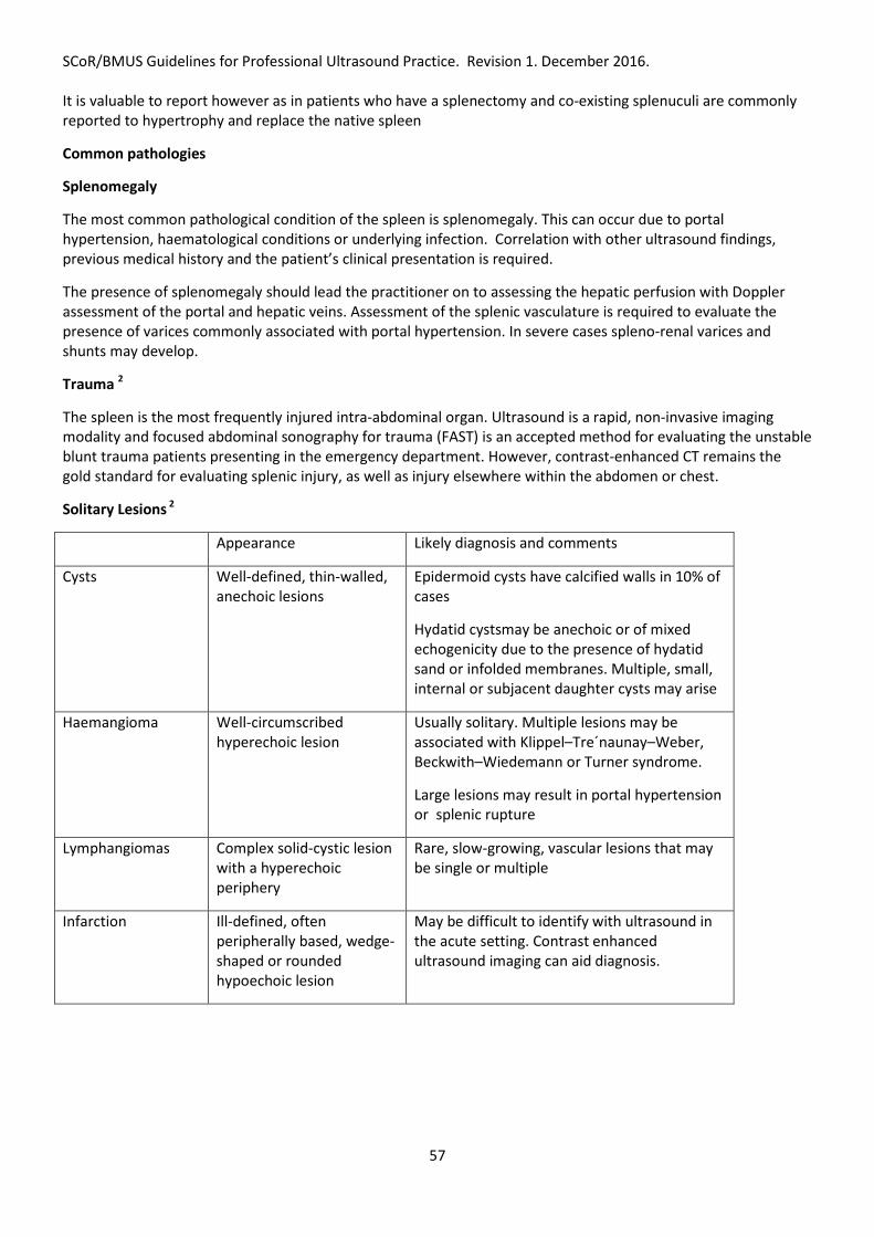
SCoR/BMUS Guidelines for Professional Ultrasound Practice. Revision 1. December 2016.
57
It is valuable to report however as in patients who have a splenectomy and co-existing splenuculi are commonly
reported to hypertrophy and replace the native spleen
Common pathologies
Splenomegaly
The most common pathological condition of the spleen is splenomegaly. This can occur due to portal
hypertension, haematological conditions or underlying infection. Correlation with other ultrasound findings,
previous medical history and the patient’s clinical presentation is required.
The presence of splenomegaly should lead the practitioner on to assessing the hepatic perfusion with Doppler
assessment of the portal and hepatic veins. Assessment of the splenic vasculature is required to evaluate the
presence of varices commonly associated with portal hypertension. In severe cases spleno-renal varices and
shunts may develop.
Trauma 2
The spleen is the most frequently injured intra-abdominal organ. Ultrasound is a rapid, non-invasive imaging
modality and focused abdominal sonography for trauma (FAST) is an accepted method for evaluating the unstable
blunt trauma patients presenting in the emergency department. However, contrast-enhanced CT remains the
gold standard for evaluating splenic injury, as well as injury elsewhere within the abdomen or chest.
Solitary Lesions 2
Appearance Likely diagnosis and comments
Cysts Well-defined, thin-walled,
anechoic lesions
Epidermoid cysts have calcified walls in 10% of
cases
Hydatid cystsmay be anechoic or of mixed
echogenicity due to the presence of hydatid
sand or infolded membranes. Multiple, small,
internal or subjacent daughter cysts may arise
Haemangioma Well-circumscribed
hyperechoic lesion
Usually solitary. Multiple lesions may be
associated with Klippel–Tre´naunay–Weber,
Beckwith–Wiedemann or Turner syndrome.
Large lesions may result in portal hypertension
or splenic rupture
Lymphangiomas Complex solid-cystic lesion
with a hyperechoic
periphery
Rare, slow-growing, vascular lesions that may
be single or multiple
Infarction Ill-defined, often
peripherally based, wedge-
shaped or rounded
hypoechoic lesion
May be difficult to identify with ultrasound in
the acute setting. Contrast enhanced
ultrasound imaging can aid diagnosis.
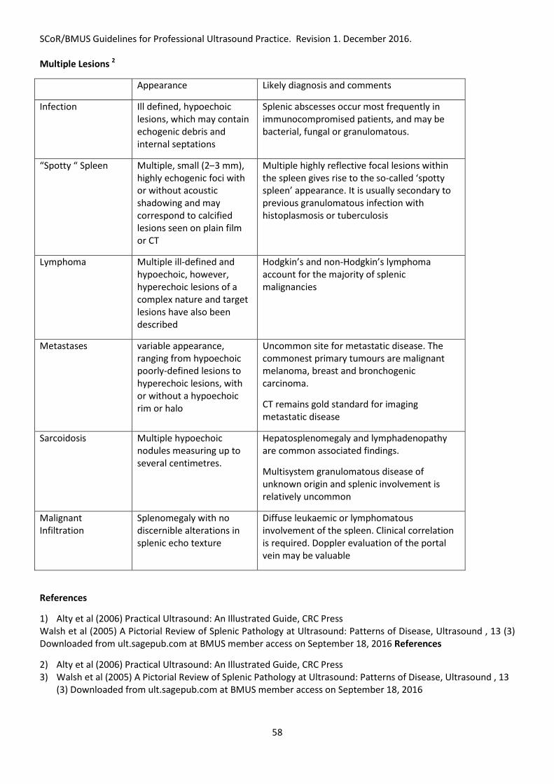
SCoR/BMUS Guidelines for Professional Ultrasound Practice. Revision 1. December 2016.
58
Multiple Lesions 2
Appearance Likely diagnosis and comments
Infection Ill defined, hypoechoic
lesions, which may contain
echogenic debris and
internal septations
Splenic abscesses occur most frequently in
immunocompromised patients, and may be
bacterial, fungal or granulomatous.
“Spotty “ Spleen Multiple, small (2–3 mm),
highly echogenic foci with
or without acoustic
shadowing and may
correspond to calcified
lesions seen on plain film
or CT
Multiple highly reflective focal lesions within
the spleen gives rise to the so-called ‘spotty
spleen’ appearance. It is usually secondary to
previous granulomatous infection with
histoplasmosis or tuberculosis
Lymphoma Multiple ill-defined and
hypoechoic, however,
hyperechoic lesions of a
complex nature and target
lesions have also been
described
Hodgkin’s and non-Hodgkin’s lymphoma
account for the majority of splenic
malignancies
Metastases variable appearance,
ranging from hypoechoic
poorly-defined lesions to
hyperechoic lesions, with
or without a hypoechoic
rim or halo
Uncommon site for metastatic disease. The
commonest primary tumours are malignant
melanoma, breast and bronchogenic
carcinoma.
CT remains gold standard for imaging
metastatic disease
Sarcoidosis Multiple hypoechoic
nodules measuring up to
several centimetres.
Hepatosplenomegaly and lymphadenopathy
are common associated findings.
Multisystem granulomatous disease of
unknown origin and splenic involvement is
relatively uncommon
Malignant
Infiltration
Splenomegaly with no
discernible alterations in
splenic echo texture
Diffuse leukaemic or lymphomatous
involvement of the spleen. Clinical correlation
is required. Doppler evaluation of the portal
vein may be valuable
References
1) Alty et al (2006) Practical Ultrasound: An Illustrated Guide, CRC Press
Walsh et al (2005) A Pictorial Review of Splenic Pathology at Ultrasound: Patterns of Disease, Ultrasound , 13 (3)
Downloaded from ult.sagepub.com at BMUS member access on September 18, 2016 References
2) Alty et al (2006) Practical Ultrasound: An Illustrated Guide, CRC Press
3) Walsh et al (2005) A Pictorial Review of Splenic Pathology at Ultrasound: Patterns of Disease, Ultrasound , 13
(3) Downloaded from ult.sagepub.com at BMUS member access on September 18, 2016
SCoR/BMUS Guidelines for Professional Ultrasound Practice. Revision 1. December 2016.
59
2.8.6 Ultrasound of the bowel
Bowel symptoms are a common cause for patients presenting to their doctors and being referred for imaging.
Suspected acute appendicitis (AA), diverticulitis and inflammatory bowel disease (IBD) are among the common
conditions where ultrasound can and should make a major contribution in diagnosis and disease monitoring.
Ultrasound technique
After a six hour fast, the small bowel is quiet with reduced bowel gas; no other preparation is required.
A general abdominal curvilinear probe is useful to quickly assess the layout of large and small bowel. Particular
attention should be given to deeper recesses (e.g .rectovesical pouch) which are inaccessible to the higher
frequency probes used for detailed interrogation of the bowel wall.
The bowel diseases of interest are those which thicken / enlarge bowel, and displace bowel gas /faeces making
them stand out against normal bowel segments.
TOP TIP: Inflamed bowel is easier to identify than normal bowel.
Focal bowel masses, segments of wall thickening, or dilated loops may be apparent even at lower frequencies but
high frequency probes are essential to characterize changes in the layers of the bowel wall.
Fortunately the common bowel diseases most frequently involve bowel segments lying in the iliac fossae which
are relatively close to the anterior abdominal wall (2-5cms) and accessible to higher frequency probes.
TOP TIP: Use Higher frequency linear probes.
• the higher frequency the better
• 2-3 focal bands close together in the target zone
• harmonics ON
• edge enhancement
• minimal compounding
• graded compression
Pressure applied to the intestines is uncomfortable. Complaining of discomfort is not an indicator of disease,
though patients with IBS may be particularly sensitive.
A gradual progressive increase in pressure applied brings the probe closer to the bowel, displacing bowel gas and
overlying bowel loops and assessing the compressibility / rigidity of normal and abnormal bowel loops and
mesenteric fat.
TOP TIP: Use Left decubitus scanning.
The caecum and appendix are on a variable length mesentery with some freedom of movement. Decubitus
scanning is routinely recommended as overlying loops may fall away and the caecum position adjusts to reveal
the appendix.
In this position the “retro caecal” appendix is often accessible.
TOP TIP: Use “Mowing the lawn” technique
Surveying the entire intestine within the abdominal cavity requires a systematic technique such as the use of
overlapping vertical sweeps of a high frequency probe up and down the abdomen like a lawnmower.
SCoR/BMUS Guidelines for Professional Ultrasound Practice. Revision 1. December 2016.
60
Normal bowel - ultrasound features
The ultrasound appearance of the bowel wall largely reflects the anatomical layering and comprises five
alternating bands of higher and lower echogenicity. The outer and inner bright bands are extremely fine and may
not be seen.
1. superficial mucosa (fine bright line)
2. deep mucosa including the lamina propria (grey)
3. submucosa (bright)
4. muscularis propria (dark)
5. serosa (fine bright line)
Abnormal bowel - ultrasound findings
Once the appendix or a suspect bowel segment has been identified, the following aspects should be assessed:
• wall thickening
• altered wall layers
• bowel lumen
• bowel plasticity/mobility/ peristalsis
• altered blood flowblood flow
• extramural mesenteric/ interloop changes
Wall thickening
Thickening of the bowel wall is the feature most commonly identified and measurements should be taken.
Stomach wall is thickest (~<7mm). Large and small bowel wall is usually <3mm. >4mm is definitely
abnormal.Thickening may be due to the presence of oedema, haemorrhage, inflammation, tumour growth or
infiltration.
Altered bowel wall layers (gut signature)
Depending on the disease process, the gut signature may be preserved, exaggerated, distorted, diminished or
obliterated.
Bowel lumen
Commonly when the bowel wall is thickened, the bowel lumen is compromised, becoming narrowed or strictured.
However ultrasound may identify a dilated, fluid- filled, obstructed appendix. In the absence of clinical or
sonographic evidence of acute inflammation, this may be a mucocele.
Bowel plasticity / mobility / peristalsis
Most diseases cause stiffening of the affected bowel segment with reduced or absent peristalsis.
Altered blood flow
As a rule, Doppler signals are not evident in healthy bowel wall. With acute inflammation, vascularity increases
and dots, lines and branches of vessels can be recorded in the wall (mainly submucosa) and in the adjacent
mesentery.
Extramural / mesenteric changes
Bowel wall disease may extend to involve peri-intestinal structures, adjacent loops or solid organs. Around
inflammatory bowel lesions, collections and abscesses, mesenteric fat becomes oedematous (swollen and
hyperechoic) displacing adjacent structures.
SCoR/BMUS Guidelines for Professional Ultrasound Practice. Revision 1. December 2016.
61
Ultrasound of the normal appendix
The normal appendix is elusive. It varies greatly in size (average length 8cm; range 1-24 cm) and position
(pelvic/descending and retrocaecal being the most common). At TAUS the appendix is identified as a thin, blind-
ending tube, with a normal gut signature, in continuity with the caecal pole, arising approximately two
centimeters from the ileocaecal valve.
The appendix wall has the same layered structure as the other parts of the intestine and the same thickness of
<2mm. The lumen is continuous with the caecum via a smaller orifice. Gas, fluid, semisolid residue and
appendicoliths are often seen in the normal appendix lumen. However obstruction of the orifice is a common
cause of appendicitis.
Acute appendicitis (AA)
Sonographic signs:
• local tenderness
• wall thickening > 2mm
• wall layers may thicken at first, then be lost in transmural low echos
• the lumen may distend
• the maximum outer diameter (MOD) includes the two walls plus the lumen. A MOD of >6mm does not
mean appendicitis but a MOD of < 6mm makes AA unlikely.
• Hypervascular on Doppler
• mesenteric oedema: the inflamed appendix may be surrounded by hyperechoic swollen fat.
• loculated periappendiceal fluid suggests imminent or actual perforation
Appendicoliths are frequently identified in asymptomatic patients with otherwise normal ultrasound
appearances and are not a reliable indicator of inflammation.
TOP TIP: In focal appendicitis the MOD may not exceed 6mm and the diagnosis may be missed if the entire
appendix is not visualized.
The perforated appendix is even more difficult to find but may most reliably be identified by loss of the
hyperechoic appendiceal wall layer (indicating transmural inflammation) and loculated periappendiceal or pelvic
fluid collections.
The appendix may lie low in the pelvis or be obscured by bowel. If a normal appendix is not identified or a
sonographic diagnosis of acute appendicitis or convincing alternative diagnosis made, further imaging (CT) is
indicated in a timely fashon.
Diverticulitis
US features of diverticula
Diverticula appear as bright “ears" outside the bowel wall with acoustic shadowing due to the presence of gas or
inspissated faeces. At higher probe frequencies, a thinned diverticular wall may be demonstrated with a reduced
gut signature owing to the absence of muscularis propria. The neck of a diverticulum may be identified as an
echogenic band traversing hypoechoic circular muscle which is often thickened.
An inflamed diverticulum is identified as an enlarged echo poor protrusion from the colon wall, with an ill-defined
margin surrounded by echogenic non-compressible fat. The gut signature is obliterated by the inflammation.
Inspissated faeces may be seen as a central shadowing echogenicity.
Inflammation will commonly extend along the bowel producing asymmetrical or circumferential hypoechoic
mural thickening that may be hyperaemic on Doppler scanning. An intramural or pericolic abscess may be
identified as an anechoic collection that may contain pockets of air or debris.
SCoR/BMUS Guidelines for Professional Ultrasound Practice. Revision 1. December 2016.
62
2.9 Imaging of the uro-genital system including testes and scrotum
Common clinical scenarios
Haematuria
The ultrasound practitioner should establish whether haematuria is microscopic or macroscopic and be aware of
the place of ultrasound in the diagnostic testing of a patient with haematuria. The investigation of haematuria
may be best performed in the context of a dedicated haematuria clinic according to local practice.
Both kidneys, ureters (if visible) and urinary bladder should be assessed.
Look for renal lesions, hydronephrosis, stones, and lesions within the urinary bladder. The practitioner should be
aware of the limitations of ultrasound in detection of transitional cell tumours of the renal pelvis. The patency of
the renal veins should be assessed when a solid renal lesion is suspected.
Renal cysts should be documented and assessed for complexity. Ultrasound of simple/minimally complex cysts is
sufficient. More complex renal cysts require formal Bosniak grading, either with contrast enhanced ultrasound or
CT according to local guidelines.
The practitioner should be aware of normal anatomical variants that may mimic renal lesions such as
hypertrophied renal columns (columns of Bertin), splenic humps etc. While the majority of normal anatomical
variants should be correctly recognised with ultrasound, contrast enhanced ultrasound, CT or MRI should be
considered where there remains doubt over a potential lesion.
The practitioner should be aware that ultrasound has limited sensitivity in assessment for transitional cell
tumours in the renal collecting system, ureters or urinary bladder. Patients with unexplained macroscopic
haematuria should be considered for further tests to better demonstrate these regions (flexible cystoscopyand CT
urogram /IVU).
Acute/chronic kidney injury (AKI – formerly acute renal failure)
The main aim of ultrasound is first to determine whether renal dysfunction is a medical or surgical (obstructive)
problem.
Assess both kidneys for size, parenchymal thickness and cortical reflectivity. The practitioner should be aware of
the changes in renal appearances with age. The urinary bladder should be assessed for distension, presence of
tumours, trabeculation, wall thickness and diverticulum formation. The size of the prostate gland should be
estimated in males and assessment of bladder emptying should be performed (where possible).
Renal cortical echogenicity should be assessed by comparison with adjacent liver/spleen and internally by
comparison with medullary pyramids. Increased renal cortical echogenicity implies ‘medical’ renal disease but is a
non-specific finding.
Where the kidneys are enlarged, the patency of the renal veins should be confirmed.
The renal collecting system should be examined for dilatation. In cases of pelvi-calyceal dilatation, the collecting
systems, ureters, and urinary bladder should be examined to determine the level and cause of any obstruction.
The practitioner should be aware of common causes of bilateral renal obstruction such as tumours of the urinary
bladder, pelvis and retroperitonum; inflammatory conditions such as retroperitoneal fibrosis, endometriosis, and
bladder outflow obstruction. The practitioner needs to be aware of physiological renal pelvis dilatation secondary
to a full bladder and, in such cases, rescanning following micturition is useful.
In cases of acute kidney injury (AKI), pre-examination preparation with fluid loading should be avoided. In some
centres, in-patients are requested to attend with an empty bladder or are catheterised.
SCoR/BMUS Guidelines for Professional Ultrasound Practice. Revision 1. December 2016.
63
Loin/renal angle pain/obstruction
Look for ultrasound features of renal obstruction and renal lesions.
The ultrasound practitioner should be aware of the limitations of ultrasound in assessing high grade urinary
obstruction. The presence of pelvi-calyceal dilatation is not always owing to urinary obstruction, neither does the
absence of pelvi-calyceal dilatation rule out high grade obstruction. In the obstructed kidney, the degree of
obstruction does not correlate well with the degree of dilatation. Chronic hydronephrosis may be associated with
loss of parenchymal thickness.
The renal pelvis and calyces should be assessed for the presence of renal calculi. While larger calculi may be
visible, ultrasound is of limited sensitivity in the detection of small calculi. The presence of acoustic shadowing is
helpful and the practitioner should be aware of image processing technologies which may reduce the presence of
shadowing, particularly spatial compounding. Colour Doppler may be useful to assess for the presence of
'twinkle’ artefact.
Assess for ureteric distension and level/cause of obstruction. Note that the mid-ureter is frequently not visible
due to overlying bowel gas but the ureter at the level of the PUJ and VUJ is usually amenable to ultrasound
assessment.
Secondary signs of obstruction may include presence/absence of ureteric jets within the bladder and increased
vascular resistance to intra-renal arterial blood flow; the practitioner should be aware that changes in intra-renal
blood flow patterns within an obstructed kidney follow a specific time schedule and the practitioner should be
conversant with this.
The practitioner should be aware of the utility of other imaging tests, plain x-ray, IVU and CT KUB and the
correlative nature of these tests.
Urinary tract infection (UTI)
In acute infection, the kidneys are frequently normal.
The kidneys should be assessed for size, position, morphology and echogenicity. The practitioner should be alert
to the signs of acute renal infection such as small amounts of perinephric fluid (renal sweat),
hydronephrosis/pyonephrosis, diffuse renal enlargement or focal cortical abnormality to suggest focal
pyelonephritis.
The kidneys should be examined for anatomical variants which may predispose to urinary tract infection such as
duplex kidneys, horseshoe kidney and renal ectopia.
The ureters should be assessed for dilatation. The urinary bladder should be examined for distension, wall
thickening, and other signs of bladder outflow obstruction. Assess prostate gland for size. Bladder emptying
should be assessed by estimating post-micturition residual bladder volume.
Renal transplant (immediate post-operative period)
The objective is to demonstrate any surgical complications which may require immediate intervention.
The size and morphology of the transplant kidney should be examined to establish a baseline for subsequent
scans. The practitioner should establish the presence/absence of perinephric haematoma/collection and exclude
pelvi-calyceal dilatation.
SCoR/BMUS Guidelines for Professional Ultrasound Practice. Revision 1. December 2016.
64
Colour Doppler of the entire kidney should be performed to establish that blood flow is present to all areas of the
kidney. Pulsed wave Doppler of the inter-lobar arteries should also be performed to assess blood flow patterns
which may give indirect evidence of arterial (inflow) or venous (outflow) problems of the graft. Doppler indices
(resistance or pulsatility indices) should be recorded. The presence/absence of reversed arterial diastolic flow
should be established and the practitioner should be aware of the possible causes for this. Finally, the renal vein
should be examined with colour Doppler ultrasound to ensure patency throughout its length.
The practitioner should be aware of ultrasound appearances which require immediate surgical intervention such
as arterial or venous occlusion of the transplant kidney and discuss with the surgical team when these are
suspected.
Imaging of the testes and scrotum
Common clinical scenarios
Scrotal mass
The aim is to localise the scrotal lump (intra- or extra-testicular) and characterise if possible.
Both testes should be assessed for size, morphology and the presence/absence of a testicular lesion. The testes
should be assessed for echogenicity by comparing both testes on a side-by-side view on the same image. The
practitioner should follow local guidelines for referral/alerting clinical teams of suspected testicular cancer.
Assessment of the epididymal heads, bodies and tails should be made for thickening, presence of lesions, cysts
etc. Be aware of the presence of normal mild epididymal thickening after vasectomy together with the common
appearances of sperm granulomas.
The tunical space should be examined for the presence of hydrocoele and the scrotal wall should be examined for
the presence of oedema or thickening.
Every effort should be made to visualise and characterise a scrotal mass. The patient should be asked to localise
the mass with direct scanning over this area where the practitioner is unable to visualise the mass during normal
scrotal scanning.
Suspected testicular torsion
In clinically suspected testicular torsion, ultrasound investigation should not delay surgical exploration to offer the
best chance of preserving testicular viability. Ultrasound cannot confidently exclude torsion but an examination
may be performed in cases of acute testicular pain. Practitioners need to be aware of the ultrasound features of
torsion and if in any doubt, urgent urological advice should be sought.
Assess the testes, epididymides and tunical space. Colour Doppler of both testes should be undertaken, assessing
presence and symmetry of blood flow within the testes.
The absence of discernible blood flow within a testis (where flow is visible on the contra-lateral normal side) is
highly predictive of testicular torsion. However, it should be noted that the presence of blood flow does not
exclude torsion, particularly intermittent torsion. The spermatic cord should be examined to assess for the
presence of twisting of vessels within the spermatic cord (the whirlpool sign).
The ultrasound practitioner should be aware that testicular torsion can also cause epididymal swelling and
hydrocoele formation, mimicking epididymo-orchitis, although the absence of intra-testicular flow may be
diagnostic. However, intermittent testicular torsion can demonstrate increased intra-testicular blood flow on
colour Doppler examination which may be indistinguishable from epididymo-orchitis.
SCoR/BMUS Guidelines for Professional Ultrasound Practice. Revision 1. December 2016.
65
2.10 Ultrasound of the adult head and neck
General principles
As in all areas of ultrasound, practitioners need to be aware of the various clinical scenarios that can propagate
ultrasound requests. In the head and neck region the clinical question posed can be unfamiliar to the occasional
operator/reporter. It is essential, in order to carry out a competent examination and issue a helpful report, that
the ultrasound practitioner understands the question that is being asked. For example, in cases where a head and
neck malignancy is suspected, a full examination of all the major lymph node territories is required in order to
accurately stage and optimally manage the patient.
The three most common masses that present to head and neck lump clinics are: lymph node, thyroid and salivary.
Examination and reporting needs to be tailored to the request received and the findings on ultrasound
examination eg if a parotid mass is identified then examination of the contralateral parotid is mandatory to look
for potential contralateral tumours (Warthins) and the findings should be recorded in the report. Examination
may be tailored to a specific area of the neck (eg a palpable mass); depending on the findings, the examination
may focus on the area in question alone, or may need to be expanded to assess the neck as a whole. For example:
in a patient with a posterior triangle mass, if the finding is that of a small superficial lipoma then the examination
can be contained to that area in question. The extent of the study should be recorded in the report eg “I have not
examined the remainder of the neck”. However, if the ultrasound examination identified a necrotic lymph node
which is potentially metastatic then a full assessment of the neck in its entirety is mandatory.
Anatomical structures
1. Lymph nodes
Knowledge of the major lymph node territories and chains is essential and an understanding of the interchange
between terminology used in the description of lymph node groups/chains (eg deep cervical /jugular chain) and
the commonly used Level classification1 that is fundamental to head and neck cancer management. Confusion in
this area can be easily created, both in requesting and reporting for the unwary. Knowledge of the typical criteria
for the ultrasound differentiation between benign and malignant lymphadenopathy is essential. Descriptive
reports that fail to classify the nodes into benign, equivocal or possibly/probably malignant are unhelpful.
Benign nodes are classically fusiform in shape, contain an echogenic hilus and possess a central hilar blood flow
pattern. Whereas malignant lymph nodes tend to be rounded, the central hilus tends to be absent, contain areas
of coagulation or cystic necrosis and exhibit deranged blood flow pattern with areas of vascular sparing and
peripheral vessels. Nodal metastases from papillary carcinoma of the thyroid are typically more echogenic and
contain punctate micro-calcification. Lymphoma classically presents as markedly hypo-echoic nodes (pseudo-
cystic appearance), rounded with plethoric blood flow which commonly displays a benign central hilar pattern. As
the architecture of lymphomatous nodes is preserved, this is mirrored in preservation of the “benign” echogenic
hilus sign.
2. Salivary masses
Ultrasound is the optimal initial investigation for a patient with a salivary mass and therefore the practitioner
needs to be aware of the common appearances of salivary tumours. 80% of salivary tumours will be benign
pleomorphic adenomas and occur in the superficial parotid. They are typically lobular and hypoechoic, often with
a pseudo-cystic appearance. Cystic change is suggestive of Warthins tumours as are multiplicity and contralateral
tumours. An irregular, spiculated outline would suggest a carcinoma.
3. Thyroid
Practitioners should be aware of the typical features that enable a diagnosis of a benign thyroid nodule to be
made and those features that indicate a potential malignancy .These have been outlined in the latest issue of the
SCoR/BMUS Guidelines for Professional Ultrasound Practice. Revision 1. December 2016.
66
British Thyroid Association Guidelines2 and how they can be used to classify the thyroid mass into a benign,
equivocal /indeterminate or (suspicious) malignant category (U1 –U5) with FNA or core biopsy required for those
in the indeterminate of malignant categories, if indicated (ie U3-U5).
Reports should therefore outline the features displayed and indicate in which category the findings sit – allowing
appropriate management.
Benign nodules may show micro-cystic or cystic change with ring down signs of colloid, egg shell calcification and
peripheral colour flow. They are typically hyper-echoic or iso-echoic in relation to the background echo texture
whereas a solid hypo-echoic nodule which contains micro-calcification is highly suggestive of a thyroid carcinoma
– typically a papillary carcinoma. The shape of the nodule (“taller rather than wide”) is also a sign of potential
malignancy. When a carcinoma is suspected, a search for potential lymph node metastases is required, together
with a decision to proceed to FNA or core biopsy in line with guidelines.
Reporting
Outlined below are examples of sample reports for various common clinical scenarios:
------------------------------------------------------------------------------------------------------------------------------------------------------
Palpable mass (a)
Clinical details
Mobile mass left posterior triangle. 2.5 cm mass. No known primary tumour, patient fit and well.
Cervical ultrasound.
The palpable mass in the left mid posterior triangle is identified as a fusiform shaped lymph node measured at 2.6
cm x 0.4 cm. It displays an echogenic hilus, the appearances are typical of a benign lymph node – no sinister
features identified.
The remainder of the left neck looked unremarkable; I have not examined the right side of the neck.
Conclusion : benign left posterior triangle lymph node.
-------------------------------------------------------------------------------------------------------------------------------------------------------
Palpable mass (b) (Primary care referral)
Clinical details
Right upper/ mid cervical mass, increasing in size for past three months. Smoker. O/E hard mass in right upper
cervical region? nodal. Nil else on examination.
Cervical ultrasound
The palpable mass is identified as a 3.4 cm diameter lymph node mass in the right upper deep cervical chain.
There are signs of coagulation necrosis and possible extra capsular spread – consistent with a metastatic lymph
node from a potential squamous cell carcinoma primary.
Further rounded suspicious nodes are seen in the right mid deep cervical chain. Nodes identified in the right
submandibular region and lower deep cervical chain and right posterior triangle - but these all appear benign.
The left side of the neck looks clear. The salivary glands and thyroid looked unremarkable. Some incidental benign
nodules are seen within the left lobe of thyroid - but no signs of anything sinister.
I have performed a FNA (21g) on the right upper deep cervical lymph node, specimen obtained and sent for
cytology. No complications identified.
I have asked the patient to contact your surgery in one weeks time to make an appointment with you to discuss
the results. I suggest that he is referred for an urgent ENT review.
SCoR/BMUS Guidelines for Professional Ultrasound Practice. Revision 1. December 2016.
67
Conclusion: probable metastatic right upper and mid deep cervical chain lymphadenopathy, FNA performed.
Urgent ENT referral recommended. Report to be faxed through to surgery.
-----------------------------------------------------------------------------------------------------------------------------------------------------
Right parotid mass. (ENT referral)
Clinical details
Patient noticed a soft lump in parotid region whilst shaving, unsure how long present. O/E 2 cm soft mass in right
parotid region, VII exam normal. Nil else to find.
Diagnosis : ? Lymph node ? salivary mass
Ultrasound cervical region
A 4.6 cm (coronal) x 3.4 cm (AP) x 3.7 cm (sup/inf) mass is identified in the posterior aspect of the tail of the right
parotid gland, it is predominantly solid with a small cystic element posteriorly. The mass is well encapsulated and
hypo-echoic. No extension into the deep aspect of the right parotid seen. Apart from some benign looking intra
parotid nodes, the remainder of the right parotid looks normal.
No significant lymphadenopathy within the right cervical region.
A contralateral tumour is identified in the inferior aspect of the left superficial parotid. Again a small cystic
element is present, the tumour measures 2.2 cm (AP) x 1.3 cm (coronal) x 2.1 cm (sup/inf) in diameter with no
deep lobe involvement. Remainder of the left parotid looks unremarkable, no significant left cervical
lymphadenopathy seen.
Both submandibular glands look normal. Nil else of note.
21g FNA performed of the right parotid tumour performed, mucoid material obtained and sent for cytology. No
complications identified.
Conclusion: probable bilateral Warthins tumours, FNA performed of the right parotid tumour.
---------------------------------------------------------------------------------------------------------------------------------------------------
Thyroid mass (a)
Clinical details
Patient complaining of fullness in lower neck, O/E ?small goitre .
Ultrasound cervical region
Multiple small iso-echoic nodules are identified in both lobes of the thyroid, several of the nodules exhibit cystic
change with ring down signs indicative of colloid. Multi-cystic change also seen. No signs of anything sinister
identified.
Thyroid is mildly increased in size, no significant associated lymphadenopathy and no retro-sternal extension
present.
Conclusion: small benign multinodular goitre. No signs of anything sinister. Radiological classification U2 – benign.
------------------------------------------------------------------------------------------------------------------------------------------------------
Thyroid mass (b) (ENT referral)
Clinical details
Left sided thyroid nodule, patient states has been present for two years. Thyroid function normal.
O/E firm nodule left lobe of thyroid, no retrosternal extension. No lymph nodes
Ultrasound thyroid
Within the mid pole region of the left lobe of the thyroid there is a 2.6 cm solid hypo-echoic ovoid mass which
contains some echogenic foci – suggestive of micro-calcification. Several smaller nodules are seen in the right lobe
but these display typical benign characteristics.
SCoR/BMUS Guidelines for Professional Ultrasound Practice. Revision 1. December 2016.
68
Within the left mid deep cervical chain there is a rounded hyperechoic lymph node just lateral to the IJV which
also displays some hyper-echoic foci. The reminder of the left neck looks clear as does the right neck.
The findings are highly suggestive of a small papillary carcinoma of the left lobe of the thyroid with a probable left
mid deep cervical lymph node metastasis. I have therefore proceeded to a FNA (21g) of both the left thyroid
nodule and the left mid cervical node.
Specimens obtained and sent for cytology. No complications identified.
Conclusion: probable papillary carcinoma of the left thyroid with left mid cervical lymph node metastasis, FNA
performed.
------------------------------------------------------------------------------------------------------------------------------------------------------
Staging ultrasound examination (Maxillofacial referral)
Clinical details
Left lateral /posterior tongue primary SCC, T3. Smoker. Left Level II nodes on examination ?? right II nodes also, ?
Bilateral metastases
Ultrasound cervical region
The tongue primary tumour can be identified on ultrasound, within the posterior left tongue. It is measured at 1.6
cm (AP) by 1.1 cm (coronal) diameter but does not cross the midline. Superior /inferior diameter difficult to assess
but measured at 1.9 cm.
There are multiple rounded lymph nodes with signs of coagulation necrosis in the left upper cervical region, largest
measured at 2.1 cm in maximum diameter. There are also similar smaller nodes in the upper mid deep cervical
chain. Features are those of metastatic lymphadenopathy. There are small benign looking nodes in the superior
left submandibular region and within the lower left jugular (deep cervical) chain and posterior triangle.
Assessment of the right neck is unremarkable, benign nodes seen in the right upper deep cervical chain but no
signs of contralateral lymph node metastases.
FNA of the largest node in the left upper deep cervical chain (Level II) performed, no complications identified.
Haemorrhagic and necrotic material obtained and sent for cytology.
Conclusion: left tongue base/mid tongue tumour, with left upper and mid deep cervical chain (levels II &III) lymph
node metastases. FNA performed on left upper deep cervical node. No contralateral lymph node metastases seen.
------------------------------------------------------------------------------------------------------------------------------------------------------
References
1) Delineation of the neck node levels for head and neck tumours: a 2013 update. DAHANCA, EORTC, HKNPCSG,
NCIC, CTG, NCRI, RTOG, TROG consensus guidelines. Gregoire V, Ang K, Budach W et al. Radiotherapy &
Oncology 110(2014) 172 -181.2014.
http://dx.doi.org/10.1016/j.radonc.2013.10.010
2) Management of thyroid cancer. British Thyroid Association Guidelines.
http://www.british-thyroid-association.org/Guidelines/
SCoR/BMUS Guidelines for Professional Ultrasound Practice. Revision 1. December 2016.
69
2.11 PAEDIATRIC ULTRASOUND EXAMINATIONS
Exam-specific guidelines and common clinical scenarios
2.11.1 Paediatric liver and biliary system
Liver: neonates and infants
Common clinical scenarios: jaundice, antenatal diagnosis of liver abnormality, RUQ mass, hepatomegaly.
Features to look for and include in report:
• liver shape, homogeneity and reflectivity;
• common duct (CD) normal or dilated (up to 1 mm in neonate), intrahepatic duct size. If CD enlarged
are there stones/inspissated bile in it, is there a mass or enlarged lymph nodes compressing the
duct?
• gallbladder (GB) shape, size and wall thickness and presence/absence of gallstones;
• portal and hepatic vein patency;
• spleen size, shape and position.
Abnormalities that may be seen:
• an abnormally shaped liver, usually associated with biliary atresia if situs solitus, ambiguous, or
inversus. Chronic liver disease features of left lobe hypertrophy and right lobe atrophy, will not have
had time to evolve in a neonate;
• a fatty liver;
• focal abnormalities; benign - calcification, haemangioendothelioma, haemangioma, focal fatty
change, cyst; malignant - hepatoblastoma;
• bile duct dilatation – owing to inspissated bile in the CD, choledochal cyst (not to be confused with a
cyst at the porta that cannot be connected to the left and right ducts – found in some cases of biliary
atresia);
• splenic size and position (? left or right) ? polysplenia. Left-sided polysplenia + situs solitus or
ambiguous is diagnostic of biliary atresia.
If any abnormality is found then a paediatric referral is necessary.
If a normal scan is found in an infant with conjugated hyperbilirubinaemia, then it is mandatory to refer the
patient to a paediatric liver specialist as soon as possible so that biliary atresia may be confirmed or excluded and
managed accordingly.
Liver: children
Examine: Liver, biliary tree, gallbladder and pancreas.
Common clinical scenarios: jaundice, painful RUQ, upper abdominal and/or back pain, abnormal LFTs,
heptomegaly.
Features to look for and include in report:
• liver shape, homogeneity and reflectivity;
• common duct (CD) , normal or dilated (1 mm in neonate, up to 6 mm in teenager, relative to height),
intrahepatic duct size. If CD enlarged are there stones in it, is there a mass or enlarged lymph nodes
compressing the duct?
SCoR/BMUS Guidelines for Professional Ultrasound Practice. Revision 1. December 2016.
70
• gallbladder (GB) shape, size and wall thickness and presence/absence of gallstones;
• portal and hepatic vein patency;
• spleen size, shape and position;
• pancreas: relatively larger and of lower reflectivity than an adult pancreas, duct > 1mm.
Abnormalities that may be seen:
• a liver of increased reflectivity (fatty liver) or decreased reflectivity (low fat content if child well,
acute hepatitis if unwell);
• an abnormally shaped liver ? CLD;
• focal abnormalities - benign: cyst, calcification, haemangioma, focal fatty change, focal nodular
hyperplasia, adenoma, abscess; malignant: hepatoblastoma, hepatocellular carcinoma (HCC )
(usually in a cirrhotic liver), fibrolamellar carcinoma, sarcoma, metastases;
• bile duct dilatation;
• enlarged pancreas +/- duct dilatation.
GP patients should be referred to a paediatric specialist if any abnormality is found or if no abnormality is found
and the patient remains jaundiced and/or has abnormal LFTs.
If the patient is pain-free and jaundice-free when the scan is performed and the CD is found to be big then an
MRCP is necessary to see if there is a choledochal cyst and common channel present.
Glossary of terms used in paediatric liver ultrasound reports
Focal lesions: benign
Abscess: an early abscess may be difficult to identify and the only clue may be posterior acoustic enhancement
and clinical symptoms. The lesion then becomes echo-poor and more clearly defined and may possibly contain
gas if the infection is caused by a gas-forming organism. Portal vein patency may be compromised.
Adenoma: uncommon in children although they are associated with glycogen storage disorders. They may be of
either increased or decreased reflectivity.
Cysts: simple, choledochal (see biliary section).
Calcification: this may be either incidental small foci that are a sequel of an intra-uterine event such as infection
or it may be part of a larger solid lesion.
Focal nodular hyperplasia (FNH): another uncommon lesion in children. They are associated with portosystemic
shunts or portal atresia.
Haemangioma: an incidental finding of a small lesion of increased reflectivity in either a subcapsular position or
adjacent to a blood vessel. The differential is an area of focal fatty change.
Haemangioendothelioma: found in neonates or infants. There may be multiple small focal lesions of reduced
reflectivity throughout the liver or one large vascular lesion. The hepatic artery is large with high velocity
intrahepatic flow and the diameter of the abdominal aorta decreases below the level of the coeliac axis. The
infant may present in heart failure as most of the arterial blood is being shunted through the liver. These usually
involute spontaneously.
Mesenchymal hamartoma: a well-defined mainly cystic mass with multiple septa.
Trauma: CT is usually the first investigation as early liver lacerations may not be visible on ultrasound. Ultrasound
can be used in follow-up to measure fluid collections but CT or contrast ultrasound is necessary to monitor for
possible development of pseudo-aneurysms.
SCoR/BMUS Guidelines for Professional Ultrasound Practice. Revision 1. December 2016.
71
Focal lesions: malignant
Embryonal sarcoma: this tumour has a variable appearance, sometimes solid and sometimes cystic.
Fibrolamellar carcinoma: a rare variant of hepatocellular carcinoma (HCC) that usually occurs in older children.
Hepatoblastoma: occurs in young children. The lesion may be solitary or multifocal, often poorly defined and
may contain calcification. Adjacent vessels may become invaded.
Hepatocellular carcinoma (HCC): often associated with a cirrhotic liver as found in biliary atresia or tyrosinaemia.
The lesion may be solitary or multifocal, often poorly defined but does not usually contain calcification.
These tumours cannot be characterised on ultrasound and cross-sectional imaging +/- biopsy is necessary to
confirm the diagnosis.
CT is used under the age of one year as MRI contrast is not licensed for use in this age group.
Metastases: these may be found in association with neuroblastoma, Wilms’ tumour, leukaemia, lymphoma.
One condition that can be diagnosed with ultrasound is neuroblastoma stage 4S. This usually occurs in infants
under one year old and the adrenal primary together with the liver metastases of decreased reflectivity are
characteristic of this condition.
Diffuse liver disease
Acute hepatitis
Sometimes described as a ‘dark liver’ or a ‘starry sky’ appearance.
The parenchyma is of reduced reflectivity causing the portal tracts to stand out more than normal.
• the liver is enlarged with rounded inferior borders
• the gallbladder wall may be oedematous
• +/- ascites.
It is possible for a liver to have a low fat content and this appearance must not be confused with acute hepatitis;
none of the secondary signs will be present and the child is usually well.
Another pitfall is acute hepatitis in a patient with a fatty liver. The parenchyma may appear of normal reflectivity,
look for the other signs.
Fatty liver
Fatty livers may be found in association with obesity, malnutrition, chemotherapy, steroids, storage disorders
leading to a metabolic disturbance (glycogen storage disorders, tyrosinaemia (increased risk of HCC).
A ‘bright’ liver; the parenchyma is of increased reflectivity and there may be small areas of focal fatty sparing.
These are not focal abnormalities, they are the only part of the liver with a normal appearance. The usual
positions for focal fatty sparing are anterior to the right portal vein and superior to the gallbladder. If focal fatty
sparing is thought to be in other positions then it is worthwhile doing a contrast ultrasound scan as a focal
abnormality may be present.
SCoR/BMUS Guidelines for Professional Ultrasound Practice. Revision 1. December 2016.
72
Chronic liver disease
The liver has a finely or coarsely heterogeneous appearance with an irregular or nodular surface. A typical
position for regenerative nodules is anterior to the right portal vein but further imaging must be performed to
exclude malignancy.
There may be lobar atrophy/hypertrophy; often the left lobe is hypertrophied and the posterior right lobe
atrophied and the para-umbilical vein may be patent. There may be an increase in periportal reflectivity due to
fibrosis around the portal tracts.
Vascular- related pathology.
Budd-Chiari Syndrome: The hepatic veins thrombose and the flow in the portal vein is reversed. There may be
ascites and splenomegaly. In chronic Budd-Chiari, the parenchyma becomes heterogeneous and small
serpiginous venous channels develop. The portal flow may revert to antegrade and the ascites may resolve.
If clinically indicated, a portosystemic shunt procedure may be performed that shunts the portal flow into the IVC.
Patent ductus venosus (DV): This normally closes soon after birth. It may remain patent because it is congenitally
abnormal. If this is the case then it will be several millimetres wide, the flow in the left portal vein is reversed and
there is no flow in the right portal vein as all the portal flow is shunted through the widely patent DV into the IVC.
If left untreated then the right lobe atrophies and FNHs may develop.
The other secondary cause for a patent DV is liver cell failure and it is the liver’s way of trying to decompress the
intrahepatic portal pressure. The lumen of the DV is much smaller, usually a millimetre or less, and if the liver
recovers the shunt will close spontaneously. The flow in both left and right branches of the portal vein is
antegrade. The para-umbilical vein may also be patent.
Portal vein thrombosis: This usually occurs if the child had an umbilical catheter inserted as a neonate. A typical
‘bag of worms’ appearance is seen at the porta anterior to the position that the portal vein should lie in.
Occasionally there is cavernous transformation where there is one venous channel that may be in the position of
the normal portal vein and this is not distinguishable on ultrasound.
Vascular malformations: There may be abnormal vascular connections between the portal vein and hepatic
artery, with a large draining hepatic vein. Multiple vascular channels may be seen with both arterial and venous
flow within them. CT +/- arterial embolisation or resection is normally performed.
Occasionally small shunts between portal and hepatic vein branches or between hepatic veins may be seen but
these are usually haemodynamically insignificant and close spontaneously.
The portal velocity should be assessed and spleen length documented.
Veno-occlusive disease: This usually occurs after chemotherapy and affects the small vessels, not the main
hepatic veins. In the acute phase there is no out-flow in the hepatic veins, they remain patent but the blood only
oscillates during respiration. The portal flow reverses and there is ascites and splenomegaly. The liver becomes
fatty. As the patient improves the hepatic vein flow re-establishes and the portal flow returns to antegrade flow.
The biliary system
Obstructive jaundice in infants and children is usually caused by calculi, choledochal malformations, pancreatic
masses, bile duct tumours (rhabdomyosarcoma), enlarged lymph nodes or sub-hepatic masses.
SCoR/BMUS Guidelines for Professional Ultrasound Practice. Revision 1. December 2016.
73
Neonates that present with conjugated hyperbilirubinaemia may have a dilated common duct due to inspissated
bile within it (haemolysis, total parenteral nutrition [TPN], cystic fibrosis). The gallbladder may also contain
inspissated bile/small calculi.
The common duct may be up to 1 mm in a neonate and 6 mm in an adolescent.
A choledochal cyst (cystic dilatation of the bile ducts):
Type I – dilatation of the extrahepatic common duct
Type II – diverticulum of the common duct
Type III – dilatation of the intra-duodenal common duct
Type IV – intra- and extrahepatic dilatation
Type V – intrahepatic dilatation only.
An MRCP scan should be performed if a choledochal cyst is found to see whether there is a common channel (the
pancreatic duct opening into the lower common duct rather than the duodenum, leading to pancreatitis).
A neonate presenting with a cyst at the porta hepatis will have either a choledochal cyst or biliary atresia. A
choledochal cyst can be seen to connect with the bile ducts, whereas the cyst found in some cases of biliary
atresia cannot be seen to connect to the bile ducts. The gallbladder also has an abnormal appearance in 90% of
cases of biliary atresia.
Cases of biliary atresia are associated with situs inversus, the liver may have an odd configuration, often lying
centrally in the epigastrium with two equal-sized lobes. The portal anatomy may be unusual and often there is
polysplenia.
Spontaneous perforation of the bile duct: the perforation occurs at the junction of the cystic duct/common
hepatic duct and the infant presents with jaundice and ascites.
2.11.2 Urinary system
Neonates
Common clinical scenarios: Antenatal diagnosis of dilated renal pelvis and/or hydronephrosis, single kidney
(pelvic kidneys are sometimes missed on antenatal scans), sepsis.
A baby should be scanned at least 48 hours after birth when following up an antenatal diagnosis of a dilated renal
pelvis, so that the baby is not dehydrated at the time of the scan, thus masking any potential renal pelvic
pathology.
Features to look for and include in report:
• two kidneys that are normal in shape, size and position with no collecting system dilatation and
normal cortical reflectivity. Neonatal kidneys may have increased cortical reflectivity up to 6 months
of age and the pyramids appear relatively dark – not to be confused with cysts. Abnormal
orientation (discussed in section on children)
• the renal pelvis can measure up to 6 mm
• a ‘cyst’ in the upper pole of a kidney – this may be an obstructed upper pole moiety in a duplex
kidney, associated with a ureterocele
• no normal kidney and several cysts in the renal position- multicystic dysplastic kidney
• a normal-shaped bladder with a normal wall thickness and no evidence of lower ureteric dilatation or
ureterocele.
If the renal pelvis is dilated then follow-up scans should be performed at 2 and 8 weeks of age. If the dilatation
continues then further imaging will be required.
SCoR/BMUS Guidelines for Professional Ultrasound Practice. Revision 1. December 2016.
74
Children
Common clinical scenarios: UTIs, loin pain, haematuria
Features to look for and include in report:
• two kidneys that are normal in shape, size and position with no collecting system dilatation and normal
cortical reflectivity.
• a normal-shaped bladder with a normal wall thickness and no evidence of lower ureteric dilatation or
ureterocele. If the patient is able to micturate when asked then a post-micturition bladder scan should
be performed to ensure complete emptying.
Abnormalities that may be seen:
• only one kidney found: if it is a normal size then there should be an ectopic kidney, often pelvic. If the
kidney is hypertrophied then the other kidney has involuted (multicystic dysplastic kidney found
antenatally) or is absent.
• abnormal orientation: horseshoe kidneys have their lower poles more medial than the upper poles and
there is a bridge of tissue connecting the two lower poles across the midline; cross-fused ectopia: both
kidneys lie on the same side with the upper pole of the lower kidney fused with the lower pole of the
upper kidney.
• a ‘cyst’ in the upper pole of a kidney, usually an obstructed upper pole moiety of a duplex kidney, often
associated with a ureterocele.
• hydronephrosis: PUJ obstruction, VUJ obstruction.
• cysts: uncommon in children, associated with tuberose sclerosis.
• infantile polycystic kidneys: autosomal recessive disorder associated with congenital hepatic fibrosis.
• big, bright kidneys with multiple cysts, (cysts may be tiny).
• adult polycystic kidney disease, autosomal dominant, may unusually present in childhood.
• the cortical reflectivity may be increased (normal finding up to 6/12 of age), a non-specific finding
associated with medical renal disease as in adults.
• Wilms’ tumour:the most common paediatric abdominal tumour.
2.11.3 Paediatric gastro-intestinal tract
Pyloric stenosis. Projectile vomiting, non-bilious vomiting in an infant
Scan the baby after a 4 hour fast.
Features to look for and include in report:
• turn the infant into the right lateral decubitus position (lying on its right side) so that any residual fluid in
the stomach lies in the antrum over the pylorus and displaces any gas from this area. If there is a
significant amount of fluid in the stomach there is impaired gastric emptying. Watch to see if the canal
opens.
• If the baby is gassy and an NG tube is present then inject 20 ml of sterile water into the stomach and
watch the pylorus; aspirate after the scan if positive for pyloric stenosis.
• Upper limits for normal measurements are:
o Single muscle wall thickness (not including mucosa or pyloric canal) = 3 mm
o Canal length = 16 mm
o TS diameter = 11 mm 1
Reference
1) de Bruyn R. Pediatric Ultrasound, How, Why and When. Elsevier, 2005 pp185-187.
SCoR/BMUS Guidelines for Professional Ultrasound Practice. Revision 1. December 2016.
75
Intussusception
Abdominal pain, redcurrant jelly stools, palpable abdominal mass.
An intussusception is usually ileo-colic. Most have no obvious pathology as the lead point, 10% are due to
Meckel’s diverticula, polyps and duplications. It is also associated with lymphoma, haematoma and cystic fibrosis
(CF).
Features to look for and include in report:
A segment of bowel prolapses into a more caudal segment and it is seen as bowel within bowel on cross section –
appearances like onion rings.
2.11.4 Neonatal hip
Breech delivery and family history of developmental hip dysplasia are the most common referral criteria.
However, local practice and Newborn and Infant Physical Examination screening programme (NIPE)1 guidelines
should be taken into consideration when justifying referrals. Note that the conduct of the ultrasound examination
of the neonatal hip is not itself part of the NIPE screening programme.
All babies with an abnormality detected on clinical examination should be scanned within two weeks of age.
Babies with a known risk factor but no detectable abnormality are to be scanned by six weeks of age.2 While not
mentioned in the NIPE standards, experience has shown that ‘abnormal’ features may be physiological before six
weeks-of-age and therefore premature babies should be age-corrected before examination.
It is helpful to have a cradle to put the baby into as it keeps them in the lateral position comfortably.
Scan longitudinally over the greater trochanter parallel to the cradle to obtain a coronal image of the acetabulum
at its maximum depth.
α angle – this gives the depth of the acetabulum. The baseline goes along the straight lateral margin of the ilium.
The second line goes from the inferior point of the iliac bone tangential to the bony acetabulum. An angle of
greater than 60° is normal, a smaller angle indicates dysplasia.
A shallow acetabulum in a baby less than 3/12 old may be physiological immaturity but if found after 3/12 of age
it signifies dysplasia.
β angle – this angle is useful in classifying the degree of dysplasia. The β angle is formed between the vertical
cortex of the ilium and the triangular labral fibrocartilage. There is considerable variability in the measurement of
this angle and it is, therefore, not always used.
Neither angle can be measured if the hip is dislocated.
Move the hip and watch it during manipulation to see if it subluxes. If so then follow up in 2 weeks. If the hip is
still unstable then referral is necessary for a harness to be fitted.
N.B.
• you can make a normal hip look abnormal but you can’t make an abnormal hip look normal
• femoral head ossification can be seen anywhere between 2 and 8 months.
Refer to textbooks/published articles for diagrams of the angles referred to above.
SCoR/BMUS Guidelines for Professional Ultrasound Practice. Revision 1. December 2016.
76
References
1) Guidance on when ultrasound examinations of the neonatal hip should be performed can be found at:
https://www.gov.uk/government/uploads/system/uploads/attachment_data/file/572685/NIPE_programme_han
dbook_2016_to_2017_November_2016.pdf (page 17)
2) Public Health England. Newborn and infant physical examination screening: Programme overview. (2013,
updated 2016)
https://www.gov.uk/guidance/newborn-and-infant-physical-examination-screening-programme-overview
2.11.5 Neonatal intracranial ultrasound
The ultrasound practitioner should be aware of the potential bio-effects of diagnostic ultrasound, particularly
heating at brain/skull interface, and ensure that the machine is set to the minimum power consistent with
obtaining a high quality diagnostic ultrasound image. Colour Doppler should not be utilised except for clearly
defined clinical reasons which provide additional diagnostic or prognostic information.
The ultrasound practitioner should be aware of common and less commonly used acoustic windows to the
neonatal brain. While the anterior fontanelle is used as standard, the posterior fontanelle can be useful to
examine the occipital horn of the lateral ventricles and the mastoid suture may be helpful in examining the
posterior fossa, cerebellum, aqueduct of Sylvius and 4th
ventricle.
A suggested approach for examining the neonatal brain is as follows:
Presence of normal anatomy
The ultrasound practitioner should be aware of normal brain anatomy in the neonate, to include changes with
age, presence of midline structures, ventricular appearances, appearances of basal ganglia, periventricular white
matter, cerebellum and extra-axial space.
Presence of intracranial haemorrhage
The ultrasound practitioner should be aware of common locations of intra-cranial haemorrhage, how this may
vary according to gestational age, and how these may present on ultrasound. Grading of haemorrhage should be
according to local agreement and protocol.
Ventricular size
The size of the lateral, 3rd
and 4th
ventricles should be assessed according to local protocol. Validated
measurement techniques (eg ventricular indices) should be utilised in cases of ventriculomegaly. This can be
useful to assess change in size over time and guide timing of intervention.
Periventricular white matter
The ultrasound practitioner should be aware of normal and abnormal appearances of the periventricular white
matter and the limitations of ultrasound in examining this region. Follow-up should be considered in cases of
periventricular flaring or suspected white matter damage.
Brain parenchyma
The ultrasound practitioner should be aware of normal sonographic appearances of the brain parenchyma and be
aware of the potential appearances of acquired abnormalities.
SCoR/BMUS Guidelines for Professional Ultrasound Practice. Revision 1. December 2016.
77
2.12 MUSCULOSKELETAL ULTRASOUND EXAMINATIONS
Notes on musculo-skeletal reports (see also recommendations for the production of an ultrasound report,
section 2.6)
The report is a recording and interpretation of observations made during the ultrasound examination. It should be
written by the person undertaking the scan and viewed in clinical context.
The report is intended to answer a clinical question and to assist with the patient journey so local opinion from
radiologists, rheumatologists, orthopaedic surgeons and physiotherapists should be taken into account in the
construction of local report templates.
As reports are now electronic, they may be available to clinicians intervening later in the clinical pathway and so
should contain all relevant and appropriate information – for example an orthopaedic surgeon may access a
report originally requested by a general practitioner or physiotherapist.
The report should include correct patient demographics; date of examination; examination type and the name
and status of the ultrasound practitioner. See also RCR satenement at https://www.rcr.ac.uk/posts/position-
statement-recording-identity-healthcare-professionals-who-report-imaging
As age-related changes1,2
are common in the musculoskeletal system and may not be the cause of the patient’s
symptoms, ultrasound appearances must always be taken in clinical context and the referrer should be made
aware of its limitations in the report. For example:
• ultrasound cannot exclude intra-articular pathology
• ultrasound cannot exclude impingement.
Comparison with the contralateral side (assuming it is asymptomatic) will help when determining the
clinical significance of age/activity related changes and should be imaged and documented in the report.
Diagnostic ultrasound is often used as a precursor to therapeutic injections and care should be taken to assist in
the direction of that injection. Some structural changes may not be currently relevant and may not be associated
with pain.
The pitfalls of ultrasound interpretation are widely documented3 and can be reduced by education and
experience of the individual ultrasound practitioner.
Examination specific guidelines and common clinical scenarios.
2.12.1 Shoulder
Purpose of scan
To evaluate the following structures:
• main rotator cuff tendons – supraspinatus, infraspinatus and subscapularis tendons
• long head of biceps tendon
• posterior glenohumeral joint
• superficial acromioclavicular joint.
Common indications
Common indications for the performance of a shoulder ultrasound scan are:
SCoR/BMUS Guidelines for Professional Ultrasound Practice. Revision 1. December 2016.
78
• ? rotator cuff tear/tendonopathy
• restricted range of movement
• pain on abduction.
Contraindications and limitations
Contraindications for diagnostic shoulder scans are unlikely; however, some limitations exist and may include the
following:
• obesity
• inability to see structures that lie deep to bone or intra-articular structures of the glenohumeral and
acromioclavicular joints
• casts, dressings, open wounds/ulcers etc can limit visualisation
• severe oedema/swelling
• patients who are unable to co-operate or provide a clinical history due to reduced cognitive functions
eg Alzheimer’s or dementia and through involuntary movements.
As age-related changes are common in the musculoskeletal system1,2
and may not be the cause of the patients
symptoms, ultrasound appearances must always be taken in clinical context and the referrer should be made
aware of its limitations in the report. For example:
• ultrasound cannot exclude impingement
• ultrasound cannot exclude intra-articular pathology.
Diagnostic ultrasound is often used as a precursor to therapeutic injections and care should be taken to assist in
the direction of that injection. Some structural changes seen on ultrasound may not be currently relevant and
may not be associated with pain. For example effusion in the long head of biceps tendon sheath does not always
reflect current tenosynovitis but may be an extension of a glenohumeral joint effusion. The addition of the site of
current symptom on a report may help.
Subacromial/subdeltoid bursal thickening may be present on the asymptomatic shoulder and does not always
reflect current ‘bursitis’ so a comparison image and addition of current symptoms may help.
The pitfalls of ultrasound interpretation are widely documented3 and can be reduced by education and
experience of the individual ultrasound practitioner.
Scan protocol
Numerous scan protocols for the shoulder are described in the literature including that from the European
Society of Musculoskeletal Radiology’s (ESSR) protocol for the shoulder.4
Training protocols also exist and may be useful to standardise scanning and reporting such as that described by
Smith et al.5
The structures that should be identified as a minimum in a standard shoulder ultrasound scan are:
• long head of biceps tendon (LHBT)– for position, tendonopathy, tears, calcification, tenosynovitis,
effusion;
• rotator interval (RI)– if LHBT is visible at the (RI) it is likely to be intact proximally;
• subscapularis tendon – for tendonopathy, tears, calcification;
• infraspinatus tendon - for tendonopathy, tears, calcification;
• supraspinatus tendon- for tendonopathy, tears, calcification;
• any visible bursae around the shoulder – subacromial/subdeltoid, subcoracoid;
SCoR/BMUS Guidelines for Professional Ultrasound Practice. Revision 1. December 2016.
79
• posterior glenohumeral joint – for effusion, cysts, joint synovitis;
• acromioclavicular joint – for effusion, cysts, joint synovitis.
Comparison with the contra-lateral side (assuming it is asymptomatic) will help when determining the clinical
significance of age/activity-related changes and should be imaged and documented in the report.
Any pathology found should be documented in two planes.
Dynamic ultrasound assessment
Dynamic assessment around the shoulder may be controversial in some centres as some indications such as
‘impingement’ have multiple potential causes (some of which will not be seen using ultrasound) and may be
considered to be a purely clinical diagnosis.
Dynamic assessment using ultrasound may be requested for the following:
• long head of biceps tendon: to assess its stability within the bicipital groove during external rotation;
• subscapularis tendon: to identify the myotendinous area which normally sits behind the coracoid
process of the scapula;
• supraspinatus tendon: to assess for bunching of the tendon and/or overlying subacromial bursa
against the acromion or coraco-acromial ligament during abduction which may be a cause of
‘impingement’;
• posterior joint recess: during internal/external rotation, to assess for a gleno-humeral joint effusion.
This will be most evident during external rotation.
Imaging protocol
A standard shoulder series should include the following minimum images for a normal scan:
• long head of biceps tendon - longitudinal and transverse;
• rotator interval showing anterior portion of supraspinatus tendon, long head of biceps and lateral
edge of subscapularis tendon;
• subscapularis tendon – longitudinal and transverse;
• supraspinatus tendon – longitudinal and transverse;
• infraspinatus tendon – longitudinal only;
• posterior glenohumeral joint recess;
• acromioclavicular joint.
Document the normal anatomy and any pathology found, including measurements and vascularity if indicated.
Report (see also recommendations for the production of an ultrasound report, section 2.6)
The report is a recording and interpretation of observations made during the ultrasound examination. It should be
written by the person undertaking the scan and viewed in clinical context.
A report is intended to answer a clinical question and to assist with the patient journey so local opinion from
radiologists, rheumatologists, orthopaedic surgeons and physiotherapists should be taken into account in the
construction of local report templates.
As reports are now electronic, they may be available to clinicians intervening later in the clinical pathway and so
should contain all relevant and appropriate information – for example an orthopaedic surgeon may access a
report originally requested by a general practitioner or physiotherapist.
The standard shoulder report should include:
SCoR/BMUS Guidelines for Professional Ultrasound Practice. Revision 1. December 2016.
80
• documentation of the normal anatomy;
• documentation of any pathology including measurements/ any increase in vascularity if appropriate;
• documentation of any limitation to range of movement and site and degree that pain or symptoms
begin;
• documentation of any difficulties with interpretation of the ultrasound appearances.
The sample reports below are intended as a guide only as reporting style may be specific to
individuals/departments.
------------------------------------------------------------------------------------------------------------------------------------------------------
Sample report for normal appearances
Normal appearances of the rotator cuff tendons. No evidence of tears or calcification or any bursal thickening or
effusions.
The long head of biceps tendon is intact and in situ.
No evidence of a gleno-humeral joint effusion.
Unremarkable ACJ noted.
If appropriate, the report may include the following:
• the rotator cuff tendons have reasonable depth and texture;
• there is evidence of subacromial/subdeltoid bursal thickening but no more so than on the
symptomatic shoulder;
• there is good, pain free subacromial movement;
• non-tender ACJ OA noted.
--------------------------------------------------------------------------------------------------------------------------------------------------------
Sample report for tendonopathy
The supraspinatus tendon appears generally thickened/thinned and tendonopathic with loss of the normal fibrillar
pattern. No tears seen.
Focal tendonopathic changes are noted at the anterior/middle/posterior portion of the supraspinatus tendon, no
tears seen.
-------------------------------------------------------------------------------------------------------------------------------------------------------
Sample report for calcific tendonopathy
A x mm intratendinous calcium deposit is (calcific flecks are) noted within the ant/mid/post aspect of the
supraspinatus tendon; no tears evident.
There is a 6mm densely shadowing calcific deposit within the supraspinatus tendon.
There is a 6mm non shadowing deposit within the supraspinatus tendon likely to be soft calcific tendonopathy.
There is marked calcific tendonopathy of the rotator cuff tendons. The largest area of calcification in the
supraspinatus tendon measures 15mm in diameter and the patient is tender on scanning.
------------------------------------------------------------------------------------------------------------------------------------------------------
Sample report for bursitis
The subacromial/subdeltoid bursa contains an effusion and the wall is hyperaemic on power Doppler. The patient
is tender to scan here and appearances are consistent with bursitis.
------------------------------------------------------------------------------------------------------------------------------------------------------
Sample reports for long head of biceps tendon
The long head of biceps tendon is intact but has dislocated from the bicipital groove medially and is sitting on the
surface of the subscapularis tendon.
SCoR/BMUS Guidelines for Professional Ultrasound Practice. Revision 1. December 2016.
81
The long head of biceps tendon has subluxed onto the medial lip of the bicipital groove. The tendon is thickened
and appears oedematous. The tendon sheath is hyperaemic and contains an effusion and the patient is tender to
scan here. Ultrasound appearances suggest an intact subluxed long head of biceps tendon with evidence of
tenosynovitis.
------------------------------------------------------------------------------------------------------------------------------------------------------
Tendon tears
Rotator cuff tendon tears are often seen in asymptomatic individuals and are not always the cause of symptoms.
It is sometimes difficult for the ultrasound practitioner to appreciate the significance of a tear on the patient’s
current clinical symptoms and while it is important to evaluate and accurately describe tears, care should be
taken if discussing scan findings with the patient.
When reporting, it is important to describe the type of tear – partial, full or complete – and the dimensions and
site of the tear. An opinion on the state of the remaining tendon tissue is also helpful. These details are
important for subsequent treatment and surgical planning as tendons with severe tendonopathy are less likely to
have a successful repair should surgery become appropriate.
Measurements
Type, size and location of tears is important and measurements of full thickness tears should be made in two
planes – anterior to posterior ( transverse) and medial to lateral ( longitudinal). The site of the tear measured
from the rotator interval is also useful.
Site of supraspinatus tendon tears
The supraspinatus tendon can be divided into anterior, mid and posterior portions. The anterior free or leading
edge, the mid-substance or footprint and the area that abuts the infraspinatus tendon - the distinction between
the two tendons can be difficult to define.
Anterior leading edge tears involve the portion of the supraspinatus tendon that lies adjacent to the long head of
biceps tendon at the rotator interval.
If there is still tendon tissue anterior to the tear, the tear is said to be midsubstance, crescent or footprint.
If the tear occurs at the supraspinatus/infraspinatus interface it is said to be posterior.
Types of tear
Partial thickness tear: A rotator cuff tendon tear that involves either the joint or bursal surface of a tendon and
does not allow communication between the two compartments. Partial thickness tears may be articular surface,
bursal surface or intrasubstance.
Articular surface tear: A partial thickness rotator cuff tear involving the articular or joint surface of the tendon.
In the supraspinatus, they can be called ‘rim rent’ or PASTA lesion – Partial Articular Supraspinatus Tendon
Avulsion.
Bursal surface tear: A partial thickness rotator cuff tear involving the bursal surface of the tendon. In the
supraspinatus tendon, this is sometimes called ‘scuffing’ as it is caused by friction from the undersurface of the
acromion.
Intrasubstance tear: A partial thickness rotator cuff tear within the substance of the tendon – sometimes called
concealed interstitial delamination (CID). A cleft is present within the tendon substance but it does not reach
either the bursal or articular surfaces of the tendon.
SCoR/BMUS Guidelines for Professional Ultrasound Practice. Revision 1. December 2016.
82
Full thickness tear: A rotator cuff tendon tear that extends across the whole depth of the tendon, involves both
surfaces and resulting in communication between the joint and bursal compartments, regardless of the width of
the tear. Some full thickness tears measure over 3cm in diameter; some look like pinholes.
Complete full thickness tear: A full thickness tendon tear that extends across the whole width and depth of the
tendon, usually resulting in retraction back from the insertion.
If the tear is complete, the entire width of the tendon will have torn, usually (but not always) at the insertion and
there is retraction of the proximal stump. The degree of retraction will vary but in the supraspinatus and
subscapularis tendons can result in the proximal stump sitting under the acromion/coracoid and so not visible
with ultrasound. This is an important finding for surgeons because it is less likely that the tendon can be pulled
back and repaired.
A supraspinatus tendon tear is often given an increased level of importance compared to the other rotator cuff
tendons as it is more often symptomatic and amenable to surgery.
------------------------------------------------------------------------------------------------------------------------------------------------------
Sample reports for tears
Partial thickness tear
There is a partial thickness tear of the bursal (or articular) surface (or intrasubstance) of the anterior aspect of the
supraspinatus tendon which measures 2mm in longitudinal section and 3 mm in transverse section.
The supraspinatus tendon appears generally tendonopathic and there is a partial thickness (articular)(bursal)
surface tear within the anterior/middle/posterior portion of this tendon. This tear measures xxmm x xxmm and
affects over (or under) 50% of the tendon depth.
There is a partial thickness tear on the articular surface of the supraspinatus tendon lying xmm posterior to the
rotator interval. It measures xxmm in width and extends over (or under) 50% of the tendon depth. The remainder
of the tendon has reasonable depth and texture.
Full thickness tear
There is a full thickness, insertional tear of the supraspinatus tendon lying xxmm posterior to the rotator interval.
The tear measures xxmm in width and there is xxmm retraction from the insertion. The remainder of the tendon
has reasonable depth and texture.
Complete full thickness tear
The supraspinatus tendon has completely ruptured and retracted; the retracted tendon end is not visualised. Cuff
arthropathy noted.
There is a complete, full thickness tear of the supraspinatus tendon with proximal retraction of x mm from the
insertion.
---------------------------------------------------------------------------------------------------------------------------------------------------
Glossary of terms
Bursitis: thickening and inflammation of a bursa, often causing an effusion. Thickening of the bursa alone does
not constitute ‘bursitis’. Comparison with the asymptomatic side is important. Hyperaemia of the bursal wall
using colour/power Doppler is often present in symptomatic bursitis.
Calcific tendonopathy: calcification within tendons. The presentation of calcific tendonopathy may vary from tiny
calcific flecks to large conglomerates which may appear ‘soft’ and non-shadowing, or denser with a dense
posterior acoustic shadow.
Cuff arthropathy: arthritis of the glenohumeral joint, particularly the humeral head due to massive rotator cuff
tears leaving a ‘bare’ humeral head.
SCoR/BMUS Guidelines for Professional Ultrasound Practice. Revision 1. December 2016.
83
Effusion: a collection of fluid in a tendon sheath, joint or bursa.
Joint synovitis: thickening and often hyperaemia of the synovium lining a joint.
LHB tendon dislocation: the long head of biceps tendon moves out of the bicipital groove medially, owing to
disruption of the transverse humeral ligament. This movement may be transient – subluxation on external
rotation of the joint, or it may stay dislocated, regardless of patient movement.
Rotator interval: the space between the subscapularis and supraspinatus tendons through which the long head of
biceps tendon passes as it exits the glenohumeral joint.
Tendonitis: not generally used now for rotator cuff tendons as this suggests an inflammatory process that has not
been proven.
Tendonopathy: A more general term for a diseased tendon.
Tendonosis: a degenerative process within a tendon, disordered biomechanics often leading to micro tears.
Tenosynovitis: inflammation of the tendon and sheath – only possible with long head of biceps tendon.
Appearances should include tendon sheath thickening and hyperaemia on Doppler, pain on palpation. May also
include effusion and tendonopathy but these may be present without current tenosynovitis.
References
1) Girish G, Lobo LG, Jacobson JA et al. Ultrasound of the shoulder: asymptomatic findings in men. Am J
Roentgenol 2011; 197: W713–9
2) Tempelhof S, Rupp S, Seil R. Age-related prevalence of rotator cuff tears in asymptomatic shoulders. J Shoulder
Elbow Surg 1999; 8: 296–9
3) Rutten MJ, Jager GJ, Blickman JG. From the RSNA refresher courses: US of the rotator cuff: pitfalls, limitations,
and artifacts. Radiographics 2006; 26: 589–604
4) Beggs I, Bianchi S, Bueno A, et al. Musculoskeletal ultrasound technical guidelines I. Shoulder. European
Society of MusculoSkeletal Radiology.
5) Smith MJ, Rogers A, Amso N, Kennedy J, Hall A, Mullaney P. A training, assessment and feedback package for
the trainee shoulder sonographer. Ultrasound 2015; 23(1):29-41
2.12.2 Elbow
Purpose of scan
To evaluate any of the following structures where appropriate and symptomatic:
• common extensor and flexor tendons;
• medial ulnar collateral ligament;
• lateral radial collateral ligament;
• distal biceps tendon;
• triceps tendon;
• cubital tunnel and ulnar nerve;
• elbow joints/olecranon fossa.
Common indications
Common indications for the performance of an elbow ultrasound scan are:
SCoR/BMUS Guidelines for Professional Ultrasound Practice. Revision 1. December 2016.
84
• ? ‘tennis’ or ‘golfers’ elbow/epicondylitis;
• restricted range of movement? effusion/synovitis;
• ? distal biceps tendon tear;
• ? ulnar nerve compression;
• olecranon bursitis.
Contraindications and limitations
Contraindications for diagnostic elbow scans are unlikely; however some limitations exist and may include the
following:
• obesity;
• inability to see intra-articular structures;
• casts, dressings, open wounds/ulcers etc can limit visualisation
• severe oedema/swelling;
• patients who are unable to cooperate or provide a clinical history due to reduced cognitive functions
eg Alzheimer’s or dementia and through involuntary movements.
As age related changes are common in the musculoskeletal system and may not be the cause of the patient’s
symptoms, ultrasound appearances must always be taken in clinical context and the referrer should be made
aware of its limitations in the report. For example:
• ultrasound cannot exclude intra articular pathology.
Diagnostic ultrasound is often used as a precursor to therapeutic injections and care should be taken to assist in
the direction of that injection. Some structural changes may not be currently relevant and may not be associated
with pain.
The pitfalls of ultrasound interpretation are widely recognised and can be reduced by education and experience
of the ultrasound practitioner.
Scan protocol
Numerous scan protocols for the elbow are described in the literature.1,2
Unlike the shoulder, diagnostic scanning of the elbow is usually focussed on a single structure, for example the
common extensor tendon for ‘? tennis elbow’, but other structures may be included if appropriate and
symptomatic and within the clinical experience of the ultrasound practitioner.2 These include:
• common extensor tendon - for tendonopathy, neovascularity, tears, calcification;
• common flexor tendon - for tendonopathy, neovascularity tears, calcification;
• distal biceps tendon - for tendonopathy, tears, calcification;
• olecranon bursa - for effusion/bursitis;
• anterior and posterior joint recesses - for effusion, joint synovitis;
• cubital tunnel - for ulnar nerve enlargement/flattening/subluxation.
Any pathology found should be documented in two planes.
SCoR/BMUS Guidelines for Professional Ultrasound Practice. Revision 1. December 2016.
85
Dynamic ultrasound assessment
Dynamic assessment using ultrasound may be requested for the following but depends on the skill and expertise
of the ultrasound practitioner:
• ? ulnar nerve subluxation
• ? medial/lateral ligament tears
Imaging protocol
There is no standard elbow series as the areas scanned are dependent on symptoms and pathology found. The
following protocols are a guide to each area:
• CEO/CFO longitudinal with colour box to show the absence of neovascularity;
• distal biceps tendon in longitudinal and transverse, distal tendon insertion in longitudinal and
myotendinous area in transverse to exclude tear/tendonopathy/calcification. Comparison with
contralateral side;
• anterior and posterior joint recess within and without colour box to show the absence of effusion or
synovitis;
• cubital tunnel with arm in flexion/extension to exclude subluxation of the ulnar nerve;
• medial/lateral ligaments in longitudinal in stressed and relaxed positions if appropriate.
Document the normal anatomy and any pathology found, including measurements and vascularity if
indicated.
Report (see also recommendations for the production of an ultrasound report, section 2.6)
The report is a recording and interpretation of observations made during the ultrasound examination. It should be
written by the person undertaking the scan and viewed in clinical context.
A report is intended to answer a clinical question and to assist with the patient journey so local opinion from
radiologists, rheumatologists, orthopaedic surgeons and physiotherapists should be taken into account in the
construction of local report templates.
As reports are now electronic, they may be available to clinicians intervening later in the clinical pathway and so
should contain all relevant and appropriate information – for example an orthopaedic surgeon may access a
report originally requested by a general practitioner or physiotherapist.
The standard elbow report should include:
• documentation of the normal anatomy;
• documentation of any pathology including measurements/ any increase in vascularity if appropriate;
• documentation of any limitation to range of movement and site and degree that pain or symptoms
begin;
• documentation of any difficulties with interpretation of the ultrasound appearances.
The sample reports below are intended as a guide only as reporting style may be specific to
individuals/departments.
------------------------------------------------------------------------------------------------------------------------------------------------------
Sample report for normal appearances
Normal appearances of the common extensor/flexor/distal biceps tendons of the elbow. No evidence of
significant tendonopathy, tears or calcification.
SCoR/BMUS Guidelines for Professional Ultrasound Practice. Revision 1. December 2016.
86
No evidence of joint effusion or synovitis but ultrasound cannot exclude intra-articular pathology.
The ulnar nerve appears normal in calibre around the elbow and is stable within the cubital tunnel on dynamic
scanning’. No solid or cystic lesions seen in the cubital tunnel.
----------------------------------------------------------------------------------------------------------------------------------------------------
Sample report for tendonopathy
The tendon appears generally thickened/thinned with loss of the normal fibrillar pattern. There is evidence of
neovascularity on Doppler and appearances are consistent with tendonopathy. No tears seen.
Focal tendonopathic changes are noted at the proximal/distal portion of the tendon, no tears seen. -
----------------------------------------------------------------------------------------------------------------------------------------------------
Sample report for calcific tendonopathy
A x mm intratendinous calcium deposit is (calcific flecks are) noted within the proximal/distal portion of the
tendon, no tears evident.
There is a 6mm densely shadowing calcific deposit within the tendon.
There is a 6mm non shadowing deposit within the tendon likely to be soft calcific tendonopathy.
------------------------------------------------------------------------------------------------------------------------------------------------------
Sample report for olecranon bursitis
The olecranon bursa contains an effusion and the wall is hyperaemic on power Doppler. The patient is tender to
scan here and appearances are consistent with bursitis.
------------------------------------------------------------------------------------------------------------------------------------------------------
Sample report for tendon tears
When reporting, it is important to describe the type of tear – partial or complete – and the dimensions and site of
the tear. An opinion on the state of the remaining tendon tissue is also helpful.
There is a well-defined, cystic area within the proximal portion of the tendon measuring XXmm in diameter and
extending across approximately xx% of the tendon depth. Appearances are consistent with cystic
degeneration/partial thickness tear.
There is a complete insertional tear of the distal biceps tendon with retraction of xxmm from the insertion. There
is loss of the normal fibrillary pattern in the detached tendon suggesting tendonopathy.
There is a large fluid collection around the distal pole of the biceps muscle likely to be a resolving haematoma.
The distal biceps tendon is intact at the insertion but appears to have completely torn at the myotendinous
junction.
------------------------------------------------------------------------------------------------------------------------------------------------------
Glossary of terms
Bursitis: thickening and inflammation of a bursa, often causing an effusion. Thickening of the bursa alone does
not constitute ‘bursitis’. Comparison with the asymptomatic side is important. Hyperaemia of the bursal wall
using colour/power Doppler is often present in symptomatic bursitis.
Calcific tendonopathy: calcification within tendons. The presentation of calcific tendonopathy may vary from tiny
calcific flecks to large conglomerates which may appear ‘soft’ and non-shadowing, or denser with a dense
posterior acoustic shadow.
Effusion: a collection of fluid in a tendon sheath, joint or bursa.
Joint synovitis: thickening and often hyperaemia of the synovium lining a joint.
SCoR/BMUS Guidelines for Professional Ultrasound Practice. Revision 1. December 2016.
87
Tendonitis: not generally used now for tendons as this suggests an inflammatory process that has not been
proven.
Tendonosis: a degenerative process within a tendon, disordered biomechanics often leading to micro tears.
Tendonopathy: a more general term for a diseased tendon.
Tenosynovitis: inflammation of the tendon and sheath: Appearances should include tendon sheath thickening
and hyperaemia on Doppler, pain on palpation. May also include effusion and tendonopathy but these may be
present without current tenosynovitis.
References
1) Beggs I, Bianchi S, Bueno A et al. Musculoskeletal ultrasound technical guidelines I. Elbow. European Society of
MusculoSkeletal Radiology.
2) Draghi F, Danesino GM, de Gautard R, Bianchi S. Ultrasound of the elbow Journal of Ultrasound (2007) 10,
76e84
2.12.3 Wrist and hand
Purpose of scan
To evaluate any of the following structures where appropriate and symptomatic:
• extensor and flexor tendons;
• joints of the hand/wrist and metacarpophalangeal (MCP), proximal interphalangeal (PIP), distal
interphalangeal (DIP) and carpometacarpal ( CMC) joints;
• ligaments of the thumb/finger joints;
• carpal tunnel;
• Guyon’s canal.
Common indications
Common indications for the performance of a wrist/hand ultrasound scan are:
• Swelling ? ganglion
• ? effusion/synovitis
• ? tendon tear
Contraindications and limitations
Contraindications for diagnostic wrist/hand scans are unlikely; however some limitations exist and may include
the following:
• obesity;
• inability to see intra-articular structures;
• casts, dressings, open wounds/ulcers etc. can limit visualisation;
• severe oedema/swelling;
• patients who are unable to cooperate or provide a clinical history due to reduced cognitive functions
eg Alzheimer’s or dementia and through involuntary movements.
As age-related changes are common in the musculoskeletal system and may not be the cause of the patient’s
symptoms, ultrasound appearances must always be taken in clinical context and the referrer should be made
aware of its limitations in the report. For example:
SCoR/BMUS Guidelines for Professional Ultrasound Practice. Revision 1. December 2016.
88
• ultrasound cannot exclude intra-articular pathology.
• The median nerve is enlarged and appears flattened in the carpal tunnel suggestive of carpal tunnel
syndrome if there is clinical correlation.
Diagnostic ultrasound is often used as a precursor to therapeutic injections and care should be taken to assist in
the direction of that injection. Some structural changes may not be currently relevant and may not be associated
with pain.
The pitfalls of ultrasound interpretation are widely documented and can be reduced by education and
experience of the individual ultrasound practitioner.
Scan protocol
Numerous scan protocols for the hand and wrist are described in the literature.1,2
Unlike the shoulder, diagnostic
scanning of the hand/wrist is usually focussed to a single area or pathology. For example, the dorsum of the wrist
for ‘? ganglion’ or the joints of the hand and wrist for ‘? synovitis’. Other structures may be included if
appropriate and symptomatic and within the clinical experience of the ultrasound practitioner. These include:
• extensor and flexor tendons for tendonopathy, tenosynovitis, tears, calcification;
• finger pulleys for triggering;
• joints of the hand/wrist for synovitis or effusion;
• bony cortex for erosions;
• ligaments of the thumb/fingers for sprains or tears;
• carpal or Guyon’s tunnel for nerve compression.
Comparison with the contralateral side (assuming it is asymptomatic) will help when determining the clinical
significance of age/activity-related changes and should be imaged and documented in the report.
Any pathology found should be documented in two planes.
Imaging protocol
There is no standard wrist and hand series as the areas scanned are dependent on symptoms and pathology
found. The following protocols are a guide to each area:
• tendons and sheaths - longitudinal with colour box to show the absence of neovascularity;
• tendons in longitudinal and transverse, distal tendon insertion in longitudinal to exclude
tear/tendonopathy/tenosynovitis/calcification. Comparison with contralateral side;
• dorsal and volar wrist joints with and without colour box to show the absence of effusion or
synovitis;
• finger ligaments in longitudinal, in stressed and relaxed positions if appropriate.
Document the normal anatomy and any pathology found, including measurements and vascularity if indicated.
Report (see also recommendations for the production of an ultrasound report, section 2.6)
The report is a recording and interpretation of observations made during the ultrasound examination; it should be
written by the person undertaking the scan and viewed in clinical context.
A report is intended to answer a clinical question and to assist with the patient journey so local opinion from
radiologists, rheumatologists, orthopaedic surgeons and physiotherapists should be taken into account in the
construction of local report templates.
SCoR/BMUS Guidelines for Professional Ultrasound Practice. Revision 1. December 2016.
89
As reports are now electronic, they may be available to clinicians intervening later in the clinical pathway and so
should contain all relevant and appropriate information – for example an orthopaedic surgeon may access a
report originally requested by a general practitioner or physiotherapist.
The report should include correct patient demographics; date of examination; examination type and the name
and status of the ultrasound practitioner.
The standard wrist/hand report should include:
• documentation of the normal anatomy;
• documentation of any pathology including measurements/ any increase in vascularity if appropriate;
• documentation of any limitation to range of movement and site and degree that pain or symptoms
begin;
• documentation of any difficulties with interpretation of the ultrasound appearances.
The sample reports below are intended as a guide only as reporting style may be specific to individuals
/departments.
------------------------------------------------------------------------------------------------------------------------------------------------------
Sample report for normal appearances
Normal appearances of compartment one of the extensor tendons. The extensor pollicis brevis and abductor
pollicis longus tendons appear normal. No evidence of De Quervain’s tenosynovitis.
No evidence of active synovitis or effusion seen arising from the wrists or within the MCP or PIPjs. No
tenosynovitis. No erosions seen.
The ulnar nerve appears normal in calibre through Guyon’s tunnel and is symmetrical with the asymptomatic side.
No evidence of compression or a solid or cystic lesion in the tunnel.
------------------------------------------------------------------------------------------------------------------------------------------------------
Sample report for tendonopathy/tenosynovitis
The tendon appears generally thickened/thinned with loss of the normal fibrillar pattern consistent with
tendonopathy. No tears seen.
There is effusion and hyperaemia of the flexor tendon sheath consistent with tenosynovitis. No evidence of a
tendon tear.
------------------------------------------------------------------------------------------------------------------------------------------------------
Sample report for joint synovitis
These scans are usually limited to rheumatology patients and there are several grading systems in use. It is
important that any grading system is discussed with rheumatology before being used.
There is a moderate degree of active synovitis arising from the right wrist and within the right and left index and
middle finger MCPjs. No evidence of active synovitis seen in the left wrist or the remainder of MCP or PIPjs. No
tenosynovitis.
-----------------------------------------------------------------------------------------------------------------------------------------------------
Sample report for tendon tears
When reporting, it is important to describe the type of tear – partial or complete – and the dimensions and site of
the tear. An opinion on the state of the remaining tendon tissue is also helpful.
There is a complete full thickness tear of the middle finger flexor digitorum profundus tendon at the distal
insertion. The tendon has retracted back to the level of the PIP joint and appears thin and tendonopathic. The
flexor digitorum superficialis tendon remains intact and appears normal.
-------------------------------------------------------------------------------------------------------------------------------------------------------
SCoR/BMUS Guidelines for Professional Ultrasound Practice. Revision 1. December 2016.
90
Glossary of terms
Calcific tendonopathy: calcification within tendons. The presentation of calcific tendonopathy may vary from tiny
calcific flecks to large conglomerates which may appear ‘soft’ and non-shadowing, or denser with a dense
posterior acoustic shadow.
Effusion: a collection of fluid in a tendon sheath, joint or bursa.
Joint synovitis: thickening and often hyperaemia of the synovium lining a joint.
Tendonitis: not generally used now for tendons as this suggests an inflammatory process that has not been
proven.
Tendonosis: a degenerative process within a tendon, disordered biomechanics often leading to micro tears.
Tendonopathy : a more general term for a diseased tendon.
Tenosynovitis: inflammation of the tendon and sheath: Appearances should include tendon sheath thickening
and hyperaemia on Doppler, pain on palpation. May also include effusion and tendonopathy but these may be
present without current tenosynovitis.
References
1) Beggs I, Bianchi S, Bueno A et al. Musculoskeletal ultrasound technical guidelines I. Wrist. European Society of
MusculoSkeletal Radiology.
2) McNally EG. Ultrasound of the small joints of the hands and feet: current status Skeletal Radiol (2008) 37:99–
113
2.12.4 Hip
Purpose of scan
To evaluate any of the following structures where appropriate and symptomatic:
• anterior hip joint;
• trochanteric bursae and gluteal tendons;
• proximal hamstrings;
• distal psoas tendon;
• adductor muscle/tendons.
Common indications
Common indications for the performance of a hip ultrasound scan are:
• ? effusion/synovitis;
• ? bursitis;
• ? hamstring tear;
• ? enthesopathy.
Contraindications and limitations
Contraindications for diagnostic hip scans are unlikely; however some limitations exist and may include the
following:
SCoR/BMUS Guidelines for Professional Ultrasound Practice. Revision 1. December 2016.
91
• obesity;
• inability to see intra-articular structures;
• casts, dressings, open wounds/ulcers etc can limit visualisation;
• severe oedema/swelling;
• patients who are unable to cooperate or provide a clinical history due to reduced cognitive functions
eg Alzheimer’s or dementia and through involuntary movements.
As age-related changes are common in the musculoskeletal system and may not be the cause of the patients
symptoms, ultrasound appearances must always be taken in clinical context and the referrer should be made
aware of its limitations in the report for example:
• ultrasound cannot exclude intra-articular pathology
Diagnostic ultrasound is often used as a precursor to therapeutic injections and care should be taken to assist in
the direction of that injection. Some structural changes may not be currently relevant and may not be associated
with pain.
The pitfalls of ultrasound interpretation are widely documented and can be reduced by education and
experience of the individual ultrasound practitioner.
Scan protocol
Hip scanning protocols are described in the literature1. Unlike the shoulder, diagnostic scanning of the hip is
usually focussed to a single area or pathology. For example the anterior hip joint for ? effusion.
Other structures may be included if appropriate and symptomatic and within the clinical experience of the
ultrasound practitioner. These include:
• anterior hip joint for synovitis or effusion;
• anterior hip/psoas tendon for tendonopathy, bursitis;
• greater trochanteric area for bursitis, gluteal enthesopathy, tendonopathy/tears/calcification;
• adductor tendons for enthesopathy, tears, tendonopathy, calcification;
• anterior thigh tendons for enthesopathy, tears, tendonopathy, calcification;
• posterior hamstring tendons enthesopathy, tears, tendonopathy, calcification.
Comparison with the contralateral side (assuming it is asymptomatic) will help when determining the clinical
significance of age/activity related changes and should be imaged and documented in the report.
Any pathology found should be documented in two planes.
Imaging protocol
There is no standard hip imaging series as the areas scanned are dependent on symptoms and pathology found.
The following protocols are a guide to each area:
• anterior hip joint longitudinal with and without colour box to show no effusion or synovitis ( ensure
Doppler settings capable of detecting deep flow);
• anterior hip/psoas tendon in longitudinal and transverse to show normal tendon with no bursal
effusion;
• greater trochanteric area in longitudinal and transverse to show normal tendons with no bursal
effusion;
• adductor tendons in longitudinal to show no tears;
SCoR/BMUS Guidelines for Professional Ultrasound Practice. Revision 1. December 2016.
92
• anterior thigh tendon insertions in longitudinal and transverse;
• posterior hamstring tendons in longitudinal and transverse.
Document the normal anatomy and any pathology found, including measurements and vascularity if indicated.
Report (see also recommendations for the production of an ultrasound report, section 2.6)
The report is a recording and interpretation of observations made during the ultrasound examination; it should be
written by the person undertaking the scan and viewed in clinical context.
A report is intended to answer a clinical question and to assist with the patient journey so local opinion from
radiologists, rheumatologists, orthopaedic surgeons and physiotherapists should be taken into account in the
construction of local report templates.
As reports are now electronic, they may be available to clinicians intervening later in the clinical pathway and so
should contain all relevant and appropriate information – for example an orthopaedic surgeon may access a
report originally requested by a general practitioner or physiotherapist.
The report should include correct patient demographics; date of examination; examination type and the name
and status of the ultrasound practitioner.
The standard hip report should include:
• documentation of the normal anatomy;
• documentation of any pathology including measurements/ any increase in vascularity if appropriate;
• document any limitation to range of movement and site and degree that pain or symptoms begin;
• document any difficulties with interpretation of the ultrasound appearances.
The sample reports below are intended as a guide only as reporting style may be specific to
individuals/departments.
------------------------------------------------------------------------------------------------------------------------------------------------------
Sample report for normal appearances
No evidence of a joint effusion or synovitis seen arising from the anterior hip joint although intra-articular
pathology cannot be excluded with ultrasound.
Normal appearances of the distal iliopsoas tendon with no evidence of bursitis.
Normal appearances of the posterior hamstring origin at the ischial tuberosity.
------------------------------------------------------------------------------------------------------------------------------------------------------
Sample report for tendonopathy
The gluteus medius tendon appears generally thickened/thinned with loss of the normal fibrillar pattern consistent
with tendonopathy. No tears seen. No evidence of a trochanteric bursal effusion.
------------------------------------------------------------------------------------------------------------------------------------------------------
Sample report for joint effusion
There is an effusion in the anterior hip joint capsule measuring xxmm in depth compared to xmm on the
asymptomatic side. The effusion does not appear to be complex but infection cannot be excluded.
------------------------------------------------------------------------------------------------------------------------------------------------------
Glossary of terms
Calcific tendonopathy: calcification within tendons. The presentation of calcific tendonopathy may vary from tiny
calcific flecks to large conglomerates which may appear ‘soft’ and non-shadowing, or denser with a dense
posterior acoustic shadow.
SCoR/BMUS Guidelines for Professional Ultrasound Practice. Revision 1. December 2016.
93
Effusion: a collection of fluid in a tendon sheath, joint or bursa.
Joint synovitis: thickening and often hyperaemia of the synovium lining a joint.
Tendonitis: not generally used now for tendons as this suggests an inflammatory process that has not been
proven.
Tendonosis: a degenerative process within a tendon, disordered biomechanics often leading to micro tears.
Tendonopathy: a more general term for a diseased tendon.
Tenosynovitis: inflammation of the tendon and sheath: Appearances should include tendon sheath thickening
and hyperaemia on Doppler, pain on palpation. May also include effusion and tendonopathy but these may be
present without current tenosynovitis.
References
1) Beggs I, Bianchi S, Bueno A, et al. Musculoskeletal ultrasound technical guidelines I. Shoulder. European Society
of MusculoSkeletal Radiology.
2) Rowbotham E, Grainger A. Ultrasound- guided intervention around the hip joint AJR 2011; 197:W122–W127
2.12.5 Knee
Purpose of scan
To evaluate any of the following structures where appropriate and symptomatic:
• quadriceps and patellar tendons;
• supra-patellar, pre-patellar, superficial and deep infra-patellar bursae;
• medial and lateral collateral ligaments;
• pes anserine tendons and bursa;
• iliotibial band;
• biceps femoris insertion;
• popliteal fossa;
• semimembranosus/medial gastrocnemius tendons/bursa.
Common indications
Common indications for the performance of a knee ultrasound scan are 1
• ? effusion/synovitis;
• ? bursitis;
• ? popliteal cyst;
• ? quadriceps/patellar tendon tear.
Contraindications and limitations
Contraindications for diagnostic knee scans are unlikely; however some limitations exist and may include the
following:
• obesity;
• inability to see intra-articular structures;
• casts, dressings, open wounds/ulcers etc. can limit visualisation;
SCoR/BMUS Guidelines for Professional Ultrasound Practice. Revision 1. December 2016.
94
• severe oedema/swelling;
• patients who are unable to cooperate or provide a clinical history due to reduced cognitive functions
e.g. Alzheimer’s or dementia and through involuntary movements.
As age-related changes are common in the musculoskeletal system and may not be the cause of the patient’s
symptoms, ultrasound appearances must always be taken in clinical context and the referrer should be made
aware of its limitations in the report for example:
• ultrasound cannot exclude intra-articular pathology
If meniscal pathology is suspected either clinically or by ultrasound, further imaging with magnetic resonance
imaging (MRI) or computed tomography (CT) if there are contraindications to MRI is advised as per local
agreement.
Diagnostic ultrasound is often used as a precursor to therapeutic injections and care should be taken to assist in
the direction of that injection. Some structural changes may not be currently relevant and may not be associated
with pain.
The pitfalls of ultrasound interpretation are widely documented and can be reduced by education and
experience of the individual ultrasound practitioner.
Scan protocol
Knee scanning protocols are described in the literature.2
Unlike the shoulder, diagnostic scanning of the knee is
usually focused to a single area or pathology. For example the popliteal fossa for ? Baker’s cyst.
Other structures may be included if appropriate and symptomatic and within the clinical experience of the
ultrasound practitioner. These include:
• anterior knee for synovitis or effusion in the suprapatellar, pre patellar or infrapatellar bursae;
• popliteal fossa for Baker’s cyst or to exclude a popliteal artery aneurysm;
• extensor tendons or distal hamstrings for tendonopathy/tears/calcification/enthesopathy;
• medial or lateral collateral ligaments for strains, tears or calcification.
Comparison with the contralateral side (assuming it is asymptomatic) will help when determining the clinical
significance of age/activity-related changes and should be imaged and documented in the report.
As in all areas: any pathology found should be imaged in two planes and the findings documented in two planes.
Imaging protocol
There is no standard knee imaging series as the areas scanned are dependent on symptoms and pathology found.
The following protocols are a guide to each area:
• anterior knee longitudinal images with and without colour box to show no effusion or synovitis in the
suprapatellar, prepatellar or infrapatellar bursae( ensure Doppler settings capable of detecting flow
in deep structures);
• longitudinal and transverse images of quadriceps and patellar tendons to show normal tendons and
their insertions;
• longitudinal images of medial and lateral collateral ligaments;
• longitudinal images of popliteal fossa to show posterior joint.
Document the normal anatomy and any pathology found, including measurements and vascularity if indicated.
SCoR/BMUS Guidelines for Professional Ultrasound Practice. Revision 1. December 2016.
95
Report (see also recommendations for the production of an ultrasound report, section 2.6)
The report is a recording and interpretation of observations made during the ultrasound examination; it should be
written by the person undertaking the scan and viewed in clinical context.
A report is intended to answer a clinical question and to assist with the patient journey so local opinion from
radiologists, rheumatologists, orthopaedic surgeons and physiotherapists should be taken into account in the
construction of local report templates.
As reports are now electronic, they may be available to clinicians intervening later in the clinical pathway and so
should contain all relevant and appropriate information – for example an orthopaedic surgeon may access a
report originally requested by a general practitioner or physiotherapist.
The report should include correct patient demographics; date of examination; examination type, site examined
(including correct side or both sides) and the name and status of the ultrasound practitioner.
The standard report should include:
• documentation of the normal anatomy;
• documentation of any pathology including measurements/ any increase in vascularity if appropriate;
• documentation of any limitation to range of movement and site and degree that pain or symptoms
begin;
• documentation of any difficulties with interpretation of the ultrasound appearances.
The sample reports below are intended as a guide only as reporting style may be specific to
individuals/departments.
------------------------------------------------------------------------------------------------------------------------------------------------------
Sample report for normal appearances
No evidence of effusion or synovitis seen within the suprapatellar, prepatellar or infrapatellar bursae although
intra-articular pathology cannot be excluded with ultrasound.
No evidence of a solid or cystic lesion seen in the popliteal fossa. No Baker’s cyst. The popliteal artery is of normal
calibre.
Normal appearances of the quadriceps and patellar tendons. Normal fibrillar pattern. No evidence of tendon
tears, calcification or enthesitis.
------------------------------------------------------------------------------------------------------------------------------------------------------
Sample report for quadriceps tendon tear
There is a complete tear of the quadriceps tendon at its insertion onto the superior pole of the patellar. With the
knee joint extended, the tendon has retracted proximally by xx mm. The patellar tendon is intact.
------------------------------------------------------------------------------------------------------------------------------------------------------
Sample report for knee effusion
There is a small/medium/large effusion in the suprapatellar bursa with evidence of synovitis. The effusion does
not appear to be complex.
----------------------------------------------------------------------------------------------------------------------------------------
Sample report for Bakers cyst/semi-membranosus
There is a well-defined, simple cystic lesion in the medial popliteal fossa measuring xxmm. It lies between the
medial head of gastrocnemius and the semi- membranosus tendon, measured at 5.6 cm in maximum axial
diameter. The site and appearances of this lesion are consistent with a Baker’s cyst.
------------------------------------------------------------------------------------------------------------------------------------------------------
SCoR/BMUS Guidelines for Professional Ultrasound Practice. Revision 1. December 2016.
96
Glossary of terms
Calcific tendonopathy: calcification within tendons. The presentation of calcific tendonopathy may vary from tiny
calcific flecks to large conglomerates which may appear ‘soft’ and non-shadowing, or denser with a dense
posterior acoustic shadow.
Effusion: a collection of fluid in a tendon sheath, joint or bursa.
Joint synovitis: thickening and often hyperaemia of the synovium lining a joint.
Tendonitis: not generally used now for tendons as this suggests an inflammatory process that has not been
proven.
Tendonosis: a degenerative process within a tendon, disordered biomechanics often leading to micro tears.
Tendonopathy: a more general term for a diseased tendon.
Tenosynovitis: inflammation of the tendon and sheath: Appearances should include tendon sheath thickening
and hyperaemia on Doppler, pain on palpation. May also include effusion and tendonopathy but these may be
present without current tenosynovitis.
References
1) Razek A, Fouda NS, Elmetwaley N, Elbogdady E. Sonography of the knee joint Journal of Ultrasound (2009) 12,
53e60
2) Beggs I, Bianchi S, Bueno A, et al. Musculoskeletal ultrasound technical guidelines V Knee. European Society of
MusculoSkeletal Radiology.
2.12.6 Foot and Ankle
Purpose of scan
To evaluate any of the following structures where appropriate and symptomatic:
• anterior ankle joint;
• medial ankle – posterior tibial, flexor digitorum longus and flexor pollicis longus tendons and
neurovascular bundle;
• lateral ankle tendons –peroneus brevis and longus;
• anterior tendons – anterior tibial extensor hallucis longus and extensor digitorum longus tendons;
• achilles tendon and insertion, retrocalcaneal and pre-Achilles bursa;
• plantar fascia;
• dorsal surface of tarsal joints;
• MTP joints;
• interdigital spaces;
• some ligaments – anterior talofibular, anterior tibiofibular, calcaneofibular, deltoid.
Common indications
Common indications for the performance of a foot/ankle ultrasound scan are:
• ? Morton’s neuroma;
• medial ankle pain. ? posterior tibial rupture/dysfunction;
SCoR/BMUS Guidelines for Professional Ultrasound Practice. Revision 1. December 2016.
97
• injury. ? Achilles tendon tear.
Contraindications and limitations
Contraindications for diagnostic foot/ankle scans are unlikely; however some limitations exist and may include
the following:
• obesity;
• inability to see intra-articular structures;
• casts, dressings, open wounds/ulcers etc. can limit visualisation;
• severe oedema/swelling;
• patients who are unable to cooperate or provide a clinical history due to reduced cognitive functions
eg Alzheimer’s or dementia and through involuntary movements.
As age-related changes are common in the musculoskeletal system and may not be the cause of the patient’s
symptoms, ultrasound appearances must always be taken in clinical context and the referrer should be made
aware of its limitations in the report for example:
• ultrasound cannot exclude intra-articular pathology
• there is evidence of complete rupture of the anterior talofibular and tibiofibular ligaments suggesting
significant instability but ultrasound cannot exclude further intra-articular injury and further imaging
is suggested.
Diagnostic ultrasound is often used as a precursor to therapeutic injections and care should be taken to assist in
the direction of that injection. Some structural changes may not be currently relevant and may not be associated
with pain.
The pitfalls of ultrasound interpretation are widely documented and can be reduced by education and
experience of the individual ultrasound practitioner.
Scan protocol
Numerous scan protocols for the foot and ankle are described in the literature.1 Unlike the shoulder, diagnostic
scanning of the foot/ankle is usually focussed to a single area or pathology. For example, interdigital spaces for ?
Morton’s neuroma. Other structures may be included if appropriate and symptomatic and within the clinical
experience of the ultrasound practitioner. These include:
• extensor and flexor tendons for tendonopathy, tenosynovitis, tears, calcification;
• joints of the foot/ankle for synovitis or effusion;
• ligaments of the ankle for sprains or tears;
• tarsal tunnel for nerve compression;
• interdigital spaces 2/3 and 3/4 for Morton’s neuromata;
• plantar fascia for fasciopathy;
• Achilles tendon for tendonopathy, enthesitis, enthesopathy, tears, calcification.
Comparison with the contralateral side (assuming it is asymptomatic) will help when determining the clinical
significance of age/activity-related changes and should be imaged and documented in the report.
Any pathology found should be documented in twoplanes.
Imaging protocol
There is no standard foot and ankle series as the areas scanned are dependent on symptoms and pathology
found. The following protocols are a guide to each area:
SCoR/BMUS Guidelines for Professional Ultrasound Practice. Revision 1. December 2016.
98
• tendons and sheaths/paratenon - longitudinal with colour box to show the absence of
neovascularity;
• tendons in longitudinal and transverse, distal tendon insertion in longitudinal to exclude
tear/tendonopathy/tenosynovitis/calcification. Comparison with contralateral side;
• anterior ankle recess or MTP joints with and without colour box to show the absence of effusion or
synovitis;
• ankle ligaments in longitudinal in stressed and relaxed positions if appropriate.
Document the normal anatomy and any pathology found, including measurements and vascularity if indicated.
Report (see also recommendations for the production of an ultrasound report, section 2.6)
The report is a recording and interpretation of observations made during the ultrasound examination; it should be
written by the person undertaking the scan and viewed in clinical context.
A report is intended to answer a clinical question and to assist with the patient journey so local opinion from
radiologists, rheumatologists, orthopaedic surgeons and physiotherapists should be taken into account in the
construction of local report templates.
As reports are now electronic, they may be available to clinicians intervening later in the clinical pathway and so
should contain all relevant and appropriate information – for example an orthopaedic surgeon may access a
report originally requested by a general practitioner or physiotherapist.
The report should include correct patient demographics; date of examination; examination type and the name
and status of the ultrasound practitioner.
The standard foot/ankle report should include:
• documentation of the normal anatomy;
• documentation of any pathology including measurements/ any increase in vascularity if appropriate;
• documentation of any limitation to range of movement and site and degree that pain or symptoms
begin;
• documentation of any difficulties with interpretation of the ultrasound appearances.
The sample reports below are intended as a guide only as reporting style may be specific to
individuals/departments.
------------------------------------------------------------------------------------------------------------------------------------------------------
Sample report for normal appearances
Normal appearances of the posterior tibial tendon. Good depth and normal fibrillary pattern with no evidence of
tears significant tendonopathy or intratendinous calcification. No evidence of tenosynovitis.
No evidence of active synovitis or effusion seen arising from the anterior ankle joint. The anterior talofibular,
calcaneofibular and tibiofibular ligaments appear intact but intra-articular pathology cannot be excluded.
------------------------------------------------------------------------------------------------------------------------------------------------------
Sample reports for tendonopathy/tenosynovitis
There is a moderate fusiform thickening (AP diameter measured at 7mm) of the Achilles tendon with loss of the
normal fibrillar pattern and a moderate degree of neovascularity within the middle third. Normal appearances of
the proximal and distal thirds of the tendon. No evidence of enthesitis or bursitis. Appearances are consistent
with a moderate middle third Achilles tendonopathy. No signs of tear identified.
The posterior tibial tendon appears generally thickened/thinned with loss of the normal fibrillar pattern but no
tear seen. There is effusion and hyperaemia within the tendon sheath and appearances are consistent with
posterior tibial tenosynovitis.
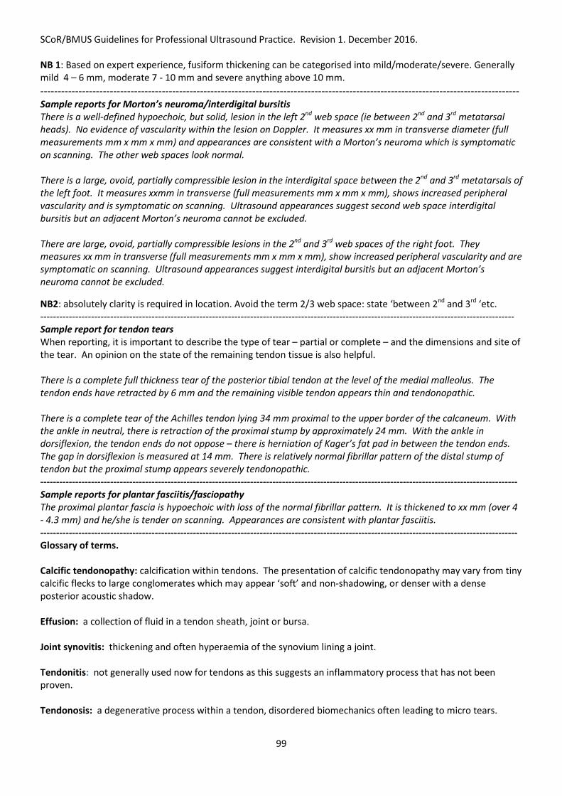
SCoR/BMUS Guidelines for Professional Ultrasound Practice. Revision 1. December 2016.
99
NB 1: Based on expert experience, fusiform thickening can be categorised into mild/moderate/severe. Generally
mild 4 – 6 mm, moderate 7 - 10 mm and severe anything above 10 mm.
------------------------------------------------------------------------------------------------------------------------------------------
Sample reports for Morton’s neuroma/interdigital bursitis
There is a well-defined hypoechoic, but solid, lesion in the left 2nd
web space (ie between 2nd
and 3rd
metatarsal
heads). No evidence of vascularity within the lesion on Doppler. It measures xx mm in transverse diameter (full
measurements mm x mm x mm) and appearances are consistent with a Morton’s neuroma which is symptomatic
on scanning. The other web spaces look normal.
There is a large, ovoid, partially compressible lesion in the interdigital space between the 2nd
and 3rd
metatarsals of
the left foot. It measures xxmm in transverse (full measurements mm x mm x mm), shows increased peripheral
vascularity and is symptomatic on scanning. Ultrasound appearances suggest second web space interdigital
bursitis but an adjacent Morton’s neuroma cannot be excluded.
There are large, ovoid, partially compressible lesions in the 2nd
and 3rd
web spaces of the right foot. They
measures xx mm in transverse (full measurements mm x mm x mm), show increased peripheral vascularity and are
symptomatic on scanning. Ultrasound appearances suggest interdigital bursitis but an adjacent Morton’s
neuroma cannot be excluded.
NB2: absolutely clarity is required in location. Avoid the term 2/3 web space: state ‘between 2nd
and 3rd
‘etc.
-----------------------------------------------------------------------------------------------------------------------------------------------------
Sample report for tendon tears
When reporting, it is important to describe the type of tear – partial or complete – and the dimensions and site of
the tear. An opinion on the state of the remaining tendon tissue is also helpful.
There is a complete full thickness tear of the posterior tibial tendon at the level of the medial malleolus. The
tendon ends have retracted by 6 mm and the remaining visible tendon appears thin and tendonopathic.
There is a complete tear of the Achilles tendon lying 34 mm proximal to the upper border of the calcaneum. With
the ankle in neutral, there is retraction of the proximal stump by approximately 24 mm. With the ankle in
dorsiflexion, the tendon ends do not oppose – there is herniation of Kager’s fat pad in between the tendon ends.
The gap in dorsiflexion is measured at 14 mm. There is relatively normal fibrillar pattern of the distal stump of
tendon but the proximal stump appears severely tendonopathic.
------------------------------------------------------------------------------------------------------------------------------------------------------
Sample reports for plantar fasciitis/fasciopathy
The proximal plantar fascia is hypoechoic with loss of the normal fibrillar pattern. It is thickened to xx mm (over 4
- 4.3 mm) and he/she is tender on scanning. Appearances are consistent with plantar fasciitis.
------------------------------------------------------------------------------------------------------------------------------------------------------
Glossary of terms.
Calcific tendonopathy: calcification within tendons. The presentation of calcific tendonopathy may vary from tiny
calcific flecks to large conglomerates which may appear ‘soft’ and non-shadowing, or denser with a dense
posterior acoustic shadow.
Effusion: a collection of fluid in a tendon sheath, joint or bursa.
Joint synovitis: thickening and often hyperaemia of the synovium lining a joint.
Tendonitis: not generally used now for tendons as this suggests an inflammatory process that has not been
proven.
Tendonosis: a degenerative process within a tendon, disordered biomechanics often leading to micro tears.
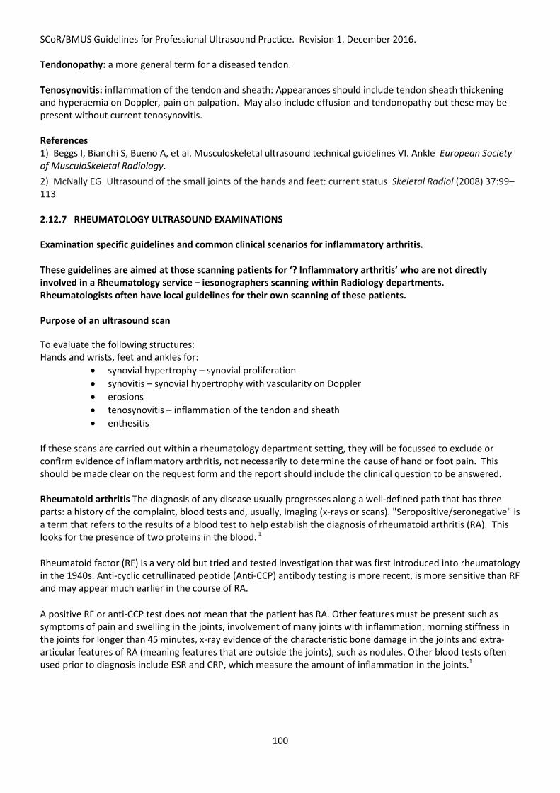
SCoR/BMUS Guidelines for Professional Ultrasound Practice. Revision 1. December 2016.
100
Tendonopathy: a more general term for a diseased tendon.
Tenosynovitis: inflammation of the tendon and sheath: Appearances should include tendon sheath thickening
and hyperaemia on Doppler, pain on palpation. May also include effusion and tendonopathy but these may be
present without current tenosynovitis.
References
1) Beggs I, Bianchi S, Bueno A, et al. Musculoskeletal ultrasound technical guidelines VI. Ankle European Society
of MusculoSkeletal Radiology.
2) McNally EG. Ultrasound of the small joints of the hands and feet: current status Skeletal Radiol (2008) 37:99–
113
2.12.7 RHEUMATOLOGY ULTRASOUND EXAMINATIONS
Examination specific guidelines and common clinical scenarios for inflammatory arthritis.
These guidelines are aimed at those scanning patients for ‘? Inflammatory arthritis’ who are not directly
involved in a Rheumatology service – iesonographers scanning within Radiology departments.
Rheumatologists often have local guidelines for their own scanning of these patients.
Purpose of an ultrasound scan
To evaluate the following structures:
Hands and wrists, feet and ankles for:
• synovial hypertrophy – synovial proliferation
• synovitis – synovial hypertrophy with vascularity on Doppler
• erosions
• tenosynovitis – inflammation of the tendon and sheath
• enthesitis
If these scans are carried out within a rheumatology department setting, they will be focussed to exclude or
confirm evidence of inflammatory arthritis, not necessarily to determine the cause of hand or foot pain. This
should be made clear on the request form and the report should include the clinical question to be answered.
Rheumatoid arthritis The diagnosis of any disease usually progresses along a well-defined path that has three
parts: a history of the complaint, blood tests and, usually, imaging (x-rays or scans). "Seropositive/seronegative" is
a term that refers to the results of a blood test to help establish the diagnosis of rheumatoid arthritis (RA). This
looks for the presence of two proteins in the blood. 1
Rheumatoid factor (RF) is a very old but tried and tested investigation that was first introduced into rheumatology
in the 1940s. Anti-cyclic cetrullinated peptide (Anti-CCP) antibody testing is more recent, is more sensitive than RF
and may appear much earlier in the course of RA.
A positive RF or anti-CCP test does not mean that the patient has RA. Other features must be present such as
symptoms of pain and swelling in the joints, involvement of many joints with inflammation, morning stiffness in
the joints for longer than 45 minutes, x-ray evidence of the characteristic bone damage in the joints and extra-
articular features of RA (meaning features that are outside the joints), such as nodules. Other blood tests often
used prior to diagnosis include ESR and CRP, which measure the amount of inflammation in the joints.1
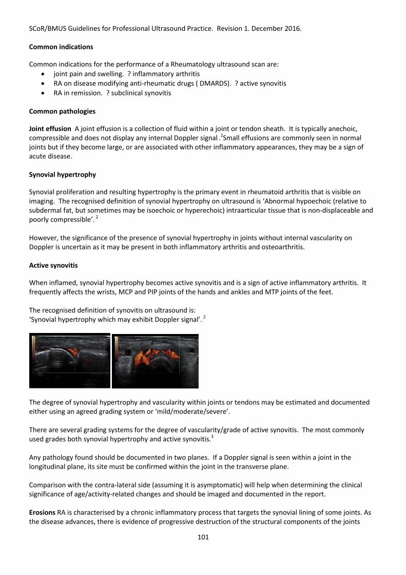
SCoR/BMUS Guidelines for Professional Ultrasound Practice. Revision 1. December 2016.
101
Common indications
Common indications for the performance of a Rheumatology ultrasound scan are:
• joint pain and swelling. ? inflammatory arthritis
• RA on disease modifying anti-rheumatic drugs ( DMARDS). ? active synovitis
• RA in remission. ? subclinical synovitis
Common pathologies
Joint effusion A joint effusion is a collection of fluid within a joint or tendon sheath. It is typically anechoic,
compressible and does not display any internal Doppler signal .2Small effusions are commonly seen in normal
joints but if they become large, or are associated with other inflammatory appearances, they may be a sign of
acute disease.
Synovial hypertrophy
Synovial proliferation and resulting hypertrophy is the primary event in rheumatoid arthritis that is visible on
imaging. The recognised definition of synovial hypertrophy on ultrasound is ‘Abnormal hypoechoic (relative to
subdermal fat, but sometimes may be isoechoic or hyperechoic) intraarticular tissue that is non-displaceable and
poorly compressible’. 2
However, the significance of the presence of synovial hypertrophy in joints without internal vascularity on
Doppler is uncertain as it may be present in both inflammatory arthritis and osteoarthritis.
Active synovitis
When inflamed, synovial hypertrophy becomes active synovitis and is a sign of active inflammatory arthritis. It
frequently affects the wrists, MCP and PIP joints of the hands and ankles and MTP joints of the feet.
The recognised definition of synovitis on ultrasound is:
‘Synovial hypertrophy which may exhibit Doppler signal’. 2
The degree of synovial hypertrophy and vascularity within joints or tendons may be estimated and documented
either using an agreed grading system or ‘mild/moderate/severe’.
There are several grading systems for the degree of vascularity/grade of active synovitis. The most commonly
used grades both synovial hypertrophy and active synovitis.3
Any pathology found should be documented in two planes. If a Doppler signal is seen within a joint in the
longitudinal plane, its site must be confirmed within the joint in the transverse plane.
Comparison with the contra-lateral side (assuming it is asymptomatic) will help when determining the clinical
significance of age/activity-related changes and should be imaged and documented in the report.
Erosions RA is characterised by a chronic inflammatory process that targets the synovial lining of some joints. As
the disease advances, there is evidence of progressive destruction of the structural components of the joints
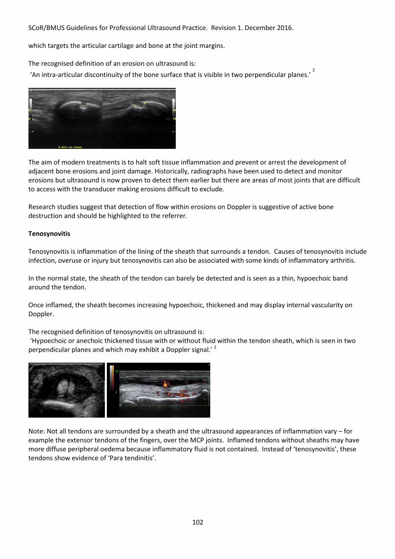
SCoR/BMUS Guidelines for Professional Ultrasound Practice. Revision 1. December 2016.
102
which targets the articular cartilage and bone at the joint margins.
The recognised definition of an erosion on ultrasound is:
‘An intra-articular discontinuity of the bone surface that is visible in two perpendicular planes.’ 2
The aim of modern treatments is to halt soft tissue inflammation and prevent or arrest the development of
adjacent bone erosions and joint damage. Historically, radiographs have been used to detect and monitor
erosions but ultrasound is now proven to detect them earlier but there are areas of most joints that are difficult
to access with the transducer making erosions difficult to exclude.
Research studies suggest that detection of flow within erosions on Doppler is suggestive of active bone
destruction and should be highlighted to the referrer.
Tenosynovitis
Tenosynovitis is inflammation of the lining of the sheath that surrounds a tendon. Causes of tenosynovitis include
infection, overuse or injury but tenosynovitis can also be associated with some kinds of inflammatory arthritis.
In the normal state, the sheath of the tendon can barely be detected and is seen as a thin, hypoechoic band
around the tendon.
Once inflamed, the sheath becomes increasing hypoechoic, thickened and may display internal vascularity on
Doppler.
The recognised definition of tenosynovitis on ultrasound is:
‘Hypoechoic or anechoic thickened tissue with or without fluid within the tendon sheath, which is seen in two
perpendicular planes and which may exhibit a Doppler signal.’ 2
Note: Not all tendons are surrounded by a sheath and the ultrasound appearances of inflammation vary – for
example the extensor tendons of the fingers, over the MCP joints. Inflamed tendons without sheaths may have
more diffuse peripheral oedema because inflammatory fluid is not contained. Instead of ‘tenosynovitis’, these
tendons show evidence of ‘Para tendinitis’.
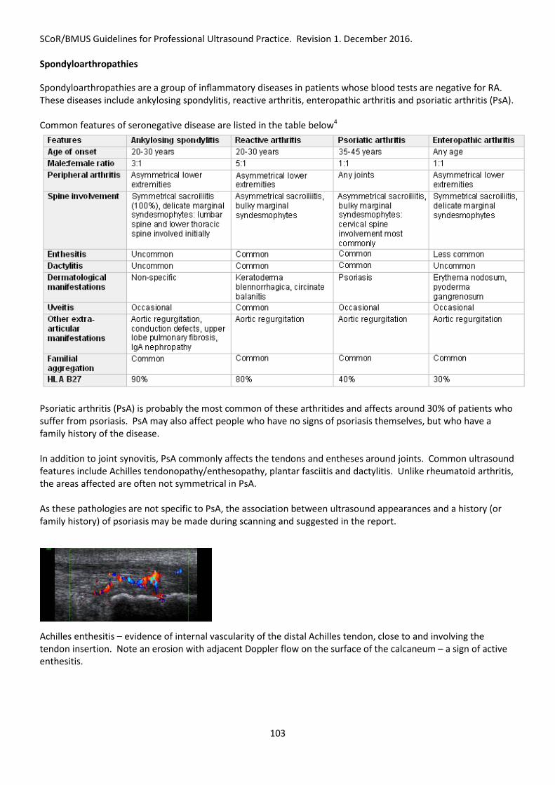
SCoR/BMUS Guidelines for Professional Ultrasound Practice. Revision 1. December 2016.
103
Spondyloarthropathies
Spondyloarthropathies are a group of inflammatory diseases in patients whose blood tests are negative for RA.
These diseases include ankylosing spondylitis, reactive arthritis, enteropathic arthritis and psoriatic arthritis (PsA).
Common features of seronegative disease are listed in the table below4
Psoriatic arthritis (PsA) is probably the most common of these arthritides and affects around 30% of patients who
suffer from psoriasis. PsA may also affect people who have no signs of psoriasis themselves, but who have a
family history of the disease.
In addition to joint synovitis, PsA commonly affects the tendons and entheses around joints. Common ultrasound
features include Achilles tendonopathy/enthesopathy, plantar fasciitis and dactylitis. Unlike rheumatoid arthritis,
the areas affected are often not symmetrical in PsA.
As these pathologies are not specific to PsA, the association between ultrasound appearances and a history (or
family history) of psoriasis may be made during scanning and suggested in the report.
Achilles enthesitis – evidence of internal vascularity of the distal Achilles tendon, close to and involving the
tendon insertion. Note an erosion with adjacent Doppler flow on the surface of the calcaneum – a sign of active
enthesitis.
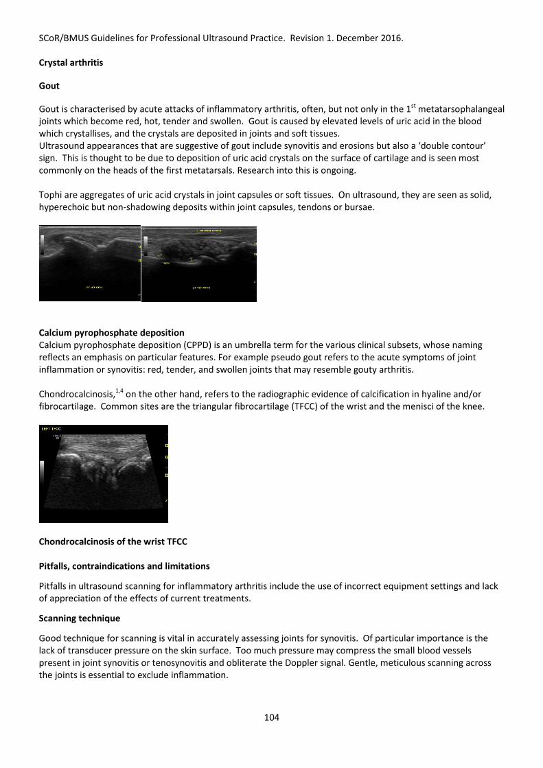
SCoR/BMUS Guidelines for Professional Ultrasound Practice. Revision 1. December 2016.
104
Crystal arthritis
Gout
Gout is characterised by acute attacks of inflammatory arthritis, often, but not only in the 1st
metatarsophalangeal
joints which become red, hot, tender and swollen. Gout is caused by elevated levels of uric acid in the blood
which crystallises, and the crystals are deposited in joints and soft tissues.
Ultrasound appearances that are suggestive of gout include synovitis and erosions but also a ‘double contour’
sign. This is thought to be due to deposition of uric acid crystals on the surface of cartilage and is seen most
commonly on the heads of the first metatarsals. Research into this is ongoing.
Tophi are aggregates of uric acid crystals in joint capsules or soft tissues. On ultrasound, they are seen as solid,
hyperechoic but non-shadowing deposits within joint capsules, tendons or bursae.
Calcium pyrophosphate deposition
Calcium pyrophosphate deposition (CPPD) is an umbrella term for the various clinical subsets, whose naming
reflects an emphasis on particular features. For example pseudo gout refers to the acute symptoms of joint
inflammation or synovitis: red, tender, and swollen joints that may resemble gouty arthritis.
Chondrocalcinosis,1,4
on the other hand, refers to the radiographic evidence of calcification in hyaline and/or
fibrocartilage. Common sites are the triangular fibrocartilage (TFCC) of the wrist and the menisci of the knee.
Chondrocalcinosis of the wrist TFCC
Pitfalls, contraindications and limitations
Pitfalls in ultrasound scanning for inflammatory arthritis include the use of incorrect equipment settings and lack
of appreciation of the effects of current treatments.
Scanning technique
Good technique for scanning is vital in accurately assessing joints for synovitis. Of particular importance is the
lack of transducer pressure on the skin surface. Too much pressure may compress the small blood vessels
present in joint synovitis or tenosynovitis and obliterate the Doppler signal. Gentle, meticulous scanning across
the joints is essential to exclude inflammation.
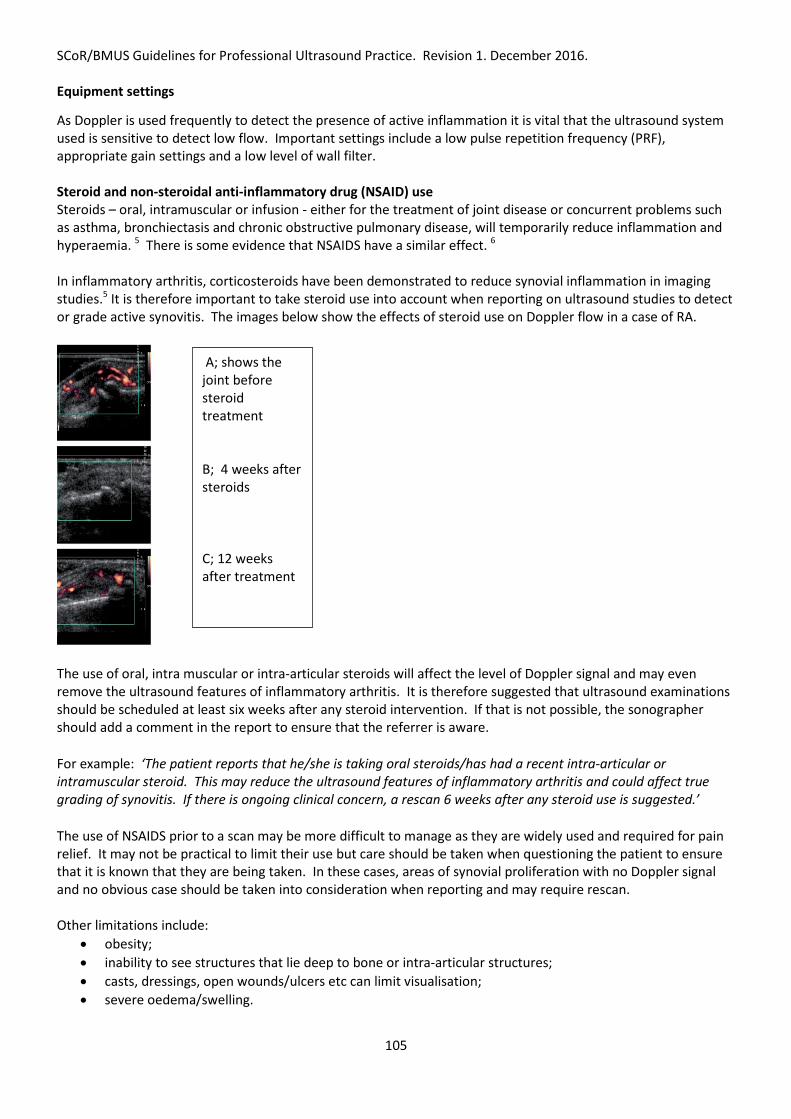
SCoR/BMUS Guidelines for Professional Ultrasound Practice. Revision 1. December 2016.
105
Equipment settings
As Doppler is used frequently to detect the presence of active inflammation it is vital that the ultrasound system
used is sensitive to detect low flow. Important settings include a low pulse repetition frequency (PRF),
appropriate gain settings and a low level of wall filter.
Steroid and non-steroidal anti-inflammatory drug (NSAID) use
Steroids – oral, intramuscular or infusion - either for the treatment of joint disease or concurrent problems such
as asthma, bronchiectasis and chronic obstructive pulmonary disease, will temporarily reduce inflammation and
hyperaemia. 5 There is some evidence that NSAIDS have a similar effect.
6
In inflammatory arthritis, corticosteroids have been demonstrated to reduce synovial inflammation in imaging
studies.5 It is therefore important to take steroid use into account when reporting on ultrasound studies to detect
or grade active synovitis. The images below show the effects of steroid use on Doppler flow in a case of RA.
The use of oral, intra muscular or intra-articular steroids will affect the level of Doppler signal and may even
remove the ultrasound features of inflammatory arthritis. It is therefore suggested that ultrasound examinations
should be scheduled at least six weeks after any steroid intervention. If that is not possible, the sonographer
should add a comment in the report to ensure that the referrer is aware.
For example: ‘The patient reports that he/she is taking oral steroids/has had a recent intra-articular or
intramuscular steroid. This may reduce the ultrasound features of inflammatory arthritis and could affect true
grading of synovitis. If there is ongoing clinical concern, a rescan 6 weeks after any steroid use is suggested.’
The use of NSAIDS prior to a scan may be more difficult to manage as they are widely used and required for pain
relief. It may not be practical to limit their use but care should be taken when questioning the patient to ensure
that it is known that they are being taken. In these cases, areas of synovial proliferation with no Doppler signal
and no obvious case should be taken into consideration when reporting and may require rescan.
Other limitations include:
• obesity;
• inability to see structures that lie deep to bone or intra-articular structures;
• casts, dressings, open wounds/ulcers etc can limit visualisation;
• severe oedema/swelling.
A; shows the
joint before
steroid
treatment
B; 4 weeks after
steroids
C; 12 weeks
after treatment
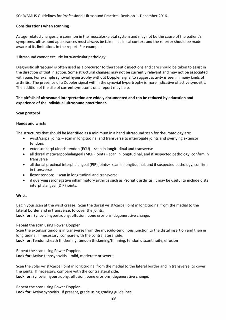
SCoR/BMUS Guidelines for Professional Ultrasound Practice. Revision 1. December 2016.
106
Considerations when scanning
As age-related changes are common in the musculoskeletal system and may not be the cause of the patient’s
symptoms, ultrasound appearances must always be taken in clinical context and the referrer should be made
aware of its limitations in the report. For example:
‘Ultrasound cannot exclude intra-articular pathology’
Diagnostic ultrasound is often used as a precursor to therapeutic injections and care should be taken to assist in
the direction of that injection. Some structural changes may not be currently relevant and may not be associated
with pain. For example synovial hypertrophy without Doppler signal to suggest activity is seen in many kinds of
arthritis. The presence of a Doppler signal within the synovial hypertrophy is more indicative of active synovitis.
The addition of the site of current symptoms on a report may help.
The pitfalls of ultrasound interpretation are widely documented and can be reduced by education and
experience of the individual ultrasound practitioner.
Scan protocol
Hands and wrists
The structures that should be identified as a minimum in a hand ultrasound scan for rheumatology are:
• wrist/carpal joints – scan in longitudinal and transverse to interrogate joints and overlying extensor
tendons
• extensor carpi ulnaris tendon (ECU) – scan in longitudinal and transverse
• all dorsal metacarpophalangeal (MCP) joints – scan in longitudinal, and if suspected pathology, confirm in
transverse
• all dorsal proximal interphalangeal (PIP) joints– scan in longitudinal, and if suspected pathology, confirm
in transverse
• flexor tendons – scan in longitudinal and transverse
• if querying seronegative inflammatory arthritis such as Psoriatic arthritis, it may be useful to include distal
interphalangeal (DIP) joints.
Wrists
Begin your scan at the wrist crease. Scan the dorsal wrist/carpal joint in longitudinal from the medial to the
lateral border and in transverse, to cover the joints.
Look for: Synovial hypertrophy, effusion, bone erosions, degenerative change.
Repeat the scan using Power Doppler
Scan the extensor tendons in transverse from the musculo-tendinous junction to the distal insertion and then in
longitudinal. If necessary, compare with the contra lateral side.
Look for: Tendon sheath thickening, tendon thickening/thinning, tendon discontinuity, effusion
Repeat the scan using Power Doppler.
Look for: Active tenosynovitis – mild, moderate or severe
Scan the volar wrist/carpal joint in longitudinal from the medial to the lateral border and in transverse, to cover
the joints. If necessary, compare with the contralateral side.
Look for: Synovial hypertrophy, effusion, bone erosions, degenerative change.
Repeat the scan using Power Doppler.
Look for: Active synovitis. If present, grade using grading guidelines.
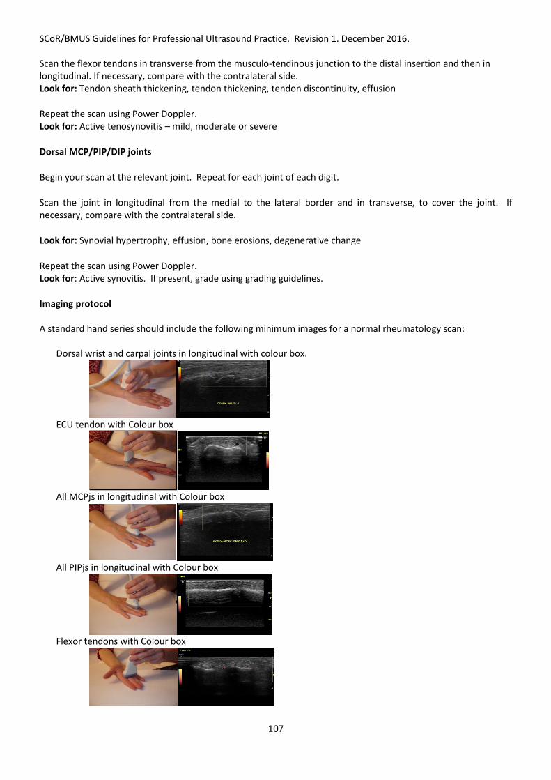
SCoR/BMUS Guidelines for Professional Ultrasound Practice. Revision 1. December 2016.
107
Scan the flexor tendons in transverse from the musculo-tendinous junction to the distal insertion and then in
longitudinal. If necessary, compare with the contralateral side.
Look for: Tendon sheath thickening, tendon thickening, tendon discontinuity, effusion
Repeat the scan using Power Doppler.
Look for: Active tenosynovitis – mild, moderate or severe
Dorsal MCP/PIP/DIP joints
Begin your scan at the relevant joint. Repeat for each joint of each digit.
Scan the joint in longitudinal from the medial to the lateral border and in transverse, to cover the joint. If
necessary, compare with the contralateral side.
Look for: Synovial hypertrophy, effusion, bone erosions, degenerative change
Repeat the scan using Power Doppler.
Look for: Active synovitis. If present, grade using grading guidelines.
Imaging protocol
A standard hand series should include the following minimum images for a normal rheumatology scan:
Dorsal wrist and carpal joints in longitudinal with colour box.
ECU tendon with Colour box
All MCPjs in longitudinal with Colour box
All PIPjs in longitudinal with Colour box
Flexor tendons with Colour box
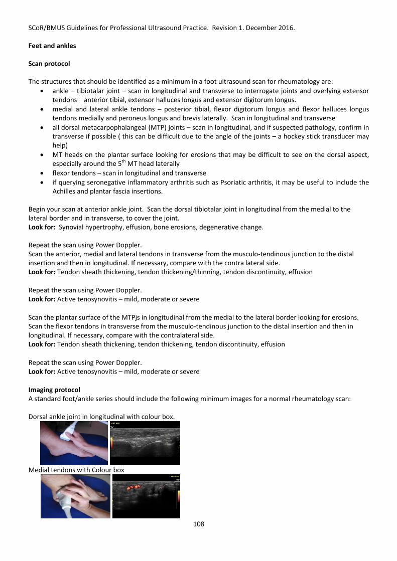
SCoR/BMUS Guidelines for Professional Ultrasound Practice. Revision 1. December 2016.
108
Feet and ankles
Scan protocol
The structures that should be identified as a minimum in a foot ultrasound scan for rheumatology are:
• ankle – tibiotalar joint – scan in longitudinal and transverse to interrogate joints and overlying extensor
tendons – anterior tibial, extensor halluces longus and extensor digitorum longus.
• medial and lateral ankle tendons – posterior tibial, flexor digitorum longus and flexor halluces longus
tendons medially and peroneus longus and brevis laterally. Scan in longitudinal and transverse
• all dorsal metacarpophalangeal (MTP) joints – scan in longitudinal, and if suspected pathology, confirm in
transverse if possible ( this can be difficult due to the angle of the joints – a hockey stick transducer may
help)
• MT heads on the plantar surface looking for erosions that may be difficult to see on the dorsal aspect,
especially around the 5th
MT head laterally
• flexor tendons – scan in longitudinal and transverse
• if querying seronegative inflammatory arthritis such as Psoriatic arthritis, it may be useful to include the
Achilles and plantar fascia insertions.
Begin your scan at anterior ankle joint. Scan the dorsal tibiotalar joint in longitudinal from the medial to the
lateral border and in transverse, to cover the joint.
Look for: Synovial hypertrophy, effusion, bone erosions, degenerative change.
Repeat the scan using Power Doppler.
Scan the anterior, medial and lateral tendons in transverse from the musculo-tendinous junction to the distal
insertion and then in longitudinal. If necessary, compare with the contra lateral side.
Look for: Tendon sheath thickening, tendon thickening/thinning, tendon discontinuity, effusion
Repeat the scan using Power Doppler.
Look for: Active tenosynovitis – mild, moderate or severe
Scan the plantar surface of the MTPjs in longitudinal from the medial to the lateral border looking for erosions.
Scan the flexor tendons in transverse from the musculo-tendinous junction to the distal insertion and then in
longitudinal. If necessary, compare with the contralateral side.
Look for: Tendon sheath thickening, tendon thickening, tendon discontinuity, effusion
Repeat the scan using Power Doppler.
Look for: Active tenosynovitis – mild, moderate or severe
Imaging protocol
A standard foot/ankle series should include the following minimum images for a normal rheumatology scan:
Dorsal ankle joint in longitudinal with colour box.
Medial tendons with Colour box
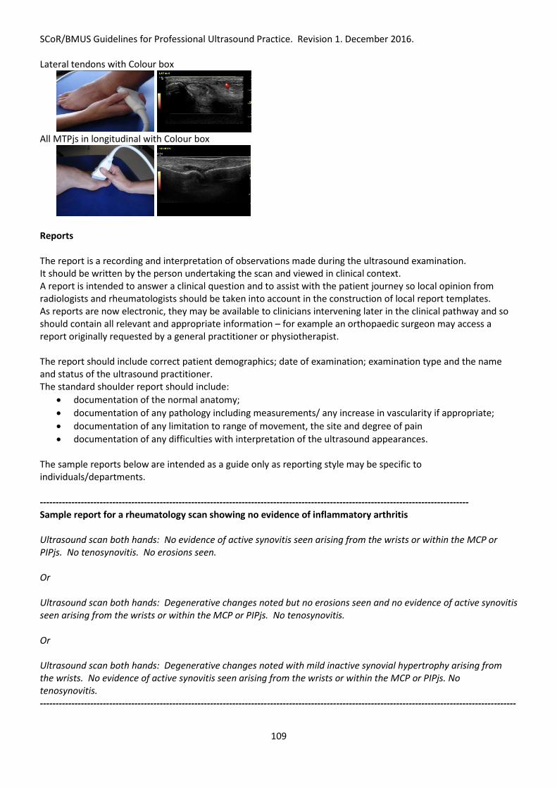
SCoR/BMUS Guidelines for Professional Ultrasound Practice. Revision 1. December 2016.
109
Lateral tendons with Colour box
All MTPjs in longitudinal with Colour box
Reports
The report is a recording and interpretation of observations made during the ultrasound examination.
It should be written by the person undertaking the scan and viewed in clinical context.
A report is intended to answer a clinical question and to assist with the patient journey so local opinion from
radiologists and rheumatologists should be taken into account in the construction of local report templates.
As reports are now electronic, they may be available to clinicians intervening later in the clinical pathway and so
should contain all relevant and appropriate information – for example an orthopaedic surgeon may access a
report originally requested by a general practitioner or physiotherapist.
The report should include correct patient demographics; date of examination; examination type and the name
and status of the ultrasound practitioner.
The standard shoulder report should include:
• documentation of the normal anatomy;
• documentation of any pathology including measurements/ any increase in vascularity if appropriate;
• documentation of any limitation to range of movement, the site and degree of pain
• documentation of any difficulties with interpretation of the ultrasound appearances.
The sample reports below are intended as a guide only as reporting style may be specific to
individuals/departments.
----------------------------------------------------------------------------------------------------------------------------------------
Sample report for a rheumatology scan showing no evidence of inflammatory arthritis
Ultrasound scan both hands: No evidence of active synovitis seen arising from the wrists or within the MCP or
PIPjs. No tenosynovitis. No erosions seen.
Or
Ultrasound scan both hands: Degenerative changes noted but no erosions seen and no evidence of active synovitis
seen arising from the wrists or within the MCP or PIPjs. No tenosynovitis.
Or
Ultrasound scan both hands: Degenerative changes noted with mild inactive synovial hypertrophy arising from
the wrists. No evidence of active synovitis seen arising from the wrists or within the MCP or PIPjs. No
tenosynovitis.
-------------------------------------------------------------------------------------------------------------------------------------------------------
SCoR/BMUS Guidelines for Professional Ultrasound Practice. Revision 1. December 2016.
110
Sample report for a rheumatology scan showing synovial hypertrophy in a patient who has had a recent
intramuscular steroid injection
Ultrasound scan both hands: There is obvious synovial hypertrophy arising from the dorsal surfaces of both wrists
but no evidence of internal Doppler signal. The patient reports an intramuscular injection of Depomedrone 2
weeks ago. This will reduce Doppler signal and may mask the ultrasound appearances of active synovitis. If
symptoms return after 4-6 weeks and there is ongoing suspicion of inflammatory arthritis, a rescan is suggested
before any further steroids are given.
-----------------------------------------------------------------------------------------------------------------------------------------
Sample report for a rheumatology scan with positive findings for inflammatory arthritis.
Ultrasound scan both hands: There is grade 2 (or moderate) active synovitis arising from the right wrist and
within the right index and middle and left index finger MCPjs. No evidence of active synovitis arising from the left
wrist or within the remainder of the MCPjs or the PIPjs. No tenosynovitis. No erosions seen.
-------------------------------------------------------------------------------------------------------------------------------------------------------
Sample report for a patient with suspected seronegative inflammatory arthritis
Ultrasound scan both feet: There is evidence of moderate synovitis arising from the left ankle joint but none seen
on the right. The distal portion of both Achilles tendons are thickened and hypoechoic with loss of the normal
fibrillar pattern. There is a moderate degree of internal vascularity within the tendons at the insertion and
erosions on the posterior aspect of the calcaneum. Appearances suggest erosive enthesitis. There is also effusion
and hyperaemia of both retro calcaneal bursae suggesting bursitis.
In the absence of an obvious biomechanical cause for these appearances, an inflammatory arthropathy should be
considered and if there is clinical correlation, a rheumatology opinion is suggested.
------------------------------------------------------------------------------------------------------------------------------------------------------
References
1) Royal College of Physicians. Rheumatoid Arthritis: National clinical guidelines for treatment in adults 2009
2) Wakefield R J, Balint PV, Szkudlarek M, et al. Musculoskeletal ultrasound including definitions for
ultrasonographic pathology. J Rheumatol 2005; 32 : 2485 – 2487
3) Berner Hammer H, Bolton-King P et al ‘Examination of intra and interrater reliability with a new
ultrasonographic reference atlas for scoring of synovitis in patients with rheumatoid arthritis’ Ann rheum Dis
doi:10.1136/ard.2011.152926
4) Kataria RK, Brent LH. Spondyloarthropathies. Am Fam Physician. 2004; 69: 2853-2260 and Gladman DD. Am J
Med Sci.1998; 316:234-2384.
5) Filippucci E, Farina A, Carotti M, Salaffi F, Grassi W. Greyscale and power Doppler sonographic changes
induced by intra-articular steroid injection treatment. Ann Rheum Dis 2004; 63: 740–743
6) Zayat S, Conoghan P, Sharif M, Freeston J. Do non-steroidal anti-inflammatory drugs have a significant effect
on detection and grading of ultrasound-detected synovitis in patients with RA? Ann Rheum Dis 2011; 70: 1746-
1751
Useful reading
Backhaus M. Ultrasound and structural changes in inflammatory arthritis: synovitis and tenosynovitis. Ann NY
Acad Sci 2009; 1154: 139-151
Backhaus M, Burmster G et al Guidelines for musculoskeletal ultrasound in rheumatology Ann Rheum Dis 60: 641-
649
Kang T, Lanni S et al The evolution of ultrasound in rheumatology. Therapeutic advances in musculoskeletal
disease 2012 Dec 4(6): 399-411
2. 13 ELASTOGRAPHY
Advances in ultrasound technologies have expanded the clinical applications of ultrasound over recent years.
Historically ultrasound has been a primary imaging modality, but it is now common for ultrasound technology to
be utilised in other techniques such as needle placement, joint injections, guided biopsies, interventional
procedures and more recently in the diagnosis and management of patients with chronic liver diseases.
SCoR/BMUS Guidelines for Professional Ultrasound Practice. Revision 1. December 2016.
111
Recent advances in elastography techniques such as acoustic radiation force impulsion (ARFI) and transient
elastography (TE) have expanded the use of ultrasound into disease management. They can also enable lesion
characterisation and the mapping of tissue stiffness. This reproduces / replaces the palpation performed by
clinicians.
At present there are two main forms of elastography which are being used routinely in clinical practice.
Strain Elastography (SE)
Quasi-static or strain elastography (SE), where the tissue deformation is produced by external palpation with the
ultrasound transducer. Most manufacturers overlay a speckle map on the ultrasound image, which is coded in a
colour or greyscale to show a pattern of strain, which is inversely related to the tissue stiffness and can be
assessed subjectively. These images are semi-quantitative and do not directly depict the elasticity which is
derived from Young’s modulus, (stress/strain ratio).
Shear Wave Elastography (SWE)
Conventional ultrasound and shear waves are both pressure waves that are conducted through soft tissue. Unlike
conventional ultrasound waves; shear waves are transverse in their directionality, they are rapidly attenuated by
soft tissue and travel more slowly (between 1 and 10 m/s). Their speed is closely related to Young’s modulus of
elasticity. Shearwaves are created naturally from muscle movements or in cardiac activity and can be induced by
ultrasound scanners which use them to measure their speed in a particular tissue or organ.
Liver Fibrosis Assessment
Most chronic liver diseases have alterations in the functional and structural appearance of the liver. The risk of
developing cirrhosis and liver related complications in viral and non viral chronic liver diseases is correlated to the
amount of liver fibrosis. The detection and classification of liver fibrosis is pivotal for assessing progression and
when to commence patients on antiviral therapies. Liver biopsy has long been the traditional approach for
fibrosis assessment and disease classification. Liver biopsy by its nature is invasive and has potentially severe
complications in up to 1% of cases 1
with associated cost implications. Recent (2015) advice from NICE (MTG27)2,
advocates elastography in the diagnosis and monitoring of fibrosis in chronic hepatitis The economic benefits of
using elastography is explored in the guideline , a saving of around £434 per patient is quoted when using Virtual
Touch Quantification (VTq) over conventional liver biopsy. The safety implications to patients should also be
considered as well as its tolerability and its ability to be undertaken in an outpatient setting. Giving the economic
challenges facing healthcare today, this technique could have the potential to deliver large savings.
http://nice.org.uk/guidance/mtg27
One difficulty with liver elastography is the different methods and multitude of ultrasound manufacturers
providing a method of stiffness assessment. This results in the varying cut-off values being ultrasound scanner
specific and not transferable across different makes of equipment.
Transient Elastography (TE)
One method of SWE is transient elastography (TE) which is being performed using the Fibroscan® (Echosens,
Paris, France). This is a mechanical system using a single element ultrasound transducer which is used to apply
light pressure on the patient’s skin, usually intercostally to assess the liver parenchyma. The resultant shear wave
travels through liver tissue along the direction of the ultrasound beam and is used to measure its speed in a
manner similar to M-mode. In patients with chronic hepatitis C, liver stiffness (LS) values are greater than 6.8-
7.6kPa. The cut off for predicting cirrhosis range between 11.0 – 13.6 kPa. Evidence suggests that TE has
limitations in differentiating between mild and significant fibrosis. Other limitations include the requirement of
dedicated equipment with specific probes and an intercostal scan approach. The technique is standardised and
SCoR/BMUS Guidelines for Professional Ultrasound Practice. Revision 1. December 2016.
112
can be employed in various settings. It is currently being used mainly outside the radiology department in
outpatient settings.
Acoustic Radiation Force Impulse (ARFI)
ARFI generates shear waves by causing small displacements in the soft tissue, causing sideways pressure waves
away from the ultrasound beam which allow ultrasound systems to measure speed with good spatial resolution.
ARFI accuracy for the assessment of liver fibrosis has been similar to TE, with some more promising results in
patients with non-alcoholic fatty liver disease (NALFD), non-alcoholic steatohepatitis (NASH) and in post
transplantation. It has the advantage of allowing a radiological assessment of the liver parenchyma and more
accurate localisation of the sample site. Therefore, fatty infiltration can be avoided so as not to not skew fibrosis
scoring.
Following the publication of the European Federation of Societies of Ultrasound in Medicine and Biology
(EFSUMB) guidelines on the clinical use of elastography, 3
it is recommended that shearwave elastography can be
used to assess the severity of patients with liver disease with viral hepatitis. The guidelines also state that
shearwave elastography is promising in NAFLD and post-transplantation.
Clinical Application
Whilst differing manufacturers have different shearwave velocities for stages of liver disease it is noted that the
following (based on the VTq imaging as described in the NICE publication 2) are given as examples of disease
staging
• Normal < 1.2 m/s
• Fibrosis ≥ 1.21 – 1.34 m/s
• Cirrhosis ≥ 1.55 – 2.00 m/s
Extreme caution is required when quoting shearwave velocities in ultrasound reports. It is useful to report the
shearwave velocity and quote the relevant reference values for the machine used to minimise confusion between
normal and abnormal readings compared to the stage of liver disease. Practitioners are advised to refer to
individual manufacturers’ reference ranges when reporting shearwave studies in liver disease.
Practical points on acquisition
• The patient should be fasted. Shear wave speed measurements are performed intercostally in the right
liver with conventional curved arrays. The probe is aligned along an intercostal space.
• 10 measurements should be acquired and the mean value and standard deviation stated.
• The recommended depth is between 3 and 7 cm. Shear wave methods can be utilised in patients with
ascites.
References
1. Seeff LB, Everson GT, Morgan TR (2010) Complication rate of percutaneous liver biopsies among persons with
advanced chronic liver disease in the HALT-C trial. Clinical Gastroenterology Hepatology, Vol 8, pp877-883.
2. National Institute for Health and Care Excellence (2015). Virtual Touch Quantification (VTq) to diagnose and
monitor liver fibrosis in chronic hepatitis B and C (MTG27) London: NICE
3. Cosgrove D, et al (2013) EFSUMB Guidelines and Recommendations on the Clinical Use of Ultrasound
Elastography. Part 2: Clinical Applications. Ultraschall in Med Vol 34 pp238-253.
Bibliography
Gennisson et al (2013) Ultrasound Elastography: Principles and Techniques, Diagnostic and Interventional
Imaging, Vol 94, pp487-95.
SCoR/BMUS Guidelines for Professional Ultrasound Practice. Revision 1. December 2016.
113
2.14 CONTRAST ENHANCED ULTRASOUND (CEUS)
Ultrasound contrast agents are formulated into a microbubble structure which performs as a blood pool tracer
when used in conjunction with contrast specific imaging modes using a low Mechanical Index (MI) technique. An
example is ‘Sonovue’ (sulphur hexafluoride). These agents are intravenously injected in order to demonstrate the
vasculature and microvasculature of organs and potential lesions.
They are primarily used for hepatic applications1
but are also proven to be useful in a variety of other
applications.2
Administration of ultrasound contrast agent
Administration is performed via an intravenous cannula.
Use of contrast agents by non-medical staff, usually sonographers, should come within the remit of a patient
group direction (PGD) or other formal local procedure which allows the prescribing and safe administration of the
drug. A sonographer must be statutorily registered to follow a PGD; voluntary registration is not sufficient.
Any contraindication to the agent used should be carefully observed, and documentation of the dose
administered recorded according to local rules.
Safety
The microbubble agents currently available are not nephrotoxic, making them a very useful alternative in patients
with renal compromise.3
They have a very low incidence of adverse reactions although there is a small risk of anaphylactoid reactions to
CEUS (rate estimated at 1:10,000) and resuscitation facilities with emergency equipment and personnel trained in
its use should be available. It is recommended to keep the patient under close medical supervision during, and for
at least 30 minutes following the administration of sulphur hexafluoride (Sonovue). A programme of annual basic
life support training should be in place for staff. 4
Microbubble agents are currently not licensed in pregnancy or in children but may be used ‘off license’ in
children. The FDA gave approval in April 2016 for the use of CEUS in the USA in children who have focal liver
lesions. Recent EFSUMB guidelines assess the current status of CEUS applications in children and make
suggestions for further development of this technique. 5
Equipment
Contrast-specific software is required to perform CEUS.
A low MI technique helps to preserve the microbubbles, enabling a longer available survey time.
The facility to record a short video clip, of all phases for at least three minutes, is essential when using CEUS to
characterise lesions. The temporal resolution of CEUS is superior to any other current imaging modality, and the
ability to review the arterial phase, frame by frame, provides a valuable contribution to the diagnostic process.
Portal washout is a hallmark of malignancy.
Main applications
• Characterisation of focal liver lesions in non-cirrhotic patients.
• Characterisation of benign lesions.
• Detection of liver metastases in patients at high risk of liver metastases (eg those with a primary
cancer who have not undergone CT staging, patients with sudden unintentional weight loss,
SCoR/BMUS Guidelines for Professional Ultrasound Practice. Revision 1. December 2016.
114
abnormal LFTs and otherwise equivocal livers on a baseline scan). Patients for pre-treatment staging
should ideally undergo CT.
• Doppler ‘rescue’ to identify flow in technically difficult examinations e.g. hepatic artery
demonstration in a difficult post-transplant scan.
• Lesion localisation pre-ablation and for US guided biopsy.
• Post ablation checks.
• Trauma, to identify soft organ damage, if CT cannot be performed.
• Trauma follow-up looking for pseudo-aneurysms and resolution of lacerations.
Cirrhotic patients with focal lesions should ideally be referred to MRI for characterisation and further exclusion of
hepatocellular carcinoma (HCC). However, if the patient is unable to undergo MRI, then CEUS may provide
valuable additional information in lesion characterisation.
Extrahepatic applications
• Characterisation of complex renal cysts 6 (eg with septa or containing debris/possible solid material).
CEUS may be offered as a first line characterisation. Complex, obviously suspicious cysts and solid
masses (Bosniak 4) should be referred urgently to CT.
• Vascular patency/ detection of infarcts in difficult renal transplant scans.
• Sentinel node detection in patients with breast cancer.
• Follow-up of endovascular aneurysm repair (EVAR) stents to establish patency and look for leaks.
• Hysterosalpingo-Contrast-Sonography (HyCoSy) for tubal patency testing.
It is the practitioner’s responsibility to be aware of the licensed use of Sonovue. Where Sonovue is used “off
license”, local agreement to its use, and by whom within the department, should be documented. The patient’s
verbal consent for the use of contrast “off license” should be sought and documented.
Further applications with less supportive evidence at this stage also include the spleen, endoscopic US including
pancreas, GI tract, scrotal and prostate.2
CEUS can also be used to facilitate ultrasound guided biopsy in difficult-
to-see lesions.
References
1) SonoVue (sulphur hexafluoride microbubbles) – contrast agent for contrast-enhanced ultrasound imaging of
the liver. NICE diagnostics guidance [DG5] August 2012. https://www.nice.org.uk/guidance/dg5
2) The EFSUMB Guidelines and Recommendations on the Clinical Practice of Contrast Enhanced Ultrasound
(CEUS): Update 2011 on non-hepatic applications Ultraschall in der Medizin/European Journal of Ultrasound
2012 Issue 1
3) Piscaglia F, Bolondi L. The safety of Sonovue in abdominal applications: retrospective analysis of 23188
investigations. Italian Society for Ultrasound in Medicine and Biology (SIUMB) Study Group on Ultrasound
Contrast Agents. Ultrasound Med Biol. 2006 Sep; 32(9): 1369-1375.
4) Royal College of Radiologists/Society and College of Radiographers. Standards for the provision of an
ultrasound service. London: RCR/SCoR (2014) Section 4.
5) Sidhu PS et al. Role of Contrast Enhanced Ultrasound (CEUS) in Paediatric Practice: An EFSUMB Position
Satement. Ultraschall Med, April 2016. http://www.ncbi.nlm.nih.gov/pubmed/27414980?dopt=Citation
6) Ascenti G et al. Complex cystic renal masses: characterization with contrast enhanced ultrasound. Radiology
April 2007; 243 (1): 158-165
2.15 INTERVENTIONAL AND ‘EXTENDED SCOPE’ EXAMINATIONS (SONOGRAPHERS)
There is a wide range of examinations that come within the overall scope of practice of ultrasound practitioners.
For those who are not medically qualified and registered with the GMC (e.g. sonographers) they are sometimes
SCoR/BMUS Guidelines for Professional Ultrasound Practice. Revision 1. December 2016.
115
termed ‘extended scope’ examinations. They include diagnostic procedures such as biopsy, fine needle
aspiration, hystero-salpingo contrast sonography (HyCoSy), drainage of body cavities and therapeutic procedures
such as joint injections.
Like all ultrasound examinations, the sonographer must be trained, competent and authorised to undertake
them.
Examples of the type of ultrasound examinations undertaken by sonographers and to which this section refers are
as follows:
i) Biopsy / fine needle aspiration eg breast, prostate, liver, thyroid.
ii) Drainage eg drainage of ascites
iii) Therapeutic egjoint injections
iv) Diagnosis e.g. HyCoSy, contrast enhanced ultrasound (CEUS).
When setting up such ‘extended scope’ services the approval of the clinical lead, service manager and Trust,
Health Board or provider organisation should be sought.
The following will all need to be considered:
• support from a lead medical practitioner
• appropriate training
• professional indemnity Insurance
• quality assurance and audit programmes
• consent procedures and acceptance that a sonographer is the person who should obtain this
• protocols available for each individual type of procedure that the sonographer will undertake
• Patient Group Directions (PGD) set up as necessary. Some sonographers, depending on professional
background, may be supplementary or independent prescribers.
2.16 PATIENT GROUP DIRECTIONS
In order to deliver safe and effective healthcare that also provides a good experience for patients, providers need
to ensure that they are maximising the full potential of their entire clinical workforce. The aim is to enable quality
improvement, innovation and greater productivity in service delivery.
Sonographers are a diverse group of healthcare professionals who deliver high quality care to patients across a
wide range of care pathways in a variety of clinical settings. Ultrasound services are having increasing demands
on their services, not least with compliance with referral to treatment targets (RTT), seven day working, and
greater access for patients. A national shortage of sonographers leading to recruitment and retention issues
further intensifies the challenge of providing a robust ultrasound service.
The existing arrangements by which sonographers, who are also statutorily registered, prescribe and supply
medicines to their patients are complex. Supplementary prescribing training is available to some professional
groups as is training to be an independent prescriber. Patient Group Directions are available to many
sonographers who are also statutorily registered as (for example) a radiographer, physiotherapist, nurse or
midwife.
Traditionally, the preferred way for patients to receive medicines they need is for a prescriber to provide care for
a patient on a one-to-one basis. This method changed following publication of the final Crown report review on
prescribing, supply and administration of medicines (1999).1
Legal frameworks were developed that have
allowed services to be re-designed and for healthcare professionals to work more flexibly for the benefits of
patients. As a result there are now several legal options for supplying and/or administering medicines including
the commonly used Patient Group Directions (PGDs).
SCoR/BMUS Guidelines for Professional Ultrasound Practice. Revision 1. December 2016.
116
There are an increasing number of PGDs that will allow medicines to be administered without the need for a
doctor to be present.
Greater flexibility of prescribing and medicines supply has the potential to reduce treatment delays, improve
specificity and responsiveness of prescribing and thereby reduce patients’ exposure to safety risks. Safety
considerations relate to training arrangements, communication of prescribing and governance arrangements.
The current situation and definitions
The law states that some groups of statutorily registered healthcare professionals are allowed to supply and /or
administer medicines using Patient Group Directions. Some professional groups can train to become
supplementary prescribers or independent prescribers.
Independent Prescribing
Independent Prescribing means that the prescriber takes responsibility for the clinical assessment of the patient,
establishing a diagnosis and the clinical management required, as well as prescribing where necessary and the
appropriateness of any prescription.
Supplementary Prescribing
Supplementary Prescribing is defined as a voluntary partnership between an independent prescriber (a doctor or
dentist) and a supplementary prescriber to implement an agreed patient-specific Clinical Management Plan with
the patient's agreement.
Patient Specific Directions (supply or administration)
A Patient Specific Direction (PSD) is a direct written instruction for a named patient and does not require
assessment of the patient by the individual instructed before administering, unlike a Patient Group Direction. In
primary care, this might be a simple instruction in the patient's notes. Examples in secondary care include
instructions on a patient's ward drug chart. If a radiologist writes the type, strength and amount of contrast agent
to be given to a named patient then that is a Patient Specific Direction. No variation is allowed.
Where a Patient Specific Direction exists, there is no need for a Patient Group Direction. Anyone can follow a
Patient Specific Direction as long as they are acting in accordance with the directions of an appropriate
practitioner (ie the radiologist) following patient assessment, and delegation of supply and/or administration.
A Patient Specific Direction can also be used where a Patient Group Direction cannot be used, eg where the
administration is carried out by a registered professional not covered by a Patient Group Direction or an
unregistered healthcare professional such as an assistant practitioner or a sonographer who is not statutorily
registered.
Patient Group Directions (supply or administration)
A Patient Group Direction (PGD) is a written instruction for the supply or administration of medicines to groups of
patients who may not be individually identified before presentation for treatment. It is not a form of prescribing
and there is no specific training that health professionals must undertake before they are able to work under a
Patient Group Direction. However, certain requirements apply to the use of Patient Group Directions.
Patient Group Directions are a way for some groups of statutorily registered healthcare professionals to be able
to supply and administer most medicines and contrast agents used in imaging and radiotherapy departments.
N.B. Only some (not all) groups of statutorily registered health care professionals can use Patient Group
Directions. Assistant practitioners are not a registered and regulated workforce and therefore are not allowed to
supply or administer under Patient Group Directions. Some sonographers cannot achieve statutory registration
SCoR/BMUS Guidelines for Professional Ultrasound Practice. Revision 1. December 2016.
117
and are instead voluntary registered. Voluntary registration is not sufficient for the use of PGDs; statutory
registration is required.
Further information on PGDs can be found at:
https://www.sor.org/practice/other-groups/prescribing
http://www.medicinesresources.nhs.uk/en/Communities/NHS/PGDs/
https://www.nice.org.uk/guidance/mpg2
PGD flow diagrams:
to pgd or not to pgd.pdf
References
1) Department of Health. Review of prescribing, supply and administration of medicines. Chair: Dr June
CrownLondon: DH, 1999
2.17 ACQUISITION, ARCHIVING AND USE OF ULTRASOUND DATA
Ultrasound data refers to ultrasound images, ultrasound reports and request forms. They include images
captured and stored in digital, video, film and thermal paper formats and written reports/requests generated
either in electronic form or on paper.
Image recording
The compilation of an appropriate number of annotated images that represent the entire ultrasound examination
is good practice as it provides the following:
• support for the written report; 1
• a second opinion to be given on those parts of the examination that have been imaged;
• a contribution to clinical governance through audit and quality assurance procedures; 2, 3
• a teaching tool;
• evidence that the examination was carried out to a competent standard;
• evidence that local guidelines and protocols were followed.
Unless the entire examination is recorded it must be recognised that the ultrasound images cannot be fully
representative of that examination. The stored images will have been chosen by the ultrasound practitioner as a
reasonable selection to support the examination report only. If an abnormality or other pathology has been
missed during the real time examination, it is unlikely to have been included on an image.4
All images should have the following demographic and machine information correctly recorded on them:
• Patient identification
• Date of Examination
• Hospital/Trust/department/provider identification.
Ultrasound practitioners should be aware that the on-screen information is not always reproduced on the
recorded images. For example, the safety indices displayed during the real time examination may not be
replicated. This will depend on the machine in use.
Managers of ultrasound services should ensure that the local protocols and guidelines address the issue of:
SCoR/BMUS Guidelines for Professional Ultrasound Practice. Revision 1. December 2016.
118
• ultrasound data acquisition
• storage and archiving of specific ultrasound data in accordance with national guidance
and current legislation including the data protection and freedom of information acts.
It is the ultrasound practitioner’s responsibility to ensure that they are familiar with local protocols for image
recording and storage.
Selection of images
Recording of images should be done in accordance with agreed local protocols and / or as required by national
protocols where these exist (eg within screening programmes). Ultrasound practitioners should ensure that
stored images are correctly annotated and can be benchmarked against the national standard.
Professional judgement should be exercised in the selection and recording of any images in addition to those
required by protocol to support the examination report or demonstrate that a measurement has been made.
As technology develops, entire examinations may be able to be routinely saved, thus removing the need to select
specific images for recording.
Screening programme requirements
The Fetal Anomaly Screening Programme and the Abdominal Aortic Aneurysm Screening programmes have
published requirements for the images that must be recorded. See:
https://www.gov.uk/government/publications/fetal-anomaly-screening-programme-handbook
(Section 5.6.4)
https://www.gov.uk/government/publications/aaa-screening-standard-operating-procedures
The equivalent screening programmes in the devolved countries will have their own recommendations (ref;
section 1.8).
Independent work (see also section 1.20)
Ultrasound practitioners working independently should be clear as to who owns the images, what their
responsibilities are for image storage and have clear governance procedures relating to image recording and
storage. If images are to be given to the patient at the end of the examination it is recommended that, at a
minimum, an identical image set is stored by the provider for future reference.
Ultrasound practitioners working in England under the Care Quality Commission (CQC) ‘practising privileges’
exemption should be aware of the wording on page 8 of the CQC ‘Scope of Registration’. This states that the
hosting provider will own all records and will have responsibility for ensuring that essential levels of quality and
safety are met.
http://www.cqc.org.uk/sites/default/files/20150326_100001_scope_registration_march_2015_updated.pdf
Only the CQC can give formal advice on registration requirements, the onus is on the independent provider to
register unless advised otherwise by the CQC.
http://www.cqc.org.uk/content/contact-us
General advice on independent practice can be found at https://www.sor.org/learning/document-
library/independent-practitioners-standards-and-guidance , within section 3.2 of these Guidelines and within the
RCR/SCoR’s 2014 ‘Standards for the provision of an ultrasound service’ document. 2
SCoR/BMUS Guidelines for Professional Ultrasound Practice. Revision 1. December 2016.
119
Storage of images and data
There are varying requirements for the storage of images and associated ultrasound data including the report and
request form. For obstetric scans this can be up to 25 years. Scans on children and young people have similarly
long retention times. Full details at http://www.nhs.uk/chq/Pages/1889.aspx?CategoryID=68
The Royal College of Radiologists also have advice at:
https://www.rcr.ac.uk/retention-and-storage-images-and-radiological-patient-data
Trusts, Health Boards, departments and independent providers should include details of image storage
requirements and responsibilities within their governance procedures.
References
1) NHS Litigation Authority. Ten years of maternity claims. An analysis of NHS Litigation Authority Data
http://www.nhsla.com/safety/Documents/Ten%20Years%20of%20Maternity%20Claims%20-
%20An%20Analysis%20of%20the%20NHS%20LA%20Data%20-%20October%202012.pdf Page 23 on.
.
2) Royal College of Radiologists, Society and College of Radiographers. Standards for the provision of an
ultrasound service. London: RCR/SCoR, 2014
http://www.sor.org/sites/default/files/document-versions/bfcr1417_standards_ultrasound.pdf
3) BMUS audit tool
https://www.bmus.org/policies-statements-guidelines/professional-guidance/bmus-recommended-audit-tool/
(BMUS member log-in required).
4) Bates J. 2011 Abdominal Ultrasound How, Why and When. (3rd
Edn) Elsevier. p14
2.18 AUDIT AND LEARNING FROM DISCREPANCY
Introduction
As outlined in the Royal College of Radiologists/Society and College of Radiographers 2014 document ’Standards
for the provision of an ultrasound service’, objective and robust audit and review of non-obstetric ultrasound
imaging is difficult owing to the very nature of the imaging specialty. Ultrasound is an operator dependent
imaging modality where image assessment and diagnosis occurs in real-time. The recorded still images are a
record of the examination that was performed but they do not necessarily reflect the quality of the examination
undertaken. That said, assessment of hard copy images can be an indication of whether any imaging parameters
have been altered and technique modified in response to the conditions found while undertaking the scan. There
are multiple limiting factors affecting the quality and outcome of any ultrasound examination. In addition to
patient factors, age and quality of the machine being used, experience of the operator and not least, an
understanding of the clinical question being asked all have an impact in the final outcome of any examination.
Undertaking quality assurance of ultrasound studies under these conditions is challenging. 1
The subjectivity and operator dependence of ultrasound imaging needs to be recognised within any audit
programme being implemented. Prior to implementation, it is essential that there is an understanding of what
the audit programme is trying to achieve. It is also essential that staff members within the team in which the audit
programme is being implemented understand the rationale for this programme, understand the process and
engage with the process itself.
Audit programmes should be viewed as a process rather than simply a means to an end. It should highlight areas
where improvements can be made while recognising that resources and support will be necessary to ensure
improvements can be implemented. Learning outcomes and actions points are an essential part of this audit
programme so that improvement to clinical practice can be monitored, focused and reviewed.

SCoR/BMUS Guidelines for Professional Ultrasound Practice. Revision 1. December 2016.
120
For any ultrasound practitioner working in isolation or within a team it is important to gain an understanding of
the expected and achievable standard of practice. Knowing and understanding this standard will provide evidence
for commissioners of any contracts but, more importantly, it will provide a benchmark against which practitioners
can be measured, and can measure themselves. While issues of failing competency are rare, they are incredibly
difficult to deal with if there is no known standard of practice within a service. 2
Reviewing images and reports is the essential first step in an ultrasound audit programme. A range of practices
are already in place which may be used to assess quality or inform services of their performance. These often
include Continuing Professional Development (CPD) activities such as follow-up of individual cases,
image/discrepancy review sessions and attendance at MDT meetings. While such CPD activities are vital in
developing the education of ultrasound practitioner they should be used in conjunction with, rather than instead
of a more formal quality assurance programme.1
When undertaking an audit programme within an ultrasound service, a holistic view of the quality of an
ultrasound examination is required. While image quality and overall report accuracy are important, it is essential
that other factors such as clarity, content, readability and relevance of the report should be assessed. A limiting
factor to the clarity of the report may be due, in part, to the quality of the referral. It is recommended that any
audit programme assesses the referral for relevance and clarity of clinical question in addition to assessing the
ultrasound examination itself.
Audit programme
There are various methods of audit process proposed in the literature and currently being undertaken in
practice1,2
however the British Medical Ultrasound Society (BMUS)3 have devised a universal audit tool that can
be used to evaluate the referral, the image quality and report. While this tool may not encompass all audit
programmes, it is recommended as a starting point from which in-house audit tools can be developed to meet
local needs.
As yet, there are no national standards for expected quality of images and reports for non-obstetric ultrasound.
The benchmark standard against which images and reports will be assessed will be limited by the individuals or
department standard. An optimum programme would be to recruit external auditors to review practice, be this
neighbouring Trusts or colleagues but it is recognised that this may encounter financial and time constraints.
In line with recent Royal College of Radiologists Standard publications it is strongly recommended that this peer
review audit is undertaken in conjunction with a discrepancy meeting. It is recommended that each service agrees
a tolerance level of acceptable quality and any cases falling below this tolerance level should be discussed openly
within a discrepancy meeting and learning points and further action agreed within the team of peers. 4,5
Recommendations for use of the BMUS audit tool
It is acknowledged that a peer review of images and reports takes time. A reasonable estimation of time required
is to allocate an average of 5 minutes per case reviewed. It is recommended that services should aim for a review
of 5% of all examinations and reports. 4,5
A timely retrospective audit of cases is required. Services may wish to allocate time on a daily, weekly or monthly
basis. Access to image and report storage facilities are required and often assistance from IT departments or PACS
managers is required to retrieve retrospective data of examinations performed.
A randomised sample of examinations will reduce bias between reviewers and users of this tool are advised to
determine a reliable method to both retrieve data and ensure it is randomised. Some users may prefer that the
cases are anonymous but this can be detrimental to the service being able to identity learning needs of individual
practitioners.
SCoR/BMUS Guidelines for Professional Ultrasound Practice. Revision 1. December 2016.
121
Individual services will need to agree who within the team is to perform reviews bearing in mind that an
individual’s inclusion into audit programme often improves compliance with them and enhances a sense of
ownership and responsibility. It is strongly suggested that all practitioners within a team participate in the
process.
A database for identifying cases that have been reviewed and storing the audit data will need to be established.
This can be an electronic database, a system linked to hospital patient records or a paper filing system. Services
need to be mindful of data protection and information governance guidance and legislation with regards to
storing patient data.
Review process
Once cases for review have been identified, the reviewer will need to access the referral information, the stored
images and the issued report.
All three aspects of the examination are reviewed.
Initially the clinical question should be reviewed. Is it clear and appropriate?
The images are then reviewed and categorised into good, acceptable or poor. These judgements will be based on
the reviewer’s own standard of practice.
The report will then be reviewed and again categorised into good, acceptable or poor. BMUS recommended
reporting standards and best practice is documented in the following article6
available from the Ultrasound
journal and which can also be found via the BMUS website.
Finally, the reviewer should determine if the clinical question has been answered and whether appropriate advice
or a conclusion has been given where appropriate. This can include a statement of normality or ‘no cause of
symptoms demonstrated’ and may be dictated by departmental practice. It is recognised that in some specialised
cases, or cases including intervention, a conclusion may not be desirable or helpful.
All scores should be recorded on an appropriate database. It is recommended that cases falling below the
departments predetermined minimum standards level are discussed with the individual practitioner before being
discussed openly at a discrepancy meeting. It is strongly recommended that any significant errors, such as
unreported pathology or significant typographical errors are rectified immediately by issuing either a
supplementary report or recalling the patient following discussion with the practitioner and / or clinician referring
the examination.
Learning from discrepancies
An ultrasound disagreement is identified by a person second reviewing images and / or a report and their opinion
is different to the original in retrospect. This review may occur as part of peer review, MDT or X-ray discussion,
in- house audit or as part or routine work.
A reporting discrepancy occurs when a retrospective review, or subsequent information about patient outcome,
leads to an opinion different from that expressed in the original report. Not all reporting discrepancies are errors.
It is recommended that local Terms of Reference are agreed when establishing the meetings. The terms of
reference should include as a minimum:
• the membership of the meeting;
• the chair of the meeting;
• confidentiality of the cases and discussions;
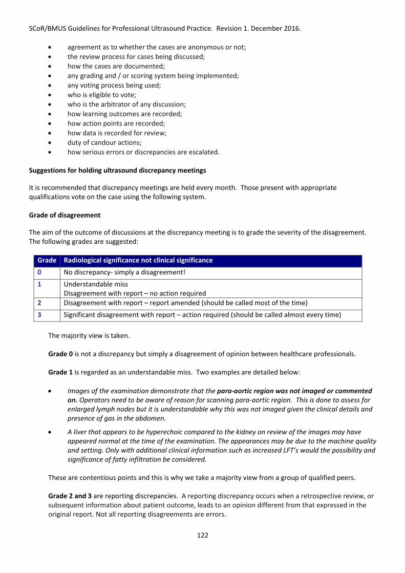
SCoR/BMUS Guidelines for Professional Ultrasound Practice. Revision 1. December 2016.
122
• agreement as to whether the cases are anonymous or not;
• the review process for cases being discussed;
• how the cases are documented;
• any grading and / or scoring system being implemented;
• any voting process being used;
• who is eligible to vote;
• who is the arbitrator of any discussion;
• how learning outcomes are recorded;
• how action points are recorded;
• how data is recorded for review;
• duty of candour actions;
• how serious errors or discrepancies are escalated.
Suggestions for holding ultrasound discrepancy meetings
It is recommended that discrepancy meetings are held every month. Those present with appropriate
qualifications vote on the case using the following system.
Grade of disagreement
The aim of the outcome of discussions at the discrepancy meeting is to grade the severity of the disagreement.
The following grades are suggested:
Grade Radiological significance not clinical significance
0 No discrepancy- simply a disagreement!
1 Understandable miss
Disagreement with report – no action required
2 Disagreement with report – report amended (should be called most of the time)
3 Significant disagreement with report – action required (should be called almost every time)
The majority view is taken.
Grade 0 is not a discrepancy but simply a disagreement of opinion between healthcare professionals.
Grade 1 is regarded as an understandable miss. Two examples are detailed below:
• Images of the examination demonstrate that the para-aortic region was not imaged or commented
on. Operators need to be aware of reason for scanning para-aortic region. This is done to assess for
enlarged lymph nodes but it is understandable why this was not imaged given the clinical details and
presence of gas in the abdomen.
• A liver that appears to be hyperechoic compared to the kidney on review of the images may have
appeared normal at the time of the examination. The appearances may be due to the machine quality
and setting. Only with additional clinical information such as increased LFT’s would the possibility and
significance of fatty infiltration be considered.
These are contentious points and this is why we take a majority view from a group of qualified peers.
Grade 2 and 3 are reporting discrepancies. A reporting discrepancy occurs when a retrospective review, or
subsequent information about patient outcome, leads to an opinion different from that expressed in the
original report. Not all reporting disagreements are errors.
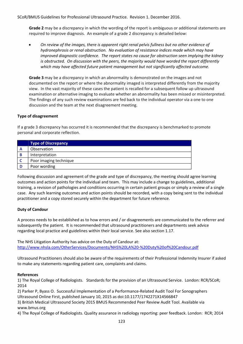
SCoR/BMUS Guidelines for Professional Ultrasound Practice. Revision 1. December 2016.
123
Grade 2 may be a discrepancy in which the wording of the report is ambiguous or additional statements are
required to improve diagnosis. An example of a grade 2 discrepancy is detailed below:
• On review of the images, there is apparent right renal pelvis fullness but no other evidence of
hydronephrosis or renal obstruction. No evaluation of resistance indices made which may have
improved diagnostic confidence. The report states no cause for obstruction seen implying the kidney
is obstructed. On discussion with the peers, the majority would have worded the report differently
which may have affected future patient management but not significantly affected outcome.
Grade 3 may be a discrepancy in which an abnormality is demonstrated on the images and not
documented on the report or where the abnormality imaged is interpreted differently from the majority
view. In the vast majority of these cases the patient is recalled for a subsequent follow up ultrasound
examination or alternative imaging to evaluate whether an abnormality has been missed or misinterpreted.
The findings of any such review examinations are fed back to the individual operator via a one to one
discussion and the team at the next disagreement meeting.
Type of disagreement
If a grade 3 discrepancy has occurred it is recommended that the discrepancy is benchmarked to promote
personal and corporate reflection.
Following discussion and agreement of the grade and type of discrepancy, the meeting should agree learning
outcomes and action points for the individual and team. This may include a change to guidelines, additional
training, a revision of pathologies and conditions occurring in certain patient groups or simply a review of a single
case. Any such learning outcomes and action points should be recorded, with a copy being sent to the individual
practitioner and a copy stored securely within the department for future reference.
Duty of Candour
A process needs to be established as to how errors and / or disagreements are communicated to the referrer and
subsequently the patient. It is recommended that ultrasound practitioners and departments seek advice
regarding local practice and guidelines within their local service. See also section 1.17.
The NHS Litigation Authority has advice on the Duty of Candour at:
http://www.nhsla.com/OtherServices/Documents/NHS%20LA%20-%20Duty%20of%20Candour.pdf
Ultrasound Practitioners should also be aware of the requirements of their Professional Indemnity Insurer if asked
to make any statements regarding patient care, complaints and claims.
References
1) The Royal College of Radiologists. Standards for the provision of an Ultrasound Service. London: RCR/SCoR;
2014
2) Parker P, Byass O. Successful Implementation of a Performance-Related Audit Tool For Sonographers
Ultrasound Online First, published January 10, 2015 as doi:10.1177/1742271X14566847
3) British Medical Ultrasound Society 2015 BMUS Recommended Peer Review Audit Tool. Available via
www.bmus.org
4) The Royal College of Radiologists. Quality assurance in radiology reporting: peer feedback. London: RCR; 2014
Type of Discrepancy
A Observation
B Interpretation
C Poor imaging technique
D Poor wording
SCoR/BMUS Guidelines for Professional Ultrasound Practice. Revision 1. December 2016.
124
5) The Royal College of Radiologists. Standards for Learning from Discrepancy Meeting. London: RCR; 2014
6) Edwards H et al What makes a good ultrasound report? Ultrasound 2014; 22 (1) 57-60
Further reading:
National Ultrasound Steering Group (NUSG), a subgroup of the National Imaging Board (2008) Ultrasound Clinical
Governance. Obtainable via:
https://www.bmus.org/static/uploads/resources/ClinicalGovernanceInUltrasound-061108.pdf
2.19 RECORDING OF IMAGES BY PATIENTS DURING ULTRASOUND EXAMINATIONS
The following advice is from the SCoR document: ‘The recording of images during diagnostic imaging including
screening and radiotherapy’ (2014).
https://www.sor.org/learning/document-library/recording-images-patients-during-diagnostic-imaging-including-
screening-and-radiotherapy
Ultrasound practitioners may be asked by a patient or person accompanying them if they will allow them to make
their own real-time recording of a diagnostic or screening examination or treatment. This request may be to
record the examination or treatment with a mobile telephone, record onto a DVD or memory device or utilise
other digital or analogue recording media. With rapid developments in communications technology, the ways in
which such recordings are able to be made are likely to become ever more varied.
There have been instances where examinations or treatments have been recorded and posted to social media
sites without the ultrasound practitioner’s consent. These recordings have included conversations between the
ultrasound practitioner and the patient. It can then prove very difficult to have these removed, especially if there
is no statement of policy placed in waiting rooms or otherwise publicly available.
The decision as to whether to allow images, an examination or treatment to be recorded should be made by the
individual departments concerned, but in general terms the SCoR and BMUS would discourage this.
Departments should have a clear policy on this issue following a risk assessment that takes account of the
following:
i) the views of members of the professional workforce both as a group and individually should clearly be
taken into account. Many do not want to be filmed or recorded and their wishes should be respected.
ii) possible medico-legal complications arising if, for example, an abnormality is recorded that is not reported
or acted upon. Conversations between the ultrasound practitioner and the patient and anyone
accompanying them may also be recorded. The employer must be aware of any local arrangements if it is
decided to allow this as they may impact on its risk strategy and insurance arrangements.
iii) it can be a great distraction and can increase stress levels for the ultrasound practitioner at a time when
they require very high levels of concentration.
iv) it has the potential to extend the time of the examination or treatment which may be contrary to published
safety guidelines and advice.
v) providers should take into consideration the possible existence of local policies allowing, for example, the
recording of a birth. Many employing authorities do try to facilitate this type of request within the
Directorates that have responsibility for maternity services.
vi) providers should consider consulting with any relevant patient liaison groups that the employing authority
may have and ensure the final agreed policy is publically available. Removal of material from social media
that has included the ultrasound practitioner without their consent may be difficult, particularly so if no
prior notice of policy has been made available.
SCoR/BMUS Guidelines for Professional Ultrasound Practice. Revision 1. December 2016.
125
vii) providers might also wish to consider how staff should respond to situations where filming begins or
continues without permission and contrary to the agreed policy.
This advice does not refer to the taking of images by ultrasound practitioners during obstetric examinations and
that is agreed procedure between the ultrasound department and the employing authority. This is often for a
previously advertised fee.
2.20 ‘HAVE YOU PAUSED AND CHECKED’ POSTERS AND PROMPT CARDS
‘Have you paused and checked?’ posters and a prompt card have been published to support ultrasound
practitioners in clinical imaging services. They are designed to act as a ready reminder of the checks that need to
be made when any ultrasound examination is undertaken. The posters are designed in PDF format to allow for
easy downloading and printing. They can then be displayed in the department as required. There are A4 and A3
sizes available and also an A6 version that is small enough to be placed close to a machine or work station. The
posters have been developed by a joint working party from the Society and College of Radiographers and the
British Medical Ultrasound Society.
https://www.sor.org/learning/document-library/have-you-paused-and-checked-ultrasound


