81
Przemysław Gałązka, MD, PhD Clinical Ward of General and Oncological Surgery for Children and Adolescents CM UMK Bydgoszcz, Poland Head of the Department: Irena Daniluk-Matraś, MD, PhD
Przemysław Gałązka, MD, PhD
Clinical Ward of General and Oncological Surgery
for Children and Adolescents
CM UMK Bydgoszcz, Poland
Head of the Department: Irena Daniluk-Matraś, MD, PhD
Chest wall deformities
Three categories of deformities:
I. There is rib overgrowth causing either depression (Pectus excavatum) or
protrusion (Pectus carinatum) of the anterior chest wall.
II. Failure of normal development (aplasia or dysplasia) - bifid sternum, (with
ectopia cordis, defects of heart, pericardium, diaphragm, abdominal wall =
Pentalogy of Cantrell) – Poland`s syndrome (unilateral absence of ribs,
pectoralis muscles and breast tissue)
III. Deformities due to trauma or pressure effects (iatrogenic after ribs resections in
pectus excavatum - aquired asphyxiating chondrodystrophy)
Frequency increased in familial connective tissue disorders such as Marfan`s
Syndrome and Ehler`s-Danlos Syndrome, systemic weakness of connective
tissue and poor muscle development = scoliosis in 20% of patients.
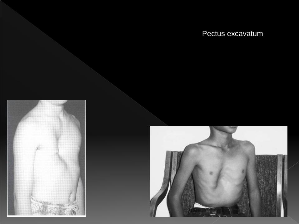
Pectus excavatum
Pectus excavatum- depression of the anterior chest wall of variable severity and
configuration (localised and deep = cup-shaped, diffused = saucer-shaped,
eccentric=grand canyon, or mixed excavatum/carinatum =horseshoe, pouter
pigeon)
Incidence: 1:300- 1:1000 (more common in European and Asian than African)
Etiology- genetic predisposition, abnormal distribution of colagen types in ribs
cartilages.
40% have a positive family history.
Boys / Girls = 4 / 1
Patients usually tall – basketball players posture, 18% have scoliosis, 16% features
of Marfan`s Syndrome, 1%- Ehlers-Danlos Syndrome
Pectus excavatum
Symptoms: exercise intolerance (80%),
chest pain (65%), lack of endurance
(65%), shortness of breath (45%), asthma-
like symptoms (30%), frequent URI (25%),
scoliosis (18%).
! Teenagers: poor body image, stop in
team sports = isolation and feelings of
worthlessness (even suicidal thoughts)!
On physical exam: „old age” or „pectus
posture”
The Haller CT index: objective measurement of deformity severity
It divides the internal transverse diameter of the chest by the
anteroposterior diameter. In normal patients the index is less than 2.
The sternum is frequently twisted so that it is at a 45% angle to the anterior chest wall
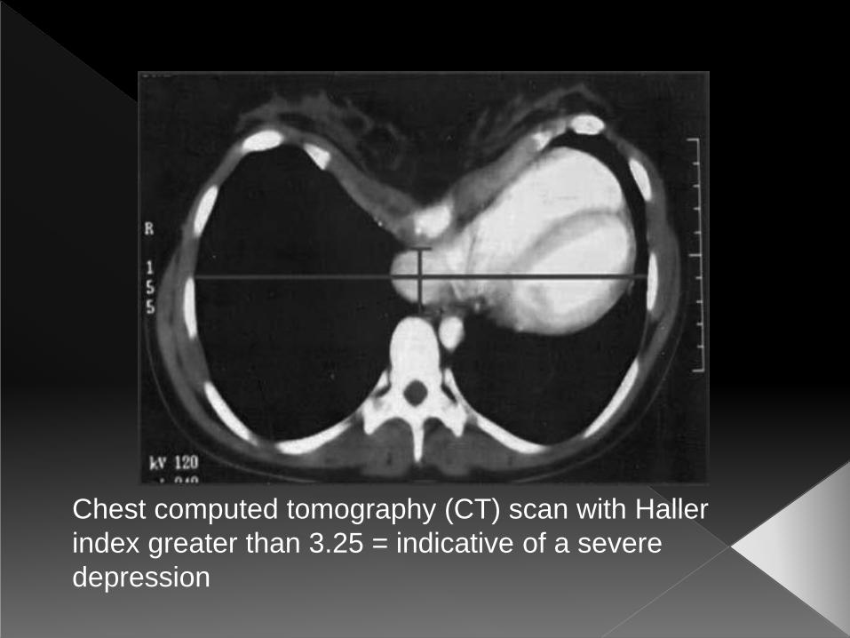
Chest computed tomography (CT) scan with Haller
index greater than 3.25 = indicative of a severe
depression
Effects of cardiac compression
(decreased cardiac output, impaired
valve function and arrhytmias)
Pulmonary effects (restrictive lung
disease, atelectasis, paradoxical
respiration)
CT index of 3,25 or greater
Failed previous repair
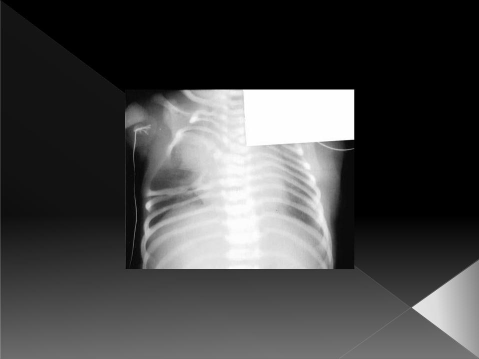
Ideal age: just before puberty
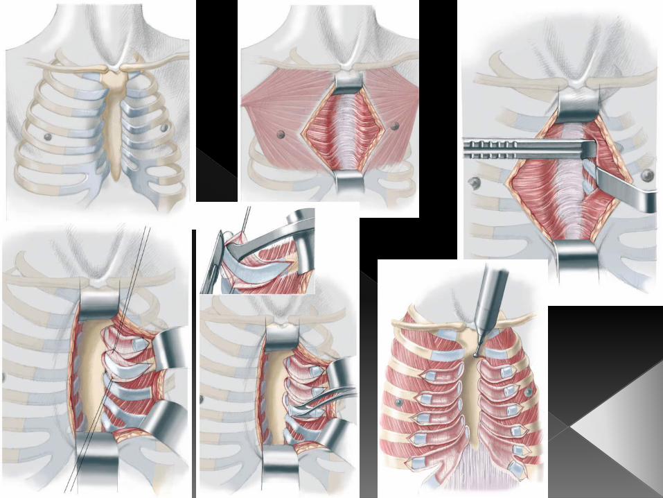
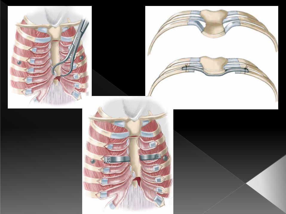
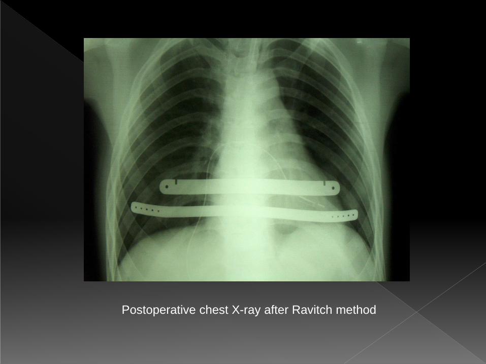
Postoperative chest X-ray after Ravitch method

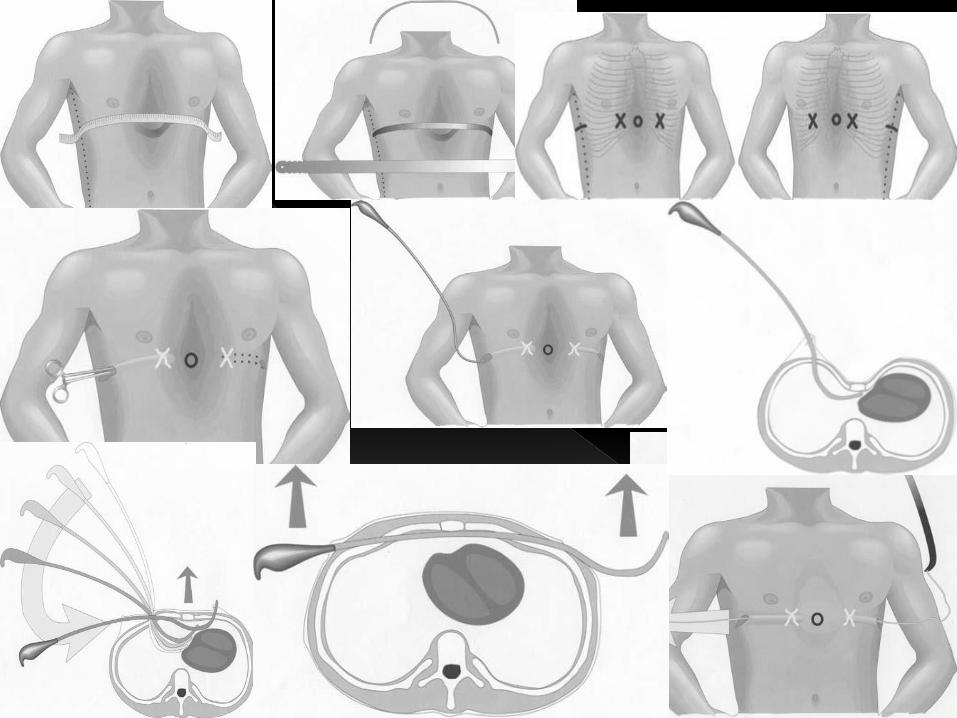
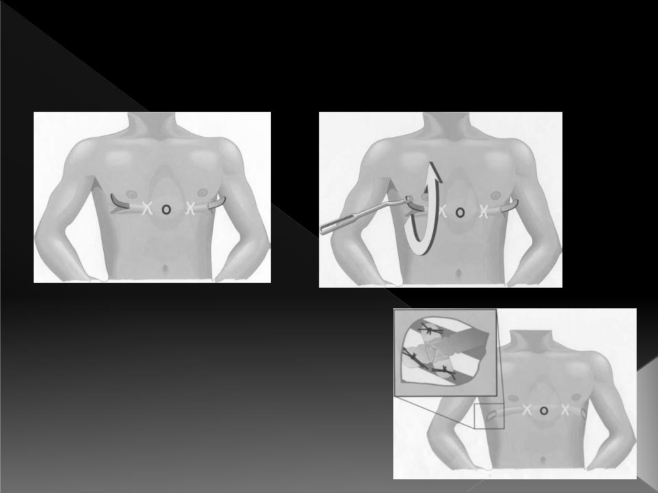
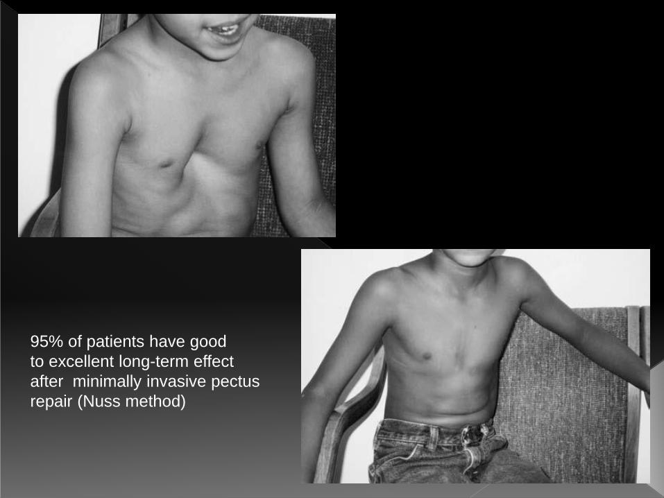
95% of patients have good
to excellent long-term effect
after minimally invasive pectus
repair (Nuss method)
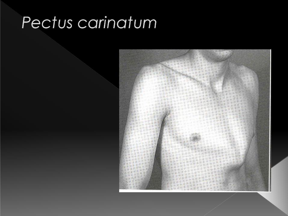
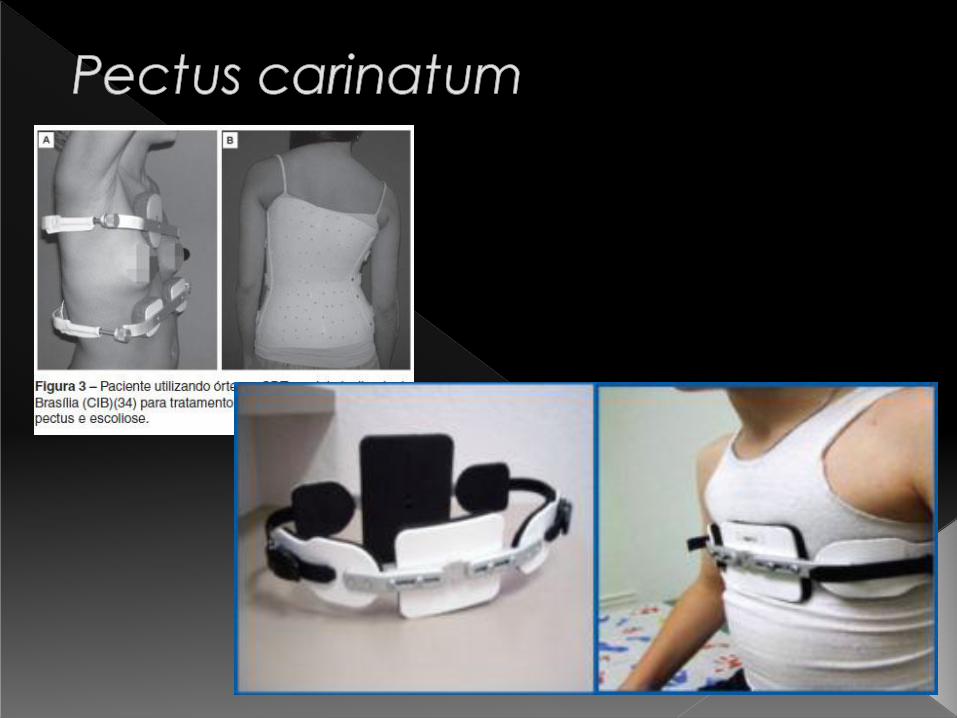
• protrusion deformity of the chest
• much less common than the excavatum deformity
• the protrusion may involve: upper chest – manubrium,
lower chest – gladiolus, unilateral, bilateral or mixed
• more common in boys (4:1), 25% have a positive family
history
• appears in late childhood and progresses rapidly during
puberty
• complains of chest pain when pressure applied on
anterior chest, no cardiac or pulmonary compression
• application of a pressure brace or surgical resection
(depending on PCI)
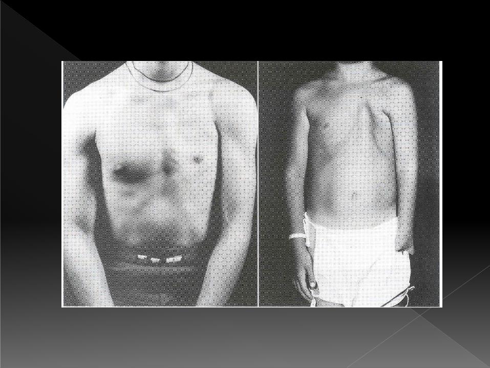
Poland`s Syndrome
Described by Alfred Poland, may include absence of all or some of:
• ribs
• pectoralis major
• pectoralis minor
• serratus anterior
• rectus abdominis
• latissimus dorsi
• nipple deformities
• limb deformities (syndactyly, brachydactyly)
• absent axillary hair
• limited subcutaneous fat
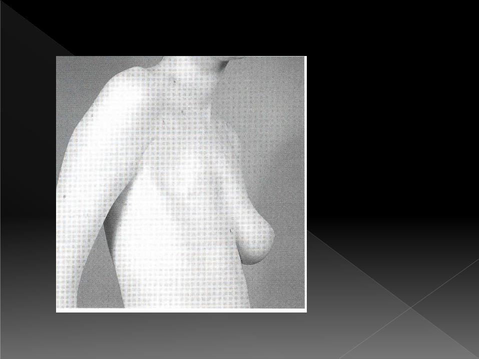
Indications for surgical intervention: large rib defects causing lung hernia,
concerns regarding injury to the heart or lungs.
Adolescent girls require breast reconstruction in caseof amastia.
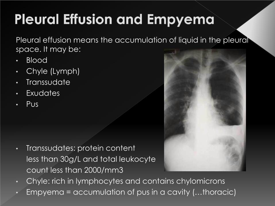
Pleural effusion means the accumulation of liquid in the pleural
space. It may be:
• Blood
• Chyle (Lymph)
• Transsudate
• Exudates
• Pus
• Transsudates: protein content
less than 30g/L and total leukocyte
count less than 2000/mm3
• Chyle: rich in lymphocytes and contains chylomicrons
• Empyema = accumulation of pus in a cavity (…thoracic)
• Primary fetal hydrothorax in 1:10000 – 15000 pregnancies
• Effusion may be secondary to mediastinal tumor,
adenomatoid tumor, pulmonary sequestration, infection, chromosomal anomaly.
• Congenital primary chylothorax – debatable
• Rarely efuusions may arise in the context of
lymphangiomatosis (present at birth or manifest later)
• Hemothorax – usually traumatic, mostly associated with
pneumothorax, may occur as a complication of the central
venous line catheterisation
• Chylothorax – damage of thoracic duct, and increased pressure in the systemic venous system, incidence is rising (from
1% to 5%)
• Hydrothorax – Iatrogenic (CVC, right-sided: transdiaphragmatic- ventriculoperitoneal shunting or
peritoneal dialysis, from urinary ascites)
- noniatrogenic (in cases of hepatic ascites,
cangestive heart disease, nephrotic syndrome,
malignancy: especially non-Hodgkin lymphoma,
metastatic disease)
• May be result of: trauma of the chest, esophageal
foreign body perforation, after surgery- leaking
bronchial stump and esophageal anastomotic leak.
Also infradiaphragmatic pathology: retained
gallstones after cholecystectomy, pancreatitis,
appendiceal perforation with peritonitis.
• Increasingly common – parapneumonic effusion
(due to pneumonic disease)- 14 in 100000
(Streptococcus pneumoniae, Haemophilus influenzae,
Ebstein-Barr virus). In third world countries:
Staphylococcus aureus. In endemic areas:
tuberculosis, echinococcus.

Clasically, three stages are distinguished: - Exudative phase (few cells in the fluid)- 24-72 hours
- Fibrinopurulent phase (accumulation of fibrinous
material, loss of lung mobility)- 7-10 days
- Organizing phase (formation of a pleural peal)
- Clinical features: respiratory symptoms
(tachypnea, dyspnea, ortopnoe); dullness on the
affected side with diminished breath sounds, signs
of inflammation (fever, tachycardia, lethargia, pain
during respiration)
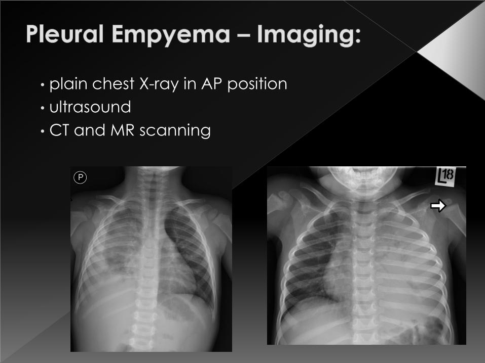
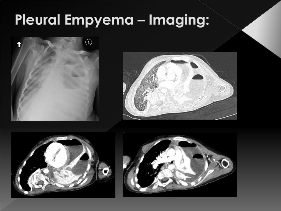
• plain chest X-ray in AP position
• ultrasound
• CT and MR scanning
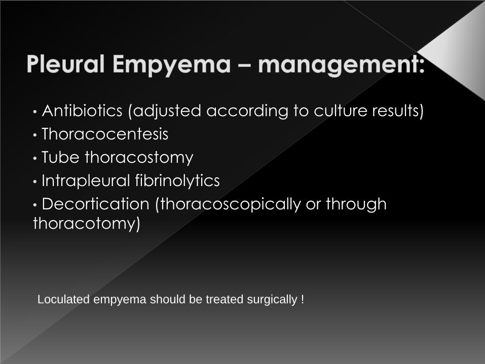
• Antibiotics (adjusted according to culture results)
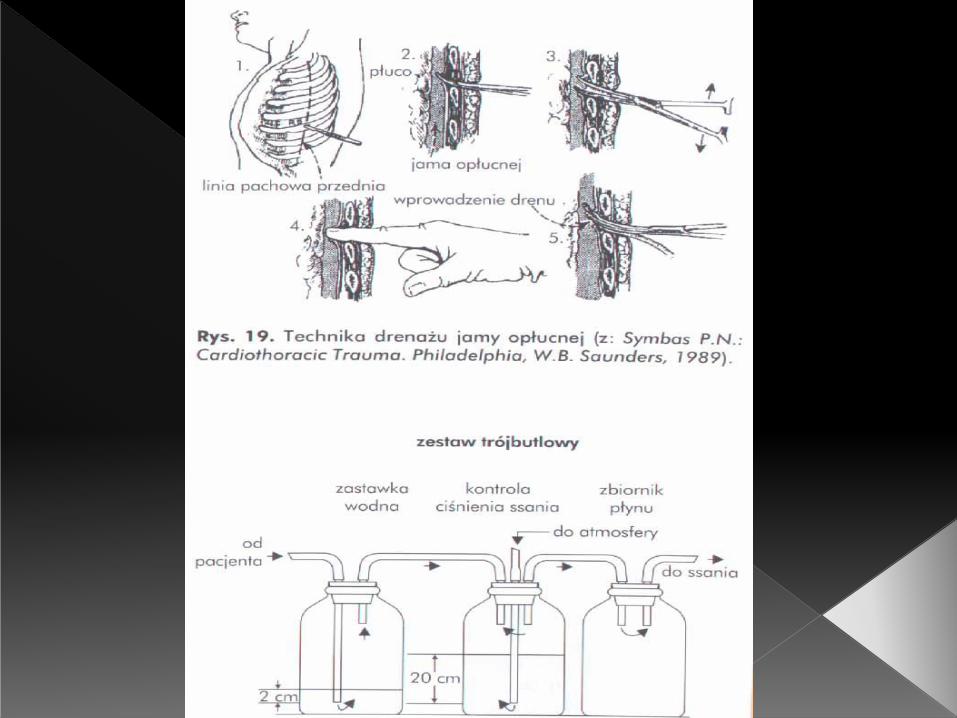
• Thoracocentesis
• Tube thoracostomy
• Intrapleural fibrinolytics
• Decortication (thoracoscopically or through
thoracotomy)
Loculated empyema should be treated surgically !
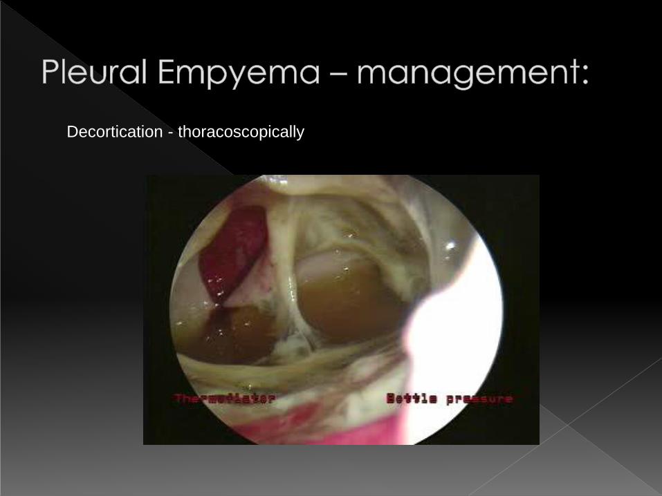
Decortication - thoracoscopically
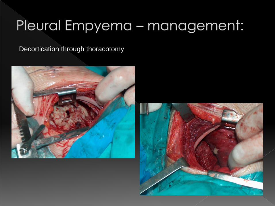
Decortication through thoracotomy
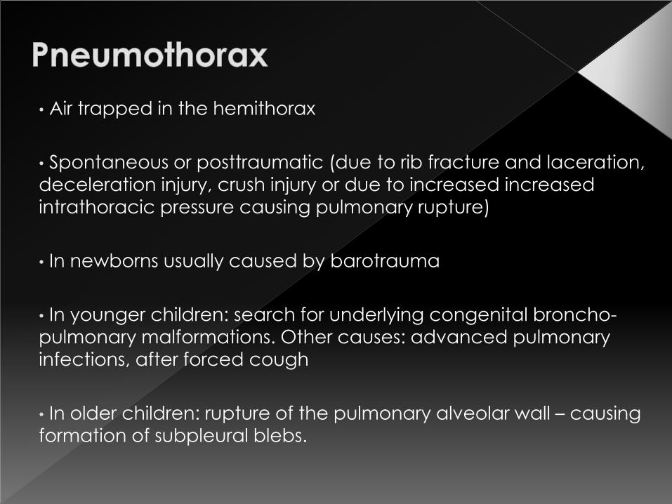
• Air trapped in the hemithorax
• Spontaneous or posttraumatic (due to rib fracture and laceration,
deceleration injury, crush injury or due to increased increased intrathoracic pressure causing pulmonary rupture)
• In newborns usually caused by barotrauma
• In younger children: search for underlying congenital broncho-
pulmonary malformations. Other causes: advanced pulmonary
infections, after forced cough
• In older children: rupture of the pulmonary alveolar wall – causing
formation of subpleural blebs.
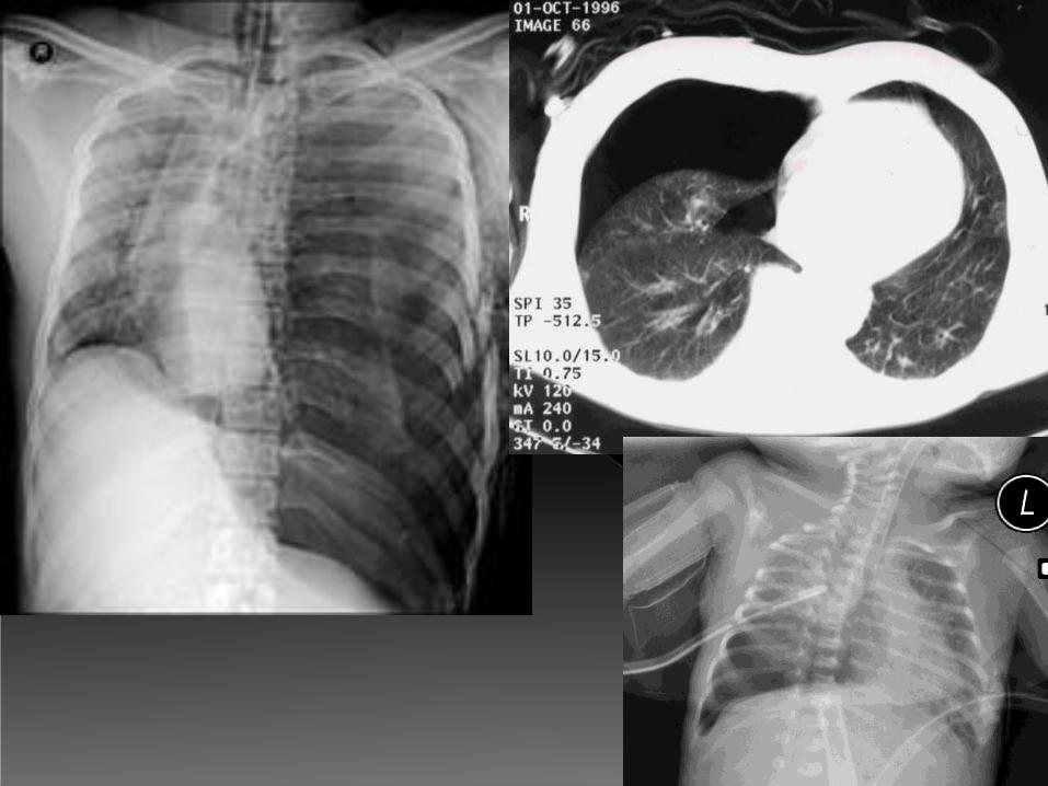
TENSION PNEUMOTHORAX
- Can quickly result in cardiovascular demise if tension within hemothorax
develops
- Findings: hyperresonance, diminished breath sounds, tracheal deviation,
hemodynamic instability
- Potentially lethal – chest x-ray not required, may waste time !
- Needle decompression followed by placement of chest tube
- Large bore angiocath in the second intercostal space along mid-clavicular line
- Alternative decompression site! – anterior axillary line just above the sixth rib
- Reexpansion of the lung, evacuation of the air and fluid, sealing of the injured
lung, resolving of the air leak…
• Tension pneumothorax – immediate decompression
• Small pneumothorax – conservative treatment
• Most often- chest tube drainage
• In case of persistent air leakage – consider operative
treatment
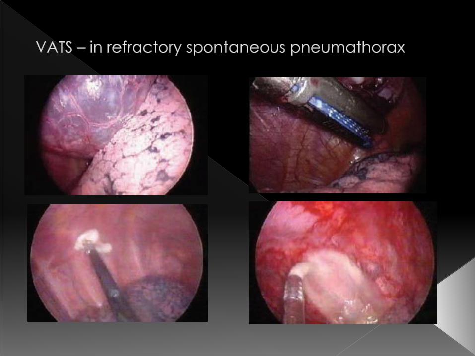
• Primary spontaneous pneumothorax- in case of second
episode: thoracoscopic pulmonary apical resection with partial pleurodesis
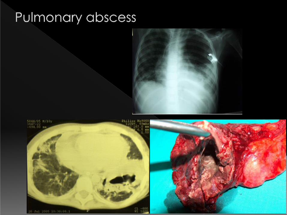
• Accumulation of purulent content in the cavity formed as a result of
pulmonary parenchyma necrosis and destruction
• Most common pathogens: aerobic (Staphylococcus aureus, Streptococcus pn, Streptococcus pyogenes, Klebsiella pn,
Escherichia coli, Pseudomonas aer), anaerobic (Fusobacterium,
Bacteroides, Peptococcus, Bacillus fragilis)
• Pathogenesis of pulmonary abscess development: aspiration of
naso-pharyngeal or gastric secretions; retention of purulent mucus in
bronchioles, bacteraemia, diffusion through continuity, complication
of the other pulmonary conditions: infections, bronchial dilatations, cysts.
• Primary vs secondary abscess
• chronic abscess – duration >6 weeks
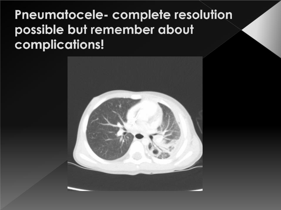
• Usually in the course of stapylococcal pneumonitis
• thin-wall spaces filled with air, as a result of the
purulent process and staphyllococcal toxins
• varies in size and localization
• Complications: pneumothorax, bronchio-pleural
fistulae, pulmonary abscess, pleural empyema

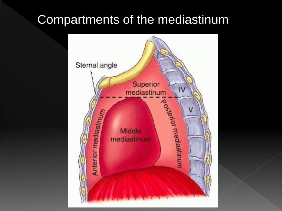
Compartments of the mediastinum
Anterior mediastinum:
<2 Years : Benign teratoma, Thymic
hyperplasia, Cystic hygroma
>2 Years: Malignant germ cell tumor,
Thymoma, Lymphoma
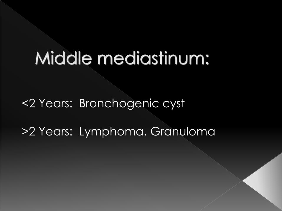
Middle mediastinum:
<2 Years: Bronchogenic cyst
>2 Years: Lymphoma, Granuloma
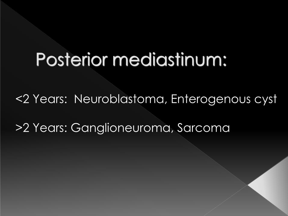
Posterior mediastinum:
<2 Years: Neuroblastoma, Enterogenous cyst
>2 Years: Ganglioneuroma, Sarcoma
Esophageal Atresia and
Tracheoesophageal Fistula
(EA TEF)
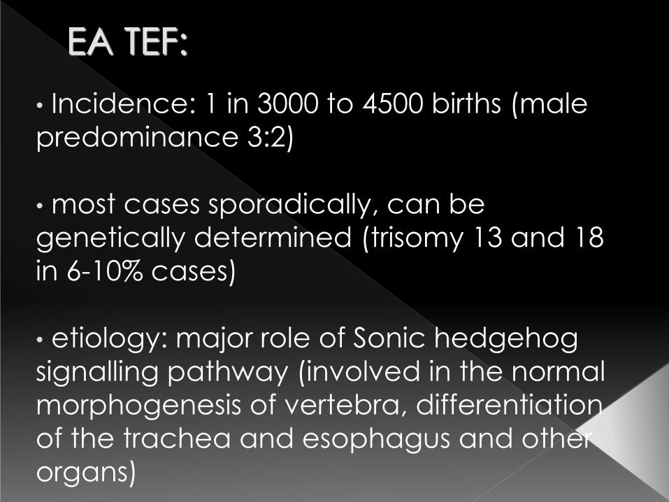
EA TEF:
• Incidence: 1 in 3000 to 4500 births (male
predominance 3:2)
• most cases sporadically, can be
genetically determined (trisomy 13 and 18
in 6-10% cases)
• etiology: major role of Sonic hedgehog
signalling pathway (involved in the normal
morphogenesis of vertebra, differentiation
of the trachea and esophagus and other
organs)
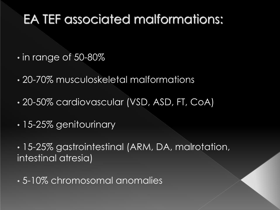
EA TEF associated malformations:
• in range of 50-80%
• 20-70% musculoskeletal malformations
• 20-50% cardiovascular (VSD, ASD, FT, CoA)
• 15-25% genitourinary
• 15-25% gastrointestinal (ARM, DA, malrotation,
intestinal atresia)
• 5-10% chromosomal anomalies
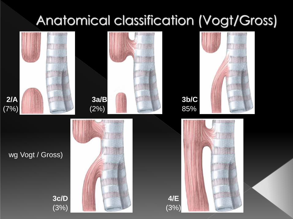
Anatomical classification (Vogt/Gross)
2/A 3a/B 3b/C
3c/D 4/E
(3%) (3%)
(7%) (2%) 85%
wg Vogt / Gross)
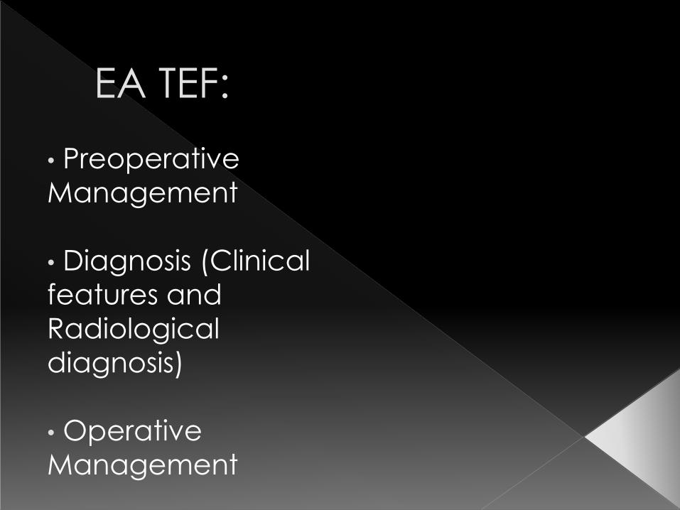
EA TEF:
• Preoperative
Management
• Diagnosis (Clinical
features and
Radiological
diagnosis)
• Operative
Management
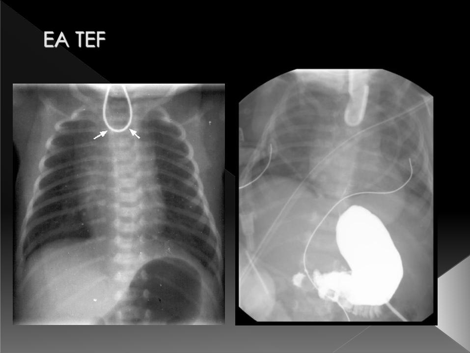
EA TEF
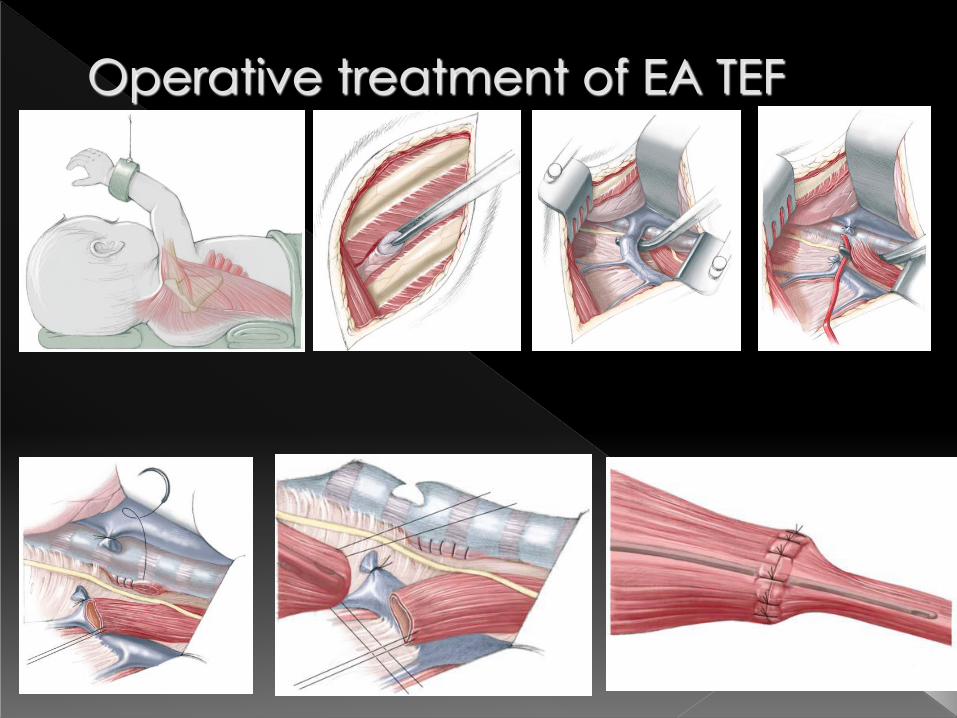
Operative treatment of EA TEF
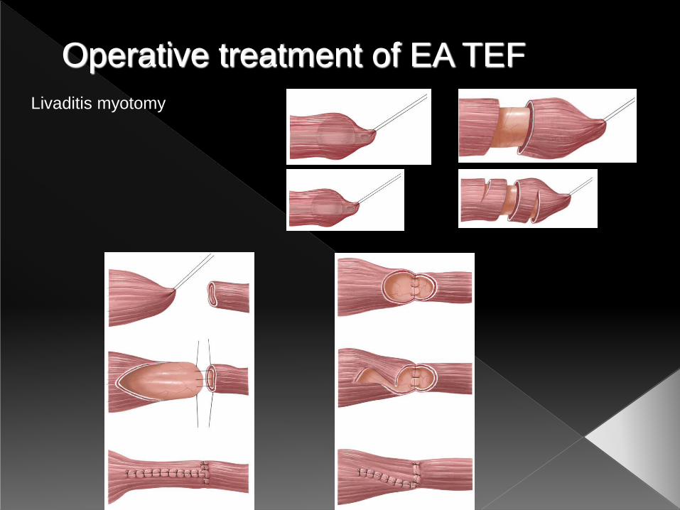
Livaditis myotomy
Operative treatment of EA TEF
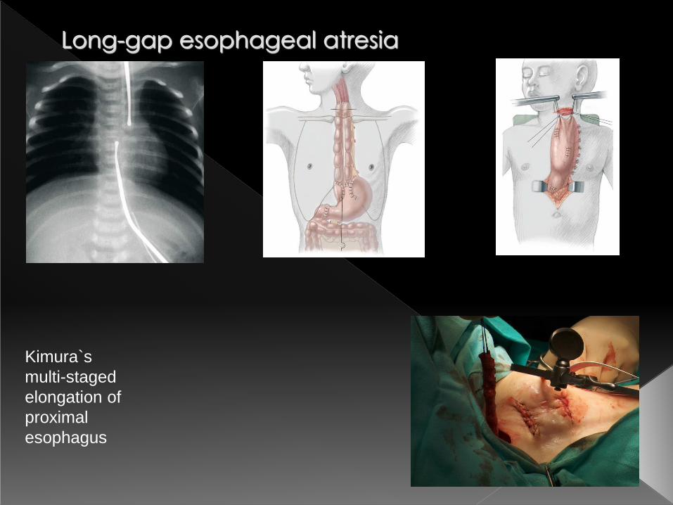
Long-gap esophageal atresia
Kimura`s
multi-staged
elongation of
proximal
esophagus
EA TEF:
• Postoperative treatment
• Early complications (recurrence of TEF,
anastomotic leak, anastomotic stenosis,
tracheomalacia)
• Late complications (GERD,
microaspirations, abnormal peristaltic
problems of the lower esophageal
segment)
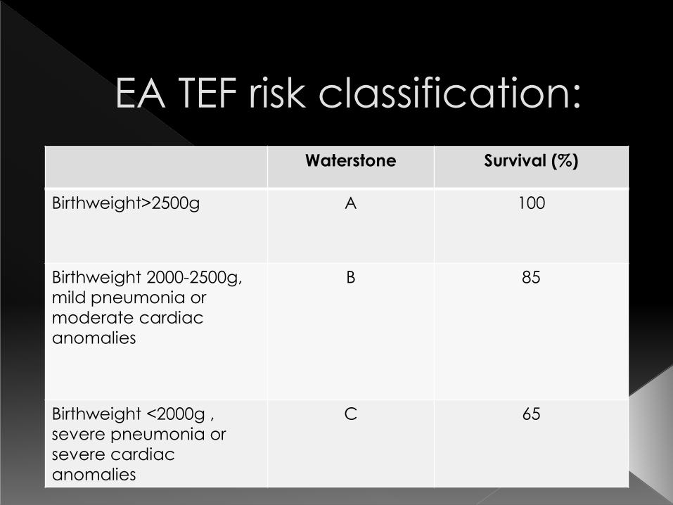
EA TEF risk classification:
Waterstone Survival (%)
Birthweight>2500g A 100
Birthweight 2000-2500g,
mild pneumonia or
moderate cardiac
anomalies
B 85
Birthweight <2000g ,
severe pneumonia or
severe cardiac
anomalies
C 65
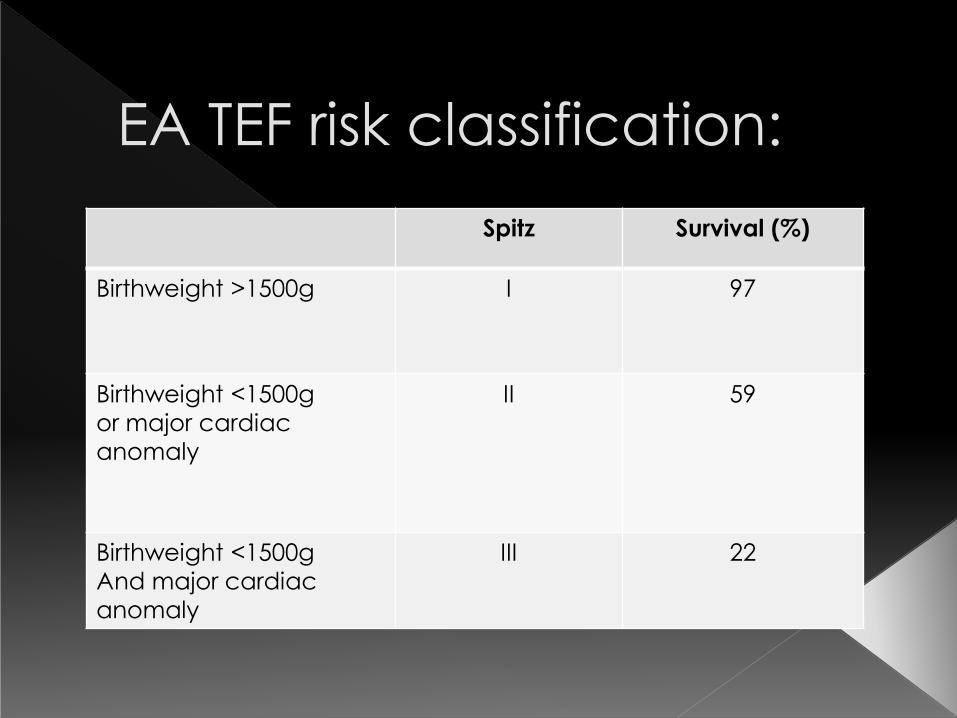
Spitz Survival (%)
Birthweight >1500g I 97
Birthweight <1500g
or major cardiac
anomaly
II 59
Birthweight <1500g
And major cardiac
anomaly
III 22
EA TEF risk classification:
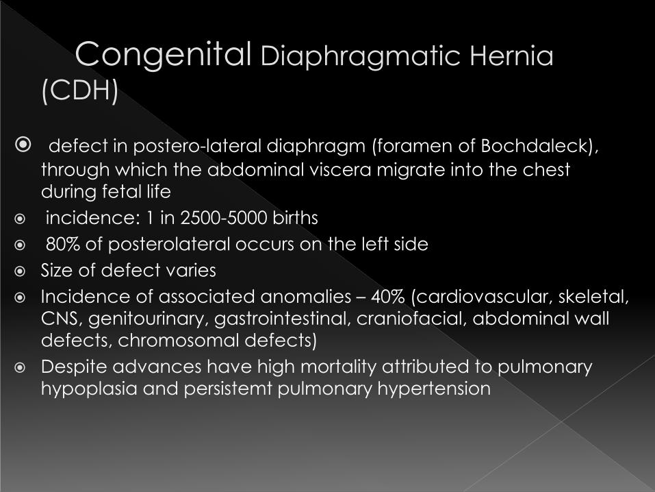
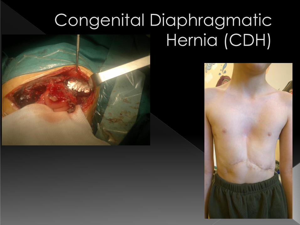
Congenital Diaphragmatic Hernia
(CDH)
defect in postero-lateral diaphragm (foramen of Bochdaleck),
through which the abdominal viscera migrate into the chest
during fetal life
incidence: 1 in 2500-5000 births
80% of posterolateral occurs on the left side
Size of defect varies
Incidence of associated anomalies – 40% (cardiovascular, skeletal,
CNS, genitourinary, gastrointestinal, craniofacial, abdominal wall
defects, chromosomal defects)
Despite advances have high mortality attributed to pulmonary
hypoplasia and persistemt pulmonary hypertension
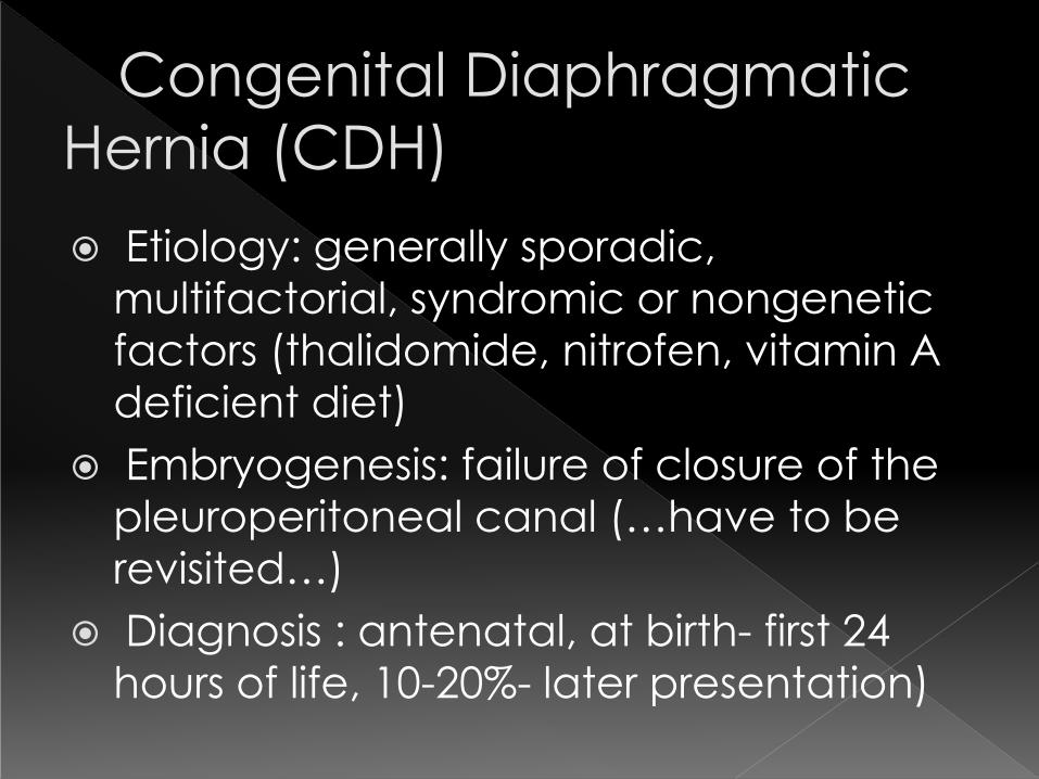
Congenital Diaphragmatic
Hernia (CDH)
Etiology: generally sporadic,
multifactorial, syndromic or nongenetic
factors (thalidomide, nitrofen, vitamin A
deficient diet)
Embryogenesis: failure of closure of the
pleuroperitoneal canal (…have to be
revisited…)
Diagnosis : antenatal, at birth- first 24
hours of life, 10-20%- later presentation)
Congenital Diaphragmatic Hernia
(CDH)
Prognostic factors of poor prognosis:
polyhydramnios, intrathoracic stomach
and liver, lung to head ratio (LHR <1,0;
>1,4)
Management: preoperative- paralysed
and sedated, gentle ventilation and
permissive hypercapnia to minimize
barotrauma, inhaled nitric oxide, High-
frequency oscillatory ventilation (HFOV),
extracorporeal membrane oxygenation
(ECMO)
Congenital Diaphragmatic Hernia
(CDH)
Timing of Surgery
Surgical Technique
Postoperative treatment
Long term follow-up: pulmonary hypoplasia,
bronchopulmonary dysplasia, persistent
pulmonary hypertension, reactive airway
disease, neurodevelopmental delay (motor
and cognitive skill), GERD< sensorineural
hearing loss, musculoskeletal: chest
assymetry, pectus deformity, recurrence)
Congenital Diaphragmatic
Hernia (CDH)
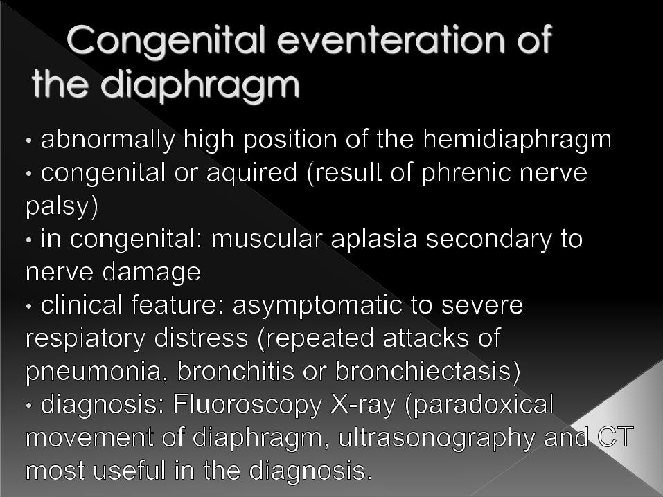
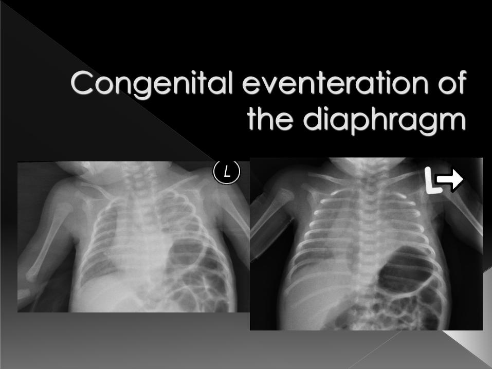
Congenital eventeration of
the diaphragm
Congenital eventeration of
the diaphragm
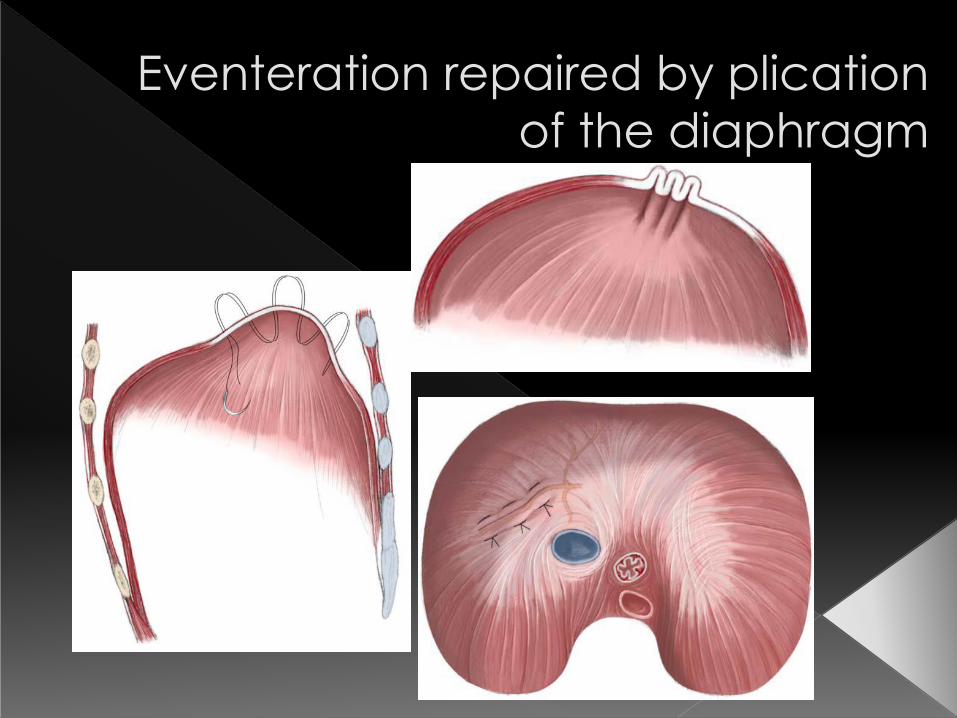
Eventeration repaired by plication
of the diaphragm

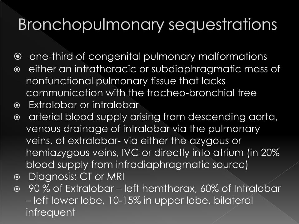
one-third of congenital pulmonary malformations
either an intrathoracic or subdiaphragmatic mass of
nonfunctional pulmonary tissue that lacks
communication with the tracheo-bronchial tree
Extralobar or intralobar
arterial blood supply arising from descending aorta,
venous drainage of intralobar via the pulmonary
veins, of extralobar- via either the azygous or
hemiazygous veins, IVC or directly into atrium (in 20%
blood supply from infradiaphragmatic source)
Diagnosis: CT or MRI
90 % of Extralobar – left hemthorax, 60% of Intralobar
– left lower lobe, 10-15% in upper lobe, bilateral
infrequent
Clinical features: extralobar often diagnosed prenatally or in infancy
(during surgery for CDH..)
intralobar often in later childhood – recurrent
pulmonary infections or haemorrhage,
both can present in the newborn period with
respiratory distress due to mass effect or congestive
heart failure because of arteriovenous shunting
Management: Resection (straight-forward in extrapulmonary, for
intralobar often lobar resection)
• benign hamartomatous or dysplastic tumors
• morphologically: overgrowth of terminal bronchioles in a glandular or
adenomatoid pattern – composed of disorganized cysts lined with ciliated
cuboidal or columnar epithelium (grossly: have both cystic and solid
components)
• CCAM constitute 10-30% of all congenital lung malformations with slight
male predominance
• Normal pulmonary arterial and venous blood supply
• Diagnosis: relatively common prenatally by sonography (echogenic
pulmonary mass), often with associated polyhydramnios, mediastinal shift,
pleural effusions and fetal hydrops…
• macrocystic or microcystic ( > 5mm<)
• differential diagnosis: CDH, pulmonary sequestration, bronchogenic cyst…
• high accuracy MRI (prenatally), contrast enhanced CT (preoperatively:
anatomy, aberrant systemic blood supply…)
• Clinical features: - newborn (respiratory distress,
asymptomatic), - childhood or adolescents (infectious
complications, pneumothorax, bronchiectasis) – adulthood
(malignancy: bronchioalveolar cancer, sacoma, pulmonary
blastoma, mesenchymoma)
Management • Fetal therapy (EXIT procedure in third trymester, ultrasound
guided thoracoamniotic shunting for a macrostystic CCAM)
• Postnatal therapy (complete spontaneous resolution in 4%,
resection should be performed in any persistent CCAM to
prevent complications)
• Resection performed through open thoracotomy or minially
invasive
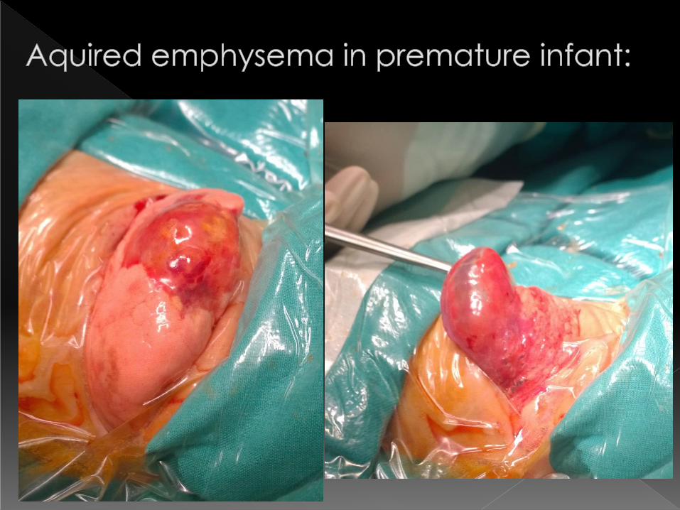
Congenital lobar overinflation – characterized by expiratory
air trapping within the affected lobe
overdistension leads to compression of adjacent lung and
mediastinal shift
etiology: focal absence of cartilaginous components,
endobronchial obstructions with secretions, granulation
tissue, ingested foreign bodies or endobronchial tumors
(intrinsic causes)
Extrinsic causes of compression (mediastinal
lymphadenopathy or aberrant pulmonary artery,
mediastinal cyst or tumor)
CLE most commonly in full term infants, but in prematures
aquired form (barotrauma, oxygen toxicity, lung
immaturity)
Lobar resection is curative, in 10-15% life-saving emergency
thoracotomy
Brochogenic cyst (BC)
• Result from abnormal budding of the bronchial tree in
which a portion of the lung bud develops independently
• The cyst walls frequently contain cartilage and are
lined with ciliated columnar epithelium
• These lesions tends to enlarge causing airway
obstruction
• Plain chest X-ray may suggest the presence of BC
but CT scan confirm the diagnosis
• Resection is curative
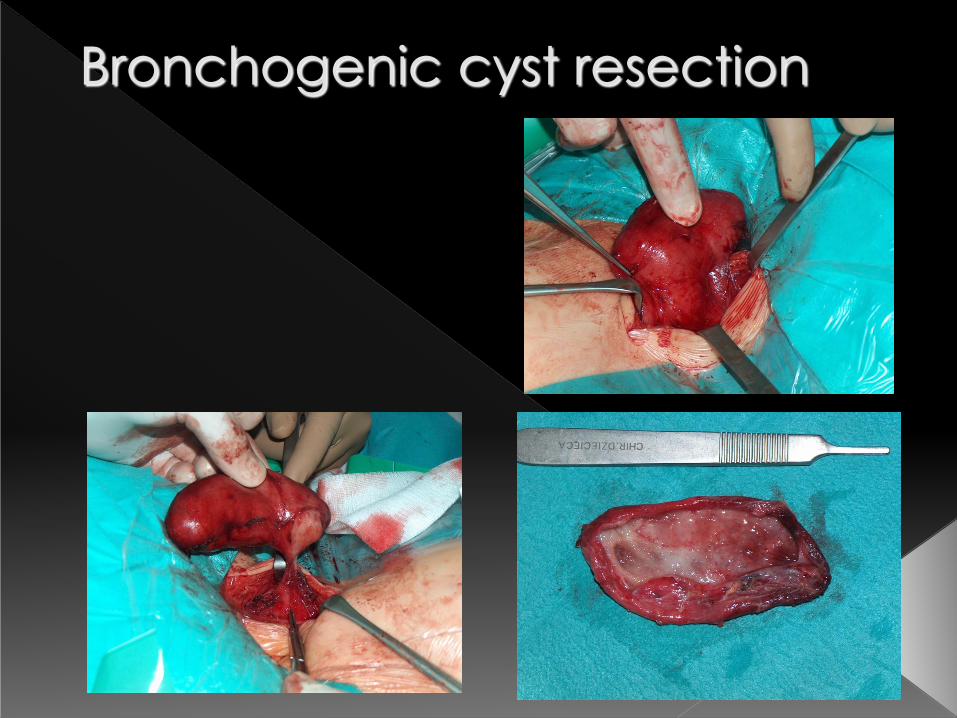
Bronchogenic cyst resection

The end…


