84
URINARY INCONTINENCE PREPARED BY : DR. ROZHAN YASIN KHALIL MBCHB , FICOG , CABOG , HDOG
| Date post: | 20-Aug-2015 |
| Category: |
Health & Medicine |
| Upload: | college-of-medicine-sulaymaniyah |
| View: | 3,419 times |
| Download: | 2 times |
URINARY INCONTINENCEPREPARED BY:
DR. ROZHAN YASIN KHALIL
MBCHB , FICOG , CABOG , HDOG

urinary incontinence:urinary incontinence:
urinary incontinence is defined as involuntary loss of urine that is objectively demonstrable and is a social or hygienic problem.
urinary incontinence is increasingly prevalent as the ageing population expands.
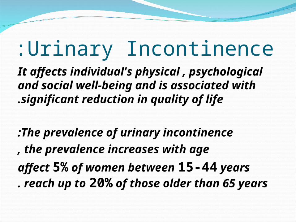
Urinary Incontinence:It affects individual's physical , psychological and social well-being and is associated with significant reduction in quality of life.
The prevalence of urinary incontinence:
the prevalence increases with age,
affect 5% of women between 15-44 years reach up to 20% of those older than 65 years.

THE ANATOMY OF URINARY TRACT:The bladder is a hollow muscular organ normally situated behind the pubic symphysis and covered superiorly and anteriorly by peritoneum .
It is composed of a syncytium of smooth muscle fibers known as the detrusor.
\\\\\\\\\\\\\\\\\\\\\\\\\\\\\\\\\contraction of this meshwork of fibers results in simultaneous reduction of the bladder in all its diameters.
The normal adult female urethra is between 3 and 5 cm in length, It is lined with pseudo-stratified transitional cell epithelium in its proximal half and distally by non-keratinized stratified sequamous epithelium.

/////////////////////////////////
beneath this is a rich vascular plexus which contributes up to one-third of the urethral pressure and which decreases with age .
Beneath this there is longitudinally oriented smooth muscle which is continuous morphologically with the detrusor .

--------------------------------------------------------contraction of this muscle layer leads to shortening and opening of the urethra.
this striated muscle is rhabdosphincter urethrae and also called the external sphincter or the intrinsic sphincter mechanism,
this muscle mass is responsible for urethral closure at rest.

````````````````````````````````````````````
The extrinsic sphincter mechanism consists of striated periurethral muscle ( levator ani )these contributes an additional closure force at times of physical effort .
\\\\\\\\\\\\\\\\\\\\\\\\\\\\\\\\\
Together the intrinsic and extrinsic sphincter mechanisms of the urethra produce a greater pressure within the urethra than in the bladder.
this is known as the positive closure pressure and is partly responsible for the maintenance of continence.

’’’’’’’’’’’’’’’’’’’’’’’’’’’’’’’’’’’’’’’‘’’’’’’’’’’’The detrusor muscle is innervated primarily
by the parasympathatic nerves S2-4 and receives a rich efferent supply .
urethral smooth muscle is innervated by sympathatic efferent fibers, the urethrae is supplied via sacral nerve roots (s2-4).

THE FUNCTION OF LOWER URINARY TRACT:
PHYSIOLOGY: The main role of the bladder is to
store the urine which continuously enters it.
thus the bladder must act as an efficient low pressure continent reservoir.

/////////////////////////////////
Urine from the kidneys enters the bladder via the ureters at a rate of
0.5-5 ml / min .
normally the first sensation of bladder filling is noted at between 150-250 ml and there is a strong desire to void at approximately
400-600 ml (bladder capacity).

\\\\\\\\\\\\\\\\\\\\\\\\\\\\\\\\\
During filling the bladder pressure should not normally rise by more than 10 cm of water to 300 ml.
In order to maintain continence the maximum urethral pressure must exceed the bladder pressure at all times except during micturition.

------------------------------------------
for continence to exist it is not only essential that the intravesical pressure remains low but also that the urethral lumen should seal completely .
\\\\\\\\\\\\\\\\\\\\\\\\\\\\\\\\\
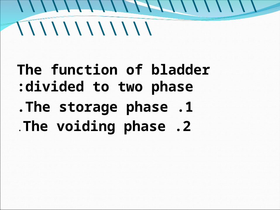
The function of bladder divided to two phase:
1 .The storage phase.2 .The voiding phase.
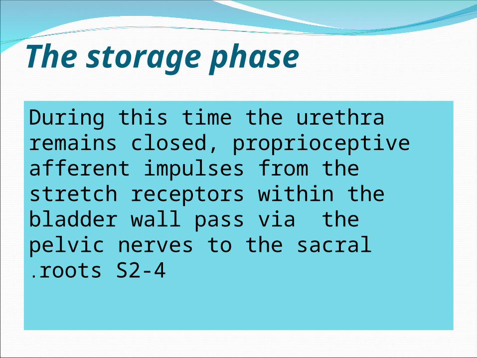
The storage phase
During this time the urethra remains closed, proprioceptive afferent impulses from the stretch receptors within the bladder wall pass via the pelvic nerves to the sacral roots S2-4.

/////////////////////////////////These impulses ascend the cord via the lateral spinothalamic tracts and the detrusor motor response is subconsciously inhibited by descending impulses from the basal ganglia.
as the bladder volume increases, afferent impulses are sent to the cerebral cortex and the first sensation of desire to void is appreciated at about half the functional bladder capacity.
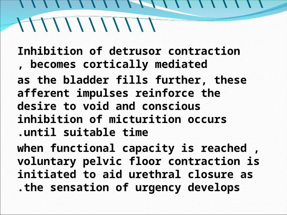
\\\\\\\\\\\\\\\\\\\\\\\\\\\\\\\\\Inhibition of detrusor contraction becomes cortically mediated,
as the bladder fills further, these afferent impulses reinforce the desire to void and conscious inhibition of micturition occurs until suitable time.
when functional capacity is reached , voluntary pelvic floor contraction is initiated to aid urethral closure as the sensation of urgency develops.

VOIDING PHASE:At a suitable time and place , cortical inhibition is released and relaxation of the pelvic floor occurs, together with relaxation of the intrinsic striated muscle of the urethra.
This results in a fall in urethral pressure which occurs a few seconds prior to increase the bladder pressure .

\\\\\\\\\\\\\\\\\\\\\\\\\\\\\\\\\a rapid discharge of efferent parasympathetic impulses via the pelvic nerve causes the detrusor to contract and also possibly to open the bladder neck and shorten the urethra .
The detrusor pressure rises normally less than
60 cm of water in women ,so that urine voided.

PATHOPHYSIOLOGY OF URINARY INCONTINENCE:
Under normal circumstances, in awomen with a healthy lower urinary tract, urine will only leave the bladder via the urethra when the intravesical pressure exceeds the maximum urethral pressure .
;;;;;;;;;;;;;;;;;;;;;;;;;;;;;;;;;;;;;;;;;;;;;;;;
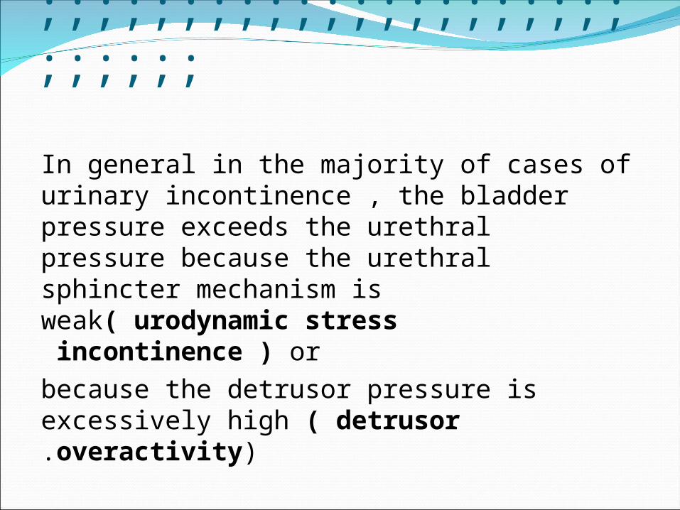
In general in the majority of cases of urinary incontinence , the bladder pressure exceeds the urethral pressure because the urethral sphincter mechanism is weak( urodynamic stress incontinence ) or
because the detrusor pressure is excessively high ( detrusor overactivity).

**************************
In urodynamic stress incontinence the factors which maintain positive urethral closure pressure at rest may be inadequate when there is an increase in intra-abdomenal pressure.

\\\\\\\\\\\\\\\\\\\\\\\\\\\\This is particularly likely to occur if the bladder neck and proximal urethra are poorly supported or descend through the pelvic floor ,as in case concomitant cystourethrocele.

------------------------------------------
An abnormally high detrusor pressure may occur in detrusor overactivity when there is inability to inhibit detrusor contractions.

THE PREVALENCE OF URINARY INCONTINENCE:
1 .50% are stress incotinence .
2 .11% are urge incontinence.
3 .36% are mixed incontinence.

Risk factor of incontinence includes:1.Age : increase with age.
2 .Race: white women are more liable than black women.
3 .pregnancy: pregnancy is responsible for marked changes in the urinary tract and consequently lower urinary symptoms are common and reflect normal physiological change due to high progesterone levels and pressure effect of gravid uterus.

\\\\\\\\\\\\\\\\\\\\\\\\\\\\\\\\\4 .Child birth: child birth may result in
damage to the pelvic floor musculature as well as injury to the pudendal and pelvic nerves .
more child birth more injury , also urinary incontinence associated with :increase exposure to oxytocic drugs ,vacum extraction , forceps delivery and fetal macrosomia.
5 .Menopause: occur more in women with menopause because of low level of oestrogen .

COMMON SYMPTOMS ASSOCIATED WITH INCONTINENCE;
_Stress incontinence is a symptom and sign mean loss of urine on physical effort , it is not a diagnosis.
_
Urgency means a sudden desire to void.
_
Urge incontinence is an involuntary loss of urine associated with strong desire to void.
**************************
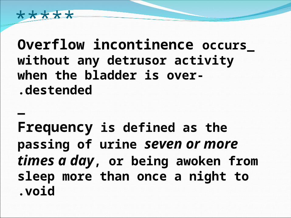
_Overflow incontinence occurs without any detrusor activity when the bladder is over-destended.
_
Frequency is defined as the passing of urine seven or more times a day, or being awoken from sleep more than once a night to void.

CLASSIFICATION OF INCONTINENCE: Urethral cause.:
1 .urodynamic stress incontinence.
2 .Detrusor over-activity.
3 .Retention with overflow.
4 .Congenital causes.
5 .Miscellaneous.
Extra- urethral causes:1 .congenital.
2 .Fistula.

URODYNAMIC STRESS INCONTINENCE: Previously called genuine stress
incontinence,
is noted during filling cystometry, and is defined as the involuntary leakage of urine during increased abdomenal pressure in the absence of a detrusor contraction.
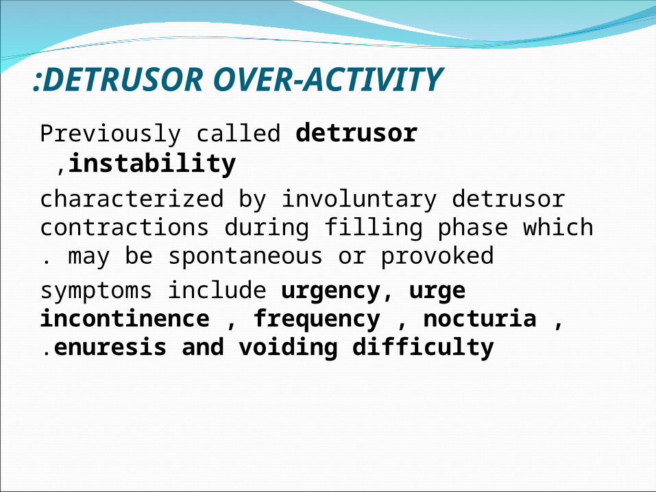
DETRUSOR OVER-ACTIVITY:
Previously called detrusor instability ,
characterized by involuntary detrusor contractions during filling phase which may be spontaneous or provoked.
symptoms include urgency, urge incontinence , frequency , nocturia , enuresis and voiding difficulty.

RETENTION WITH OVERFLOW:
Insidious failure of bladder emptying may lead to chronic retention and finally , when normal voiding is ineffective to overflow incontinence.
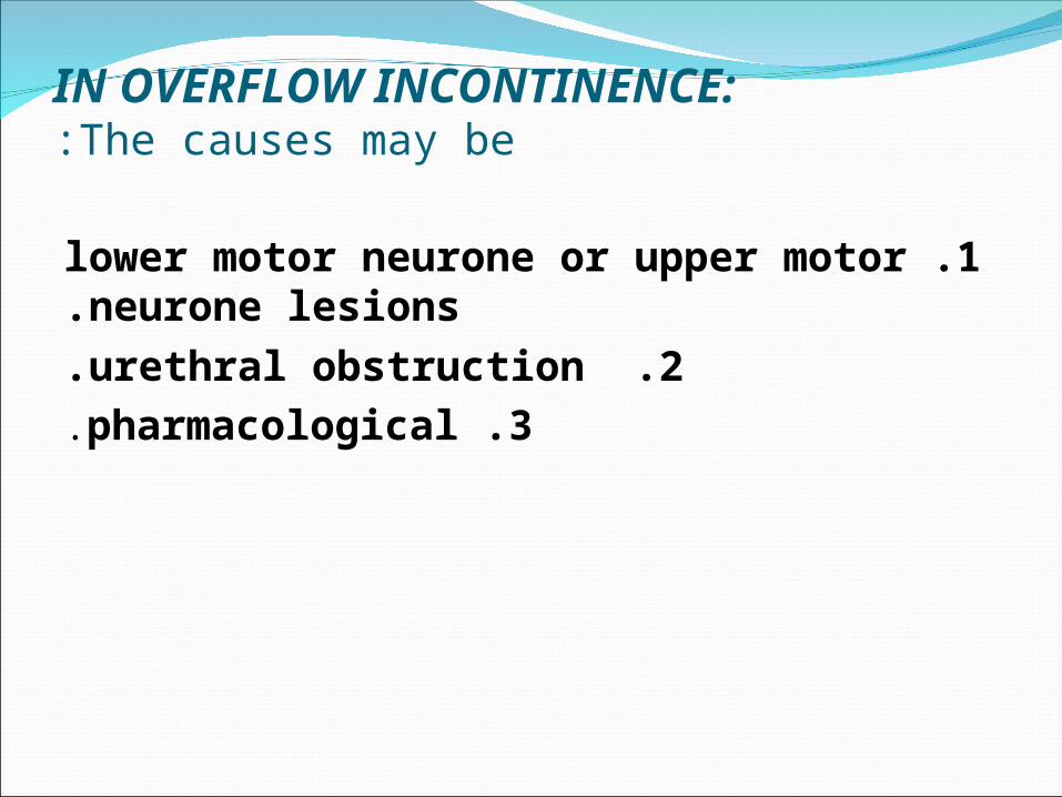
IN OVERFLOW INCONTINENCE:The causes may be:
1 .lower motor neurone or upper motor neurone lesions.
2 .urethral obstruction.
3 .pharmacological.
CONGENITAL CAUSES OF INCONTINENCE:
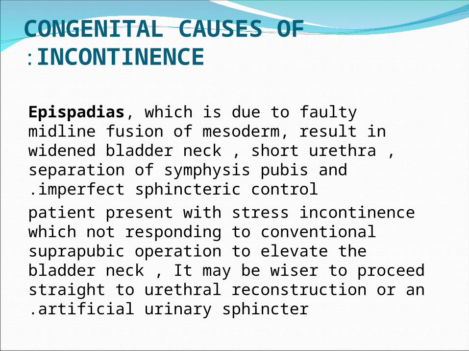
Epispadias, which is due to faulty midline fusion of mesoderm, result in widened bladder neck , short urethra , separation of symphysis pubis and imperfect sphincteric control.
patient present with stress incontinence which not responding to conventional suprapubic operation to elevate the bladder neck , It may be wiser to proceed straight to urethral reconstruction or an artificial urinary sphincter.

MISCELLANEOUS CAUSES:
Acute urinary tract infection or faecal impaction in the elderly may lead to temporary incontinence.
A urethral diverticulum may lead to post-micturition dribble, as urine collects within the divrticulum and escapes as the patient stands up.

EXTR-URETHRAL CAUSES OF INCONTINENCE:
1 .Congenital: bladder extrophy and ectopic ureter.
2 .Fistula : a urinary fistula is an abnormal opening between the urinary tract and the outside,
urinary fistula have an obstetric and gynaecological causes, The former include obstructive labour with compression of the bladder between the presenting head and bony pelvis.

/////////////////////////////////
The gynaecological causes are associated with pelvic surgery or pelvic malignancy or radiotherapy, what ever the cause the fistula must be accurately localized.
It can be treated by primary closure or by surgery which can be delayed until tissue inflammation and oedema have resolved at about 4 weeks.
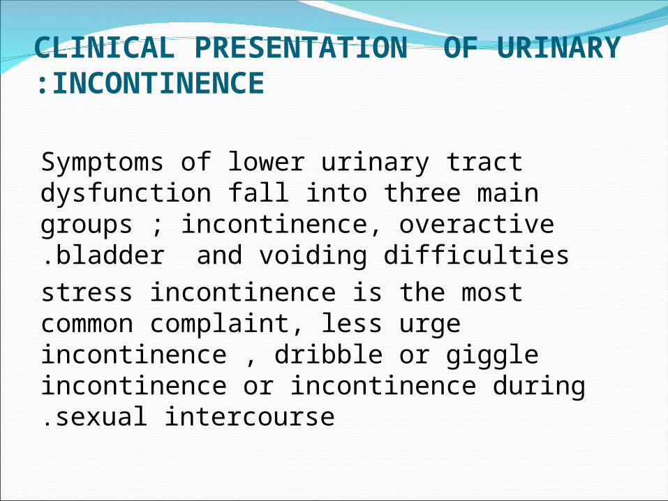
CLINICAL PRESENTATION OF URINARY INCONTINENCE:
Symptoms of lower urinary tract dysfunction fall into three main groups ; incontinence, overactive bladder and voiding difficulties.
stress incontinence is the most common complaint, less urge incontinence , dribble or giggle incontinence or incontinence during sexual intercourse.

………………………………....……………
nocturnal enuresis(bed wetting) may occur, symptoms of voiding difficulty include hesitancy, a poor stream, straining to void .
Apart from the symptoms of lower urinary tract dysfunction, it is important to take a full history from all women who present with urinary incontinence. other gynaecological symptoms such as prolapse or menstrual disturbances may be relevant.

/////////////////////////////////
A fibroid uterus may compress the bladder and cause urinary frequency and urgency.
There is an increase incidence of stress incontinence amongest women who have large babies particularly following instrumental vaginal delivery so obstetric history is important.

\\\\\\\\\\\\\\\\\\\\\\\\\\\\\\\\\
urological problems such as urinary tract infections , acute urinary retention and childhood enuresis should be sought.
history of diabetes and neurological problem (multiple sclerosis) should be checked, and drug history also important such as use of diuretics drugs ( frusimide).

Investigation of urodynamics incontinenceWill be discussed in next lecture

\\\\\\\\\\\\\\\\\\\\\\\\\\\\
THANK YOU
\\\\\\\\\\\\\\\\\\\\\\\\\\\\
INVESTIGATIONS OF URINARY INCONTINENCE
PRESENTED BY :Dr. ROZHAN YASIN KHALILMBCHB , FICOG , CABOG , HDOG

INVESTIGATIONS OF URINAYY INCONTINENCE:
Accurate and detailed history and examenation provide a framework for
the diagnosis,
but often there is discrepancy between the patients symptoms and urodynamic findings.

THE AIM OF URODYNAMIC INVESTIGATION: The aim of urodynamic investigations is to
provide accurate diagnosis of disorders of micturition and they involve investigation of lower urinary tract and pelvic floor dysfunction these investigation range from simple procedure to sophisticated studies only available in tertiary referal centres.

These investigation includes:
1. mid stream urine specimen
2 .urinary diary ( frequency / volume chart) .
3 .pad test.
4 .Uroflowmetry.
5 .Cystometry.
6 .Videocystourethrography.
7 .Urethral pressure profilometry.

INVESTIGATION:8 .Cystourethroscopy.
9 .Ultrasound.
10 .Intraveneous urography.
11 .Cystourethrography.
12 .Electromyography.
13 .Ambulatory urodynamics.
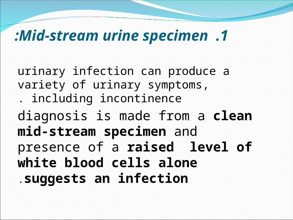
1 .Mid-stream urine specimen:
urinary infection can produce a variety of urinary symptoms, including
incontinence.
diagnosis is made from a clean mid-stream specimen and presence of a raised level of white blood cells alone suggests an infection.

2 .Urinary diary: A urinary diary is a simple record of
the patients fluid intake and output, episodes of urgency and leakage are recorded.
a ssugested practice is one week for follow up, these diaries recall provide an assessment of functional bladder capacity and to monitor conservative treatment e.g. bladder re-education, electrical stimulation and drug therapy.

: 3 .Pad test pad test are used to verifyand quantify urine loss.
it takes 1 hour , the patient wears a pre-weighed sanitary towel , drinks 500 ml of water and rest for 15 minutes .
After a series of defined manoeuvres the pad re-weighed, a urine loss of more than 1 g is considered significant . if indicated,
methylene blue solution can be instillated intravesically prior to the pad test to differentiate between urine and other loss , e.g. vaginal discharge.
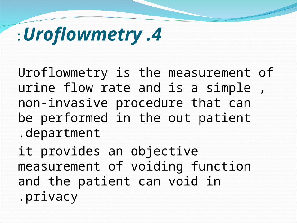
4 .Uroflowmetry:
Uroflowmetry is the measurement of urine flow rate and is a simple , non-invasive procedure that can be performed in the out patient department.
it provides an objective measurement of voiding function and the patient can void in privacy.
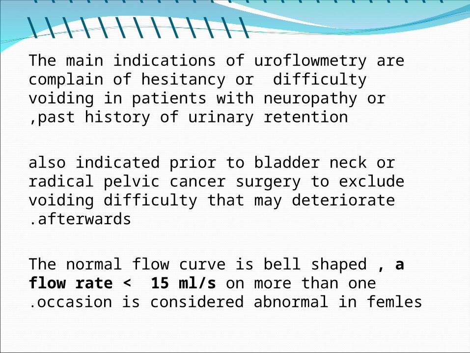
\\\\\\\\\\\\\\\\\\\\\\\\\\\\\\\\\\\\\ The main indications of uroflowmetry are
complain of hesitancy or difficulty voiding in patients with neuropathy or past history of urinary retention,
also indicated prior to bladder neck or radical pelvic cancer surgery to exclude voiding difficulty that may deteriorate afterwards.
The normal flow curve is bell shaped , a flow
rate < 15 ml/s on more than one occasion is considered abnormal in femles.

5 .Cystometry: Cystometry involves the
measurement of the pressure-volume relationship of the bladder,
it involves simultaneous abdominal pressure recording in addition to intravesical pressure monitoring during bladder filling and voiding .

------------------------------------------
Electronic subtraction of abdominal from intravesical pressure enables determination of the detrusor pressure.
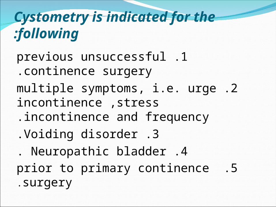
Cystometry is indicated for the following:
1 .previous unsuccessful continence surgery.
2 .multiple symptoms, i.e. urge incontinence ,stress incontinence and frequency.
3 .Voiding disorder.4 .Neuropathic bladder.
5 .prior to primary continence surgery.
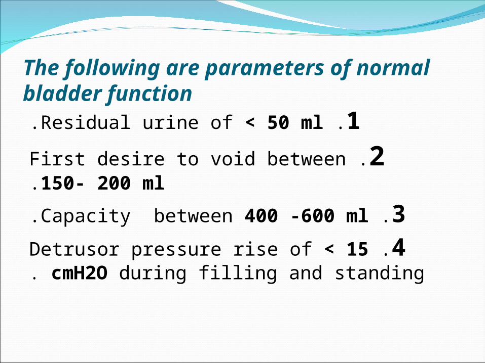
The following are parameters of normal
bladder function 1 .Residual urine of < 50 ml.
2 .First desire to void between 150- 200 ml.
3 .Capacity between 400 -600 ml.
4 .Detrusor pressure rise of < 15 cmH2O during filling and standing .
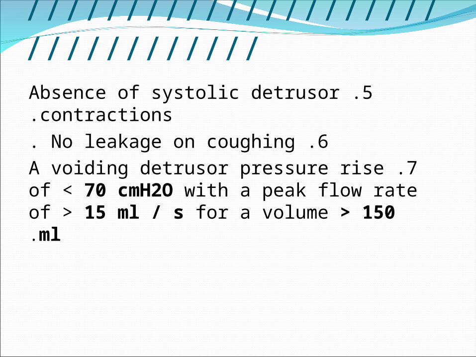
///////////////////////////////// 5 .Absence of systolic detrusor
contractions. 6 .No leakage on coughing.
7 .A voiding detrusor pressure rise of < 70 cmH2O with a peak flow rate of > 15 ml / s for a volume > 150 ml.

6 .Videocystourethrography:
Is a radio-opaque filling medium used during cystometry ,the urinary tract can be visualized by X- ray screening.
Videocystourethrography can provide more information to detect vesico-ureteric reflux, any detrusor contraction and leakage can be noted, also trabeculation and bladder,urethral diverticulae can be seen.

7 .Intravenous urography:
: Is indicated if patients present with- haematuria,
- neuropathic bladder and suspected uretero-vaginal fistula.

8 .Ultrasound:
ultrasound used to detect post-micturition urine residual estimation to detect voiding difficulty, urethral cyst and diverticula can also be examined using this technique .
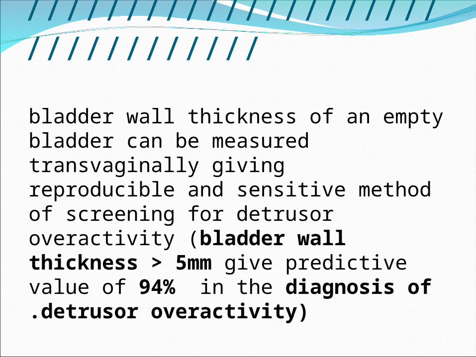
/////////////////////////////////
bladder wall thickness of an empty bladder can be measured transvaginally giving reproducible and sensitive method of screening for detrusor overactivity (bladder wall thickness > 5mm give predictive value of 94% in the diagnosis of detrusor overactivity).
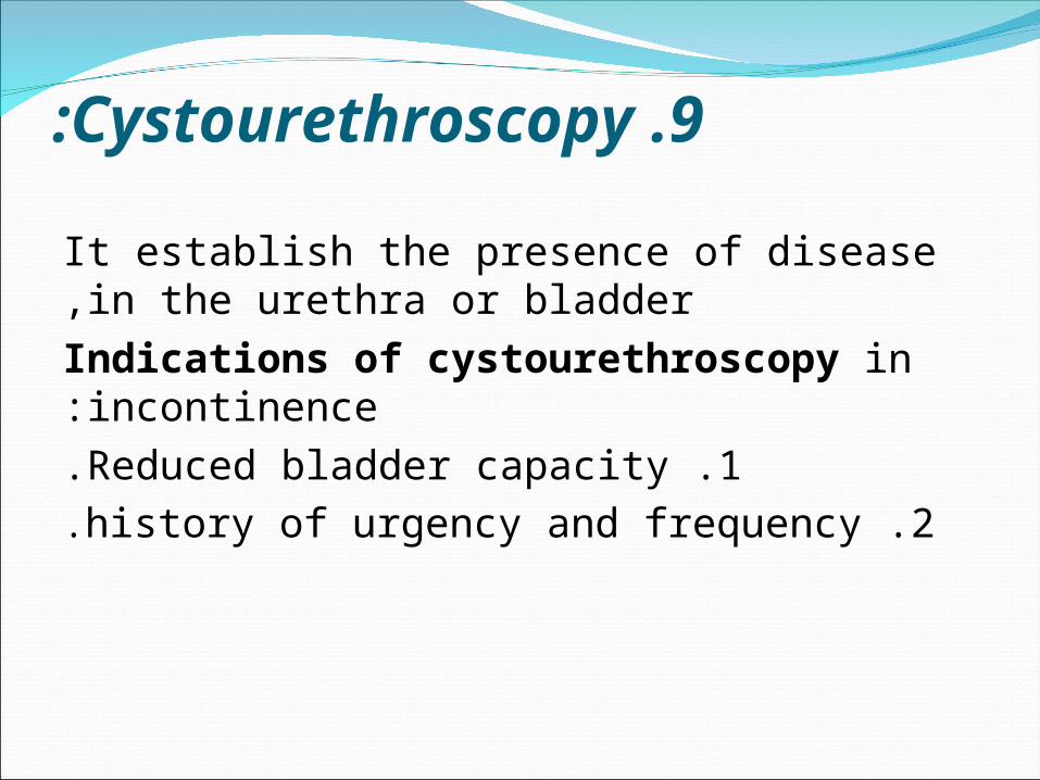
9 .Cystourethroscopy:
It establish the presence of disease in the urethra or bladder,Indications of cystourethroscopy in incontinence:
1 .Reduced bladder capacity. 2 .history of urgency and frequency.

/////////////////////////////////
3 .Suspected urethrovaginal or vesicovaginal fistula.
4 .haematuria or abnormal cytology. 5 .persistent urinary tract infection.

10 .Urethral pressure profilometry To maintain continence ,
the urethral pressure must remain higher than the intravesical pressure, the urethral pressure profiles can be obtained using a catheter tip dual sensor microtransducer.

TREATMENT OF URINARY INCONTINENCE:
In general simple measures such as exclusion of
urinary tract infection, restriction of fluid intake,
modifying medications ( e.g. diuretics ) and treating chronic cough and constipation play an important role in the management of most types of urinary incontinence.

TREATMENT OF URODYNAMIC STRESS INCONTINENCE:
Includes:
1 .Prevention. 2 .conservative management.
3 .surgery.
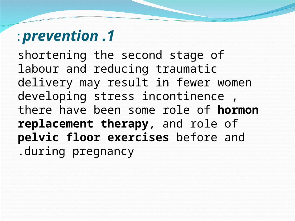
1 .prevention: shortening the second stage of labour and
reducing traumatic delivery may result in fewer women developing stress incontinence , there have been some role of hormon replacement therapy, and role of pelvic floor exercises before and during pregnancy.

2 .conservative treatment: Physiotherapy is the mainstay of conservative
treatment and is indicated when:1 .the incontinence is mild.
2 .the patient is medically unfit for surgery.3 .patient does not wish surgery under go
surgery. 4 .women have not yet complete her family.
5 .useful in patients name on long waitng list.

conservative treatment include: 1 .kegel ( pelvic floor ) exercise.
2 .perineometry. 3 .Vaginal cones.
4 .maximum electrical stimulation. 5 .drugs as duloxetine which is alpha-
adrenocepter agonist. 6 .vaginal devices.

3 .SURGICAL TREATMENT OF URODYNAMICS STRESS INCONTINENCE:
For women seeking cure , the mainstay of treatment is surgery. the aim of surgery are:
1. Restoration of the proximal urethra and bladder neck to the zone of intra-
abdominal pressure transmission. 2 .to increase urethral resistance.
3 .a combinationof both.

\\\\\\\\\\\\\\\\\\\\\\\\\\\\\\\\\
surgery have a cure rate of 90% if properly performed as primary procedure,
these operation include:

1 .vaginal route : a. ANTERIOR COLPORRHAPHY +/- KELLY
SUTURE. B. URETHROCLIESIS
C. URETHRAL BULKING AGENTS. D. RETROPUBIC TAPE PROCEDURES.
E. TRANSOBTURATOR TAPE PROCEDURES.
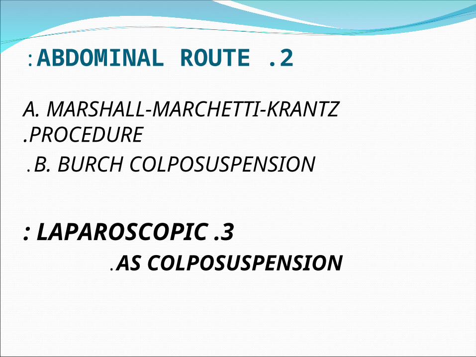
2 .ABDOMINAL ROUTE:
A. MARSHALL-MARCHETTI-KRANTZ PROCEDURE.
B. BURCH COLPOSUSPENSION.
3 .LAPAROSCOPIC: AS COLPOSUSPENSION.
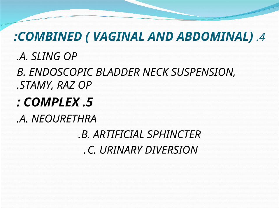
4 .COMBINED ( VAGINAL AND ABDOMINAL):
A. SLING OP. B. ENDOSCOPIC BLADDER NECK SUSPENSION, STAMY,
RAZ OP.
5 .COMPLEX: A. NEOURETHRA.
B. ARTIFICIAL SPHINCTER.C. URINARY DIVERSION.

\\\\\\\\\\\\\\\\\\\\\\\\\\\\IN COLPOSUSPENSION OPERATION
THE SUCCESS RATE IS OVER 95% . HOW EVER, FOR THE ELDERLY PATIENT WITH A
SCARRED, NARROWED VAGINA, A BLADDER NECK BULKING INJECTION
MAY BE MORE APPROPRIATE BECAUSE IT IS LESS INVASIVE AND PERFORMED AS A DAY-CASE PROCEDURE.

\\\\\\\\\\\\\\\\\\\\\\\\\\\\\\\\\ A NEW OPERATION CALLED A TENSION-FREE
VAGINAL TAPE WHICH IS BASED ON THE THEORY OF SUBURETHRAL SUPPORT, HAS
BEEN DEVELOPED.
THIS INVOLVE INSERTION OF A PROLENE TAPE UNDERNEATH THE MID-URETHRA, THROUGH A VERY SMALL VAGINAL INCISION WITH 2 TINY ABDOMINAL INCISIONS.
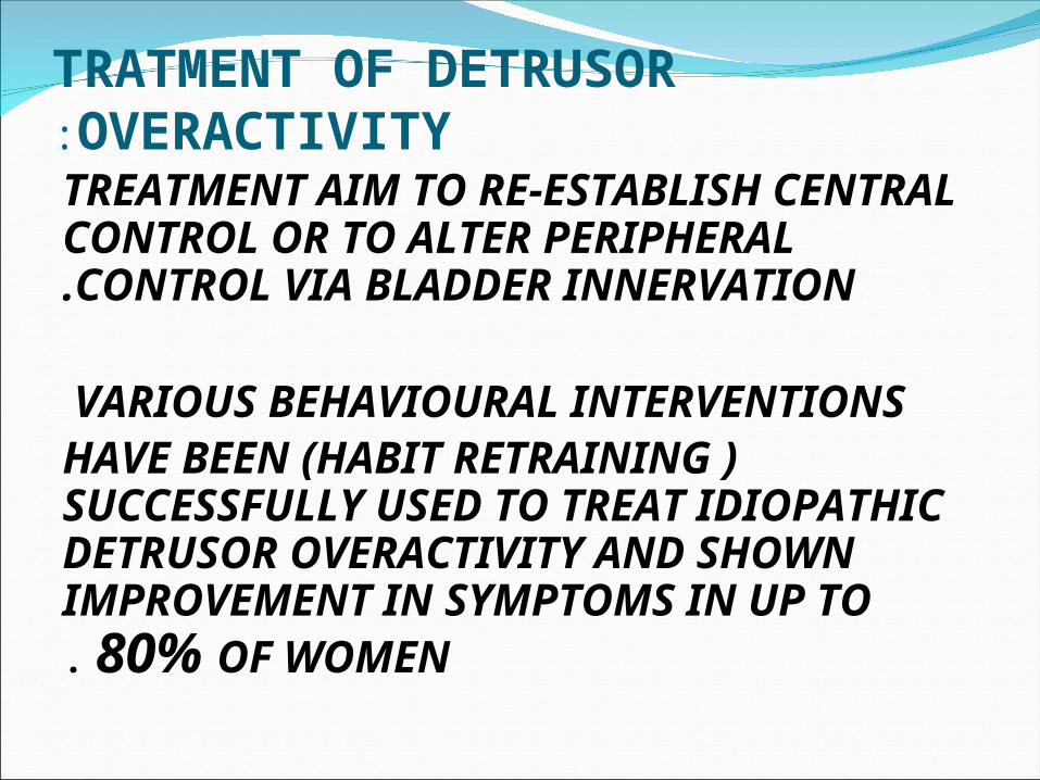
TRATMENT OF DETRUSOR OVERACTIVITY: TREATMENT AIM TO RE-ESTABLISH CENTRAL
CONTROL OR TO ALTER PERIPHERAL CONTROL VIA BLADDER INNERVATION.
VARIOUS BEHAVIOURAL INTERVENTIONS ( HABIT RETRAINING )HAVE BEEN
SUCCESSFULLY USED TO TREAT IDIOPATHIC DETRUSOR OVERACTIVITY AND SHOWN IMPROVEMENT IN SYMPTOMS IN UP TO 80% OF WOMEN .

TREATMENT INCLUDES:1 .PSYCHOTHERAPY INCLUDE:
A. BLADDER DRILL. B. BIOFEEDBACK.
C. HYPNOTHERAPY. D. ACUPUNCTURE.

\\\\\\\\\\\\\\\\\\\\\\\\\\\\\\\\\
2 .DRUG THERAPY INCLUDE:
A.INHIBITE BLADDER CONTRACTIONS , AS TRICYCLIC ANTIDEPRESSANTS, ANTICHOLINERGIC AGENTS.
B. IMPROVE LOCAL TISSUES (OESTROGEN).
C. REDUCE URINE PRODUCTION ( SYNTHETIC VASOPRSSIN).
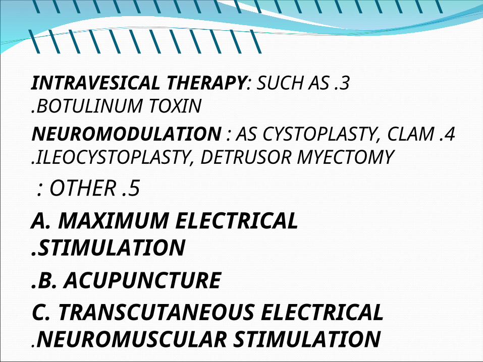
\\\\\\\\\\\\\\\\\\\\\\\\\\\\\\\\\3 .INTRAVESICAL THERAPY: SUCH AS BOTULINUM
TOXIN.4 .NEUROMODULATION : AS CYSTOPLASTY, CLAM
ILEOCYSTOPLASTY, DETRUSOR MYECTOMY.
5 .OTHER : A. MAXIMUM ELECTRICAL STIMULATION.B. ACUPUNCTURE.
C. TRANSCUTANEOUS ELECTRICAL NEUROMUSCULAR STIMULATION.

\\\\\\\\\\\\\\\\\\\\\\\\\\\\
THANK YOU


