Neurological: Mental status, strength, tendon reflexes,sensory testing.Laboratory Evaluation: Electrolytes, glucose, liver function tests, INR/PTT, CBC with differential; X-rays,ECG (if >35 yrs or cardiovascular disease), urinalysis.
Assessment and Plan: Assign a number to each prob-lem. Discuss each problem, and describe surgical plansfor each numbered problem, including preoperativetesting, laboratory studies, medications, and antibiotics.
Discharge Summary
Patient's Name:
Chart Number:Date of Admission: Date of Discharge:
Admitting Diagnosis: Discharge Diagnosis:
Name of Attending or Ward Service:Surgical Procedures:History and Physical Examination and LaboratoryData: Describe the course of the disease up to the timethe patient came to the hospital, and describe the physi-cal exam and laboratory data on admission.Hospital Course: Describe the course of the patient's
8/20/2019 Gynecology and Obstetricsvvvvvvvvvvvvvvvvvvvvvvvvvvvvvvvvvvvvvvvvv
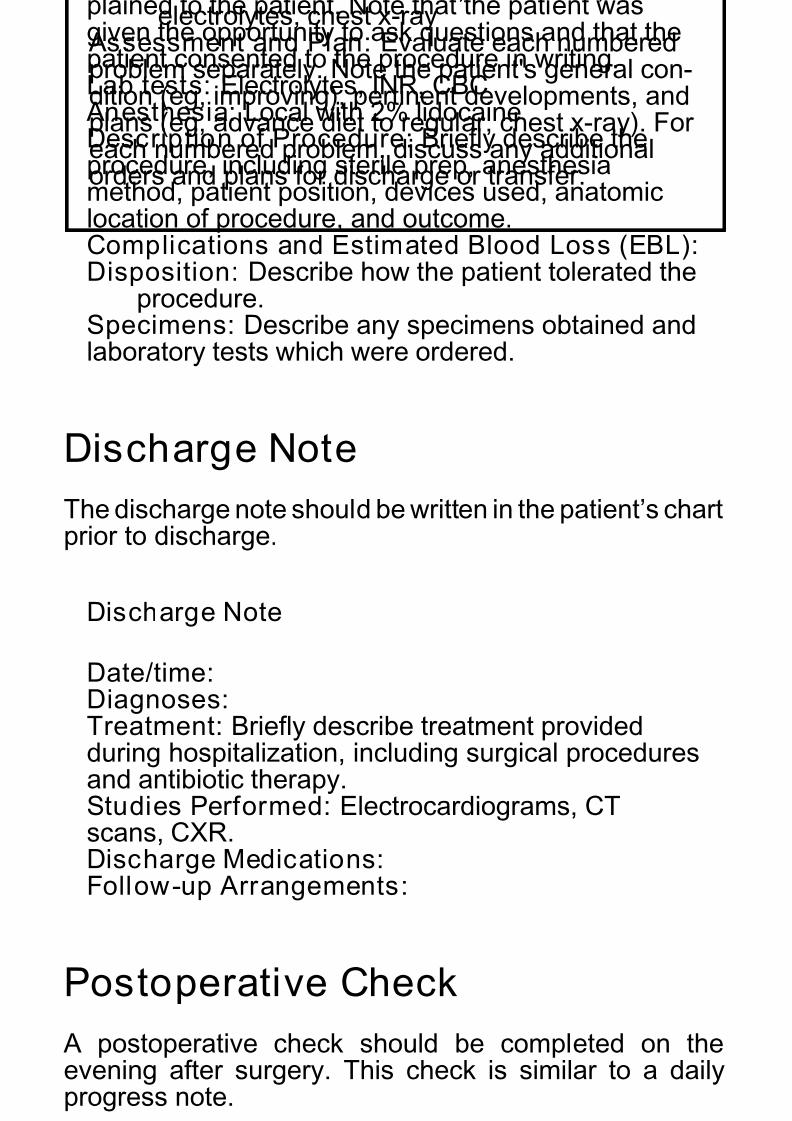
A procedure note should be written in the chart when aprocedure is performed. Procedure notes are brief operative notes.
Procedure Note
Date and time:Procedure:
Indications:Patient Consent: Document that the indications,risks and alternatives to the procedure were ex-plained to the patient. Note that the patient wasgiven the opportunity to ask questions and that the
patient consented to the procedure in writing.Lab tests: Electrolytes, INR, CBC
Anesthesia: Local with 2% lidocaineDescription of Procedure: Briefly describe theprocedure, including sterile prep, anesthesiamethod, patient position, devices used, anatomiclocation of procedure, and outcome.Complications and Estimated Blood Loss (EBL):Disposition: Describe how the patient tolerated the
procedure.Specimens: Describe any specimens obtained andl b t t t hi h d d
8/20/2019 Gynecology and Obstetricsvvvvvvvvvvvvvvvvvvvvvvvvvvvvvvvvvvvvvvvvv
sent to pathology.Description of Operative Procedure: After obtaining
informed consent, the patient was taken to the operatingroom and placed in the supine position, given generalanesthesia, and prepped and draped in sterile fashion.
A Pfannenstiel incision was made 2 cm above thesymphysis pubis and extended sharply to the rectus
fascia. The fascial incision was bilaterally incised withcurved Mayo scissors, and the rectus sheath was sepa-rated superiorly and inferiorly by sharp and blunt dissec-tion. The peritoneum was grasped between two Kellyclamps, elevated, and incised with a scalpel. The pelvis
was examined with the findings noted above. A Balfour retractor was placed into the incision, and the bowel waspacked away with moist laparotomy sponges. Two Kocher clamps were placed on the cornua of the uterus and usedfor retraction.
The round ligaments on both sides were clamped,sutured with #0 Vicryl, and transected. The anterior leaf of the broad ligament was incised along the bladder reflection to the midline from both sides, and the bladder
was gently dissected off the lower uterine segment andcervix with a sponge stick.The retroperitoneal space was opened and the ureters
were identified bilaterally. The infundibulopelvic ligamentson both sides were then doubly clamped, transected, and
doubly ligated with #O Vicryl. Excellent hemostasis wasobserved. The uterine arteries were skeletonized bilater-
8/20/2019 Gynecology and Obstetricsvvvvvvvvvvvvvvvvvvvvvvvvvvvvvvvvvvvvvvvvv
placed into the posterior vagina, a Deaver or rightangle retractor is positioned anterior to the cervix,
and then the anterior and posterior lips of the cervixare grasped with a single- or double-toothedtenaculum.
D. Traction is placed on the cervix to expose theposterior vaginal mucosa. Using Mayo scissors, the
posterior cul-de-sac is entered sharply, and theperitoneum identified. A figure-of-eight suture isthen used to attach the peritoneum to the posterior vaginal mucosa.
E. A Steiner-Anvard weighted speculum is inserted
into the posterior cul-de-sac after this space isopened. The uterosacral ligaments are clamped,with the tip of the clamp incorporating the lower portion of the cardinal ligaments. The clamp isplaced perpendicular to the uterine axis, and thepedicle cut so that there is 0.5 cm of tissue distal tothe clamp. A transfixion suture is then placed at thetip of the clamp. Once ligated, the uterosacralligaments are transfixed to the posterior lateral
vaginal mucosa. This suture is held with ahemostat.F. Downward traction is placed on the cervix to provide
countertraction for the vaginal mucosa and theanterior vaginal mucosa is incised at the level of the
cervicovaginal junction. The bladder is advancedupward using an open, moistened gauze sponge. At
8/20/2019 Gynecology and Obstetricsvvvvvvvvvvvvvvvvvvvvvvvvvvvvvvvvvvvvvvvvv
cancer remains (eg, hyperplasia with atypia, pres-ence of necrosis, or pyometra).
II. Dilation and curettage A. Dilation and curettage is performed as either adiagnostic or therapeutic procedure. Indications for diagnostic D&C include:1. A nondiagnostic office biopsy in women who are
at high risk of endometrial carcinoma.2. Insufficient tissue for analysis on office biopsy.3. Cervical stenosis prevents the completion of an
office biopsy.B. Diagnostic D&Cs are usually performed with
hysteroscopy to obtain a visual image of theendometrial cavity, exclude focal disease, andprevent missing unsuspected polyps.
C. Examination under anesthesia. After anesthesiahas been administered, the size, shape, and positionof the uterus are noted, with particular attention tothe axis of the cervix and flexion of the fundus. Thesize, shape, and consistency of the adnexa aredetermined. The perineum, vagina, and cervix are
then prepared with an aseptic solution and vaginalretractors are inserted into the vagina.D. Operative technique. A D&C is performed with the
woman in the dorsal lithotomy position.1. Endocervical curettage (ECC) is performed
before dilation of the cervix. A Kevorkian-Youngecurette is introduced into the cervical canal up to
8/20/2019 Gynecology and Obstetricsvvvvvvvvvvvvvvvvvvvvvvvvvvvvvvvvvvvvvvvvv
of metaplastic and endocervical cells indicatesadequate sampling of the transformation zone of the cervix, the area at risk for neoplasia. Mostwomen without an endocervical/transformationzone component present should be screenedwith a repeat Pap test in 12 months. However,repeat testing in six months is advised in the
following situations:a. A previous Pap smear result of ASC-US or worse without three subsequent negative Papsmears.
b. A previous Pap smear with an unexplained
glandular abnormality.c. An HPV test result positive for a high-risk typewithin the previous 12 months.
d. Inability to clearly visualize or sample theendocervical canal.
e. Immunosuppression.f. Insufficient frequency of previous screening
(eg, failure to be screened at least biennially).3. Blood or inflammation present. Women with
partially obscuring blood or inflammation shouldhave a repeat test in six months if they meet anyof the above criteria.
Negative/NegativeNegative/Negative ASCUS/Negative ASCUS/PositiveGreater than ASCUS/ Posi-
tive or negative
Routine screening in 3yearsRepeat combined test in 6-12 months*Repeat cytology in 12
months**ColposcopyColposcopy
*If Negative/Negative, then resume screening in 3 years
If ASCUS/Negative, then repeat combined test in 12 monthsIf greater than ASCUS/Negative, then colposcopyIf any cytology result/Positive, the colposcopy
**Follow-up depends on cytology results
HPV = Human Papillomarvirus. Positive means high-risktypes are present. Negative means high-risk types are notpresent
C. Special circumstances1 Infection or reactive changes When an
8/20/2019 Gynecology and Obstetricsvvvvvvvvvvvvvvvvvvvvvvvvvvvvvvvvvvvvvvvvv
ogy seven days after completion of therapy,with referral to colposcopy if an abnormality
persists. If repeat cytology is normal, thenanother cytology test should be obtained in four to six months. The woman can return to routinesurveillance if both tests are normal, but shouldbe referred for colposcopy if either test is ASC-
US or worse.2. Adolescents. Initial colposcopy may be de-ferred in adolescents. Instead, they may bemanaged with HPV DNA testing at 12 monthswith referral to colposcopy for positive results
(high-risk HPV DNA types).3. Pregnant women with LSIL are managed in a
similar fashion to those with HSIL (see below).Colposcopy should be performed, with biopsyand endocervical curettage performed for anylesion suspicious for HSIL or more severedisease.
C. High-grade squamous intraepithelial lesions1. HSIL may also be referred to as CIN II or III,
severe dysplasia, or carcinoma in situ (CIS). Allwomen with HSIL should be referred for colposcopy and endocervical curettage.
2. If colposcopy reveals no lesion or only biopsyproven CIN I, then cytology, colposcopy, and
biopsies should be reviewed. A cytologicaldiagnosis of HSIL without colposcopic or
8/20/2019 Gynecology and Obstetricsvvvvvvvvvvvvvvvvvvvvvvvvvvvvvvvvvvvvvvvvv
Cervical intraepithelial abnormalities are usually firstdetected by cytology screening. Treatment of cervicalintraepithelial abnormalities is typically undertaken after a histologic abnormality has been proven by tissuebiopsy.
I. Atypical squamous cells (ASC) is a cytologicalscreening diagnosis that does not require treatment.
ASC does require further evaluation to exclude the
presence of higher- grade disease that might requiretreatment. Treatment may be initiated if there is biopsyproven dysplasia.
II. Low-grade lesions. Low-grade precursors of cervicalcancer have been called low-grade squamous
intraepithelial lesions (LSIL), low-grade cervicalintraepithelial neoplasia (CIN I), and mild dysplasia. A. Management
1. Expectant management is preferred for thereliable patient with biopsy-confirmed CIN I in
whom the entire lesion and limits of the transfor-mation zone are completely visualized (ie,satisfactory colposcopic examination). If treat-ment is desired, ablative or excisional modalitiesare appropriate. An excisional procedure is thepreferred diagnostic/therapeutic approach in allwomen if colposcopic examination is unsatisfac
8/20/2019 Gynecology and Obstetricsvvvvvvvvvvvvvvvvvvvvvvvvvvvvvvvvvvvvvvvvv
endometrial, or glandular cells not otherwisespecified (NOS).
2. AGC, favor neoplastic, endocervical,endometrial, or NOS.
3. Endocervical adenocarcinoma in situ (AIS).4. Adenocarcinoma.
B. Cold-knife conization is the best method for
diagnosis of AIS. Adenocarcinoma in situ (AIS) of the cervix is characterized by endocervical glandslined by atypical columnar epithelial cells.
C. If conization margins are positive, repeatconization should be performed in patients who
wish to maintain fertility and who understand therisk of leaving residual disease. Repeatconizations should also be considered if conemargins are negative in the setting of a positiveECC. If fertility is not desired, hysterectomy shouldbe performed.
V. Recommendations for initial management of cervical intraepithelial lesions
A. CIN I. Expectant management is recommended
for the reliable patient in whom the entire lesionand limits of the transformation zone are com-pletely visualized. Expectant management con-sists of repeat cytology at 6 and 12 months or HPV testing at 12 months.
B. CIN II, III, squamous carcinoma in situ. Loopelectrosurgical excision procedure (LEEP) is the
8/20/2019 Gynecology and Obstetricsvvvvvvvvvvvvvvvvvvvvvvvvvvvvvvvvvvvvvvvvv
the endocervical margin following cone biopsy, LSILbut no visible lesion, and those with an unsatisfac-tory colposcopic examination. A long straightcurette is used to scrape the four quadrants of theendocervical canal and an endocervical brush isemployed to remove any exfoliated tissue.Endocervical curettage in not performed in preg-
nant women.References: See page 184.
Contraception Approximately 31 percent of births are unintended; about22 percent were "mistimed," while 9 percent were "un-wanted."
I. Hormonal contraceptive methods other than oralcontraceptives
A. Contraceptive vaginal ring (NuvaRing) delivers15 :g ethinyl estradiol and 120 :g of etonogestrel
daily.1. Advantages of the ring include rapid return to
ovulation after discontinuation, lower doses of hormones, ease and convenience, and im-proved cycle control. Benefits, risks, and contra-indications to use are similar to those with com-bined oral contraceptive pills except for the
8/20/2019 Gynecology and Obstetricsvvvvvvvvvvvvvvvvvvvvvvvvvvvvvvvvvvvvvvvvv
bleeding may be treated with 50 :g of ethinylestradiol for 14 days.
H. Medr ox ypro gesterone acetate/est radio lcypionate (MPA/E2C, Lunelle) is a combined (25mg MPA and 5 mg E2C), injectable contraceptive.1. Although monthly IM injections are required,
MPA/E2C has several desirable features:
a. It has nearly 100 percent effectiveness inpreventing pregnancy.b. Fertility returns within three to four months
after it is discontinued.c. Irregular bleeding is less common than in
women given MPA alone.2. Weight gain, hypertension, headache,
mastalgia, or other nonmenstrual complaints arecommon.
3. Lunelle should be considered for women whoforget to take their birth control pills or those whowant a discreet method of contraception. Theinitial injection should be given during the first 5days of the menstrual cycle or within 7 days of
stopping oral contraceptives. Lunelle injectionsshould be given every 28 to 30 days; 33 days atthe most.
II. Oral contraceptives A. Combined (estrogen-progestin) oral contraceptives
are reliable, and they have noncontraceptivebenefits, which include reduction in dysmenorrhea,
8/20/2019 Gynecology and Obstetricsvvvvvvvvvvvvvvvvvvvvvvvvvvvvvvvvvvvvvvvvv
implantation and making the cervical mucus lesspermeable to penetration by sperm.
D. Contraindications1. Absolute contraindications to OCs:
a. Previous thromboembolic event or strokeb. History of an estrogen-dependent tumorc. Active liver disease
d. Pregnancye. Undiagnosed abnormal uterine bleedingf. Hypertriglyceridemiag. Women over age 35 years who smoke heavily
(greater than 15 cigarettes per day)
2. Screening requirements. Hormonal contracep-tion can be safely provided after a careful medi-cal history and blood pressure measurement.Pap smears are not required before a prescrip-tion for OCs.
E. Efficacy. When taken properly, OCs are a veryeffective form of contraception. The actual failurerate is 2 to 3 percent due primarily to missed pills or failure to resume therapy after the seven-day pill-
free interval.
Noncontraceptive Benefits of Oral ContraceptivePills
Dysmenorrhea Functional ovarian cysts
8/20/2019 Gynecology and Obstetricsvvvvvvvvvvvvvvvvvvvvvvvvvvvvvvvvvvvvvvvvv
H. Recommendations1. Monophasic OCs containing the second genera-
tion progestin, norethindrone (Ortho-Novum1/35) are recommended when starting a patienton OCs for the first time. This progestin has verylow androgenicity when compared to other sec-ond generation progestins, and also compares
favorably to the third generation progestins inandrogenicity.2. The pill should be started on the first day of the
period to provide the maximum contraceptiveeffect in the first cycle. However, most women
start their pill on the first Sunday after the periodstarts. Some form of back-up contraception isneeded for the first month if one chooses theSunday start, because the full contraceptiveeffect might not be provided in the first pill pack.
Factors to Consider in Starting or Switching OralContraceptive Pills
Objective ActionProducts thatachieve the ob-
jective
To minimizehigh risk of
Select a productwith a lower dosage
Alesse, Aviane,Loestrin 1/20,
8/20/2019 Gynecology and Obstetricsvvvvvvvvvvvvvvvvvvvvvvvvvvvvvvvvvvvvvvvvv
1. Consider pretreatment one hour before each oral con-traceptive pill dose, using one of the following orallyadministered antiemetic agents:
Prochlorperazine (Compazine), 5 to 10 mgPromethazine (Phenergan), 12.5 to 25 mgTrimethobenzamide (Tigan), 250 mg
Meclizine (Antivert) 50 mg2. Administer the first dose of oral contraceptive pill within
72 hours of unprotected coitus, and administer thesecond dose 12 hours after the first dose. Brand name
options for emergency contraception include the follow-ing:Preven Kit – two pills per dose (0.5 mg of levonorgestrel and 100 µg of ethinyl estradiol per dose)
Plan B – one pill per dose (0.75 mg of levonorgestrel per dose)Ovral – two pills per dose (0.5 mg of levonorgestreland 100 µg of ethinyl estradiol per dose)Nordette – four pills per dose (0.6 mg of levonorgestrel and 120 µg of ethinyl estradiol per
dose)Triphasil – four pills per dose (0.5 mg of levonorgestrel and 120 µg of ethinyl estradiol per dose)
VIII. Sterilization
8/20/2019 Gynecology and Obstetricsvvvvvvvvvvvvvvvvvvvvvvvvvvvvvvvvvvvvvvvvv
scribed previously.4. Antibiotics are used prophylactically .
Doxycycline is the best agent because of abroad spectrum of antimicrobial effect. D-nega-tive patients should receive D (Rho[D]) immuneglobulin.
C. Complications
1. The most common postabortal complicationsare pain, bleeding, and low-grade fever. Mostcases are caused by retained gestational tissueor a clot in the uterine cavity. These symptomsare best managed by a repeat uterine evacua-
tion, performed under local anesthesia2. Cervical shock. Vasovagal syncope produced
by stimulation of the cervical canal can be seenafter paracervical block. Brief tonic-clonic activityrarely may be observed and is often confusedwith seizure. The routine use of atropine withparacervical anesthesia or the use of conscioussedation prevents cervical shock.
3. Perforation
a. The risk of perforation is less than 1 in every1,000 first-trimester abortions. It increaseswith gestational age and is greater for parouswomen than for nulliparous women. Perfora-tion is best evaluated by laparoscopy to
determine the extent of the injury.b. Perforations at the junction of the cervix and
8/20/2019 Gynecology and Obstetricsvvvvvvvvvvvvvvvvvvvvvvvvvvvvvvvvvvvvvvvvv
A. Dilation and evacuation1. Transcervical dilation and evacuation of the
uterus (D&E) is the method most commonlyused for mid-trimester abortions before 21menstrual weeks. In the one-stage technique,forcible dilation is performed slowly and carefullyto sufficient diameter to allow insertion of large,
strong ovum forceps for evacuation. The better approach is a two-stage procedure in whichmultiple Laminaria are used to achieve gradualdilatation over several hours before extraction.Uterine evacuation is accomplished with long,
heavy forceps, using the vacuum cannula to rup-ture the fetal membranes, drain amniotic fluid,and ensure complete evacuation.
2. Preoperative ultrasonography is necessary for all cases 14 weeks and beyond. Intraoperativereal-time ultrasonography helps to locate fetalparts within the uterus.
3. Dilation and evacuation becomes progressivelymore difficult as gestational age advances, and
instillation techniques are often used after 21weeks. Dilation and evacuation can be offered inthe late mid-trimester, but two sets of Laminariatents for a total of 36-48 hours is recommended.
After multistage Laminaria treatment, urea is in-
jected into the amniotic sac. Extraction is thenaccomplished after labor begins and after fetal
8/20/2019 Gynecology and Obstetricsvvvvvvvvvvvvvvvvvvvvvvvvvvvvvvvvvvvvvvvvv
first missed menses.B. Transvaginal ultrasound is most useful for identi-
fying an intrauterine gestation. An extrauterinepregnancy will be visualized in only 16 to 32 per-cent of cases, thus a pelvic ultrasound showing "nointrauterine or extrauterine gestation" does notexclude the diagnosis of EP.
1. The identification of an intrauterine pregnancyeffectively excludes the possibility of an ectopicin almost all cases. However, pregnancies con-ceived with assisted reproductive technology arean exception, since the incidence of combined
intrauterine and extrauterine pregnancy may beas high as 1/100 pregnancies.
2. An early intrauterine pregnancy is identifiedsonographically by the presence of a true gesta-tional sac. Using TVS, the gestational sac isusually visible at 4.5 to 5 weeks of gestation withthe double decidual sign at 5.5 to 6 weeks, theyolk sac appears at 5 to 6 weeks and remainsuntil 10 weeks, and a fetal pole with cardiac
activity is first detected at 5.5 to 6 weeks.C. beta-hCG concentration. The gestational sac isusually identified at beta-hCG concentrations above1500 to 2000 IU/L. The absence of an intrauterinegestational sac at beta-hCG concentrations above
2000 IU/L strongly suggests an EP.D. Progesterone concentrations are higher in
8/20/2019 Gynecology and Obstetricsvvvvvvvvvvvvvvvvvvvvvvvvvvvvvvvvvvvvvvvvv
percent.B. Methotrexate is a folic acid antagonist, which
inhibits DNA synthesis and cell reproduction.
Criteria for Receiving Methotrexate
Absolute indicationsHemodynamically stable without active bleeding or signs of hemoperitoneumNonlaparoscopic diagnosisPatient desires future fertilityGeneral anesthesia poses a significant riskPatient is able to return for follow-up carePatient has no contraindications to methotrexate
Relative indicationsUnruptured mass <3.5 cm at its greatest dimensionNo fetal cardiac motion detectedPatients whose bet-hCG level does not exceed 6,000-15,000 mlU/mL
Contraindications to Methotrexate Therapy
Absolute contraindicationsBreast feeding
Overt or laboratory evidence of immunodeficiency Alcoholism, alcoholic liver disease, or other chronic liver di
8/20/2019 Gynecology and Obstetricsvvvvvvvvvvvvvvvvvvvvvvvvvvvvvvvvvvvvvvvvv
Salpingectomy is the procedure of choice if the tuberequires removal.
References: See page 184.
Acute Pelvic Pain
I. Clinical evaluation A. Assessment of acute pelvic pain should determine
the patient’s age, obstetrical history, menstrualhistory, characteristics of pain onset, duration, andpalliative or aggravating factors.
B. Associated symptoms may include urinary or gastrointestinal symptoms, fever, abnormal bleed-ing, or vaginal discharge.
C. Past medical history. Contraceptive history, surgi-
cal history, gynecologic history, history of pelvicinflammatory disease, ectopic pregnancy, sexuallytransmitted diseases should be determined. Currentsexual activity and practices should be assessed.
D. Method of contraception
1. Sexual abstinence in the months preceding theonset of pain lessons the likelihood of pregnancy-related etiologies.
2. The risk of acute PID is reduced by 50% in pa-tients taking oral contraceptives or using a barrier method of contraception. Patients taking oralcontraceptives are at decreased risk for an
8/20/2019 Gynecology and Obstetricsvvvvvvvvvvvvvvvvvvvvvvvvvvvvvvvvvvvvvvvvv
adnexal mass is characteristic. There is often ahistory of repetitive, transitory pain. Pelvicsonography often confirms the diagnosis. Laparo-scopic diagnosis and surgical intervention areindicated.
D. Ruptured or hemorrhagic corpus luteal cystusually causes bilateral pain, but it can cause
unilateral tenderness in 35%. Ultrasound aids indiagnosis.E. Endometriosis usually causes chronic or recurrent
pain, but it can occasionally cause acute pelvic pain.There usually is a history of dysmenorrhea and deep
dyspareunia. Pelvic exam reveals fixed uterineretrodisplacement and tender uterosacral and cul-de-sac nodularity. Laparoscopy confirms the diagno-sis.
References: See page 184.
Chronic Pelvic Pain
Chronic pelvic pain (CPP) is menstrual or nonmenstrualpain of at least six months' duration, located below theumbilicus and severe enough to cause functional disabil-ity or require treatment. Gynecologic conditions accountfor 90 percent of cases of CPP. Gastrointestinal diseases,such as irritable bowel syndrome, are the next mostcommon category
8/20/2019 Gynecology and Obstetricsvvvvvvvvvvvvvvvvvvvvvvvvvvvvvvvvvvvvvvvvv
A. Surgical scars, hernias, and masses should besought. Pelvic examination should include anevaluation for physical findings consistent withendometriosis, adenomyosis, or leiomyomata.Tender areas should be identified.
B. Phys ical f i nd ings charac ter i s t i c of endometriosis are uterosacral ligament abnormal-
ities (eg, nodularity or thickening, focal tender-ness), lateral displacement of the cervix caused byendometriosis, and cervical stenosis.
C. Adnexal enlargement may be palpable if anendometrioma is present.
D. Nongynecologic physical findings that are ob-served more frequently among women withendometriosis are red hair color, scoliosis, anddysplastic nevi.
E. Adenomyosis and leiomyomata. Women withadenomyosis can have a slightly enlarged, globu-lar, tender uterus. Uterine myomas are character-ized by enlarged, mobile uterus with an irregular contour.
F. Chronic pelvic inflammatory disease is charac-terized by uterine tenderness or cervical motiontenderness. Adhesions resulting from a surgicalprocedure can cause pain, especially with move-ment of viscera. An adnexal mass suggests an
ovarian neoplasm. Adnexa tenderness suggests aninflammatory process. In women with uterine
8/20/2019 Gynecology and Obstetricsvvvvvvvvvvvvvvvvvvvvvvvvvvvvvvvvvvvvvvvvv
2. Breast development should be assessed byTanner staging.
3. The genital examination should evaluate clitoralsize, pubertal hair development, intactness of the hymen, depth of the vagina, and presenceof a cervix, uterus, and ovaries. If the vaginacan not be penetrated with a finger, rectal
examination may allow evaluation of the internalorgans. Pelvic ultrasound is also useful todetermine the presence or absence of müllerianstructures.
4. The skin should be examined for hirsutism,
acne, striae, increased pigmentation, andvitiligo.5. Classic physical features of Turner syndrome
include low hair line, web neck, shield chest,and widely spaced nipples.
C. Step III: Basic laboratory testing1. If a normal vagina or uterus are not obvi-
ously present on physical examination, pelvicultrasonography should be performed to confirm
the presence or absence of ovaries, uterus, andcervix. Ultrasonography can be useful to ex-clude vaginal or cervical outlet obstruction inpatients with cyclic pain.a. Uterus absent
(1) If the uterus is absent, evaluation shouldinclude a karyotype and serum testoster-
8/20/2019 Gynecology and Obstetricsvvvvvvvvvvvvvvvvvvvvvvvvvvvvvvvvvvvvvvvvv
and fertility and prevent of endometrial hyperplasia,obesity, and metabolic defects.
E. Functional hypothalamic amenorrhea canusually be reversed by weight gain, reduction in theintensity of exercise, or resolution of illness or emotional stress. For women who want to continueto exercise, estrogen-progestin replacement
therapy should be given to those not seekingfertility to prevent osteoporosis. Women who wantto become pregnant can be treated with gonado-tropins or pulsatile GnRH.
F. Hypothalamic or pituitary dysfunction that is not
reversible (eg, congenital GnRH deficiency) istreated with either exogenous gonadotropins or pulsatile GnRH if the woman wants to becomepregnant.
References: See page 184.
Secondary Amenorrhea
Amenorrhea (absence of menses) can be a transient,intermittent, or permanent condition resulting fromdysfunction of the hypothalamus, pituitary, ovaries,uterus, or vagina. Amenorrhea is classified as either
primary (absence of menarche by age 16 years) or secondary (absence of menses for more than three cycles
8/20/2019 Gynecology and Obstetricsvvvvvvvvvvvvvvvvvvvvvvvvvvvvvvvvvvvvvvvvv
5. Normal or low serum gonadotropin concen-trations and all other tests normal a. This result is one of the most common out-
comes of laboratory testing in women withamenorrhea. Women with hypothalamicamenorrhea (caused by marked exercise or weight loss to more than 10 percent below
the expected weight) have normal to lowserum FSH values. Cranial MRI is indicatedin all women without an a clear explanationfor hypogonadotropic hypogonadism and inmost women who have visual field defects or
headaches. No further testing is required if the onset of amenorrhea is recent or is easilyexplained (eg, weight loss, excessive exer-cise) and there are no symptoms suggestiveof other disease.
b. High serum transferrin saturation may indi-cate hemochromatosis, high serumangiotensin-converting enzyme values sug-gest sarcoidosis, and high fasting bloodglucose or hemoglobin A1c values indicatediabetes mellitus.
6. Normal serum prolactin and FSH concentra-tions with history of uterine instrumentationpreceding amenorrhea
a. Evaluation for Asherman's syndrome shouldbe completed. A progestin challenge should
8/20/2019 Gynecology and Obstetricsvvvvvvvvvvvvvvvvvvvvvvvvvvvvvvvvvvvvvvvvv
E. In women with no symptoms of estrogen deficiencybut with dysfunctional uterine bleeding who smokeor have other reasons to avoid an oral contracep-tive, monthly withdrawal bleeding can be inducedwith medroxyprogesterone acetate (5 to 10 mgdaily for 10 to 14 days per month).
II. Menopause occurs at a mean age of 51 years in
normal women. Menopause occurring after age 55 isdefined as late menopause. The age of menopause isreduced by about two years in women who smoke.
III. Short-term effects of estrogen deficiency A. Hot flashes. The most common symptom of
menopause is the hot flash, which occurs in 75percent of women. Flashes are self-limited, with 50to 75 percent of women having cessation of hotflashes within five years.
B. Hot flashes typically begin as the sudden sensation
of heat centered on the face and upper chest,which rapidly becomes generalized. The sensationof heat lasts from two to four minutes, is oftenassociated with profuse perspiration and occasion-ally palpitations, and is often followed by chills andshivering. Hot flashes usually occur several timesper day.
C. Treatment of menopausal symptoms withestrogen
1. Data from the WHI and the HERS trials hasdetermined that continuous estrogen-progestin
8/20/2019 Gynecology and Obstetricsvvvvvvvvvvvvvvvvvvvvvvvvvvvvvvvvvvvvvvvvv
are more effective than lubricants that be-come more viscous after application such asK-Y jelly. A more effective treatment is vagi-nal estrogen therapy.
b. Low-dose vaginal estrogen(1) Vaginal ring estradiol (Estring), a
silastic ring impregnated with estradiol, isthe preferred means of delivering estro-gen to the vagina. The silastic ring deliv-ers 6 to 9 µg of estradiol to the vaginadaily for a period of three months. Therings are changed once every three
months by the patient. Concomitantprogestin therapy is not necessary.(2) Conjugated estrogens (Premarin), 0.5
gm of cream, or one-eighth of anapplicatorful daily into the vagina for
three weeks, followed by twice weeklythereafter. Concomitant progestin ther-apy is not necessary.
(3) Estrace cream (estradiol) can also bygiven by vaginal applicator at a dose of
one-eighth of an applicator or 0.5 g(which contains 50 µg of estradiol) dailyinto the vagina for three weeks, followedby twice weekly thereafter. Concomitantprogestin therapy is not necessary.
(4) Estradiol (Vagifem). A tablet containing25 micrograms of estradiol is available
8/20/2019 Gynecology and Obstetricsvvvvvvvvvvvvvvvvvvvvvvvvvvvvvvvvvvvvvvvvv
Temper outburstsEnergeticFluid retention Breast tenderness or swell-ingWeight gain Abdominal bloating or swell-ingSwelling of extremitiesGeneral somatic Fatigue or tiredness
Dizziness or vertigoNauseaInsomnia
C. Other common findings include acne, oversensitivityto environmental stimuli, anger, easy crying, and
t i t ti l t H t fl h h t l it
8/20/2019 Gynecology and Obstetricsvvvvvvvvvvvvvvvvvvvvvvvvvvvvvvvvvvvvvvvvv
5. The patient should be asked to record symp-toms prospectively for two months. If the patientfails to demonstrate a symptom free interval inthe follicular phase, she should be evaluated for a mood or anxiety disorder.
II. Nonpharmacologic therapy A. Relaxation therapy and cognitive behavioral ther-
apy have shown some benefit. Behavioral mea-sures include keeping a symptom diary, gettingadequate rest and exercise, and making dietarychanges.
B. Sleep disturbances, ranging from insomnia to
excessive sleep, are common. A structured sleepschedule with consistent sleep and wake times isrecommended. Sodium restriction may minimizebloating, fluid retention, and breast swelling andtenderness. Caffeine restriction and aerobic exer-
cise often reduce symptoms.III. Dietary Supplementation
A. Vitamin E supplementation is a treatment for mastalgia. The administration of 400 IU per day of
vitamin E during the luteal phase improves affectiveand somatic symptoms.B. Calcium carbonate in a dosage of 1200 mg per day
for three menstrual cycles results in symptomimprovement in 48 percent of women with PMS.
IV. Pharmacologic TherapyA. Fluoxetine (Sarafem) and sertraline (Zoloft) have
8/20/2019 Gynecology and Obstetricsvvvvvvvvvvvvvvvvvvvvvvvvvvvvvvvvvvvvvvvvv
E. Treatment1. Medical protocols for anovulatory bleeding (dys-
functional uterine bleeding) are similar to thosedescribed above for adolescents.
2. Hormonal therapya. In women who do not desire immediate fertil-
ity, hormonal therapy may be used to treat
menorrhagia.b. A 21-day package of oral contraceptives isused. The patient should take one pill threetimes a day for 7 days. During the 7 days of therapy, bleeding should subside, and, follow-
ing treatment, heavy flow will occur. After 7days off the hormones, another 21-day pack-age is initiated, taking one pill each day for 21days, then no pills for 7 days.
c. Alternatively, medroxyprogesterone (Provera),
10-20 mg per day for days 16 through 25 of each month, will result in a reduction of men-strual blood loss. Pregnancy will not beprevented.
d. Patients with severe bleeding may havehypotension and tachycardia. These patientsrequire hospitalization, and estrogen(Premarin) should be administered IV as 25mg q4-6h until bleeding slows (up to a maxi-
mum of four doses). Oral contraceptivesshould be initiated concurrently as described
8/20/2019 Gynecology and Obstetricsvvvvvvvvvvvvvvvvvvvvvvvvvvvvvvvvvvvvvvvvv
B. Risk factors for endometrial hyperplasia are thesame as those for endometrial cancer. The risk for both disorders is increased tenfold in women whouse unopposed estrogen-replacement therapy.
Women Who Should Undergo Evaluation for Endometrial Hyperplasia or Endometrial Cancer
Over age 40 years with abnormal uterine bleedingUnder age 40 years with abnormal uterine bleedingand risk factors (eg, chronic anovulation, obesity,
tamoxifen)Failure to respond to medical treatment of abnormaluterine bleedingPostmenopausal women with uterus in situ receiv-ing unopposed estrogen replacement therapy
Presence of atypical glandular cells onPapanicolaou smear Presence of endometrial cells on Papanicolaousmear in a woman >40 year of ageWomen with hereditary nonpolyposis colorectal
cancer
IV. Treatment A. Premenopausal women
1. No atypia. Endometrial hyperplasia withoutatypia is treated with medroxyprogesteroneacetate (MPA) 10 mg daily for 12 to 14 dayseach month for three to six months.
2. With atypia. Endometrial hyperplasia with
atypia on endometrial biopsy is further evalu-ated by hysteroscopy with dilatation and curet
8/20/2019 Gynecology and Obstetricsvvvvvvvvvvvvvvvvvvvvvvvvvvvvvvvvvvvvvvvvv
Breast cancer is the second most commonly diagnosedcancer among women, after skin cancer. Approximately182,800 new cases of invasive breast cancer are diag-
nosed in the United States per year. The incidence of breast cancer increases with age. White women are morelikely to develop breast cancer than black women. Theincidence of breast cancer in white women is about 113cases per 100,000 women and in black women, 100
cases per 100,000.
I. Risk factors
Risk Factors for Breast Cancer
Age greater than 50 yearsPrior history of breast can-cer
Family historyEarly menarche, beforeage 12Late menopause, after age 50
Nulliparity
Age greater than 30 at firstbirthObesity
High socioeconomic sta-tus Atypical hyperplasia onbiopsyIonizing radiation expo-
sure
V Methods of breast biopsy
8/20/2019 Gynecology and Obstetricsvvvvvvvvvvvvvvvvvvvvvvvvvvvvvvvvvvvvvvvvv
V. Methods of breast biopsy A. Palpable masses. Fine-needle aspiration biopsy
(FNAB) has a sensitivity ranging from 90-98%.
Nondiagnostic aspirates require surgical biopsy.1. The skin is prepped with alcohol and the lesionis immobilized with the nonoperating hand. A 10mL syringe, with a 14 gauge needle, is intro-duced in to the central portion of the mass at a
90° angle. When the needle enters the mass,suction is applied by retracting the plunger, andthe needle is advanced. The needle is directedinto different areas of the mass while maintain-ing suction on the syringe.
2. Suction is slowly released before the needle iswithdrawn from the mass. The contents of theneedle are placed onto glass slides for patho-logic examination.
3. Excisional biopsy is done when needle biopsies
are negative but the mass is clinically suspectedof malignancy.
B. Stereotactic core needle biopsy. Using acomputer-driven stereotactic unit, the lesion islocalized in three dimensions, and an automatedbiopsy needle obtains samples. The sensitivity andspecificity of this technique are 95-100% and 94-98%, respectively.
C. Nonpalpable lesions
1. Needle localized biopsya. Under mammographic guidance, a needle
8/20/2019 Gynecology and Obstetricsvvvvvvvvvvvvvvvvvvvvvvvvvvvvvvvvvvvvvvvvv
sclerosing adenosis, epithelial calcification, or papillary apocrine changes.
5. Atypical hyperplasia is associated with a four to sixfold increased risk of breast cancer.
6. Radial scars are benign breast lesions of uncertain pathogenesis that are occasionallydetected by mammography. Thus, histologic
confirmation is required to exclude spiculatedcarcinoma.II. Symptoms and signs of benign breast disease
A. Women with fibrocystic changes can have breasttenderness during the luteal phase of the men-
strual cycle. Fibrocystic disease is characterized bymore severe or prolonged pain.B. Women in their 30s sometimes present with multi-
ple breast nodules 2 to 10 mm in size as a result of proliferation of glandular cells.
C. Women in their 30s and 40s present with solitary or multiple cysts. Acute enlargement of cysts maycause severe, localized pain of sudden onset.Nipple discharge is common, varying from palegreen to brown.
III. Differential diagnosis A. Breast pain
1. Women with mastitis usually complain of thesudden onset of pain,fever, erythema, tender-
ness, and induration.2. Large pendulous breasts may cause pain due to
8/20/2019 Gynecology and Obstetricsvvvvvvvvvvvvvvvvvvvvvvvvvvvvvvvvvvvvvvvvv
3. Round dense lesions on mammography oftenrepresent cystic fluid. Solid and cystic lesionscan often be distinguished by ultrasonographyand mammography, and needle aspirationunder ultrasound guidance further documents
the cystic nature of the lesion.D. Breast pain. Women who present with breast painas their only symptom often undergo mammogra-phy. Only 0.4 percent of women with breast painhave breast cancer. The vast majority of women
have normal findings (87 percent); benign abnor-malities are noted in 9 percent.E. Ductal lavage. The cytologic detection of cellular
atypia can identify women with a higher risk of developing breast cancer.
V. Treatment A. Fibrocystic disease. The major aim of therapy in
fibrocystic disease is to relieve breast pain or discomfort. Symptomatic relief also may beachieved with a soft brassiere with good support,acetaminophen or a nonsteroidal anti-inflammatorydrug, or both.1. Breast pain or discomfort may be relieved with
a thiazide diuretic.
2. Avoidance of caffeine may provide somepatients with relief of pain.
8/20/2019 Gynecology and Obstetricsvvvvvvvvvvvvvvvvvvvvvvvvvvvvvvvvvvvvvvvvv
Document injuriesCollect samples (pubic hair, fingernail scrapings,vaginal secretions, saliva, blood-stained clothing)Report to authorities as required Assure chain of evidence
C. Previous obstetric and gynecologic conditionsshould be sought, particularly infections, preg-nancy, use of contraception, and date of the last
menstrual period. Preexisting pregnancy, risk for pregnancy, and the possibility of preexisting infec-tions should be assessed.
D. Physical examination of the entire body andphotographs or drawings of the injured areas
should be completed. Bruises, abrasions, andlacerations should be sought. Superficial or exten-sive lacerations of the hymen and vagina, injury tothe urethra, and occasionally rupture of the vaginalvault into the abdominal cavity may be noted. Bitemarks are common.1. Pelvic examination should assess the status of
the reproductive organs, collect samples fromthe cervix and vagina, and test for Neisseria
gonorrhoeae and Chlamydia trachomatis.2. A Wood light should be used to find semen on
8/20/2019 Gynecology and Obstetricsvvvvvvvvvvvvvvvvvvvvvvvvvvvvvvvvvvvvvvvvv
Screening and Treatment of Sexually Transmis-sible Infections Following Sexual Assault
Init ial Examination
Infection
• Testing for and gonorrhea and chlamydia from speci-mens from any sites of penetration or attempted penetra-tion
• Wet mount and culture or a vaginal swab specimen for Trichomonas
• Serum sample for syphilis, herpes simplex virus, hepatitis
B virus, and HIVPregnancy PreventionProphylaxis• Hepatitis B virus vaccination and hepatitis B immune
globulin.
• Empiric recommended antimicrobial therapy for chlamydial, gonococcal, and trichomonal infections andfor bacterial vaginosis:Ceftriaxone, 125 mg intramuscularly in a single dose,plusMetronidazole, 2 g orally in a single dose, plus
Doxycycline 100 mg orally two times a day for 7 days Azithromycin (Zithromax) is used if the patient is unlikely
to comply with the 7 day course of doxycycline; singledose of four 250 mg caps.
If the patient is penicillin-allergic, ciprofloxacin 500 mg
PO or ofloxacin 400 mg PO is substituted for ceftriaxone. If the patient is pregnant, erythromycin 500
8/20/2019 Gynecology and Obstetricsvvvvvvvvvvvvvvvvvvvvvvvvvvvvvvvvvvvvvvvvv
D. Dual x-ray absorptiometry. In dual x-rayabsorptiometry (DXA), two photons are emitted froman x-ray tube. DXA is the most commonly usedmethod for measuring bone density because it givesvery precise measurements with minimal radiation.DXA measurements of the spine and hip are recom-mended.
E. Biochemical markers of bone turnover . Urinarydeoxypyridinoline (DPD) and urinary alpha-1 toalpha-2 N-telopeptide of collagen (NTX) are themost specific and clinically useful markers of boneresorption. Biochemical markers are not useful for
the screening or diagnosis of osteoporosis becausethe values in normal and osteoporosis overlapsubstantially.
II. Recommendations for screening for osteoporosisof the National Osteoporosis Foundation
A. All women should be counseled about the riskfactors for osteoporosis, especially smoking cessa-tion and limiting alcohol. All women should beencouraged to participate in regular weight-bearingand exercise.
B. Measurement of BMD is recommended for allwomen 65 years and older regardless of risk fac-tors. BMD should also be measured in all womenunder the age of 65 years who have one or more
risk factors for osteoporosis (in addition to meno-pause). The hip is the recommended site of mea-
t f t i V th b b li
8/20/2019 Gynecology and Obstetricsvvvvvvvvvvvvvvvvvvvvvvvvvvvvvvvvvvvvvvvvv
ment of osteoporosis. Venous thromboembolismis a risk.
Treatment Guidelines for Osteoporosis
Calcium supplements with or without vitamin D supplementsor calcium-rich dietWeight-bearing exercise Avoidance of alcohol tobacco products Alendronate (Fosamax)Risedronate (Actonel)Raloxifene (Evista)
Agents for Treating Osteoporosis
Medication Dosage Route
Calcium 1,000 to 1,500 mg per day Oral
Vitamin D 400 IU per day (800 IU per day in winter in northern lati-tudes)
Oral
Alendronate
(Fosamax)
Prevention: 5 mg per day or
35 mg once-a-weekTreatment: 10 mg per day or
Oral
2 P l i i ti h ld i l d
8/20/2019 Gynecology and Obstetricsvvvvvvvvvvvvvvvvvvvvvvvvvvvvvvvvvvvvvvvvv
2. Pelvic examination should include aPapanicolaou smear and bimanual examinationto assess uterine size and any ovarian masses.
3. Testing for Chlamydia trachomatis, Mycoplasmahominis, and Ureaplasma urealyticum are rec-ommended.
C. Physical examination for the man1. Height, weight, and hair distribution,
gynecomastia, palpable lymph nodes or thyromegaly should be sought.
2. The consistency, size, and position of bothtesticles and the presence of varicocele or
abnormal location of the urethral meatus on thepenis should be noted. Testing for Chlamydia,Ureaplasma, and Mycoplasma should be com-pleted.
D. The cornerstone of any infertility evaluation relies
on the assessment of six basic elements: (1) semenanalysis, (2) sperm-cervical mucus interaction, (3)ovulation, (4) tubal patency, and (5) uterine and (6)peritoneal abnormalities. Couples of reproductiveage who have intercourse regularly without contra-ception have approximately a 25-30% chance of conceiving in a given menstrual cycle and an 85%chance of conceiving within 1 year.
E. Semen analysis. The specimen is routinely ob-
tained by masturbation and collected in a cleanglass or plastic container. It is customary to have
8/20/2019 Gynecology and Obstetricsvvvvvvvvvvvvvvvvvvvvvvvvvvvvvvvvvvvvvvvvv
II. Differential diagnosis and treatment A. The differential diagnosis of infertility includes
ovarian (20%), pelvic (25%), cervical (10%), and
male (35%) factors. In approximately 10% of casesno explanation is found. Optimal frequency of coitusis every other day around the time of ovulation;however, comparable pregnancy rates are achievedby 3-4 times weekly intercourse throughout thecycle.
B. Ovarian factor infertility1. An ovarian factor is suggested by irregular
cycles, abnormal BBT charts, midluteal phase
serum progesterone levels less than 3 ng/mL, or luteal phase defect documented by endometrial
lit th f ti
8/20/2019 Gynecology and Obstetricsvvvvvvvvvvvvvvvvvvvvvvvvvvvvvvvvvvvvvvvvv
mucus quality, or the presence of antispermantibodies.
2. Patients with an abnormal PCT should be
screened for an infectious etiology. The pres-ence of immotile sperm or sperm shaking inplace and not demonstrating forward motion issuggestive of immunologically related infertility.
Sperm-cervical mucus and antisperm antibodytesting are indicated when PCTs are repeatedlyabnormal, despite normal-appearing cervicalmucus and normal semen analysis.
E. Male factor infertility includes conditions that
affect sperm production, sperm maturation, andsperm delivery. Intrauterine insemination is fre-quently used to treat men with impaired semenparameters.
F. Unexplained Infertility
1. The term unexplained infertility should be usedonly after a thorough infertility investigation hasfailed to reveal an identifiable source and theduration of infertility is 24 months or more. His-tory, physical examination, documentation of ovulation, endometrial biopsy, semen analyses,PCT, hysterosalpingogram, and laparoscopyshould have been completed.
2. Because couples with unexplained infertility lack
an identifiable causative factor of their infertility,empirical treatment with clomiphene therapy
i f th fifth fi ) i l d i th i
8/20/2019 Gynecology and Obstetricsvvvvvvvvvvvvvvvvvvvvvvvvvvvvvvvvvvvvvvvvv
size of the fifth finger) is placed in the vaginaby the woman. As each dilator is replacedwith the next larger size without pain, muscle
relaxation occurs.b. Muscle awareness exercises
(1) The examiner places one finger inside thevaginal introitus, and the woman is in-
structed to contract the muscle that sheuses to stop urine flow. The woman theninserts her own finger into the vagina andcontracts. The process is continued athome.
(2) Once a woman can identify the appropri-ate muscles, vaginal contractions can bedone without placing a finger in the va-gina.
E. Medications that interfere with sexual function.
The most common of medications that interfere withsexual function are antihypertensive agents, anti-psychotics, and antidepressants.
Medications Associated With Sexual Dysfunc-tion in Women
Medication Decreased
Libido
Delayed or No
Orgasm
8/20/2019 Gynecology and Obstetricsvvvvvvvvvvvvvvvvvvvvvvvvvvvvvvvvvvvvvvvvv
Key Questions in Evaluating Patients for UrinaryIncontinence
Do you leak urine when you cough, laugh, lift something or sneeze? How often?Do you ever leak urine when you have a strong urge on theway to the bathroom? How often?
How frequently do you empty your bladder during the day?How many times do you get up to urinate after going tosleep? Is it the urge to urinate that wakes you?Do you ever leak urine during sex?Do you wear pads that protect you from leaking urine? How
often do you have to change them?Do you ever find urine on your pads or clothes and wereunaware of when the leakage occurred?Does it hurt when you urinate?Do you ever feel that you are unable to completely emptyyour bladder?
vagina by contracting the pelvic muscles for 15minutes at a time.
C. Pelvic floor electrical stimulation with a vaginal or
anal probe produces a contraction of the levator animuscle. Cure or improvement in 48 percent of treated patients, compared with 13 percent of control subjects.
D.Occlusive devices, such as pessaries, can mimicthe effects of a retropubic urethropexy. A properlyfitted pessary prevents urine loss during vigorouscoughing in the standing position with a full bladder.
E. Medications such as estrogens and al-
pha-adrenergic drugs may also be effective intreating women with stress incontinence. Stressincontinence may be treated with localized estro-gen replacement therapy (ERT). Localized ERTcan be given in the form of estrogen cream or an
estradiol-impregnated vaginal ring (Estring).
Medications Used to Treat Urinary Incontinence
Drug Dosage
Stress Incontinence
Pseudoephedrine(Sudafed)
15 to 30 mg, three timesdaily
References: See page 184
8/20/2019 Gynecology and Obstetricsvvvvvvvvvvvvvvvvvvvvvvvvvvvvvvvvvvvvvvvvv
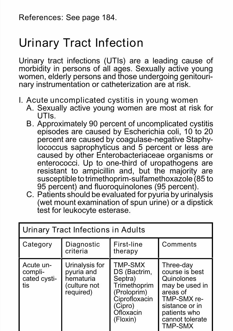
Urinary tract infections (UTIs) are a leading cause of morbidity in persons of all ages. Sexually active young
women, elderly persons and those undergoing genitouri-nary instrumentation or catheterization are at risk.
I. Acute uncomplicated cystitis in young women A. Sexually active young women are most at risk for
UTIs.B. Approximately 90 percent of uncomplicated cystitis
episodes are caused by Escherichia coli, 10 to 20percent are caused by coagulase-negative Staphy-
lococcus saprophyticus and 5 percent or less arecaused by other Enterobacteriaceae organisms or enterococci. Up to one-third of uropathogens areresistant to ampicillin and, but the majority aresusceptible to trimethoprim-sulfamethoxazole (85 to
95 percent) and fluoroquinolones (95 percent).C. Patients should be evaluated for pyuria by urinalysis(wet mount examination of spun urine) or a dipsticktest for leukocyte esterase.
Urinary Tract Infections in Adults
8/20/2019 Gynecology and Obstetricsvvvvvvvvvvvvvvvvvvvvvvvvvvvvvvvvvvvvvvvvv
B. Women who have more than three UTI recurrenceswithin one year can be managed using one of threepreventive strategies.1. Acute self-treatment with a three-day course of
standard therapy.2. Postcoital prophylaxis with one-half of a
3. Continuous daily prophylaxis for six months withtrimethoprim-sulfamethoxazole, one-half tabletper day (40/200 mg); nitrofurantoin, 50 to 100mg per day; norfloxacin (Noroxin), 200 mg per
day; cephalexin (Keflex), 250 mg per day; or trimethoprim (Proloprim), 100 mg per day.III. Complicated UTI
A. A complicated UTI is one that occurs because of enlargement of the prostate gland, blockages, or
the presence of resistant bacteria.B. Accurate urine culture and susceptibility are neces-
sary. Treatment consists of an oral fluoroquinolone.In patients who require hospitalization, parenteraladministration of ceftazidime (Fortaz) or cefoperazone (Cefobid), cefepime (Maxipime),aztreonam (Azactam), imipenem-cilastatin(Primaxin) or the combination of anantipseudomonal penicillin (ticarcillin [Ticar],
mezlocillin [Mezlin], piperacillin [Pipracil]) with anaminoglycoside.
B Diagnosis is confirmed by visualization of burrows
8/20/2019 Gynecology and Obstetricsvvvvvvvvvvvvvvvvvvvvvvvvvvvvvvvvvvvvvvvvv
B. Diagnosis is confirmed by visualization of burrowsand observation of parasites, eggs, larvae, or redfecal compactions under microscopy.
C. Treatment. Permethrin 5% cream (Elimite) ismassaged in from the neck down and remove bywashing after 8 hours.
References: See page 184.
Sexually Transmissible Infections
Approximately 12 million patients are diagnosed with asexually transmissible infection (STI) annually in theUnited States. Sequella of STIs include infertility, chronicpelvic pain, ectopic pregnancy, and other adverse preg-nancy outcomes.
Diagnosis and Treatment of Bacterial SexuallyTransmissible Infections
Organ-ism Diagnos-tic Meth-ods
RecommendedTreatment Alternative
Chlam
ydiatracho-
Direct flu-
orescentantibody,
Doxycycline 100
mg PO 2 times aday for 7 days or
Ofloxacin (Floxin)
300 mg PO 2 timesa day for 7 days
8/20/2019 Gynecology and Obstetricsvvvvvvvvvvvvvvvvvvvvvvvvvvvvvvvvvvvvvvvvv
tients have perihepatitis (Fitz-Hugh Curtis syn-drome).
B. Purulent endocervical discharge and/or acute
cervical motion and adnexal tenderness bybimanual examination is strongly suggestive of PID. Rectovaginal examination should reveal theuterine adnexal tenderness.
III. Diagnosis A. Diagnostic criteria and guidelines. The index of
suspicion for the clinical diagnosis of PID should behigh, especially in adolescent women.
B. The CDC has recommended minimum criteria
required for empiric treatment of PID. These major determinants include lower abdominal tenderness,adnexal tenderness, and cervical motion tender-ness. Minor determinants (ie, signs that mayincrease the suspicion of PID) include:
with or without metronidazole (Flagyl, 500 mgtwice daily) for 14 days. An alternative is aninitial single dose of ceftriaxone (Rocephin, 250
mg IM), cefoxitin (Mefoxin, 2 g IM plusprobenecid 1 g orally), or another parenteralthird-generation cephalosporin, followed bydoxycycline (100 mg orally twice daily) with or without metronidazole for 14 days. Quinolonesare not recommended to treat gonorrhea ac-quired in California or Hawaii. If the patient mayhave acquired the disease in Asia, Hawaii, or California, cefixime or ceftriaxone should be
used.2. Another alternative is azithromycin (Zithromax,1 g PO for Chlamydia coverage) andamoxicillin-clavulanate (Amoxicillin, 875 mgPO) once by directly observed therapy, followed
by amoxicillin-clavulanate (Amoxicillin, 875 mgPO BID) for 7 to 10 days.
C. Inpatient therapy1. For inpatient treatment, the CDC suggests
either of the following regimens:a. Cefotetan (Cefotan), 2 g IV Q12h, or
cefoxitin (Mefoxin, 2 g IV Q6h) plusdoxycycline (100 mg IV or PO Q12h)
b. Clindamycin (Cleocin), 900 mg IV Q8h,
plus gentamicin (1-1.5 mg/kg IV q8h)2. Alternative regimens:
III Diagnostic studies
8/20/2019 Gynecology and Obstetricsvvvvvvvvvvvvvvvvvvvvvvvvvvvvvvvvvvvvvvvvv
III. Diagnostic studies A. Vaginal pH. Measurement of vaginal pH should
always be determined. The pH of the normal vaginal
secretions is 4.0 to 4.5. A pH above 4.5 suggestsbacterial vaginosis or trichomoniasis (pH 5 to 6),and helps to exclude candida vulvovaginitis (pH 4 to4.5).
B. Saline microscopy should look for candidal buds or hyphae, motile trichomonads, epithelial cells stud-ded with adherent coccobacilli (clue cells), andpolymorphonuclear cells (PMNs). The addition of 10% potassium hydroxide to the wet mount is
helpful in diagnosing candida vaginitis. Culture for candida and trichomonas may be useful if micros-copy is negative.
C. Cervical culture. A diagnosis of cervicitis, typicallydue to Neisseria gonorrhoeae or Chlamydia
trachomatis, must always be considered in womenwith purulent vaginal discharge. The presence of high-risk behavior or any sexually transmitteddisease requires screening for HIV, hepatitis B, andother STDs.
Clinical Manifestations of Vaginitis
Candidal Vagi-
nitis
Nonmalodorous, thick, white, "cottagecheese-like" discharge that adheres to vagi-nal walls
5 Topical vaginal therapy with 2% clindamycin
8/20/2019 Gynecology and Obstetricsvvvvvvvvvvvvvvvvvvvvvvvvvvvvvvvvvvvvvvvvv
5. Topical vaginal therapy with 2% clindamycincream (5 g once daily for 7 days) appears to beless effective than the metronidazole regimens but
is a reasonable choice. Pseudomembranouscolitis has been reported with topical clindamycin.Clindamycin cream should not be used withcondoms, which may be weakened.
G. Relapse1. Approximately 30% of patients have a recurrence
within three months. Recurrence usually reflectsa failure to eradicate the offending organisms.Management of symptomatic relapse includes
prolonged therapy for 10 to 14 days.2. Most women with a history of recurrent infectionbenefit from suppressive therapy withmetronidazole gel 0.75% for 10 days, followed bytwice-weekly applications for three to six months.
V. Candida vulvovaginitis A. Incidence. Candida vulvovaginitis accounts for one-
third of vaginitis. Up to 75% of women report havinghad at least one episode of candidiasis. The condi-tion is rare before menarche. It is less common inpostmenopausal women, unless they are takingestrogen replacement therapy.
B. Microbiology and risk factors. Candida albicans isresponsible for 80-92% of vulvovaginal candidiasis.
Sporadic attacks of vulvovaginal candidiasis usuallyoccur without an identifiable precipitating factor.
*S it i b d if i fl ti i d i tl
8/20/2019 Gynecology and Obstetricsvvvvvvvvvvvvvvvvvvvvvvvvvvvvvvvvvvvvvvvvv
*Suppositories can be used if inflammation is predominantlyvaginal; creams if vulvar; a combination if both. Cream-suppository combination packs available: clotrimazole
(Gyne-Lotrimin, Mycelex); miconazole (Monistat, M-Zole). If diagnosis is in doubt, consider oral therapy to avoid amelio-ration of symptoms with use of creams. Use 1-day or 3-dayregimen if compliance is an issue. Miconazole nitrate may beused during pregnancy.
**Nonprescription formulation. If nonprescription therapiesfail, use terconazole 0.4% cream or 80-mg suppositories atbedtime for 7 days.
4. Complicated infections. Factors that predisposeto complicated infection include uncontrolleddiabetes, immunosuppression, and a history of recurrent vulvovaginal candidiasis. Women withsevere inflammation or complicated infectionrequire seven to 14 days of topical therapy or twodoses of oral therapy 72 hours apart.
Management options for complicated or recur-rent yeast vaginitis
Extend any 7-day regimen to 10 to 14 daysEliminate use of nylon or tight-fitting clothing
Invasive cervical carcinoma is the third most commoncancer in the United States. The International Federationof Gynecology and Obstetrics (FIGO) recently revised itsstaging criteria. Survival rates for women with cervical
cancer improve when radiotherapy is combined withcisplatin-based chemotherapy.
I. Clinical evaluation A. Human papillomavirus is the most important factor
contributing to the development of cervicalintraepithelial neoplasia and cervical cancer. Other epidemiologic risk factors associated with cervicalintraepithelial neoplasia and cervical cancer include
history of sexual intercourse at an early age, multi-ple sexual partners, sexually transmitted diseases(including chlamydia), and smoking. Additional riskfactors include a male partner or partners who havehad multiple sexual partners; previous history of
squamous dysplasias of the cervix, vagina, or vulva;and immunosuppression
8/20/2019 Gynecology and Obstetricsvvvvvvvvvvvvvvvvvvvvvvvvvvvvvvvvvvvvvvvvv
Stage II Cervical carcinoma invades beyond the uterus,but not to the pelvic wall or to the lower third of the
vaginaIla No obvious parametrial involvement
IIb Obvious parametrial involvement
Stage III The carcinoma has extended to the pelvic wall. Onrectal examination, there is no cancer-free space betweenthe tumor and the pelvic wall. The tumor involves the lower third of the vagina. All cases with hydronephrosis or nonfunctioning kidney are included, unless they are known
to be due to other causes.IIIa Tumor involves lower third of the vagina, with no
extension to the pelvic wallIIIb Extension to the pelvic wall or hydronephrosis or
nonfunctioning kidney
Stage IV The carcinoma has extended beyond the truepelvis, or has involved (biopsy proved) the mu-cosa of the bladder or rectum. Bullous edema, assuch, does not permit a case to be allotted to
Stage IV.IVa Spread of the growth to adjacent organs (bladder or rectum or both)
IVb Spread to distant organs
Guidelines for Clinical Staging of Invasive Cervi-
VII. Treatment of early-stage (Ib-lla) carcinoma
8/20/2019 Gynecology and Obstetricsvvvvvvvvvvvvvvvvvvvvvvvvvvvvvvvvvvvvvvvvv
y g ( ) A. Both treatment strategies for stage Ib and early-
stage IIa invasive carcinoma include 1) a primary
surgical approach with radical hysterectomy andpelvic lymphadenectomy or 2) primary radiationtherapy with external beam radiation and either high-dose-rate or low-dose-rate brachytherapy. The5-year survival rate is 87-92% using either ap-proach.
B. Radical surgery leaves the vagina in more func-tional condition, while radiation therapy results in areduction in length, caliber, and lubrication of the
vagina. In premenopausal women, ovarian functioncan be preserved with surgery. The surgical ap-proach also provides the opportunity for pelvic andabdominal exploration and provides better clinicaland pathologic information with which to individual-
ize treatment.VIII. Adjuvant therapy following primary surgery in
early-stage carcinoma A. Patients with histologically documented
extracervical disease (pelvic nodal involvement,positive margins, or parametrial extension) aretreated with concurrent pelvic radiation therapy andcisplatin-based chemotherapy. The use of com-bined adjuvant chemotherapy and radiation therapy
in these high-risk patients following primary surgerysignificantly improves relapse-free survival and
II. Clinical evaluation
8/20/2019 Gynecology and Obstetricsvvvvvvvvvvvvvvvvvvvvvvvvvvvvvvvvvvvvvvvvv
A. Ninety percent of patients with endometrial cancer have abnormal vaginal bleeding, usually presenting
as menometrorrhagia in a perimenopausal womanor menstrual-like bleeding in a woman past meno-pause. Perimenopausal women relate a history of intermenstrual bleeding, excessive bleeding lastinglonger than seven days or an interval of less than21 days between menses. Heavy, prolonged bleed-ing in patients known to be at risk for anovulatorycycles should prompt histologic evaluation of theendometrium. The size, contour, mobility and
position of the uterus should be noted.B. Patients who report abnormal vaginal bleeding andhave risk factors for endometrial cancer shouldhave histologic evaluation of the endometrium.Premenopausal patients with amenorrhea for more
than six to 12 months should be offeredendometrial sampling, especially if they have riskfactors associated with excessive estrogen expo-sure. Postmenopausal women with vaginal bleedingwho either are not on hormonal replacement ther-apy or have been on therapy longer than sixmonths should be evaluated by endometrial sam-pling.
C. Endometrial sampling
1. In-office sampling of the endometrial lining maybe accomplished with a Novak or Kevorkian
B. For most patients whose cancers have progressed
8/20/2019 Gynecology and Obstetricsvvvvvvvvvvvvvvvvvvvvvvvvvvvvvvvvvvvvvvvvv
p p gbeyond stage IB grade 2, postoperative radiationtherapy is recommended. Because tumor response
to cytotoxic chemotherapy has been poor, chemo-therapy is used only for palliation.
C. Endometrial hyperplasia with atypia should betreated with hysterectomy except in extraordinarycases. Progestin treatment is a possibility in womenyounger than 40 years of age who refuse hysterec-tomy or who wish to retain their childbearing poten-tial, but an endometrial biopsy should be performedevery three months. Treatment of atypical hyperpla-
sia and well-differentiated endometrial cancer withprogestins in women younger than 40 years of ageresults in complete regression of disease in 94percent and 75 percent, respectively.
D. Patients found to have hyperplasia without atypia
should be treated with progestins and have anendometrial biopsy every three to six months.IV. Serous and clear cell adenocarcinomas
A. These cancers are considered in a separate cate-gory from endometrioid adenocarcinomas. Theyhave a worse prognosis overall. Patients withserious carcinomas have a poorer survival. The 3year survival is 40% for stage I disease.
B. Serous and clear cell carcinomas are staged like
ovarian cancer. A total abdominal hysterectomy andbilateral salpingo-oophorectomy, lymph node
nancy has a sensitivity of 62 to 100 percent and a
8/20/2019 Gynecology and Obstetricsvvvvvvvvvvvvvvvvvvvvvvvvvvvvvvvvvvvvvvvvv
C. It is reasonable to pursue a period of observation
in a premenopausal woman with an adnexal massif the mass is not clinically suspicious onultrasonography. Adnexal masses that are mobile,purely cystic, unilateral, less than 8 to 10 cm indiameter, and have smooth internal and externalcontours by ultrasound are highly unlikely to bemalignant and can be followed for two months; themajority of physiologic cysts will regress during thistime.
D. Exploration is indicated if there is no resolutionwithin two months. However, women who havesolid, fixed, irregularly shaped, or large massesshould undergo surgery. A mass that increases insize or does not regress must be presumed to be
neoplastic and should be removed surgically.E. The threshold for surgical intervention is lower inpostmenopausal women; those with cysts greater than 3 cm should undergo exploratory surgery,laparotomy, or laparoscopy.
F. Tumor markers. CA 125: The preoperative evalu-ation of a woman with suspected ovarian cancer should include measurement of the CA 125 con-centration. The serum CA 125 (normal <35 U/mL)
is elevated (>65 U/mL) in 80 percent of womenwith epithelial ovarian cancer. It is also increased
St D fi iti
8/20/2019 Gynecology and Obstetricsvvvvvvvvvvvvvvvvvvvvvvvvvvvvvvvvvvvvvvvvv
IV Growth involves one or both ovaries withdistant metastases; if pleural effusion ispresent, there must be positive cytology
findings to assign a case to stage IV;parenchymal liver metastasis equals stageIV
B. Procedure
1. The staging procedure is usually approachedthrough a laparotomy incision. Any free fluid inthe cul-de-sac is submitted for cytologic evalua-tion. Washings of the peritoneal cavity are ob-
tained by instilling and removing 50 to 100 mL of saline. The affected adnexa should be removedintact and a frozen section obtained to determineor confirm the diagnosis. Thorough surgicalstaging should be carried out in the absence of
obvious stage IV disease. Preservation of theuterus and a normal appearing contralateraladnexa is an option in women desirous of main-taining future fertility.
2. All intraperitoneal surfaces should be carefully
inspected and suspicious areas or adhesionsshould be biopsied If there is no evidence of
Ri k F t f B t C
8/20/2019 Gynecology and Obstetricsvvvvvvvvvvvvvvvvvvvvvvvvvvvvvvvvvvvvvvvvv
Other Risk FactorsLate menopauseObesityWeight gainIncreased intra-abdominal
fat (android body habitus)Lack of regular exerciseElevated serum estradiolElevated free testosteronelevels A previous premalignantbreast biopsyRadial scars in benignbreast biopsies
A history of breast cancer Exposure to ionizing radia-tionHigher bone mineral den-
More than 50% of women in family have breast cancer Breast cancer present in more than I generationMultiple occurrences of breast cancer (>3) in close relativesOnset at less than age 45 yearsHistory of bilateral breast cancer
High rate of co-existing ovarian cancer BRCA1 gene mutation
B. Nulliparity and increased age at first pregnancy areassociated with an increased risk for breast can-cer. Nulliparity alone accounts for 16% of new
Symptoms of nipple discharge, pain, skin changes,h
8/20/2019 Gynecology and Obstetricsvvvvvvvvvvvvvvvvvvvvvvvvvvvvvvvvvvvvvvvvv
or rashes may occur.B. On physical examination, the breast mass should
be palpated for size, position, adherence of thetumor to the skin or chest wall, density, fluctuance,and tenderness. In addition, both breasts andaxillae should be examined for other tumors andany lymph nodes. A search for supraclavicular lymph nodes should also be conducted.
C. Any evidence of skin changes, ulceration, peaud'orange (thickening of skin to resemble an orangeskin), or lymphedema is suspicious for locally
advanced cancer.D. Immediate mammography should be obtained. Awhite blood count, hematocrit, and erythrocytesedimentation rate may be needed if cancer isfound.
IV. Diagnosis A. The definitive diagnosis is made by pathological
evaluation of tissue.B. A combination of clinical breast examination,
mammography, and fine-needle aspiration and
biopsy may be sufficient to make a diagnosis. If allstudies are "benign," there is a greater than 99%chance that a benign breast lesion is present.
C. Open biopsy in the operating room or wire-localiza-
tion of a suspicious lesion noted on mammographymay be necessary if fine-needle aspiration and
with aggressive biological behavior of the cancer d li i l t
8/20/2019 Gynecology and Obstetricsvvvvvvvvvvvvvvvvvvvvvvvvvvvvvvvvvvvvvvvvv
and a poor clinical outcome.C. The staging of breast cancer dictates not only the
prognosis but also directs treatment modalityrecommendations. The prognosis for women isbased on their age, tumor type, initial tumor size,presence of nodes and staging, and hormone-re-ceptor status. The overall 10-year survival ratesfor the more common breast cancer stages aregreater than 90% for stage 0, greater than 75% for stage I, greater than 50% for stage IIA, and ap-proximately 50% for stage IIB.
VI. Treatment of breast cancer A. Treatment choices for ductal carcinoma in situ, astage 0 cancer, include 1) mastectomy, 2)lumpectomy followed by radiation therapy, or 3)lumpectomy followed by radiation therapy and
then tamoxifen if the tumor is estrogen-receptor test positive.B. Surgical Treatment
1. Several long-term studies show that conserva-tive therapy and radiation result in at least asgood a prognosis as radical mastectomy. Skin-sparing mastectomy involves removing all thebreast tissue, the nipple, and the areolar com-plex. The remainder of the surface skin tissue
remains intact. Reconstruction is then com-pleted with a natural-appearing breast. This
Obstetrics
8/20/2019 Gynecology and Obstetricsvvvvvvvvvvvvvvvvvvvvvvvvvvvvvvvvvvvvvvvvv
A. Diagnosis of pregnancy1. Amenorrhea is usually the first sign of concep-
tion. Other symptoms include breast fullnessand tenderness, skin changes, nausea, vomit-ing, urinary frequency, and fatigue.
2. Pregnancy tests. Urine pregnancy tests maybe positive within days of the first missed men-strual period. Serum beta human chorionicgonadotropin (HCG) is accurate up to a few
days after implantation.3. Fetal heart tones can be detected as early as11-12 weeks from the last menstrual period(LMP) by Doppler. The normal fetal heart rate is120-160 beats per minute.
4. Fetal movements ("quickening") are first felt bythe patient at 17-19 weeks.5. Ultrasound will visualize a gestational sac at 5-
6 weeks and a fetal pole with movement andcardiac activity by 7-8 weeks. Ultrasound can
estimate fetal age accurately if completed be-f 24 k
Initial Prenatal Assessment of past Obstetrical
8/20/2019 Gynecology and Obstetricsvvvvvvvvvvvvvvvvvvvvvvvvvvvvvvvvvvvvvvvvv
activity and heart rate), listeria precautions,toxoplasmosis precautions (eg, hand washing,
eating habits, cat care) should be discussed.B. Abstinence from alcohol, cigarettes, illicit drugs
should be assessed. Information on the safety of commonly used nonprescription drugs, signs andsymptoms to be reported should be discussed, asappropriate for gestational age (eg, vaginal bleed-ing, ruptured membranes, contractions, decreasedfetal activity).
C. Headache and backache. Acetaminophen
(Tylenol) 325-650 mg every 3-4 hours is effective. Aspirin is contraindicated.D. Nausea and vomiting. First-trimester morning
sickness may be relieved by eating frequent, smallmeals, getting out of bed slowly after eating a few
crackers, and by avoiding spicy or greasy foods.Promethazine (Phenergan) 12.5-50 mg PO q4-6hprn or diphenhydramine (Benadryl) 25-50 mg tid-qid is useful.
E. Constipation. A high-fiber diet with psyllium
(Metamucil), increased fluid intake, and regular exercise should be advised. Docusate (Colace)100 mg bid may provide relief.
IV. Nutri tion, vitamins, and weight gain A. All pregnant women should be encouraged to eat
a well-balanced diet. Folic acid is recommended in
than 140 mg/dL, a 3-hour glucose tolerance test isnecessar
8/20/2019 Gynecology and Obstetricsvvvvvvvvvvvvvvvvvvvvvvvvvvvvvvvvvvvvvvvvv
should be repeated in the third trimester in anywoman at high risk for acquiring these infections;all women under age 25 years should be retestedfor Chlamydia trachomatis late in pregnancy.
G. Screening for group B streptococcus coloniza-tion at 35-37 weeks . All pregnant women should
8. Review of systems. Severe headaches,scotomas, hand and facial edema, or epigastricpain (preeclampsia) should be sought. Dysuria,
8/20/2019 Gynecology and Obstetricsvvvvvvvvvvvvvvvvvvvvvvvvvvvvvvvvvvvvvvvvv
pain (preeclampsia) should be sought. Dysuria,urinary frequency or flank pain may indicatecystitis or pyelonephritis.
C. Obstetrical history. Past pregnancies, durationsand outcomes, preterm deliveries, operative deliver-ies, prolonged labors, pregnancy-induced hyperten-sion should be assessed.
D. Past medical history of asthma, hypertension, or renal disease should be sought.
II. Physical examination
A. Vital signs are assessed.B. Head. Funduscopy should seek hemorrhages or exudates, which may suggest diabetes or hyperten-sion. Facial, hand and ankle edema suggestpreeclampsia.
C. Chest. Auscultation of the lungs for wheezes andcrackles may indicate asthma or heart failure.
D. Uterine Size. Until the middle of the third trimester,the distance in centimeters from the pubicsymphysis to the uterine fundus should correlatewith the gestational age in weeks. Toward term, themeasurement becomes progressively less reliablebecause of engagement of the presenting part.
E. Estimation of fetal weight is completed by palpa-
tion of the gravid uterus.F. Leopold's maneuvers are used to determine theposition of the fetus.1. The first maneuver determines which fetal pole
occupies the uterine fundus. The breech moveswith the fetal body. The vertex is rounder andharder, feels more globular than the breech, and
can be moved separately from the fetal body.2. Second maneuver. The lateral aspects of theuterus are palpated to determine on which sidethe fetal back or fetal extremities (the smallparts) are located.
3. Third maneuver. The presenting part is movedfrom side to side. If movement is difficult, en-
gagement of the presenting part has occurred.4. Fourth maneuver. With the fetus presenting by
test for syphilis, rubella antibody titer, urinalysis,culture, Pap smear, cervical cultures for gonor-rhea and Chlamydia, and hepatitis B surface
8/20/2019 Gynecology and Obstetricsvvvvvvvvvvvvvvvvvvvvvvvvvvvvvvvvvvvvvvvvv
2. During labor, the CBC, urinalysis and RPR arerepeated. The HBSAG is repeated for high-riskpatients. A clot of blood is placed on hold.
J. Fetal heart rate. The baseline heart rate, variability,accelerations, and decelerations are recorded.
III. Normal labor A. Labor is characterized by uterine contractions of
sufficient frequency, intensity, and duration to resultin effacement and dilatation of the cervix.
B. The first stage of labor starts with the onset of regular contractions and ends with complete di-latation (10 cm). This stage is further subdividedinto the latent and an active phases.1. The latent phase starts with the onset of regular
uterine contractions and is characterized by slowcervical dilatation to 4 cm. The latent phase is
variable in length.2. The active phase follows and is characterized by
more rapid dilatation to 10 cm. During the activephase of labor, the average rate of cervicaldilatation is 1.5 cm/hour in the multipara and 1.2cm/hour in the nullipara.
C. The second stage of labor begins with complete
dilatation of the cervix and ends with delivery of theinfant. It is characterized by voluntary and involun-tary pushing. The average second stage of labor isone-half hour in a multipara and 1 hour in theprimipara.
D. The third stage of labor begins with the delivery of the infant and ends with the delivery of the placenta.
E. Intravenous fluids. IV fluid during labor is usuallyRinger's lactate or 0.45% normal saline with 5%dextrose. Intravenous fluid infused rapidly or givenas a bolus should be dextrose-free because mater-nal hyperglycemia can occur.
F. Activity. Patients in the latent phase of labor areusually allowed to walk.
G. Narcotic and analgesic drugs1. Nalbuphine (Nubain) 5 to 10 mg SC or IV q2-3h.
IV Q4h). In women with non-immediate-typepenicillin allergy cefazolin (Ancef 2 g initial
8/20/2019 Gynecology and Obstetricsvvvvvvvvvvvvvvvvvvvvvvvvvvvvvvvvvvvvvvvvv
penicillin-allergy, cefazolin (Ancef, 2 g initialdose, then 1 g Q8h) is recommended.
Clindamycin (900 mg IV Q8h) or erythromycin(500 mg IV Q6h) are recommended for patientsat high risk for anaphylaxis to penicillins as longas their GBS isolate is documented to be suscep-tible to both clindamycin and erythromycin.
IV. Normal spontaneous vaginal delivery A. Preparation. As the multiparous patient ap-
proaches complete dilatation or as the nulliparouspatient begins to crown the fetal scalp, preparations
are made for delivery.B. Maternal position. The mother is usually placed inthe dorsal lithotomy position with left lateral tilt.
C. Delivery of a fetus in an occiput anterior posi-tion
1. Delivery of the heada. The fetal head is delivered by extension asthe flexed head passes through the vaginalintroitus.
b. Once the fetal head has been delivered,
external rotation to the occiput transverseposition occurs.
c. The oropharynx and nose of the fetus aresuctioned with the bulb syringe. A finger ispassed into the vagina along the fetal neck tocheck for a nuchal cord. If one is present, it is
Delivery Note
8/20/2019 Gynecology and Obstetricsvvvvvvvvvvvvvvvvvvvvvvvvvvvvvvvvvvvvvvvvv
5. Note lacerations of cervix, vagina, rectum, perineum.6. Estimated blood loss:7. Disposition: Mother to recovery room in stable condition. Infant to nursery in stable
condition.
Routine Postpartum Orders
Transfer: To recovery room, then postpartum ward when stable.Vitals: Check vitals, bleeding, fundus q15min x 1 hr or until stable, then q4h. Activi ty: Ambulate in 2 hours if stable
Nursing Orders: If unable to void, straight catheterize; sitz baths prn with 1:1000 Beta-dine prn, ice pack to perineum prn, record urine output.Diet: Regular IV Fluids: D5LR at 125 cc/h. Discontinue when stable and taking PO diet.Medications:
Oxytocin (Pitocin) 20 units in 1 L D5LR at 100 drops/minute or 10 U IM.FeS04 325 mg PO bid-tid.
Symptomatic Medications: Acetaminophen/codeine (Tylenol #3) 1-2 tab PO q3-4h prn OROxycodone/acetaminophen (Percocet) 1 tab q6h prn pain.Milk of magnesia 30 mL PO q6h prn constipation.Docusate Sodium (Colace) 100 mg PO bid.Dulcolax suppository PR prn constipation. A and D cream or Lanolin prn if breast feeding.Breast binder or tight brazier and ice packs prn if not to breast feed.
Labs: Hemoglobin/hematocrit in AM. Give rubella vaccine if titer <1:10.
Active Management of Labor
The active management of labor refers to active control
fails to descend during the second stage of labor.Three possible causes for failure to progress are
8/20/2019 Gynecology and Obstetricsvvvvvvvvvvvvvvvvvvvvvvvvvvvvvvvvvvvvvvvvv
E. Inefficient uterine action is the most common causeof dystocia in the nulliparous gravida, especiallyearly in labor. Secondary arrest of labor after previ-ously satisfactory progress may be due to anocciput-posterior position or cephalopelvic dispro-
portion. It is often difficult for the clinician to differen-tiate among these entities, thus oxytocin is adminis-tered in all cases of failure to progress (unless acontraindication exists).
F. In the first stage, progressive cervical dilatation of at
least 1 cm per hour should occur within one hour of establishing efficient uterine contractions (five toseven contractions within 15 minutes) with oxytocin.The second stage is considered prolonged if itextends longer than two hours in women without
epidural anesthesia and longer than three hours inwomen with epidural anesthesia despite adequatecontractions and oxytocin augmentation.
References: See page 184.
B. External fetal monitoring. The FHR is measuredby focusing an ultrasound beam on the fetal heart
8/20/2019 Gynecology and Obstetricsvvvvvvvvvvvvvvvvvvvvvvvvvvvvvvvvvvvvvvvvv
by focusing an ultrasound beam on the fetal heart.The fetal monitor interprets Doppler signals.
C. Internal fetal monitoring of FHR is an invasiveprocedure. A spiral electrode is insertedtranscervically into the fetal scalp. The internalelectrode detects the fetal (ECG) and calculates thefetal heart rate based upon the interval between Rwaves. This signal provides accurate measurementof beat-to-beat and baseline variability.
D. Biophysical profile. The biophysical profile (BPP)consists of electronic fetal heart rate evaluation
combined with sonographically assessed fetalbreathing movements, motor movement, gross fetaltone, and amniotic fluid volume.
II. Fetal heart rate patterns A. The fetal heart rate pattern recorded by an elec-
tronic fetal monitor is categorized as reassuring or nonreassuring.B. Reassuring fetal heart rate patterns
1. A baseline fetal heart rate of 120 to 160 bpm2. Absence of FHR decelerations
3. Age appropriate FHR accelerations4. Normal FHR variability (5 to 25 bpm).
C. Early decelerations (ie, shallow symmetricaldecelerations in which the nadir of the decelerationoccurs simultaneously with the peak of the contrac-tion) and mild bradycardia of 100 to 119 bpm are
Fetal scalp sampling is recommended to further evaluate positive test results
8/20/2019 Gynecology and Obstetricsvvvvvvvvvvvvvvvvvvvvvvvvvvvvvvvvvvvvvvvvv
evaluate positive test results.2. Fetal scalp blood sampling. Capillary blood
collected from the fetal scalp typically has a pHlower than arterial blood. A pH of 7.20 wasinitially thought to represent the critical value for identifying serious fetal stress and an increase inthe incidence of low Apgar scores. The degree of technical skill required prohibits widespread useof this modality.
IV. Management of nonreassuring FHR patternsduring labor
1. Determine the cause of the abnormality (eg, cordprolapse, maternal medication, abruption pla-centa)
2. Attempt to correct the problem or initiate mea-sures to improve fetal oxygenation (eg, change
maternal position, administer oxygen and intrave-nous fluids, consider amnioinfusion or tocolysis)3. If the nonreassuring pattern does not resolve
within a few minutes, perform ancillary tests todetermine the fetal condition
B. The presence of accelerations almost always as-sures the absence of fetal acidosis. Therefore, if such accelerations are not observed, they should beelicited by manual or vibroacoustic stimulation.
C. Nonstress test1 A NST is performed using an electronic fetal
8/20/2019 Gynecology and Obstetricsvvvvvvvvvvvvvvvvvvvvvvvvvvvvvvvvvvvvvvvvv
1. A NST is performed using an electronic fetalmonitor. Testing is generally begun at 32 to 34
weeks. Testing is performed at daily to weeklyintervals as long as the indication for testingpersists.
2. The test is reactive if there are two or more fetalheart rate accelerations of 15 bpm above thebaseline rate lasting for 15 seconds in a 20minute period. A nonreactive NST does notshow such accelerations over a 40 minuteperiod. Nonreactivity may be related to fetal
immaturity, a sleep cycle, drugs, fetal anomalies,or fetal hypoxemia.3. If the NST is nonreactive, it is considered
nonreassuring and further evaluation or deliveryof the fetus is indicated. At term, delivery rather
than further evaluation is usually warranted. Anonreassuring NST preterm usually should beassessed with ancillary tests, since the falsepositive rate of an isolated NST may be 50 to 60percent.
D. Fetal movement assessment (“ kick counts” )1. A diminution in the maternal perception of fetal
movement often but not invariably precedes fetaldeath, in some cases by several days.
2. The woman lies on her side and counts distinctfetal movements. Perception of 10 distinct move-
Components of the Biophysical Profi le
8/20/2019 Gynecology and Obstetricsvvvvvvvvvvvvvvvvvvvvvvvvvvvvvvvvvvvvvvvvv
at 1 min and 5 min; normal uterus, tubes, ovaries.Cord pH:
Specimens: Placenta, cord blood (type and Rh).Estimated Blood Loss: 800 cc; no blood replaced.Fluids, blood and urine output: Drains: Foley to gravity.Complications: NoneDisposition: Patient sent to recovery room in stablecondition.
B. Discontinue IV when taking adequate PO fluids.C. Discontinue Foley, and I and O catheterize prn.D. Ambulate tid with assistance; incentive spirometer
q1h while awake.E. Check hematocrit, hemoglobin, Rh, and rubellastatus.
F. Medications1. Acetaminophen/codeine (Tylenol #3) 1-2 PO q4-
Assistant: Anesthesia: EpiduralFindings At Surgery: Normal fallopian tubes bilaterallyDescription of Operative Procedure:
After assuring informed consent, the patient wastaken to the operating room and spinal anesthesiaadministered. A small, transverse, infraumbilical skinincision was made with a scalpel, and the incision wascarried down through the underlying fascia until the
peritoneum was identified and entered. The left fallopiantube was identified, brought into the incision and graspedwith a Babcock clamp. The tube was then followed out tothe fimbria. An avascular midsection of the fallopian tubewas grasped with a Babcock clamp and brought into a
knuckle. The tube was doubly ligated with an O-plainsuture and transected. The specimen was sent to pathol-ogy. Excellent hemostasis was noted, and the tube wasreturned to the abdomen. The same procedure wasperformed on the opposite fallopian tube.
The fascia was then closed with O-Vicryl in a singlelayer. The skin was closed with 3-O Vicryl in asubcuticular fashion. The patient tolerated the procedurewell. Needle and sponge counts were correct times 2.Estimated Blood Loss (EBL): <20 ccSpecimens: Segments of right and left tubes
be D negative, antepartum prophylaxis is notnecessary.
8/20/2019 Gynecology and Obstetricsvvvvvvvvvvvvvvvvvvvvvvvvvvvvvvvvvvvvvvvvv
1. D immunoglobulin is given to the D negativemother as soon after delivery as cord bloodfindings indicate that the baby is Rh positive.
2. A woman at risk who is inadvertently not given Dimmunoglobulin within 72 hours after deliveryshould still receive prophylaxis at any time upuntil two weeks after delivery. If prophylaxis isdelayed, it may not be effective.
3. A quantitative Kleihauer-Betke analysis should
be performed in situations in which significantmaternal bleeding may have occurred (eg, after maternal abdominal trauma, abruptio placentae,external cephalic version). If the quantitativedetermination is thought to be more than 30 mL,
D immune globulin should be given to the mother in multiples of one vial (300 mcg) for each 30 mLof estimated fetal whole blood in her circulation,unless the father of the baby is known to be Dnegative.
G. Abruptio placentae, placenta previa, cesareandelivery, intrauterine manipulation, or manualremoval of the placenta may cause more than 30mL of fetal-to-maternal bleeding. In these condi-tions, testing for excessive bleeding (Kleihauer-Betke test) or inadequate D immunoglobulin dos-
Drug Therapy for Nausea and Vomiting of Preg-
8/20/2019 Gynecology and Obstetricsvvvvvvvvvvvvvvvvvvvvvvvvvvvvvvvvvvvvvvvvv
2. Disorders of pregnancya. Hydatidiform mole may present with early
pregnancy bleeding, passage of grape-likevesicles, and a uterus that is enlarged inexcess of that expected from dates. An ab-sence of heart tones by Doppler after 12weeks is characteristic. Hyperemesis,preeclampsia, or hyperthyroidism may bepresent. Ultrasonography confirms the diagno-
sis.b. Ectopic pregnancy should be excluded whenfirst trimester bleeding is associated withpelvic pain. Orthostatic light-headedness,syncope or shoulder pain (from diaphragmatic
irritation) may occur.(1) Abdominal tenderness is noted, and pelvicexamination reveals cervical motion ten-derness.
(2) Serum beta-HCG is positive.
E. Laboratory tests1. Complete blood count. The CBC will not reflect
acute blood loss.2. Quantitative serum beta-HCG level may be
positive in nonviable gestations since beta-HCGmay persist in the serum for several weeks after
6. Methylergonovine (Methergine), 0.2 mg POq4h for 6 doses, is given if there is continued
8/20/2019 Gynecology and Obstetricsvvvvvvvvvvvvvvvvvvvvvvvvvvvvvvvvvvvvvvvvv
IV. Complete abortion A. A complete abortion is diagnosed when complete
passage of products of conception has occurred.The uterus is well contracted, and the cervical osmay be closed.
B. Differential diagnosis1. Incomplete abortion2. Ectopic pregnancy. Products of conception
should be examined grossly and submitted for
pathologic examination. If no fetal tissue or villiare observed grossly, ectopic pregnancy mustbe excluded by ultrasound.
C. Management of complete abortion1. Between 8 and 14 weeks, curettage is necessary
because of the high probability that the abortionwas incomplete.2. D immunoglobulin (RhoGAM) is administered to
Rh-negative, unsensitized patients.3. Beta-HCG levels are obtained weekly until zero.
Incomplete abortion is suspected if beta-HCGlevels plateau or fail to reach zero within 4weeks.
V. Missed abortion is diagnosed when products of conception are retained after the fetus has expired. If products are retained, a severe coagulopathy with
IV. Cystitis occurs in 0.3 to 1.3 percent of pregnantwomen. Bacteria are confined to the lower urinary
8/20/2019 Gynecology and Obstetricsvvvvvvvvvvvvvvvvvvvvvvvvvvvvvvvvvvvvvvvvv
A. Acute cystitis should be considered in any gravidawith frequency, urgency, dysuria, hematuria, or suprapubic pain in the absence of fever and flankpain. Urine culture with a CFU count >102/mLshould be considered positive on a midstream
urine specimen with pyuria.B. Empiric treatment regimens:
C. Each of these drugs is given for three to sevendays.D. Other regimens which have a broader spectrum of
activity include amoxicill in-clavulanate(Augmentin) 500 mg BID or 250 mg TID,
trimethoprim-sulfamethoxazole (Bactrim) 1 DSBID but not in the third trimester of pregnancy,cefpodoxime proxetil (Vantin) 100 mg BID, andcefixime (Suprax) 400 mg QD. All of these drugscan be used for three to seven days.
Fluoroquinolones should be avoided in preg-nancy.
E. Monthly urine cultures should be performedbeginning one to two weeks after completion of treatment.
V. Pyelonephritis complicates 1 to 2 percent of all
Trauma During Pregnancy
8/20/2019 Gynecology and Obstetricsvvvvvvvvvvvvvvvvvvvvvvvvvvvvvvvvvvvvvvvvv
Trauma is the leading cause of nonobstetric death inwomen of reproductive age. Six percent of all pregnan-cies are complicated by some type of trauma.
I. Mechanism of injury
A. Blunt abdominal trauma1. Blunt abdominal trauma secondary to motor vehicle accidents is the leading cause of nonobstetric-related fetal death during preg-nancy, followed by falls and assaults. Uterine
rupture or laceration, retroperitoneal hemor-rhage, renal injury and upper abdominal injuriesmay also occur after blunt trauma.
2. Abruptio placentae occurs in 40-50% of pa-tients with major traumatic injuries and in up to5% of patients with minor injuries.
3. Clinical findings in blunt abdominal trauma.Vaginal bleeding, uterine tenderness, uterinecontractions, fetal tachycardia, late decelerations,
fetal acidosis, and fetal death.4. Detection of abruptio placentae. Beyond 20weeks of gestation, external electronic monitoringcan detect uterine contractile activity. The pres-ence of vaginal bleeding and tetanic or
hypertonic contractions is presumptive evidenceof abruptio placentae
2. A complete blood count, urinalysis (hematuria),blood type and screen (to check Rh status), and
8/20/2019 Gynecology and Obstetricsvvvvvvvvvvvvvvvvvvvvvvvvvvvvvvvvvvvvvvvvv
INR, PTT, fibrinogen and fibrin split products,should be obtained. The coagulation panel isuseful if any suspicion of abruption exists.
3. The Kleihauer-Betke (KB) testa. This test detects fetal red blood cells in the
maternal circulation. A KB stain should beobtained routinely for any pregnant traumapatient whose fetus is over 12 weeks.
b. Regardless of the patient's blood type and Rhstatus, the KB test can help determine if fetomaternal hemorrhage has occurred.
c. The KB test can also be used to determinethe amount of Rho(D) immunoglobulin(RhoGAM) required in patients who are Rh-
negative.d. A positive KB stain indicates uterine trauma,and any patient with a positive KB stainshould receive at least 24 hours of continuousuterine and fetal monitoring and a coagulation
panel.4. Ultrasonography is less sensitive for diagnosing
abruption than is the finding of uterine contrac-tions on external tocodynamometry. Absence of sonographic evidence of abruption does notcompletely exclude an abruption.
Criteria for Gestational Diabetes with ThreeHour Oral Glucose Tolerance Test
8/20/2019 Gynecology and Obstetricsvvvvvvvvvvvvvvvvvvvvvvvvvvvvvvvvvvvvvvvvv
1. Moderate caloric restriction may be useful intreating obese women (body mass index greater than 30 km/m2) with gestational diabetes. How-ever, ketosis should be avoided.
2. Caloric allotment. The recommended caloric
intake is 30 kcal per present weight in kg per dayin pregnant women who are 80 to 120 percent of ideal body weight at the start of pregnancy. 24kcal per present weight in kg per day in over-
weight pregnant women (120 to 150 percent of ideal body weight). 12 to 15 kcal per presentweight in kg per day for morbidly obese pregnantwomen (>150 percent of ideal body weight). 40kcal per present weight in kg per day in pregnant
women who are less than 80 percent of idealbody weight.3. Carbohydrate intake. Recommendations for
calorie and carbohydrate distribution are 40percent carbohydrate, 20 percent protein, and
40 percent fat.4. Calorie distribution
a. Distribution of calories should be three mealsand three snacks. In overweight women,however, the snacks are eliminated.
b. The remaining calories should be distributed
3. Macrosomia and cesarean delivery. The riskof macrosomia among women with untreatedGDM i 17 t 29 t C d li f
8/20/2019 Gynecology and Obstetricsvvvvvvvvvvvvvvvvvvvvvvvvvvvvvvvvvvvvvvvvv
the prevention of shoulder dystocia is recom-mended when the estimated fetal weight isgreater than 4.5 kg.
E. Delivery. The great majority of women with gesta-tional diabetes proceed to term and have a sponta-
neous vaginal delivery. The maternal blood glucoseconcentration should be maintained between 70and 90 mg/dL. Insulin can usually be withheldduring delivery, and an infusion of normal saline isusually sufficient to maintain normoglycemia.
F. Postpartum concerns1. Nearly all women with gestational diabetes are
normoglycemic after delivery. However, they areat risk for recurrent gestational diabetes, im-
paired glucose tolerance, and overt diabetes.One-third to two-thirds of women will havegestational diabetes in a subsequent pregnancy.Women with gestational diabetes have anincidence of type 2 diabetes in the first five years
postpartum of 47 to 50 percent.2. After delivery, blood glucose should be mea-
sured to ensure that the mother no longer hashyperglycemia. Fasting blood glucose concen-trations should be below 115 mg/dL and one-hour postprandial concentrations should be
Class Description
8/20/2019 Gynecology and Obstetricsvvvvvvvvvvvvvvvvvvvvvvvvvvvvvvvvvvvvvvvvv
supplement of ferrous sulfate (30 mg) andfolate (400 µg) is also recommended.4. Insulin regimen. Most women with type 1
diabetes require at least three injections of insulin per day.a. The average insulin requirement in pregnant
women with type 1 diabetes is 0.7 units/kg inthe first trimester, often increasing to 0.8U/kg for weeks 18 to 26, 0.9 U/kg for weeks27 to 36, and 1.0 U/kg for weeks 37 to term.
b. Women with type 2 diabetes also should betreated with insulin for blood glucose control,preferably started during the preconceptionperiod. During the first trimester, insulin
requirements are similar in women with type1 and type 2 diabetes. However, as thepregnancy proceeds into the third trimester,insulin requirements increase proportion-ately more in women with type 2 than type 1
diabetes; in one study, for example, therespective insulin doses are 1.6 and 1.2U/kg per day.
c. Administer a combination of regular insulinand intermediate-acting insulin (such asNPH insulin) The insulin is initially distrib-
1. Diabetic gravida should be seen every one totwo weeks until 32 weeks of gestation and then
kl til d li
8/20/2019 Gynecology and Obstetricsvvvvvvvvvvvvvvvvvvvvvvvvvvvvvvvvvvvvvvvvv
a. Antepartum fetal testing is recommendedfor pregnancies complicated bypregestational diabetes starting with weeklyNSTs at 32 weeks when there is suboptimalglycemic control (hemoglobin A1c values >7
percent) and by 35 weeks with good control(hemoglobin A1c <7 percent), increasing thefrequency of testing to two times per weekas indicated. In complicated patients withi n t r au te r i ne g row th res t r i c t i on ,oligohydramnios, preeclampsia, or poorlycontrolled blood sugars, testing may start at26 weeks.
b. Assessment of fetal growth. The ultra-
sound examination is repeated at 28 to 30weeks. If there is evidence of intrauterinegrowth restriction, tests of fetal well-beingare initiated. The last sonographic examina-tion is performed at 38 weeks to estimate
fetal weight.G. Labor and Delivery
1. Timing of delivery. There is little benefit incontinuing pregnancy beyond 39 weeks if thecervix is favorable. Induction can be safelydelayed until 40 weeks in women with excellent
Group B Streptococcal Infection in
8/20/2019 Gynecology and Obstetricsvvvvvvvvvvvvvvvvvvvvvvvvvvvvvvvvvvvvvvvvv
PregnancyGroup B streptococcus (GBS; Streptococcus agalactiae),a Gram positive coccus, is an important cause of infectionin neonates, causing sepsis, pneumonia, and meningitis.
GBS infection is acquired in utero or during passagethrough the vagina. Vaginal colonization with GBS duringpregnancy may lead to premature birth, and GBS is afrequent cause of maternal urinary tract infection,chorioamnionitis, postpartum endometritis, andbacteremia.
I. Clinical evaluation A. The primary risk factor for GBS infection is maternal
GBS genitourinary or gastrointestinal colonization.B. The rate of transmission from colonized mothers toinfants is approximately 50 percent. However, only1 to 2 percent of all colonized infants develop early-onset GBS disease.
C. Maternal obstetrical factors associated withneonatal GBS disease:1. Delivery at less than 37 weeks of gestation2. Premature rupture of membranes3. Rupture of membranes for 18 or more hours
before delivery
References: See page 184.
8/20/2019 Gynecology and Obstetricsvvvvvvvvvvvvvvvvvvvvvvvvvvvvvvvvvvvvvvvvv
Premature Rupture of MembranesPremature rupture of the membranes (PROM) refers torupture of membranes prior to the onset of labor or
regular uterine contractions. It can occur at term or prior to term, in which case it is designated preterm prematurerupture of the membranes (PPROM). The frequencies of term, preterm, and midtrimester PROM are 8, 1 to 3, andless than 1 percent of pregnancies, respectively.
The incidence of this disorder to be 7-12%. In pregnan-cies of less than 37 weeks of gestation, preterm birth (andits sequelae) and infection are the major concerns after PROM.
I. Pathophysiology A. Premature rupture of membranes is defined as
rupture of membranes prior to the onset of labor.B. Preterm premature rupture of membranes is
defined as rupture of membranes prior to term.C. Prolonged rupture of membranes consists of rupture of membranes for more than 24 hours.
D. The latent period is the time interval from ruptureof membranes to the onset of regular contractions
or laborE Many cases of preterm PROM are caused by
B. The patient should be evaluated for the presence of chorioamnionitis [fever (over 38°C), leukocytosis,maternal and fetal tachycardia uterine tenderness
8/20/2019 Gynecology and Obstetricsvvvvvvvvvvvvvvvvvvvvvvvvvvvvvvvvvvvvvvvvv
maternal and fetal tachycardia, uterine tenderness,
foul-smelling vaginal discharge].C. The patient should be evaluated for labor, and asterile speculum examination should assess cervi-cal change.
D. The fetus should be evaluated with heart rate
monitoring because PROM increases the risk of umbilical cord prolapse and fetal distress caused byoligohydramnios.
V. Management of premature rupture of membranes A. Management of term patients
1. At 36 weeks and beyond, management of PROMconsists of delivery. Patients in active labor should be allowed to progress.
2. Patients with chorioamnionitis, who are not in
labor, should be immediately induced withoxytocin (Pitocin).3. Patients who are not yet in active labor (in the
absence of fetal distress, meconium, or clinicalinfection) may be discharged for 48 hours, and
labor usually follows. If labor has not begunwithin a reasonable time after rupture of mem-branes, induction with oxytocin (Pitocin) is ap-propriate. Use of prostaglandin E2 is safe for cervical ripening.
B. Management of Preterm Premature Rupture of
Sample Antibiotic Regimens Used for Prophy-laxis in Women with PPROM
8/20/2019 Gynecology and Obstetricsvvvvvvvvvvvvvvvvvvvvvvvvvvvvvvvvvvvvvvvvv
Ampicillin 1 or 2 g IV every 6 hours for 24hours, then 500 mg PO every 6hours until delivery
AmpicillinGentamicin
Clindamycin
Amoxicillin plusclavulanic acid(Augmentin)
2 g IV every 6 hours for 4 dosesand90 mg IV initially, then 60 mg IVevery 8 hours for three doses and900 mg IV every 8 hours for threedoses, followed by500 mg PO TID for 7 days
Erythromycin base 333 mg PO every 8 hours untildelivery
Piperacillin 3 g IV every 6 hours for 3 days
Ampicillin-sulbactam(Unasyn)
3 g every 6 hours for 7 days
Ampicillin 2 g IV every 6 hours for 7 days
Preterm Labor, Threatened or Actual
8/20/2019 Gynecology and Obstetricsvvvvvvvvvvvvvvvvvvvvvvvvvvvvvvvvvvvvvvvvv
beta-agonist for labor inhibition. It is typically
given as a continuous intravenous infusionstarted at 2.5 to 5 µg/min; increased by 2.5to 5 µg/min every 20 to 30 minutes to amaximum of 25 µg/min, or until the contrac-tions have abated. At this point, the infusion
may be reduced by decrements of 2.5 to 5µg/min to the lowest dose that maintainsuterine quiescence.
b. Terbutaline can also be administered subcu-taneously as 0.25 mg every 20 to 30 min-utes for up to four doses or until tocolysis isachieved. It should be withheld if the mater-nal heart rate exceeds 120 beats/min. Oncelabor is inhibited, 0.25 mg can be adminis-
tered every 3 to 4 hours until the uterus isquiescent for 24 hours.C. Calcium channel blockers
1. Nifedipine is more effective than betamimeticsfor delaying delivery at least 48 hours. Dosage
is 30 mg orally, followed by 20 mg orally in 90minutes, followed by 20 mg orally every four toeight hours.
2. Maternal side effects. Nifedipine may causenausea, flushing, headache, dizziness, andpalpitations Nifedipine also decreases mean
IX. Intrapartum antibiotic prophylaxis againstgroup B streptococcus is recommended for thefollowing:
8/20/2019 Gynecology and Obstetricsvvvvvvvvvvvvvvvvvvvvvvvvvvvvvvvvvvvvvvvvv
A. Pregnant women with a positive screening cultureunless a planned Cesarean section is performed inthe absence of labor or rupture of membranes
B. Pregnant women who gave birth to a previousinfant with invasive GBS disease
C. Pregnant women with documented GBS bacteriuriaduring the current pregnancy
D. Pregnant women whose culture status is unknown(culture not performed or result not available) andwho also have delivery at <37 weeks of gestation,amniotic membrane rupture for >18 hours, or intrapartum temperature >100.4ºF (>38ºC)
E. The recommended IAP regimen is penicillin G (5million units IV initial dose, then 2.5 million units IV
Q4h). In women with non-immediate-type penicillin-allergy, cefazolin (Ancef, 2 g initial dose, then 1 gQ8h) is recommended.
References: See page 184.
Bleeding in the Second Half of Pregnancy
Bleeding in the second half of pregnancy occurs in 4% of
4. Ultrasonography has a sensitivity in detectingplacental abruption of only 15%.
D Management of placental abruption
8/20/2019 Gynecology and Obstetricsvvvvvvvvvvvvvvvvvvvvvvvvvvvvvvvvvvvvvvvvv
1. Mild placental abruptiona. If maternal stability and reassuring fetalsurveillance are assured and the fetus isimmature, close expectant observation withfetal monitoring is justified.
b. Maternal hematologic parameters are moni-tored and abnormalities corrected.
c. Tocolysis with magnesium sulfate is initiatedif the fetus is immature.
2. Moderate to severe placental abruptiona. Shock is aggressively managed.b. Coagulopathy
(1) Blood is transfused to replace blood loss.(2) Clotting factors may be replaced using
cryoprecipitate or fresh-frozen plasma.One unit of fresh-frozen plasma increasesfibrinogen by 10 mg/dL. Cryoprecipitatecontains 250 mg fibrinogen/unit; 4 gm(15-20 U) is an effective dose.
(3) Platelet transfusion is indicated if theplatelet count is less than 50,000/mcL.One unit of platelets raises the plateletcount 5000-10,000/mcL; 4 to 6 U is thesmallest useful dose.
c Oxygen should be administered and urine
I. Clinical evaluation A. Screening. Pregnant women are routinely
screened for signs and symptoms of preeclampsia
8/20/2019 Gynecology and Obstetricsvvvvvvvvvvvvvvvvvvvvvvvvvvvvvvvvvvvvvvvvv
at each prenatal visit. Women at high risk for preeclampsia should be seen in early pregnancy toassess blood pressure, establish accurate preg-nancy dating, and perform baseline laboratory tests.
B. Risk factors for preeclampsia:
1. Primigravid state.2. History of preeclampsia.3. A higher blood pressure at the initiation of
pregnancy and a large body size.4. A family history of preeclampsia is associated
with a two to fivefold increase in risk.5. Multiple pregnancy.6. Preexisting maternal hypertension.7. Pregestational diabetes.
8. Antiphospholipid antibody syndrome.9. Vascular or connective tissue disease.10. Advanced maternal age (>35 to 40 years).
C. Late pregnancy screening. Measurement of bloodpressure and urine protein at regular intervals in the
late second and third trimesters is critical for diag-nosis of preeclampsia. A rising blood pressure isusually the first sign of disease. Women shouldreport possible signs of preeclampsia, such aspersistent or severe headache, visual changes,right upper quadrant or epigastric pain sudden
H. Liver involvement may present as right upper quadrant or epigastric pain, elevated liver en-zymes and subcapsular hemorrhage or hepatic
8/20/2019 Gynecology and Obstetricsvvvvvvvvvvvvvvvvvvvvvvvvvvvvvvvvvvvvvvvvv
rupture.I. Central nervous system. Headache, blurredvision, scotomata, and, rarely, cortical blindness aremanifestations of preeclampsia; seizures in apreeclamptic woman are defined as eclampsia.
J. Fetus and placenta. The fetal consequences arefetal growth restriction and oligohydramnios.Severe or early onset preeclampsia result in thegreatest decrements in birth weight.
II. Management of preeclampsia A. The definitive treatment of preeclampsia is deliv-
ery. Delivery is recommended for women with mildpreeclampsia at or near term and for most womenwith severe preeclampsia or severe gestational
hypertension regardless of gestational age.Exceptions may be made for women remote fromterm (less than 32 to 34 weeks of gestation) whoimprove after hospitalization and do not havesignificant end-organ dysfunction or fetal deterio-
ration.B. Fetal assessment consists of daily fetal move-
ment counts and nonstress testing and/or bio-physical profiles at periodic intervals. Asonographic estimation of fetal weight should beperformed to look for growth restriction and
Indications for Delivery in Preeclampsia
8/20/2019 Gynecology and Obstetricsvvvvvvvvvvvvvvvvvvvvvvvvvvvvvvvvvvvvvvvvv
Maternal indications Gestational age greater than or equal to 38 weeks of gestationPlatelet count less than 100,000cells per mm3
Deteriorating liver functionProgressive deterioration in re-nal function Abruptio placentaePersistent severe headaches or visual changesPersistent severe epigastric
pain, nausea, or vomiting
Fetal indications Severe fetal growth restrictionNonreassuring results from fetaltesting
Oligohydramnios
III. Severe preeclampsia A. All women with severe preeclampsia should be
delivered or hospitalized for the duration of preg-nancy. Prolonged antepartum management may beconsidered in women under 32 to 34 weeks of gestation who have:1. Severe proteinuria (greater than 5 g in 24
hours)2 Mild i t t i f t l th t i ti (fifth t
2. Intravenous labetalol is both effective and safe(beginning with 20 mg intravenously followed at10- to 15-minute intervals by 40 mg then 80 mg
8/20/2019 Gynecology and Obstetricsvvvvvvvvvvvvvvvvvvvvvvvvvvvvvvvvvvvvvvvvv
up to a maximum total cumulative dose of 220mg).3. Occasionally, preeclamptic women with severe
hypertension are stabilized and not delivered. Inthese patients, oral antihypertensive therapy is
often indicated. The only oral drugs that havebeen proven to be safe in pregnant women aremethyldopa (250 mg twice daily orally, maximumdose 4 g/day), and beta-blockers, such aslabetalol (100 mg twice daily orally, maximumdose 2400 mg/day).
4. Blood pressure goal. The goal of therapy is asystolic pressure of 140 to 155 mm Hg anddiastolic pressure of 90 to 105 mm Hg.
Treatment of Severe Hypertension inPreeclampsia
The goal is a gradual reduction of blood pressure toa level below 160/105 mm Hg. Sudden and severehypotension should be avoided.
Hydralazine: 5 mg IV repeat 5 to 10 mg IV every 20minutes to maximum cumulative total of 20 mg or
D. Choice of drug1. Methyldopa and hydralazine have been most
widely used in pregnant women and their long-
8/20/2019 Gynecology and Obstetricsvvvvvvvvvvvvvvvvvvvvvvvvvvvvvvvvvvvvvvvvv
term safety for the fetus has been demonstrated. ACE inhibitors should not be continued in preg-nancy.
2. Beta-blockers are generally considered to besafe, although they may impair fetal growth when
used early in pregnancy, particularly atenolol.Labetalol is the preferred agent. Nifedipine (30to 90 mg once daily as sustained-release tablet,increase at 7 to 14 day intervals, maximum dose120 mg/day) has been used.
3. The normal fall in blood pressure during thesecond trimester may allow a reduction in drugdosage or even cessation of therapy.
4. Start treatment with either labetolol or
methyldopa. A long-acting calcium channelblocker (eg, nifedipine or amlodipine) can beadded as either second- or third-line treatment.
5. Blood pressure goal. The goal of therapy inwomen without end-organ damage is systolic
pressure between 140 and 150 mm Hg anddiastolic pressure between 90 and 100 mm Hg.However, in women with end-organ damage, theblood pressure should be below 140/90 mm Hg.
E. Other management issues1 Frequent prenatal visits for monitoring maternal
Clinical Designation of Genital Herpes SimplexVirus Infection
8/20/2019 Gynecology and Obstetricsvvvvvvvvvvvvvvvvvvvvvvvvvvvvvvvvvvvvvvvvv
Primary genital HSV infection Antibodies to both HSV-1 and HSV-2 are ab-sent at the time the patient acquires genitalHSV due to HSV-1 or HSV-2
Nonprimary first episode genital HSV infection Acquisition of genital HSV-1 with pre-existingantibodies to HSV-2 or acquisition of genital
HSV-2 with pre-existed antibodies rto HSV-1
Recurrent genital HSV infectionReactivation of genital HSV in which the HSVtype recovered from the lesion is the sametype as antibodies in the serum
III. Maternal treatment A. Primary infection. Acyclovir therapy (200 mg PO
five times per day or 400 mg PO TID for 7 to 14days) is recommended. Acyclovir is safe in preg-nancy. Acyclovir should be administered to preg-nant women experiencing a first episode of HSVduring pregnancy to reduce the duration of activelesions Suppressive therapy (400 mg PO BID) for
III. Assessment of labor abnormalities A. Labor abnormalities caused by inadequate
uterine contractil ity (powers). The minimal uterine
8/20/2019 Gynecology and Obstetricsvvvvvvvvvvvvvvvvvvvvvvvvvvvvvvvvvvvvvvvvv
contractile pattern of women in spontaneous labor consists of 3 to 5 contractions in a 10-minute period.B. Labor abnormalities caused by fetal characteris-
tics (passenger)1. Assessment of the fetus consists of estimating
fetal weight and position. Estimations of fetal size,even those obtained by ultrasonography, arefrequently inaccurate.
2. In the first stage of labor, the diagnosis of dystocia can not be made unless the active phaseof labor and adequate uterine contractile forceshave been present.
3. Fetal anomalies such as hydrocephaly,encephalocele, and soft tissue tumors may ob-
struct labor. Fetal imaging should be consideredwhen malpresentation or anomalies are sus-pected based on vaginal or abdominal examina-tion or when the presenting fetal part is persis-tently high.
C. Labor abnormalities due to the pelvic passage(passage)1. Inefficient uterine action should be corrected
before attributing dystocia to a pelvic problem.2. The bony pelvis is very rarely the factor that limits
vaginal delivery of a fetus in cephalic presenta-
Fetal Macrosomia
Excessive birth weight is associated with an increased
8/20/2019 Gynecology and Obstetricsvvvvvvvvvvvvvvvvvvvvvvvvvvvvvvvvvvvvvvvvv
Excessive birth weight is associated with an increasedrisk of maternal and neonatal injury. Macrosomia isdefined as a fetus with an estimated weight of more than4,500 grams, regardless of gestational age.
I. Diagnosis of macrosomia A. Clinical estimates of fetal weight based on Leopold'smaneuvers or fundal height measurements are ofteninaccurate.
B. Diagnosis of macrosomia requires ultrasound
evaluation; however, estimation of fetal weightbased on ultrasound is associated with a largemargin of error.
C. Maternal weight, height, previous obstetric history,fundal height, and the presence of gestational
diabetes should be evaluated.II. Factors influencing fetal weight
A. Gestational age. Post-term pregnancy is a riskfactor for macrosomia. At 42 weeks and beyond,2.5% of fetuses weigh more than 4,500 g. Ten totwenty percent of macrosomic infants are post-termfetuses.
B. Maternal weight. Heavy women have a greater riskof giving birth to excessively large infants. Fifteen to
35% of women who deliver macrosomic fetusesweigh 90 kg or more
been reported to be 13.8%, nearly seven times theprimary rate. Shoulder dystocia occurs in 5.1% of obese women. In the antepartum period, risk factors
8/20/2019 Gynecology and Obstetricsvvvvvvvvvvvvvvvvvvvvvvvvvvvvvvvvvvvvvvvvv
include gestational diabetes, excessive weight gain,short stature, macrosomia, and postterm preg-nancy. Intrapartum factors include prolongedsecond stage of labor, abnormal first stage, arrestdisorders, and instrumental (especially midforceps)
delivery. Many shoulder dystocias will occur in theabsence of any risk factors.
II. Management A. Shoulder dystocia is a medical and possibly surgical
emergency. Two assistants should be called for if not already present, as well as an anesthesiologistand pediatrician. A generous episiotomy should becut. The following sequence is suggested:1. McRoberts maneuver: The legs are removed
from the lithotomy position and flexed at thehips, with flexion of the knees against the abdo-men. Two assistants are required. This maneu-ver may be performed prophylactically in antici-pation of a difficult delivery.
2. Suprapubic pressure: An assistant is re-quested to apply pressure downward, above thesymphysis pubis. This can be done in a lateraldirection to help dislodge the anterior shoulder from behind the pubic symphysis. It can also be
performed in anticipation of a difficult delivery
dates; and, at 20 weeks, a fundal height 20 cmabove the symphysis pubis or at the umbilicus.
8/20/2019 Gynecology and Obstetricsvvvvvvvvvvvvvvvvvvvvvvvvvvvvvvvvvvvvvvvvv
B. In patients without reliable clinical data, ultrasoundis beneficial. Ultrasonography is most accurate inearly gestation. The crown-rump length becomesless accurate after 12 weeks in determining gesta-tional age because the fetus begins to curve.
III. Management of the postdates pregnancy A. A postdates patient with a favorable cervix should
receive induction of labor Only 8 2% of pregnan-cies at 42 weeks have a ripe cervix (Bishop score
Induction of Labor
8/20/2019 Gynecology and Obstetricsvvvvvvvvvvvvvvvvvvvvvvvvvvvvvvvvvvvvvvvvv
Induction of labor refers to stimulation of uterine contrac-tions prior to the onset of spontaneous labor. Between1990 and 1998, the rate of labor induction doubled from10 to 20 percent.
I. Indications for labor induction: A. Preeclampsia/eclampsia, and other hypertensive
diseasesB. Maternal diabetes mellitus
C. Prelabor rupture of membranesD. ChorioamnionitisE. Intrauterine fetal growth restriction (IUGR)F. IsoimmunizationG. In-utero fetal demise
H. Postterm pregnancyII. Absolute contraindications to labor induction:
A. Prior classical uterine incisionB. Active genital herpes infectionC. Placenta or vasa previaD. Umbilical cord prolapseE. Fetal malpresentation, such as transverse lie
II. Requirements for induction A. Prior to undertaking labor induction, assessments
of gestational age fetal size and presentationclinical pelvimetry and cervical examinationshould
Weekly membrane stripping beginning at 38weeks of gestation results in delivery within ashorter period of time (8.6 versus 15 days).
8/20/2019 Gynecology and Obstetricsvvvvvvvvvvvvvvvvvvvvvvvvvvvvvvvvvvvvvvvvv
2. Amniotomy is an effective method of labor induc-tion when performed in women with partiallydilated and effaced cervices. Caution should beexercised to ensure that the fetal vertex is well-applied to the cervix and the umbilical cord or
other fetal part is not presenting.3. Foley catheter. An uninflated Foley catheter can
be passed through an undilated cervix and theninflated. This technique is as effective as prosta-glandin E2 gel. The use of extra-amniotic salineinfusion with a balloon catheter or a double ballooncatheter (Atad ripener) also appears to be effectivefor cervical ripening.
C. Prostaglandins
1. Local administration of prostaglandins to thevagina or the endocervix is the route of choicebecause of fewer side effects and acceptableclinical response. Uncommon side effects includefever, chills, vomiting, and diarrhea.
2. Prepidil contains 0.5 mg of dinoprostone in 2.5mL of gel for intracervical administration. The dosecan be repeated in 6 to 12 hours if there is inade-quate cervical change and minimal uterine activityfollowing the first dose. The maximum cumulative
dose is 1 5 mg (ie 3 doses) within a 24-hour
bleed. The cervix and vagina should be inspected torule out lacerations. If no laceration is found but bleed-ing is still profuse, the uterus should be manually
8/20/2019 Gynecology and Obstetricsvvvvvvvvvvvvvvvvvvvvvvvvvvvvvvvvvvvvvvvvv
examined to exclude rupture.C. The placenta and uterus should be examined for retained placental fragments. Placenta accreta isusually manifest by failure of spontaneous placentalseparation.
D. Bleeding from non-genital areas (venous puncturesites) suggests coagulopathy. Laboratory tests thatconfirm coagulopathy include INR, partialthromboplastin time, platelet count, fibrinogen, fibrinsplit products, and a clot retraction test.
E. Medical management of postpartum hemorrhage1. Oxytocin (Pitocin) is usually given routinely imme-
diately after delivery to stimulate uterine firmnessand diminish blood loss. 20 units of oxytocin in 1,000
mL of normal saline or Ringer's lactate is adminis-tered at 100 drops/minute. Oxytocin should not begiven as a rapid bolus injection because of thepotential for circulatory collapse.
2. Methylergonovine (Methergine) 0.2 mg can be
given IM if uterine massage and oxytocin are noteffective in correcting uterine atony and providedthere is no hypertension.
3. 15-methyl prostaglandin F2-alpha (Hemabate),one ampule (0.25 mg), can be given IM, with repeat
injections every 20min up to 4 doses can be given
with extension into the myometrium (endomyometritis)and parametrial tissues (parametritis).
B. The single most important risk factor for postpartum
8/20/2019 Gynecology and Obstetricsvvvvvvvvvvvvvvvvvvvvvvvvvvvvvvvvvvvvvvvvv
endometritis is route of delivery. The incidence of endometritis after a vaginal birth is less than threepercent, but is 5 to 10 times higher after cesareandelivery.
C. Other proposed risk factors include prolonged labor,
prolonged rupture of membranes, multiple vaginalexaminations, internal fetal monitoring, maternaldiabetes, presence of meconium, and low socioeco-nomic status.
D. Microbiology. Postpartum endometritis is usually apolymicrobial infection, produced by a mixture of aerobes and anaerobes from the genital tract.
Type and Frequency of Bacterial Isolates inPostpartum Endometritis*
Isolate Frequency (percent)
Gram positive Group B streptococciEnterococciS. epidermidisLactobacilli
DiphtheroidsS Aureus
8794
21
is generally not used in mothers who will be breast-feeding.
References: See page 184.
8/20/2019 Gynecology and Obstetricsvvvvvvvvvvvvvvvvvvvvvvvvvvvvvvvvvvvvvvvvv
exams; breasts, abdomen. Costovertebral angle tenderness,uterine tenderness, phlebitis, calf tenderness; wound exam.Speculum exam.Differential Diagnosis: UTI, upper respiratory infection,atelectasis, pneumonia, wound infection, mastitis, episiotomyb t i i f ti d i th b i