1
…By the way, where is the fornix???
An introduction to gross neuroanatomy
Marco L. Loggia, [email protected]
Brigham and Women’s Hospital (Anesthesiology)Mass General Hospital (Psychiatry)
Harvard Medical School
Resources– H. Blumenfeld. Neuroanatomy through clinical cases
(Sinauer 2002).– Digital anatomist:
• http://www9.biostr.washington.edu/da.html– Sylvius:
• http://www.sylvius.com/
Some slides kindly provided by E. Duerden, UMontreal.
All images and animations included in this presentation are from the Digital Anatomist website, unless otherwise specified.
VENTRAL = towards the belly (=‘ventrum’ in latin)DORSAL = towards the back (=‘dorsum’ in latin)ROSTRAL = towards the snout (‘rostrum’=beak in latin)CAUDAL = towards the tail (=‘cauda’ in latin)
Orientation
Blumenfeld, 2002. © Sinauer (2002)
In animals with a linear organization of the CNS, terminology is straightforward:
=
Sylvius.com
Humans, however, have an upright posture…
Watch out! ‘Superior’=‘Dorsal’ above the midbrain; =‘Rostral’ in the midbrain or below
Orientation
Blumenfeld (adapted). © Sinauer (2002)
ABOVE M-D junction
BELOW M-D junction
2
Orientation
VENTRAL
MEDIAL = close to the midlineLATERAL = close to the sides
MEDIALLATERAL LATERAL
Horizontal Sagittal Coronal
Horizontal (axial/transverse)
Think about the horizon!
Coronal
Imagine a tiara-like crown!
Sagittal
Think about the bow of an archer!
Orientation
Blumenfeld. © Sinauer (2002)
Sylvius.com
Horizontal Sagittal Coronal
Horizontal (axial/transverse)
Think about the horizon!
Coronal
Imagine a tiara-like crown!
Sagittal
Think about the bow of an archer!
Orientation Major subdivisions of the encephalon
Telencephalon-Cereb. Hemispheres(including cortex and subcortical structures)
Diencephalon-thalamus-hypothalamus-other associated structures
Brainstem-Midbrain (mesencephalon)-Pons*-Medulla oblongata (myelencephalon)
Cerebellum** Pons+cerebellum = metencephalon
3
The brainstem
Midbrain
Pons
Medulla Oblongata
The brainstem
Midbrain
Pons
Medulla Oblongata
Inferior Olives
Pyramids
Cerebral peduncles
Cranial nerves
Cerebellum
The brainstem The brainstem
Cerebellar Peduncles
Sup. colliculus
Inf. colliculusMidbrain
Pons
Medulla Oblongata
4
The brainstem
Midbrain
Pons
Medulla Oblongata
Cerebellum
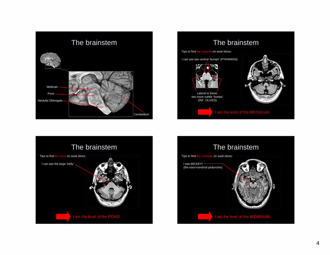
The brainstemTips to find the medulla on axial slices:
I can see two ventral ‘bumps’ (PYRAMIDS)
Lateral to these, two more subtle ‘bumps’
(INF. OLIVES)
I am the level of the MEDULLA!
The brainstem
I can see the large ‘belly’
I am the level of the PONS!
Tips to find the pons on axial slices:
The brainstem
I see MICKEY!(the ears=cerebral peduncles)
I am the level of the MIDBRAIN!
Tips to find the midbrain on axial slices:
5
The diencephalon
Thalamus
Hypothalamus
Hypothalamic sulcus
The diencephalonThe ICECREAM tip: Thalamus is the SCOOP, the hypothalamus the CONE!
The diencephalon
Infundibulum of the Pituitary
gland
Mammillarybodies
Opticchiasm
Hypothalamus liesdorsal to these structures
The diencephalon
Infundibulum of the pituitary
gland
Mammillarybodies
Opticchiasm
6
The diencephalonThalami
The diencephalon
The ventricular system
Lateral v.
Third v.
Fourth v.
Cerebral aqueduct
Fourth ventricle
(between pons/upper medulla and cerebellum)
Third ventricle(between the 2 thalami;
& at the center of the hypothalamus)
The ventricular system
Cerebral aqueduct(tiny canal inside the midbrain)
Lateral ventricles(inside the hemispheres)
7
Midbrain
Pons
Medulla Oblongata Cerebellum
Fourth ventricleCerebral aqueduct
The ventricular systemThird ventricleForamen of Monro
Thalamus
Hypothalamus
Cerebral Hemispheres• 2 hemispheres, interconnected by: corpus callosum, anterior commissure, posterior commissure and (in some individuals) interthalamic adhesion (aka massa intermedia).
AnteriorCommissure
(tip: rostral to the fornix!)
Corpus callosum
Posterior commissure(tip: dorsal to the top of the aqueduct!)
Cerebral Hemispheres• 2 hemispheres, interconnected by: corpus callosum, anterior commissure, posterior commissure and (in some individuals) interthalamic adhesion.
• In each hemisphere: cortex (gyri, sulci), white matter and subcortical structures (including hippocampus, amygdala and basal ganglia).
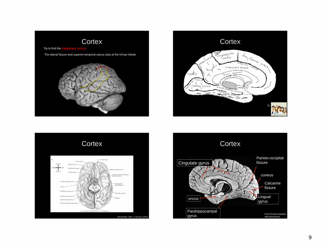
Cortex
• Composed of GYRI (the ‘HILLS’) and SULCI (the ‘VALLEYS’)
• If a sulcus is very deep FISSURE (E.g. Lateral fissure)
• Some sulci run LONGITUDINALLY, others in a MEDIAL-LATERAL direction
• 4 ‘undisputed’ lobes (frontal, parietal, temporal and occipital) + insula (sometimes
referred to as ‘the fifth lobe’).
8
CortexCentral (Rolandic) fissure
Lateral (Sylvian) fissure
Beal, LSU (2005). Modified
Parieto-occipital fissure
Preoccipital notch
FRONTAL LOBE
PARIETAL LOBE
TEMPORAL LOBE
OCCIPITAL LOBE
Cortex
©
Cortex
Superior frontal gyrus
middle frontal gyrus
inferior frontal gyrus
Inferior temporal gyrus
superior temporal gyrus
Inferior parietal lobule
middle temporal gyrusmiddle temporal gyrus
superior parietal lobule precentral gyrus
Post central gyrus
Courtesy of Digital Anatomist Project at Univ of Washington
From Emma Duerden,with permission (modified)
Lateral sulcus
Central sulcus
Intraparietal fissure
CortexTips to find the central sulcus:
1) SFS stops at the precentral gyrus;2) Look for the ‘Omega shape’ (motor hand area)
9
CortexTip to find the intraparietal sulcus:
The lateral fissure and superior temporal sulcus stop at the Inf par lobule
Cortex
©
Cortex
Blumenfeld, 2002. © Sinauer (2002)
Cortex
Calcarinefissure
Parieto-occipital fissure
cuneus
Lingual gyrus
Cingulate gyrus
uncus
Parahippocampalgyrus Courtesy of Digital Anatomist Project at Univ. of Washington
From Emma Duerden,with permission
10
Cortex
Digital Anatomist Project at Univ. of WashingtonFrom Emma Duerden,modified, with permission
Insula
Heschl’s(transverse) gyri
Cortex
Digital Anatomist Project at Univ. of WashingtonFrom Emma Duerden,with permission (modified)
HippocampusTip to find the hippocampus: Look below the inferior horn of the lateral ventricle!
Lateral ventricle
Inferior horn of the lateral ventricle
Hippocampus
HippocampusHippocampus
Fornix (the axons of thehippocampal neurons)
The fornix connects the hippocampus to the mammillary bodies
Blumenfeld, 2002. © Sinauer (2002)
11
Hippocampus and Amygdala
Hippocampus
Amygdala
Tip to find the amygdala: Look rostral to the hippocampus!
The basal ganglia
Lentiform nucleus(=putamen+globus pallidus)
Caudate nucleus
Blumenfeld, 2002. © Sinauer (2002)
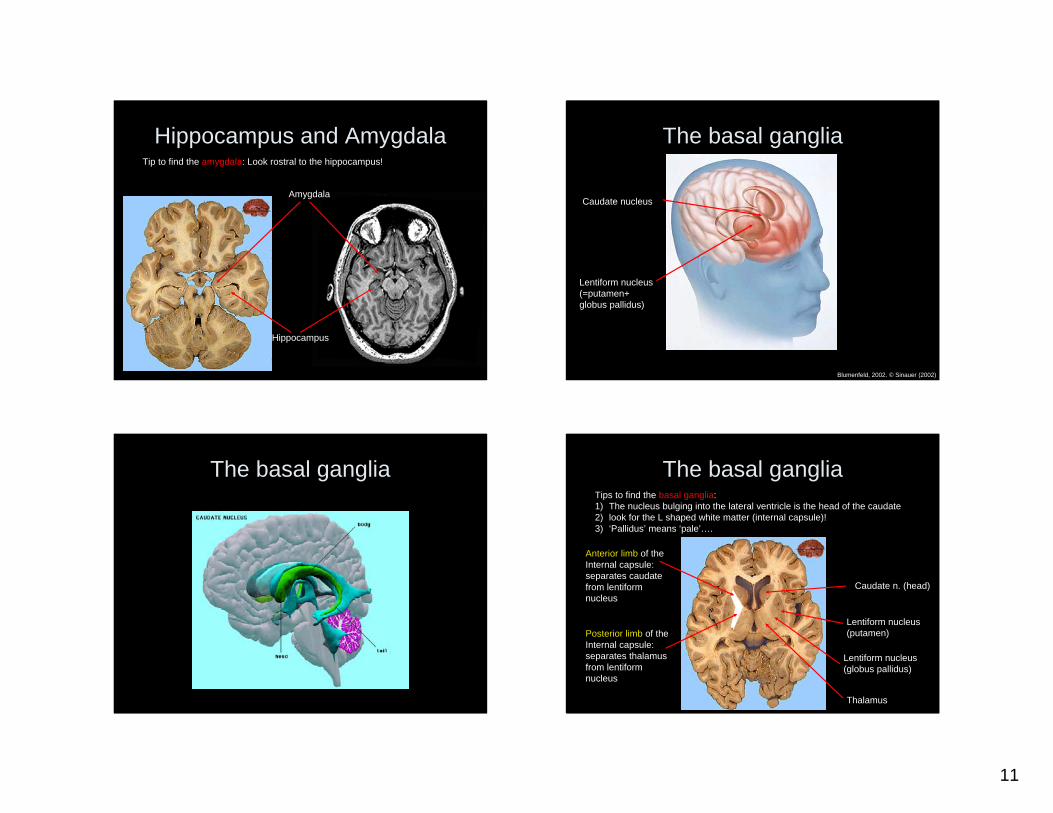
The basal ganglia The basal gangliaTips to find the basal ganglia: 1) The nucleus bulging into the lateral ventricle is the head of the caudate 2) look for the L shaped white matter (internal capsule)!3) ‘Pallidus’ means ‘pale’….
Anterior limb of theInternal capsule: separates caudate from lentiform nucleus
Caudate n. (head)
Lentiform nucleus (putamen)
Lentiform nucleus (globus pallidus)
Posterior limb of theInternal capsule: separates thalamus from lentiform nucleus
Thalamus
12
The basal gangliaTips to find the basal ganglia: 1) The nucleus bulging into the lateral ventricle is the head of the caudate 2) look for the L shaped white matter (internal capsule)!3) ‘Pallidus’ means ‘pale’….
Anterior limb of theInternal capsule: separates caudate from lentiform nucleus
Caudate n. (head)
Lentiform nucleus (putamen)
Lentiform nucleus (globus pallidus)
Posterior limb of theInternal capsule: separates thalamus from lentiform nucleus
Thalamus
Thanks!