H2-M3–restricted CD8+ T cells are not required for MHC class Ib–restricted immunity against Listeria monocytogenes
Sarah E.F. D’Orazio,1 Christine A. Shaw,2 and Michael N. Starnbach2
1Department of Microbiology, Immunology, and Molecular Genetics, University of Kentucky College of Medicine, Lexington, KY 40536
2Department of Microbiology and Molecular Genetics, Harvard Medical School, Boston, MA 02115
Studies using major histocompatibility complex (MHC)-Ia–defi cient mice have shown that MHC-Ib–restricted CD8+ T cells can clear infections caused by intracellular pathogens such as Listeria monocytogenes. M3-restricted CD8+ T cells, which recognize short hydrophobic N-formylated peptides, appear to comprise a substantial portion of the MHC-Ib–restricted T cell response in the mouse model of L. monocytogenes infection. In this study, we isolated formyltransferase (fmt) mutant strains of L. monocytogenes that lacked the ability to add formyl groups to nascent polypeptides. These fmt mutant Listeria strains did not produce antigens that could be recognized by M3-restricted T cells. We showed that immunization of MHC-Ia–defi cient mice with fmt mutant Listeria resulted in stimulation of a protective memory response that cleared subsequent challenge with wild-type L. monocytogenes, despite the fact that M3-restricted CD8+ T cells did not proliferate in these mice. These data suggest that M3-restricted T cells are not required for protection against L. monocy-togenes and underscore the importance of searching for new antigen-presenting molecules among the large MHC-Ib family of proteins.
Although innate immune responses are essential to limit the spread of intracellular bacterial in-fections, it is the adaptive immune response that allows the body to completely clear tremendous bacterial burdens. Memory CD8+ T cells that recognize antigens presented by MHC-I on the surface of infected cells are generally thought to be required for clearance of intracellular path-ogens. Mice express two groups of MHC-I proteins: (a) the classically studied MHC-Ia mol-ecules (K, D, and L) and (b) the nonclassical or MHC-Ib proteins, including Q, T, and M gene products. Much eff ort has been devoted to iden-tifying peptide antigens within bacterial proteins that are predicted to bind to classical MHC-I molecules. For example, at least four dominant Kd-binding epitopes derived from secreted viru-lence factors are known to be presented during Listeria monocytogenes infection, and several other subdominant epitopes have also been identifi ed (1, 2). However, in recent years, it has become increasingly clear that the nonclassical MHC-Ib molecules can also present antigens to T cells during bacterial infection (3).
Nonclassical MHC-I molecules are a widely diverse set of proteins encoded both within and outside the MHC locus that have been collec-tively referred to as MHC-Ib. It is diffi cult to make generalizations about MHC-Ib mole-cules because of the diversity of the members of this protein family; however, many diff erences from classical MHC-I have been observed. Cell surface levels of many MHC-Ib proteins are lower than MHC-Ia and some MHC-Ib molecules display a tissue-specifi c expression pattern, unlike MHC-Ia molecules that are ubiquitously expressed (4, 5). MHC-Ib mole-cules have also been shown to bind a more di-verse set of antigens than MHC-Ia, which bind only 8–10 mer peptides. For example, during Mycobacterium tuberculosis infection, CD1 pro-teins present lipid molecules to T cells (6, 7). When mice are infected with L. monocytogenes, CD8+ T cells recognize formylated peptides bound to M3 (8).
Although our knowledge of most MHC-Ib molecules is limited, M3 has been studied in great detail. The peptide-binding groove of M3 is shallower than classical MHC-I molecules and it has been demonstrated to specifi cally
bind short (5–6 mer) hydrophobic peptides with an NH2- terminal formylated methionine (9–11). It has been suggested that M3 evolved specifi cally to present bacterial antigens to the mammalian immune system because N-formylation is used as a signal to initiate protein synthesis only in prokary-otes and for a limited set of mitochondrial and chloroplast proteins. At least three formylated peptides derived from L. monocytogenes (fMIVIL, fMIGWII, and fMIVTLF) have been shown to bind M3, and T cells that recognize each of these peptides are known to be activated during Listeria infec-tion (12–14). In fact, in BALB/c mice, the magnitude of the fMIGWII-specifi c T cell response is nearly as great as the im-munodominant Kd-binding LLO91-99 T cell response (8).
Using MHC-Ia–defi cient mice, we and others have shown that MHC-Ib–restricted CD8+ T cells alone are suffi -cient to clear infection with the intracellular bacterial patho-gen L. monocytogenes (15, 16). It has been suggested that M3-restricted T cells are largely responsible for this protec-tive immunity in mice (17). If this were true, use of the L. monocytogenes murine infection model would be more lim-ited as a tool to study the role of MHC-Ib–restricted T cells in human immunity against intracellular pathogens because no human homologue for M3 has yet been discovered.
The purpose of this study was to determine whether MHC-Ib molecules other than M3 could mediate protective immunity against L. monocytogenes infection. We used an ac-tinonin resistance selection strategy to generate mutant strains of L. monocytogenes that lacked the necessary enzyme systems to incorporate formylmethionine at the NH2 terminus of newly synthesized proteins. Mice infected with these mutant strains were therefore unable to present formylated peptide antigens derived from L. monocytogenes to naive M3-restricted CD8+ T cells. We found that activation and proliferation of M3-restricted CD8+ T cells was not required for protective immunity against Listeria in MHC-Ia–defi cient mice, sug-gesting that other MHC-Ib must be capable of presenting
antigens to T cells and stimulating a memory CD8+ T cell response. These data underscore the importance of discovering new classes of MHC-Ib molecules and identifying the types of antigens presented by these MHC-Ib family members.
RESULTSSelection of L. monocytogenes strains containing formyltransferase (fmt) mutationsPrevious studies have shown that formylation is not essential for initiation of protein synthesis in either Pseudomonas aerugi-nosa (18) or Staphylococcus aureus (19). We predicted that for-mylation would similarly not be required for protein synthesis in L. monocytogenes and that it would be possible to select for a mutant strain incapable of formylating nascent polypep-tides. Such a strain could then be used as a tool to determine the role of M3-restricted T cells in MHC-Ib–mediated anti-Listeria immunity. We used actinonin, a naturally occurring peptide deformlyase (PDF) inhibitor (20), to select for sponta-neous fmt mutants in Listeria. Failure to remove formyl groups from the NH2-terminal methionine of newly synthesized proteins is toxic to most bacteria; however, if formyl groups are never added, then PDF activity would not be required, and bacteria should grow readily in the presence of actinonin (Fig. 1). Margolis et al. previously showed that growth of S. aureus in the presence of actinonin resulted in the appear-ance of actinonin-resistant (ActR) colonies that had sponta-neous mutations in the S. aureus fmt gene (19). We used a similar selection strategy to isolate Listeria fmt mutants.
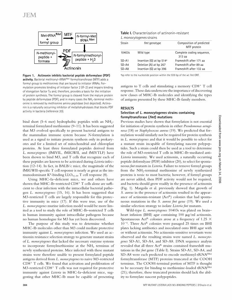
Wild-type L. monocytogenes 10403s was plated on brain–heart infusion (BHI) agar containing 100 μg/ml actinonin. Spontaneous ActR colonies arose at a frequency of 1.25 × 10⁻⁶. Three ActR colonies were passaged 25 times on BHI plates lacking antibiotics and inoculated onto BHI agar with or without actinonin. No actinonin-sensitive revertants were observed and the resulting strains were named L. monocyto-genes SD-A1, SD-A4, and SD-A8. DNA sequence analysis revealed that all three ActR strains contained frameshift mu-tations in the fmt gene (Table I). Strains SD-A1, SD-A4, and SD-A8 were each predicted to encode methionyl-tRNAfMet formyltransferase (MTF) proteins truncated at the COOH terminus. The COOH-terminal portion of MTF is thought to be necessary for binding to methionine-loaded tRNAfMet (21); therefore, these truncated proteins should lack the abil-ity to formylate nascent polypeptides.
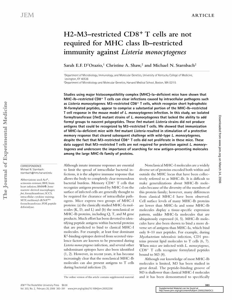
Figure 1. Actinonin inhibits bacterial peptide deformylase (PDF) activity. Bacterial methionyl-tRNAfMet formyltransferase (MTF) adds a formyl group to methionines that are bound to initiator tRNAs. For-mylation promotes binding of initiator factor 2 (IF-2) and impairs binding of elongation factor Tu and, therefore, provides a basis for the initiation of protein synthesis. The formyl group is cleaved from the mature protein by peptide deformylase (PDF), and in many cases the NH2-terminal methi-onine is removed by methionine amino peptidase (not depicted). Actino-nin is a naturally occurring inhibitor of metallohydrolases that blocks PDF activity in bacteria (reference 20).
Table I. Characterization of actinonin-resistant L. monocytogenes strainsStrain fmt genotype Composition of predicted
MTF protein
10403s Wild type Complete coding sequence, 313 aa
SD-A1 Insertion (G) at bp 514a Frameshift after 171 aaSD-A4 Deletion (A) at bp 267 Frameshift after 89 aaSD-A8 Insertion (G) at bp 396 Frameshift after 132 aa
abp refer to the nucleotide position within the 939 bp of the wt fmt ORF.
fmt mutant L. monocytogenes have an increased doubling time compared with wild-type L. monocytogenesAfter 24–48 h of incubation at 37°C on BHI agar, colonies of the fmt mutant L. monocytogenes grew to only approximately half the size of wild-type Listeria colonies, suggesting that the fmt mutants had a slower growth rate. In fact, all three fmt mutant strains had a twofold increase in doubling time when grown in BHI broth (Fig. 2). Even after 48 h of incubation, growth of the fmt mutant strains did not reach the same den-sity as wild-type L. monocytogenes (unpublished data). Slower growth rates have also been observed for fmt mutant strains of P. aeuruginosa (18) and S. aureus (19). Collectively, these re-sults suggest that using formylation to initiate protein syn-thesis enhances bacterial growth, but that formylation is not essential for in vitro growth of these bacteria.
No intracellular growth defect is observed for fmt mutant L. monocytogenesWild-type L. monocytogenes encode several virulence factors that allow the bacteria to readily enter mammalian cells, es-cape from endocytic vesicles, and grow within the cytoplasm of infected cells. As shown in Fig. 3, we tested a variety of diff erent cell types and found no diff erence in the ability of either fmt mutant or wild-type Listeria strains to replicate in-side cells. In both L2 fi broblasts (Fig. 3 A) and Hepa1-6 liver epithelial cells (Fig. 3 B), the growth rate for strains SD-A1 and SD-A8 was indistinguishable from the wild-type strain 10403s. In J774 macrophage-like cells, we recovered fewer intracellular fmt mutant Listeria than wild-type Listeria 1 h after infection (Fig. 3 C). However, the growth rate over the next 7 h in J774 cells was the same for all three strains. These data demonstrate that intracellular growth of the fmt mutant Liste-ria strains was not impaired compared with wild-type Listeria.
N-formylated peptides do not accumulate in the supernatants of fmt mutant Listeria culturesFormylated MIGWII (f-MIGWII), an M3-binding peptide derived from the L. monocytogenes LemA protein, has been shown to accumulate over time in the culture supernatant when L. monocytogenes is grown in broth (22). The mecha-nism for this accumulation of formylated peptide is not known, but it is thought that extracellular proteases could cleave the NH2-terminal portion of LemA, which is pre-dicted to have an “Nout” topology. Presumably, this orienta-tion protects the formyl group on the peptide from PDF activity in the bacterial cytoplasm. To ensure that formylation was not occurring in the fmt mutant strains, we used a T cell cytotoxicity assay to measure the amount of fMIGWII pep-tide found in either wild-type or fmt mutant Listeria culture supernatants. Target cells were pretreated with Listeria cul-ture supernatant and exposed to the fMIGWII-specifi c CD8+ T cell line S172. Line S172 is able to lyse target cells coated
Figure 2. The doubling time of fmt mutant Listeria is increased compared with wild-type Listeria during growth in BHI broth. Freshly streaked wild-type (Lm 10403s) or fmt mutant (SD-A1, SD-A4, SD-A8) L. monocytogenes strains were incubated overnight at 37°C with aeration in BHI broth. The next day, each culture was diluted to an OD550 of �0.015 in fresh BHI broth and further incubated at 37°C with aeration. Bacterial growth was assessed by determining the OD550 at various time points as indicated. Representative data from one of two different experiments are shown.
Figure 3. fmt mutant Listeria do not have a replication defect during intracellular growth. Monolayers of L2 fi broblasts, Hepa1-6 epithelial cells, or J774 macrophage-like cells plated on glass coverslips were infected with the Listeria strains indicated for 30 min, washed ex-tensively, and incubated in media containing 25 μg/ml gentamicin to kill extracellular bacteria. At each of the time points indicated, the cells at-tached to each coverslip were lysed in sterile water to release intracellular bacteria. Serial dilutions of each lysate were plated on BHI agar to deter-mine the total number of bacterial colony forming units (CFU) associated with each coverslip. Data shown represent the average values ± SD for triplicate coverslips from one of two separate experiments.
with picomolar or greater concentrations of formylated MIGWII peptide but not unformylated peptide (Fig. 4 B). As shown in Fig. 4 A, line S172 CD8+ T cells lysed target cells treated with supernatant from wild-type Listeria, but did not lyse targets treated with either SD-A1 or SD-A8 supernatant. In addition, the CD8+ T cell clone 2.5, which recognizes an as yet unidentifi ed Listeria antigen bound to M3 (unpublished data) was also not capable of lysing target cells treated with supernatant from fmt mutant Listeria. The lower limit of de-tection for the recognition of formylated peptides by CD8+ T cells using the cytotoxicity assay was 10−11M (Fig. 4 B). These results suggested that N-formylated proteins were not present in the fmt mutant Listeria strains to provide a source of M3-binding peptides.
fmt mutant Listeria establish infection in mouse spleen, but have an impaired ability to replicate in mouse liverBefore characterizing the adaptive immune response against the fmt mutant Listeria in vivo, we fi rst tested whether or not the fmt mutant strains were virulent enough to establish a sustained infection in mice that would allow for processing
and presentation of bacterial antigens to the immune system. We used a competitive index assay to compare the virulence of the fmt mutant strains to wild-type L. monocytogenes DP-L3903. BALB/c mice were infected with a 1:1 mixture of wild-type (erythromycin-resistant) and fmt mutant L. monocy-togenes (erythromycin-sensitive). 3 d later, liver and spleen homogenates from each mouse were diff erentially plated on media with or without erythromycin to determine the total number of CFU of each Listeria strain present. As shown in Fig. 5 A, approximately equal numbers of wild-type and fmt mutant L. monocytogenes were recovered from the spleen of each mouse, indicating that fmt mutant Listeria were fully able to compete with wild-type Listeria for growth in the spleen. However, at least 10-fold fewer SD-A1 or SD-A8 L. monocy-togenes were recovered from mouse livers compared with wild-type Listeria (Fig. 5 A), suggesting that the fmt mutant strains were less virulent in the liver and not able to compete with wild-type L. monocytogenes for growth.
A growth defect in the liver, but not the spleen, has also been observed for L. monocytogenes strains with mutations in the actA gene (23). ActA mutants are unable to spread effi -ciently from cell to cell and form smaller plaques than wild-type L. monocytogenes when assayed on cell monolayers. Similarly, we have observed that fmt mutant Listeria formed
Figure 4. Formylated peptides do not accumulate in the culture supernatants of fmt mutant Listeria strains. T cell recognition of tar-get cells was measured as a function of cytotoxicity (51Cr release into the supernatant) and is shown as percentage of specifi c lysis. (A) 51Cr-loaded EL-4 target cells were pretreated with media alone (BHI broth) or culture supernatant derived from either wild-type (wt Lm) or fmt mutant (SD-A1, SD-A8) L. monocytogenes strains. CD8+ T cells were added at various effector to target ratios and the cells were coincubated for 4 h. Line S172 T cells recognize N-formylated MIGWII peptide bound to M3; clone 2.5 is an M3-restricted CD8+ T clone of unknown specifi city. (B) 51Cr-loaded EL-4 target cells were pretreated with synthetic peptide at the fi nal concentrations indicated. Line S172 T cells were added at an effector: target ratio of 45:1.
Figure 5. fmt mutant Listeria are less virulent in the liver than in the spleen. (A) A competitive index assay was used to determine the relative virulence of fmt mutant Listeria strains SD-A1 and SD-A8 com-pared with wild-type Listeria. BALB/c mice were sublethally infected intra-venously with a 1:1 mixture of wild-type (erythromycin-resistant) and mutant Listeria (erythromycin-sensitive). Mice were killed 3 d later and the spleens and livers were homogenized and differentially plated to de-termine the total number of each bacterial strain present. Squares repre-sent competitive indices for individual mice; thick black lines represent average indices for groups of mice. (B) A plaque assay was used to assess the degree of cell-to-spread achieved by fmt mutant Lm SD-A1 compared with wild-type Lm 10403s. Confl uent monolayers of L2 fi broblasts were infected at low MOI (0.1) for 1 h and agarose overlays were added. After 3 d of incubation at 37°C in 7% CO2, neutral red overlays were added to visualize the plaques. Plaque size indicates the relative degree of cell-to-cell spread for each strain. Infections were performed in duplicate wells in two separate experiments.
plaques in cell monolayers that were only half the size of plaques formed by wild-type L. monocytogenes (Fig. 5 B). In L2 fi broblasts, strains SD-A1 and SD-A8 formed plaques that were 63% and 54% of wild-type size, respectively. Similar re-sults were observed when monolayers of Hepa1-6 epithelial cells were infected with L. monocytogenes fmt mutants (55% for SD-A1 and 61% for SD-A8). Collectively, these results sug-gest that fmt mutant Listeria are readily able to infect single cells but that their ability to spread to adjacent cells is im-paired compared with wild-type L. monocytogenes.
M3-restricted CD8+ T cells are not activated in mice immunized with fmt mutant L. monocytogenesTo confi rm that M3-restricted CD8+ T cells were not acti-vated in mice infected with fmt mutant Listeria, we harvested splenocytes from mice 7 d after infection and determined the number of fMIGWII-specifi c CD8+ T cells present. In naive BALB/c mice, �0.3% of all CD8+ T cells secreted IFNγ after incubation with fMIGWII, the immunodominant M3-binding peptide epitope derived from L. monocytogenes (Fig. 6 A and Fig. S1, available at http://www.jem.org/cgi/ content/full/jem.20052256/DC1). 7 d after infection with wild-type Listeria, those T cells had expanded �10-fold. However, no increase in the number of fMIGWII-specifi c T cells was observed in spleens of BALB/c mice infected with either SD-A1 or SD-A8. Similar results were observed when MHC-Ia–defi cient mice were used (unpublished data). As a control, we also counted the number of LLO91-99-specifi c CD8+ T cells found in the spleens of BALB/c mice infected with either wild-type or fmt mutant Listeria. A signifi cant in-crease in the number of LLO91-99-specifi c CD8+ T cells was observed after infection with all three Listeria strains (Fig. 6 A). Therefore, the fmt mutant Listeria were able to infect enough cells to allow for suffi cient processing and presentation of a MHC-Ia (Kd)-binding peptide to naive CD8+ T cells.
Although the fmt mutant Listeria did not appear to be pro-ducing formylated MIGWII peptide antigen, signifi cant quan-tities of unformylated MIGWII peptide could be present in the cytoplasm of cells infected with either strain SD-A1 or SD-A8. Unformylated peptides bind to M3 with a much lower af-fi nity than formylated peptides, and it has been thought that unformylated peptide bound to M3 would not stimulate naive CD8+ T cells in vivo. To verify that infection with fmt mu-tant Listeria did not result in activation of any M3-restricted MIGWII-specifi c CD8+ T cells, we also counted the number of T cells present that could recognize unformylated MIGWII peptide. Splenocytes harvested from mice infected with wild-type or fmt mutant Listeria 7 d earlier were exposed to high concentrations of either fMIGWII or MIGWII and the num-ber of CD8+ T cells that produced IFNγ in response to these stimuli was assessed by intracellular cytokine staining (ICCS). As shown in Fig. 6 B, the number of MIGWII-specifi c T cells present in the spleens of mice infected with either strain SD-A1 or SD-A8 did not change over background levels. These results suggested that M3-restricted naive CD8+ T cells are not activated in mice infected with fmt mutant L. monocytogenes.
To determine whether CD8+ T cells restricted by other MHC-Ib molecules were being activated in the absence of an M3-restricted T cell response, we infected MHC-Ia–defi cient mice with a sublethal dose of L. monocytogenes SD-A1. 7 d later, splenocytes from these mice were incubated with either uninfected bone marrow–derived macrophages (BMMΦ) or cells that had been infected with fmt mutant Listeria strain SD-A1. As shown in Fig. 7 A and Fig. S2 (available at http://www.jem.org/cgi/content/full/jem.20052256/DC1), a sig-nifi cant number of CD8+ T cells capable of recognizing an-tigens derived from L. monocytogenes SD-A1 were found in the spleens of MHC-Ia–defi cient mice. Recognition of these antigens was not blocked by the addition of either anti-M3 or anti–Qa-1b antibody. Control groups of mice infected with wild-type Listeria also displayed signifi cant T cell recognition of infected BMMΦ. However, as expected, pretreatment with anti-M3 antibody blocked recognition and subsequent IFNγ expression for a large fraction of the CD8+ T cells found in these mice (Fig. 7 B). These results suggest that other antigens besides formylated peptides were presented
Figure 6. M3-restricted CD8+ T cells are not activated during infection with fmt mutant Listeria. Mice were immunized with PBS (naive group) or a sublethal dose of either wild-type (10403s) or fmt mu-tant (SD-A1, SD-A8) L. monocytogenes. 7 d later, the mice were killed and the number of antigen-specifi c CD8+ T cells present in the spleen was determined by IFNγ ICCS. (A) BALB/c splenocytes were exposed to either 1 μM LLO91-99 peptide, which binds to Kd (black bars) or 1 μM fMIGWII peptide, which binds to M3 (white bars). (B) MHC-Ia–defi cient splenocytes were incubated with either no peptide (gray bars), 1 μM formylated fMIGWII peptide (black bars), or 1 μM unformylated MIGWII peptide (white bars). Data shown represent the average number of CD8β
+ cells that were IFNγ+ within each group of mice ± SE from one of three separate experiments.
to CD8+ T cells by novel MHC-Ib molecules during both wild-type and fmt mutant Listeria infection of mice.
Class Ia MHC-defi cient mice immunized with fmt mutant Listeria develop protective immunity against infection with wild-type ListeriaMHC-Ia–defi cient mice are able to clear secondary lethal challenge with L. monocytogenes as rapidly as wild-type mice (15, 16) and it has been suggested that M3-restricted CD8+ T cells are largely responsible for this protective immunity (17). We used the fmt mutant strains of Listeria to determine whether activation of M3-restricted T cells was required for protection against secondary Listeria infection in MHC-Ia–defi cient mice. Groups of MHC-Ia–defi cient mice were im-munized with a sublethal dose of either wild-type or fmt mutant L. monocytogenes; a control group (naive mice) re-ceived PBS. 3 wk later, all groups of mice were challenged with 5 LD50 of wild-type L. monocytogenes 10403s. 3 d later, the total number of Listeria CFU present in the spleen or liver was determined. Mice immunized with wild-type Listeria had at least two logs fewer bacteria in the spleen or liver com-pared with naive mice (Fig. 8). Surprisingly, mice immunized with either SD-A1 or SD-A8 showed the same degree of protection against high dose secondary infection as was ob-served after immunization with wild-type L. monocytogenes. This result indicates that activation of M3-restricted CD8+ T cells was not essential for protective immunity against L. monoytogenes in these mice and suggests that other MHC-Ib–restricted T cells may have a signifi cant role in the clear-ance of Listeria infection.
D I S C U S S I O N We isolated ActR fmt mutant strains of L. monocytogenes to use as a tool to address the role of M3-restricted CD8+ T cells in
MHC-Ib–mediated protective immunity against intracellular bacterial pathogens. While performing assays to characterize these fmt mutant strains, we discovered several interesting phenotypes. For example, although the fmt mutant strains were able to compete with wild-type L. monocytogenes for growth in the spleen, signifi cantly fewer fmt mutant Listeria were isolated from the liver. This diff erence in virulence can be at least partially explained by the observation that fmt mu-tant Listeria have a reduced ability to spread from cell-to-cell. One mechanism for Listeria to prolong survival in the host is thought to be the ability of the bacteria to spread to a neigh-boring cell without encountering the extracellular environ-ment, where the bacteria are susceptible to phagocytosis by activated macrophages or neutrophils. Hepatocytes, which are readily infected by L. monocytogenes, are connected by cell junctions within the tissue, and this cell-to-cell contact pre-sumably facilitates spread of the bacteria within the liver. In contrast, L. monocytogenes infects primarily circulating im-mune cells (monocytes, macrophages, and dendritic cells) in the spleen. Thus, the apparent increased virulence of the fmt mutant L. monocytogenes in the spleen may simply refl ect the fact that effi cient cell-to-cell spread of bacteria is not an im-portant feature for growth in the spleen.
Interestingly, the fmt mutant L. monocytogenes had a slight growth defect in vitro but not in vivo. It has long been thought that formylation of initiator Met-tRNAs is essential for protein synthesis in bacteria and in eukaryotic organelles such as chloroplasts and mitochondria. This belief is based largely on studies that showed a marked (>10-fold) decrease in growth of Escherichia coli containing mutations in fmt (24) and the identifi cation of formylmethionine at the NH2 ter-minus of several mitochondrial proteins. However, we show here that mutations in the L. monocytogenes fmt gene have
Figure 7. CD8+ T cells from fmt mutant Listeria-immune mice recognize antigens presented by an MHC-Ib molecule other than M3 or Qa-1b. Groups of MHC-Ia–defi cient mice were infected with 2,000 CFU of either (A) fmt mutant (Lm SD-A1) or (B) wild-type (Lm 10403s) L. monocytogenes. 7 d after infection, splenocytes from these mice were harvested and exposed to MHC-Ia–defi cient BMMΦ (either uninfected or cells that had been infected for 3 h with the same strain of Listeria used to infect the mice). As indicated, some samples of Listeria-infected BMMΦ were pretreated with either anti-M3 or anti–Qa-1b monoclonal antibody before the addition of immune splenocytes. Data shown represent the average number of TCRβ+ CD8+ cells that stained IFNγ+ within each group of mice ± SE from one out of two experiments.
Figure 8. Immunization with fmt mutant Listeria results in full protective immunity against secondary challenge with wild-type Listeria. Groups of MHC-Ia–defi cient mice were immunized with PBS (naive) or 103 CFU of either 10403s (wild type) or SD-A1 (fmt mutant) L. monocytogenes. 3 wk later, all groups of mice were challenged intrave-nously with 5 × 10⁴ wild-type Listeria. The total number of CFU per organ was determined 3 d after infection. Similar results were observed for groups of mice immunized with strain SD-A8 (not depicted). Data shown represent average values ± SD for groups of mice from one of two independent experiments.
only a modest eff ect on bacterial growth in BHI broth. Other groups have shown a similar growth phenotype in P. aerugi-nosa (18) and S. aureus (19). These observations suggest that formylation may confer a slight advantage for in vitro growth of most bacterial species, but that only in E. coli has a strict requirement for fmt activity been demonstrated. Li et al. showed that protein synthesis could also be initiated in yeast mitochondria in the absence of formylated methionine (25). A comparative analysis of the structural requirements for binding of IF-2 to fMet-tRNA vs. Met-tRNA in each of these organisms may elucidate the mechanism whereby pro-tein synthesis can readily initiate in the absence of formyl-methionine in prokaryotes or in mitochondria.
It is possible that during infection of mice, secondary mu-tations occurred that allowed the fmt mutant L. monocytogenes to replicate faster. However, we found that the fmt mutant strains were replicating at the same rate as wild-type L. mono-cytogenes after just 1–2 h of infection of tissue culture cells. It does not seem likely that selective pressures could have re-sulted in secondary mutations during this short time period. These data suggest that the cytoplasm of host cells may be an environment in which the bacterial enzymes involved in for-mylation are not necessary. Even if secondary mutations did occur in vivo that resulted in a low level of formylation in the fmt mutant bacteria, insuffi cient antigen was presented to ac-tivate the immunodominant M3-restricted Listeria-specifi c CD8+ T cell population.
During primary Listeria infection, M3-restricted T cells expand faster than Kd-restricted T cells, with peak numbers occurring by 5–6 d after infection (8) and it has been shown that M3-restricted CD8+ T cells alone can clear sublethal Listeria infections within 5 d (26). The role of M3-restricted T cells during secondary infection, however, has been less clear. Kerksiek et al. showed that M3-restricted memory CD8+ T cell populations are generated and maintained after primary infection with L. monocytogenes. However, despite the fact that these memory CD8+ T cells were activated in the same manner as Kd-restricted T cells, the M3-restricted CD8+ cells failed to undergo a dramatic proliferative burst upon rechallenge with Listeria (27). It has been diffi cult to determine the precise role of M3-restricted CD8+ T cells during secondary Listeria infections because of the apparent paradox that M3-restricted T cells are a dominant fraction of the MHC-Ib–restricted T cell population (17), yet the recall expansion of these cells is small and perhaps not signifi cant enough to clear large bacterial burdens. Hamilton et al. re-cently suggested that it is the MHC-Ia–restricted T cell re-sponse that limits the expansion of M3-restricted memory CD8+ T cells during secondary challenge (26).
In this report, we show that immunization with an fmt mutant Listeria strain that lacks formylated peptides still results in the generation of protective immunity against secondary Listeria infection. These data argue that activation of M3- restricted CD8+ T cells is not essential for protective immu-nity against Listeria and suggests that presentation of antigen in the context of other MHC-Ib molecules is suffi cient to
elicit a protective memory CD8+ T cell response. Bouwer et al. previously showed that Qa-1b–restricted Listeria-specifi c cytotoxic T lymphocytes increase in number during L. mono-cytogenes infection (28). However, like M3-restricted CD8+ T cells, Qa-1b–restricted T cells do not expand signifi cantly during secondary infection (29). We have previously shown that in MHC-Ia–defi cient mice, the residual population of MHC-Ib–restricted CD8+ T cells does expand signifi cantly after both primary and secondary Listeria infection, and that protective immunity is mediated by CD8+ T cells in these mice (15). Because neither M3-restricted nor Qa-1b– restricted CD8+ T cells expand signifi cantly during secondary Listeria infection, the activated population of CD8+ T cells must rec-ognize L. monocytogenes antigens presented by novel MHC-Ib molecules. These MHC-Ib–restricted CD8+ T cells appear to be necessary for clearance of Listeria in the absence of MHC-Ia–restricted and M3-restricted T cell responses be-cause adoptive transfer of L. monocytogenes SD-A1 immune splenocytes depleted of CD8+ T cells failed to protect naive mice from challenge with L. monocytogenes (Fig. S3, available at http://www.jem.org/cgi/content/full/jem.20052256/DC1). Further studies will be required to determine whether these additional MHC-Ib–restricted CD8+ T cell specifi cities are involved in protective immunity in wild-type mice.
In the past decade, the majority of studies examining MHC-Ib–restricted T cells have focused on the recognition of formylated peptides presented by M3 molecules. The re-sults presented here make a strong argument that a search for new MHC-Ib molecules capable of presenting antigen to CD8+ T cells would result in the identifi cation of new anti-genic specifi cities able to protect against secondary Listeria infection. Further characterization of the unique types of an-tigens that can be recognized by protective CD8+ T cells will greatly facilitate the rational design of eff ective therapeutic strategies to protect against other intracellular bacteria, in-cluding prevalent pathogens such as Mycobacterium tuberculosis and Chlamydia trachomatis.
MATERIALS AND METHODSBacteria. L. monocytogenes fmt mutant strains SD-A1, SD-A4, and SD-A8
were derived from wild-type L. monocytogenes 10403s. L. monocytogenes
10403s has a LD50 of �104 CFU in BALB/c mice. Bacteria were grown in
BHI broth (Difco) supplemented with 100 μg/ml actinonin (Sigma- Aldrich)
as indicated. DNA fragments encoding fmt were amplifi ed from each Listeria
strain using Platinum PCR Supermix (Invitrogen) with the following prim-
ers: 3′-G A C A T G A G C G G A C A G G G G A A G T -5′ and 3′-C A C C C C G T T G-
A C G A A T T T A G T T A C -5′. The DNA sequence of each PCR product was
directly determined using internal primers. Bacterial culture supernatants
were prepared by inoculating BHI broth and incubating with aeration at
37°C for 24 h. Bacteria were centrifuged at 13,000 revolutions/min for
20 min and the supernatant was collected. Aliquots were stored at −20°C
Plaque assay. Cells were grown to confl uence in six-well dishes, infected
with L. monocytogenes for 1 h at various multiplicities of infection, and washed
three times with warm PBS. Overlays consisting of MEM (GIBCO BRL)
containing 2.5% FBS, 1% agarose, and 10 μg/ml gentamicin sulfate were
added and the cells were incubated for an additional 3 d. Plaques were visu-
alized by the addition of neutral red overlays (1% agarose, 0.2% neutral red
in MEM-2.5). Plaque size was measured by analyzing digital images of the
overlays.
ICCS. IFNγ ICCS was performed using the Cytofi x/Cytoperm Plus (with
GolgiPlug) kit (BD Biosciences). In brief, splenocytes were harvested from
Listeria-infected mice and 5 × 10⁶ cells/well were added to 24-well dishes.
Synthetic peptides (Bio-Synthesis) corresponding to antigenic epitopes
were added to some wells at a fi nal concentration of 1 μM. In experiments
where J774 cells were used as targets, 4 × 10⁵ J774 cells per well were in-
fected with L. monocytogenes SD-A1 at a multiplicity of infection of 1.0 for
2 h before the addition of splenocytes. Listeria culture supernatant (50 μl)
was added directly to some wells. After 5–6 h of incubation in the pres-
ence of GolgiPlug at 37°C in 7% CO2, cells were stained with anti-CD8α
(53–6.7), anti-CD8β (53–5.8), and anti-TCRb (H57-597) antibodies. The
cells were fi xed, permeabilized, and stained with anti-IFNγ (XMG1.2) an-
tibody according to the manufacturer’s instructions. Cells were suspended
in FACS buff er (0.5% BSA, 0.1% NaN3 in HBSS) before fl ow cytometric
analysis. Fluorescence intensities were measured using a FACSCalibur fl ow
cytometer (Becton Dickinson) and analysis was performed using CELL-
Quest software. Dead cells and monocytes were excluded using forward
and side scatter gating. Typically, 100,000 gated events were collected for
each analysis.
Antibody-blocking experiments. CD8+ cells were enriched from whole
splenocytes by negative selection using antibody-conjugated IMag magnetic
beads (BD Biosciences). Typically, the enrichment protocol removed 90–
95% of the splenocytes from MHC-Ia–defi cient mice. Listeria-infected or
uninfected MHC-Ia–defi cient BMMΦ were pretreated with anti-M3 (clone
130) or anti–Qa-1b (6A8.6F10.1A6) monoclonal antibodies (fi nal concentra-
tion 500 μg/ml) for 30 min at room temperature. The antibodies were
maintained at a concentration of 20 μg/ml for 5–6 h during the incubation
of CD8-enriched splenocytes with infected or uninfected macrophages. The
number of IFNγ-secreting CD8+ T cells was determined by ICCS as de-
scribed in the previous paragraph.
Online supplemental material. Fig. S1 displays primary dot plots from
individual mice representative of the data shown in Fig. 6 A. Fig. S2 includes
primary FACS plots for individual mice representative of the data shown in
Fig. 7. Fig. S3 shows that adoptive transfer of fmt mutant Listeria-immune
spleen depleted of CD8+ T cells does not protect mice as well as whole im-
mune spleen. Online supplemental material is available at http://www.jem.
org/cgi/content/full/jem.20052256/DC1.
We would like to thank J. Mekalanos for helpful discussions about actinonin resistance, Z. Balsara and D. Higgins for critical review of the manuscript, and J.N. Lafave and D. McElroy for technical assistance.
This work was supported by National Institutes of Health (NIH) grant nos. AI055962 and AI41526 (to M.N. Starnbach) and by a pilot grant from the Harvard Digestive Diseases Center, NIH grant no. DK34854 (to S.E.F. D’Orazio).
The authors have no confl icting fi nancial interests.
Submitted: 9 November 2005Accepted: 5 January 2006
R E F E R E N C E S 1. Busch, D.H., K. Kerksiek, and E.G. Pamer. 1999. Processing of Listeria
monocytogenes antigens and the in vivo T-cell response to bacterial infec-
tion. Immunol. Rev. 172:163–169.
2. Geginat, G., S. Schenk, M. Skoberne, W. Goebel, and H. Hof. 2001. A
novel approach of direct ex vivo epitope mapping identifi es dominant
and subdominant CD4 and CD8 T cell epitopes from Listeria monocyto-
genes. J. Immunol. 166:1877–1884.
3. Soloski, M.J., M.E. Szperka, A. Davies, and S.L. Wooden. 2000. Host
immune response to intracellular bacteria: a role for MHC-linked class-Ib
antigen-presenting molecules. Proc. Soc. Exp. Biol. Med. 224:231–239.