Hadoop for EEG Storage and Processing: a Feasibility Study Ghita Berrada, Maurice van Keulen, and Mena B. Habib University of Twente, The Netherlands {g.berrada,m.vankeulen,m.badiehhabibmorgan}@utwente.nl Abstract. Lots of heterogeneous complex data are collected for diag- nosis purposes. Such data should be shared between all caregivers and, often at least partly automatically processed, due to its complexity, for its full potential to be harnessed. This paper is a feasibility study that assesses the potential of Hadoop as a medical data storage and processing platform using EEGs as example of medical data. Keywords: EEG,Hadoop, medical data storage and processing 1 Introduction The diagnosis process often involves multiple clinicians/specialists and a large number of ordered tests. As a result, huge amounts of heterogeneous data are gathered and scattered in many locations (or islands of data). Table 1 shows the scale of data produced in and spread across the healthcare system. To further compound the problem, different locations often use non-interoperable systems and file formats, if the data is indeed digitized. A McKinsey Global Institute (MGI) report on the US healthcare system ([1]) shows that up to 30% of data that includes medical records, laboratory and surgery reports, is not digitized and that the video and monitor feeds that make up most of the clinical data produced are not stored but used real time. Such a setting makes it hard for caregivers to access a patient’s full history and get a full picture of his/her con- dition. As it stands, needless tests may be ordered and diagnoses delayed and/or missed, not to mention data security more easily breached. The prevalence of misdiagnoses is estimated to be up to 15% in most areas of medicine ([2]). And a study of physician-reported diagnosis errors ([3]) finds most cases are due to testing (44%) or clinician assessment errors (32%). A case from The Washington Post exposes all those issues ([4]). A patient strug- gling with depression is diagnosed with a meningioma 1 , unrelated with the patient’s depression and not in need of monitoring, according to the attending clinician at the time. Four years, many moves across US states and many consul- tations (with other clinicians) later, and with her condition steadily worsening, the patient is hospitalized and the meningioma, gone under the radar for years, is finally rediscovered and pinpointed as the cause of the patient’s near-fatal 1 a brain tumor
Transcript
Hadoop for EEG Storage and Processing: aFeasibility Study
Ghita Berrada, Maurice van Keulen, and Mena B. Habib
University of Twente, The Netherlands{g.berrada,m.vankeulen,m.badiehhabibmorgan}@utwente.nl
Abstract. Lots of heterogeneous complex data are collected for diag-nosis purposes. Such data should be shared between all caregivers and,often at least partly automatically processed, due to its complexity, forits full potential to be harnessed. This paper is a feasibility study thatassesses the potential of Hadoop as a medical data storage and processingplatform using EEGs as example of medical data.
Keywords: EEG,Hadoop, medical data storage and processing
1 Introduction
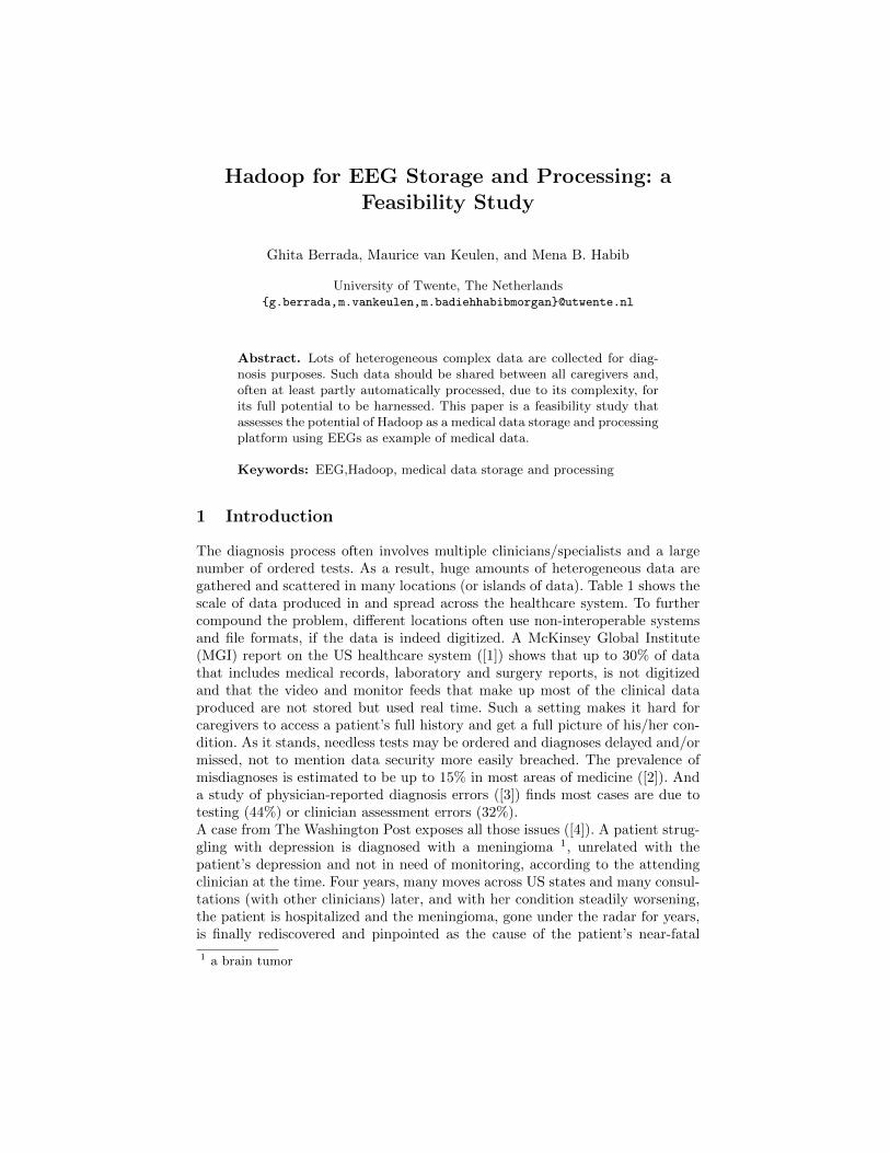
The diagnosis process often involves multiple clinicians/specialists and a largenumber of ordered tests. As a result, huge amounts of heterogeneous data aregathered and scattered in many locations (or islands of data). Table 1 shows thescale of data produced in and spread across the healthcare system. To furthercompound the problem, different locations often use non-interoperable systemsand file formats, if the data is indeed digitized. A McKinsey Global Institute(MGI) report on the US healthcare system ([1]) shows that up to 30% of datathat includes medical records, laboratory and surgery reports, is not digitizedand that the video and monitor feeds that make up most of the clinical dataproduced are not stored but used real time. Such a setting makes it hard forcaregivers to access a patient’s full history and get a full picture of his/her con-dition. As it stands, needless tests may be ordered and diagnoses delayed and/ormissed, not to mention data security more easily breached. The prevalence ofmisdiagnoses is estimated to be up to 15% in most areas of medicine ([2]). Anda study of physician-reported diagnosis errors ([3]) finds most cases are due totesting (44%) or clinician assessment errors (32%).A case from The Washington Post exposes all those issues ([4]). A patient strug-gling with depression is diagnosed with a meningioma 1, unrelated with thepatient’s depression and not in need of monitoring, according to the attendingclinician at the time. Four years, many moves across US states and many consul-tations (with other clinicians) later, and with her condition steadily worsening,the patient is hospitalized and the meningioma, gone under the radar for years,is finally rediscovered and pinpointed as the cause of the patient’s near-fatal
1 a brain tumor
2 Ghita Berrada, Maurice van Keulen, Mena B. Habib
condition. This case stresses the necessity of care continuity and easy access topatient history: had the meningioma been known to clinicians after the initialdiagnosis, the patient may have been spared years of misery, a possible fataloutcome and enjoyed a better quality of life.To solve such issues, authorized caregivers need fast and reliable access to ashared medical data repository containing tests’ data and their interpretations.An international data repository would ideally be needed but is unlikely to becreated in the foreseeable future for legal reasons. So national scale repositoriesshould at least be created. The MGI report cited earlier ([1]) argues that sharingmedical data offers huge premiums such as a drastic reduction of healthcare costsand waste and improved patient outcomes and quality of life through allowingremote patient monitoring, easing comparative effectiveness studies and clinicaldecision systems deployment and increasing data transparency.Sharing data would also provide a trove of data on which competing automatedmedical data interpretation methods can easily be tested, compared, interpretedand reproduced. So far, the automated medical data interpretation methods aim-ing at reducing the clinicians’ workload and easing the diagnosis process havebeen of limited use as they are tested on distinct, usually small data, makingthem hard to reproduce and interpret with any certainty.The MGI report ([1]) also points out there are critical technical hurdles to over-come before medical data can be shared, analyzed properly and its full potentialuncovered,e.g standardizing formats, ensuring systems’ interoperability, integrat-ing pre-existing, fragmented and heterogenous datasets and providing sufficientstorage. So any potential design for a medical repository should take into ac-count the distributed nature of the data 2, its heterogeneity and size and thediversity of file formats and platforms used across healthcare institutions. Thedata should also be easy to access for further, complex processing.In this paper, we show that a rather low cost technical solution (and possiblestorage platform for medical data) that fits those constraints and requires mini-mal changes to current state of the art storage and processing techniques alreadyexists: the Hadoop platform. In what follows, we will take EEG data as exampleof medical data.The rest of this paper is organized as follows. We introduce Hadoop, explainwhy it is a good fit for medical data storage and show how EEGs can be storedwith Hadoop (Section 2). The example of EEG feature selection by exhaustivesearch is then used to lay out why complex data processing should also be donewith Hadoop (Sections 3 and 4).Contributions In this paper, we give a proof of concept for an EEG repositoryby :
– explaining why Hadoop fits the constraints imposed on potential medicaldata repositories
– showing how to store EEG data in a Hadoop framework– proving that EEG data can be analyzed on national scale on Hadoop by
designing and benchmarking a representative machine-learning algorithm
2 healthcare institutions are unlikely to let their data be stored externally
Hadoop for EEG Storage and Processing: a Feasibility Study 3
Table 1: Medical data statistics (2009 data, last year for which records are avail-able) from [5]
Fig. 1: EEG showing an adult’s normal eyes-closed EEG segment
1.1 Related work
Hadoop has been found a viable solution for storing and processing big datasimilar to medical data, such as images in astronomy ([6]) or power grid timeseries, which unlike medical time series, are unidimensional time series ([7]).[8] is, to the best of our knowledge, the first paper to consider storing medicaldata and EEGs in particular with Hadoop and show it is a promising solution inneed of more testing. [8] suggest exploring the ”design and benchmarking of ma-chine learning algorithms on [the Hadoop] infrastructure and pattern matchingfrom large scale EEG data.” and this is one of the goals of our paper.
2 Hadoop: a good fit for medical repositories’ constraints
2.1 Introduction to Hadoop
Hadoop, an open source platform managed by the Apache open source com-munity, has 2 core components: the Hadoop Distributed File System (HDFS)and the job management framework or MapReduce framework. The HDFS isdesigned to reliably store huge files on all cluster machines. Each HDFS file is
2 Assuming standard 20-minute EEGs in EDF+ format. File average size: 13.7MB3 Assuming average size of 23MB per MRI and 35MB per CT4 Based on data from OECD countries with available data from exams performed
in and outside of hospitals i.e the USA, Greece, France, Belgium, Turkey, Iceland,Luxembourg, the Netherlands, Canada, Denmark, Estonia,the Czech Republic, theSlovak Republic, Chile, Israel and South Korea
4 Ghita Berrada, Maurice van Keulen, Mena B. Habib
cut into blocks and each block then replicated and stored at different physicallocations in the cluster to ensure fault tolerance. The HDFS has a master/slavearchitecture with one master server called Namenode managing the filesystemnamespace and regulating the file access by clients and multiple slave servers(one per cluster node) called Datanodes managing the storage in the nodes theyrun on. The Namenode maps the file blocks to the Datanodes and gives theDatanodes instructions to perform operations on blocks and serve filesystemclients’ read and write requests. The Hadoop MapReduce framework also hasa master/slave architecture with a single master called jobtracker and severalslave servers (one per cluster node) called tasktrackers. MapReduce jobs are sub-mitted to the jobtracker, which puts the jobs in a queue and executes them onfirst come/first serve basis. The jobtracker assigns tasks to the tasktrackerswithinstructions on how to execute them.
2.2 Hadoop and parallel data processing: the MapReduce model
MapReduce is a programming model for data-intensive parallelizable processingtasks (introduced in [9]) designed to process large volumes of data in parallel,with the workload split between large numbers of low level commodity ma-chines. The MapReduce framework, unlike parallel databases, hides the complexand messy details of load balancing, data distribution, parallelization and fault-tolerance from the user in a library, thus making it simpler to use the resourcesof a large distributed system to process big datasets. The MapReduce model re-lies on 2 successive functions to transform lists of input data elements into listsof output data elements: a mapper function and a reducer function. Each inputdata element is transformed into a new output data element by the mapper.The transformed elements are then aggregated by the reducer to return a singleoutput value. A simple example is files word count: in this case, the mapper as-sociates a number of words to each of the input files while the reducer functionsums the values obtained during the mapping step.
2.3 Hadoop for medical data storage
The Hadoop platform provides a solution to the technical hurdles outlined bythe MGI report ([1]) described earlier (Section 1).First of all, Hadoop was designed to scale with large data. It is currently be-ing used at Facebook to store about 100PB of user data, i.e data much biggerthan national scale medical data which ranges from dozens of terabytes (eg theNetherlands) to petabytes of data (eg the USA) annually as shown in Table 1.So Hadoop can easily handle national scale amount of medical data.Moreover, Hadoop can store heterogeneous formats of data, in particular un-structured data, and if there is a method to extract the data from the files thatstore it 5, the data can then be fed to Hadoop MapReduce for further analysis
5 Such methods currently exist at the sites where the different types of data are stored.There is,at most, a need to translate those methods into Java, Python, Perl or anyother language that can be interfaced with Hadoop.
Hadoop for EEG Storage and Processing: a Feasibility Study 5
and processing.Hadoop is also tolerant to node failure. The HDFS relies on replication (by de-fault 3 copies on 3 Datanodes per file block) to ensure file blocks are not lostif a data server fails. If a Datanode fails and some data blocks have less thana set minimum of copies, the Namenode orders the replication of the affectedblocks in some available Datanodes to bring back the replication factor of theblocks to safer levels. The probability of losing a block in a 4000 nodes’ clusterin a day (respectively in a year) in the case of uncorrelated failures of multiplenodes is about 5.7 × 10⁻⁷ (respectively 2.1 × 10⁻⁴) ([10]). At Yahoo! in 2009for example, only 641 blocks were lost out of 329 million on 17720 nodes i.e aloss rate of 1.9 × 10⁻⁴% ([10]). The only problem left is the Namenode as theHDFS is unusable if the Namenode fails. Namenode crashes rarely occur though([11])(1 in 4 years at Facebook) and solutions limiting the crash impact are al-ready being deployed. One such solution is the AvatarNodes in use at Facebook:2 AvatarNodes, an active and standby one, replace the unique Namenode andreceive the Datanodes messages in its stead. The Standby AvatarNode thus con-tains up-to-date information about block locations and can be started in under aminute to replace the Namenode (or Active AvatarNode) if it fails. This solutioncuts cluster planned downtime by 50%. Data stored with Hadoop will thereforebe constantly available.Hadoop was built for parallel processing (via MapReduce described in Section2.2) and we study the feasibility EEG data processing with Hadoop with theexample of feature selection by exhaustive search in Section 3.
2.4 Hadoop and EEG storage
An EEG is a multidimensional time series obtained by capturing the brain’selectric activity with scalp electrodes. Figure 1 shows an example of EEG. Theincreasingly popular EDF+ format is used to store EEGs and contains all theinformation about the EEG recording, both metadata in a header encoded inUTF-8 and raw data in binary format. The metadata includes patient informa-tion and EEG signal technical attributes (eg equipment details and samplingrate). Annotations on the EEG, such as context of recording or EEG eventslabels, may also be stored in the EDF+ file. See [12] for format details.HDFS does not call for any set file format, so we store EEGs in EDF+ inHDFS. We anonymize EEGs before storage for security reasons. Keeping EEGsas EDF+ files has many advantages. No additional data formatting is neededand existing tools for EDF+ files, eg. visualization tools, can still be used. Andas EDF+ files are mainly binary files, the size of the stored EEGs is small: 2500EDF+ files (dataset 1 in Section 4 and Table 2(a)) i.e to about 2 years of EEGdata at the local hospital take up 46.5GB whereas the same data 6 would takeup 1TB when in a relational database.
6 with one table for metadata, one table for raw data and one tuple per raw data point
6 Ghita Berrada, Maurice van Keulen, Mena B. Habib
3 EEG feature selection with Hadoop
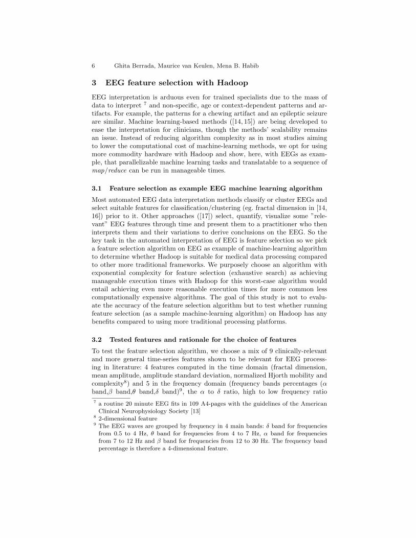
EEG interpretation is arduous even for trained specialists due to the mass ofdata to interpret 7 and non-specific, age or context-dependent patterns and ar-tifacts. For example, the patterns for a chewing artifact and an epileptic seizureare similar. Machine learning-based methods ([14, 15]) are being developed toease the interpretation for clinicians, though the methods’ scalability remainsan issue. Instead of reducing algorithm complexity as in most studies aimingto lower the computational cost of machine-learning methods, we opt for usingmore commodity hardware with Hadoop and show, here, with EEGs as exam-ple, that parallelizable machine learning tasks and translatable to a sequence ofmap/reduce can be run in manageable times.
3.1 Feature selection as example EEG machine learning algorithm
Most automated EEG data interpretation methods classify or cluster EEGs andselect suitable features for classification/clustering (eg. fractal dimension in [14,16]) prior to it. Other approaches ([17]) select, quantify, visualize some ”rele-vant” EEG features through time and present them to a practitioner who theninterprets them and their variations to derive conclusions on the EEG. So thekey task in the automated interpretation of EEG is feature selection so we picka feature selection algorithm on EEG as example of machine-learning algorithmto determine whether Hadoop is suitable for medical data processing comparedto other more traditional frameworks. We purposely choose an algorithm withexponential complexity for feature selection (exhaustive search) as achievingmanageable execution times with Hadoop for this worst-case algorithm wouldentail achieving even more reasonable execution times for more common lesscomputationally expensive algorithms. The goal of this study is not to evalu-ate the accuracy of the feature selection algorithm but to test whether runningfeature selection (as a sample machine-learning algorithm) on Hadoop has anybenefits compared to using more traditional processing platforms.
3.2 Tested features and rationale for the choice of features
To test the feature selection algorithm, we choose a mix of 9 clinically-relevantand more general time-series features shown to be relevant for EEG process-ing in literature: 4 features computed in the time domain (fractal dimension,mean amplitude, amplitude standard deviation, normalized Hjorth mobility andcomplexity8) and 5 in the frequency domain (frequency bands percentages (αband,β band,θ band,δ band)9, the α to δ ratio, high to low frequency ratio
7 a routine 20 minute EEG fits in 109 A4-pages with the guidelines of the AmericanClinical Neurophysiology Society [13]
8 2-dimensional feature9 The EEG waves are grouped by frequency in 4 main bands: δ band for frequencies
from 0.5 to 4 Hz, θ band for frequencies from 4 to 7 Hz, α band for frequenciesfrom 7 to 12 Hz and β band for frequencies from 12 to 30 Hz. The frequency bandpercentage is therefore a 4-dimensional feature.
Hadoop for EEG Storage and Processing: a Feasibility Study 7
EEGdataset
featuresset
511 datasets(1 per possible
featurecombination)
performance of eachfeature combination
(classificationaccuracy)
com
puting
featu
res
buildin
g1
datas
et
perdist
inct
set of
featu
res (t
otal
:511
)
apply
ing
class
ifica
tion
onea
chdat
aset
Fig. 2: EEG feature selection steps
(high frequency being frequencies above 25Hz), brain symmetry index (BSI) andspectral entropy). These features detect many pathologies and patterns: EEGasymmetries as in focal seizures or hemispheric ischemia with the BSI defined in[18], temporal lobe seizure with the Hjorth mobility and complexity([15]), highfrequency artifacts with the high to low frequency ratio ([19]), hypofunctionalpatterns with the α to δ ratio and iso-electric ([19]), low-voltage EEGs withthe mean amplitude ([19]). The fractal dimension separates normal sequencesand other sequence types ([16]) and normal EEGs and Alzheimer patients EEGs([14]). An extra feature, the nearest neighbour synchronization (mNNC) (definedin [17]), used to detect seizures ([19]), sleep or encephalopathies ([17]) 10 is com-puted in the feature computation step (to measure scalability) but not used forclassification.Each of the 9 features can be picked alone or in combination witha variable number of the other features. So there are
∑9i=1 C
i9 = 511 distinct
possible ways to pick a feature set from the 9 features. This paper doesn’t aimto assess the classification performance of the chosen features. The features wereonly picked as sample EEG features for scalability tests so others may have beenselected for this study.
3.3 Performing EEG feature selection with exhaustive search
We evaluate each of the 511 possible feature combinations to select the best fea-ture combination for our classification problem. Figure 2 summarizes the featureevaluation steps. For simplicity, we choose KNN as classifier but the same prin-ciple applies to other classifiers. We then implement this algorithm in 4 steps inMapReduce:
1. Map: Extract the segments of interest from the original EEG files and com-pute all features for each of the segments
2. Reduce: Build one dataset per feature combination3. Map: Train the classifier and assess its performance for each feature set4. Reduce: Choose the feature set that maximizes mean accuracy (for all classes).
Details on the classifier and EEG segments of interest are found in Section 4.1.
10 the mNNC value increases in seizures and decreases in sleep or encephalopathies
8 Ghita Berrada, Maurice van Keulen, Mena B. Habib
4 Experiments
This section describes the experiments performed and their setup. Table 2 sum-marises the hardware and software properties of the experimental servers.
4.1 Details on EEG classification
EEG labeling hinges on properties such as sequence type and patient age sofeature selection can be done only on segments of similar properties. Only eyes-closed segments from adult EEGs are used in this paper. The feature selectionprinciple is unchanged for other age groups and segment types. We use KNNas a classifier. We assess a feature set’s performance by the mean classificationaccuracy (mean of the accuracy for all classes) and run 3 rounds of the Shuffleand Split cross-validation, with 30% of the data used as training set per iteration,to reduce overfitting and minimize the prediction error. We have 3 EEG classes:normal, normal but for increased β wave (often due to medication) and abnormal.
4.2 Dataset description
We use a dataset of 2500 EEGs for the experiments. This amount of data isabout 30% of the EEG data collected monthly11 in the Netherlands and about2 years of data from the local hospital12. All EEGs in the dataset were recordedon patients in a hospital setting following the International 10/20 System withAg/AgCl electrodes and using a common average reference. Only the 19 channelscommon to all EEGs are kept for calculations, with each channel sampled at250Hz. All 9 features from Section 3.2 and mNNC are computed on the wholedataset (hereafter named dataset 1-Table 2(a)) to check the scalability of featurecomputation. To test feature selection by exhaustive search, we use a subset of1000 files from dataset 1 for which the class label is known precisely (hereafternamed dataset 2). The EEGs in both datasets predominantly represent standardEEGs (15 to 40 minutes’ EEGs) i.e the most common EEGs in clinical practice(91.6% of the EEGs recorded per year at the local hospital).
4.3 Benchmarking the EEG exhaustive search feature selection
Setup We test EEG feature selection with python and with Hadoop Streaming.To speed up the python code, we use the joblib library to parallelize parts of thefeature selection: features are computed EEG by EEG with several tasks runningconcurrently and several feature combinations are tested for classification at thesame time. The number of jobs running concurrently is RAM-bound.We selected Hadoop Streaming as Hadoop interface as we can write python codewith it. This allows us to reuse most of the code from the python with joblibapproach, thus easing the performance comparison between both approaches
11 and about a third of the annual Dutch data in filesize12 Medisch Spectrum Twente, Enschede, The Netherlands
Hadoop for EEG Storage and Processing: a Feasibility Study 9
Table 2: Server and EEG test file characteristics
(a) Characteristics of experimental datasets
Dataset Number Total size Minimum Maximum Number of files of duration Number ofof files of files EEG EEG <15mn 15 40mn 1 >2h values
duration duration to 40 mn to 1h to 2h
dataset 1 2500 46.51GB 10s 3h 9mn 204 2201 90 253 19(feature computation (7.4% (79.5% (3.25% (9.14% (0.69% 578,648,474,500only) of files) of files) of files) of files) of files)
dataset 2 1000 16.06GB 10s 2h 8mn 50s 73 909 33 35 1for classification (5.6% (69.9% (2.54% (2.69% (0.08% 6,828,505,000subset of dataset1) of files) of files) of files) of files) of files)
(b) Characteristics of the servers used in the experiments
Server OS Software used Processor RAM Number of nodes
tested. There are 30 available map slots in the Hadoop cluster (2 maps pernode) so that up to 30 maps run at the same time until the Hadoop map jobsare done. Similarly there are 30 possible reduce slots. Unless otherwise stated,we run 2 maps per node for the Hadoop Streaming jobs. We compute all featuresover windows of 1800 ms in both Hadoop and Python approaches. 1800 ms ofEEG data equals 450 points per channel with the standard frequency of EEGsignal i.e 250Hz and about 9 eye blink artifacts (shortest known EEG events).
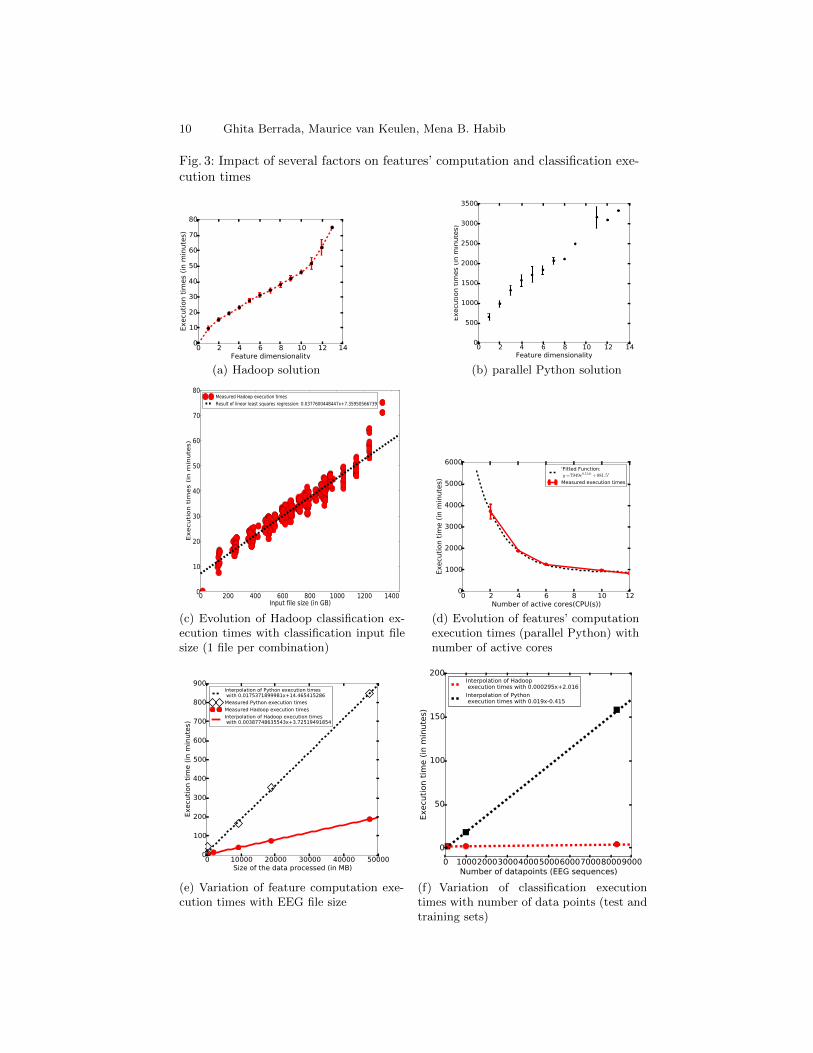
Experiment 1: Feature computation In the first set of experiments, we only per-form the first step of feature selection (described in Section 3.3), i.e EEG segmentextraction and feature computation, on part or all of dataset 1. For each exper-iment, execution times are recorded. Figures 4(a), 4(b), and 4(d) were obtainedusing all of dataset 1. Figure 4(d) explores the evolution of feature computationtimes when the number of cores of the Python server is made to vary. Fea-ture computation execution times grow linearly with the size of processed filesfor both Hadoop and Python solutions (Figure 4(e)) but the Python executiontimes grow 4.5 times faster than the Hadoop ones. Therefore, feature extractionwith Hadoop is especially beneficial for large files and scales to a national scaleamount of data. Based on the interpolations of Figure 4(e), extracting the 10features from Section 3.2 for the whole annual Dutch EEG data(i.e 167GB-Table1) would take about 11 hours and 7 minutes with Hadoop compared to morethan 2 days with Python. The Python execution time decreases exponentiallywith the number of active cores/CPUs (Figure 4(d)) but an infinite number ofCPUs would be needed to reach the same performance as Hadoop!
10 Ghita Berrada, Maurice van Keulen, Mena B. Habib
Fig. 3: Impact of several factors on features’ computation and classification exe-cution times
Result of linear least squares regression: 0.0377600448447x+7.35950566739
(c) Evolution of Hadoop classification ex-ecution times with classification input filesize (1 file per combination)
0 2 4 6 8 10 12Number of active cores(CPU(s))
0
1000
2000
3000
4000
5000
6000
Execu
tion t
ime (
in m
inute
s)
'Fitted Function: y=7949e0.514t +881.5'
Measured execution times
(d) Evolution of features’ computationexecution times (parallel Python) withnumber of active cores
0 10000 20000 30000 40000 50000Size of the data processed (in MB)
0
100
200
300
400
500
600
700
800
900
Execu
tion t
ime (
in m
inute
s)
Interpolation of Python execution times with 0.0175371899981x+14.465415286
Measured Python execution times
Measured Hadoop execution times
Interpolation of Hadoop execution times with 0.00387748635543x+3.72519491854
(e) Variation of feature computation exe-cution times with EEG file size
0 100020003000400050006000700080009000Number of datapoints (EEG sequences)
0
50
100
150
200
Execu
tion t
ime (
in m
inute
s)
Interpolation of Hadoop execution times with 0.000295x+2.016
Interpolation of Python execution times with 0.019x-0.415
(f) Variation of classification executiontimes with number of data points (test andtraining sets)
Hadoop for EEG Storage and Processing: a Feasibility Study 11
Experiment 2: Brute-force classification and feature selection Experiments de-scribed here all use dataset 2 (see Section 4.2 and Table 2(a) for details) andtest the time it takes to assess the classification performance of all possible 511feature combinations 13. 253295 EEG segments are extracted from dataset 2,i.e 113,982,750 values or 1.67% of the total values in the original files. Table 3summarises the results of implementing the feature selection algorithm describedin Section 3.3 with Hadoop Streaming and Python. Due to recurrent memoryerrors, only 154 feature combinations out of 511 (30.14%) were tested for clas-sification with Python. The execution times for Python classification in Table3 are estimates based on available data. Insufficient RAM per Hadoop node ledto all 511 combinations being tested with 37 successive jobs 14 instead of one sothat only 1 map would run per node and not 2. The current implementation isclearly subpar as map slots become available as the job runs but are unusableuntil the job ends and the next starts. This is however easily fixed, with theright user privileges, by setting the maximum number of maps per node to 15so that at any time only one map runs per node: all 511 classifications can thenrun in a single Hadoop job. Table 3 shows that even this suboptimal solutionevaluates the classification performance of all feature sets faster than Python.The gap in classification execution times between Hadoop and Python widenswith the size of datasets to classify (Figure 4(f)). For very small datasets (33training and 67 test points), Python outperforms Hadoop slightly (1.82 minutesfor Python and 2.4 minutes with Hadoop to test all 511 combinations). Hadoophas overall a clear edge over Python as dataset size rises: the classification runsabout 64.76 times faster on Hadoop. Classifying dataset 2’s sequences, even insuboptimal conditions with Hadoop, runs 29.9 to 34.1615 times faster than withPython (see Table 3). So Hadoop is more suited for large datasets’ classification.Hadoop also scales linearly with the size of classification input files16 (Figure4(c)) and handles feature dimensions’ increase better than Python (about 2 or-ders of magnitude faster than Python (Figures 4(a) and 4(b))).
4.4 Discussion
The experiments (Section 4.3) show Hadoop as a scalable and promising solutionto process EEGs if the task at hand it parallelizable (eg feature computation)even if it is CPU-intensive and RAM-bound (classification with all possible fea-ture combinations). It goes to prove that a cluster of commodity hardware (15machines with Dual core processors and only 7.8GB of RAM here) is better atprocessing complex data than a single highly specialized powerful server if thetask is a series of (semi-)independent steps that can run in parallel. Hadoop hasalso been shown to be able to process a national scale amount of data with a
13 all features except nearest neighbor synchronization14 36 testing 14 combinations at a time and 1 testing 7 combinations at a time15 compared to the estimated upper and lower bounds for the Python job respectively16 files obtained by extracting all eyes closed segments from the original EDF+ files
and applying each of the 9 tested features on the extracted segments15 Result of 37 successive jobs instead of only one job testing all 511 combinations16 estimates based on data available
12 Ghita Berrada, Maurice van Keulen, Mena B. Habib
Table 3: Execution times for whole feature selection process on dataset 2 andeach of its steps
Segment extraction& Feature computation Classification only Complete featurefeature computation and formatting selectiononly for classification selection
estimated upper bound: estimated upper bound:12 days 14h34min 12 days 16h2 min
quite small number of cluster machines. This is also a rather cheap solution: acluster like the experimental one costs 10000 to 20000 euros i.e 1000-1500 eu-ros per machine as compared to above 3000 euros per machine for the type ofserver used in the Python experiments. Owning a Hadoop cluster is in theory notneeded as web services like Amazon Elastic Map Reduce (EMR) offer access toHadoop clusters tailored for diverse processing needs. This is not doable, though,given the sensitivity of medical data. And we can boost the Hadoop performancefurther by optimizing the code we wrote by mostly reusing the Python one, viafor example, changing the Hadoop configuration parameters to solve memory is-sues or using other Hadoop Python frameworks like mrjob or Dumbo that don’trequire map/reduce inputs and outputs to be strings passed via stdin/stdoutand should thus need less processing RAM or using machine-learning algorithmsoptimized for the platform (Mahout library).
5 Conclusions
Hadoop is a promising solution for EEG storage and processing. Computationtimes for complex parallelizable machine-learning algorithms are notably reducedcompared to more traditional means of computation and become manageable.The gain in computation times grows with data amount to process, Hadoopscaling easily with national scale data. So it would seem that it is better toprocess data with many commodity machines rather than with one extremelypowerful server, when the processing task is parallelizable. In future, we wouldlike to extend this work to other medical data types such as MRI or CT andstudy how to integrate data from computations run on diverse types of medicaldata (eg MRI and EEG). We would also like to run more tests on medical dataquerying (especially natural language querying). And Hadoop data security alsoneeds to be explored further.
Acknowledgments The EEGs used in this paper were kindly provided by Prof.Dr. Ir. Michel van Putten (Dept. of Neurology and Clinical Neurophysiology,Medisch Spectrum Twente and MIRA, University of Twente, Enschede, TheNetherlands), who we also thank for useful comments on the paper.
Hadoop for EEG Storage and Processing: a Feasibility Study 13
References
1. Manyika, J., Chui, M., Brown, B., Bughin, J., Dobbs, R., Roxburgh, C., Byers,A.H.: Big data: The next frontier for innovation, competition, and productivity.Technical report, Mc Kinsey Global Institute (June 2011)
2. Eta S. Berner, M.L.G.: Overconfidence as a cause of diagnostic error in medicine.Am. J. Med 121(5) (May 2008) S2–S23 (Supplement)
3. Schiff, G.D., Hasan, O., Kim, S., Abrams, R., Cosby, K., Lambert, B.L., Elstein,A.S., Hasler, S., Kabongo, M.L., Krosnjar, N., Odwazny, R., Wisniewski, M.F.,McNutt, R.A.: Diagnostic error in medicine: Analysis of 583 physician-reportederrors. Arch Intern Med 169(20) (2009) 1881–1887
4. Boodman, S.G.: Medical mystery: Depression and possible dementia masked thereal problem. The Washington Post (Dec. 23 2013)
5. OECD: Medical technologies. In: Health at a Glance 2011: OECD Indicators .OECD Publishing (2011)
6. Wiley, K., Connolly, A., Gardner, J.P., Krughof, S., Balazinska, M., Howe, B.,Kwon, Y., Bu, Y.: Astronomy in the cloud: Using mapreduce for image coaddition.CoRR abs/1010.1015 (2010)
7. Bach, F., Cakmak, H.K., Maass, H., Kuehnapfel, U.: Power Grid Time Series DataAnalysis with Pig on a Hadoop Cluster compared to Multi Core Systems. In: Proc.of PDP 2013. (2013)
8. Dutta, H., Kamil, A., Pooleery, M., Sethumadhavan, S., Demme, J.: Distributedstorage of large-scale multidimensional electroencephalogram data using hadoopand hbase. In: Grid and Cloud Database Management. Springer (2011) 331–347
9. Dean, J., Ghemawat, S.: Mapreduce: Simplified data processing on large clusters.In: OSDI, USENIX Association (2004) 137–150
10. Chansler, R.J.: Data availability and durability with the Hadoop Distributed FileSystem. 37(1) (February 2012) 16–22
11. Borthakur, D., Gray, J., Sarma, J.S., Muthukkaruppan, K., Spiegelberg, N., Kuang,H., Ranganathan, K., Molkov, D., Menon, A., Rash, S., Schmidt, R., Aiyer, A.:Apache hadoop goes realtime at facebook. In: Proc. of SIGMOD. (2011) 1071–1080
12. Kemp, B., Olivan, J.: European data format ’plus’ (EDF+), an EDF alike standardformat for the exchange of physiological data. Clin Neurophysiol 114 (2003) 1755–61
13. American Clinical Neurophysiology Society: Guideline 8: Guidelines for recordingclinical EEG on digital media. J Clin Neurophysiol 23 (Apr. 2006) 122–124
14. Goh, C., Hamadicharef, B., Henderson, G.T., Ifeachor, E.C.: Comparison of FractalDimension Algorithms for the Computation of EEG Biomarkers for Dementia. In:Proc. of CIMED. (2005)
15. Cecchin, T., Ranta, R., Koessler, L., Caspary, O., Vespignani, H., Maillard, L.:Seizure lateralization in scalp EEG using Hjorth parameters. Clin Neurophysiol121(3) (Mar. 2010) 290–300
16. Berrada, G., de Keijzer, A.: An IFS-based similarity measure to index electroen-cephalograms. In: Proc. of PAKDD. (2011) 457–468
17. van Putten, M.J.: The Colorful Brain: Visualization of EEG Background Patterns.J Clin Neurophysiol 25(2) (2008) 63–68
18. van Putten, M.J.: Extended BSI for continuous EEG monitoring in carotid en-darterectomy. Clin Neurophysiol 117(12) (2006) 2661–2666
19. Cloostermans, M., de Vos, C., Heida, T., de Keijzer, A., van Putten, M.: Moni-toring the brain in the adult ICU. In: Proc. of the 4th Annual Symposium of theIEEE/EMBS Benelux Chapter. (Nov. 2009) 128–130