119


Haematologic Technologies, Inc. (HTI) is proud to present its research reagent catalog. Inside this catalog you will find a comprehensive line of human, bovine and murine co-agulation reagents along with antibodies, factor deficient plasmas and customized blood collection tubes. Firmly into our third decade of business, we remain focused on our mission to manufacture and deliver the highest quality research reagents available, and to strive for the highest customer satisfaction possible.This is exemplified by our ISO 9001 certification, which we obtained in September of 2007, and reflected in our quality policy: “HTI is committed to achieving the highest level of customer satisfaction, and to the continual improvement of its products, services and quality management system.”
ServicesHaematologic Technologies is proud to offer contract services through our Haemtech Biopharma Services division (www.haemtechbiopharma.com). HTI can provide solutions for your biopharmaceutical, diagnostic or research needs, and our extensive expertise in protein chemistry, state-of-the-art instrumentation and rigorous quality systems will deliver the results you need to go from bench-top to market. Our services include:
• Assay services• Characterization• Formulation• Immunogenicity assay development and testing• Methods optimization• OEM manufacturing• Process development• Protein purification• Qualification and validation• Release testing• Stability testing
Please inquire directly about these or other services that you may require. We can be reached at 802-878-1777 or via the Web at http://www.haemtech.com.

TABLE OF CONTENTSGENERAL INFORMATIONORDERING (Terms and Conditions) . . . . . . . . . . . . . . . . . . . . . . . . . . . . . . . . . . . . . . . . . . . . . . . . . . . . A-1
TECHNICAL NOTES . . . . . . . . . . . . . . . . . . . . . . . . . . . . . . . . . . . . . . . . . . . . . . . . . . . . . . . . . . . . . . . . A-2
HUMAN PROTEINSHUMAN ZYMOGENSFACTOR VII . . . . . . . . . . . . . . . . . . . . . . . . . . . . . . . . . . . . . . . . . . . . . . . . . . . . . . . . . . . . . . . . . . . . . . . B-1
FACTOR IX . . . . . . . . . . . . . . . . . . . . . . . . . . . . . . . . . . . . . . . . . . . . . . . . . . . . . . . . . . . . . . . . . . . . . . . . B-3
FACTOR X . . . . . . . . . . . . . . . . . . . . . . . . . . . . . . . . . . . . . . . . . . . . . . . . . . . . . . . . . . . . . . . . . . . . . . . . B-5
FACTOR X (modified) . . . . . . . . . . . . . . . . . . . . . . . . . . . . . . . . . . . . . . . . . . . . . . . . . . . . . . . . . . . . . . . . B-5
FACTOR XI . . . . . . . . . . . . . . . . . . . . . . . . . . . . . . . . . . . . . . . . . . . . . . . . . . . . . . . . . . . . . . . . . . . . . . . . B-7
FACTOR XII . . . . . . . . . . . . . . . . . . . . . . . . . . . . . . . . . . . . . . . . . . . . . . . . . . . . . . . . . . . . . . . . . . . . . . . B-9
FACTOR XIII . . . . . . . . . . . . . . . . . . . . . . . . . . . . . . . . . . . . . . . . . . . . . . . . . . . . . . . . . . . . . . . . . . . . . . B-11
glu-PLASMINOGEN . . . . . . . . . . . . . . . . . . . . . . . . . . . . . . . . . . . . . . . . . . . . . . . . . . . . . . . . . . . . . . . . B-13
glu-PLASMINOGEN (affinity forms I and II) . . . . . . . . . . . . . . . . . . . . . . . . . . . . . . . . . . . . . . . . . . . . . . B-13
lys-PLASMINOGEN . . . . . . . . . . . . . . . . . . . . . . . . . . . . . . . . . . . . . . . . . . . . . . . . . . . . . . . . . . . . . . . . B-13
PROTEIN C . . . . . . . . . . . . . . . . . . . . . . . . . . . . . . . . . . . . . . . . . . . . . . . . . . . . . . . . . . . . . . . . . . . . . . B-15
PROTHROMBIN . . . . . . . . . . . . . . . . . . . . . . . . . . . . . . . . . . . . . . . . . . . . . . . . . . . . . . . . . . . . . . . . . . . B-17
TAFI . . . . . . . . . . . . . . . . . . . . . . . . . . . . . . . . . . . . . . . . . . . . . . . . . . . . . . . . . . . . . . . . . . . . . . . . . . . . E-13
HUMAN ENZYMESACTIVATED PROTEIN C . . . . . . . . . . . . . . . . . . . . . . . . . . . . . . . . . . . . . . . . . . . . . . . . . . . . . . . . . . . . C-1
ACTIVATED PROTEIN C (active site blocked) . . . . . . . . . . . . . . . . . . . . . . . . . . . . . . . . . . . . . . . . . . . . C-1
a-THROMBIN . . . . . . . . . . . . . . . . . . . . . . . . . . . . . . . . . . . . . . . . . . . . . . . . . . . . . . . . . . . . . . . . . . . . . . C-3
a-THROMBIN (active site blocked) . . . . . . . . . . . . . . . . . . . . . . . . . . . . . . . . . . . . . . . . . . . . . . . . . . . . . C-3
b-THROMBIN . . . . . . . . . . . . . . . . . . . . . . . . . . . . . . . . . . . . . . . . . . . . . . . . . . . . . . . . . . . . . . . . . . . . . . C-5
g-THROMBIN . . . . . . . . . . . . . . . . . . . . . . . . . . . . . . . . . . . . . . . . . . . . . . . . . . . . . . . . . . . . . . . . . . . . . . C-5
FACTOR VIIa . . . . . . . . . . . . . . . . . . . . . . . . . . . . . . . . . . . . . . . . . . . . . . . . . . . . . . . . . . . . . . . . . . . . . . C-7
FACTOR IXa . . . . . . . . . . . . . . . . . . . . . . . . . . . . . . . . . . . . . . . . . . . . . . . . . . . . . . . . . . . . . . . . . . . . . . . C-9
FACTOR IXa (active site blocked) . . . . . . . . . . . . . . . . . . . . . . . . . . . . . . . . . . . . . . . . . . . . . . . . . . . . . . C-9
FACTOR Xa . . . . . . . . . . . . . . . . . . . . . . . . . . . . . . . . . . . . . . . . . . . . . . . . . . . . . . . . . . . . . . . . . . . . . . C-11
FACTOR Xa (modified or active site blocked) . . . . . . . . . . . . . . . . . . . . . . . . . . . . . . . . . . . . . . . . . . . . C-11
FACTOR XIa . . . . . . . . . . . . . . . . . . . . . . . . . . . . . . . . . . . . . . . . . . . . . . . . . . . . . . . . . . . . . . . . . . . . . . C-13
FACTOR XIIIa . . . . . . . . . . . . . . . . . . . . . . . . . . . . . . . . . . . . . . . . . . . . . . . . . . . . . . . . . . . . . . . . . . . . . C-15
PLASMIN . . . . . . . . . . . . . . . . . . . . . . . . . . . . . . . . . . . . . . . . . . . . . . . . . . . . . . . . . . . . . . . . . . . . . . . . C-17
I

II
HUMAN COFACTORSFACTOR V . . . . . . . . . . . . . . . . . . . . . . . . . . . . . . . . . . . . . . . . . . . . . . . . . . . . . . . . . . . . . . . . . . . . . . . . D-1
FACTOR Va . . . . . . . . . . . . . . . . . . . . . . . . . . . . . . . . . . . . . . . . . . . . . . . . . . . . . . . . . . . . . . . . . . . . . . . D-3
FIBRONECTIN . . . . . . . . . . . . . . . . . . . . . . . . . . . . . . . . . . . . . . . . . . . . . . . . . . . . . . . . . . . . . . . . . . . . . D-5
PROTEIN S . . . . . . . . . . . . . . . . . . . . . . . . . . . . . . . . . . . . . . . . . . . . . . . . . . . . . . . . . . . . . . . . . . . . . . . D-7
von WILLEBRAND FACTOR . . . . . . . . . . . . . . . . . . . . . . . . . . . . . . . . . . . . . . . . . . . . . . . . . . . . . . . . . D-11
PROTEIN Z . . . . . . . . . . . . . . . . . . . . . . . . . . . . . . . . . . . . . . . . . . . . . . . . . . . . . . . . . . . . . . . . . . . . . . D-13
HUMAN PROTEASE INHIBITORSANTITHROMBIN III . . . . . . . . . . . . . . . . . . . . . . . . . . . . . . . . . . . . . . . . . . . . . . . . . . . . . . . . . . . . . . . . . E-3
HEPARIN COFACTOR II . . . . . . . . . . . . . . . . . . . . . . . . . . . . . . . . . . . . . . . . . . . . . . . . . . . . . . . . . . . . E-11
TAFI (thrombin-activatable fibrinolysis inhibitor) . . . . . . . . . . . . . . . . . . . . . . . . . . . . . . . . . . . . . . . . . . . E-13
ANGIOSTATIN . . . . . . . . . . . . . . . . . . . . . . . . . . . . . . . . . . . . . . . . . . . . . . . . . . . . . . . . . . . . . . . . . . . . E-15
a2-PLASMIN INHIBITOR . . . . . . . . . . . . . . . . . . . . . . . . . . . . . . . . . . . . . . . . . . . . . . . . . . . . . . . . . . . . E-17
HUMAN PLATELET AND OTHER PLASMA DERIVED PROTEINSFIBRINOGEN AND FIBRINOGEN FRAGMENTS . . . . . . . . . . . . . . . . . . . . . . . . . . . . . . . . . . . . . . . . . . F-1
b2-GLYCOPROTEIN I . . . . . . . . . . . . . . . . . . . . . . . . . . . . . . . . . . . . . . . . . . . . . . . . . . . . . . . . . . . . . . . F-3
HUMAN PLATELET OSTEONECTIN . . . . . . . . . . . . . . . . . . . . . . . . . . . . . . . . . . . . . . . . . . . . . . . . . . . .G-3
PLATELET FACTOR 4 . . . . . . . . . . . . . . . . . . . . . . . . . . . . . . . . . . . . . . . . . . . . . . . . . . . . . . . . . . . . . . . F-5
b-THROMBOGLOBULIN . . . . . . . . . . . . . . . . . . . . . . . . . . . . . . . . . . . . . . . . . . . . . . . . . . . . . . . . . . . . . F-7
THROMBOSPONDIN . . . . . . . . . . . . . . . . . . . . . . . . . . . . . . . . . . . . . . . . . . . . . . . . . . . . . . . . . . . . . . . . F-9
VITRONECTIN . . . . . . . . . . . . . . . . . . . . . . . . . . . . . . . . . . . . . . . . . . . . . . . . . . . . . . . . . . . . . . . . . . . . F-11
HUMAN BONE RELATED PROTEINSOSTEOCALCIN . . . . . . . . . . . . . . . . . . . . . . . . . . . . . . . . . . . . . . . . . . . . . . . . . . . . . . . . . . . . . . . . . . . .G-1
BOVINE PROTEINSBOVINE ZYMOGENSFACTOR IX . . . . . . . . . . . . . . . . . . . . . . . . . . . . . . . . . . . . . . . . . . . . . . . . . . . . . . . . . . . . . . . . . . . . . . . . B-3
FACTOR X . . . . . . . . . . . . . . . . . . . . . . . . . . . . . . . . . . . . . . . . . . . . . . . . . . . . . . . . . . . . . . . . . . . . . . . . B-5
glu-PLASMINOGEN . . . . . . . . . . . . . . . . . . . . . . . . . . . . . . . . . . . . . . . . . . . . . . . . . . . . . . . . . . . . . . . . B-13
PROTEIN C . . . . . . . . . . . . . . . . . . . . . . . . . . . . . . . . . . . . . . . . . . . . . . . . . . . . . . . . . . . . . . . . . . . . . . B-15
PROTHROMBIN . . . . . . . . . . . . . . . . . . . . . . . . . . . . . . . . . . . . . . . . . . . . . . . . . . . . . . . . . . . . . . . . . . . B-17
BOVINE ENZYMESACTIVATED PROTEIN C . . . . . . . . . . . . . . . . . . . . . . . . . . . . . . . . . . . . . . . . . . . . . . . . . . . . . . . . . . . . . C-1
ACTIVATED PROTEIN C (active site blocked) . . . . . . . . . . . . . . . . . . . . . . . . . . . . . . . . . . . . . . . . . . . . C-1
a-THROMBIN . . . . . . . . . . . . . . . . . . . . . . . . . . . . . . . . . . . . . . . . . . . . . . . . . . . . . . . . . . . . . . . . . . . . . . C-3

III
a-THROMBIN (active site blocked) . . . . . . . . . . . . . . . . . . . . . . . . . . . . . . . . . . . . . . . . . . . . . . . . . . . . . C-3
FACTOR IXa . . . . . . . . . . . . . . . . . . . . . . . . . . . . . . . . . . . . . . . . . . . . . . . . . . . . . . . . . . . . . . . . . . . . . . . C-9
FACTOR IXa (active site blocked) . . . . . . . . . . . . . . . . . . . . . . . . . . . . . . . . . . . . . . . . . . . . . . . . . . . . . . C-9
FACTOR Xa . . . . . . . . . . . . . . . . . . . . . . . . . . . . . . . . . . . . . . . . . . . . . . . . . . . . . . . . . . . . . . . . . . . . . . C-11
FACTOR Xa (modified or active site blocked) . . . . . . . . . . . . . . . . . . . . . . . . . . . . . . . . . . . . . . . . . . . . C-11
BOVINE COFACTORSFACTOR V . . . . . . . . . . . . . . . . . . . . . . . . . . . . . . . . . . . . . . . . . . . . . . . . . . . . . . . . . . . . . . . . . . . . . . . . D-1
FACTOR Va . . . . . . . . . . . . . . . . . . . . . . . . . . . . . . . . . . . . . . . . . . . . . . . . . . . . . . . . . . . . . . . . . . . . . . . D-3
BOVINE BONE-RELATED AND OTHER PROTEINSOSTEOCALCIN . . . . . . . . . . . . . . . . . . . . . . . . . . . . . . . . . . . . . . . . . . . . . . . . . . . . . . . . . . . . . . . . . . . .G-1
BOVINE BONE OSTEONECTIN . . . . . . . . . . . . . . . . . . . . . . . . . . . . . . . . . . . . . . . . . . . . . . . . . . . . . . .G-3
LACTADHERIN . . . . . . . . . . . . . . . . . . . . . . . . . . . . . . . . . . . . . . . . . . . . . . . . . . . . . . . . . . . . . . . . . . . .G-5
MURINE PROTEINS (please inquire about those not listed)MOUSE PROTHROMBIN . . . . . . . . . . . . . . . . . . . . . . . . . . . . . . . . . . . . . . . . . . . . . . . . . . . . . . . . . . . B-17
MOUSE FACTOR IX . . . . . . . . . . . . . . . . . . . . . . . . . . . . . . . . . . . . . . . . . . . . . . . . . . . . . . . . . . . . . . . . B-3
MOUSE FACTOR X . . . . . . . . . . . . . . . . . . . . . . . . . . . . . . . . . . . . . . . . . . . . . . . . . . . . . . . . . . . . . . . . . B-5
MOUSE PLASMINOGEN . . . . . . . . . . . . . . . . . . . . . . . . . . . . . . . . . . . . . . . . . . . . . . . . . . . . . . . . . . . . B-13
MOUSE THROMBIN . . . . . . . . . . . . . . . . . . . . . . . . . . . . . . . . . . . . . . . . . . . . . . . . . . . . . . . . . . . . . . . . C-3
MOUSE FACTOR XA . . . . . . . . . . . . . . . . . . . . . . . . . . . . . . . . . . . . . . . . . . . . . . . . . . . . . . . . . . . . . . . C-11
MOUSE ANTITHROMBIN III . . . . . . . . . . . . . . . . . . . . . . . . . . . . . . . . . . . . . . . . . . . . . . . . . . . . . . . . . . E-3
MOUSE PROTEIN C . . . . . . . . . . . . . . . . . . . . . . . . . . . . . . . . . . . . . . . . . . . . . . . . . . . . . . . . . . . . . . . B-15
MOUSE ACTIVATED PROTEIN C . . . . . . . . . . . . . . . . . . . . . . . . . . . . . . . . . . . . . . . . . . . . . . . . . . . . . . C-1
MOUSE FIBRINOGEN . . . . . . . . . . . . . . . . . . . . . . . . . . . . . . . . . . . . . . . . . . . . . . . . . . . . . . . . . . . . . . . F-1
PROTEINS AND PRODUCTS FROM OTHER SPECIESRABBIT THROMBOMODULIN . . . . . . . . . . . . . . . . . . . . . . . . . . . . . . . . . . . . . . . . . . . . . . . . . . . . . . . . . D-9
FOR RABBIT, RAT, CANINE, PORCINE, ETC, SEE INFORMATION PAGE . . . . . . . . . . . . . . . . . .PAGE VI
INHIBITORS AND SUBSTRATES ANSN-BASED FLUOROGENIC SUBSTRATES . . . . . . . . . . . . . . . . . . . . . . . . . . . . . . . . . . . . . . . . . . . E-1
BIOTINYLATED EGR-CHLOROMETHYLKETONE . . . . . . . . . . . . . . . . . . . . . . . . . . . . . . . . . . . . . . . . . E-5
FLUORESCEIN EGR-CHLOROMETHYLKETONE . . . . . . . . . . . . . . . . . . . . . . . . . . . . . . . . . . . . . . . . . E-5
BIOTINYLATED FPR-CHLOROMETHYLKETONE . . . . . . . . . . . . . . . . . . . . . . . . . . . . . . . . . . . . . . . . . E-5
FLUORESCEIN FPR-CHLOROMETHYLKETONE . . . . . . . . . . . . . . . . . . . . . . . . . . . . . . . . . . . . . . . . . E-5

IV
FPR-CHLOROMETHYLKETONE . . . . . . . . . . . . . . . . . . . . . . . . . . . . . . . . . . . . . . . . . . . . . . . . . . . . . . E-5
EGR-CHLOROMETHYLKETONE . . . . . . . . . . . . . . . . . . . . . . . . . . . . . . . . . . . . . . . . . . . . . . . . . . . . . . E-5
CORN TRYPSIN INHIBITOR . . . . . . . . . . . . . . . . . . . . . . . . . . . . . . . . . . . . . . . . . . . . . . . . . . . . . . . . . . E-7
DAPA (dansylarginine-N-[3-ethyl-1,5-pentanediyl]amide) . . . . . . . . . . . . . . . . . . . . . . . . . . . . . . . . . . . . E-9
VENOM PROTEASESRVV-X ACTIVATOR . . . . . . . . . . . . . . . . . . . . . . . . . . . . . . . . . . . . . . . . . . . . . . . . . . . . . . . . . . . . . . . . C-19
RVV-V ACTIVATOR . . . . . . . . . . . . . . . . . . . . . . . . . . . . . . . . . . . . . . . . . . . . . . . . . . . . . . . . . . . . . . . . C-19
E . CARINATUS II PROTEASE . . . . . . . . . . . . . . . . . . . . . . . . . . . . . . . . . . . . . . . . . . . . . . . . . . . . . . . . C-19
ANTIBODIESMONOCLONAL ANTIBODIESANTI-FACTOR V . . . . . . . . . . . . . . . . . . . . . . . . . . . . . . . . . . . . . . . . . . . . . . . . . . . . . . . . . . . . . . . . . . . H-1
ANTI-HUMAN FACTOR VII . . . . . . . . . . . . . . . . . . . . . . . . . . . . . . . . . . . . . . . . . . . . . . . . . . . . . . . . . . . H-1
ANTI-HUMAN FACTOR IX . . . . . . . . . . . . . . . . . . . . . . . . . . . . . . . . . . . . . . . . . . . . . . . . . . . . . . . . . . . . H-1
ANTI-HUMAN FACTOR X . . . . . . . . . . . . . . . . . . . . . . . . . . . . . . . . . . . . . . . . . . . . . . . . . . . . . . . . . . . . H-1
ANTI-MURINE FACTOR X . . . . . . . . . . . . . . . . . . . . . . . . . . . . . . . . . . . . . . . . . . . . . . . . . . . . . . . . . . . . H-2
ANTI-HUMAN PROTEIN C . . . . . . . . . . . . . . . . . . . . . . . . . . . . . . . . . . . . . . . . . . . . . . . . . . . . . . . . . . . H-2
ANTI-MURINE PROTEIN C . . . . . . . . . . . . . . . . . . . . . . . . . . . . . . . . . . . . . . . . . . . . . . . . . . . . . . . . . . . H-2
ANTI-HUMAN PROTEIN S . . . . . . . . . . . . . . . . . . . . . . . . . . . . . . . . . . . . . . . . . . . . . . . . . . . . . . . . . . . . H-2
ANTI-HUMAN THROMBIN . . . . . . . . . . . . . . . . . . . . . . . . . . . . . . . . . . . . . . . . . . . . . . . . . . . . . . . . . . . . H-2
ANTI-HUMAN PROTHROMBIN . . . . . . . . . . . . . . . . . . . . . . . . . . . . . . . . . . . . . . . . . . . . . . . . . . . . . . . . H-2
ANTI-MURINE PROTHROMBIN . . . . . . . . . . . . . . . . . . . . . . . . . . . . . . . . . . . . . . . . . . . . . . . . . . . . . . . H-2
ANTI-MURINE PLASMINOGEN . . . . . . . . . . . . . . . . . . . . . . . . . . . . . . . . . . . . . . . . . . . . . . . . . . . . . . . . H-2
ANTI-HUMAN FACTOR XI . . . . . . . . . . . . . . . . . . . . . . . . . . . . . . . . . . . . . . . . . . . . . . . . . . . . . . . . . . . . H-2
ANTI-HUMAN FACTOR XII . . . . . . . . . . . . . . . . . . . . . . . . . . . . . . . . . . . . . . . . . . . . . . . . . . . . . . . . . . . H-2
ANTI-TAFI . . . . . . . . . . . . . . . . . . . . . . . . . . . . . . . . . . . . . . . . . . . . . . . . . . . . . . . . . . . . . . . . . . . . . . . . . H-3
ANTI-OSTEONECTIN . . . . . . . . . . . . . . . . . . . . . . . . . . . . . . . . . . . . . . . . . . . . . . . . . . . . . . . . . . . . . . . H-3
ANTI-OSTEOCALCIN . . . . . . . . . . . . . . . . . . . . . . . . . . . . . . . . . . . . . . . . . . . . . . . . . . . . . . . . . . . . . . . H-3
ANTI-HUMAN TISSUE FACTOR . . . . . . . . . . . . . . . . . . . . . . . . . . . . . . . . . . . . . . . . . . . . . . . . . . . . . . . H-3
ANTI-HUMAN TISSUE FACTOR PATHWAY INHIBITOR . . . . . . . . . . . . . . . . . . . . . . . . . . . . . . . . . . . . . H-3
POLYCLONAL ANTIBODIESANTI-HUMAN PROTHROMBIN . . . . . . . . . . . . . . . . . . . . . . . . . . . . . . . . . . . . . . . . . . . . . . . . . . . . . . . . H-4
ANTI-HUMAN THROMBIN . . . . . . . . . . . . . . . . . . . . . . . . . . . . . . . . . . . . . . . . . . . . . . . . . . . . . . . . . . . . H-4
ANTI-HUMAN FACTOR V . . . . . . . . . . . . . . . . . . . . . . . . . . . . . . . . . . . . . . . . . . . . . . . . . . . . . . . . . . . . H-4
ANTI-HUMAN FACTOR Va . . . . . . . . . . . . . . . . . . . . . . . . . . . . . . . . . . . . . . . . . . . . . . . . . . . . . . . . . . . H-4

V
ANTI-BOVINE FACTOR V . . . . . . . . . . . . . . . . . . . . . . . . . . . . . . . . . . . . . . . . . . . . . . . . . . . . . . . . . . . . H-4
ANTI-HUMAN FACTOR VI . . . . . . . . . . . . . . . . . . . . . . . . . . . . . . . . . . . . . . . . . . . . . . . . . . . . . . . . . . . . H-4
ANTI-HUMAN FACTOR VIIIc . . . . . . . . . . . . . . . . . . . . . . . . . . . . . . . . . . . . . . . . . . . . . . . . . . . . . . . . . . H-4
ANTI-HUMAN FACTOR IX . . . . . . . . . . . . . . . . . . . . . . . . . . . . . . . . . . . . . . . . . . . . . . . . . . . . . . . . . . . . H-4
ANTI-HUMAN FACTOR X . . . . . . . . . . . . . . . . . . . . . . . . . . . . . . . . . . . . . . . . . . . . . . . . . . . . . . . . . . . . H-4
ANTI-HUMAN FACTOR XI . . . . . . . . . . . . . . . . . . . . . . . . . . . . . . . . . . . . . . . . . . . . . . . . . . . . . . . . . . . . H-4
ANTI-HUMAN FACTOR XII . . . . . . . . . . . . . . . . . . . . . . . . . . . . . . . . . . . . . . . . . . . . . . . . . . . . . . . . . . . H-4
ANTI-HUMAN FACTOR XIII . . . . . . . . . . . . . . . . . . . . . . . . . . . . . . . . . . . . . . . . . . . . . . . . . . . . . . . . . . . H-4
ANTI-HUMAN PROTEIN C . . . . . . . . . . . . . . . . . . . . . . . . . . . . . . . . . . . . . . . . . . . . . . . . . . . . . . . . . . . H-4
ANTI-HUMAN PROTEIN S . . . . . . . . . . . . . . . . . . . . . . . . . . . . . . . . . . . . . . . . . . . . . . . . . . . . . . . . . . . . H-4
ANTI-HUMAN PLASMINOGEN . . . . . . . . . . . . . . . . . . . . . . . . . . . . . . . . . . . . . . . . . . . . . . . . . . . . . . . . H-4
ANTI-HUMAN ANTITHROMBIN III . . . . . . . . . . . . . . . . . . . . . . . . . . . . . . . . . . . . . . . . . . . . . . . . . . . . . . H-4
ANTI-HUMAN HEPARIN COFACTOR II . . . . . . . . . . . . . . . . . . . . . . . . . . . . . . . . . . . . . . . . . . . . . . . . . H-4
ANTI-TAFI . . . . . . . . . . . . . . . . . . . . . . . . . . . . . . . . . . . . . . . . . . . . . . . . . . . . . . . . . . . . . . . . . . . . . . . . . H-4
ANTI-HUMAN von WILLEBRAND FACTOR . . . . . . . . . . . . . . . . . . . . . . . . . . . . . . . . . . . . . . . . . . . . . . H-4
ANTI-HUMAN TISSUE FACTOR . . . . . . . . . . . . . . . . . . . . . . . . . . . . . . . . . . . . . . . . . . . . . . . . . . . . . . . H-4
ANTI-HUMAN TISSUE FACTOR PATHWAY INHIBITOR . . . . . . . . . . . . . . . . . . . . . . . . . . . . . . . . . . . . . H-4
ANTI-HUMAN PROTEIN Z . . . . . . . . . . . . . . . . . . . . . . . . . . . . . . . . . . . . . . . . . . . . . . . . . . . . . . . . . . . . H-4
ANTI-MURINE FACTOR X . . . . . . . . . . . . . . . . . . . . . . . . . . . . . . . . . . . . . . . . . . . . . . . . . . . . . . . . . . . . H-4
ANTI-MURINE PROTHROMBIN . . . . . . . . . . . . . . . . . . . . . . . . . . . . . . . . . . . . . . . . . . . . . . . . . . . . . . . H-5
ANTI-MURINE PLASMINOGEN . . . . . . . . . . . . . . . . . . . . . . . . . . . . . . . . . . . . . . . . . . . . . . . . . . . . . . . . H-5
ANTI-MURINE PROTEIN C . . . . . . . . . . . . . . . . . . . . . . . . . . . . . . . . . . . . . . . . . . . . . . . . . . . . . . . . . . . H-5
ANTI-MURINE ATIII . . . . . . . . . . . . . . . . . . . . . . . . . . . . . . . . . . . . . . . . . . . . . . . . . . . . . . . . . . . . . . . . . H-5
ANTI-MURINE FIBRINOGEN . . . . . . . . . . . . . . . . . . . . . . . . . . . . . . . . . . . . . . . . . . . . . . . . . . . . . . . . . . H-5
ANTI-RAT FACTOR IX . . . . . . . . . . . . . . . . . . . . . . . . . . . . . . . . . . . . . . . . . . . . . . . . . . . . . . . . . . . . . . H-5
DEFICIENT PLASMASALL PLASMAS . . . . . . . . . . . . . . . . . . . . . . . . . . . . . . . . . . . . . . . . . . . . . . . . . . . . . . . . . . . . . . . . . . . . . .I-1
COLLECTION TUBESSAMPLE COLLECTION/ANTICOAGULANT TUBES . . . . . . . . . . . . . . . . . . . . . . . . . . . . . . . . . . . . . . . J-1
CONTRACT RESEARCH, CONTRACT MANUFACTURING AND CUSTOM PROTEIN PURIFICATIONSEE INFORMATION PAGE . . . . . . . . . . . . . . . . . . . . . . . . . . . . . . . . . . . . . . . . . . . . . . . . . . . . . . .PAGE VI

ADDITIONAL PRODUCTS AND SERVICES INFORMATIONCUSTOM PROTEIN PURIFICATION AND CHARACTERIZATION
Frequently researchers require plasma proteins that do not appear in our catalog (e .g . other human pro-teins or proteins from other species), or require a modification of those that do . Haematologic Technologies has the ability to meet these needs, and will work with you to provide the protein that you require—fully characterized to your specifications . Some examples of non-listed proteins we have produced include:
• Porcine Factor V • Rabbit Factor Xa• Canine Fibrinogen • Rat Factor X• Rabbit Factor X • Rabbit Thrombin• Human Glutathione Peroxidase • And many more…
CONTRACT RESEARCH
Haematologic Technologies’ contract research division specializes in performing all levels of coagulation research for our clients . From ELISA development to complex product design, HTI can assist you with your research outsourcing needs . HTI offers dedicated staff and research/manufacturing suites to keep your project on target . And of course, your project will be treated with the utmost respect and confidentiality .
CONTRACT MANUFACTURING
In addition to research, Haematologic Technologies offers contract manufacturing for those who need a continuous supply of specially developed research reagents for in vitro use, and find it to be more cost-effective to outsource the production of those reagents . HTI will manufacture your reagents to your specifi-cations, and will perform the necessary quality control in-house . And, with our dedication to quality you can be assured that product consistency will never be compromised .
ABOUT HAEMATOLOGIC TECHNOLOGIES
Haematologic Technologies, Inc . (HTI) is a primary manufacturer that specializes in the isolation and char-acterization of high quality, human plasma proteins which are intended for in vitro research use . HTI’s emphasis is focused on proteins involved in the regulation of blood coagulation and fibrinolysis, as well as the regulation of bone metabolism . The HTI product line consists of nearly 150 highly purified and well-characterized proteins including zymogens, enzymes, cofactors, inhibitors, and platelet proteins as well as a complementary line of monoclonal and polyclonal antibodies . Additionally, HTI offers a broad line of ser-vices, which includes: custom protein purification, protein modification, assay development, and contract research .
VI

ORDERING, DELIVERYPlease direct orders to:
Haematologic Technologies, Inc .
Telephone: +1 (802) 878-1777Telefax: +1 (802) 878-1776e-mail: hti@haemtech .comWeb: http://www .haemtech .com
All domestic orders are shipped promptly via overnight courier . Items requiring blue or dry ice are routinely shipped Monday through Thursday only . Shipping and handling charges are prepaid by Haematologic Technologies, Inc . and subsequently added to the total invoiced amount .
International orders are routinely shipped on Mondays . The procurement of any necessary import permits and all duties, import and license fees are the responsibility of the customer .
OUR PRODUCTS ARE ALSO AVAILABLE FROM OUR NETwORk OF INTERNATIONAL DISTRIBUTORS, wHICH CAN BE FOUND AT www.haemtech.com.
Visit us on the internet at http://www.haemtech.comPlease inquire about custom formulation, modification and conjugation.
A
A-1
Specify:
• Purchase Order Number or Credit Card Number (MC/VISA)• Shipping Address• Billing Address• Catalog Number and Product Name• Size and Quantity
PRICING, TERMS OF PAYMENTPrices are subject to change without notice . Bulk discounts are available based on the quantity of the order . Please inquire when the order is placed . All orders are binding if confirmed in writing or through delivery of the ordered products .
Payment in U .S . dollars is due within 30 days of the invoice date . Wire transfers are accepted with customer paying all associated bank fees . Inquire for details . We also accept VISA and MasterCard payments .
All prices are FOB Essex Junction . Our shipping responsibility ceases with safe delivery to the trans-porta-tion company . Any shipment received damaged must be immediately reported to the shipping company . In addition, Haematologic Technologies, Inc . should be informed immediately . No products may be returned without authorization from Haematologic Technologies, Inc .
wARRANTYHaematologic Technologies, Inc . warrants its products to the original purchaser against defects in materials under normal use or application . This warranty applies to products in original containers and does not apply to a product which has been subjected to alteration .
All products are developed and sold for research purposes and/or in vitro use only . Preparations with known toxicity are shipped with an information sheet describing, to the best of our knowledge, any known hazards in handling . All products should be handled by trained personnel only . Proper procedures for handling potential viral contaminants should be followed for all products isolated from human, bovine and murine source materials .

TECHNICAL NOTES
Our “Technical Notes” section is a new addition to the catalog . It is not meant to serve as a methods section, but instead focuses on some of the most common questions that we receive in our technical services department . Many of the recommendations that follow are generalized, and may differ from many established protocols . If you have technical questions that we do not address in this section, or if you require further assistance, please do not hesitate to call (802) 878-1777 and ask for our technical services department .
PRODUCT STORAGE AND HANDLING
All orders shipped from Haematologic Technologies, Inc . are accompanied by product information sheets which describe proper storage conditions . In order that we may warrant product stability, it is imperative that these storage conditions be maintained at all times . Many of our protein preparations are formulated in 50% (vol/vol) glycerol/H2O which will remain in fluid phase during storage at -20°C . This preferred method of storage yields the greatest protein stability while still allowing access to the stock protein sample without repeated thawing and freezing steps . For some of our new customers who may not be accustomed to handling proteins in glycerol/H2O, here are a few basic tips:
• All products which are formulated with either glycerol/H2O or aqueous buffer are delivered in microcentrifuge tubes . By briefly centrifuging the samples in their original containers, complete recovery of the sample at the bottom of the tube will be accomplished .
• All products which are formulated with glycerol/H2O should be stored at -20°C . Temperatures lower than -30°C should be avoided in order to prevent a phase transition .
• When preparing to make a dilution of the stock sample, remove the sample from storage at -20°C and place on ice for a brief period of time (5-10 min) . The sample will become less viscous and thus easier to pipette .
• Never allow protein solutions to remain at room temperature for excessive periods of time . Elevated temperatures may enhance the rate of protein degradation .
• Avoid storing or maintaining dilute protein samples for a long period of time . In general, purified proteins are inherently more stable in concentrated form .
• Many proteins are “sticky” by nature . To avoid losing protein due to adsorption, extremely dilute protein samples should be prepared in buffers containing excipients such as bovine serum albumin, poly(ethylene glycol), or gelatin .
Visit us on the internet at http://www.haemtech.comPlease inquire about custom formulation, modification and conjugation.A-2

ENZYME ASSAYS AND ACTIVITY UNITS
Data sheets: Data sheets documenting the specific activity of HTI products are included in all shipments . The type of assay performed and an explanation of the units of activity are included in footnotes .
Assays and units: In general, the activities of HTI products are measured in standard PT and APTT based clotting assays where one unit of activity is defined as the amount of activity present in 1 ml of normal pooled human plasma . Exceptions to this rule include the following:
NIH Units . Thrombin Activity is reported in NIH units . This activity is determined by comparison to U .S . Standard Thrombin, Lot J .
Chromogenic Substrate Hydrolysis . The activity of some enzymes (ex . APC, plasmin) are determined by the rate of hydrolysis of chromogenic substrates . 1 unit of activity is defined as the hydrolysis of 1 µmole of substrate per minute, under defined reaction conditions . It should be noted that this activity is highly dependent on the particular substrate used, the concentration of substrate, pH, ionic strength and temperature . The conditions are specified on the product data sheet included in all shipments .
wESTERN BLOTTING
Basic buffers: TBS: 8 .76 grams of NaCl and 2 .42 grams of Tris base in 900 ml of deionized water . Adjust pH to 7 .4 with 6N HCl . Adjust the volume to 1 liter with deionized water .
PBS: 8 .0 grams of NaCl, 0 .2 grams of KCl, 1 .44 grams of Na2HPO4 and 0 .24 grams of KH2PO4 in 900 ml of deionized water . Adjust pH to 7 .4 . Adjust volume to 1 liter with deionized water .
Blocking buffer: TBS or PBS containing one of the following: 2%(w/v) BSA, ovalbumin or casein, or 5%(w/v) non-fat powdered milk .
BSA/Tween buffer: TBS or PBS containing 0 .02%(w/v) BSA and 0 .05% (v/v) Tween 20 .
Blocking conditions: After the protein samples have been transferred from the gel to either a nitrocellulose or PVDF membrane, remaining binding sites are blocked by incubating the blot in blocking buffer for 2 hours at room temperature or overnight at 4°C . Be sure to gently mix or agitate the blot during all blocking and incubation steps .
wash steps: Wash the blot at least three times (incubating for 3 minutes with each wash) with BSA/Tween buffer between each procedural step .
Primary antibody: Primary antibodies should be diluted in BSA/Tween buffer . Dilute monoclonals and affinity purified polyclonals to 5 micrograms per ml . Dilute non-affinity purified polyclonals to 25 micrograms per ml . Incubate the blot with the primary antibody for 2 hours at room temperature, or overnight at 4°C .
Visit us on the internet at http://www.haemtech.comPlease inquire about custom formulation, modification and conjugation.
A
A-3

Visit us on the internet at http://www.haemtech.comPlease inquire about custom formulation, modification and conjugation.A-4
Secondary antibody: Use a peroxidase conjugated secondary antibody that is appropriate for your primary antibody (i .e ., if you are using a murine IgG monoclonal, you may select a peroxidase conjugated goat anti-mouse IgG) . Dilute the secondary antibody according to the manufacturer’s suggestions . Incubate with the blot for 1 to 2 hours at room temperature .
Development: Development may be done using the DAB (3,3’-diaminobenzidine) detection system (see Sigma catalog #D4418) or by chemiluminescence (see Amersham ECL Western Blotting Detection Reagents) . Follow the manufacturer’s instructions for either application .
CLEAVAGE OF FUSION PROTEINS
Conditions: Because of the subtle changes in secondary and/or tertiary structure which occur among different fusion proteins we cannot recommend a precise set of conditions that will optimize the factor Xa or thrombin cleavage of all fusion proteins . Instead, we offer some basic recommendations for starting conditions, and then suggest changes that may optimize the cleavage of your protein .
Enzymes: Bovine factor Xa (HTI catalog #BCXA-1060) is employed for removal of affinity tags from fusion proteins which contain a factor Xa recognition site .
Human alpha-thrombin (HTI catalog #HCT-0020) is employed for removal of affinity tags from fusion proteins which contain a thrombin recognition site .
Concentrations: Start with a 1:50 molar ratio of enzyme to substrate (this is assuming that your substrate protein is in solution in the range of 1 to 50 micromolar) . To make this calculation, you can use a molecular weight of 45,300 for bovine factor Xa, and 36,700 for both human or bovine thrombin .
Buffer choices: Thrombin and factor Xa are serine proteases that function best when the pH is between 7 .0 and 8 .5, and the ionic strength is near or equivalent to that of a 0 .15M NaCl solution . TBS or HBS (20mM Tris (or Hepes), pH 7 .5, containing 0 .15M NaCl) are suitable buffers . For factor Xa, we also recommend including 2 mM CaCl2 .
Temperature: Generally these experiments are done at room temperature, although elevated temperatures (i .e ., 37°C) which increase the hydrolysis rate will also work .
Optimization: To optimize the cleavage of fusion proteins a time-course experiment followed by SDS-PAGE analysis is often useful . Data from the time course experiment should be examined for both completeness of cleavage, as well as specificity . Generally reactions can be accelerated by increasing: a) the enzyme concentration; b) the temperature; and c) the pH (but not over pH 8 .5), however an increased rate may also be accompanied by non-specific cleavage patterns .

Visit us on the internet at http://www.haemtech.comPlease inquire about custom formulation, modification and conjugation.
A
A-5
IMMUNOHISTOCHEMISTRY
Buffer: HBS: 8 .76 grams of NaCl and 4 .8 grams of Hepes in 900 ml of deionized water . Adjust pH to 7 .4 with 10 N NaOH . Adjust the volume to 1 liter with deionized water . For antibody/antigen interactions that are calcium dependent, include 2 mM CaCl2 in the buffer . The frequent need for calcium containing buffers is the reason we do not use PBS for this and many other applications .
Antibody: Start with an antibody concentration of 100 nM and then adjust this concentration to optimize your specific application .
Incubations: Recommended incubation times are 30 to 60 minutes at room temperature or overnight (16 hours) at 4°C .
Detection: For detection, we recommend using a FITC-labeled secondary antibody . These are readily available from a number of different suppliers, and we make no specific recommendations . Follow the manufacturer’s instructions for use of the FITC conjugate .
ELISA
Basic buffer: TBS: 8 .76 grams of NaCl and 2 .42 grams of Tris base in 900 ml of deionized water . Adjust pH to 7 .4 with 6N HCl . Adjust the volume to 1 liter with deionized water .
Coating buffer: 50 mM Sodium Carbonate, pH 9 .6 . 1 .7 grams of Na2CO3, 2 .86 grams of NaHCO3 in 900 ml of deionized water . Check the pH to assure that it is 9 .6 and adjust if necessary . Adjust the final volume to 1 liter using deionized water .
Blocking buffer: TBS containing 2 .0% (w/v) BSA .
wash buffer: TBS containing 0 .05% (v/v) Tween 20
Assay buffer: TBS containing 0 .02%(w/v) BSA and 0 .05% (v/v) Tween 20 .
Coating plates: Select assay plates that have been certified for use in ELISA applications . Dilute the coating protein (generally an antibody, however some competitive assay formats may require the antigen to be immobilized on the plate) to
the desired concentration in coating buffer . For monoclonal antibodies and affinity purified polyclonal antibodies, we recommend a coating concentration of 10 micrograms per ml . For non-affinity purified polyclonals, we recommend a coating concentration of 25 micrograms per ml . Add the coating solution to the assay plate (0 .1 ml per well is standard practice), cover the plate and allow it to incubate for 2 hours at room temperature, or overnight (approximately 16 hours) at 4°C .

Visit us on the internet at http://www.haemtech.comPlease inquire about custom formulation, modification and conjugation.A-6
Blocking plates: Remove the coating solution from the plate and fill the wells to 90% of their capacity (approximately 400 microliters for a standard 96 well plate) with blocking buffer . Incubate the plate for 2 hours at room temperature, or overnight (approximately 16 hours) at 4°C .
Standards: Standard curves are typically generated by diluting the standard to 100 nanograms per ml in assay buffer, and then serially diluting to make eight standard concentrations in the range of 100 to 0 .78 nanograms per ml . Apply 0 .1 mls of each dilution to appropriate wells in the plate . The standard is typically run in duplicate . Samples: Samples are diluted in assay buffer so that they will fall within the working range of the standard curve (0 .78 to 100 nanograms per ml) . Apply 0 .1 mls of each sample to the appropriate wells in the plate . The samples are typically run in duplicate at two or more dilutions .
Incubation times: Primary incubations (i .e ., those involving application of the standard or samples to the plate) are generally performed for either 2 hours at room temperature or overnight (16 hours) at 4°C . Secondary incubations (those involving the application of secondary antibodies or antibody conjugates) are generally done for 1 hour at room temperature . Incubation times may be further shortened by performing steps at 37°C when possible .
Development: The two most common enzymes utilized for detection are horseradish peroxidase and alkaline phosphatase . For horseradish peroxidase the most common substrates are tetramethylbenzidine (TMB) and o-phenylenediamine (OPD) . The common substrate for alkaline phosphatase is p-nitrophenyl phosphate (PNPP) . We recommend following the manufacturers instructions for the use of these substrates .
Optimization: Developing a sensitive and reproducible ELISA requires optimization of many conditions including choice of antibodies, antibody concentrations, incubation times, incubation temperatures, wash conditions, etc . The choice of antibody and the concentration of antibody used may affect both sensitivity and background noise . In addition, more (or less) stringent buffer conditions may be required to achieve optimal results .

B
ZYMOGENS

Human factor VII is a single chain, vitamin K-dependent, plasma glycoprotein which is synthesized in the liver (1-3) . Prior to secretion into the blood, post translational modification by a vitamin K-dependent carboxylase produces ten g-carboxyglutamic acid (gla) residues located in the NH2-terminal portion of the molecule, which facilitate cell membrane binding . Factor VII is proteolytically activated to the serine protease, factor VIIa, during coagulation . Factor VII can be activated by thrombin, factor IXa, factor Xa or factor XIIa . The activation results in cleavage of the single chain molecule on the COOH-terminal side of arginine-152, to produce an NH2-terminal derived light chain (Mr=20,000) and a COOH-terminal derived heavy chain (Mr=30,000) which remain covalently associated by a single disulfide bond . The light chain region contains the gla domain, as well as two growth factor domains which are homologous to human epidermal growth factor (EGF) . A single b-hydroxyaspartic acid identified in factor VII is also located in the light chain region . The heavy chain region of factor VIIa contains the catalytic domain . Factor VIIa and the cofactor, tissue factor, may combine on negatively charged cell surfaces in a calcium dependent manner to form the extrinsic factor Xase enzyme comp lex . This enzyme complex catalyzes the conversion of both factor IX to factor IXa and factor X to factor Xa . The cDNA for factor VII has been isolated and the nucleo-tide sequence determined (4) . Factor VII shares extensive sequence homology with other serine proteases including factor IX, factor X and protein C .
Human factor VII is purified using a combination of conventional techniques (2) and immunoaffinity chromatography (5) . The purified protein is supplied in 50% (vol/vol) glycerol/H2O and should be stored at -20°C . Purity is determined by SDS-PAGE analysis and activity is measured in a factor VII clotting assay .
Visit us on the internet at http://www.haemtech.comPlease inquire about custom formulation, modification and conjugation.
B
B-1
FACTOR VII
DOMAIN STRUCTURE OF HUMAN FACTOR VII. The domain structure of human factor VII is represented, where: GLA = region containing g-carboxyglutamic acid residues, EGF = region containing sequences homologous to human epidermal growth factor, CATALYTIC DOMAIN = region containing the serine protease catalytic triad. The site at which factor Xa cleaves factor VII to form factor VIIa is indicated with an arrow.

PROPERTIES OF FACTOR VII
Localization: Plasma
Plasma concentration: 0 .5 µg/mla (2)
Mode of action: Zymogen; precursor to the serine protease factor VIIa
Molecular weight: 50,000 (2)
Extinction coefficient: E1 cm, 280 nm = 13 .9
Isoelectric point: 4 .8-5 .1b (6)
Structure: single chain, NH2-terminal gla-domain, two EGF domains
Percent carbohydrate: 13%b (7)
Post-translational modifications: one b-hydroxyaspartate (8), ten gla residues (9)
a based upon activity measurementsb based upon analysis of bovine factor VII
References1 . Davie, E .W ., et al., Adv . Enzymol ., 48, 277 (1979) .2 . Bajaj, S .P ., et al., J . Biol . Chem ., 256, 253 (1981) .3 . Broze, G .J ., et al., J . Biol . Chem ., 255, 1242 (1980) .4 . Hagen, F .S ., et al. ., Proc . Natl . Acad . Sci ., USA, 83, 2412 (1986) .5 . Jenny, R .J ., et al., Prep . Biochem ., 16, 227 (1986) .6 . Discipio, R .G ., et al., Biochemistry, 18, 899 (1979) .7 . Kisiel, W ., et al., Biochemistry, 14, 4928 (1975) .8 . McMullen, B .A ., et al., BBRC, 115, 8 (1983) .9 . Discipio, R .G ., et al., Biochemistry, 16, 698 (1977) .
Visit us on the internet at http://www.haemtech.comPlease inquire about custom formulation, modification and conjugation.B-2
1%
Catalog NumberHuman Factor VII HCVII-0030
Visit us on the internet at http://www.haemtech.comPlease inquire about custom formulation, modification and conjugation.
B
B-3
FACTOR IX
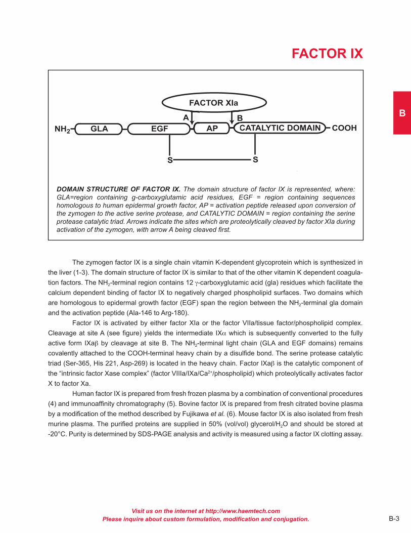
DOMAIN STRUCTURE OF FACTOR IX. The domain structure of factor IX is represented, where: GLA=region containing g-carboxyglutamic acid residues, EGF = region containing sequences homologous to human epidermal growth factor, AP = activation peptide released upon conversion of the zymogen to the active serine protease, and CATALYTIC DOMAIN = region containing the serine protease catalytic triad. Arrows indicate the sites which are proteolytically cleaved by factor XIa during activation of the zymogen, with arrow A being cleaved first.
The zymogen factor IX is a single chain vitamin K-dependent glycoprotein which is synthesized in the liver (1-3) . The domain structure of factor IX is similar to that of the other vitamin K dependent coagula-tion factors . The NH2-terminal region contains 12 g-carboxyglutamic acid (gla) residues which facilitate the calcium dependent binding of factor IX to negatively charged phospholipid surfaces . Two domains which are homologous to epidermal growth factor (EGF) span the region between the NH2-terminal gla domain and the activation peptide (Ala-146 to Arg-180) .
Factor IX is activated by either factor XIa or the factor VIIa/tissue factor/phospholipid complex . Cleavage at site A (see figure) yields the intermediate IXa which is subsequently converted to the fully active form IXab by cleavage at site B . The NH2-terminal light chain (GLA and EGF domains) remains covalently attached to the COOH-terminal heavy chain by a disulfide bond . The serine protease catalytic triad (Ser-365, His 221, Asp-269) is located in the heavy chain . Factor IXab is the catalytic component of the “intrinsic factor Xase complex” (factor VIIIa/IXa/Ca2+/phospholipid) which proteolytically activates factor X to factor Xa .
Human factor IX is prepared from fresh frozen plasma by a combination of conventional procedures (4) and immunoaffinity chromatography (5) . Bovine factor IX is prepared from fresh citrated bovine plasma by a modification of the method described by Fujikawa et al. (6) . Mouse factor IX is also isolated from fresh murine plasma . The purified proteins are supplied in 50% (vol/vol) glycerol/H2O and should be stored at -20°C . Purity is determined by SDS-PAGE analysis and activity is measured using a factor IX clotting assay .
Visit us on the internet at http://www.haemtech.comPlease inquire about custom formulation, modification and conjugation.B-4
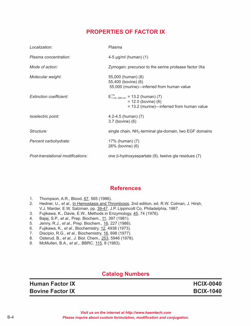
PROPERTIES OF FACTOR IX
Localization: Plasma
Plasma concentration: 4-5 µg/ml (human) (1)
Mode of action: Zymogen; precursor to the serine protease factor IXa
Molecular weight: 55,000 (human) (8) 55,400 (bovine) (6) 55,000 (murine)—inferred from human value
Extinction coefficient: E1 cm, 280 nm = 13 .2 (human) (7) = 12 .0 (bovine) (6) = 13 .2 (murine)—inferred from human value Isoelectric point: 4 .2-4 .5 (human) (7) 3 .7 (bovine) (6)
Structure: single chain, NH2-terminal gla-domain, two EGF domains
Percent carbohydrate: 17% (human) (7) 26% (bovine) (6)
Post-translational modifications: one b-hydroxyaspartate (9), twelve gla residues (7)
References1 . Thompson, A .R ., Blood, 67, 565 (1986) .2 . Hedner, U ., et al., In Hemostasis and Thrombosis, 2nd edition, ed . R .W . Colman, J . Hirsh, V .J . Marder, E .W . Salzman, pp . 39-47 . J .P . Lippincott Co, Philadelphia, 1987 .3 . Fujikawa, K ., Davie, E .W ., Methods in Enzymology, 45, 74 (1976) .4 . Bajaj, S .P ., et al., Prep . Biochem ., 11, 397 (1981) .5 . Jenny, R .J ., et al., Prep . Biochem ., 16, 227 (1986) .6 . Fujikawa, K ., et al., Biochemistry, 12, 4938 (1973) .7 . Discipio, R .G ., et al., Biochemistry, 16, 698 (1977) .8 . Osterud, B ., et al., J . Biol . Chem ., 253, 5946 (1978) .9 . McMullen, B .A ., et al.,, BBRC, 115, 8 (1983) .
Catalog NumbersHuman Factor IX HCIX-0040Bovine Factor IX BCIX-1040
1%

Visit us on the internet at http://www.haemtech.comPlease inquire about custom formulation, modification and conjugation.
B
B-5
FACTOR X
DOMAIN STRUCTURE OF FACTOR X. The domain structure of factor X is represented, where: GLA = region containing g-carboxyglutamic acid residues, EGF = region containing sequences homologous to human epidermal growth factor, AP = activation peptide released upon conversion of the zymogen to the active serine protease, CATALYTIC DOMAIN = region containing the serine protease catalytic triad. The arrow indicates the site which is proteolytically cleaved by factor Xase during activation of the zymogen.
Factor X is a vitamin K-dependent protein zymogen which is synthesized in the liver and circulates in plasma as a two chain molecule linked by a disulfide bond (1,2) . Prior to secretion into plasma, post-translational modifications produce 11 gamma-carboxyglutamic acid (gla) residues and a single b-hydroxy-aspartic acid residue, which are located within the NH2-terminal light chain . The light chain also contains two epidermal growth factor (EGF) homology domains . The COOH-terminal heavy chain of factor X con-tains most of the carbohydrate moieties, as well as the latent serine protease domain . The activation of factor X is catalyzed by either the intrinsic factor Xase complex (factor IXa, factor VIIIa, cellular surface and calcium ions) or the extrinsic factor Xase complex (factor VIIa, tissue factor, cellular surface and calcium ions) . Activation of human factor X by either complex results in cleavage at Arg52-Ile53 of the COOH-termi-nal heavy chain and subsequent release of a 52 amino acid activation glycopeptide . Factor Xa then serves as the enzyme component of the prothrombinase complex which is responsible for the rapid conversion of prothrombin to thrombin . The gla residues enable factor X/Xa to bind phospholipid (i .e . cell surfaces) in a calcium dependent manner; a requirement for assembly of the prothrombinase complex . The first EGF homology domain contains a Ca2+ binding site which acts as a hinge to fold the EGF and GLA domains towards each other (12) . This region of the molecule is involved in recognition of cellular binding domains .
Human factor X is isolated from fresh frozen human plasma by a combination of conventional techniques (3) and immunoaffinity chromatography (4) . In addition to the standard human factor X prepara-tion, Gla-domainless human factor X is also available . Bovine factor X is isolated from fresh bovine plasma using a modification of the procedure reported by Bajaj et al. (5,6) . Mouse factor X is also isolated from fresh murine plasma .The purified zymogen is supplied in 50% (vol/vol) glycerol/H2O and should be stored at -20°C . Purity is determined by SDS-PAGE analysis and activity is measured in a factor X clotting assay .
Visit us on the internet at http://www.haemtech.comPlease inquire about custom formulation, modification and conjugation.B-6
PROPERTIES OF FACTOR X
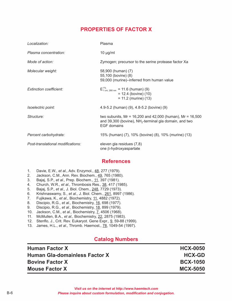
Localization: Plasma
Plasma concentration: 10 µg/ml
Mode of action: Zymogen; precursor to the serine protease factor Xa
Molecular weight: 58,900 (human) (7) 55,100 (bovine) (8) 59,000 (murine)–inferred from human value
Extinction coefficient: E1 cm, 280 nm = 11 .6 (human) (9) = 12 .4 (bovine) (10) = 11 .2 (murine) (13) Isoelectric point: 4 .9-5 .2 (human) (9), 4 .8-5 .2 (bovine) (9)
Structure: two subunits, Mr = 16,200 and 42,000 (human), Mr = 16,500 and 39,300 (bovine), NH2-terminal gla domain, and two EGF domains
Percent carbohydrate: 15% (human) (7), 10% (bovine) (8), 10% (murine) (13)
Post-translational modifications: eleven gla residues (7,8) one b-hydroxyaspartate
References1 . Davie, E .W ., et al., Adv . Enzymol ., 48, 277 (1979) .2 . Jackson, C .M ., Ann . Rev . Biochem ., 49, 765 (1980) .3 . Bajaj, S .P ., et al., Prep . Biochem ., 11, 397 (1981) .4 . Church, W .R ., et al., Thrombosis Res ., 38, 417 (1985) .5 . Bajaj, S .P ., et al., J . Biol . Chem ., 248, 7729 (1973) .6 . Krishnaswamy, S ., et al., J . Biol . Chem ., 261, 8997 (1986) .7 . Fujikawa, K ., et al., Biochemistry, 11, 4882 (1972) .8 . Discipio, R .G ., et al., Biochemistry, 16, 698 (1977) .9 . Discipio, R .G ., et al., Biochemistry, 18, 899 (1979) .10 . Jackson, C .M ., et al., Biochemistry, 7, 4506 (1968) .11 . McMullen, B .A ., et al., Biochemistry, 22, 2875 (1983) .12 . Stenflo, J ., Crit . Rev . Eukaryot . Gene Expr ., 9, 59-88 (1999) .13 . James, H .L ., et al., Thromb . Haemost ., 78, 1049-54 (1997) .
Catalog NumbersHuman Factor X HCX-0050Human Gla-domainless Factor X HCX-GDBovine Factor X BCX-1050Mouse Factor X MCX-5050
1%

Visit us on the internet at http://www.haemtech.comPlease inquire about custom formulation, modification and conjugation. B-7
B
FACTOR XI
DOMAIN STRUCTURE OF FACTOR XI. The domain structure of the factor XI monomer is represent-ed. The areas identified as R1 through R4 correspond to four tandem amino acid sequence repeats which ultimately comprise the heavy chain of factor XIa. During proteolytic activation by factor XIIa, the “CATALYTIC DOMAIN”, which comprises the light chain of factor XIa, is cleaved from the heavy chain region. The heavy and light chains of factor XIa remain associated through a disulfide bond. The mature factor XI molecule is a homodimer composed of two apparently identical monomers which remain associated through disulfide bonds.
Factor XI is a plasma glycoprotein which circulates in a non-covalent complex with high molecular weight kininogen (1) . The mature molecule is synthesized in the liver and is a two-chain homodimer with a molecular weight of approximately 160,000 (2,3) . It is estimated that 5% of the total mass is attributable to carbohydrate (2) . The two identical monomers have molecular weights of 80,000, and are joined together by disulfide bonds . Thus by SDS-PAGE analysis, factor XI appears as a single band both non-reduced (Mr = 160,000), and reduced (Mr = 80,000) .
Factor XI circulates as a zymogen and requires proteolytic activation to acquire serine protease activity . The conversion of factor XI to factor XIa is catalyzed by factor XIIa, and results in cleavage of the Arg369-Ile370 bond in each monomer (3) . Factor XIa consists of two NH2-terminal derived heavy chains, and two COOH-terminal derived light chains, all of which are held together by disulfide bonds . Factor XIa participates within the intrinsic pathway of coagulation by catalyzing the conversion of factor IX to factor IXa . A bleeding disorder called plasma thromboplastin antecedent deficiency results from a lack of factor XI procoagulant activity (4,5) . The variable bleeding tendencies observed in factor XI deficient patients do not correlate with either factor XI activity or antigen levels . This latter observation may be related to the ability of the tissue factor/factor VIIa complex to also activate factor IX to IXa .
Historically, factor XI has been difficult to purify due to its relatively low concentration in plasma, and its susceptibility to proteolysis (6) . Factor XI is purified from fresh frozen plasma that is stabilized by added inhibitors . The plasma is first treated with BaCl2 to remove the vitamin K-dependent proteins, and factor XI is then isolated by affinity chromatography . A final chromatography step on heparin sepharose yields a homogeneous preparation of intact factor XI . The finished product is supplied in 50% (vol/vol) glycerol/H2O and should be stored at -20°C .

Visit us on the internet at http://www.haemtech.comPlease inquire about custom formulation, modification and conjugation.B-8
PROPERTIES OF FACTOR XI
Localization: Plasma; in association with high molecular weight kininogen
Plasma concentration: 2-7 µg/ml (2,3,7,8)
Mode of action: Zymogen; precursor to the serine protease factor XIa
Molecular weight: 160,000 (human) (2,3)
Extinction coefficient: E1 cm, 280 nm = 13 .4 (human) (2)
Isoelectric point: 8 .9-9 .1
Structure: homodimer consisting of two apparently identical subunits (Mr ~ 80,000) held together by disulfide bonds . Monomers contain four tandem amino acid repeats that share homology with plasma prekallikrein (9) .
Percent carbohydrate: 5% (human) (2)
References1 . Thompson, R .E ., et al., J . Clin . Invest ., 60, 1376 (1977) .2 . Kurachi, K . and Davie, E .W ., Biochemistry, 16, 5831 (1977) .3 . Bouma, B .N . and Griffen, J .H ., J . Biol . Chem ., 252, 6432 (1977) .4 . Rosenthal, R .L ., et al., Proc . Soc . Exp . Biol . Med ., 82, 171 (1953) .5 . Forbes, C .D . and Ratnoff, J ., J . Lab . Clin . Med ., 79, 113 (1972) .6 . Kurachi, K . and Davie, E .V ., Methods Enzymol ., 80, 211 (1981) .7 . Wuepper, K .D ., Fed . Proc ., 31, 624 (1972) .8 . Saito, H . and Goldsmith, G ., Blood, 50, 377 (1977) .9 . Fujikawa, K ., et al., Biochemistry, 25, 2417 (1986) .
Catalog NumberHuman Factor XI HCXI-0150
1%

Visit us on the internet at http://www.haemtech.comPlease inquire about custom formulation, modification and conjugation. B-9
B
FACTOR XII
DOMAIN STRUCTURE OF FACTOR XII. The organization of the factor XII domain structure based on sequence homology is represented, where: EGF = epidermal growth factor, Type I and Type II = domains homologous to those found in fibronectin, solid arrows = kallikrein cleavage sites that form a- and b-factor XIIa, dashed arrows=cleavages leading to intermediate forms of factor XIIa.
Factor XII (XII) (Hageman Factor) is a single chain (Mr = 78,000) glycoprotein zymogen that circu-lates in plasma at a concentration of 40 µg/ml (1-5) . Reciprical activation of XII to the active serine protease factor XIIa (XIIa) by kallikrein is central to initiation of the intrinsic coagulation pathway . Surface bound a-XIIa in turn activates factor XI to XIa . Secondary cleavage of a-XIIa by kallikrein yields b-XIIa, which cata-lyzes solution phase activation of kallikrein, factor VII and the classical complement cascade . The ability of a variety of negatively charged substances, both physiological and nonphysiological to promote XII activa-tion and, thus, initiation of the intrinsic pathway has led to the psuedonym “contact activation .” Binding to anionic surfaces induces a conformational change, making the XII zymogen more susceptible to cleavage by a variety of proteases (6,7) . It is unlikely that binding to negatively charged surfaces alone is sufficient to activate XII, since highly purified preparations of XII and plasma deficient in prekallikrein and high molecular weight kininogen do not undergo this “autocatalysis” (8-11) .
A single cleavage by kallikrein at R353-Val354 of XII yields a-XIIa, a 2 chain protease (Mr = 80,000) held together by disulfide bonds . The COOH-terminal light chain (Mr = 28,000) contains the catalytic triad (His-40, Asp-89, Ser-191), while the NH2-terminal heavy chain (Mr = 52,000) contains the anionic surface binding portion of the molecule . A secondary cleavage of a-XIIa by kallikrein outside the disulfide bond yields b-XIIa (XIIf, BHFa, HFf, hageman factor fragments) (Mr = 28,000), which no longer binds anionic surfaces (12) . b-XIIa can activate prekallikrein, but has little procoagulant activity (13,14) . Several other minor intermediate forms of XIIa are indicated in the figure above .
Inhibitors of XIIa include C1-INH, a2-antiplasmin, a2-macroglobulin and antithrombin III . At physi-ological concentrations, the relative effectiveness of these inhibitors is 91 : 4 .5 : 3 : 1 .5, respectively (10, 16-19) . The ratio of C1-INH to XII has been implicated in the “cold activation” of factor VII and the conver-sion of prorenin to renin on storage of plasma (20,21) .
Human factor XII is prepared from fresh frozen plasma by immunoaffinity chromatography and supplied in 50% (vol/vol) glycerol/H2O for storage at -20°C .

Visit us on the internet at http://www.haemtech.comPlease inquire about custom formulation, modification and conjugation.B-10
PROPERTIES OF FACTOR XII
Localization: Plasma
Plasma concentration: 40 µg/ml (3)
Mode of action: Zymogen; precursor to the serine protease factor XIIa; activated by kallikrein/HMWK/anionic surface complex to intitiate the intrinsic pathway
Molecular weight: 80,000 (2)
Extinction coefficient: E1 cm, 280 nm = 14 .0
Isoelectric point: 6 .8
Structure: single chain (Mr = 80,000), organized into 6 domains based on sequence homology (5)
Percent carbohydrate: 17%
References1 . Schmaier, A .H ., et al., in Hemostasis and Thrombosis, ed . R .W . Colman, J . Hirsh, V .J . Marder and E .W . Salzman, pp 18-38, J .B . Lippincott Company, Philadelphia, 1987 .2 . Griffin, J .H . and Cochrane, C .G ., Methods Enzymol ., 45, 56-65 (1976) .3 . Saito, H ., et al., J Lab Clin ., 88, 506 (1976) .4 . McMullen, B .A . and Fujikawa, N ., J . Biol . Chem ., 260, 5328 (1985) .5 . Davie, E .W ., in Hemostasis and Thrombosis, ed . R .W . Colman, J . Hirsh, V .J . Marder and E .W . Salzman, pp 242-267, J .B . Lippincott Company, Philadelphia, 1987 .6 . Griffin, J .H ., Proc . Natl . Acad . Sci . USA, 75, 1998 (1978) .7 . McMillin, L .R ., et al., J . Clin . Invest ., 54, 1312 (1974) .8 . Revak, S .D ., et al., J . Clin . Invest ., 59, 1167 (1977) .9 . Fujikawa, K ., et al., Biochemistry, 16, 4182 (1977) .10 . Bonno, N ., et al., in New Comprehensive Biochemistry, ed . R .F .A . Zwaal and H .C .Hemker, pp . 103-128, Elsevier, Amsterdam, 1986 .11 . Claeys, H . and Collen, D . (1978) Eur . J . Biochem ., 87, 69 (1978) .12 . Revak, S .D . and Cochrane, C .G ., J . Clin . Invest ., 57, 852 (1976) .13 . Cochrane, C .G ., et al., J . Exp . Med ., 138, 1564 (1973) .14 . Revak, S .D ., et al., J . Exp . Med ., 147, 719 (1978) .15 . Ghebrehiwet, B ., et al., J . Clin . Invest ., 71, 1458 (1983) .16 . DeAgostini, A ., et al., J . Clin . Invest ., 73, 1542 (1984) .17 . Rathoff, O .D ., et al., J . Exp . Med ., 129, 315 (1969) .18 . Stead, N ., et al., J . Biol Chem ., 251, 6481 (1976) .19 . Pixley, R .A ., et al., J . Biol Chem ., 260, 1723 (1985) .20 . Gjonnass, H ., Thromb . Diath . Haemorrh ., 28, 182 (1972) .21 . Radcliffe, R ., et al., Blood, 59, 611 (1977) .
Catalog NumberHuman Factor XII HCXII-0155
1%

Visit us on the internet at http://www.haemtech.comPlease inquire about custom formulation, modification and conjugation. B-11
B
FACTOR XIII
THROMBIN CATALYZED ACTIVATION OF FACTOR XIII. The zymogen, factor XIII, is a tetramer (A2B2) composed of 2 identical A subunits and 2 identical B subunits. Thrombin cleaves a Mr = 4000 peptide from the NH2-terminal of the light chain, exposing the active site sulfhydryl. Full expression of activity is achieved following Ca2+-dependent dissociation of the B chain dimer.
Factor XIII is the zymogenic form of the glutaminyl-peptide g-glutamyl transferase factor XIIIa (fibrinoligase, plasma transglutaminase, fibrin stabilizing factor, E .C . 2 .3 .2 .13) (1-3) . Factor XIII is unique among transamidases in that it is a zymogen in vivo (2) . Factor XIII is found both extracellularly in plasma and intracellularly in platelets, megakaryocytes, monocytes, placenta, uterus, liver and prostrate tissues . Plasma factor XIII is synthesized in the liver and circulates as a tetramer (Mr = 320,000), composed of 2 pairs of nonidentical subunits (A2B2) (4) . The intra-cellular forms are synthesized in the tissues where they reside as dimers (Mr = 146,000) of 2 identical A chains (A2) (7-11) . The A subunits of plasma and intracel-lular forms of factor XIII are functionally identical . The A subunit contains 6 free sulfhydryl groups one of which is the active site (12) .
The concentration of factor XIII in plasma (A2B2) is approximately 30 µg/ml (8) . It is the last of the zymogens to become activated in the coagulation cascade and it is the only enzyme in this system that is not a serine protease . The conversion of plasma factor XIII (A2B2) to the active transamidase factor XIIIa (A2') results from hydrolysis of the Arg36-Gly37 at the NH2-terminus of the A subunit by thrombin (13) . Full expression of activity is achieved only after the Ca2+ (Kd = 10-3M) and fibrin(ogen) (Kd = 10-8M) dependent dissociation of the B subunit dimer from the A2' dimer (14-16) .
In the coagulation cascade, factor XIIIa functions to stabilize the fibrin clot by crosslinking the a
and g-chains of fibrin . Other proteins known to be substrates for Factor XIIIa which may be hemostatically important include fibronectin (17), a2-antiplasmin (18), collagen (19), factor V (20), von Willebrand Factor (19) and thrombospondin (21,22) .
Factor XIII is purified from fresh frozen human plasma by a modification of the procedures described by Folke (2) and Lorand (10) involving barium citrate, ammonium sulfate and glycine precipi-tations, ion exchange chromatography and gel filtration . Factor XIII is homogeneous as judged by SDS PAGE, with a specific activity of ~50 units/mg . Factor XIII is supplied in 50% glycerol containing 0 .5 mM EDTA, for storage at -20°C .
Visit us on the internet at http://www.haemtech.comPlease inquire about custom formulation, modification and conjugation.B-12
PROPERTIES OF FACTOR XIII
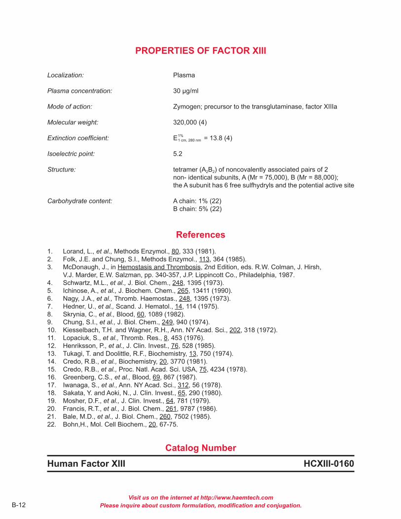
Localization: Plasma
Plasma concentration: 30 µg/ml
Mode of action: Zymogen; precursor to the transglutaminase, factor XIIIa
Molecular weight: 320,000 (4)
Extinction coefficient: E1 cm, 280 nm = 13 .8 (4)
Isoelectric point: 5 .2
Structure: tetramer (A2B2) of noncovalently associated pairs of 2 non- identical subunits, A (Mr = 75,000), B (Mr = 88,000); the A subunit has 6 free sulfhydryls and the potential active site
Carbohydrate content: A chain: 1% (22) B chain: 5% (22)
References1 . Lorand, L ., et al., Methods Enzymol ., 80, 333 (1981) .2 . Folk, J .E . and Chung, S .I ., Methods Enzymol ., 113, 364 (1985) .3 . McDonaugh, J ., in Hemostasis and Thrombosis, 2nd Edition, eds . R .W . Colman, J . Hirsh, V .J . Marder, E .W . Salzman, pp . 340-357, J .P . Lippincott Co ., Philadelphia, 1987 .4 . Schwartz, M .L ., et al., J . Biol . Chem ., 248, 1395 (1973) .5 . Ichinose, A ., et al., J . Biochem . Chem ., 265, 13411 (1990) .6 . Nagy, J .A ., et al., Thromb . Haemostas ., 248, 1395 (1973) .7 . Hedner, U ., et al., Scand . J . Hematol ., 14, 114 (1975) .8 . Skrynia, C ., et al., Blood, 60, 1089 (1982) .9 . Chung, S .I ., et al., J . Biol . Chem ., 249, 940 (1974) .10 . Kiesselbach, T .H . and Wagner, R .H ., Ann . NY Acad . Sci ., 202, 318 (1972) .11 . Lopaciuk, S ., et al., Thromb . Res ., 8, 453 (1976) .12 . Henriksson, P ., et al., J . Clin . Invest ., 76, 528 (1985) .13 . Tukagi, T . and Doolittle, R .F ., Biochemistry, 13, 750 (1974) .14 . Credo, R .B ., et al., Biochemistry, 20, 3770 (1981) .15 . Credo, R .B ., et al., Proc . Natl . Acad . Sci . USA, 75, 4234 (1978) .16 . Greenberg, C .S ., et al., Blood, 69, 867 (1987) .17 . Iwanaga, S ., et al., Ann . NY Acad . Sci ., 312, 56 (1978) .18 . Sakata, Y . and Aoki, N ., J . Clin . Invest ., 65, 290 (1980) .19 . Mosher, D .F ., et al., J . Clin . Invest ., 64, 781 (1979) .20 . Francis, R .T ., et al., J . Biol . Chem ., 261, 9787 (1986) .21 . Bale, M .D ., et al., J . Biol . Chem ., 260, 7502 (1985) .22 . Bohn,H ., Mol . Cell Biochem ., 20, 67-75 .
Catalog NumberHuman Factor XIII HCXIII-0160
1%

Visit us on the internet at http://www.haemtech.comPlease inquire about custom formulation, modification and conjugation. B-13
B
PLASMINOGEN
HUMAN PLASMINOGEN. The domain structure of human plasminogen is represented where: K1-K5 = the 5 kringle domains, B-CHAIN = catalytic domain of plasmin , and the arrows indicate the sites of proteolytic cleavage by plasmin, elastase, and plasminogen activators (PA’S).
Plasminogen is a single chain glycoprotein zymogen which is synthesized in the liver and circulates in plasma at a concentration of approximately 2 .4 µM (1,2) . The plasminogen molecule contains 790 amino acids, 24 disulfide bridges, no free sulfhydryls and 5 regions of internal sequence homology, known as kringles, between Lys77 and Arg560 . These five triple-looped, three disulfide bridged, kringle regions are homologous to the kringle domains in t-PA, u-PA and prothrombin . Plasminogen contains one high affinity (Kd = 9x10⁻⁶M) and four low affinity (Kd = 5x10⁻³M) lysine binding sites . The high affinity binding site resides within the first kringle region of plasminogen . The interaction of plasminogen with fibrin and
a2-antiplasmin is mediated by these lysine binding sites . Native glu-plasminogen (Mr = 88,000) is readily converted to Lys-77-plasminogen (Mr = 83,000) by plasmin hydrolysis of the Lys76-Lys77 peptide bond . Elastase catalyzed cleavage of the Val441-Val442 peptide bond of glu-plasminogen yields a function-ally active zymogen termed Val-442 plasminogen or mini-plasminogen .
The conversion of plasminogen to plasmin occurs by a variety of mechanisms, but all result in hydrolysis of the Arg560-Val561 peptide bond of plasminogen, yielding two chains which remain covalently associated by a disulfide bond .
Native glu-plasminogen is prepared from fresh frozen human plasma by a modification of the pro-cedure of Castellino (3), utilizing gel filtration and affinity chromatography . The two carbohydrate variants of glu-plasminogen (CHOI and CHOII) are isolated by gradient elution from lysine-Sepharose using the lysine analog, e-aminocaproic acid (3) . The plasminogen is supplied in 50% (vol/vol) glycerol/H2O for storage at -20°C . Purity is determined by SDS-PAGE analysis .

Visit us on the internet at http://www.haemtech.comPlease inquire about custom formulation, modification and conjugation.B-14
PROPERTIES OF PLASMINOGEN
Localization: Plasma
Plasma concentration: 210 µg/ml (human) (4)
Mode of action: Zymogen; precursor to the serine protease plasmin
Molecular weight: 88,000 (glu plasminogen) (5) 83,000 (lys-plasminogen) (5) 38,000 (val-plasminogen) (6)
Extinction coefficient: E1 cm, 280 nm = 17 .0 (5) Isoelectric point: 6 .2 (glu-plasminogen) (1) 6 .7-8 .3 (lys-plasminogen) (1)
Structure: single chain, 24 intra chain disulfide bridges, 5 kringle regions
Percent carbohydrate: Approximately 2%
References1 . Robbins, K .C ., Methods in Enzymology, 45, 257 (1976) .2 . Collen, D . in Blood Coagulation, Zwaal, R .E .A . and Hemker, H .C ., eds ., pp . 243-258, New York, Elsevier (1986) .3 . Castellino, F .J ., et al., Methods in Enzymology, 80, 365 (1981) .4 . Wohl, R .C ., et al.,Thromb . Res ., 27, 523 (1982) .5 . Barlow, G .H ., et al., Biochemistry, 23, 2384 (1984) .6 . Sottrup-Jensen, L ., et al., in Progress in Chemical Fibrinolysis and Thrombolysis, Vol . 3, ed . J .F . Davidson, R .M . Rowan, M .M . Samana, P .C . Desnoyers, pp . 197-228, New York: Raven Press (1975) .
Catalog NumbersHuman glu-Plasminogen HCPG-0130Human glu-Plasminogen CHOI HCPG-0131Human glu-Plasminogen CHOII HCPG-0132Human lys-Plasminogen HCPG-0133Bovine Plasminogen BCPG-1130Mouse Plasminogen MCPG-5130
1%
Visit us on the internet at http://www.haemtech.comPlease inquire about custom formulation, modification and conjugation. B-15
B
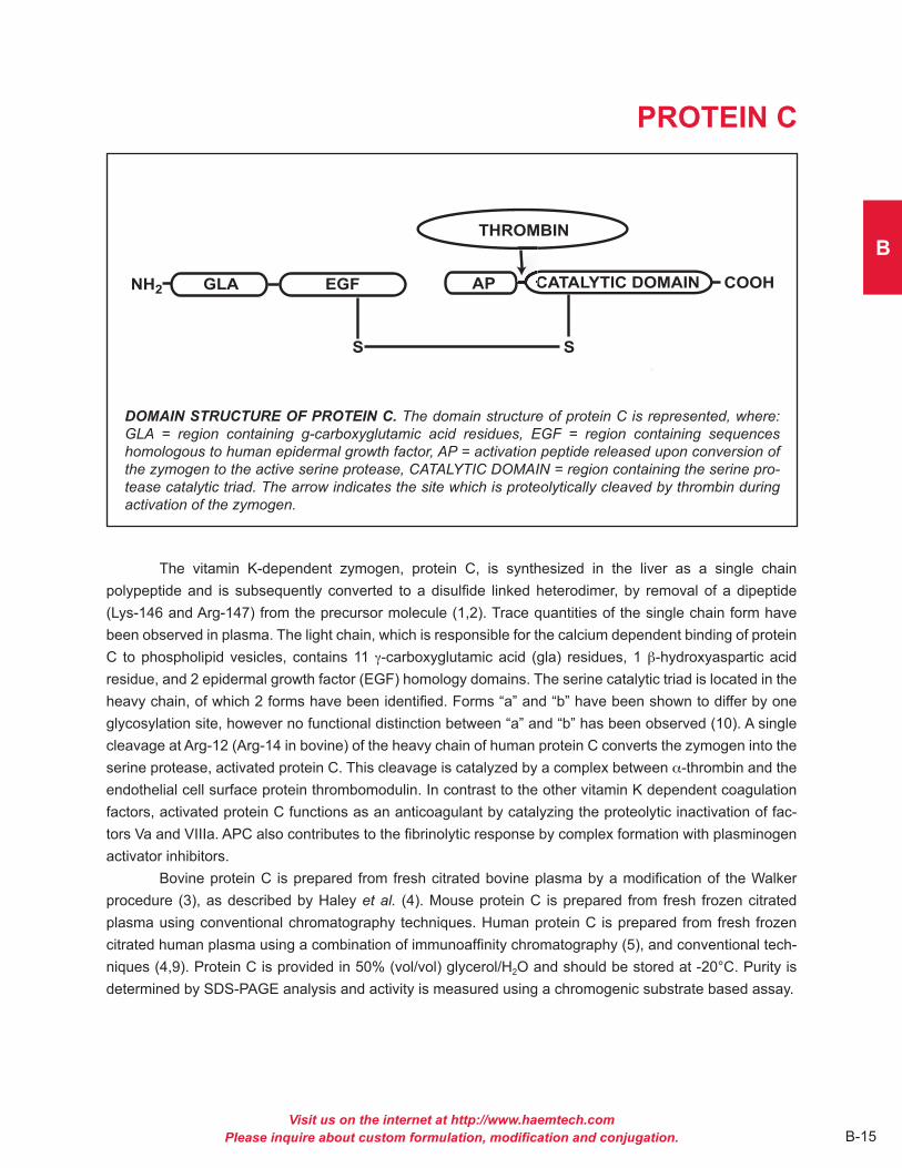
PROTEIN C
DOMAIN STRUCTURE OF PROTEIN C. The domain structure of protein C is represented, where: GLA = region containing g-carboxyglutamic acid residues, EGF = region containing sequences homologous to human epidermal growth factor, AP = activation peptide released upon conversion of the zymogen to the active serine protease, CATALYTIC DOMAIN = region containing the serine pro-tease catalytic triad. The arrow indicates the site which is proteolytically cleaved by thrombin during activation of the zymogen.
The vitamin K-dependent zymogen, protein C, is synthesized in the liver as a single chain polypeptide and is subsequently converted to a disulfide linked heterodimer, by removal of a dipeptide (Lys-146 and Arg-147) from the precursor molecule (1,2) . Trace quantities of the single chain form have been observed in plasma . The light chain, which is responsible for the calcium dependent binding of protein C to phospholipid vesicles, contains 11 g-carboxyglutamic acid (gla) residues, 1 b-hydroxyaspartic acid residue, and 2 epidermal growth factor (EGF) homology domains . The serine catalytic triad is located in the heavy chain, of which 2 forms have been identified . Forms “a” and “b” have been shown to differ by one glycosylation site, however no functional distinction between “a” and “b” has been observed (10) . A single cleavage at Arg-12 (Arg-14 in bovine) of the heavy chain of human protein C converts the zymogen into the serine protease, activated protein C . This cleavage is catalyzed by a complex between a-thrombin and the endothelial cell surface protein thrombomodulin . In contrast to the other vitamin K dependent coagulation factors, activated protein C functions as an anticoagulant by catalyzing the proteolytic inactivation of fac-tors Va and VIIIa . APC also contributes to the fibrinolytic response by complex formation with plasminogen activator inhibitors .
Bovine protein C is prepared from fresh citrated bovine plasma by a modification of the Walker procedure (3), as described by Haley et al. (4) . Mouse protein C is prepared from fresh frozen citrated plasma using conventional chromatography techniques . Human protein C is prepared from fresh frozen citrated human plasma using a combination of immunoaffinity chromatography (5), and conventional tech-niques (4,9) . Protein C is provided in 50% (vol/vol) glycerol/H2O and should be stored at -20°C . Purity is determined by SDS-PAGE analysis and activity is measured using a chromogenic substrate based assay .

Visit us on the internet at http://www.haemtech.comPlease inquire about custom formulation, modification and conjugation.B-16
PROPERTIES OF PROTEIN C
Localization: Plasma
Plasma concentration: 4-5 µg/ml (human) (6) 5-10 µg/ml (bovine) (2)
Mode of action: Zymogen; precursor to the serine protease activated protein C (APC)
Molecular weight: 62,000 (human) (7) 58,000 (bovine) (7) 62,000 (murine)–inferred from human value
Extinction coefficient: E1 cm, 280 nm = 14 .5 (human) (7) = 13 .7 (bovine) (7) = 14 .5 (murine)–inferred from human value Isoelectric point: 4 .4-4 .8 (human) (8) 4 .2-4 .5 (bovine) (8)
Structure: two chains, Mr = 41,000 and 21,000, disulfide linked, NH2-terminal gla domain two EGF domains
Percent carbohydrate: 23 % (human) (7) 14 % (bovine) (7)
Post-translational modifications: eleven gla residues (bovine), nine gla residues (human), one b-hydroxyaspartate
References1 . Esmon, C .T ., Progress in Thromb . and Hemostas ., 10, 25 (1984) .2 . Stenflo, J ., Semin . in Thromb . and Hemostas ., 10, 109 (1984) .3 . Walker, F .J ., et al., Biochim . Biophys . Acta, 571, 333 (1979) .4 . Haley, P .E ., et al., J . Biol . Chem ., 264, 16303 (1989) .5 . Jenny, R .J ., et al., Prep . Biochem ., 16, 227 (1986) .6 . Griffen, J .H ., et al.,Blood, 60, 261 (1982) .7 . Kisiel, W ., et al., Methods Enzymol ., 80, 320 (1981) .8 . Discipio, R .G ., et al., Biochemistry, 18, 899 (1979) .9 . Bajaj, S .P ., et al., Prep . Biochem ., 11, 397 (1981) .10 . Kalafatis, M . et al., J . Biol . Chem ., 291, 1578 (2016)
Catalog NumbersHuman Protein C HCPC-0070Bovine Protein C BCPC-1070Mouse Protein C MCPC-5070
1%

Visit us on the internet at http://www.haemtech.comPlease inquire about custom formulation, modification and conjugation. B-17
B
PROTHROMBIN
DOMAIN STRUCTURE OF PROTHROMBIN. The domain structure of prothrombin is represented, where: GLA = region containing g-carboxyglutamic acid residues, KRINGLE = regions of internal sequence homology, CATALYTIC DOMAIN = region containing the serine protease catalytic triad. Arrows indicate the sites which are proteolytically cleaved by factor Xa during activation of the zymogen.
Prothrombin is a vitamin K-dependent plasma protein which is synthesized in the liver (1) . Prior to secretion into plasma, prothrombin undergoes post-translational modification by a vitamin K-dependent carboxylase which converts ten specific glutamic acid residues to g-carboxyglutamic acid (gla) . The ten gla residues are located within the first 40 amino acids of the mature protein and contribute to the ability of prothrombin to bind to negatively charged phospholipid membranes . Prothrombin contains two regions of internal homology which are referred to as “kringle” structures . These regions of conspicuous second-ary structure are located between residues 40 and 270 of the mature plasma protein and replace the growth factor domains found in several other plasma serine proteases . Thus far, no function has been ascribed to these regions, but there is suspicion that they may play a role in one of several binary protein interactions involving prothrombin . The mature single chain protein circulates in plasma as a zymogen and, during coagulation, is proteolytically activated to the potent serine protease a-thrombin . This proteolysis is catalyzed by the prothrombinase enzyme complex . During activation, prothrombin is cleaved at Arg271-Thr272 (human) / Arg273-Thr274 (bovine) and at Arg 320-Ser321 (human) / Arg323-Ser324 (bovine) to yield a “pro” fragment (fragment 1 .2) and thrombin, the latter of which is composed of two chains covalently linked by a disulfide bond . In the case of human prothrombin/thrombin, there is an additional thrombin feed-back cleavage at Arg284-Thr285 resulting in an additional 13 amino acids being removed from the mature thrombin “A” chain .
Human prothrombin is prepared from fresh frozen human plasma as described by Bajaj and coworkers (2) . Bovine prothrombin is prepared from fresh bovine plasma using a modification of the procedure described by Owen and coworkers (3) . Mouse prothrombin is prepared from fresh frozen murine plasma using conventional chromatography techniques . Purified prothrombin is supplied in 50% (vol/vol) glycerol/H2O and should be stored at -20°C . Purity is determined by SDS-PAGE analysis, and activity is measured by clotting and/or chromogenic substrate assay, following conversion of prothrombin to thrombin .
Visit us on the internet at http://www.haemtech.comPlease inquire about custom formulation, modification and conjugation.B-18
PROPERTIES OF PROTHROMBIN
Localization: Plasma
Plasma concentration: 100 µg/ml (1)
Mode of action: Zymogen; precursor to the serine protease a-thrombin
Molecular weight: 72,000 (1,4,5)
Extinction coefficient: E1 cm, 280 nm = 13 .8 (human) (4) = 14 .4 (bovine) (1) = 13 .8 (murine)–inferred from human value
Isoelectric point: 4 .7-4 .9 (human) (6) 4 .4-4 .9 (bovine) (6)
Structure: single chain, NH2-terminal gla domain, two kringle regions
Percent carbohydrate: 8 .2 % (human) (4) 10 .0 % (bovine) (5)
Post-translational modifications: ten gla residues (4,5)
References1 . Mann, K .G ., et al., Methods in Enzymology, 45, 156 (1976) .2 . Bajaj, S .P ., et al., Prep . Biochem ., 11, 397 (1981) .3 . Owen, W .G ., et al., J . Biol . Chem ., 249, 594 (1974) .4 . Kisiel, W ., et al., Biochem . Biophys . Acta, 304, 103 (1973) .5 . Magnusson, S ., et al., In Proteases in Biological Control, ed . E . Reich, D .B . Rifkin, E . Shaw, pp . 123-149 . New York: Cold Spring Harbor Laboratories, 1975 .6 . Discipio, R .G ., et al., Biochemistry, 18, 899 (1979) .
Catalog NumbersHuman Prothrombin HCP-0010Human Prothrombin Fragment 1 HCP1-0010Human Prothrombin Fragment 1 - 2 HCP12-0010Human Prothrombin Fragment 2 HCP2-0010Human Prethrombin-1 HCP1-0011Human Prethrombin-2a HCP2-0011Bovine Prothrombin BCP-1010Bovine Prothrombin Fragment 1 BCP1-1010Mouse Prothrombin MCP-5010
1%
aNaja cleavage of human II is between E262 and D263 . This cleavage yields a “pre-2” product that is 9 amino acids longer than native pre-2 .
C
ENZYMES
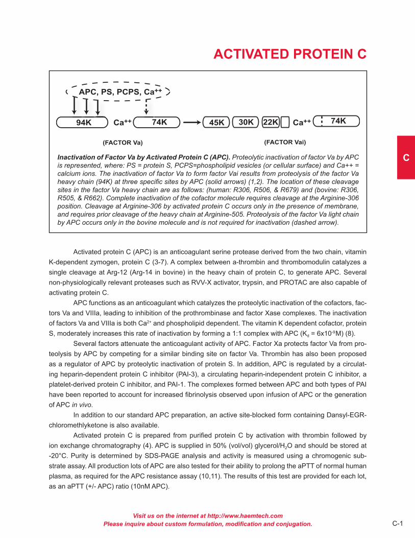
Visit us on the internet at http://www.haemtech.comPlease inquire about custom formulation, modification and conjugation. C-1
C
ACTIVATED PROTEIN C
Inactivation of Factor Va by Activated Protein C (APC). Proteolytic inactivation of factor Va by APC is represented, where: PS = protein S, PCPS=phospholipid vesicles (or cellular surface) and Ca++ = calcium ions. The inactivation of factor Va to form factor Vai results from proteolysis of the factor Va heavy chain (94K) at three specific sites by APC (solid arrows) (1,2). The location of these cleavage sites in the factor Va heavy chain are as follows: (human: R306, R506, & R679) and (bovine: R306, R505, & R662). Complete inactivation of the cofactor molecule requires cleavage at the Arginine-306 position. Cleavage at Arginine-306 by activated protein C occurs only in the presence of membrane, and requires prior cleavage of the heavy chain at Arginine-505. Proteolysis of the factor Va light chain by APC occurs only in the bovine molecule and is not required for inactivation (dashed arrow).
Activated protein C (APC) is an anticoagulant serine protease derived from the two chain, vitamin K-dependent zymogen, protein C (3-7) . A complex between a-thrombin and thrombomodulin catalyzes a single cleavage at Arg-12 (Arg-14 in bovine) in the heavy chain of protein C, to generate APC . Several non-physiologically relevant proteases such as RVV-X activator, trypsin, and PROTAC are also capable of activating protein C .
APC functions as an anticoagulant which catalyzes the proteolytic inactivation of the cofactors, fac-tors Va and VIIIa, leading to inhibition of the prothrombinase and factor Xase complexes . The inactivation of factors Va and VIIIa is both Ca2+ and phospholipid dependent . The vitamin K dependent cofactor, protein S, moderately increases this rate of inactivation by forming a 1:1 complex with APC (Kd = 6x10⁻⁹M) (8) .
Several factors attenuate the anticoagulant activity of APC . Factor Xa protects factor Va from pro-teolysis by APC by competing for a similar binding site on factor Va . Thrombin has also been proposed as a regulator of APC by proteolytic inactivation of protein S . In addition, APC is regulated by a circulat-ing heparin-dependent protein C inhibitor (PAI-3), a circulating heparin-independent protein C inhibitor, a platelet-derived protein C inhibitor, and PAI-1 . The complexes formed between APC and both types of PAI have been reported to account for increased fibrinolysis observed upon infusion of APC or the generation of APC in vivo .
In addition to our standard APC preparation, an active site-blocked form containing Dansyl-EGR-chloromethlyketone is also available .
Activated protein C is prepared from purified protein C by activation with thrombin followed by ion exchange chromatography (4) . APC is supplied in 50% (vol/vol) glycerol/H2O and should be stored at -20°C . Purity is determined by SDS-PAGE analysis and activity is measured using a chromogenic sub-strate assay . All production lots of APC are also tested for their ability to prolong the aPTT of normal human plasma, as required for the APC resistance assay (10,11) . The results of this test are provided for each lot, as an aPTT (+/- APC) ratio (10nM APC) .

Visit us on the internet at http://www.haemtech.comPlease inquire about custom formulation, modification and conjugation.C-2
PROPERTIES OF ACTIVATED PROTEIN C
Localization: Plasma
Mode of action: Anticoagulant, inactivates factors Va and VIIIa
Molecular weight: 56,200 (human) (5) 52,650 (bovine) (5) 56,200 (murine)—inferred from human value
Extinction coefficient: E1 cm, 280 nm = 14 .5 (human) (5) = 13 .7 (bovine) (5) = 14 .5 (murine)—inferred from human value
Isoelectric point: 4 .4-4 .8 (human) (9) 4 .2-4 .5 (bovine) (9)
Structure: two chains, Mr = 35,000 and 21,000, disulfide linked, NH2-terminal gla domain two EGF domains
Percent carbohydrate: 23 % (human) (5) 14 % (bovine) (5)
Post-translational modifications: eleven gla residues (bovine), nine gla residues (human), one b-hydroxyaspartate
Complimentary fluorogenic substrate(s): HTI Catalog # SN-54 and SN-59
References1 . Kalafatis, M ., and Mann, K .G ., J . Biol . Chem ., 268, 27246 (1993) .2 . Kalafatis, M ., et al., J . Biol . Chem ., 269, 31869 (1994) .3 . Esmon, C .T ., Progress in Thromb . and Hemostas ., 10, 25 (1984) .4 . Esmon, C .T ., J . Biol . Chem ., 264, 4743 (1989) .5 . Kisiel, W ., et al., Methods Enzymol ., 80, 320 (1981) .6 . Stenflo, J ., Semin . in Thromb . and Hemostas ., 10, 109 (1984) .7 . Marlar, R .A ., Semin . in Thromb . and Hemostas ., 11, 387 (1985) .8 . Walker, F .J ., et al., J . Biol . Chem ., 256, 11128 (1981) .9 . Discipio, R .G ., et al., Biochemistry, 18, 899 (1979) .10 . Dahlback, B ., and Hildebrand, B ., Proc . Natl . Acad . Sci . USA, 91, 1396 (1994) .11 . Svensson, P .J ., and Dahlback, B ., New Engl . J . Med ., 330, 517 (1994) .
Catalog NumbersHuman Activated Protein C HCAPC-0080Human Actitated Protein C - DEGR HCAPC-DEGRBovine Activated Protein C BCAPC-1080Bovine Activated Protein C - DEGR BCAPC-DEGRMouse Activated Protein C MCAPC-5080
1%

Visit us on the internet at http://www.haemtech.comPlease inquire about custom formulation, modification and conjugation. C-3
C
a-THROMBIN
PROTEOLYTIC ACTIVATION OF PROTHROMBIN. The serine protease a-thrombin is produced by proteolytic activation of the zymogen, prothrombin. The enzyme complex, prothrombinase, catalyzes the proteolysis of two peptide bonds in prothrombin, which gives rise to an NH2-terminal derived F1.2 region and the heterodimer, a-thrombin. Alpha-thrombin is composed of an “A” chain (Mr = 6000) which is covalently linked to a “B” chain (Mr = 31,000) through a single disulfide bond.
Alpha-thrombin is a highly specific serine protease generated by proteolytic activation of the zymo-gen prothrombin (1) . During coagulation, thrombin cleaves fibrinogen to form fibrin, leading to the ultimate step in coagulation, the formation of a fibrin clot . Thrombin is also responsible for feedback activation of the procofactors factor V and factor VIII . Thrombin has also been reported to activate factor XIII and plate-lets, and also functions as a vasoconstrictor protein . The procoagulant activity of thrombin is arrested in two ways: 1) inhibition by either heparin cofactor II or the antithrombin III/heparin complex; or 2) complex formation with thrombomodulin . Formation of the thrombin/thrombomodulin complex results in the inability of thrombin to cleave fibrinogen and activate factors V and VIII, but increases the efficiency of thrombin for activation of the anticoagulant, protein C .
Thrombin is a two chain enzyme composed of an NH2-terminal “A” chain (Mr = 6,000) and a COOH-terminal “B” chain (Mr = 31,000) which remain covalently associated through a single disulfide bond . Human thrombin is 13 amino acids shorter than the bovine thrombin due to a thrombin cleavage site on the human protein that is not present in the bovine protein .
Thrombin is also utilized for site specific cleavage of fusion proteins expressed in bacteria (9-11) . A thrombin sensitive site is incorporated between the recombinant protein of interest and peptides or proteins which facilitate purification and/or expression . The target protein is released from the expressed hybrid by cleavage with thrombin . Thrombin can then be easily removed by affinity chromatography .
Human, bovine, and murine thrombin are prepared from purified prothrombin using a modification of the Lundblad procedure (1) as described by Nesheim et al. (2) . Thrombin is supplied in 50% (vol/vol) glycerol/H2O and should be stored at -20°C . Purity is determined by SDS-PAGE analysis and activity is measured in a thrombin specific clotting assay, and compared to standardized NIH thrombin . Thrombin is also available with the active site blocked with either DFP, FPRck, or biotinlyated FPRck .

Visit us on the internet at http://www.haemtech.comPlease inquire about custom formulation, modification and conjugation.C-4
PROPERTIES OF a-THROMBIN
Localization: Plasma
Mode of action: Serine protease which cleaves fibrinogen to form fibrin; also responsible for activation of protein C, platelet activation and feedback activation of the procofactors, factor V and factor VIII
Molecular weight: 36,700 (3-6)
Extinction coefficient: E1 cm, 280 nm = 18 .3 (human) (6) = 19 .5 (bovine) (7) = 18 .3 (murine)–inferred from human value
Specific Activity: approximately 3800 NIH units/mg
Isoelectric point: 7 .0-7 .6 (human) (3)
Structure: two subunits, approximately Mr = 6,000 and 31,000
Percent carbohydrate: approximately 5%
Complimentary fluorogenic substrate(s): HTI Catalog # SN-17a, SN-20 and SN-59
References1 . Lundblad, R .L ., et al., Methods Enzymol ., 45, 156 (1976) .2 . Nesheim, M .E ., et al., J . Biol . Chem ., 258, 5386 (1983) .3 . Fenton, J .W ., et al., in Chemistry and Biology of Thrombin, ed . R .L . Lundblad, J .W . Fenton, K .G . Mann, pp . 43-70 . Ann Arbor, MI: Ann Arbor Science Publishers, Inc ., 1977 .4 . Braughman, D .J ., et al., J . Biol . Chem ., 242, 5252 (1967) .5 . Winzor, D .J ., et al., Arch . Biochem . Biophys ., 104, 202 (1964) .6 . Fenton, J .W ., et al.,J . Biol . Chem ., 252, 3587 (1977) .7 . Winzor, D .J ., et al., J . Phys . Chem ., 68, 338 (1964) .8 . Magnusson, S ., in The Enzymes, ed . P .D . Boyer, vol . III, pp . 277-321 . New York: Academic Press, 1971 .9 . Gaun, K .L . and Dixon, J .E ., Anal . Biochem ., 192, 262 (1991) .10 . Germino, J . and Bastia, D ., Proc . Natl . Acad . Sci . USA, 81, 4692 (1984) .11 . Chang, J .-Y ., Eur . J . Biochem ., 151, 217 (1985) .
Catalog NumbersHuman a-Thrombin HCT-0020Human a-Thrombin-DFP HCT-DFPHuman a-Thrombin-FPRCk HCT-FPRCkHuman a-Thrombin-BFPRCk HCT-BFPRCkBovine a-Thrombin BCT-1020Bovine a-Thrombin-DFP BCT-DFPBovine a-Thrombin-FPRCk BCT-FPRCkBovine a-Thrombin-BFPRCk BCT-BFPRCkMouse Thrombin MCT-5020
1%

Visit us on the internet at http://www.haemtech.comPlease inquire about custom formulation, modification and conjugation. C-5
C
b-THROMBINg-THROMBIN
The degradation pattern of human a-thrombin to less active forms is illustrated above. b-thrombin is generated by cleavage of the intact B-chain at the Arg106-Tyr107 bond to yield the B1 and B2 fragments. g-thrombin is generated by further cleavage of the b-thrombin B2-chain at the Lys190-Gly191 bond to yield the B4 and B5 fragments. Although not illustrated, the B3 fragment is a potential intermediate that is derived by cleavage of the intact B-chain at the Lys190-Gly191 bond, resulting in a product that is often referred to as b'-thrombin. (The amino acid numbering used here begins at the NH2-terminal end of the A-chain and continues through the B-chain).
- THROMBIN
- THROMBIN
- THROMBIN
36 37 106 107
107 190 191 295
295
295
106
36 37
36 37 B5
Alpha-thrombin is a highly specific serine protease generated by proteolytic activation of the zymo-gen prothrombin (1) . It has been shown that purified forms of topical grade bovine a-thrombin are subject to proteolytic degradation to less active forms upon long term storage (2-4) . Similar degradation patterns were observed upon limited digestion of a-thrombin with trypsin (5) . These proteolyzed forms of a-thrombin have been termed b-thrombin and g-thrombin . Human b-thrombin is generated by cleavage of the B-chain at the Arg106-Tyr107 bond . g-thrombin is generated by further cleavage of the b-thrombin B2-chain at the Lys190-Gly191 bond . These cleavages cause release of peptides (B1 and B4) that are no longer covalently attached to the thrombin molecule, but remain associated through ion exchange and gel filtration chroma-tography . These proteolyzed forms of thrombin retain their ability to cleave small synthetic substrates (6,7) and some protein substrates such as factor XIII (8), antithrombin III (9) and prothrombin (10) . Their ability to clot fibrinogen (11), cleave thrombospondin (12) or activate protein C (9) have been markedly decreased .
Human b-thrombin and g-thrombin are prepared from purified a-thrombin by limited proteolysis with TPCK-treated trypsin, essentially by the method of Braun et al . (5) . b-thrombin is supplied in 10 mM sodium phosphate, 0 .3 M NaCl, pH 6 .5, and g-thrombin is supplied in 100 mM sodium phosphate, 0 .1% PEG, pH 6 .5, and both should be stored at -80°C . Purity is assessed by SDS-PAGE and activity is assessed using a fibrinogen clotting assay .

Visit us on the internet at http://www.haemtech.comPlease inquire about custom formulation, modification and conjugation.C-6
PROPERTIES OF b- AND g-THROMBIN
Mode of action: Proteolyzed forms of a-thrombin which retain activity toward small substrates, factor XIII and prothrombin, but have reduced activity toward fibrinogen, protein C activation and antithrombin III binding
Molecular weight: 35,400 (b-thrombin) 35,400 (g-thrombin)
Extinction coefficient: E1 cm, 280 nm = 18 .3
4,000 (A chain)* 34,300 (B chain)* 6,000 (B1 chain)* 10,400 (B5 chain)* 11,800 (B4 chain)*
* Apparent molecular weight as determined by SDS-PAGE analysis
Structure: b-thrombin: three chains (A, B1, B2), disulfide link between the A and the B2 chains . g-thrombin: four chains (A, B1, B5, B4) with a disulfide link between the A peptide and the B5 peptide
References1 . Lundblad, R .L ., et al., Methods Enzymol ., 45, 156 (1976) .2 . Lundblad, R .L ., et al., J . Biol . Chem ., 254, 8524 (1979) .3 . Fenton, J .W ., et al., in Chemistry and Biology of Thrombin, ed . R .L . Lundblad, J .W . Fenton, K .G . Mann, pp . 43-70, Ann Arbor, MI: Ann Arbor Science Publishers, Inc ., 1977 .4 . Boissel, J .P ., et al., J . Biol . Chem ., 259, 5691 (1984) .5 . Braun, P .J ., et al., Thromb . Res ., 50, 273 (1988) .6 . Lottenberg, R ., et al., Thromb . Res ., 28, 313 (1982) .7 . Witting, et al ., Thromb . Res ., 46, 567 (1987) .8 . Lorand, L . and Credo, R .B ., in Chemistry and Biology of Thrombin, ed . R .L . Lundblad, J .W . Fenton, K .G . Mann, pp . 311-323, Ann Arbor, MI: Ann Arbor Science Publishers, Inc ., 1977 .9 . Bezeaud, A ., et al., Eur . J . Chem ., 153, 491 (1985) .10 . Seegers, W .H ., et al., Semin . Thromb . Haemostasis, 1, 211 (1975) .11 . Lundblad, R .L ., et al., J . Biol . Chem ., 259, 6991 (1984) .12 . Takahashi, K ., et al., Biochem . J ., 224, 673 (1984) .
Catalog NumbersHuman b-thrombin HCBT-0022Human g-thrombin HCGT-0021
1%

Visit us on the internet at http://www.haemtech.comPlease inquire about custom formulation, modification and conjugation. C-7
C
FACTOR VIIa
PARTICIPATION OF FACTOR VIIa IN THE EXTRINSIC FACTOR Xase COMPLEX. The components of the extrinsic factor Xase enzyme complex are illustrated. Factor VIIa and its cofactor tissue factor (TF) assemble on a negatively charged membrane surface in a calcium dependent manner to form an enzyme complex which proteolytically converts factor X to factor Xa. The complex is referred to as the extrinsic factor Xase complex, because the tissue factor component is presumably derived from a source “extrinsic” to the plasma environment, which is accessible following vascular injury.
Human factor VII is a single chain plasma glycoprotein, and is a zymogen in its native form (1-4) . Proteolytic activation of factor VII, yields the enzyme factor VIIa, which when bound to the integral membrane protein tissue factor, forms an enzyme complex that proteolytically converts factor X to factor Xa . This enzyme complex is best known as the extrinsic factor Xase (pronounced ten-ase) complex, since by virtue of its tissue factor component, it is composed of protein normally extrinsic to the plasma environment .
The conversion of factor VII to factor VIIa is catalyzed by a number of proteases including thrombin, factor IXa, factor Xa, factor XIa, and factor XIIa . Rapid activation also takes place when factor VII is combined with tissue factor in the presence of calcium (5-9) . This latter event is consistent with autocatalysis, and is likely initiated by a small amount of pre-existing factor VIIa . The activation reaction results in cleavage of the peptide bond between arginine 152 and isoleucine 153 .
The resulting factor VIIa consists of an NH2-derived light chain (Mr = 20,000), and a COOH- terminal derived heavy chain (Mr = 30,000), which remain associated through a single disulfide bond (cys 135 - cys 262) . The light chain contains the membrane binding “Gla domain”, while the heavy chain contains the catalytic domain .
Unlike other serine proteases, factor VIIa alone is not readily inhibited by the antithrombin III/heparin complex . However, in the presence of tissue factor, antithrombin III/heparin exhibits significant inhibition of factor VIIa (6) .
Human factor VIIa is prepared from affinity purified factor VII, and purified by ion exchange chroma-tography . The purified protein is supplied in 50% (vol/vol) glycerol/H2O and should be stored at -20°C . The purity is determined by SDS-PAGE analysis, and activity is measured in a factor VII clotting assay .

Visit us on the internet at http://www.haemtech.comPlease inquire about custom formulation, modification and conjugation.C-8
PROPERTIES OF FACTOR VIIa
Localization: Plasma
Mode of action: Enzyme component of the extrinsic factor X activating complex; also activates factor IX, thus by-passing the contact activation system
Molecular weight: 50,000 (human) (2)
Extinction coefficient: E1 cm, 280 nm = 13 .9a (2)
Specific activity: approximately 16,000 units/mgb
Structure: 2 subunits; NH2-terminal derived light chain (Mr = 20,000), COOH-terminal derived heavy chain (Mr = 30,000), NH2-terminal gla-domain, two EGF domains
Percent carbohydrate: 13%c (10)
Post-translational modifications: one b-hydroxyaspartate (11), ten gla residues (12)
Complimentary fluorogenic substrate(s): HTI Catalog # SN-17a and SN-17c
a inferred from the zymogen, factor VIIb determined from single stage clotting assayc based upon analysis of bovine factor VII
References1 . Broze, G .J . and Majerus, P .W ., Methods Enzymol ., 80, 228 (1981) .2 . Bajaj, S .P ., et al., J . Biol . Chem ., 256, 253 (1981) .3 . Williams, E .B ., et al., J . Biol . Chem ., 264, 7536 (1989) .4 . Kisiel, W . and McMullen, B .A ., Thrombosis Res ., 22, 375 (1981) .5 . Radcliffe, R . and Nemerson, Y ., J . Biol . Chem ., 250, 388 (1975) .6 . Lawson, J .H ., et al.,J . Biol . Chem ., 268, 767 (1993) .7 . Kisiel, W ., et al., Biochemistry, 16, 4189 (1977) .8 . Radcliffe, R ., et al., Blood, 50, 611 (1977) .9 . Seligsohn, U ., et al., J . Clin . Invest ., 64, 1056 (1979) .10 . Kisiel, W ., et al., Biochemistry, 14, 4928 (1975) .11 . McMullen, B .A ., et al., BBRC, 115, 8 (1983) .12 . Discipio, R .G ., et al., Biochemistry, 16, 698 (1977) .
Catalog NumberHuman Factor VIIa HCVIIA-0031
1%

Visit us on the internet at http://www.haemtech.comPlease inquire about custom formulation, modification and conjugation. C-9
C
FACTOR IXa
ACTIVATION OF FACTOR X BY FACTOR IXa. The activation of factor X by the intrinsic factor Xase complex is shown. The intrinsic factor Xase complex consists of an enzyme, factor IXa, and a cofac-tor, factor VIIIa, assembled on a phospholipid surface in the presence of calcium ions. The enzyme complex proteolytically cleaves a 10,000 molecular weight activation peptide from the NH2-terminal of the heavy chain of factor X, thus expressing the active-site of factor Xa.
Factor IXa is produced from its inactive precursor, factor IX, via proteolytic cleavage by factor XIa or the tissue factor/factor VIIa/phospholipid complex . The activation results from the cleavage of two peptide bonds in the factor IX molecule, releasing an activation glycopeptide with an apparent molecular weight of 10,000 . The heavy chain of factor IXa (Mr = 28,000) contains the serine protease catalytic domain, while the light chain (Mr = 17,000) contains the membrane binding domain .
Factor IXa functions as a serine protease involved in the activation of the zymogen, factor X, to form the enzyme, factor Xa . The factor IXa enzymatic activity is greatly enhanced by inclusion of its cofac-tor, factor VIIIa, in the presence of calcium ions on a phospholipid surface . Factor IXa is readily inhibited by antithrombin III, and this inhibition is greatly accelerated by the presence of heparin . Factor IXa is not inhibited by DFP .
Factor IXa is prepared from highly purified factor IX by activation with factor XIa, as described by Lindquist et al . (5) . The factor IXa is further purified by gel filtration, followed by immunoaffinity purification . Factor IXa is also available with the active site irreversibly blocked by the tripeptide chloromethylketone, EGRck, or by the fluorescent inhibitor, Dansyl-EGRck . Our standard factor IXa is supplied in 50% (vol/vol) glycerol/H2O for storage at -20°C . Purity is assessed by SDS-PAGE analysis . Activity is determined in a one-stage clotting assay .

Visit us on the internet at http://www.haemtech.comPlease inquire about custom formulation, modification and conjugation.C-10
PROPERTIES OF FACTOR IXA
Localization: Plasma
Mode of action: Enzyme component of the Factor Xase complex
Molecular weight: 45,000 (7)
Extinction coefficient: E1 cm, 280 nm = 14 .0 (5)
Structure: 2 subunits, Mr = 28,000 and 17,000 (5), NH2-terminal gla-domain, two EGF domains
Post-translational modifications: one b-hydroxyaspartate (3), twelve gla residues (4)
References1 . Thompson, A .R ., Blood, 67, 565 (1986) .2 . Hedner, U ., et al., In Hemostasis and Thrombosis, 2nd edition, ed . R .W . Colman, J . Hirsh, V .J . Marder, E .W . Salzman, pp ., 39-47 . J .P . Lippincott Co, Philadelphia, 1987 .3 . Discipio, R .G ., et al., Biochemistry, 16, 698 (1977) .4 . McMullen, B .A ., et al., BBRC, 115, 8 (1983) .5 . Lindquist, P .A ., et al., J . Biol . Chem ., 253, 1902 (1978) .
Catalog NumbersHuman Factor IXa HCIXA-0050Human Factor IXa-EGR HCIXA-EGRHuman Factor IXa-DEGR HCIXA-DEGRBovine Factor IXa BCIXA-1050Bovine Factor IXa-EGR BCIXA-EGRBovine Factor IXa-DEGR BCIXA-DEGR
1%

Visit us on the internet at http://www.haemtech.comPlease inquire about custom formulation, modification and conjugation. C-11
C
FACTOR Xa
PARTICIPATION OF FACTOR Xa IN PROTHROMBINASE. The participation of the serine protease factor Xa in the prothrombinase complex is illustrated. Membrane bound factor Xa binds to membrane bound factor Va to form the prothrombinase complex. This complex effectively converts the zymogen prothrombin (II) to the active serine protease thrombin (IIa) by proteolytic removal of the fragment 1.2 (F1.2) portion of prothrombin.
Activation of the zymogen, factor X, by either the intrinsic or extrinsic factor Xase complexes pro-duces the active serine protease factor Xa (1,2) . The activation of factor X requires proteolytic cleavage of the heavy chain, resulting in the release of an activation glycopeptide . The heavy chain region in factor Xa contains the serine protease catalytic domain, while the light chain, as in the zymogen, contains the membrane binding domain .
Factor Xa participates in the prothrombinase complex, which catalyzes the rapid conversion of prothrombin to thrombin . Prothrombinase is an enzyme complex composed of factor Xa (enzyme) and fac-tor Va (cofactor) assembled on a cellular surface in the presence of calcium ions . Although factor Xa can independently catalyze the activation of prothrombin, the rate at which this reaction occurs is increased nearly 300,000-fold with complete assembly of the prothrombinase complex . The clotting activity of factor Xa in vivo is terminated by either inactivation of the cofactor, factor Va, or by direct inhibition of factor Xa by inhibitors, such as ATIII, after disassembly of the prothrombinase complex .
In recent years, molecular biologists have utilized factor Xa for site specific cleavage of fusion proteins expressed in bacteria (9-12) . A factor Xa-sensitive site is incorporated between the recombinant protein of interest and peptides or proteins which facilitate purification and/or expression . The target protein is released from the expressed hybrid by cleavage with factor Xa . The factor Xa can then be easily removed by affinity chromatography .
Factor Xa is prepared by activating purified factor X with the factor X activator isolated from Rus-sell’s viper venom . Factor Xa is purified from the activation mixture by chromatography over benzamidine-Sepharose followed by gel filtration (1,3) . Several modified forms of factor Xa are also available including: A) active-site blocked factor Xa containing tripeptide chloromethylketone inhibitors EGRck, Dansyl-EGRck, or BEGRck; B) b-Factor Xa; and C) human Gla-domainless b-factor Xa . The standard enzyme is supplied in 50% (vol/vol) glycerol/H2O and should be stored at -20°C . Purity is determined by SDS-PAGE analysis and activity is measured in a factor Xa clotting assay and/or chromogenic substrate assay .

C-12
PROPERTIES OF FACTOR Xa
Localization: Plasma
Mode of action: Enzyme component of the prothrombinase complex
Molecular weight: 46,000 (human) (4), 45,300 (bovine) (5) 46,000 (murine)–inferred from human value
Extinction coefficient: E1 cm, 280 nm = 11 .6 (human) (6) = 12 .4 (bovine) (7) = 11 .2 (murine) (12)
Specific activity: approximately 1000 units/mg
Structure: two subunits, Mr = 16,200 and 29,000 (human) (6), Mr = 16,500 and 28,800 (bovine) (5), NH2-terminal gla domain, two EGF domains
Percent carbohydrate: 3 .0% (human) (8), 2 .1% (bovine) (8)
Post-translational modifications: eleven gla residues, one b-hydroxyaspartate
Complimentary fluorogenic substrate(s): HTI Catalog # SN-7
References1 . Jesty, J ., et al., Methods Enzymol ., 45, 95 (1976) .2 . Jackson, C .M ., Ann . Re v . Biochem ., 49, 765 (1980) .3 . Krishnaswamy, S ., et al., J .Biol . Chem ., 262, 3291 (1987) .4 . Discipio, R .G ., et al., Biochemistry, 16, 5253 (1977) .5 . Fujikawa, K . and Davie, E .W ., Methods Enzymol ., 45, 89 (1976) .6 . Discipio, R .G ., et al., Biochemistry, 16, 698 (1977) .7 . Jackson, C .M ., et al., Biochemistry, 7, 4506 (1968) .8 . Fujikawa, K ., Biochemistry, 13, 5290 (1974) .9 . Nagai, K ., et al., Proc . Natl . Acad . Sci . USA, 82, 7252 (1985) .10 . Aurell, L ., et al., Thromb . Res ., 11, 595 (1984) .11 . Nagai, K . and Thogersen, H ., Nature, 309, 810 (1984) .12 . James, H .L . et al., Thromb . Haemost ., 78, 1049-54 (1997) .
Catalog NumbersHuman Factor Xa HCXA-0060Human b-Factor Xa HCBXA-0061Human Gla-domainless b-Factor Xa HCXA-GDHuman Factor Xa-EGR HCXA-EGRHuman Factor Xa-DEGR HCXA-DEGRHuman Factor Xa-BEGR HCXA-BEGRBovine Factor Xa BCXA-1060Bovine Factor Xa-EGR BCXA-EGRBovine Factor Xa-DEGR BCXA-DEGRMouse Factor Xa MCXA-5060
1%

Visit us on the internet at http://www.haemtech.comPlease inquire about custom formulation, modification and conjugation. C-13
C
FACTOR XIa
PROTEOLYTIC ACTIVATION OF FACTOR XI. The activation of factor XI by factor XIIa is illustrated. Although factor XI can bind independently to negatively charged surfaces, proteolysis by factor XIIa can only occur in the presence of surface- bound high molecular weight kininogen (HMWKa). The bound HMWKa is susceptible to proteolysis by the newly generated factor XIa which results in a loss of its biological activity (HMWKi). Factor XIa catalyzes the proteolytic conversion of factor IX to factor IXa either in solution phase or when bound to a negatively charged surface.
Proteolysis of XI by factor XIIa in the presence of high molecular weight kininogen (HMWK), yields the enzyme factor XIa (1-3) . Factor XIa is a serine protease which participates in the intrinsic pathway of coagulation by catalyzing the conversion of factor IX to factor IXa . Because factor XI is a homodimer, the enzyme, factor XIa, is composed of two identical heavy chains (Mr = 50,000), and two identical light chains (Mr = 30,000), all of which are held together by disulfide bonds . This latter property makes factor XIa unique among the serine proteases, since it contains two active sites per molecule (1-3) .
Factor XIa, like its precursor factor XI, remains in complex with HMWK (4) . In complex form, factor XIa/HMWK is capable of activating factor XII to factor XIIa and prekallikrein to kallikrein . Additionally, in the complexed form, factor XIa is less affected by protease inhibitors (5) . The major plasma inhibitor of factor XIa is a1-antitrypsin and then to a much lesser extent, antithrombin-III (5,6) . While in the complexed form, factor XIa may catalyze the proteolysis of HMWK, yielding a biologically inactive form of HMWK (HMWKi), thus allowing dissociation of the Factor XIa/HMWKi complex . Either free factor XIa or the factor XIa/HMWK complex catalyzes the proteolytic conversion of factor IX to factor IXa .
Factor XIa is prepared by activating purified factor XI with factor XIIa . Following activation, factor XIa is purified to homogeneity by a combination of affinity chromatography techniques . The factor XIa is supplied in 50% (vol/vol) glycerol/H2O and should be stored at -20°C . Purity is assessed by SDS PAGE analysis . Activity is determined in a clotting assay using factor XI deficient plasma .

Visit us on the internet at http://www.haemtech.comPlease inquire about custom formulation, modification and conjugation.C-14
PROPERTIES OF FACTOR XIa
Localization: Plasma
Mode of action: A serine protease that converts factor IX to factor IXa
Molecular weight: 160,000 (1,2)
Extinction coefficient: E1 cm, 280 nm = 13 .4a (human) (2)
Structure: two apparently identical heavy chains (Mr ~ 50,000) and two apparently identical light chains (Mr ~ 30,000) held together by disulfide bonds . Each light chain contains a catalytic domain (1,3)
Percent carbohydrate: 5% (human) (1)
Complimentary fluorogenic substrate(s): HTI Catalog # SN-13a and SN-45
a inferred from the zymogen, factor XI
References1 . Kurachi, K . and Davie, E .W ., Biochemistry, 16, 5831 (1977) .2 . Bouma, B .N . and Griffen, J .H ., J . Biol . Chem ., 252, 6432 (1977) .3 . Kurachi, K ., et al., Biochemistry, 19, 1330 (1980) .4 . Thompson, R .E ., et al., J . Clin . Invest ., 60, 1376 (1977) .5 . Scott, C .F ., et al., J . Clin . Invest ., 69, 844 (1982) .6 . Heck, L .W . and Kaplan, A .P ., J . Exp . Med ., 140, 1615 (1974) .
Catalog NumberHuman Factor XIa HCXIA-0160
1%

Visit us on the internet at http://www.haemtech.comPlease inquire about custom formulation, modification and conjugation. C-15
C
FACTOR XIIIa
STABILIZATION OF FIBRIN. The conversion of soluble fibrin to an insoluble fibrin clot is catalyzed by the transamidase factor XIIIa. Lysine and glutamine side chains of a- and g-chains of fibrin are crosslinked to form homopolymers.
Factor XIIIa (FXIIIa) (fibrinoligase, plasma transglutaminase, fibrin stabilizing factor, E .C . 2 .3 .2 .13) is a glutaminyl-peptide g-glutamyl transferase functioning in the final stages of the coagulation cascade, stabilizing the fibrin clot by crosslinking the a and g chains of fibrin to form homopolymers (1-4) . Several unique features of FXIIIa set it apart from proteins with which it is generally associated . Factor XIIIa is the only non-proteolytic enzyme of the coagulation cascade . Unlike other transamidases, it exists totally in zymogenic form and is the only sulfhydrylase enzyme which functions extracellularly .
Plasma FXIIIa is a tetramer, (Mr = 312,000), composed of 2 pairs of non-identical subunits (A2B2) (4-7) . Platelet FXIII is a dimer (Mr = 46,000), composed of only a pair of identical A subunits (A2') as is factor XIII from all intracellular sources (7-11) . The A subunit contains 6 free sulfhydryl groups, one of which is the active center (12) .
The conversion of plasma FXIII (A2B2) to the active transglutaminase, FXIIIa (A2') results from the thrombin catalyzed hydrolysis of the Arg36-Gly37 peptide bond at the NH2-terminal of the A subunit (13) . The conformational change induced by this cleavage results in exposure of the active site cysteine . Full activity is achieved only after the Ca2+ dependent (Kd = 10-3M) dissociation of the B subunit dimer from the A2' dimer (14-16) . Dissociation of the A2'B2 complex is enhanced by binding to the region of fibrin(ogen) containing residues 242-424 of the Aa chain (14-16) . Thus, at normal plasma concentrations of FXIII (90 nM), fibrinogen (8 µM) and Ca2+ (2 .5 mM free), all the FXIIIa generated would be in the fully activated (A2') molecular form . Other physiologically relevant crosslinked complexes generated by FXIIIa include fibrin-fibrinogen (17), fibrin-fibronectin (18), fibrin-vWF (19), fibrin-a2antiplasmin (20), fibrin-actin (21), fibrin-thrombospondin, fibronectin-collagen (19), vWF-collagen (19), actin-myosin (21,22), factor V-factor V (23) . In addition to clot stabilization, FXIIIa is thought to function in various processes involving cell proliferation, such as wound healing, tissue remodeling, atherosclerosis, and tumor growth .
Plasma FXIIIa is generated by cleavage of homogenous plasma FXIII by a-thrombin, in the presence of EDTA . The A2'B2 subunit is purified by ion and affinity chromatography and supplied in 50% glycerol,0 .5 mM EDTA for storage at -20°C .

Visit us on the internet at http://www.haemtech.comPlease inquire about custom formulation, modification and conjugation.C-16
PROPERTIES OF FACTOR XIIIa
Localization: Plasma
Mode of action: Plasma transglutaminase
Molecular weight: 312,000 (4)
Extinction coefficient: E1 cm, 280 nm = 13 .8 (4)a
Isoelectric point: 5 .2a
Structure: tetramer (A2'B2) in the absence of calcium, two identical A chains (Mr = 71,000) each containing 6 free sulfhydryls and an active site, two identical B subunits (Mr = 88,000)
Carbohydrate content: A chain: 1% B chain: 5%
a inferred from factor XIII
References1 . McDonaugh, J ., in Hemostasis and Thrombosis, 2nd Edition, eds . R .W . Colman, J . Hirsh, V .J . Marder, E .W . Salzman, pp . 340-357, Philadelphia: J .P . Lippincott Co ., 1987 .2 . Lorand, L ., et al., Methods Enzymol ., 80, 333 (1981) .3 . Folk, J .E . and Chung, S .I ., Methods Enzymol ., 113, 364 (1985) .4 . Ichinose, A ., et al., J . Biochem . Chem ., 265, 13411 (1990) .5 . Schwartz, M .L ., et al., J . Biol . Chem ., 248, 1395 (1973) .6 . Nagy, J .A ., et al., Thromb . Haemostas ., 248, 1395 (1973) .7 . Hedner, U ., et al., Scand . J . Hematol ., 14, 114 (1975) .8 . Skrynia, C ., et al., Blood, 60, 1089 (1982) .9 . Chung, S .I ., et al., J . Biol . Chem ., 249, 940 (1974) .10 . Kiesselbach, T .H . and Wagner, R .H ., Ann . NY Acad . Sci ., 202, 318 (1972) .11 . Lopacink, S ., et al.,Thromb . Res ., 8, 453 (1976) .12 . Henriksson, P ., et al., J . Clin . Invest ., 76, 528 (1985) .13 . Takagi, T . and Doolittle, R .F ., Biochemistry, 13, 750 (1974) .14 . Credo, R .B ., et al., Biochemistry, 20, 3770 (1981) .15 . Credo, R .B ., et al., Proc . Natl . Acad . Sci . USA, 75, 4234 (1978) .16 . Greenberg, C .S ., et al., Blood, 69, 867 (1987) .17 . Kamaide, H . and Shainoff, J .R ., J . Lab . Med ., 85, 574 (1975) .18 . Iwanaga, S ., et al., Ann . NY Acad . Sci ., 312, 56 (1978) .19 . Mosher, D .F ., et al., J . Clin . Invest ., 64, 781 (1979) .20 . Sakata, Y . and Aoki, N ., J . Clin . Invest ., 65, 290 (1980) .21 . Mui, P .K . and Ganguly, P ., Am . J ., Physiol ., 233, H346 .22 . Cohen, L ., et al., Arch . Biochem . Biophys ., 192, 100 (1979) .23 . Francis, R .T ., et al., J . Biol . Chem ., 261, 9787 (1986) .
Catalog NumberHuman Factor XIIIa HCXIIIA-0165
1%

Visit us on the internet at http://www.haemtech.comPlease inquire about custom formulation, modification and conjugation. C-17
C
PLASMIN
HUMAN PLASMINOGEN. The domain structure of human plasminogen is represented where: K1-K5 = the 5 kringle domains, B-CHAIN = catalytic domain of plasmin, and the arrows indicate the sites of proteolytic cleavage by plasmin, elastase, and plasminogen activators (PA’S).
Plasminogen (whether Glu-1, Lys-77 or Val-442) is converted to the active serine protease plasmin by hydrolysis of the Arg560-Val561 peptide bond yielding an NH2-terminal heavy (A) chain and a COOH-terminal light (B) chain linked by 2 disulfide bonds (1-3) . This conversion is catalyzed by a variety of physi-ological and pathological activators, including urinary type plasminogen activators, tissue type plasminogen activators, streptokinase, staphylokinase, kallikrein, factors IXa and XIIa . The COOH-terminal derived light chain (Mr = 26,000) contains the catalytic triad (His42, Asp85 and Ser180) as well as the streptokinase binding site . The NH2-terminal derived heavy chain ranges in molecular weight from 63,000 to 12,000 depending on the type of plasminogen from which it originated . In the absence of inhibitors, plasmin cleaves the amino-terminal Glu1 to Lys76 peptide from plasmin (plasminogen) to yield Lys-plasmin, which has a greater affinity for fibrin than the Glu form . The heavy chain of Lys-plasminogen contains 5 triple loop disulfide bridged regions of internal sequence homology known as kringles . Kringles 1-4 contain the v-aminocarboxylic acid and fibrin binding sites .
Plasmin is a serine protease with broad specificity which, in addition to cleavage of fibrin, is capable of activation and/or degradation of compounds of the coagulation, kinin generation and comple-ment systems . Although plasmin can be inhibited by a number of plasma protease inhibitors in vitro, regula-tion of plasmin in vivo is thought to occur mainly through its interaction with a2-antiplasmin, and to a lesser extent, a2-macroglobulin .
Human Lys-plasmin is prepared from homogeneous Glu-plasminogen using urokinase, as described by Robbins et al. (3) . Plasmin is supplied in 50% (vol/vol) glycerol/H2O and should be stored at -20°C . Activity is measured by chromogenic substrate assay and purity is judged by SDS gel electro- phoresis .

Visit us on the internet at http://www.haemtech.comPlease inquire about custom formulation, modification and conjugation.C-18
PROPERTIES OF PLASMIN
Localization: Plasma
Mode of action: Enzyme involved in fibrinolysis
Molecular weight: 83,000 (lys-plasmin) (4)
Extinction coefficient: E1 cm, 280 nm = 17 .0 (4)
Isoelectric point: 6 .7-8 .3 (3)
Structure: Two subunits, Mr = 57,000 and Mr= 26,000, disulfide linked, 5 kringle domains, 22 disulfide bridges, NH2-terminal lysine .
Percent carbohydrate: Approximately 2% (2)
Complimentary fluorogenic substrate(s): HTI Catalog # SN-5
References1 . Robbins, K .C . in Hemostasis and Thrombosis, 2nd edition, ed . R .W . Colman, J . Hirsh, V .J . Marder, E .W . Salzman, pp . 340-357, J .P . Lippincott Co ., Philadelphia, 1987 .2 . Collen, D . in Blood Coagulation, eds . R .F .P . Zwaal and H . C . Hemker, pp . 243-258, Elsevier, New York, (1986) .3 . Robbins, K .C ., Methods in Enzymology, 80, 379 (1981) .4 . Barlow, G .H ., et al., Biochemistry, 23, 2384 (1984) .
Catalog NumbersHuman Plasmin HCPM-0140Mouse Plasmin MCPM-5140
1%

Visit us on the internet at http://www.haemtech.comPlease inquire about custom formulation, modification and conjugation. C-19
VENOM PROTEASES
Snake venom proteases are useful tools for studying coagulation reactions . The venom proteases offered are highly purified, homogenous preparations with the indicated activities . All of our venom products are supplied in 50% glycerol for storage at -20°C .
RVV-V RVV-X ECV-PROTHROMBIN ACTIVATOR ACTIVATOR ACTIVATOR
CATALOG # RVVV-2000 RVVX-2010 ECVII-2011
SOURCE Vipera Russelli Vipera Russelli Vipera Echis Russelli Russelli Carinatus
MOLECULAR Mr = 27,200 single Mr = 79,000 Mr = 56,000 singlewEIGHT chain glycoprotein glycoprotein 2 chain disulfide linked subunits (Mr = 67kD, 26kD)
CLASS serine protease Zn+2 dependant metaloendopeptidase endopeptidase
INHIBITORS DFP EDTA, EDTA, DTT, o-phenanthroline o-phenanthroline
APPLICATION Converts single chain Conversion of factor Cleavage of factor V to an active X to Xa, factor IX to Arg323-Ile324 bond in two chain form IXaa prothrombin to form meizothrombin
REFERENCES 2,3 1 4
1 . Kisiel, W ., et.al., Biochem ., 15, 4901-4905 (1976) .2 . Kisiel, W . and Canfield, R ., Meth .Enzymol ., 80, 275-285 (1980) .3 . Kane, W . and Majerus, P .J ., J .Biol .Chem ., 256, 1002-1007 (1987) .4 . Morita, T . and Iwanaga, S ., Meth .Enzymol ., 80, 303-311 (1980) .
C

D
COFACTORS

Visit us on the internet at http://www.haemtech.comPlease inquire about custom formulation, modification and conjugation.
D
D-1
FACTOR V
ACTIVATION OF FACTOR V BY a-THROMBIN. The activation of single chain factor V (330,000) by a-thrombin is represented. Arrows indicate the site at which factor V is cleaved by thrombin to yield two activation peptides (71,000 and 120,000), in addition to the heavy (94,000) and light (74,000) chains which form factor Va. The heavy and light chains of factor Va remain non-covalently associ-ated in the presence of calcium ions.
Factor V is a large, single chain, plasma glycoprotein which is an essential component in the blood coagulation cascade (1) . During coagulation, the procofactor, factor V, is converted to the active cofactor, factor Va, via limited proteolysis by the serine protease a-thrombin (illustrated above), and less efficient-ly by factor Xa . The active cofactor is composed of an NH2-terminal derived heavy chain (Mr = 94,000) and a COOH-terminal derived light chain (Mr = 74,000) which remain non-covalently associated in the presence of calcium ions . Factor Va serves as a cofactor for the serine protease factor Xa . Factor Va and Xa assemble on a phospholipid surface in a non-covalent and calcium ion-dependent manner, to form the prothrombinase complex . The prothrombinase complex is responsible for the rapid conversion of the zymogen prothrombin to the active serine protease, a-thrombin . Assembly of the prothrombinase complex increases the rate at which prothrombin is converted to thrombin by nearly 300,000-fold relative to the rate with factor Xa alone . Down regulation of the prothrombinase complex is accomplished partly through the inactivation of factor Va by activated protein C .
Human factor V is prepared from fresh frozen human plasma using immunoaffinity chromatography as described by Katzmann and coworkers (2) . Bovine factor V is prepared from fresh bovine plasma using conventional chromatographic techniques as described by Nesheim and coworkers (3) . Purified factor V is supplied in 50% (vol/vol) glycerol/H2O, and should be stored at -20°C . Purity is determined by SDS-PAGE analysis and activity is measured in a factor V clotting assay .

Visit us on the internet at http://www.haemtech.comPlease inquire about custom formulation, modification and conjugation.D-2
PROPERTIES OF FACTOR V
Localization: Plasma and platelets
Plasma concentration: 7 .0 µg/ml (human) (4,5) 35 µg/ml (bovine) (5)
Mode of action: Procofactor; activated by thrombin to form the active cofactor, factor Va
Molecular weight: 330,000a (3)
Extinction coefficient: E1 cm, 280 nm = 9 .6a (3)
Structure: one subunit, 2196 amino acidsb (1,6)
Percent carbohydrate: Approximately 25%c
a determined for bovine factor Vb determined for human factor Vc based upon the calculated molecular weight of human factor V
References1 . Mann, K .G ., et al., Ann . Rev . Biochem ., 57, 915 (1988) .2 . Katzmann, J .A ., et al., Proc . Natl . Acad . Sci . USA, 78, 162 (1981) .3 . Nesheim, M .E ., et al., J . Biol . Chem ., 254, 508 (1979) .4 . Tracy, P .B ., et al., Blood, 60, 59 (1982) .5 . Nesheim, M .E ., et al., Methods Enzymol ., 80, 249 (1981) .6 . Jenny, R .J ., et al., Proc . Natl . Acad . Sci . USA, 84, 4846 (1987) .
Catalog NumbersHuman Factor V HCV-0100Bovine Factor V BCV-1100
1%

Visit us on the internet at http://www.haemtech.comPlease inquire about custom formulation, modification and conjugation. D-3
D
FACTOR Va
PARTICIPATION OF FACTOR Va IN PROTHROMBINASE. Assembly and expression of the pro-thrombinase complex is illustrated. Factor Va, composed of a heavy (VaH ) and light (VaL ) chain, binds to negatively charged phospholipid membranes and effectively serves as a receptor for membrane bound factor Xa (Xa). The enzyme complex serves to convert the zymogen prothrombin (II) to the active serine protease thrombin (IIa), in a proteolytic event which removes the fragment 1.2 (F1.2) portion of prothrombin.
Factor Va is a cofactor for the serine protease factor Xa, and in the presence of calcium ions they collectively assemble on a phospholipid surface to form the prothrombinase complex (1) . The prothrom-binase complex is responsible for the rapid conversion of prothrombin to thrombin . Factor Va is derived from the pro-cofactor, factor V, upon limited proteolysis by a-thrombin . The thrombin cleavage of factor V liberates two heavily glycosylated activation peptides from the central portion of the molecule which have no cofactor function . Factor Va is comprised of an NH2-terminal derived heavy chain (Mr = 94,000) and a COOH-terminal derived light chain (Mr = 74,000) which remain associated in the presence of calcium ions . The cofactor binds to phospholipid (cell membrane) surfaces and effectively serves as a receptor for membrane bound factor Xa . Complete assembly of the prothrombinase complex (factor Xa, factor Va, phospholipid, and calcium) results in a 300,000-fold increase in the rate of prothrombin conversion relative to the rate observed with factor Xa alone . The interaction between factor Va and factor Xa is mediated by both the heavy and light chain of factor Va, while the binding of prothrombin to factor Va is mediated solely by the heavy chain .
Factor Va is prepared by activating purified factor V with thrombin and is subsequently purified by immunoaffinity chromatography (2) . This process results in cofactor preparations which are free of both activation peptides and thrombin . Purified factor Va is supplied in 50% glycerol (vol/vol), 5 .0 mM CaCl2, and should be stored at -20°C . Purity is determined by SDS-PAGE analysis and activity is measured in a factor Va clotting assay .

Visit us on the internet at http://www.haemtech.comPlease inquire about custom formulation, modification and conjugation.D-4
PROPERTIES OF FACTOR Va
Localization: Plasma and membrane surfaces
Mode of action: cofactor for factor Xa in the prothrombinase complex
Molecular weight: 168,000a
Extinction coefficient: E1 cm, 280 nm = 17 .4 (3)
Structure: two subunits, Mr ~ 94,000 (heavy chain) and 74,000 (light chain) (4)
Percent carbohydrate: Approximately 8%b
a based upon the combined molecular weight of the subunitsb based upon the calculated molecular weight of human factor Va
References1 . Mann, K .G ., et al., Ann . Rev . Biochem ., 57, 915 (1988) .2 . Odegaard, B ., et al., J . Biol . Chem ., 262, 11233 (1987) .3 . Krishnaswamy, S ., et al., J . Biol . Chem ., 263, 5714 (1988) .4 . Nesheim, M .E ., et al., J . Biol . Chem ., 259, 3187 (1984) .
Catalog NumbersHuman Factor Va HCVA-0110Bovine Factor Va BCVA-1110
1%

Visit us on the internet at http://www.haemtech.comPlease inquire about custom formulation, modification and conjugation. D-5
D
FIBRONECTIN
DOMAIN STRUCTURE OF FIBRONECTIN. The domain structure of the fibronectin monomer is represented. Native fibronectin is composed of two subunits which are linked through two interchain disulfide bonds near the COOH-terminal of each subunit. The transglutaminase (factor XIIIa) cross-linking site is indicated by the arrow. Various macromolecular binding domains, including heparin (HEP), fibrin (FBN), and collagen are shown. The cell adhesion site (CELL) which possesses an RGDS sequence is depicted.
Fibronectin (cold-insoluble globulin) is a high molecular weight, adhesive, glycoprotein found in both plasma (1) and the extracellular matrix (2) . Fibronectin is composed of two peptide chains of approxi-mately 275,000 molecular weight which are linked through two interchain disulfide bonds at the COOH-terminal end of the molecule (3) . The structure of fibronectin is characterized by three different types of repeating homologous sequence units (4,5) . The 45 amino acid type-I repeat constitutes the NH2- and COOH-terminal ends of the protein . The two 60 amino acid type-II segments follow the first nine type-I repeats at the NH2-terminus . The 90 amino acid type-III segments occupy the central region of the fibro-nectin molecule . Structural differences between plasma and cellular fibronectin (6,7) as well as between the two subunits of plasma fibronectin (8,9) have been identified . These differences likely originate due to transcriptional and posttranscriptional events involving mRNA splicing (10) .
The apparent function of fibronectin is to mediate cell attachment by interacting with cell surface receptors and extracellular matrix components (10-12) . Fibronectin contains an Arg-Gly-Asp-Ser (RGDS) cell attachment-promoting sequence within one of the type-III homology segments in the middle of the molecule (13) . This RGDS site is recognized by specific RGDS-dependent cell receptors (13) which are members of the integrin family of proteins (14) . Fibronectin binding to activated platelets (15) is mediated through the RGDS cell adhesion sequence (16) . Other binding domains specific for such extracellular macromolecules as heparin, fibrin(ogen), and collagen (10-12) have been identified and may be important in the fibronectin-mediated adhesion of platelets to the extracellular matrix of endothelial cells (17) . Fibrin-fibronectin complexes are stabilized by the factor XIIIa-catalyzed covalent cross-linking of fibronectin to the a-chain of fibrin (18) .
Fibronectin is isolated from human plasma by thermal precipitation, ion exchange, and gelatin-agarose affinity chromatography . The purified protein is supplied as a lyophilizate . Purity is assessed by SDS-PAGE analysis .

Visit us on the internet at http://www.haemtech.comPlease inquire about custom formulation, modification and conjugation.D-6
PROPERTIES OF FIBRONECTIN
Localization: plasma, 0 .3 mg/ml (1) extracellular matrix (2)
Mode of action: mediates cell attachment to extracellular matrix (9-11)
Molecular weight: 550,000 (3)
Extinction coefficient: E1 cm, 280 nm = 14 .0
Structure: heterodimer (3)
Percent carbohydrate: 4-10%, depending on source (12)
References1 . Mosesson, M .W ., and Umfleet, R .A ., J . Biol . Chem ., 245, 5728 (1970) .2 . Stenman, S . and Vaheri, A ., J . Exp . Med ., 147, 1054 (1978) .3 . Skorstengaard, K ., et al., Eur . J . Biochem ., 161, 441 (1986) .4 . Kornblihtt, A .R ., et al., EMBO J ., 4, 1755 (1985) .5 . Odermatt, E ., et al., Proc . Natl . Acad . Sci . USA, 82, 6571 (1985) .6 . Hayashi, M . and Yamada, K .M ., J . Biol . Chem ., 256, 11292 (1981) .7 . Atherton, B .T . and Hynes, R .O ., Cell, 25, 133 (1981) .8 . Hayashi, M . and Yamada, K .M ., J . Biol . Chem ., 258, 3332 (1983) .9 . Sekiguchi, K . and Hakomori, S-I ., J . Biol . Chem ., 258, 3967 (1983) .10 . Hynes, R .O ., Ann . Rev . Cell Biol ., 1, 67 (1985) .11 . Mosher, D .F ., Ann . Rev . Med ., 35, 561 (1984) .12 . Rouslahti, E . Ann . Rev . Biochem ., 57, 375 (1988) .13 . Rouslahti, E . and Pierschbacher, M .D ., Cell, 44, 517 (1986) .14 . Hynes, R .O ., Cell, 48, 549 (1987) .15 . Plow, E .F . and Ginsberg, M .H ., J . Biol . Chem ., 256, 9477 (1981) .16 . Ginsberg, M ., et al., J . Biol . Chem ., 260, 3931 (1985) .17 . Houdjik, W .P .M ., et al., Arteriosclerosis, 6, 24 (1986) .18 . Mosher, D .F ., J . Biol . Chem ., 250, 6614 (1975) .
Catalog NumberHuman Fibronectin HCFN-0170
1%

Visit us on the internet at http://www.haemtech.comPlease inquire about custom formulation, modification and conjugation. D-7
D
PROTEIN S
DOMAIN STRUCTURE OF PROTEIN S. The domain structure of protein S is represented, where: GLA = region containing g-carboxyglutamic acid residues, EGF = region containing sequences ho-mologous to human epidermal growth factor, TSR = thrombin sensitive region, ? = region of unknown function which replaces the catalytic triad found in vitamin K-dependent serine proteases.
Protein S is a single chain vitamin K-dependent protein which is thought to function in both the coagulation and complement cascades (1,2) . Approximately 60% of protein S circulating in plasma is complexed to C4b binding protein (C4BP) . It has been suggested that g-carboxyglutamic acid (gla) depen-dent binding of protein S to negatively charged phospholipids may function to concentrate C4BP at cell surfaces following injury .
In the coagulation system, protein S functions as an anticoagulant cofactor protein . Activated protein C (APC) forms a 1:1 stoichiometric complex with protein S in the presence of Ca2+ and phospho-lipid vesicles (Kd = 6x10⁻⁹M) (3) . In the presence of protein S, a moderate increase (3-10 fold) in the rate of factor Va and factor VIIIa inactivation by APC is observed in plasma and on the surface of unstimulated platelets . Protein S bound to C4BP does not possess APC cofactor activity . Recently, an additional binding protein which enhances the activity of protein S has been described (4) . Proteolytic inactivation of protein S by thrombin has been proposed as a regulatory mechanism in this system . A single cleavage by thrombin abolishes protein S cofactor activity by removing an NH2-terminal peptide (Mr = 8000) which contains the gla domain .
The domain structure of protein S is similar to that of the other vitamin K-dependant coagulation factors with the exception that protein S does not possess the catalytic triad . Protein S is a single chain pro-tein containing 10 gla residues in the NH2-terminal domain and 4 epidermal growth factor (EGF) domains .
Human protein S is isolated from fresh frozen plasma by a combination of conventional methods (9) and immunoaffinity chromatography as described by Jenny et al. (5) . Purified protein S is supplied in 50% (vol/vol) glycerol/H2O and should be stored at -20°C . Purity is determined by SDS-PAGE analysis .

Visit us on the internet at http://www.haemtech.comPlease inquire about custom formulation, modification and conjugation.D-8
PROPERTIES OF PROTEIN S
Localization: Plasma, free and complexed to C4BP
Plasma concentration: 10 µg/ml (free) (6)
Mode of action: Cofactor for activated protein C
Molecular weight: 69,000 (7)
Extinction coefficient: E1 cm, 280 nm = 9 .5 (7)
Isoelectric point: 5 .0-5 .5 (7)
Structure: single chain, NH2-terminal gla domain, four EGF domains
Percent carbohydrate: 7% (7)
Post-translational modifications: one b-hydroxyaspartate (8) ten gla residues (7), three b-hydroxyasparagine (8)
References1 . Walker, F .J ., Semin . Thromb . Hemostas ., 10, 131 (1984) .2 . Dahlback, B ., et al., Semin . Thromb . Hemostas ., 10, 139 (1984) .3 . Walker, F .J ., J . Biol . Chem ., 256, 11128 (1981) .4 . Walker, F .J ., J . Biol . Chem ., 261, 10941 (1986) .5 . Jenny, R .J ., et al., Prep . Biochem ., 16, 227 (1986) .6 . Dahlback, B ., Biochem . J ., 209, 837 (1983) .7 . Discipio, R .G ., et al., Biochemistry, 18, 899 (1979) .8 . Stenflo, J .,et al., Proc . Natl . Acad . Sci . USA, 84, 368 (1987) .9 . Bajaj, S .P ., et al., Prep . Biochem ., 13, 191 (1983) .
Catalog NumberHuman Protein S HCPS-0090
1%

Visit us on the internet at http://www.haemtech.comPlease inquire about custom formulation, modification and conjugation. D-9
D
THROMBOMODULIN
DOMAIN STRUCTURE OF THROMBOMODULIN. The domain structure of thrombomodulin is rep-resented, where: EGF = regions containing sequence homologous to human epidermal growth factor, O-GLYCOS = region rich in O-glycosylation sites, MEM = membrane spanning region, CYTO = cyto-plasmic domain containing a free cysteine.
Thrombomodulin (TM) is an integral membrane glycoprotein expressed on the surface of endo-thelial cells . Its discovery by Esmon and Owen has focused attention on the importance of the protein C anticoagulant pathway (1,2) . TM serves as a cofactor for protein C activation by forming a 1:1 stoichiometric complex with thrombin (Kd = 10-10M), which accelerates the rate of protein C activation by 1000-fold relative to the rate with thrombin alone . In addition to facilitating protein C activation, the binding of thrombin to TM drastically alters the procoagulant activity of thrombin . When bound to TM, thrombin no longer clots fibrino-gen, activates factor V, inactivates protein S or triggers platelet aggregation .
TM is a single chain protein composed of 5 distinct domains . The domain structure of TM is simi-lar to the low density lipoprotein (LDL) receptor . A short cytoplasmic domain containing a free cysteine is located at the COOH-terminal end and is joined by a membrane spanning region to an O-glycosylation rich domain . The latter is followed by an epidermal growth factor (EGF) homology region and the NH2-terminal hydrophobic domain . The EGF homology region contains 6 EGF-like domains and contains the binding sites for both thrombin and protein C .
Thrombomodulin is isolated from rabbit lung by a modification (3) of the procedure described by Galvin et al. (4) and is supplied as an aqueous solution in 0 .02 M Tris, 0 .15 M NaCl, pH 7 .4, containing 0 .05% polidocanol, and can be stored at -80°C for up to a year . Purity is determined by SDS-PAGE analysis and activity is determined using a chromogenic assay for protein C activation .

Visit us on the internet at http://www.haemtech.comPlease inquire about custom formulation, modification and conjugation.D-10
PROPERTIES OF THROMBOMODULIN
Localization: Endothelial cell membrane, traces of degraded, yet functional, TM detected in human urine and plasma (7,8)
Mode of action: Cofactor; enhances the proteolytic activation of protein C by thrombin
Molecular weight: 74,000 (5)
Extinction coefficient: E1 cm, 280 nm = 8 .8 (5)
Isoelectric point: 2 .5 (2)
Structure: Single chain, NH2-terminal hydrophobic domain, six EGF domains, one O-glycosylation rich domain, one transmembrane domain, COOH-terminal cytoplasmic domain
Post-translational modifications: b-hydroxyasparagine (0 .5 mol/mol of protein) (6)
References 1 . Esmon, C .T ., et al., Proc . Natl . Acad . Sci . USA, 78, 2249 (1981) .2 . Esmon, N .L ., Semin . Thromb . Hemostas ., 13, 454 (1987) .3 . Haley, P .E ., et al., J . Biol . Chem ., 264, 16303 (1989) .4 . Galvin, J .B ., et al., J . Biol . Chem ., 262, 2199 (1987) .5 . Esmon, N .L ., et al., J . Biol . Chem ., 257, 859 (1982) .6 . Stenflo, J ., et al., J . Biol . Chem ., 263, 21 (1988) .7 . DeBault, L .E ., et al., Lab Invest ., 54, 172 (1986) .8 . Ishi, J ., et al., J . Clin . Invest . ,76, 2178 (1985) .
Catalog NumberRabbit Thrombomodulin RABTM-4202
1%

Visit us on the internet at http://www.haemtech.comPlease inquire about custom formulation, modification and conjugation. D-11
D
VON wILLEBRAND FACTOR
MULTIMERIC STRUCTURE OF VON WILLEBRAND FACTOR. The formation of a 1.8 MDa von Wil-lebrand factor (vWF) multimer with bound factor VIII is illustrated. Each vWF monomer (Mr = 260,000) contains a factor VIII binding site near the NH2-terminal end of the molecule. The monomers are joined end-to-end (NH2 to NH2 and COOH to COOH) by disulfide bonds to form large multimers. The mature multimers can bind one factor VIII molecule per monomeric subunit.
Von Willebrand factor (vWF) is a multimeric plasma glycoprotein that is required for normal hemo-static platelet plug formation (1-8) . The mature plasma protein is composed of apparently identical subunits (Mr = 260,000) which are held together by disulfide bonds . The circulating vWF molecule ranges in size from dimers (Mr = 520,000) to extremely large multimers (Mr > 10,000,000) . During normal hemostasis, the larger multimers of vWF are responsible for facilitating platelet plug formation by forming a bridge between platelet glycoprotein IB and exposed collagen in the subendothelium (9-14) . Either a lack of vWF protein or the presence of abnormalities which result in decreased polymerization may cause a loss of biological activity which is characteristic of von Willebrand's disease .
In addition to its role in platelet plug formation, vWF is also responsible for the binding and trans-port of factor VIII (antihemophilic factor) in plasma (15) . It appears that this latter event is responsible for both the stability and effective delivery of functional factor VIII . Studies indicate that factor VIII binds to the NH2-terminal portion of the mature vWF subunit with a stoichiometry of one factor VIII molecule per vWF monomer (16,17) .
The single chain vWF monomer contains a large number of cysteine residues at both the NH2-terminal and COOH-terminal ends, which are involved in the multimer formation . Carbohydrate analyses indicate that nearly 15% of the mass of vWF is contributed by carbohydrate (18) . It appears that the carbo-hydrate serves to protect vWF from proteolysis, but is not necessary for functional activity or multimer formation .
vWF is prepared from citrated human plasma using a combination of the procedures described by Thorell (19), and Lollar (20) . A factor VIII free vWF preparation, further purified to ensure removal of factor VIII procoagulant activity, is also available . The preparations are >95% pure as judged by SDS-PAGE under reducing conditions, and consist of large multimers as determined by electrophoresis in SDS/ agarose gels . The protein is shipped frozen in 0 .025M sodium citrate, 0 .1M glycine, 0 .1M NaCl, pH 6 .8, for storage at -70°C .

Visit us on the internet at http://www.haemtech.comPlease inquire about custom formulation, modification and conjugation.D-12
PROPERTIES OF VON wILLEBRAND FACTOR
Localization: Plasma and subendothelium
Mode of action: facilitates platelet plug formation by forming a bridge between platelet glycoprotein IB and exposed collagen in the subendothelium; also binds and transports factor VIII .
Molecular weight: 260,000 to >10,000,000 (1-8)
Extinction coefficient: Not Applicable; concentration determined by total protein assay .
Structure: multimeric protein composed of identical 260,000 molecular weight subunits
Percent carbohydrate: approximately 15% (18)
References1 . Hoyer, L .W ., Blood, 58, 1 (1981) .2 . Ruggeri, Z .M . and Zimmerman, T .S ., J . Clin . Invest ., 65, 1318 (1980) .3 . Zimmerman, T .S ., et al., Proc . Natl . Acad . Sci . USA, 72, 5121 (1975) .4 . Hoyer, L .W . and Shainoff, J .R ., Blood, 55, 1056 (1980) .5 . Counts, R .B ., et al., J . Clin . Invest ., 62, 702 (1979) .6 . Perret, B .A ., et al., Biochim . Biophys . Acta, 578, 164 (1979) .7 . Weinstein, M . and Deykin, D ., Blood, 53, 1095 (1979) .8 . Meyer, D ., et al., J . Lab . Clin . Invest ., 95, 590 (1980) .9 . Okumura, T . and Jameison, G .A ., Thromb . Res ., 8, 701 (1976) .10 . Nachman, R .L ., et al., J . Clin . Invest ., 59, (1977) .11 . Jenkins, C .S .P ., et al., J . Clin . Invest ., 57, 112 (1976) .12 . Santoro, S .A ., Thromb . Res ., 21, 689 (1981) .13 . Santoro, S .A . and Cowan, J .F ., Collagen Relat . Res ., 2, 31 (1982) .14 . Morton, L .F ., et al., Thromb . Res ., 32, 545 (1983) .15 . Tuddenham, E .G .D ., et al., Br . J . Haematol ., 52, 259 (1982) .16 . Foster, P .A ., et al., J . Biol . Chem ., 262, 8443 (1987) .17 . Parker, C .G . and Lollar, P ., J . Biol . Chem ., 262, 17572 (1987) .18 . Federici, A .B ., et al., J . Clin . Invest ., 74, 2049 (1984) .19 . Thorell, L . and Blomback, B ., Thromb . Res ., 35, 431 (1984) .20 . Lollar, P ., et al., J . Biol . Chem ., 263, 10451 (1988) .
Catalog NumbersHuman von willebrand Factor HCVwF-0190Human von willebrand Factor (VIII free) HCVwF-0191

Visit us on the internet at http://www.haemtech.comPlease inquire about custom formulation, modification and conjugation. D-13
D
PROTEIN Z
DOMAIN STRUCTURE OF PROTEIN Z. The domain structure of protein Z is represented, where GLA = the region containing g-carboxyglutamic acid residues; EGF = the region containing sequences homologous to human epidermal growth factor; pseudo catalytic domain = region homologous to the catalytic chain of serine proteases which lacks the active site His and Ser residues normally con-served in the catalytic triad.
Human protein Z (PZ) is a single chain, vitamin K-dependant plasma protein (1,2) . Analogous with the majority of the coagulation proteins, protein Z is synthesized in the liver . The mature protein contains 360 amino acids (4) . Based on amino acid sequence homology the domain structure is similar to that of other vitamin K-dependant zymogens which include; factor VII, factor IX, factor X, and protein C (3,4) . The N-terminal region contains a g-carboxyglutamic acid (Gla) domain important in its phospholipid membrane binding ability (5) . Following the N-terminal Gla domain are two EGF domains and a region which connects to a catalytic-like domain (3,4) . The C-terminal region has been shown to lack the “typical” serine protease activation site as well as the His and Ser residues from the catalytic triad (3,4) . Protease activity has not been detected in either the full-length protein or cleavage products of protein Z (2) . Functionally, protein Z has been shown to be a direct requirement for the binding of thrombin to endothelial phospholipids (6,7) . Protein Z also serves as a cofactor for the inhibition of coagulation factor Xa by a plasma serpin called protein Z-dependant protease inhibitor (ZPI) (8) . Inhibition is dependant upon complex formation between factor Xa-PZ-ZPI on the phospholipid surface (8) .
The physiological function of protein Z is still rather ill defined . As is the case with other coagula-tion proteins and inhibitors, protein Z is consumed during disseminated intravascular coagulation (DIC) (9) . Furthermore, patients diagnosed with a protein Z deficiency present abnormal bleeding diathesis during and after surgical events (10) . These findings provide direct evidence as to the importance of protein Z in blood coagulation .
Human protein Z is prepared from fresh frozen plasma similar to the procedure described by Broze and Miletich (2) . The purified protein Z is supplied in 50% (vol/vol) glycerol/H2O and should be stored at -20°C . Purity is assessed by SDS-PAGE analysis .

Visit us on the internet at http://www.haemtech.comPlease inquire about custom formulation, modification and conjugation.D-14
PROPERTIES OF PROTEIN Z
Localization: Plasma
Plasma concentration: 2 .7 µg/ml
Mode of action: Required for PZ dependant inhibition of factor Xa .
Molecular weight: 62,000 (2)
Extinction coefficient: E1 cm, 280 nm = 12 .0 (2)
Structure: Single chain, (Mr = 62,000), displays structural similarity to other vitamin K-dependant coagulation factors based on sequence homology, most notably factor X (3,4)
References1 . Prowse, C .V . and Esnouf, M .P ., Biochem . Soc . Trans ., 5, 255 (1973) . 2 . Broze, G .J . and Miletech, J .P ., J . Clin . Invest .,73, 933, (1984) . 3 . Ichinose, A . et al., Biochem . Biophysics Res . Comm ., 173, 1139 (1990) . 4 . Sejima, H . et al., Biochem . Biophysics Res . Comm ., 171, 661 (1990) . McDonald, J .F . et al., Biochem ., 36, 5120 (1997) . 5 . Hogg, P .J . and Stenflo, J ., J . Biol . Chem ., 266, 10953 (1991) . 6 . Hogg, P .J . and Stenflo, J ., Biochem . Biophysics Res . Comm ., 17, 801 (1991) . 7 . Han, X . et al., Biochem ., 38, 11073 (1999) . 8 . Kemkes-Matthes, B . et al., Onkologie, 18, 195 (1995) . 9 . Kemkes-Matthes, B . and Matthes, K .J ., Thromb . Res ., 79, 49 (1995) . 10 . Kemkes-Matthes, B . and Matthes, K .J ., Biomed Progress, 13, 51 (2000) .
Catalog NumberHuman Protein Z HCPZ-0220
1%

INHIbITORS &
SubSTRATESE

Visit us on the internet at http://www.haemtech.comPlease inquire about custom formulation, modification and conjugation.
E
E-1
ANSN-BASEDFLUOROGENIC SUBSTRATES
GENERAL STRUCTURE OF ANSN-BASED FLUOROGENIC SUBSTRATES. The general structureof 6-amino-1-naphthalenesulfonamide-based (ANSN) fluorogenic substrates is illustrated. In the present family of compounds, R1 is a tripeptide of which the COOH-terminal residue is typically an arginine. R2 and R3 may be either a hydrogen, alkyl, aryl, or cycloalkyl group. The dotted line indicates the site of hydrolysis by the various serine proteases.
Substrates containing the fluorescent reporter group 6-amino-1-naphthalene-sulfonamide (ANSN) are useful compounds for monitoring the enzyme activity of various serine proteases (1-12) . In this class of is compounds, the ANSN reporter group linked (in the R1 position) to short tri-peptide sequences . The peptide sequences are designed to optimize the interaction between the enzyme and substrate . Additional components which may be added to the R2 and R3 positions reflect changes in the P' subsite positions, and generally affect the kinetic parameters of the substrates . Compounds which are effective substrates are hydrolyzed between the tri-peptide and the ANSN group . Once cleaved from the peptide moiety, the ANSN group exhibits about a 1000 fold increase in relative fluorescence .
The kinetic properties identified on the following page will aid in the selection of an appropriate sub-strate . The ANSN substrates have proved especially useful for the analyses of factor VIIa (1-5,7, 9,10,12) . Although the substrate hydrolysis rates are relatively slow for factor VIIa alone, several compounds such as compound SN-17a exhibit a large change in kcat when tissue factor is incorporated into the assay system .
The ANSN-based substrates are provided as 10mM stock solutions in DMSO . Assays are typically conducted in physiologic buffers containing Hepes or Tris, with substrate concentrations ranging from 1 to 100 µM . The relative change in fluorescence is monitored at a 470 nm emission wavelength with a 352 nm excitation wavelength . Light artifacts can be minimized by employing a 390 to 450 nm long-pass cutoff filter in the emission beam .
The stock substrate solutions in DMSO should be stored frozen at 4°C or colder, and should be protected from light . Under these conditions the compounds will remain stable for over one year .

Visit us on the internet at http://www.haemtech.comPlease inquire about custom formulation, modification and conjugation.E-2
References1 . Butenas, S ., et al., Biochemistry, 31, 5399 (1992) .2 . Lawson, J .H ., et al., J . Biol . Chem ., 267, 4834 (1992) .3 . Lawson, J .H ., et al., Methods in Enzymology, 222, 177 (1993) .4 . Butenas, S ., et al., Biochemistry, 32, 6531 (1993) .5 . Butenas, S ., et al., J . Biol . Chem ., 269, 25838 (1994) .6 . Butenas, S ., et al., Anal . Biochem ., 225, 231 (1995) .7 . Butenas, S ., and Mann, K .G ., Biochemistry, 35, 1904 (1996) .8 . Butenas, S ., et al., Biochemistry, 36, 2123 (1997) .9 . Butenas, S ., et al., J . Biol . Chem ., 272, 21527 (1997) .10 . Butenas, S ., et al., Thromb . Haemost ., 78, 1193 (1997) .11 . Hockin,M .F ., et al., Arterioscler .Thromb .Vasc .Biol .,in press (1997) .12 . Butenas, S ., et al., U .S .A . patent # 5,399,487 .
Fluorogenic Substrate Catalog Numbers and Properties
CAT. # STRUCTURE kINETIC PROPERTIES (KM-µM; kcat-s-1; kcat/KM-M-1s-1x10-5)
SN-5
SN-7
SN-13a
SN-17a
SN-17c
SN-18
SN-20
SN-45
SN-54
SN-59
D-AFK-ANSNH-iC4H9•2HBrF .W . = 786 .6(useful for plasmin)
Mes-D-LGR-ANSN(C2H5)2F .W . = 682 .8(useful for factor Xa)
D-LPR-ANSNH-C3H7•2 HClF .W . = 721 .74(useful for factor XIa)
D-FPR-ANSNH-C6H11•2 HClF .W . = 777 .81(useful for IIa & factor VIIa)
D-FPR-ANSNH-C4H9•2 HClF .W . = 751 .76(useful for factor VIIa)
Boc-L-(p-F)FPR-ANSNH-C2H5F .W . = 768 .92(useful for tPA)
Boc-L-FPR-ANSNH-C2H5F .W . = 750 .90(useful for thrombin)
L-EGR-ANSNH-C3H7•2 HBrF .W . = 724 .6(useful for factor XIa)
BOC-D-VLR-ANSNH-C4H9F .W . = 746 .98(useful for aPC)
D-VPR-ANSNH-C4H9•2 HClF .W . = 703 .73(useful for thrombin and aPC)
KMkcatkcat/KM
KMkcatkcat/KM
KMkcatkcat/KM
KMkcatkcat/KM
KMkcatkcat/KM
KMkcatkcat/KM
KMkcatkcat/KM
KMkcatkcat/KM
KMkcatkcat/KM
KMkcatkcat/KM
IIa
NDNDND
310 .630 .2
0 .519380
0 .417430
NDNDND
3 .744120
17 .05834
1002 .50 .25
190 .0550 .03
2 .0110550
VIIa
NDNDND
1800 .0070 .0004
3000 .070 .002
1500 .050 .004
1860 .110 .006
500 .0080 .002
NDNDND
NDNDND
420 .0070 .002
890 .0190 .002
VIIa/TF
NDNDND
2000 .790 .04
3004 .50 .15
3308 .40 .25
1022 .70 .26
2170 .880 .04
NDNDND
NDNDND
1701 .60 .09
520 .760 .14
Xa
NDNDND
125362 .9
1713 .30 .19
1500 .320 .02
NDNDND
NDNDND
11000 .310 .003
1100 .200 .02
190 .0550 .03
1603 .30 .21
XIa
NDNDND
580150 .26
75537 .1
NDNDND
NDNDND
NDNDND
NDNDND
225823 .6
NDNDND
520921 .8
aPC
NDNDND
1130 .0550 .005
455212
7 .86 .68 .6
53214 .0
NDNDND
5402 .20 .04
440170 .39
3 .92 .15 .2
547213
tPA
NDNDND
NDNDND
980 .310 .03
360 .0740 .02
NDNDND
711 .030 .15
470 .0110 .002
NDNDND
NDNDND
1100 .710 .065
Plasmin
1303 .70 .28
NDNDND
NDNDND
NDNDND
NDNDND
NDNDND
NDNDND
NDNDND
NDNDND
NDNDND
ND = NOT DETERMINED

Visit us on the internet at http://www.haemtech.comPlease inquire about custom formulation, modification and conjugation. E-3
E
ANTITHROMBIN III
DOMAIN STRUCTURE OF ANTITHROMBIN III. Antithrombin III contains three intra-chain disulfide bonds (-S-S-), a carbohydrate rich domain (CHO), an NH2-terminal heparin binding domain, and a COOH-terminal serine protease binding domain.
Antithrombin III (ATIII) is a single chain glycoprotein with a molecular weight of 58,000 . It is a mem-ber of the serpin (serine protease inhibitor) super family and is considered to be the most important inhibitor in the coagulation cascade (1,2) . ATIII inhibits a wide spectrum of serine proteases including thrombin, fac-tors IXa, Xa and XIa, kallikrein, plasmin, urokinase, C1-esterase, and trypsin . The mechanism of inhibition involves the formation of a stable 1:1 complex between the active site of the protease and the scissile bond (Arg 385-Ser 386) of ATIII . The active site serine of thrombin has been shown to form a covalent intermedi-ate with the P1 amino acid (Arg 385) of ATIII . The rate of inhibition of serine proteases by ATIII is increased to varying degrees by heparin . In the case of thrombin or factor Xa, the interaction with ATIII is enhanced 3 orders of magnitude in the presence of heparin . The interaction between ATIII and heparin involves a unique sequence of sulfated and non-sulfated monosaccharide units on heparin, and critical lysine residues on ATIII . The binding of ATIII to heparinoid structures on vascular endothelium has been demonstrated and shown to enhance the inhibition of factors IXa, Xa, and thrombin .
ATIII may also function in the complement cascade . The binding of ATIII to fluid phase complement attack-complexes in sera has been demonstrated . In addition, the S protein of complement (an inhibitor of the membrane attack-complex) interferes with the ATIII/thrombin interaction .
ATIII is prepared from fresh frozen plasma by heparin-agarose affinity chromatography (3) . The purified protein is supplied in 50% (vol/vol) glycerol/H2O and should be stored at -20°C . Purity is determined by SDS-PAGE analysis .

Visit us on the internet at http://www.haemtech.comPlease inquire about custom formulation, modification and conjugation.E-4
PROPERTIES OF ANTITHROMBIN II
Localization: Plasma
Plasma concentration: 150 µg/ml
Mode of action: Serine protease inhibitor
Molecular weight: 58,000 (3)
Extinction coefficient: E1 cm, 280 nm = 6 .2 (4)
Isoelectric point: 4 .9-5 .3 (5)
Structure: Single chain, three intra chain disulfide bonds (Cys 8-Cys 128, Cys 21-Cys95, Cys 239-Cys 422) (4), 10% a-helix, 30-40% b-structure, 50% random coil (5), scissile bond (Arg 385-Ser 386)
Percent carbohydrate: 9 % (5,7)
References1 . Griffith, M .J ., In Blood Coagulation, ed . R .F .P . Zwaal, H .C . Hemker, pp . 259-283, Amersterdam: Elsevier, 1986 .2 . Rosenberg, R .D ., et al., J . Clin . Invest ., 74, 1 (1984) .3 . Damus, R .S . and Rosenberg, R .D ., Methods Enzymol ., 45, 653-69 (1976) .4 . Petersen, T .E ., et al., In The Physiological Inhibitors of Coagulation and Fibrinolysis, ed . D . Collen, M . Verstrate, pp . 43-54, Amsterdam: Elsevier, 1979 .5 . Nordenman, B ., et al., J . Biochem ., 78, 195 (1977) .6 . Conrad, J ., et al., Hemostasis, 13, 363 (1983) .7 . Kurachi, W ., et al., Biochemistry, 15, 368 (1976) .
Catalog NumbersHuman Antithrombin III HCATIII-0120Mouse Antithrombin III MCATIII-5120
1%
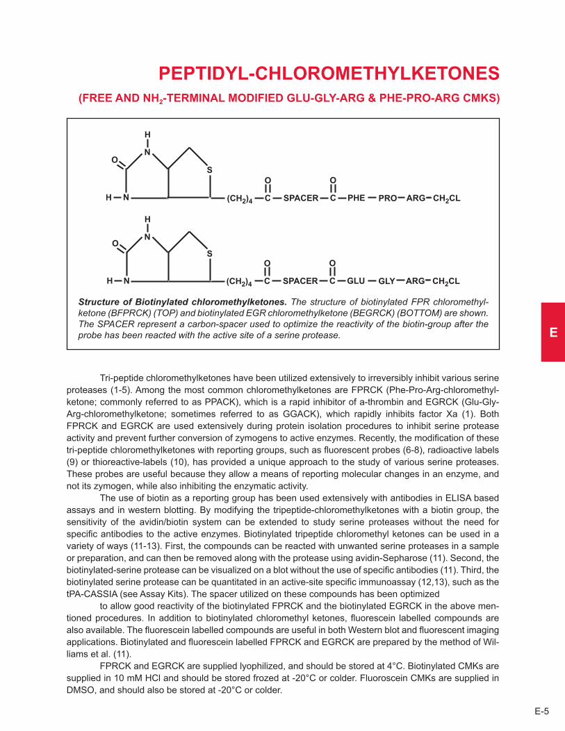
E-5
E
PEPTIDYL-CHLOROMETHYLkETONES (FREE AND NH2-TERMINAL MODIFIED GLU-GLY-ARG & PHE-PRO-ARG CMkS)
Structure of Biotinylated chloromethylketones. The structure of biotinylated FPR chloromethyl-ketone (BFPRCK) (TOP) and biotinylated EGR chloromethylketone (BEGRCK) (BOTTOM) are shown. The SPACER represent a carbon-spacer used to optimize the reactivity of the biotin-group after the probe has been reacted with the active site of a serine protease.
Tri-peptide chloromethylketones have been utilized extensively to irreversibly inhibit various serine proteases (1-5) . Among the most common chloromethylketones are FPRCK (Phe-Pro-Arg-chloromethyl-ketone; commonly referred to as PPACK), which is a rapid inhibitor of a-thrombin and EGRCK (Glu-Gly-Arg-chloromethylketone; sometimes referred to as GGACK), which rapidly inhibits factor Xa (1) . Both FPRCK and EGRCK are used extensively during protein isolation procedures to inhibit serine protease activity and prevent further conversion of zymogens to active enzymes . Recently, the modification of these tri-peptide chloromethylketones with reporting groups, such as fluorescent probes (6-8), radioactive labels (9) or thioreactive-labels (10), has provided a unique approach to the study of various serine proteases . These probes are useful because they allow a means of reporting molecular changes in an enzyme, and not its zymogen, while also inhibiting the enzymatic activity .
The use of biotin as a reporting group has been used extensively with antibodies in ELISA based assays and in western blotting . By modifying the tripeptide-chloromethylketones with a biotin group, the sensitivity of the avidin/biotin system can be extended to study serine proteases without the need for specific antibodies to the active enzymes . Biotinylated tripeptide chloromethyl ketones can be used in a variety of ways (11-13) . First, the compounds can be reacted with unwanted serine proteases in a sample or preparation, and can then be removed along with the protease using avidin-Sepharose (11) . Second, the biotinylated-serine protease can be visualized on a blot without the use of specific antibodies (11) . Third, the biotinylated serine protease can be quantitated in an active-site specific immunoassay (12,13), such as the tPA-CASSIA (see Assay Kits) . The spacer utilized on these compounds has been optimized
to allow good reactivity of the biotinylated FPRCK and the biotinylated EGRCK in the above men-tioned procedures . In addition to biotinylated chloromethyl ketones, fluorescein labelled compounds are also available . The fluorescein labelled compounds are useful in both Western blot and fluorescent imaging applications . Biotinylated and fluorescein labelled FPRCK and EGRCK are prepared by the method of Wil-liams et al . (11) .
FPRCK and EGRCK are supplied lyophilized, and should be stored at 4°C . Biotinylated CMKs are supplied in 10 mM HCl and should be stored frozed at -20°C or colder . Fluoroscein CMKs are supplied in DMSO, and should also be stored at -20°C or colder .

Visit us on the internet at http://www.haemtech.comPlease inquire about custom formulation, modification and conjugation.E-6
PROPERTIES OF CHLOROMETHYLkETONE COMPOUNDS
Formula & Molecular Weight:: FPR-CK•2HCl C21H33O3N6Cl3 524 BFPR-CK•1HCl C43H68O7N10S1Cl2 940 FFPR-CK•1HCl C42H42O9N6Cl2 846
EGR-CK•2HCl C14H27O5N6Cl3 466 BEGR-CK•1HCl C36H62O9N10S1Cl2 882 FEGR-CK•1HCl C35H36O11N6Cl2 788
Special properties: Tri-peptide chloromethyl ketones are very potent and irreversible inhibitors of serine proteases . BFPRCK is especially useful for inhibition of thrombin and tPA, while BEGRCK is useful for inhibition of factor Xa . The biotin moiety provides the ability to use the peptide-chloromethyl- ketones as specific probes for detection and/or capture of serine protases via the avidin/biotin interaction .
References1 . Kettner, C . and Shaw, E ., Methods Enzymol ., 80, 826 (1981) . 2 . Ganu, V .S . and Shaw, E ., Thromb . Res ., 45, 1 (1987) . 3 . Kettner, C . and Shaw, E ., Biochim . Biophys . Acta, 569, 31 (1979) . 4 . Kettner, C ., et al., Arch . Biochem . Biophys ., 202, 420 (1980) . 5 . Kettner, C . and Shaw, E ., Bochemistry, 17, 4778 (1978) . 6 . Kettner, C . and Shaw, E ., Thromb . Res ., 22, 645 (1981) . 7 . Lollar, P . and Fass, D .H ., Arch . Biochem . Biophys ., 233, 438 (1984) . 8 . Boskovic, D .S ., et al., J . Biol . Chem ., 265, 10497 (1990) . 9 . Rauber, P ., et al., Anal . Biochem ., 168, 259 (1988) . 10 . Bock, P .E ., Biochemistry, 27, 6633 (1988) . 11 . Williams, E .B ., et al., J . Biol . Chem ., 264, 7536, (1989) . 12 . Mann, K .G ., et al., Blood, 76, 755 (1990) . 13 . Hartshorn, J . et al., Blood, 78, No 10, Suppl 1, abstract 833, p 211a (ABSTRACT)14 . Williams, E .B and Mann, K .G ., Methods Enzymol ., 222, 503 (1993)15 . Tracy, R .P . et al. Methods Enzymol ., 222, 514 (1993)
Catalog NumbersFPR-chloromethylketone (PPACk) FPRCk-01Biotinylated FPR-chloromethylketone BFPRCk-06Fluorescein-FPR-chloromethylketone FFPRCk-06EGR-chloromethylketone (GGACk) EGRCk-01Biotinylated EGR chloromethylketone BEGRCk-06Fluorescein-EGR-chloromethylketone FEGRCk-06

E-7
E
CORN TRYPSIN INHIBITOR
EFFECT OF CTI ON THE APTT AND PT OF NORMAL HUMAN PLASMA. The graph illustrates the effect of added CTI on the PT and aPTT of normal human plasma. CTI was added to normal pooled human plasma to various final concentrations, and the mixtures were allowed to incubate for a brief period on ice prior to conducting the assays. The PT remains unaffected while the aPTT doubles with the addition of approximately 60 micrograms of CTI per ml of plasma.
Corn trypsin inhibitor (CTI) is a small protein that is localized in the kernels of most species of corn . CTI is not only an inhibitor of trypsin, but is also a specific inhibitor of human factor XIIa when tested in blood clotting experiments (1-6) . The inhibitor forms a one-to-one complex with either trypsin or factor XIIa, and when added to plasma, prolongs the activated partial thromboplastin time without affecting the PT assay (6) . The specificity for factor XIIa makes the inhibitor useful for the segregation and study of coagulation reactions (6) .
The protein is comprised of 112 amino acids which yields a calculated molecular weight of 12,028 (7) . The calculated molecular weight and that which has been determined by sedimentation equilibrium analyses of the purified protein (12,500) are in good agreement (3) . A comparison of the amino acid sequence of CTI to that of other trypsin or serine protease inhibitors reveals little or no similarities (7) .
CTI is purified using a combination of published procedures (3,4) . To begin the purification process, CTI is extracted from the kernels of fresh sweet corn into a physiologic buffer . The extract is then de-fatted using acetone, and the protein is further purified by employing gel filtration and ion-exchange chromato-graphy . The final CTI preparation appears as a single band by SDS-PAGE analyses under both reducing and non-reducing conditions .
Preparations of CTI are tested for the ability to prolong the aPTT assay without affecting the PT assay . The specific activity of each lot of CTI is determined, and one unit is defined as the amount of CTI required to double the aPTT of normal human plasma .
Purified CTI is formulated in 20 mM Tris, 0 .3 M NaCl, pH 7 .4 and should be stored frozen at -20°C or colder .Blood Collection Tubes
To simplify the process of collecting samples with added CTI, HTI has developed blood collection tubes containing CTI . These tubes simplify the process of conducting TF-dependent studies by allowing you to draw blood directly onto an anticoagulant containing CTI . You may choose to use our standard CTI/Ci-trate formulation (11mM Citrate, 50 ug/mL CTI) or you may create your own custom formulation . HTI’s blood collection tubes are not sterile and are manufactured and sold for research use only .

Visit us on the internet at http://www.haemtech.comPlease inquire about custom formulation, modification and conjugation.E-8
PROPERTIES OF CORN TRYPSIN INHIBITOR
Localization: corn kernels
Mode of action: forms a one-to-one complex with either trypsin or factor XIIa and inhibits their respective catalytic activity
Molecular weight: 12,500 (3)
Extinction coefficient: E1 cm, 280 nm = 20 .0a (3)
Structure: single chain protein comprised of 112 amino acids (3,7)
a determined by ultracentrifugation studies
References1 . Hochstrasser, K ., et al., Z . Physiol . Chem ., 348, 1337 (1967) . 2 . Halim, R .K ., et al., Crop . Sci ., 13, 405 (1973) .3 . Swartz, M .J ., et al., J . Biol . Chem ., 252, 8105 (1977) .4 . Hojima, Y ., et al., Thromb . Res ., 20, 149 (1980) .5 . Ratnoff, O .D . and Moneme, V ., Proc . Soc . Exp . Biol . Med ., 166, 297 (1981) .6 . Rand, M .D . et al., Blood, 88, 3432 (1996) .7 . Mahoney, W .C ., et al., J . Biol . Chem ., 259, 8412 (1984) .
Catalog NumbersCorn Trypsin Inhibitor CTI-01CTI/Citrate Collection Tube SCAT-27-4.5/5
1%
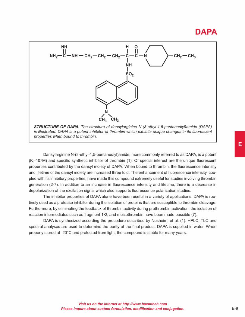
Visit us on the internet at http://www.haemtech.comPlease inquire about custom formulation, modification and conjugation. E-9
E
DAPA
STRUCTURE OF DAPA. The structure of dansylarginine N-(3-ethyl-1,5-pentanediyl)amide (DAPA) is illustrated. DAPA is a potent inhibitor of thrombin which exhibits unique changes in its fluorescent properties when bound to thrombin.
Dansylarginine N-(3-ethyl-1,5-pentanediyl)amide, more commonly referred to as DAPA, is a potent (Ki=10-7M) and specific synthetic inhibitor of thrombin (1) . Of special interest are the unique fluorescent properties contributed by the dansyl moiety of DAPA . When bound to thrombin, the fluorescence intensity and lifetime of the dansyl moiety are increased three fold . The enhancement of fluorescence intensity, cou-pled with its inhibitory properties, have made this compound extremely useful for studies involving thrombin generation (2-7) . In addition to an increase in fluorescence intensity and lifetime, there is a decrease in depolarization of the excitation signal which also supports fluorescence polarization studies .
The inhibitor properties of DAPA alone have been useful in a variety of applications . DAPA is rou-tinely used as a protease inhibitor during the isolation of proteins that are susceptible to thrombin cleavage . Furthermore, by eliminating the feedback of thrombin activity during prothrombin activation, the isolation of reaction intermediates such as fragment 1•2, and meizothrombin have been made possible (7) .
DAPA is synthesized according the procedure described by Nesheim, et al . (1) . HPLC, TLC and spectral analyses are used to determine the purity of the final product . DAPA is supplied in water . When properly stored at -20°C and protected from light, the compound is stable for many years .

Visit us on the internet at http://www.haemtech.comPlease inquire about custom formulation, modification and conjugation.E-10
PROPERTIES OF DAPA
Formula: C25H39O3N6S1Cl1 (1) (Dansylarginine N-(3-ethyl-1,5-pentanediyl)amide•HCl)
Molecular weight: 539 g/mole (1)
Extinction coefficient: E1 cm, 330 nm = 4010 (1)
Special properties: A specific and potent inhibitor of thrombin (Ki = 10-7M) . Exhibits a 3-fold increase in fluorescence intensity and lifetime when bound to thrombin
References1 . Nesheim, M .E ., et al., Biochemistry, 18, 996 (1979) .2 . Nesheim, M .E ., et al., J . Biol . Chem ., 254, 10952 (1979) . 3 . Hibbard, L .S ., et al., Biochemistry, 21, 2285 (1982) .4 . Nesheim, M .E . and Mann, K .G ., J . Biol . Chem ., 258, 5386 (1983) .5 . Krishnaswamy, S ., et al., J . Biol . Chem ., 261, 8977 (1986) .6 . Krishnaswamy, S ., et al., J . Biol . Chem ., 262, 3291 (1987) .7 . Doyle, M .F . and Mann, K .G ., J . Biol . Chem ., 265, 10693 (1990) .
Catalog NumberDansylarginine N-(3-ethyl-1,5-pentanediyl)amide•HCl DAPA
1M

Visit us on the internet at http://www.haemtech.comPlease inquire about custom formulation, modification and conjugation. E-11
E
HEPARIN COFACTOR II
INHIBITION OF THROMBIN BY HEPARIN COFACTOR II. Thrombin (IIa), heparin (HEP) and heparin cofactor II (HCII) interact to form a ternary complex via a random order bireactant mechanism. Heparin subsequently dissociates from the complex as thrombin forms a covalent complex with the heparin cofactor II.
Heparin cofactor II (HCII) (heparin cofactor A, antithrombin BM, dermatan sulfate cofactor, human leuserpin-2) is a single chain glycoprotein (Mr = 65,600, 10% carbohydrate) member of the serine protease inhibitor (serpin) family (1-3) . Amino acid sequence analysis of heparin cofactor II reveals 3 potential N-glycosylation sites and 2 sulfated tyrosines located in two internal repeats of 7 residues located near the NH2-terminal (4-6) . The plasma concentration is estimated to be 90 µg/ml (2) .
In the coagulation cascade, heparin cofactor II inhibits thrombin by formation of a bimolecular complex in the presence (k2 = 4 .5x108 M-1min-1) and absence (k2 = 5 .0x105 M-1min-1) of heparin (2,7-9) . This complex is stable to denaturants and essentially irreversible . In addition to heparin, a wide variety of polyanionic compounds accelerate inhibition of thrombin by HCII (3) . Noteworthy among these compounds is dermatan sulfate, which accelerates inhibition of thrombin by HCII, but not antithrombin III (1,6) This feature has been utilized to develop a plasma based assay specific for heparin cofactor II . Although HCII shows only ~25% sequence homology to other serpins, the reactive site peptide (COOH-terminal 36 amino acids) released on inhibition of thrombin is 53% homologous to that released from antithrombin III (ATIII) by thrombin (4,9) . The scissile bond in HCII is Leu-Ser which is unusual for an arginine specific protease inhibitor (4,9) . In contrast to ATIII, the only coagulation protease inhibited by HCII is thrombin . Other proteases inhibited by HCII include a-chymotrypsin, neutrophil cathepsin G and streptomyces griseus protease B (3) . The major physiologic role of heparin cofactor II may remain to be discovered .
Heparin cofactor II is prepared from fresh frozen plasma by a modification of the procedure of Griffith, et al., (9) . Purity is assessed on SDS PAGE and activity is determined based on thrombin inhibition in the presence of dermatan sulfate . HCII is supplied in 50% (vol/vol) glycerol/H20 for storage at -20°C .

Visit us on the internet at http://www.haemtech.comPlease inquire about custom formulation, modification and conjugation.E-12
PROPERTIES OF HEPARIN COFACTOR II
Localization: Plasma
Plasma concentration: 90 µg/ml (2)
Mode of action: serine protease inhibitor; inhibits thrombin, a-chymotrypsin, neutrophil cathepsin G, streptomyces griseus protease B
Molecular weight: 65,600 (2)
Extinction coefficient: E1 cm, 280 nm = 5 .93 (5)
Isoelectric point: 4 .95-5 .15 (2)
Structure: single chain glycoprotein, 3 potential N-glycosylation sites, two 7-amino acid residue repeats, reactive site sequence: TVTTVGFMPL-STQVRFTVDR (4)
Percent carbohydrate: 9 .9% (2)
Post-translational modifications: 3 sulfated tyrosines
References1 . Bringinshaw, G .W . and Shanberge, J .M ., Arch . Biochem . Biophys ., 161, 683 (1974) .2 . Tollefsen, D .M ., et al., J . Biol . Chem ., 257, 2162 (1982) .3 . Pratt, C .W ., et al., Ann . N .Y . Acad . Sci ., 556, 104 (1989) .4 . Ragg, H ., Nucleic Acids Res ., 14, 1073 (1986) .5 . Church, F .C ., et al., Arch . Biochem . Biophys ., 259, 331 (1987) .6 . Hortin, G ., et al., J . Biol . Chem ., 261, 15827 (1986) .7 . Church, F .C ., et al., Proc . Natl . Acad . Sci . USA, 82, 6431 (1985) .8 . Tollefsen, D .M . and Blank, M .K ., J . Clin . Invest ., 68, 589 (1981) .9 . Griffith, M .J ., et al., J . Biol . Chem ., 260, 2218 (1985) .10 . Tollefsen, D .M ., et al., J . Biol . Chem ., 258, 6713 (1983) .11 . Griffith, M .J . and Marbet, G .A ., Biochem . Biophys . Res . Comm ., 112, 663 (1983) .12 . Abildgaard, U . and Larsen, M .L ., Thromb . Res ., 35, 257 (1984) .13 . Griffith, M .J ., et al., Biochemistry, 24, 6777 (1985) .14 . Parker, K . and Tollefsen, D .M ., J . Biol . Chem ., 260, 3501 (1985) .
Catalog NumberHeparin Cofactor II HCII-0190
1%

Visit us on the internet at http://www.haemtech.comPlease inquire about custom formulation, modification and conjugation. E-13
E
TAFI
DOMAIN STRUCTURE OF TAFI. The N-linked glycosylation sites (N22, N51, N63. N86) are represented by N. The active site Zn2+ and residues (S299, G336, D344) involved in substrate binding are shown.
Thrombin Activatable Fibrinolysis Inhibitor (TAFI, Plasma pro-carboxypeptidase B, carboxy- peptidase U) is a single chain glycoprotein zymogen (Mr = 60,000) synthesized in the liver and circulating at a plasma concentration of 50 nM (1-4) . Thrombin (plasmin, trypsin) cleavage of the zymogen releases a 92 amino acid N-terminal activation peptide containing 4 N-linked glycosylation sites (N22, N51, N63, N86) and the proposed plasminogen recognition site . The rate of thrombin catalyzed activation of TAFI is increased 1250 fold by formation of a ternary complex with thrombomodulin (5) . The 309 amino acid C-terminal (Mr = 35,783) catalytic domain (TAFIa, pCPB) displays the properties of a basic carboxy-peptidase, hydrolyzing lysine and arginine from the C-terminal position of polypeptides . This portion of the molecule is homologous to tissue carboxypeptidase B and contains 7 conserved cystine residues (64,77,136,151, 160,165, 291), the active site Zn2+ coordination site (H67, E69, H196) and the basic C-terminal amino acid substrate binding pocket (D257, G244, S207) .
TAFI is proposed to play a key role in the interaction between procoagulant, anticoagulant and fibrinolytic systems (5-9) . Effective fibrinolysis results from the formation of a ternary complex between tPA, plasminogen and C-terminal lysine residues on fibrin . Plasminogen bound to fibrin is more effectively converted to plasmin, thereby localizing the lytic activity to the area of the clot . Plasmin degradation of fibrin generates additional C-terminal lysine residues thereby amplifying the system locally . The ability of TAFI to bind specifically to plasminogen and to cleave C-terminal lysines on fibrin (and cell surfaces) results in down-regulation of fibrinolysis by reducing the number of plasminogen and tPA binding sites on fibrin . The activation of TAFI by the thrombin/thrombomodulin complex couples both the phenomenon of coagulation induced inhibition of fibrinolysis and the profibrinolytic effect of activated protein C .
TAFI is prepared from fresh frozen human plasma by a modification of the method of Bajzar, et. al. (10), and supplied in HBS for storage at -80°C . Activity is determined measuring the rate of hydrolysis of hipuryl-L- Arg following activation with the thrombin/thrombomodulin complex (11) .

Visit us on the internet at http://www.haemtech.comPlease inquire about custom formulation, modification and conjugation.E-14
PROPERTIES OF TAFI
Localization: Plasma
Plasma concentration: 2 .5 µg/ml
Mode of action: Basic carboxypeptidase, cleaves C-terminal lysine and arginine residues . Inhibition of fibrinolysis by removal of plasminogen binding sites on fibrin .
Molecular weight: 60,000
Extinction coefficient: E1 cm, 280 nm = 14 .9 (calculated from cDNA)
Isoelectric point: 5 .0
Structure: Single chain glycoprotein, 92 a .a . N-terminal activation peptide, 309 a .a . catalytic domain, 1 zinc binding site
Percent carbohydrate: 19%
Post-translational modifications: 4 N-linked glycosylation sites located at residues N22, N51, N63, and N86 of the activation peptide
References1 . Eaton, D .L ., et.al., J .Biol .Chem ., 266, 21833-21838 (1991) .2 . Hendriks, D ., et.al., BBA, 1034, 86-92 (1990) .3 . Hendriks, D ., et.al., J .Clin .Chem .Clin .Biochem ., 27, 277 (1989) .4 . Campbell, W ., Okada, H ., BBRC, 162, 933-939 (1989) .5 . Bajzar, L . et.al., J . Biol . Chem ., 271, 16603-16608 (1996) .6 . Redlitz, A ., et.al., J .Clin .Invest ., 96, 2534-2538 (1995) .7 . Broze, G .J . and Higuchi, D .A ., Blood, 88, 3815-3823 (1996) .8 . Bajzar, L . and Nesheim, M ., J .Biol .Chem ., 268, 8608-8616 (1993) .9 . Collen, D . and Lijnew, H .R ., Blood, 78, 3114-3124 (1992) .10 . Bajzar, L . et.al., J .Biol .Chem ., 270, 14477-14484 (1995) .11 . Folk, J .E . et.al., J . Biol . Chem . 235, 2272-2282 (1960) .
Catalog NumberThrombin Activatable Fibrinolysis Inhibitor TAFI-01
1%

E-15
E
“ANGIOSTATIN”
HUMAN “ANGIOSTATIN.” Selected proteolytic cleavage sites within the human plasminogen mol-ecule are illustrated. Cleavage of plasminogen by elastase yields “angiostatin” (kringles 1-4) or angio-statin-like fragments (kringles 1-3). HTI’s “angiostatin” product is derived from a single cleavage within the plasminogen molecule resulting in a product that includes Kringles 1-4 as well as the NH2-terminal 78 amino acids that are absent in the elastase generated product. [K1-K5 = the 5 kringle domains, B-CHAIN = catalytic domain of plasmin, and the arrows indicate the sites of proteolytic cleavage by plasmin, elastase, HTI’s proprietary enzyme, and plasminogen activators (PA’S)].
Angiostatin is a single-chain, proteolytic fragment of glu-plasminogen which has a molecular weight of about 38,000 . It is a potent inhibitor of angiogenesis and was first identified and isolated from the serum and urine of tumor bearing mice (1) . The mouse protein reportedly extends from threonine-98 through valine-440 (numbering from the NH2-terminus of glu-plasminogen) . This fragment of mouse plasminogen, as well as the human equivalent, includes four out of the five kringle domains of plasminogen (1) .
The inhibition of angiogenesis by angiostatin is directly related to inhibiting the proliferation of endothelial cells (1,2) . Because tumor growth is known to be angiogenesis dependent, it was initially hypothesized that the inhibitory properties of angiostatin would have clinical utility in arresting various cancers that are expressed as solid tumors . Studies performed in animal model systems have since demonstrated that recombinant angiostatin effectively suppresses tumor growth and metastasis (3,4) .
Numerous enzymes have been identified which will convert plasminogen to angiostatin or at least to angiostatin-like fragments . The enzymes include several matrix metalloproteinases as well as urokinase and a tumor cell-derived reductase (5-10) . The precise enzyme or mechanism which is responsible for the formation of angiostatin in vivo is unknown and it is believed that there may be multiple pathways for the conversion of plasminogen to angiostatin .
Structure/function studies have indicated that the first three kringle domains (and not the fourth) are responsible for the inhibitory properties of angiostatin, and that removal of the fourth kringle domain may actually yield a more potent inhibitor (11) .
HTI’s “angiostatin” product is produced by limited proteolysis of purified human glu-plasminogen using a proprietary enzyme preparation . The NH2-terminal sequence of this fragment is identical to human glu-plasminogen (Glu-1) and the molecule terminates at proline-452 thus making it larger than the “natural” or elastase derived product with an apparent molecular weight of about 50,000 . Like authentic angiostatin, this product demonstrates an antiproliferative effect when tested in a growth factor-induced endothelial cell proliferation assay . The product is formulated in 20mM Hepes, 0 .15M NaCl, pH 7 .4, and should be stored frozen at -80°C .

Visit us on the internet at http://www.haemtech.comPlease inquire about custom formulation, modification and conjugation.E-16
PROPERTIES OF “ANGIOSTATIN”
Mode of action: Inhibits endothelial cell proliferation
Molecular weight: approximately 50,000 by SDS-PAGE (appears as a doublet due to the presence of two carbohydrate variants)
Extinction coefficient: E1 cm, 280 nm = 17 .4
Structure: single chain, 4 kringle regions, 14 disulfide bonds, no free sulfhydryls
Percent carbohydrate: Approximately 3%
References1 . O’Reilly, M .S ., et al., Cell, 79, 315 (1994) .2 . Folkman, J ., Nat . Med ., 1, 27 (1995) .3 . Sim, S .K ., et al., Cancer Res ., 57, 1329 (1997) .4 . Wu, Z ., et al., Biochem . Biophys . Res . Commun ., 236, 651 (1997) .5 . Dong, Z ., et al., Cell, 88, 801 (1997) .6 . Lijnen, H .R ., et al., Biochemistry, 37, 4699 (1998) .7 . Patterson, B .C ., et al., J . Biol . Chem ., 272, 28823 (1997) .8 . Stathakis, P ., et al., J . Biol . Chem ., 272, 20641 (1997) .9 . Gately, S ., et al., Cancer Res ., 56, 4887 (1996) .10 . Gately, S ., et al., Proc . Nat . Acad . Sci . USA, 94, 10868 (1997) .11 . Cao, Y ., et al., J . Biol . Chem ., 271, 29461 (1996) .
Catalog NumberHuman “Angiostatin” ANG-01
1%
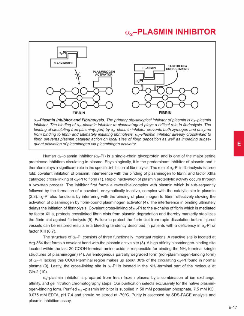
E-17
E
a2–PLASMIN INHIBITOR
a2–Plasmin Inhibitor and Fibrinolysis. The primary physiological inhibitor of plasmin is a2–plasmin inhibitor. The binding of a2–plasmin inhibitor to plasmin(ogen) plays a critical role in fibrinolysis. The binding of circulating free plasmin(ogen) by a2–plasmin inhibitor prevents both zymogen and enzyme from binding to fibrin and ultimately initiating fibrinolysis. a2–Plasmin inhibitor already crosslinked to fibrin prevents plasmin catalytic action on local sites of fibrin deposition as well as impeding subse-quent activation of plasminogen via plasminogen activator.
Human a2–plasmin inhibitor (a2-PI) is a single-chain glycoprotein and is one of the major serine proteinase inhibitors circulating in plasma . Physiologically, it is the predominant inhibitor of plasmin and it therefore plays a significant role in the specific inhibition of fibrinolysis . The role of a2-PI in fibrinolysis is three fold: covalent inhibition of plasmin; interference with the binding of plasminogen to fibrin; and factor XIIIa catalyzed cross-linking of a2-PI to fibrin (1) . Rapid inactivation of plasmin proteolytic activity occurs through a two-step process . The inhibitor first forms a reversible complex with plasmin which is sub-sequently followed by the formation of a covalent, enzymatically inactive, complex with the catalytic site in plasmin (2,3) . a2-PI also functions by interfering with the binding of plasminogen to fibrin, effectively slowing the activation of plasminogen by fibrin-bound plasminogen activator (4) . The interference in binding ultimately delays the initiation of fibrinolysis . Covalent cross-linking of a2-PI to the a-chains of fibrin which is mediated by factor XIIIa, protects crosslinked fibrin clots from plasmin degradation and thereby markedly stabilizes the fibrin clot against fibrinolysis (5) . Failure to protect the fibrin clot from rapid dissolution before injured vessels can be restored results in a bleeding tendency described in patients with a deficiency in a2-PI or factor XIII (6,7) .
The structure of a2-PI consists of three functionally important regions . A reactive site is located at Arg-364 that forms a covalent bond with the plasmin active site (8) . A high affinity plasminogen-binding site located within the last 20 COOH-terminal amino acids is responsible for binding the NH2-terminal kringle structures of plasmin(ogen) (4) . An endogenous partially degraded form (non-plasminogen-binding form) of a2-PI lacking this COOH-terminal region makes up about 30% of the circulating a2-PI found in normal plasma (9) . Lastly, the cross-linking site in a2-PI is located in the NH2-terminal part of the molecule at Gln-2 (10) .
a2–plasmin inhibitor is prepared from fresh frozen plasma by a combination of ion exchange, affinity, and gel filtration chromatography steps . Our purification selects exclusively for the native plasmin-ogen-binding form . Purified a2 –plasmin inhibitor is supplied in 50 mM potassium phosphate, 7 .5 mM KCl, 0 .075 mM EDTA, pH 7 .4 and should be stored at -70°C . Purity is assessed by SDS-PAGE analysis and plasmin inhibition assay .

Visit us on the internet at http://www.haemtech.comPlease inquire about custom formulation, modification and conjugation.E-18
a2–PLASMIN INHIBITOR
Localization: Plasma
Mode of action: Inhibits plasmin by forming an irreversible complex with the catalytic active site . Prevents the binding of plasmin to fibrin . Cross-linked by factor XIIIa to fibrin .
Plasma Concentration: 69 µg/ml (1)
Molecular weight: 58,700 single-chain (determined from amino acid sequence and 14% carbohydrate) and 67,000 (determined by SDS-PAGE) (1)
Extinction coefficient: E1 cm, 280 nm = 7 .03 (2)
Isoelectric Point: Unknown
Structure: Circulates as single chain molecule consisting of 452 amino acids (1) .
Carbohydrate Content: 14% (w/w) (1)
References1 Aoki, N . et al., Methods Enzymol ., 223, 185-197 (1993) .2 . Moroi, M . and Aoki, N ., J . Biol . Chem ., 251, 5956 (1976) .3 . Wiman, B . and Collen, D ., J . Biol . Chem ., 254, 9291 (1979) .4 . Moroi, M . and Aoki, N ., Thromb . Res ., 10, 851 (1977) .5 . Sakata, Y . and Aoki, N ., J . Clin . Invest ., 69, 536 (1982) .6 . Aoki, N . et al., J . Clin . Invest ., 63, 877 (1979) .7 . Israels, E . D . et al., J . Clin . Invest ., 52, 2398 (1973) .8 . Holmes, W . E . et al., J . Biol . Chem ., 262, 1659 (1987) .9 . Kluft, C . and Los, N ., Thromb . Res ., 21, 65 (1981) .10 . Tamaki, T . and Aoki, N ., J . Biol . Chem ., 257, 14767 (1982) .
Catalog Numbera2-Plasmin Inhibitor HA2AP-0230
1%
F
pLATELET & OTHER
pLASMA dERIvEd
pROTEINS

Visit us on the internet at http://www.haemtech.comPlease inquire about custom formulation, modification and conjugation. F-1
F
FIBRINOGEN
DOMAIN STRUCTURE OF FIBRINOGEN. The trinodular arrangement of the Aa, Bb, and g chains, the IIa cleavage sites (IIa) releasing FPA and FPB, as well as the plasmin cleavage sites (Pm) yielding Fragment D and Fragment E are shown.
The thrombin (IIa) catalyzed cleavage of soluble fibrinogen (Fbg) to form fibrin (Fbn) is the terminal proteolytic event in the coagulation cascade . These soluble Fbn monomers spontaneously polymerize to form an insoluble Fbn network which is stabilized by the factor XIIIa catalyzed crosslinking of lys and glu residues of a and g chains . This Fbn network is the major protein component of the hemostatic plug .
Plasma fibrinogen is large glycoprotein (Mr = 340,000) synthesized in the liver and circulating at a concentration of 2 .6 mg/ml . It is a disulfide linked dimer composed of 3 pairs of disulfide linked non-identical polypeptide chains (Aa, Bb and g) . Notable features of the Aa chain are the N-terminal peptide (fibrinopep-tide A (FPA, 1-16)), factor XIIIa crosslinking sites and 2 phosphorylation sites . When synthesized, Fbg is fully phosphorylated, but circulates at only 20-30% phosphorylation . The Bb chain contains fibrinopeptide B (FPB, 1-14), one of the 3 N-linked carbohydrate moieties (Mr = 2500) and an N-terminal pyroglutamic acid . The g chain contains the other N-linked glycosylation site and a factor XIIIa crosslinking sites . The 2 elon-gated subunits ((AaBbg)2) are aligned in an antiparallel manner forming a trinodular arrangement of the six chains . The nodes are formed by disulfide rings between the 3 parallel chains . The central node (n-disulfide knot, E domain) is formed by the N-termini of all six chains held together by 11 disulfide bonds . This region contains the 2 IIa-sensitive sites . The release of FPA by cleavage at R16-G17 generates Fbn I, exposing a polymerization site (17-20) on the Aa chain . These regions bind to complimentary regions on the D domain of Fbn to form protofibrils . Subsequent IIa cleavage of FPB (R14-G15) from the Bb chain exposes additional polymerization sites and promotes lateral growth of the Fbn network .
Each of the 2 domains between the central node (E domain) and the C-terminal nodes (D domain) is composed of parallel a-helical regions of the Aa, Bb and g chains coiled around each other to form a “coiled coil” with polar residues directed outward and nonpolar residues forming a hydrophobic core . In this region, all 3 chains possess a protease (plasmin) sensitive site . The other major plasmin sensitive site is in the hydrophilic preturbance of the a-chain from the C-terminal node . Controlled plasmin degradation at these sites converts Fbg into fragment D and fragment E . The individual fragments are isolated by salt fraction-ation, gel filtration and ion exchange chromatography . The fragments are supplied lyophilized for storage at 4°C . Highly purified research grade fibrinogen (>95% clottable) is prepared by a combination of conventional and affinity techniques . It is supplied as a frozen solution in citrate-phosphate buffer for storage at -70°C .

Visit us on the internet at http://www.haemtech.comPlease inquire about custom formulation, modification and conjugation.F-2
PROPERTIES OF FIBRINOGEN
Localization: Plasma, platelets
Plasma concentration: 2 .6 mg/ml
Mode of action: Precursor molecule which is cleaved by thrombin to form fibrin clot .
Molecular weight: 340,000
Extinction coefficient: E1 cm, 280 nm = 15 .1
Isoelectric Point: 5 .1-6 .3
Structure: Dimer of 3 pairs of non-identical chains Aa (Mr= 66,800), Bb (Mr = 52,000) and g (Mr = 46,500), Elongated trinodular molecule with 2 terminal D domains and one central E domain . Factor XIIIa cross-linking sites at Q328, Q366, K508, K584 in Aa chain; at Q398 and K406 in g chain .
Percent carbohydrate: 3%
Post-translational modifications: Aa chain: 2 phosphorylated serines, S3, S346 Bb chain: N364 glycosylation site Mr = 2500, N-terminal pyroglutamic acid g chain: glycosylated N52
References1 . Hantgan, R .R ., et.al., in Haemostasis and Thrombosis, 2nd edition, pp 269-289, Bloom, A .L ., Forbes, C .D ., Thomas, D .P . and Tuddenham, E .G .D ., eds, Churchill Livingstone, 1991 .2 . Doolittle, R .F . in Haemostasis and Thrombisos, 3rd edition, 491-513, Bloom, A .L ., Forbes, C .D ., Thomas, D .P . and Tuddenham, E .G .D ., eds, Churchill Livingstone, 1994 .3 . Shaefer, J .A . and Higgins, D .L ., CRC Crit .Rev .Clin .Lab .Sci ., 26, 1-41 (1988) .4 . Hoeprich, P .D . and Doolittle, R .F ., Biochemistry, 22, 2049 (1983) .5 . Doolittle, R .F . et.al., J .Mol .Biol ., 120, 311-325 (1978) .6 . Marder, V .J ., et.al., J .Biol .Bhem ., 244, 2111-2119 (1969) .7 . Budzynski, A .Z ., et.al., J .Biol .Chem ., 249, 2294-2302 (1974) .8 . Furlan, M . and Beck, E .A ., Biochim .Biophys . Acta, 310, 205-216 (1973) .9 . Furlan, M ., in Human Protein Data, Fibrinogen, Haeberli,A ., ed ., VCH Publishers, N .Y . (1995) .
Catalog NumbersResearch Grade Fibrinogen HCI-0150RFibrinogen Fragment D HCI-0150DFibrinogen Fragment E HCI-0150EMouse Fibrinogen MCI-5150
1%

Visit us on the internet at http://www.haemtech.comPlease inquire about custom formulation, modification and conjugation. F-3
F
b2-GLYCOPROTEIN I
DOMAIN STRUCTURE OF b2-GLYCOPROTEIN I. The five contiguous Sushi domains (I-V) of b2I are shown. The 5 carbohydrate side chains at N73, N143, N164, N174, and N224 are represented (CHO).
b2-Glycoprotein I (b2I, Apolipoprotein H) is a highly glycosylated single chain protein (Mr = 54,200) which is synthesized in the liver and circulates in plasma at a concentration of 100-200 µg/ml (1-4) . Ap-proximately 40% of the plasma b2I is associated with lipoproteins, which led to the designation apolipopro-tein H (5) . The 326 amino acid protein contains five repeating mutually homologous domains consisting of approximately 60 amino acids which are disulfide bonded to form Short Consensus Repeats (SCR) or Sushi domains . The third, C-terminal sushi domain contains three of the five N-linked carbo-hydrate chains, at positions N143, N164, N174 . The other two sites are at N73 and N234 in domains II and IV, respectively . There are 38 basic amino acid residues distributed unevenly among the five domains; eight in domain I, five in domain II, five in domain III, five in domain IV and fifteen in domain V . The direct binding of domains I and V has been demonstrated and likely results from folding of the molecule in a manner that expresses a net cationic surface charge (5) .
Although the physiological role of b2I is still being studied, the ability to bind to anionic surfaces has been the focus of intense investigation . b2I has been shown to bind to anionic vesicles (3), platelets (6), DNA (7), mitochondria (8) and heparin (9) . It has been suggested that the binding of b2I to negatively charged surfaces can inhibit the contact activation pathway in blood coagulation . The binding to activated platelets is reported to inhibit platelet associated prothrombinase and adenylate cyclase activities . Studies on physiologic roles for binding to DNA, mitochondria, heparanoids and phospholipids have focused on the area of autoimmune disorders . In particular, the complexes between b2I and cardiolipin have been implicated in the anti-phospholipid related disorders LAC and SLE (10-13) .
The human b2-Glycoprotein I is purified from fresh frozen human plasma, using a combination of ion exchange and affinity chromatography . It is greater than 95% pure by SDS PAGE . The human b2-Glyco-protein I is lyophilized from a glycine-NaCl buffer and should be stored at 4°C .

Visit us on the internet at http://www.haemtech.comPlease inquire about custom formulation, modification and conjugation.F-4
PROPERTIES OF b2-GLYCOPROTEIN I
Localization: Plasma
Plasma concentration: 100-200 µg/ml
Mode of action: Cationic residues on the surface of sushi domains I and V bind to anionic phospholipids . Inhibition of contact activation, platelet membrane dependant prothrombinase and adenylate cyclase activities . Complexes with DNA, mitochondria, and cardiolipin augments antigenicity in autoimmune disorders .
Molecular weight: 54,200 (SDS PAGE), 48,000 (Sed . Equil)
Extinction coefficient: E1 cm, 280 nm = 10 .0
Structure: 326 amino acid single chain protein with 5 contiguous Short Consensus Repeats or sushi domains .
Percent carbohydrate: 25%
Post-translational modifications: 5 N-linked glycosylation sites, 1 in domain II (N73), 3 in domain III (N143, N164, N174) and 1 in domain IV (N224) .
References1 . Lozier, J ., et.al., Proc .Natl .Acad . Sci .,USA, 81, 2640-3644 (1984) .2 . Kato, H . and Enjyoi, K ., Biochem ., 30, 11687-11694 (1997) .3 . Wurm, H ., Int .J .Biochem ., 16, 511-515 (1984) .4 . Bendixen, E ., et.al., Biochem ., 31, 3611-3617 (1992) .5 . Steinkasserer, A ., et.al., Biochem .J ., 277, 387-391 (1991) .6 . Nimpf, J . et.al., Biochim .Biophys .Acta, 884, 142-149 (1986) .7 . Kroll, J . et.al., Biochim .Biophys .Acta, 434, 490-501 (1986) .8 . Schousboe, I ., Biochim .Biophys .Acta, 679, 396-408 (1979) .9 . Polz, E ., et.al., Int .J .Biochim ., 11, 265-273 (1076) .10 . McNeil, H .P . et.al., Proc .Natl .Acad .USA, 87, 4120-4124 (1990) .11 . Galli, M . et.al., Lancet, I, 1544-1547 (1990) .12 . Matsuuna, E ., et.al., Lancet, II, 177-178 (1990) .13 . Pengo, V . et.al., Thrombosis and Haemostasis, 73, 29-34 (1995) .
Catalog NumberHuman b2-Glycoprotein I B2GI-0001
1%

Visit us on the internet at http://www.haemtech.comPlease inquire about custom formulation, modification and conjugation. F-5
F
PLATELET FACTOR-4
DOMAIN STRUCTURE OF PLATELET FACTOR-4. The domain structure of the platelet factor-4 monomer is represented. The platelet factor-4 monomer is a 7,800 molecular weight peptide. The heparin-binding domain is presumably localized within a COOH-terminal region of the molecule where several lysine residues are clustered. In its native state, platelet factor-4 is a homotetramer which ex-ists in complex with a high molecular weight proteoglycan carrier protein.
Platelet factor-4 (PF-4) is a low molecular weight, heparin-binding protein which is secreted from agonist-activated platelets (1,2) . PF-4 is localized within the platelet a-granule (2) where its concentration ranges from 11 .2-12 .4 µg per 109 platelets making it, on a molar basis, one of the most abundant proteins in the platelet (1,2) . Since the relative concentration of PF-4 in platelets exceeds that of plasma by 280,000-fold (3), PF-4 levels in plasma have been utilized as a measure of platelet activation in vivo . PF-4 is secreted from platelets in complex with a high molecular weight, proteoglycan, carrier protein (4,5) . Sedimentation equilibrium experiments have shown that in the absence of its proteoglycan carrier, the molecular weight of PF-4 is 27,000-29,000 (4,5) . Subsequent amino acid sequencing of PF-4 revealed a molecular weight of 7,800 (6-8) indicating that native PF-4 is a homotetramer . Functionally, PF-4 neutralizes the anticoagu-lant activity of heparin in plasma . The heparin binding site within PF-4 is presumably located within the lysine-rich, COOH-terminal region of the molecule (6-8) . Since soluble heparin is a therapeutic agent, the physiological significance of the anti-heparin activity of PF-4 is not known . However, by interacting with cell surface expressed heparin-like glycosaminoglycans on endothelial cells, PF-4 may exert its procoagulant effect (9,10) . PF-4 binding to cell surface glycosaminoglycans may also be a mechanism through which PF-4 stimulates the release of histamine from basophils (11) . The chemotactic activity of PF-4 toward neutrophils and monocytes (12) has also been localized to the COOH-terminal, presumed heparin-binding domain of PF-4 (13) . While PF-4 is primarily a secreted protein, PF-4 binding sites on the platelet surface have been identified which may be important for platelet aggregation (14) .
Human PF-4 is prepared from the supernatant of thrombin-activated platelets by heparin-agarose affinity chromatography (15) . The purified protein is supplied in 25 mM Hepes pH 7 .4, 2 M NaCl and should be stored at -20°C . Purity is assessed by SDS-PAGE analysis and heparin-neutralizing activity is verified by clotting assay .

Visit us on the internet at http://www.haemtech.comPlease inquire about custom formulation, modification and conjugation.F-6
PROPERTIES OF PLATELET FACTOR-4
Localization: platelet a-granule (2)
Mode of action: neutralizes the anticoagulant activity of heparin . Plasma concentration is used as a marker of platelet activation .
Molecular weight: 29,000 (4)
Extinction coefficient: E1 cm, 280 nm = 2 .6a
Structure: homotetramer (monomer, Mr-7800) (5-7)
a calculated based upon amino acid sequence and molecular weight
References1 . Rucinski, B ., et al., Blood, 53, 47 (1979) .2 . Kaplan, K .L ., et al., Blood, 53, 604 (1979) .3 . George, J .N ., Blood, 76, 859 (1990) .4 . Barber, A .G ., et al., Biochim . Biophys . Acta, 286, 316 (1972) .5 . Moore, S ., et al., Biochim . Biophys . Acta, 379, 370 (1975) .6 . Hermodson, M ., et al., J . Biol . Chem ., 252, 6276 (1977) .7 . Deuel, T .F ., et al., Proc . Natl . Acad . Sci . USA, 74, 2256 (1977) .8 . Walz, D .A ., et al., Thromb . Res ., 11, 833 (1977) .9 . Busch, C ., et al., Thromb . Res ., 19, 129 (1980) .10 . Rao, A .K ., et al., Blood, 61, 1208 (1983) .11 . Brindley, L .L ., et al., J . Clin . Invest ., 72, 1218 (1983) .12 . Deuel, T .F ., et al., Proc . Natl . Acad . Sci . USA, 74, 2256 (1981) .13 . Osterman, D .G ., et al., Biochem . Biophys . Res . Commun ., 107, 130 (1982) .14 . Capitanio, A .M ., et al., Biochim . Biophys . Acta, 839, 161 (1985) .15 . Jordon, R .E ., et al., J . Biol . Chem ., 257, 400 (1982) .
Catalog NumberHuman Platelet Factor-4 HPF4-0180
1%
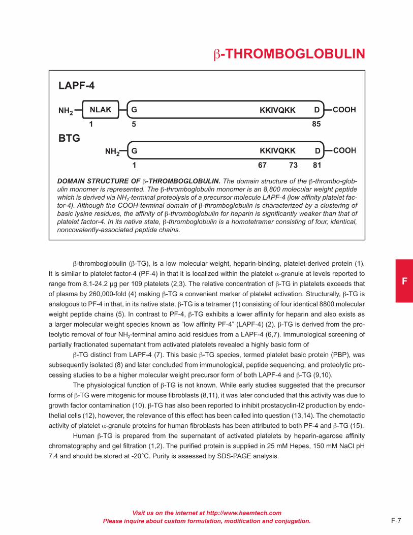
Visit us on the internet at http://www.haemtech.comPlease inquire about custom formulation, modification and conjugation. F-7
F
b-THROMBOGLOBULIN
DOMAIN STRUCTURE OF b-THROMBOGLOBULIN. The domain structure of the b-thrombo-glob-ulin monomer is represented. The b-thromboglobulin monomer is an 8,800 molecular weight peptide which is derived via NH2-terminal proteolysis of a precursor molecule LAPF-4 (low affinity platelet fac-tor-4). Although the COOH-terminal domain of b-thromboglobulin is characterized by a clustering of basic lysine residues, the affinity of b-thromboglobulin for heparin is significantly weaker than that of platelet factor-4. In its native state, b-thromboglobulin is a homotetramer consisting of four, identical, noncovalently-associated peptide chains.
b-thromboglobulin (b-TG), is a low molecular weight, heparin-binding, platelet-derived protein (1) . It is similar to platelet factor-4 (PF-4) in that it is localized within the platelet a-granule at levels reported to range from 8 .1-24 .2 µg per 109 platelets (2,3) . The relative concentration of b-TG in platelets exceeds that of plasma by 260,000-fold (4) making b-TG a convenient marker of platelet activation . Structurally, b-TG is analogous to PF-4 in that, in its native state, b-TG is a tetramer (1) consisting of four identical 8800 molecular weight peptide chains (5) . In contrast to PF-4, b-TG exhibits a lower affinity for heparin and also exists as a larger molecular weight species known as “low affinity PF-4” (LAPF-4) (2) . b-TG is derived from the pro-teolytic removal of four NH2-terminal amino acid residues from a LAPF-4 (6,7) . Immunological screening of partially fractionated supernatant from activated platelets revealed a highly basic form of
b-TG distinct from LAPF-4 (7) . This basic b-TG species, termed platelet basic protein (PBP), was subsequently isolated (8) and later concluded from immunological, peptide sequencing, and proteolytic pro-cessing studies to be a higher molecular weight precursor form of both LAPF-4 and b-TG (9,10) .
The physiological function of b-TG is not known . While early studies suggested that the precursor forms of b-TG were mitogenic for mouse fibroblasts (8,11), it was later concluded that this activity was due to growth factor contamination (10) . b-TG has also been reported to inhibit prostacyclin-I2 production by endo-thelial cells (12), however, the relevance of this effect has been called into question (13,14) . The chemotactic activity of platelet a-granule proteins for human fibroblasts has been attributed to both PF-4 and b-TG (15) .
Human b-TG is prepared from the supernatant of activated platelets by heparin-agarose affinity chromatography and gel filtration (1,2) . The purified protein is supplied in 25 mM Hepes, 150 mM NaCl pH 7 .4 and should be stored at -20°C . Purity is assessed by SDS-PAGE analysis .

Visit us on the internet at http://www.haemtech.comPlease inquire about custom formulation, modification and conjugation.F-8
PROPERTIES OF b-THROMBOGLOBULIN
Localization: platelet a-granule (3)
Mode of action: heparin-binding protein: Plasma concentration used as a marker of platelet activation .
Molecular weight: 35,800 (1)
Extinction coefficient: E1 cm, 280 nm = 2 .6a
Structure: homotetramer (monomer, Mr ~ 8800) (5)
a calculated based upon amino acid sequence and molecular weight
References1 . Moore, S ., et al., Biochim . Biophys . Acta, 379, 360 (1975) .2 . Rucinski, B ., et al., Blood, 53, 47 (1979) .3 . Kaplan, K .L ., et al., Blood, 53, 604 (1979) .4 . George, J .N ., Blood, 76, 859 (1990) .5 . Begg, S ., et al., Biochemistry, 17, 1739 (1978) .6 . Holt, J .C . and Niewiarowski, S ., Biochim . Biophys . Acta, 632, 284 (1980) . 7 . Niewiarowski, S ., et al., Blood, 55, 453 (1980) . 8 . Paul, D ., et al., Proc . Natl . Acad . Sci . USA, 77, 5914 (1980) . 9 . Varma, K .G ., et al., Biochim . Biophys . Acta, 701, 7 (1982) .10 . Holt, J .C ., et al., Biochemistry, 25, 1988 (1986) .11 . Niewiarowski, S . and Paul, D ., in Platelets in Biology and Pathology, Vol . 2, pp . 91-106, (Gordon, J .L . ed .) Elsevier/North-Holland Biomedical Press (1981) .12 . Hope, W ., et al., Nature, 282, 210 (1979) . 13 . Ager, A . and Gordon, J .L ., Thromb . Res ., 24, 95 (1981) .14 . Poggi, A . et al., Proc . Soc . Exp . Biol . Med ., 172, 543 (1983) .15 . Senior, R .M ., et al., J . Cell . Biol ., 96, 382 (1983) .
Catalog Numberb-Thromboglobulin HBTG-0210
1%

Visit us on the internet at http://www.haemtech.comPlease inquire about custom formulation, modification and conjugation. F-9
F
THROMBOSPONDIN
DOMAIN STRUCTURE OF THROMBOSPONDIN. The domain structure of the thrombospondin monomer is represented. The thrombospondin monomer is a 150,000 molecular weight glycopep-tide. Regions containing amino acid sequence homologous to procollagen, properdin and epidermal growth factor (EGF) are shown. Heparin, calcium ion, and cell binding domains are indicated. Na-tive thrombospondin is a 450,000 molecular weight homotrimer consisting of three identical peptide chains linked through interchain disulfide bonds.
Thrombospondin is a high-molecular weight, heparin-binding glycoprotein constituent of human platelets (1) . Ranging from 30-50 µg per 109 platelets, thrombospondin is one of the most abundant proteins in the platelet a-granule (2,3) . Thrombospondin was initially termed “thrombin-sensitive protein” based upon its release by thrombin-activated platelets (4,5) . Structurally, thrombospondin is a 450,000 molecular weight glycoprotein (1) consisting of three, identical, disulfide-linked polypeptide chains (6-8) . The binding of throm-bospondin to the surface of both resting and thrombin-activated platelets has been reported (9) . A thrombos-pondin-specific membrane receptor has also been partially characterized (10) . Functionally, platelet-derived thrombospondin may play a role in platelet adherence and aggregation (11) .
Thrombospondin is not an exclusive product of platelets and megakaryoctes . The synthesis of thrombospondin by endothelial cells (12), fibroblasts (13), monocytes and macrophages (14), and osteo-blasts (15) has been reported . Thrombospondin is also an integral component of the basement membrane in a number of different tissues (16) . Thrombospondin interacts with a variety of extracellular macromolecules including heparin (1,17), collagen (18), fibrinogen and fibronectin (19), plasminogen (20), plasminogen acti-vator (21), and osteonectin (22) . Through the collective efforts of a number of different investigators employ-ing both peptide chemistry and cDNA analytical techniques, distinct heparin, Ca2+-ion, platelet, and protein binding domains within thrombospondin have been identified (23) . Based upon its specific interactions with both cells and extracellular matrix components, thrombospondin has been hypothesized to be a member of a class of extracellular proteins which may modulate cell-matrix interactions (24) .
Human thrombospondin is prepared from the supernatant of activated platelets by heparin-agarose affinity and gel filtration chromatography (25) . The purified protein is supplied in 50% (vol/vol) glycerol/H2O and should be stored at -20°C . Purity is assessed by SDS-PAGE analysis .

Visit us on the internet at http://www.haemtech.comPlease inquire about custom formulation, modification and conjugation.F-10
PROPERTIES OF THROMBOSPONDIN
Localization: platelet a-granule, extracellular matrix
Mode of action: platelet adherence/aggregation, cell-matrix interactions
Molecular weight: 450,000 (1)
Extinction coefficient: E1 cm, 280 nm = 10 .5 (1)
Isoelectric point: 4 .7 (1)
Structure: Homotrimer (monomer, Mr~150,000) (6-8)
Percent carbohydrate: 4% (1)
References1 . Lawler, J .W ., et al., J . Biol . Chem ., 253, 8609 (1978) .2 . Dawes, J ., et al., Thromb . Res ., 29, 569 (1983) .3 . Switalska, H .I ., et al., J . Lab . Clin . Med ., 106, 690 (1985) .4 . Baenziger, N .L ., et al., Proc . Natl . Acad . Sci . USA, 68, 240 (1971) .5 . Baenziger, N .L ., et al., J . Biol . Chem ., 247, 2723 (1972) .6 . Margossian, S ., et al., J . Biol . Chem ., 256, 7495 (1981) .7 . Coligan, J .E ., and Slayter, H .S ., J . Biol . Chem ., 259, 3944 (1984) .8 . Lawler, J ., et al., J . Biol . Chem ., 260, 3762 (1985) .9 . Wolff, R ., et al., J . Biol . Chem ., 261, 6840 (1986) .10 . Asch, A .S ., et al., J . Clin . Chem ., 79, 1054 (1987) .11 . Jaffe, E .A ., et al., Nature, 295, 246 (1982) .12 . Mosher, D .F ., et al., J . Cell Biol ., 93, 343 (1982) .13 . Jaffe, E .A ., et al., Proc . Natl . Acad . Sci . USA, 80, 998 (1983) .14 . Jaffe, E .A ., et al., Blood, 65, 79 (1985) .15 . Gehron-Robey, P ., et al., J . Cell Biol ., 108, 719 (1989) .16 . Wright, T .N ., et al., J . Histochem . Cytochem ., 33, 295 (1985) .17 . Dixit, V .M ., et al., J . Biol . Chem ., 259, 10100 (1984) .18 . Mumby, S .M ., et al., J . Cell . Biol ., 98, 646 (1984) .19 . Lahav, J ., et al., Eur . J . Biochem ., 145, 151 (1984) .20 . Silverstein, R .L ., et al., J . Clin . Invest ., 74, 1625 (1984) .21 . Silverstein, R .L . et al., J . Biol . Chem ., 260, 10346 (1985) .22 . Clezardin, P . et al., Eur . J . Biochem ., 175, 275 (1988) .23 . Asch, A .S . and Nachman, R .L ., Prog . Hemostasis Thromb ., 9, 157 (1989) .24 . Sage, E .H . and Bornstein, P ., J . Biol . Chem ., 266, 14831 (1991) .25 . Santoro, S .A . and Frazier, W .A ., Meth . Enzymol ., 144, 438 (1987) .
Catalog NumberHuman Thrombospondin HCTP-0200
1%

Visit us on the internet at http://www.haemtech.comPlease inquire about custom formulation, modification and conjugation. F-11
F
VITRONECTIN
DOMAIN STRUCTURE OF VITRONECTIN. The domain structure of human vitronectin is represent-ed, where: RGD sequence represents the major cell attachment site, an acidic (---) region near the NH2-terminus represents a number of macromolecular binding sites, the shaded region represents the hemopexin-type domain, the basic (+++) region near the COOH-terminus represents the heparin-binding site, the arrow represents an endogenous protease cleavage site, PO4 is the site representing phosphorylation due to protein kinase A.
Vitronectin is a plasma glycoprotein that circulates in the blood at a concentration range of 200 to 400 micrograms/ml constituting 0 .2 to 0 .5% of total plasma proteins (1,2) . It is detected as a mixture of both 75 kDa and 65 kDa forms (3) . The 65 kDa form is a product of proteolytic processing of the former which appears to be endogenously present . Vitronectin is involved in a number of physiological processes that include blood coagulation, fibrinolysis, complement cascade, and cell adhesion . Vitronectin (Serum spread-ing factor) is one of the two major cell-adhesive glycoproteins . It is a major ligand for the vitronectin receptor (aV/b3) or aV/b5 integrin on adhesive cells (4) . Other functions include heparin-binding; collagen-binding; osteonectin-binding; complement lysis inhibitor and “scavenger” protein for macromolecular products of the complement and haemostasis cascade systems (5) . It also functions as a substrate for trans glutamin-ase (Factor XIIIa) crosslinking, cAMP-dependent protein kinase, and tyrosyl protein sulfo-transferase (4,5) . Vitronectin has implications for thrombosis due to its heparin binding properties . It has been postulated that vitronectin prevents the acceleration of clot formation inhibition by acting as a heparin scavenger ultimately protecting thrombin and factor Xa from heparin-dependant inactivation by antithrombin III or heparin cofactor II (6,7) . Vitronectin is also thought to play an important role in fibrinolysis by its ability to bind active PAI-1 (4) . Extracellular binding of vitronectin to plasminogen activator inhibitor-1 (PAI-1) and plasminogen, stabilize the inhibitor and thus affect tissue plasminogen activator-mediated plasmin formation (4) .
The vitronectin domain structure starting at the NH4-terminus consists of a somatomedin-B region (residues 1 to 44), a connecting segment containing the major cell attachment RGD sequence (residues 45 to 47) and a highly acidic region (residues 53 to 64), followed by hemopexin-like repeats (homology to hemopexin which is a heme-binding plasma protein), a glycosaminoglycan-binding site which is represented by a 40-amino acid stretch rich in basic residues, a protease-sensitive region susceptible to high concentra-tions of thrombin, and a COOH-terminal protein kinase A-dependent phosphorylation site, which is adjacent to the protease cleavage site (4,5) .
Human vitronectin is prepared from fresh frozen plasma similar to the procedure described by Yatohgo and coworkers (8) . Purified vitronectin is supplied in 50 mM sodium phosphate, 150 mM NaCl, pH 7 .4 and should be stored at -70°C . Purity is assessed by SDS-PAGE analysis .

Visit us on the internet at http://www.haemtech.comPlease inquire about custom formulation, modification and conjugation.F-12
PROPERTIES OF VITRONECTIN
Localization: Plasma
Plasma Concentration: 200-400 µg/ml (1,2)
Mode of action: Vitronectin provides a molecular link important for cell adhesion and regulation of defense mechanisms in the terminal steps of blood coagulation and complement cascades
Molecular weight: 75,000 single-chain form and a two-chain form composed of 10,000 and 65,000 (SDS-PAGE) (4)
Extinction coefficient: E1 cm, 280 nm = 13 .8 (9)
Isoelectric point: 4 .75 – 5 .25 (8M urea) (4)
Structure: Circulates in monomeric, dimeric and possibly higher oligomeric forms . Monomer: 459 amino acid, single chain polypeptide with seven intrachain disulfides and one free sulfhydryl
Carbohydrate Content: 10-15% (w/w)
References1 . Shaffer, M .C . et al., J . Lab . Clin . Med ., 103, 783 (1984) .2 . Conlan, S .V . et al., Blood ., 72, 185 (1988) .3 . Tollefsen D .M . et al., J Biol Chem ., 265, 9778-9781 (1990) .4 . Preissner, K .T . and Seiffert, D ., Thrombosis Research ., 89, 1-21 (1998) .5 . Preissner, K .T ., Annu . Rev . Cell Biol ., 7, 275 (1991) .6 . Preissner, K .T . and Muller-Berghaus, G ., Eur . J .Biochem ., 262, 12247-12253 (1987) .7 . Preissner, K .T . and Sie, P ., Thromb . Haemostasis ., 60, 309-406 (1988) .8 . Yatohgo, T . et al., Cell Structure . and Function ., 13, 281-292 (1988) .9 . Dahlback, B . and Podack, E .R ., Biochemistry ., 24, 2368 (1985) .
Catalog NumberHuman Vitronectin HVN-0230
1%

G
bONE RELATEd
pROTEINS

Visit us on the internet at http://www.haemtech.comPlease inquire about custom formulation, modification and conjugation. G-1
G
OSTEOCALCIN
AMINO ACID SEQUENCE OF BOVINE OSTEOCALCIN. The amino acid sequence of bovine bone osteocalcin. The positions of the gamma-carboxyglutamic acid residues are indicated by g.
Osteocalcin, also known as Bone-Gla-Protein (BGP), is a single chain (Mr = 5800) vitamin K dependent protein produced by osteoblasts and found in high concentrations in bone (1-4) . Post- translational modification by a vitamin K dependent carboxylase produces three g-carboxyglutamic acid residues at positions 17, 21 and 24 . The mature protein contains 49 amino acids and a single intrachain disulfide bond joining Cys23 to Cys29 . The secondary structure is highly calcium dependent and contains 14% a-helix, 20% b-sheet and 67% random form in the presence of calcium, and 1% a-helix, 20% b-sheet and 79% random form in the absence of calcium (4) .
BGP binds to phospholipid vesicles in the presence of calcium ions (Kd = 6 x 10⁻⁶ M) (5) . BGP also binds hydroxyapatite and is an efficient inhibitor of hydroxyapatite seeded crystal growth, suggesting a regulatory role in bone mineralization (6) . Although the fundamental role of BGP in vivo remains elusive, in vitro experimentation suggests roles in both bone resorption and bone formation . BGP has been shown to function as a chemo-attractant in an in vitro macrophage mediated bone resorption system . The serum concentration of BGP has been linked to bone formation rates, and BGP levels are currently used in research assessment of osteoporosis and other metabolic bone disease states (7) .
Osteocalcin is prepared from bovine bone generally as described by Price et al. (1) . This involves demineralization in EDTA, size exclusion and ion exchange chromatography . The purified protein is supplied 50 % (vol/vol) glycerol/0 .01 M Tris, 0 .075 M NaCl, pH 7 .4, and should be stored at -20°C . Human osteocalcin is prepared from extracts of acid-demineralized bone, using immunoaffinity chromatography . The human protein is supplied in 0 .02 M Tris, 0 .15 M NaCl, 2 mM CaCl2, pH 7 .4 and should be stored at -80°C . Purity is determined by SDS-PAGE .

Visit us on the internet at http://www.haemtech.comPlease inquire about custom formulation, modification and conjugation.G-2
PROPERTIES OF OSTEOCALCIN
Localization: Bone, Plasma
Mode of action: Unknown
Molecular weight: 5,800 (1)
Extinction coefficient: E1 cm, 280 nm = 13 .3 (7)
Isoelectric point: 4 .0-4 .5 (8)
Structure: Single chain, one intrachain disulfide bond, Cys 23-29
Percent carbohydrate: 0 %
Post-translational modifications: Three gla residues, 17, 21, 24
References1 . Price, P .A ., et al., in Bone and Mineral Research, Vol . 1, ed . Peck, W ., pp . 157-190, Ansterdam: Escerpta Medica (1983) .2 . Hauschka, P .V ., et al., Proc . Natl . Acad . Sci ., USA, 73, 1447 (1975) .3 . Gundberg, C .M ., et al.,, Methods Enzymol ., 107, 516 (1984) .4 . Delmas, P .D ., et al., Biochemistry, 23, 4720 (1984) .5 . Gendreau, M .A ., et al., J . Biol . Chem ., 264, 6972 (1989) .6 . Romberg, R .W ., et al., Biochemistry, 25, 1176 (1986) .7 . Tracy, R .P ., et al., J . Bone Min . Res ., 5, 451 (1990) .8 . Delmas, P .D ., et al., Calcif . Tissue Int ., 36, 308 (1984) .
Catalog NumbersBovine Osteocalcin BOC-3020Human Osteocalcin HOC-0302
1%

Visit us on the internet at http://www.haemtech.comPlease inquire about custom formulation, modification and conjugation. G-3
G
OSTEONECTIN
DOMAIN STRUCTURE OF HUMAN OSTEONECTIN. The domain structure of human osteonectin is illustrated, where: CHO = potential glycosylation sites, OP = potential serine phophorylation sites, EF-Hand = regions homologous to calcium binding domains in calmodulin, and acidic = NH2-terminal acidic domain.
Osteonectin is an acidic, noncollagenous glycoprotein (Mr = 29,000) originally isolated from fetal and adult bovine bone matrix (1-5) . In vitro bovine bone osteonectin binds type I collagen, calcium (Kd = 3x10⁻⁷ M) and hydroxyapatite (Kd = 8x10⁻⁸ M) and has been shown to be a potent inhibitor of hydroxyapatite seeded crystal growth (6) . In this context it has been suggested that osteonectin may play an important role in the regulation of bone metabolism by binding hydroxyapatite to collagen .
Recently, proteins homologous to osteonectin have been identified in a number of cell types, most of which are associated with extracellular matrix production (7) . The amino acid sequence (from cDNA sequences) of one of these proteins, human placental SPARC is identical to human bone osteonectin .
Osteonectin has also been identified as an alpha granule component of human platelets and is secreted during activation . A small portion of the secreted osteonectin is expressed on the platelet cell surface in an activation dependent manner (8) .
Purified platelet osteonectin is a single chain molecule which exhibits a slightly larger apparent molecular weight than that of osteonectin derived from bone (8) . The NH2-terminal sequences of platelet and bone-derived osteonectin are identical, but the two proteins differ with regard to the extent ot glycosylation (8-10) .
Human platelet osteonectin is isolated from thrombin activated platelets as described by Kelm, et al. (8) . Bovine bone osteonectin is isolated from demineralized bone by the method of Romberg et al . (2) . The human protein is supplied in 0 .02 M Tris, 0 .15 M NaCl, pH 7 .4 and the bovine protein is supplied in 0 .02 M Hepes, 0 .15 M NaCl, pH 7 .4 . Both should be stored at -80°C . Purity is judged by SDS-PAGE analysis .

Visit us on the internet at http://www.haemtech.comPlease inquire about custom formulation, modification and conjugation.G-4
PROPERTIES OF OSTEONECTIN
Localization: Bone, platelets, plasma (0 .9 µg/ml)a (5), serum (2 .6 µg/ml)b (5) .
Mode of action: Unknown
Molecular weight: 32,700 (human bone, from cDNA) (1) 29,000 (bovine bone, by sedimentation equilibrium) (3)
Extinction coefficient: E1 cm, 280 nm = 8 .0
Isoelectric point: 5 .5c (3)
Structure: Single chain, NH2 terminal acidic domain, cysteine rich serpin homology domain, 2 EF hand domains .
Post-translational modifications: Phosphoserine (11)
a Determined for Human Plasmab Determined for Human Serumc Determined for Bovine Bone
References1 . Villarreal, X .C ., et al., Biochemistry, 28, 6483 (1989) .2 . Termine, J .D ., et al., J . Biol . Chem ., 256, 10403 (1981) .3 . Romberg, R .W ., et al., J . Biol . Chem ., 260, 2728 (1985) .4 . Tracy, R .P ., et al., Int . J . Biochem ., 29, 653 (1988) .5 . Stenner, D .D ., et al., Proc . Natl . Acad . Sci . USA, 83, 6892 (1986) .6 . Romberg, R .W ., et al., Biochemistry, 25, 1176 (1986) .7 . Sage, E .H . and Bornstein, P .J ., J . Biol . Chem ., 266, 14831 (1991) .8 . Kelm, R .J . and Mann, K .G ., J . Bone Min . Res ., 4, 5245 (1989) .9 . Kelm, R .J . and Mann, K .G ., Blood, 75, 1105 (1990) .10 . Kelm, R .J ., et al., Blood, 80, 3112 (1992) .11 . Fisher, L .W ., et al., J . Biol . Chem ., 262, 9702 (1987) .
Catalog NumbersHuman Osteonectin HON-0303Bovine Osteonectin BON-3010
1%

Visit us on the internet at http://www.haemtech.comPlease inquire about custom formulation, modification and conjugation. G-5
G
LACTADHERIN
THE STRUCTURE OF LACTADHERIN. Lactadherin binds to phosphatidyl-L-serine via the C2 domain and binds the avb3 and avb5 integrins via the second EGF domain. The one letter amino acid codes refer to identified amino acids that participate in binding. For the C2 domain the capital letters refer to water-exposed hydrophobic residues.
Lactadherin is a widely distributed glycoprotein (~ 50 kDa), which was originally characterized due to its association with milk fat/lipid globule membranes . Synonymous names are PAS-6/7, bovine-associated mucoprotein, BA-46, P47, and MFG-E8 . Structural hallmarks of lactadherin are the presence of two epider-mal growth factor (EGF) homology domains (with an RGD peptide motif in the second EGF domain), and two C domains sharing homology with the discoidin family of lectin domains including the phospholipid-binding domains of blood clotting factors V and VIII . Lactadherin shows preferential binding to phosphatidylserine (L-form) in a calcium independent manner .
Purified lactadherin functions as an anticoagulant by blocking phosphatidylserine-containing membrane sites for blood coagulation proteins (10) . Fluoresence-labeled lactadherin functions as a sensi-tive probe for exposed phosphatidylserine on nucleated cells and on stimulated platelets (8, 9) . Lactadherin will bind to membranes that have phosphatidylserine content below the threshold for annexin V binding .
Lactadherin is purified from un-pasteurized bovine milk (11) .

Visit us on the internet at http://www.haemtech.comPlease inquire about custom formulation, modification and conjugation.G-6
PROPERTIES OF LACTADHERIN
Localization: Milk (1) , also secreted by stimulated macrophages (2) , vascular smooth muscle (3) , endothelial cells (4)
Plasma concentration: unknown in normal adults, measurable in women with metastatic breast cancer (5) , measurable in newborn calves after milk meal (1)
Mode of action: Physiologic function as opsonin or bridge ligand (6, 7) . The lectin-like domains engage phosphatidyl-L-serine, the second EGF domain engages avb3 and/or avb5 integrin leading to phagocytosis of apoptotic cells .
Molecular weight: 52 kDa and 47 kDa (glycosylation variants, PAS-6 and PAS-7 respectively)
Extinction coefficient: E1 cm, 280 nm = 16 .5 (calculated)
Structure: single chain with two EGF domains and two C domains . The mouse isoform contains a central proline rich region and the human molecule contains a single EGF domain
Percent carbohydrate: 6%, 13% for glycosylation variants
Post-translational modification: glycosylation
References1 . Butler, J .E ., et al., Differentiation, 17, 31 (1980) .2 . Hanayama, R ., et al., Nature, 417, 182 (2002) .3 . Haggqvist, B ., et al., Proc . Natl . Acad . Sci . USA, 96, 8669 (1999) .4 . Silvestre, J .S ., et al., Nat . Med ., 11, 499 (2005) . 5 . Ceriani, R .L ., et al., Proc . Natl . Acad . Sci . USA ,79, 5420 (1982) . 6 . Hanayama, R ., et al., Science, 304, 1147 (2004) .7 . Hanayama, R ., and Nagata, S ., Proc . Natl . Acad . Sci . USA, 102, 16886 (2005) .8 . Shi, J ., et al., Cytometry A, 69, 1193 (2006) . 9 . Dasgupta, S .K ., et al., Transl . Res ., 148, 19 (2006) . 10 . Shi, J ., and Gilbert, G .E ., Blood, 101, 2628 (2003) . 11 . Hvarregaard et al., Eur . J . Biochem ., 240 (1996) .
Catalog NumbersBovine Lactadherin BLAC-1200FITC-labeled B.Lact. BLAC-FITC
1%

H
ANTIbOdIES

MONOCLONAL ANTIBODIESCATALOG # ANTIGEN RECOGNIZED SUBCLASS PROPERTIES
AHV-5101 Light chain of human factor V, IgG1 Kd = 3x10⁻⁹M, RIA (+), Blot (-), human factor V, human factor inhibitory Va, bovine factor Va
AHV-5102 120,000 activation peptide of human IgG Kd = 4x10⁻⁹M, RIA (+), Blot (+) factor V, not bovine factor V or Va . AHV-5108 Human factor V and Va, light chain IgG RIA (+), Blot (+) (fragment E, 74,000) of factor Va AHV-5110 120,000 dalton activation peptide IgG1 RIA (+), Blot (+), useful for of human factor V purification of activation peptide
AHV-5112 Human factor Va light chain IgG1 RIA (+), Blot (+) (fragment E, 74,000), reacts preferably to factor Va over factor V AHV-5146 Human factor V and Va, heavy chain IgG Blot (+), ELISA (+) (fragment D, 94,000) of factor Va ABV-5103 Bovine factor V, epitope on the IgG1 RIA (+), Blot (+) 120,000 activation peptide of bovine factor V
ABV-5104 Bovine factor Va heavy chain, IgG1 RIA (+), Blot (+), ELISA (+), weakly with intact bovine factor V inhibitory
ABV-5105 Bovine factor Va light chain, bovine IgG1 RIA (+), Blot (+) factor V in the absence of Ca2+
ABV-5106 Bovine factor Va heavy chain, IgG1 Blot (+), ELISA (+) weakly with intact bovine factor V
ABV-5107 Bovine factor Va light chain, IgG1 Blot (+), ELISA (+) bovine factor V
AHVII-5031 Human factor VII, factor VIIa, IgG1 RIA (+), Blot (+), ELISA (+) BFPRck factor VIIa
AHIX-5041 Human factor IX, human factor IXa, IgG1 RIA (+), Blot (+), ELISA (+), heavy chain of human factor IX and IXa Immunohistochemistry (+)
AHX-5050 Heavy chains of human factors IgG1 Inhibits PT, prothrombinase and Xa and X; does not bind bovine partially APTT, but not amidase factor X or BEGRCK-Xa activity . Useful for purification .
ABX-5051 Heavy chain of factor X and Xa IgG1 Kd = 9x10-11M, RIA (+), Blot (+), (reactive toward human, bovine, inhibitory, partial calcium rabbit, sheep, porcine and canine dependence factor X), BEGRck factor Xa
Visit us on the internet at http://www.haemtech.comPlease inquire about custom formulation, modification and conjugation.
H
H-1

Visit us on the internet at http://www.haemtech.comPlease inquire about custom formulation, modification and conjugation.H-2
CATALOG # ANTIGEN RECOGNIZED SUBCLASS PROPERTIES
AMX-9050 Heavy chain of mouse factor X and Xa . Unknown ELISA (+), Blot (+), Does not Reactive toward human X and Xa inhibit PT
AMX-9051 Heavy chain of mouse factor X . Unknown ELISA (+), Blot (+), Does not Does not react with human inhibit PT
AHXI-5061 Human factor XI, 125I-factor XI, Human IgG Inhibitory in clotting assay . factor XIa, BEGRck-factor XIa Useful for purification . Blot (+) nonreduced only, RIA (+), ELISA (+)
AHXII-5155 Human factor XII IgG1 Useful for purification
AHPC-5071 Human protein C and activated IgG1 RIA (+), Blot (+) protein C
AHPC-5011 Human protein C IgG1 ELISA (+), Blot (+)
AHPC-5072 Human protein C IgG2b ELISA (+)
AMPC-9071 Mouse protein C Unknown Blot (+) (PC but not APC,reduced & non-reduced, not cross-reactive with human PC/APC), ELISA (+) (PC but not APC), APC Inhibition (APTT) (-), Inhibition of PC Activation (RTM Assay) (+), Inhibition of PC Activation (Protac Assay) (+) (less inhibition than RTM assay)
AMPC-9072 Mouse protein C Unknown Blot (+) (PC but not APC,reduced & non-reduced, not cross-reactive with human PC/APC), ELISA (+) (PC and APC), APC Inhibition (APTT) (-), Inhibition of PC Activation (RTM Assay) (-) (possibly slight inhibition), Inhibition of PC Activation (Protac Assay) (-)
AHPS-5091 Human protein S and protein IgG2b RIA (+), Blot (+) non-reduced, S/C4BP complex calcium dependent, ELISA (+)
AHPS-5092 Human protein S IgG1 Requires Ca2+, ELISA (+), Blot (+) non-reduced, RIA (+)
AHT-5020 Human thrombin, thrombin-ATIII IgG1 Inhibits clotting but not amidase complex, thrombin-PPACK activity . Blot (-), ELISA (+), Kd (IIa) = 1 .4 x 10-8M, Kd (IIa-AT III) = 1 .5 x 10-8M
AMPG-9130 Mouse plasminogen Unknown Western Blot: plasminogen (+), plasmin (NR only), ELISA: plasminogen (+), plasmin (+)
AHP-5013 Human prothrombin, prethrombin-1, IgG2a ELISA (+) . Blot (+), inhibits fragment 2, meizothrombin clotting and prothrombin activation .

CATALOG # ANTIGEN RECOGNIZED SUBCLASS PROPERTIES
AMP-9013 Mouse prothrombin Unknown Non-inhibitory in clotting assay . Western Blot (prothrombin): NR (+), R (+), Western Blot (thrombin): negative, ELISA: prothrombin (+), thrombin (-) .
AHTAFI-5024 Human TAFI, activated TAFI IgG1 Blot (+), ELISA (+), Inhibits TAFI activation . Inhibits activated TAFI
AHTAFI-5026 Human TAFI, activated TAFI IgG1 Blot (+), ELISA (+) Inhibits TAFI activation . Does not inhibit activated TAFI
AHTAFI-5081 Human TAFI IgG2b Blot (+), ELISA (+), Non-inhibitory .
AON-5031 Human, rat and bovine bone IgG1 RIA (+), Blot (+), ELISA (+), osteonectin, human platelet Immunohistochemistry (+), osteonectin calcium dependent
ABOC-5021 Human and bovine bone IgG1 RIA (+), Blot (+), ELISA (+), osteocalcin Immunohistochemistry (+), calcium dependent
AHTF-5264 Human Tissue Factor IgG Blot (+), ELISA (+)
AHTFPI-5138 Human Tissue Factor IgG Blot (+), ELISA (+) Pathway Inibitor
Visit us on the internet at http://www.haemtech.comPlease inquire about custom formulation, modification and conjugation.
H
H-3

POLYCLONAL ANTIBODIES(PURIFIED IgG FRACTIONS)
CATALOG # ANTIGEN RECOGNIZED HOST ANIMAL
PAHFII-S Human Prothrombin Sheep
PAHT-S Human Thrombin Sheep
PAHFV-S Human Factor V Sheep
PAHFVA-S Human Factor Va Sheep
PAHFV-H Human Factor V Horse
PABFV-S Bovine Factor V Sheep
PAHFVII-S Human Factor VII Sheep
PAHFVIIA-R Human factor VIIa Rabbit
PAHFVIII-S Human Factor VIIIc Sheep
PAHFIX-S Human Factor IX Sheep
PARFIX-S Rat Factor IX Sheep
PAHFX-S Human Factor X Sheep
PAHFX-G (while supplies last) Human Factor X Goat
PAHFXI-S Human Factor XI Sheep
PAHFXII-S Human Factor XII Sheep
PAHFXIII-S Human Factor XIII Sheep
PAHPC-S Human Protein C Sheep
PAHPC-C Human Protein C Chicken
PAHPC-H Human Protein C Horse
PAMPC-S Murine Protein C Sheep
PAHPS-S Human Protein S Sheep
PAHPG-S Human Plasminogen Sheep
PAHAT-S Human Antithrombin III Sheep
PAMAT-S Mouse Antithrombin III Sheep
PAHCII-S Human Heparin Cofactor II Sheep
PATAFI-S Human TAFI Sheep
PAHVWF-S Human von Willebrand Factor Sheep
PAHTF-S Human Tissue Factor Sheep
PAHTFPI-S Human Tissue Factor Pathway Inhibitor Sheep
PAHPZ-S Human Protein Z Sheep
PAMFX-S Murine Factor X Sheep
PAMFX-SIA (Immuno adsorbed) Murine Factor X Sheep
Visit us on the internet at http://www.haemtech.comPlease inquire about custom formulation, modification and conjugation.H-4

(PURIFIED IgG FRACTIONS cont.)
CATALOG # ANTIGEN RECOGNIZED HOST ANIMAL
PAMFII-S Murine Prothrombin Sheep
PAMPG-S Murine Plasminogen Sheep
PAMFGN-S Mouse Fibrinogen Sheep
PAPFGN-S Porcine Fibrinogen Sheep
(AFFINITY PURIFIED IgG FRACTIONS)
CATALOG # ANTIGEN RECOGNIZED HOST ANIMAL
PAHFII-SAP Human Prothrombin Sheep
PAHFVII-SAP Human factor VII Sheep
PAHFIX-SAP Human Factor IX Sheep
PAHFXI-SAP Human Factor XI Sheep
Please inquire about availability and pricing of other affinity purified fractions and peroxidase conjugates.
Visit us on the internet at http://www.haemtech.comPlease inquire about custom formulation, modification and conjugation.
H
H-5

I
FACTOR dEFICIENT
pLASMA

Visit us on the internet at http://www.haemtech.comPlease inquire about custom formulation, modification and conjugation.
I
I-1
FACTOR DEFICIENT PLASMA
Haematologic Technologies’ factor deficient plasmas are for research use or for further manu-facture into in vitro diagnostic reagents . Our deficient plasmas are manufactured from citrated “coagulation normal” human plasma, and are immunodepleted of the target antigen .* Each lot of plasma is assayed to ensure > 50% activity of the remaining factors, and is tested for fibrinogen level, PT, APTT, and clarity .
The deficient plasmas currently available are: factors II, V, VII, VIII, IX, X, XI, XII . They are available in 1 mL vials as well as in bulk quantities . Please inquire about custom aliquot sizes .
Haematologic Technologies’ deficient plasmas are shipped frozen on dry ice, and have a five (5)-year expiration when stored continuously at –60ºC or colder .
*Our factor VIII deficient plasma is chemically depleted of factor VIII activity while retaining normal antigen content (i.e. CRIM+ plasma).
Catalog NumbersProthrombin Deficient FII-IDFactor V Deficient FV-IDFactor VII Deficient FVII-IDFactor VIII Deficient FVIII-CDFactor IX Deficient FIX-IDFactor X Deficient FX-IDFactor XI Deficient FXI-IDFactor XII Deficient FXII-ID

J
COLLECTION TubES

Visit us on the internet at http://www.haemtech.comPlease inquire about custom formulation, modification and conjugation. J-1
J
CUSTOMIZED COLLECTION TUBES
OVERVIEwMany non-routine tests and applications which require the collection of blood or other body fluids, also require the use of special anti-coagulant or proteinase inhibitor cocktails to preserve the integrity of the sample . Good examples of such tests include the measurements of Fibrinopeptide-A (FPA), Prothrombin Fragment 1•2 (F1•2), Fibrinogen Degradation Products (FDP) and the Thrombin/Antithrombin III complex (TAT), all of which are highly influenced by persistent protease activity in blood or plasma samples (1-8) . The SCAT series of collection tubes (Sample Collection/Anticoagulant Tubes) were developed specifically to minimize in vitro artifact by rapidly quenching unwanted protease activity .
SCAT tubes are carefully formulated to yield a reproducible concentration of inhibitors with rapid dissolution properties . The tubes are evacuated and stoppered under controlled conditions so that the tubes will automatically fill to the proper volume .
The three most common and widely used formulations of SCAT tubes are designated as SCAT-1, SCAT-2 and SCAT-875B . These tubes are formulated to yield the following concentrations of inhibitors in whole blood:
SCAT-1: SCAT-2:25 µM PPACK (FPR-chloromethylketone) 25 µM PPACK (FPR-chloromethylketone)200 KIU/ml Aprotinin 11 mM NaCitrate4 .5 mM EDTA Mild hemolysis may occur using this tube SCAT-875B: SCAT-27:75 µM PPACK (FPR-chloromethylketone) 50 µg/mL Corn Trypsin Inhibitor 11 mM Citrate (please see page E-7)
CUSTOM TUBESMany clinical researchers have the need for sample collection tubes with special cocktails specific to their studies . However, finding commercial sources and/or having them manufactured on a custom basis is often associated with high volume and cost requirements . Therefore, in addition to our stock formulation tubes

J-2
SPECIAL PRECAUTIONS
The SCAT series of sample collection tubes are to be used for investigational research applications only . They must not be used for in-vitro diagnostic applications . Although the SCAT tubes may resemble standard phlebotomy collection tubes, it should be noted that these tubes are NOT STERILE . To collect samples into the SCAT tubes it is necessary to use a catheter that is at least five inches in length, to elimi-nate the possibility of a back-flush from the non-sterile tube to the sample donor . Direct phlebotomy using a multi-sample luer adapter (MSLA) should only be done when the adapter is coupled to a catheter of at least five inches in length .
References1 . Tracy, R .P ., et al., Blood, 78, suppl . 1, abstract 376 (1988) .2 . Bovill, E .G ., et al., Annals of Int . Med ., 115, 256 (1991) .3 . Geffken, D .F ., et al., Arch . of Path . and Lab . Med ., 118, 1106 (1994) .4 . Becker, R .C ., et al., J . Thrombosis and Thrombolysis, 1, 101 (1994) .5 . Granger, C .B ., et al., Circulation, 91, 1929 (1995) .6 . Cannon, C .P ., et al., Circulation, 95, 351 (1997) .7 . Tracy, R .P ., et al., J . Am . Col . Card ., 30, 716 (1997) .8 . Tracy, R .P ., et al., Am . Heart Journal, In Press (1997) .
Catalog Numbers 3ml SCAT-1 tube SCAT-1-3/3 5ml SCAT-1 tube SCAT-1-5/510ml SCAT-1 tube SCAT-1-10/10 3ml SCAT-2 tube SCAT-2-3/3 5ml SCAT-2 tube SCAT-2-5/5 10ml SCAT-2 tube SCAT-2-10/10 3ml SCAT-875B tube SCAT-875B-3/5 2ml SCAT-27 SCAT-27-1.8/5 3ml SCAT-27 SCAT-27-2.8/5 5ml SCAT-27 SCAT-27-4.5/5
Some examples of custom tube formulations:
• Sodium citrate (custom formulation) • CPD anticoagulant• Citrate, CTI • Diprotin-A• PPACK, mannitol • Glycine• EDTA, aprotinin • Aprotinin
Haematologic Technologies can provide custom formulated tubes to meet your specifications in batch sizes as small as one . With an almost unlimited reagent selection, custom draw volumes, and customer-desig-nated batch size you can get your tubes the way you want, and in the exact quantity you need .
Haematologic Technologies also offers OEM contract manufacturing of custom tubes for those who need a continuous supply for their process or product . Tubes will be manufactured to your specifications, on your time schedule, and with all the appropriate quality documentation . GMP compliant production and tube sterilization services are available .


