22 HEAD AND NECK TUMOURS Introductory Notes The following sites are included: ● Lip, Oral cavity ● Pharynx: Oropharynx, Nasopharynx, Hypopharynx ● Larynx ● Maxillary sinus ● Nasal cavity and Ethmoid sinus ● Mucosal Malignant Melanoma ● Major Salivary glands ● Thyroid gland Carcinomas arising in minor salivary glands of the upper aerodigestive tract are classified according to the rules for tumours of their anatomic site of origin, e.g., oral cavity. Each site is described under the following headings: ● Rules for classification with the procedures for assessing T, N, and M categories; additional meth- ods may be used when they enhance the accuracy of appraisal before treatment ● Anatomical sites and subsites where appropriate ● Definition of the regional lymph nodes ● TNM Clinical classification ● pTNM Pathological classification ● G Histopathological grading ● Stage grouping ● Summary COPYRIGHTED MATERIAL
Carcinomas arising in minor salivary glands of the upper aerodigestive tract are classified according to the rules for tumours of their anatomic site of origin, e.g., oral cavity.
Each site is described under the following headings:
● Rules for classification with the procedures for assessing T, N, and M categories; additional meth-ods may be used when they enhance the accuracy of appraisal before treatment
● Anatomical sites and subsites where appropriate● Definition of the regional lymph nodes● TNM Clinical classification● pTNM Pathological classification● G Histopathological grading● Stage grouping● Summary
COPYRIG
HTED M
ATERIAL
Regional Lymph Nodes
The definitions of the N categories for all head and neck sites except nasopharynx and thyroid are the same.
Midline nodes are considered ipsilateral nodes except in the thyroid.
Distant Metastasis
The definitions of the M categories for all head and neck sites are the same.
The categories M1 and pM1 may be further speci-fied according to the following notation:
Pulmonary PUL Bone marrow MAROsseous OSS Pleura PLEHepatic HEP Peritoneum PERBrain BRA Adrenals ADRLymph nodes LYM Skin SKIOthers OTH
Head and Neck Tumours 23
24 Head and Neck Tumours
Histopathological Grading
The definitions of the G categories apply to all head and neck sites except thyroid and mucosal malignant melanoma. These are:
G – Histopathological Grading
GX Grade of differentiation cannot be assessedG1 Well differentiatedG2 Moderately differentiatedG3 Poorly differentiatedG4 Undifferentiated
Oral Cavity (C02–06)1. Buccal mucosa (i) Mucosa of upper and lower lips (C0.3, 4) (ii) Cheek mucosa (C06.0)
The classification applies to carcinomas of the vermilion surfaces of the lips and of the oral cav-ity, including those of minor salivary glands.
There should be histological confirmation of the disease.
The following are the procedures for assessing T, N, and M categories:
T categories Physical examination and imagingN categories Physical examination and imagingM categories Physical examination and imaging
26 Head and Neck Tumours
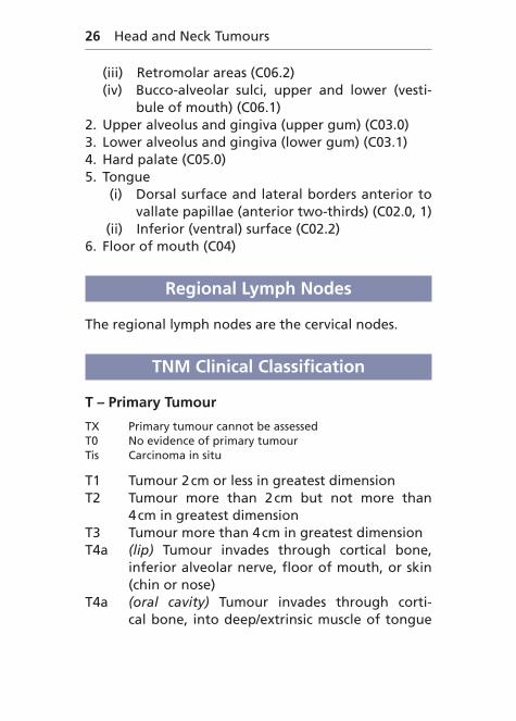
(iii) Retromolar areas (C06.2) (iv) Bucco-alveolar sulci, upper and lower (vesti-
bule of mouth) (C06.1)2. Upper alveolus and gingiva (upper gum) (C03.0)3. Lower alveolus and gingiva (lower gum) (C03.1)4. Hard palate (C05.0)5. Tongue (i) Dorsal surface and lateral borders anterior to
vallate papillae (anterior two-thirds) (C02.0, 1) (ii) Inferior (ventral) surface (C02.2)6. Floor of mouth (C04)
Regional Lymph Nodes
The regional lymph nodes are the cervical nodes.
TNM Clinical Classification
T – Primary Tumour
TX Primary tumour cannot be assessedT0 No evidence of primary tumourTis Carcinoma in situ
T1 Tumour 2 cm or less in greatest dimensionT2 Tumour more than 2 cm but not more than
4 cm in greatest dimensionT3 Tumour more than 4 cm in greatest dimensionT4a (lip) Tumour invades through cortical bone,
inferior alveolar nerve, floor of mouth, or skin (chin or nose)
T4a (oral cavity) Tumour invades through corti-cal bone, into deep/extrinsic muscle of tongue
Lip and Oral Cavity 27
(genioglossus, hyoglossus, palatoglossus, and styloglossus), maxillary sinus, or skin of face
T4b (lip and oral cavity) Tumour invades mastica-tor space, pterygoid plates, or skull base, or encases internal carotid artery
Note: Superficial erosion alone of bone/tooth socket by gingi-val primary is not sufficient to classify a tumour as T4.
N – Regional Lymph Nodes
NX Regional lymph nodes cannot be assessedN0 No regional lymph node metastasisN1 Metastasis in a single ipsilateral lymph node,
3 cm or less in greatest dimensionN2 Metastasis as described below:
N2a Metastasis in a single ipsilateral lymph node, more than 3 cm but not more than 6 cm in greatest dimension
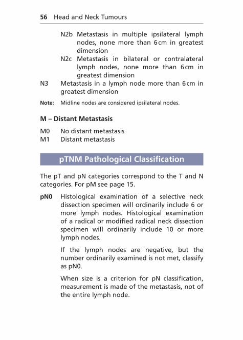
N2b Metastasis in multiple ipsilateral lymph nodes, none more than 6 cm in greatest dimension
N2c Metastasis in bilateral or contralateral lymph nodes, none more than 6 cm in greatest dimension
N3 Metastasis in a lymph node more than 6 cm in greatest dimension
Note: Midline nodes are considered ipsilateral nodes.
M – Distant Metastasis
M0 No distant metastasisM1 Distant metastasis
28 Head and Neck Tumours
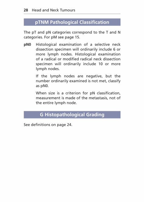
pTNM Pathological Classification
The pT and pN categories correspond to the T and N categories. For pM see page 15.
pN0 Histological examination of a selective neck dissection specimen will ordinarily include 6 or more lymph nodes. Histological examination of a radical or modified radical neck dissection specimen will ordinarily include 10 or more lymph nodes.
If the lymph nodes are negative, but the number ordinarily examined is not met, classify as pN0.
When size is a criterion for pN classification, measurement is made of the metastasis, not of the entire lymph node.
(C13.0): extends from the level of the arytenoid car-tilages and connecting folds to the inferior border of the cricoid cartilage, thus forming the anterior wall of the hypopharynx
2. Piriform sinus (C12.9): extends from the pharyngo-epiglottic fold to the upper end of the oesopha-gus. It is bounded laterally by the thyroid cartilage and medially by the hypopharyngeal surface of the aryepiglottic fold (C13.1) and the arytenoid and cricoid cartilages
3. Posterior pharyngeal wall (C13.2): extends from the superior level of the hyoid bone (or floor of the vallecula) to the level of the inferior border of the cricoid cartilage and from the apex of one piriform sinus to the other
32 Head and Neck Tumours
Regional Lymph Nodes
The regional lymph nodes are the cervical nodes.The supraclavicular fossa (relevant to classifying
nasopharyngeal carcinoma) is the triangular region defined by three points:
1. The superior margin of the sternal end of the clavicle
2. The superior margin of the lateral end of the clavicle
3. The point where the neck meets the shoulder. This includes caudal portions of Levels IV and V
TNM Clinical Classification
T – Primary Tumour
TX Primary tumour cannot be assessedT0 No evidence of primary tumourTis Carcinoma in situ
OropharynxT1 Tumour 2 cm or less in greatest dimensionT2 Tumour more than 2 cm but not more than
4 cm in greatest dimensionT3 Tumour more than 4 cm in greatest dimension
or extension to lingual surface of epiglottisT4a Tumour invades any of the following: larynx,
deep/extrinsic muscle of tongue (genioglossus, hyoglossus, palatoglossus, and styloglossus), medial pterygoid, hard palate, or mandible*
Pharynx 33
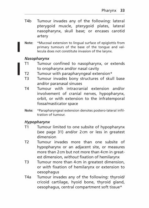
T4b Tumour invades any of the following: lateral pterygoid muscle, pterygoid plates, lateral nasopharynx, skull base; or encases carotid artery
Note: *Mucosal extension to lingual surface of epiglottis from primary tumours of the base of the tongue and val-lecula does not constitute invasion of the larynx.
NasopharynxT1 Tumour confined to nasopharynx, or extends
to oropharynx and/or nasal cavityT2 Tumour with parapharyngeal extension*T3 Tumour invades bony structures of skull base
and/or paranasal sinusesT4 Tumour with intracranial extension and/or
involvement of cranial nerves, hypopharynx, orbit, or with extension to the infratemporal fossa/masticator space
Note: *Parapharyngeal extension denotes postero-lateral infil-tration of tumour.
HypopharynxT1 Tumour limited to one subsite of hypopharynx
(see page 31) and/or 2 cm or less in greatest dimension
T2 Tumour invades more than one subsite of hypopharynx or an adjacent site, or measures more than 2 cm but not more than 4 cm in great-est dimension, without fixation of hemilarynx
T3 Tumour more than 4 cm in greatest dimension, or with fixation of hemilarynx or extension to oesophagus
T4a Tumour invades any of the following: thyroid/cricoid cartilage, hyoid bone, thyroid gland, oesophagus, central compartment soft tissue*
Note: *Central compartment soft tissue includes prelaryngeal strap muscles and subcutaneous fat.
N – Regional Lymph Nodes (Oro- and Hypopharynx)
NX Regional lymph nodes cannot be assessedN0 No regional lymph node metastasisN1 Metastasis in a single ipsilateral lymph node,
3 cm or less in greatest dimensionN2 Metastasis as described below:
N2a Metastasis in a single ipsilateral lymph node, more than 3 cm but not more than 6 cm in greatest dimension
N2b Metastasis in multiple ipsilateral lymph nodes, none more than 6 cm in greatest dimension
N2c Metastasis in bilateral or contralateral lymph nodes, none more than 6 cm in greatest dimension
N3 Metastasis in a lymph node more than 6 cm in greatest dimension
Note: Midline nodes are considered ipsilateral nodes.
N – Regional Lymph Nodes (Nasopharynx)
NX Regional lymph nodes cannot be assessedN0 No regional lymph node metastasisN1 Unilateral metastasis, in cervical lymph node(s),
and/or unilateral or bilateral metastasis in retro-pharyngeal lymph nodes, 6 cm or less in great-est dimension, above the supraclavicular fossa
Pharynx 35
N2 Bilateral metastasis in cervical lymph node(s), 6 cm or less in greatest dimension, above the supraclavicular fossa
N3 Metastasis in cervical lymph node(s) greater than 6 cm in dimension or in the supraclavicular fossaN3a greater than 6 cm in dimensionN3b extension in the supraclavicular fossa
Note: Midline nodes are considered ipsilateral nodes.
M – Distant Metastasis
M0 No distant metastasisM1 Distant metastasis
pTNM Pathological Classification
The pT and pN categories correspond to the T and N categories. For pM see page 15.
pN0 Histological examination of a selective neck dis-section specimen will ordinarily include 6 or more lymph nodes. Histological examination of a radi-cal or modified radical neck dissection specimen will ordinarily include 10 or more lymph nodes.
If the lymph nodes are negative, but the number ordinarily examined is not met, classify as pN0.
When size is a criterion for pN classification, measurement is made of the metastasis, not of the entire lymph node.
G Histopathological Grading
See definitions on page 24.
36 Head and Neck Tumours
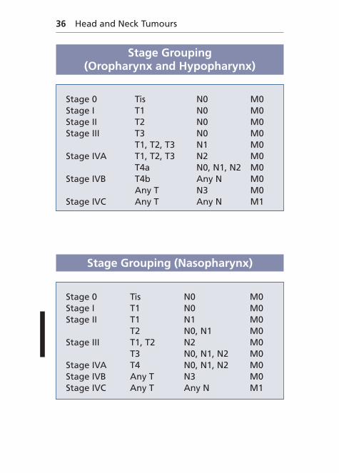
Stage Grouping (Oropharynx and Hypopharynx)
Stage 0 Tis N0 M0Stage I T1 N0 M0Stage II T2 N0 M0Stage III T3 N0 M0
T1, T2, T3 N1 M0Stage IVA T1, T2, T3 N2 M0
T4a N0, N1, N2 M0Stage IVB T4b Any N M0
Any T N3 M0Stage IVC Any T Any N M1
Stage Grouping (Nasopharynx)
Stage 0 Tis N0 M0Stage I T1 N0 M0Stage II T1 N1 M0
T2 N0, N1 M0Stage III T1, T2 N2 M0
T3 N0, N1, N2 M0Stage IVA T4 N0, N1, N2 M0Stage IVB Any T N3 M0Stage IVC Any T Any N M1
pterygoid, hard palate, mandibleT4b Lateral pterygoid muscle, pterygoid plates,
lateral nasopharynx, skull base, carotid artery
HypopharynxT1 �2 cm and limited to one subsiteT2 �2–4 cm or more than one subsiteT3 �4 cm or with hemilarynx fixation, extension
to oesophagusT4a Thyroid/cricoid cartilage, hyoid bone, thyroid
gland, central compartment soft tissueT4b Prevertebral fascia, carotid artery, mediastinal
structures
Oropharynx and HypopharynxN1 Ipsilateral single �3 cmN2 (a) Ipsilateral single �3–6 cm (b) Ipsilateral multiple �6 cm (c) Bilateral, contralateral �6 cmN3 �6 cm
38 Head and Neck Tumours
Summary
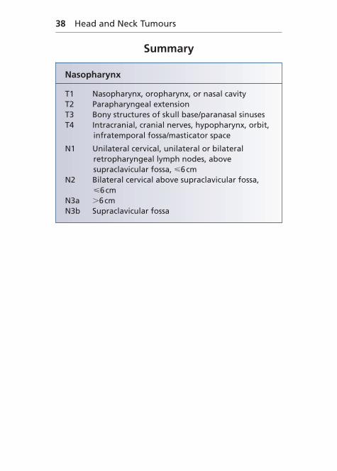
Nasopharynx
T1 Nasopharynx, oropharynx, or nasal cavityT2 Parapharyngeal extensionT3 Bony structures of skull base/paranasal sinusesT4 Intracranial, cranial nerves, hypopharynx, orbit,
infratemporal fossa/masticator space
N1 Unilateral cervical, unilateral or bilateral retropharyngeal lymph nodes, above supraclavicular fossa, �6 cm
N2 Bilateral cervical above supraclavicular fossa, �6 cm
The classification applies to carcinomas. There should be histological confirmation of the disease.
The following are the procedures for assessing T, N, and M categories:
T categories Physical examination, laryngoscopy, and imaging
N categories Physical examination and imagingM categories Physical examination and imaging
Epilarynx (including marginal zone)
Supraglottis excluding epilarynx
}}
40 Head and Neck Tumours
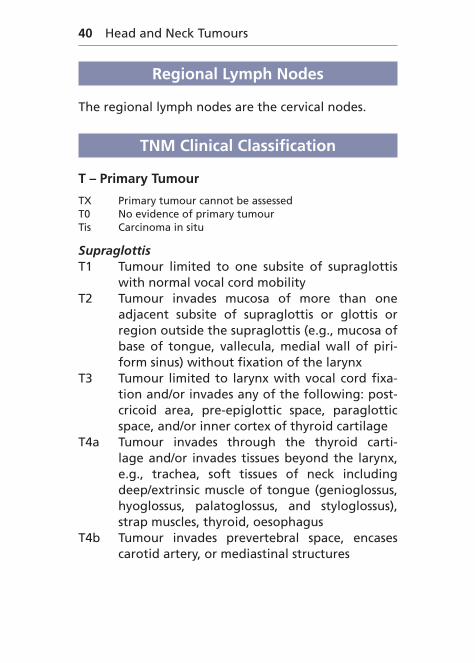
Regional Lymph Nodes
The regional lymph nodes are the cervical nodes.
TNM Clinical Classification
T – Primary Tumour
TX Primary tumour cannot be assessedT0 No evidence of primary tumourTis Carcinoma in situ
SupraglottisT1 Tumour limited to one subsite of supraglottis
with normal vocal cord mobilityT2 Tumour invades mucosa of more than one
adjacent subsite of supraglottis or glottis or region outside the supraglottis (e.g., mucosa of base of tongue, vallecula, medial wall of piri-form sinus) without fixation of the larynx
T3 Tumour limited to larynx with vocal cord fixa-tion and/or invades any of the following: post-cricoid area, pre-epiglottic space, paraglottic space, and/or inner cortex of thyroid cartilage
T4a Tumour invades through the thyroid carti-lage and/or invades tissues beyond the larynx, e.g., trachea, soft tissues of neck including deep/extrinsic muscle of tongue (genioglossus, hyoglossus, palatoglossus, and styloglossus), strap muscles, thyroid, oesophagus
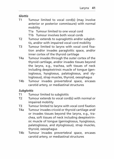
GlottisT1 Tumour limited to vocal cord(s) (may involve
anterior or posterior commissure) with normal mobilityT1a Tumour limited to one vocal cordT1b Tumour involves both vocal cords
T2 Tumour extends to supraglottis and/or subglot-tis, and/or with impaired vocal cord mobility
T3 Tumour limited to larynx with vocal cord fixa-tion and/or invades paraglottic space, and/or inner cortex of the thyroid cartilage
T4a Tumour invades through the outer cortex of the thyroid cartilage, and/or invades tissues beyond the larynx, e.g., trachea, soft tissues of neck including deep/extrinsic muscle of tongue (gen-ioglossus, hyoglossus, palatoglossus, and sty-loglossus), strap muscles, thyroid, oesophagus
SubglottisT1 Tumour limited to subglottisT2 Tumour extends to vocal cord(s) with normal or
impaired mobilityT3 Tumour limited to larynx with vocal cord fixationT4a Tumour invades cricoid or thyroid cartilage and/
or invades tissues beyond the larynx, e.g., tra-chea, soft tissues of neck including deep/extrin-sic muscle of tongue (genioglossus, hyoglossus, palatoglossus, and styloglossus), strap muscles, thyroid, oesophagus
NX Regional lymph nodes cannot be assessedN0 No regional lymph node metastasisN1 Metastasis in a single ipsilateral lymph node,
3 cm or less in greatest dimensionN2 Metastasis as described below:
N2a Metastasis in a single ipsilateral lymph node, more than 3 cm but not more than 6 cm in greatest dimension
N2b Metastasis in multiple ipsilateral lymph nodes, none more than 6 cm in greatest dimension
N2c Metastasis in bilateral or contralateral lymph nodes, none more than 6 cm in greatest dimension
N3 Metastasis in a lymph node more than 6 cm in greatest dimension
Note: Midline nodes are considered ipsilateral nodes.
M – Distant Metastasis
M0 No distant metastasisM1 Distant metastasis
pTNM Pathological Classification
The pT and pN categories correspond to the T and N categories. For pM see page 15.
pN0 Histological examination of a selective neck dissection specimen will ordinarily include 6 or more lymph nodes. Histological examination of a radical or modified radical neck dissection
Larynx 43
specimen will ordinarily include 10 or more lymph nodes.
If the lymph nodes are negative, but the number ordinarily examined is not met, classify as pN0.
When size is a criterion for pN classification, measurement is made of the metastasis, not of the entire lymph node.
G Histopathological Grading
See definitions on page 24.
Stage Grouping
Stage 0 Tis N0 M0Stage I T1 N0 M0Stage II T2 N0 M0Stage III T1, T2 N1 M0
T3 N0, N1 M0Stage IVA T1, T2, T3, N2 M0
T4a N0, N1, N2 M0Stage IVB T4b Any N M0
Any T N3 M0Stage IVC Any T Any N M1
44 Head and Neck Tumours
Summary
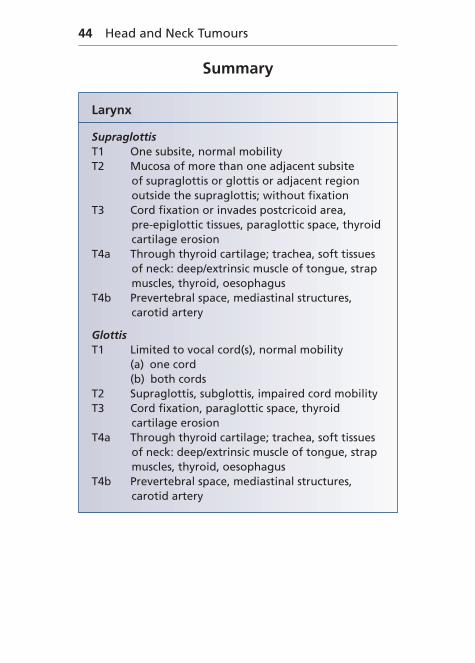
Larynx
SupraglottisT1 One subsite, normal mobilityT2 Mucosa of more than one adjacent subsite
of supraglottis or glottis or adjacent region outside the supraglottis; without fixation
T3 Cord fixation or invades postcricoid area, pre-epiglottic tissues, paraglottic space, thyroid cartilage erosion
T4a Through thyroid cartilage; trachea, soft tissues of neck: deep/extrinsic muscle of tongue, strap muscles, thyroid, oesophagus
All SitesN1 Ipsilateral single �3 cmN2 (a) Ipsilateral single �3–6 cm (b) Ipsilateral multiple �6 cm (c) Bilateral, contralateral �6 cmN3 �6 cm
Nasal Cavity and Paranasal Sinuses
(C30.0, 31.0, 1)
Rules for Classification
The classification applies to carcinomas. There should be histological confirmation of the disease.
The following are the procedures for assessing T, N, and M categories:
T categories Physical examination and imagingN categories Physical examination and imagingM categories Physical examination and imaging
Anatomical Sites and Subsites
● Nasal Cavity (C30.0) Septum Floor Lateral wall Vestibule● Maxillary sinus (C31.0)● Ethmoid sinus (C31.1) Left Right
Regional Lymph Nodes
The regional lymph nodes are the cervical nodes.
Nasal Cavity and Paranasal Sinuses 47
TNM Clinical Classification
T – Primary Tumour
TX Primary tumour cannot be assessedT0 No evidence of primary tumourTis Carcinoma in situ
Maxillary SinusT1 Tumour limited to the mucosa with no erosion
or destruction of boneT2 Tumour causing bone erosion or destruction,
including extension into the hard palate and/or middle nasal meatus, except extension to poste-rior wall of maxillary sinus and pterygoid plates
T3 Tumour invades any of the following: bone of posterior wall of maxillary sinus, subcutaneous tissues, floor or medial wall of orbit, pterygoid fossa, ethmoid sinuses
T4a Tumour invades any of the following: ante-rior orbital contents, skin of cheek, pterygoid plates, infratemporal fossa, cribriform plate, sphenoid or frontal sinuses
T4b Tumour invades any of the following: orbital apex, dura, brain, middle cranial fossa, cranial nerves other than maxillary division of trigemi-nal nerve (V2), nasopharynx, or clivus
Nasal Cavity and Ethmoid SinusT1 Tumour restricted to one subsite of nasal cavity
or ethmoid sinus, with or without bony invasionT2 Tumour involves two subsites in a single site or
extends to involve an adjacent site within the nasoethmoidal complex, with or without bony invasion
48 Head and Neck Tumours
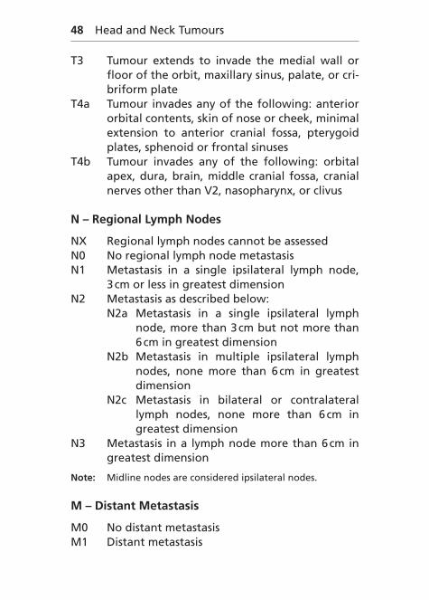
T3 Tumour extends to invade the medial wall or floor of the orbit, maxillary sinus, palate, or cri-briform plate
T4a Tumour invades any of the following: anterior orbital contents, skin of nose or cheek, minimal extension to anterior cranial fossa, pterygoid plates, sphenoid or frontal sinuses
T4b Tumour invades any of the following: orbital apex, dura, brain, middle cranial fossa, cranial nerves other than V2, nasopharynx, or clivus
N – Regional Lymph Nodes
NX Regional lymph nodes cannot be assessedN0 No regional lymph node metastasisN1 Metastasis in a single ipsilateral lymph node,
3 cm or less in greatest dimensionN2 Metastasis as described below:
N2a Metastasis in a single ipsilateral lymph node, more than 3 cm but not more than 6 cm in greatest dimension
N2b Metastasis in multiple ipsilateral lymph nodes, none more than 6 cm in greatest dimension
N2c Metastasis in bilateral or contralateral lymph nodes, none more than 6 cm in greatest dimension
N3 Metastasis in a lymph node more than 6 cm in greatest dimension
Note: Midline nodes are considered ipsilateral nodes.
M – Distant Metastasis
M0 No distant metastasisM1 Distant metastasis
Nasal Cavity and Paranasal Sinuses 49
pTNM Pathological Classification
The pT and pN categories correspond to the T and N categories. For pM see page 15.
pN0 Histological examination of a selective neck dis-section specimen will ordinarily include 6 or more lymph nodes. Histological examination of a radi-cal or modified radical neck dissection specimen will ordinarily include 10 or more lymph nodes.
If the lymph nodes are negative, but the number ordinarily examined is not met, classify as pN0.
When size is a criterion for pN classification, measurement is made of the metastasis, not of the entire lymph node.
G Histopathological Grading
See definitions on page 24.
Stage Grouping
Stage 0 Tis N0 M0Stage I T1 N0 M0Stage II T2 N0 M0Stage III T3 N0 M0
T1, T2, T3 N1 M0Stage IVA T1, T2, T3 N2 M0
T4a N0, N1, N2 M0Stage IVB T4b Any N M0
Any T N3 M0Stage IVC Any T Any N M1
50 Head and Neck Tumours
Summary
Nasal Cavity and Paranasal Sinuses
Maxillary SinusT1 MucosaT2 Bone erosion/destruction, hard palate, middle
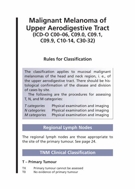
The classification applies to mucosal malignant melanomas of the head and neck region, i. e., of the upper aerodigestive tract. There should be his-tological confirmation of the disease and division of cases by site.
The following are the procedures for assessing T, N, and M categories:
T categories Physical examination and imagingN categories Physical examination and imagingM categories Physical examination and imaging
Regional Lymph Nodes
The regional lymph nodes are those appropriate to the site of the primary tumour. See page 24.
TNM Clinical Classification
T – Primary Tumour
TX Primary tumour cannot be assessedT0 No evidence of primary tumour
52 Head and Neck Tumours
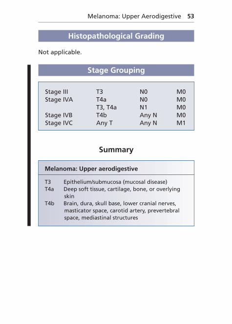
T3 Tumour limited to the epithelium and/or sub-mucosa (mucosal disease)
T4a Tumour invades deep soft tissue, cartilage, bone, or overlying skin
T4b Tumour invades any of the following: brain, dura, skull base, lower cranial nerves (IX, X, XI, XII), masticator space, carotid artery, preverte-bral space, mediastinal structures
Note: Mucosal melanomas are aggressive tumours, therefore T1 and T2 are omitted as are stages I and II.
N – Regional Lymph Nodes
NX Regional lymph nodes cannot be assessedN0 No regional lymph node metastasisN1 Regional lymph node metastasis
M – Distant Metastasis
M0 No distant metastasisM1 Distant metastasis
pTNM Pathological Classification
The pT and pN categories correspond to the T and N categories. For pM see page 15.
pN0 Histological examination of a regional lym-phadenectomy specimen will ordinarily include 6 or more lymph nodes.
If the lymph nodes are negative, but the number ordinarily examined is not met, classify as pN0.
Stage Grouping
Stage III T3 N0 M0Stage IVA T4a N0 M0
T3, T4a N1 M0Stage IVB T4b Any N M0Stage IVC Any T Any N M1
Summary
Melanoma: Upper aerodigestive
T3 Epithelium/submucosa (mucosal disease)T4a Deep soft tissue, cartilage, bone, or overlying
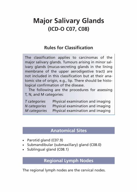
The classification applies to carcinomas of the major salivary glands. Tumours arising in minor sal-ivary glands (mucus-secreting glands in the lining membrane of the upper aerodigestive tract) are not included in this classification but at their ana-tomic site of origin, e.g., lip. There should be histo-logical confirmation of the disease.
The following are the procedures for assessing T, N, and M categories:
T categories Physical examination and imagingN categories Physical examination and imagingM categories Physical examination and imaging
Major Salivary Glands 55
TNM Clinical Classification
T – Primary Tumour
TX Primary tumour cannot be assessedT0 No evidence of primary tumour
T1 Tumour 2 cm or less in greatest dimension with-out extraparenchymal extension*
T2 Tumour more than 2 cm but not more than 4 cm in greatest dimension without extraparen-chymal extension*
T3 Tumour more than 4 cm and/or tumour with extraparenchymal extension*
T4b Tumour invades base of skull, and/or pterygoid plates, and/or encases carotid artery
Note: *Extraparenchymal extension is clinical or macroscopic evidence of invasion of soft tissues or nerve, except those listed under T4a and 4b. Microscopic evidence alone does not constitute extraparenchymal extension for classification purposes.
N – Regional Lymph Nodes
NX Regional lymph nodes cannot be assessedN0 No regional lymph node metastasisN1 Metastasis in a single ipsilateral lymph node,
3 cm or less in greatest dimensionN2 Metastasis as described below:
N2a Metastasis in a single ipsilateral lymph node, more than 3 cm but not more than 6 cm in greatest dimension
56 Head and Neck Tumours
N2b Metastasis in multiple ipsilateral lymph nodes, none more than 6 cm in greatest dimension
N2c Metastasis in bilateral or contralateral lymph nodes, none more than 6 cm in greatest dimension
N3 Metastasis in a lymph node more than 6 cm in greatest dimension
Note: Midline nodes are considered ipsilateral nodes.
M – Distant Metastasis
M0 No distant metastasisM1 Distant metastasis
pTNM Pathological Classification
The pT and pN categories correspond to the T and N categories. For pM see page 15.
pN0 Histological examination of a selective neck dissection specimen will ordinarily include 6 or more lymph nodes. Histological examination of a radical or modified radical neck dissection specimen will ordinarily include 10 or more lymph nodes.
If the lymph nodes are negative, but the number ordinarily examined is not met, classify as pN0.
When size is a criterion for pN classification, measurement is made of the metastasis, not of the entire lymph node.
Major Salivary Glands 57
G Histopathological Grading
See definitions on page 24.
Stage Grouping
Stage I T1 N0 M0Stage II T2 N0 M0Stage III T3 N0 M0
T1, T2, T3 N1 M0Stage IVA T4a N0, N1 M0
T1, T2, T3, T4a N2 M0Stage IVB T4b Any N M0
Any T N3 M0Stage IVC Any T Any N M1
Summary
Salivary Glands
T1 �2 cm, without extraparenchymal extensionT2 �2–4 cm, without extraparenchymal extensionT3 �4 cm and/or extraparenchymal extensionT4a Skin, mandible, ear canal, facial nerveT4b Skull, pterygoid plates, carotid artery
N1 Ipsilateral single �3 cmN2 (a) Ipsilateral single �3–6 cm (b) Ipsilateral multiple �6 cm (c) Bilateral, contralateral �6 cmN3 �6 cm
Thyroid Gland(ICD-O C73)
Rules for Classification
The classification applies to carcinomas. There should be microscopic confirmation of the dis-ease and division of cases by histological type.
The following are the procedures for assessing T, N, and M categories:
T categories Physical examination, endoscopy, and imaging
N categories Physical examination and imagingM categories Physical examination and imaging
Regional Lymph Nodes
The regional lymph nodes are the cervical and upper/superior mediastinal nodes.
TNM Clinical Classification
T – Primary Tumour
TX Primary tumour cannot be assessedT0 No evidence of primary tumour
T1 Tumour 2 cm or less in greatest dimension, lim-ited to the thyroid
Thyroid Gland 59
T1a Tumour 1 cm or less in greatest dimen-sion, limited to the thyroid
T1b Tumour more than 1 cm but not more than 2 cm in greatest dimension, limited to the thyroid
T2 Tumour more than 2 cm but not more than 4 cm in greatest dimension, limited to the thyroid
T3 Tumour more than 4 cm in greatest dimension, limited to the thyroid or any tumour with mini-mal extrathyroid extension (e.g., extension to sternothyroid muscle or perithyroid soft tissues)
T4a Tumour extends beyond the thyroid capsule and invades any of the following: subcutane-ous soft tissues, larynx, trachea, oesophagus, recurrent laryngeal nerve
All anaplastic carcinomas are considered T4 tumoursT4a* (anaplastic carcinoma only) Tumour (any size)
limited to the thyroidT4b* (anaplastic carcinoma only) Tumour (any size)
extends beyond the thyroid capsules
Notes: Multifocal tumours of all histological types should be designated (m) (the largest determines the classifica-tion), e.g., T2(m).
N – Regional Lymph Nodes
NX Regional lymph nodes cannot be assessedN0 No regional lymph node metastasis
60 Head and Neck Tumours
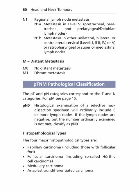
N1 Regional lymph node metastasisN1a Metastasis in Level VI (pretracheal, para-
tracheal, and prelaryngeal/Delphian lymph nodes)
N1b Metastasis in other unilateral, bilateral or contralateral cervical (Levels I, II II, IV, or V) or retropharyngeal or superior mediastinal lymph nodes
M – Distant Metastasis
M0 No distant metastasisM1 Distant metastasis
pTNM Pathological Classification
The pT and pN categories correspond to the T and N categories. For pM see page 15.
pN0 Histological examination of a selective neck dissection specimen will ordinarily include 6 or more lymph nodes. If the lymph nodes are negative, but the number ordinarily examined is not met, classify as pN0.
Histopathological Types
The four major histopathological types are:
● Papillary carcinoma (including those with follicular foci)
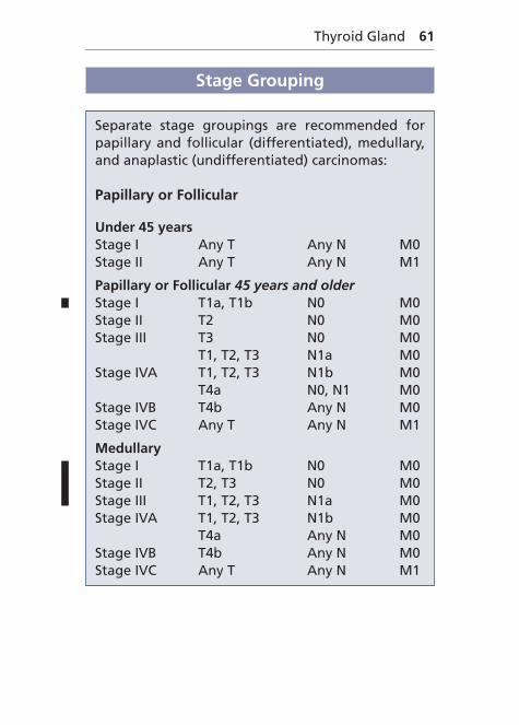
Separate stage groupings are recommended for papillary and follicular (differentiated), medullary, and anaplastic (undifferentiated) carcinomas:
Papillary or Follicular
Under 45 yearsStage I Any T Any N M0Stage II Any T Any N M1
Papillary or Follicular 45 years and olderStage I T1a, T1b N0 M0Stage II T2 N0 M0Stage III T3 N0 M0
T1, T2, T3 N1a M0Stage IVA T1, T2, T3 N1b M0
T4a N0, N1 M0Stage IVB T4b Any N M0Stage IVC Any T Any N M1
MedullaryStage I T1a, T1b N0 M0Stage II T2, T3 N0 M0Stage III T1, T2, T3 N1a M0Stage IVA T1, T2, T3 N1b M0
T4a Any N M0Stage IVB T4b Any N M0Stage IVC Any T Any N M1
62 Head and Neck Tumours
Anaplastic CarcinomaAll anaplastic carcinoma are stage IV
Stage IVA T4a Any N M0Stage IVB T4b Any N M0Stage IVC Any T Any N M1
Thyroid Gland
Papillary, follicular, and medullary carcinomaT1 �2 cm, intrathyroidalT2 �2–4 cm, intrathyroidalT3 �4 cm or minimal extrathyroidal extensionT4a Subcutaneous, larynx, trachea, oesophagus,