89
Head & Neck Surgery Course Dr Pierfrancesco PELLICCIA Pr Benjamin LALLEMANT Service ORL et CMF CHU de Nîmes CH de Arles Oral cavity: surgical anatomy www.orl-nimes.fr

Head & Neck Surgery Course
Dr Pierfrancesco PELLICCIA
Pr Benjamin LALLEMANT
Service ORL et CMF
CHU de Nîmes
CH de Arles
Oral cavity: surgical anatomy
www.orl-nimes.fr

A: philtrum; B: upper labial frenulum; C: opening of Stensen's duct; D: labial commissure; E: hard palate; F: soft palate; G: intermaxillary commissure; H: base of tongue; I: lateral border of tongue, dorsal view; J: tip of tongue, dorsal view; K: tip of tongue, ventral view; L: lateral border of tongue, ventral view; M:
ventral surface of tongue; N: lingual frenulum; O: floor of mouth; P: opening of Wharton's duct; Q:
vestibular gingiva; R: vestibule.
Introduction Schematic representation of oral cavity and floor of mouth
www.orl-nimes.fr

Introduction Mucosal features of oral cavity
www.orl-nimes.fr

Introduction Coronal section
www.orl-nimes.fr

Functions • Speech
• Mastication
• bolus preparation and initiation of deglutition
Introduction Physiology
www.orl-nimes.fr
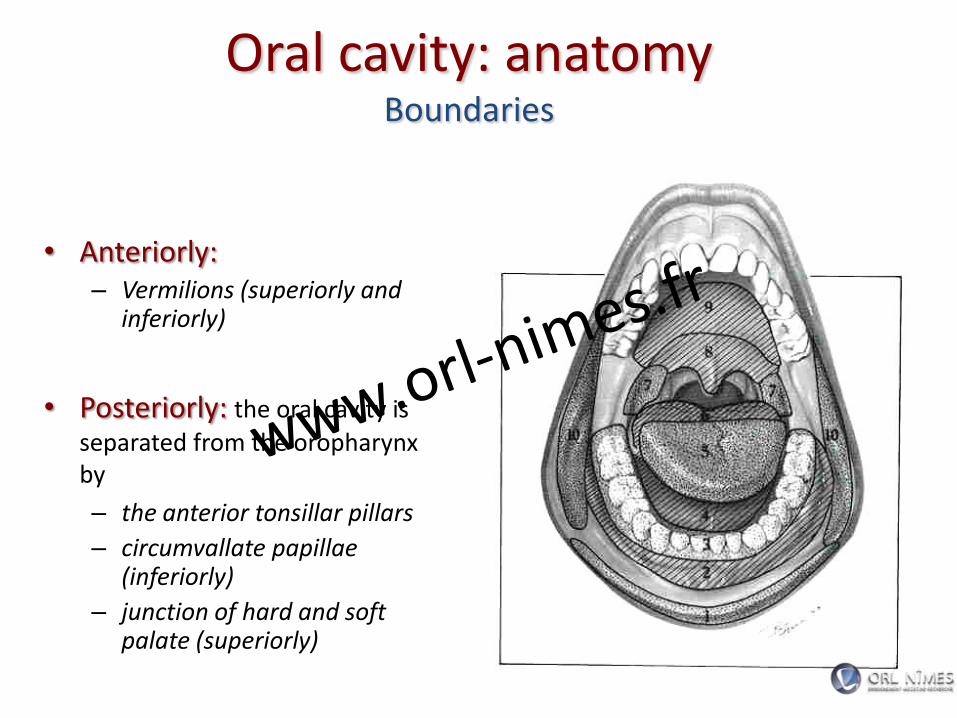
• Anteriorly: – Vermilions (superiorly and
inferiorly)
• Posteriorly: the oral cavity is
separated from the oropharynx by
– the anterior tonsillar pillars
– circumvallate papillae (inferiorly)
– junction of hard and soft palate (superiorly)
Oral cavity: anatomy Boundaries
www.orl-nimes.fr

Oral cavity: anatomy Skeleton
• Mandible
• Maxilla
• Palatine bone www.orl-nimes.fr

Oral cavity: anatomy Skeleton
www.orl-nimes.fr

Oral cavity: anatomy Skeleton
www.orl-nimes.fr

• Masticatory muscles – Temporal
– Masseter
– Internal pterygoid
– External pterygoid
• Muscles of facial expression
Oral cavity: anatomy Muscles
www.orl-nimes.fr

Oral cavity: arterial supply
www.orl-nimes.fr

Oral cavity: arterial supply
www.orl-nimes.fr

Oral cavity: venous supply
www.orl-nimes.fr

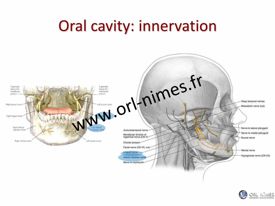
Oral cavity: innervation
• Greater palatine nerve (V2)
• Lingual nerve (V3 and VII)
• XII CN
• Inferior alveolar nerve (V3)
• Posterior, midldle and anterior superior alveolar nerve (V2)
www.orl-nimes.fr

Oral cavity: innervation
www.orl-nimes.fr
Oral cavity: innervation
www.orl-nimes.fr

Oral cavity: innervation
www.orl-nimes.fr

Oral cavity: innervation
www.orl-nimes.fr

• 1st echelon nodes:
level I,II,III
• Then IV, V
• Skip metastasis!
Oral cavity: lymphatics
www.orl-nimes.fr

• lips
• alveolar ridges
• buccal mucosa
• retromolar trigone
• hard palate
• floor of mouth
• mobile tongue
Sites of oral cavity
www.orl-nimes.fr

•The sulcus terminalis divide the anterior and posterior tongue
•Tongue base ends at the vallecula
•Foramen cecum-area(where the thyroid descends)
Tongue: anatomy
www.orl-nimes.fr

•There are 8 muscles of the tongue
•They are classified as intrinsic and extrinsic muscles
Tongue: muscles
www.orl-nimes.fr

There are 4 paired intrinsic muscles of the tongue
Superior Longitudinal
Inferior longitudinal
Verticalis
Transversus muscle
Tongue: muscles
www.orl-nimes.fr

Function of the intrinsic muscles
Inferior and superior longitudinal muscles - Move tip up and down
Transverse muscle -Narrows and lengthens the tongue
Vertical Muscle - Flattens and depresses the tongue
Tongue: muscles
www.orl-nimes.fr

The 4 extrinsic muscles are Genioglossus-from the mandible
Hyoglossus- from the hyoid bone
Styloglossus- from the styloid process
Palatoglossus- from the palatine aponeurosis
Tongue: muscles
www.orl-nimes.fr
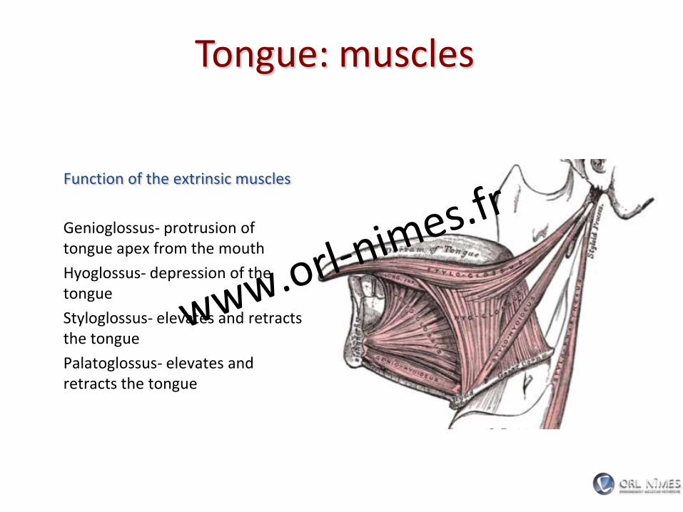
Function of the extrinsic muscles
Genioglossus- protrusion of tongue apex from the mouth
Hyoglossus- depression of the tongue
Styloglossus- elevates and retracts the tongue
Palatoglossus- elevates and retracts the tongue
Tongue: muscles
www.orl-nimes.fr

•lingual artery
•Other contributors include the ascending palatine and tonsillar branch of the facial artery
Tongue: arterial supply
www.orl-nimes.fr

•lingual vein
Tongue: venous supply
www.orl-nimes.fr

Sensory nerves
Motor innervation
Tongue: innervation
www.orl-nimes.fr

Sensory nerves Lingual branch of V2-General sensation for the anterior two thirds of tongue
Chorda tympani of CN VII- taste for anterior 2/3
Lingual branch of CN IX- General sensation and taste for posterior 1/3
Superior laryngeal CN X- root of tongue and lingual base sensation.
Tongue: innervation
www.orl-nimes.fr

Motor innervation
All tongue muscles are innervated by XII except the palatoglossus- innervated by X
Tongue: innervation
www.orl-nimes.fr

Hard palate: anatomy
• The palate forms the roof of the mouth and intervenes between the nasal and oral cavities.
• It consists of the palatine process of the maxilla, the horizontal plates of the palatine bone.
www.orl-nimes.fr

Hard palate: anatomy
• Three foramina open on the oral aspect of the hard palate
www.orl-nimes.fr

Foramina that open on the oral aspect of the hard palate
Incisive Fossa – Slight depression posterior
to central incisor teeth
– Nasopalatine nerve
Greater palatine foramina – Medial to 3rd Molar
– Greater palatine vessels and nerve
Lesser palatine foramina – Lesser Palatine nerves and
vessels to soft palate
www.orl-nimes.fr

Greater palatine artery
• branch of the third part of the maxillary artery
• it descends with its accompanying nerve in the palatine canal.
Superior Alveolar Arteries • terminal branches of the
internal maxillary artery
Hard palate: arterial supply
www.orl-nimes.fr

• The greater palatine emerges on
the hard palate from the greater
palatine foramen
• runs forward in a groove on the
inferior surface of the bony palate
almost to the incisor teeth
• supplies the gums and the
mucosa and glands of the hard
palate.
Hard palate: arterial supply
www.orl-nimes.fr

• The superior alveolar
arteries (anterior, middle,
posterior) provide blood
supply to the maxillary
gingiva, alveolar ridge, and
dentition
Hard palate: arterial supply
www.orl-nimes.fr

• The venous drainage is to
the pterygoid plexus and subsequently to the internal jugular venous system.
Hard palate: venous supply
www.orl-nimes.fr

• The nasopalatine nerves
• Greater Palatine Nerves
Hard palate: innervation
www.orl-nimes.fr

• The nasopalatine nerves – branches of V2
– They enter the palate at the incisive foramen
– they supply the anterior part of the hard palate behind the incisor teeth.
Hard palate: innervation
www.orl-nimes.fr

• Greater Palatine Nerves
– Greater (and Lesser) Palatine run through the palatine canal and exit at the Great and Lesser Palatine Foramens, respectively.
Hard palate: innervation
www.orl-nimes.fr

• Greater Palatine Nerves
– Parasympathetic postganglionic secretomotor fibres from the pterygopalatine ganglion run with the nerves to supply the palatine mucous glands.
Hard palate: innervation
www.orl-nimes.fr

Perineural Spread
• Tumors spreading by perineural extension can be discovered by radiographic enlargement of the palatine foramina or widening of the palatine canals or the foramen rotundum.
www.orl-nimes.fr
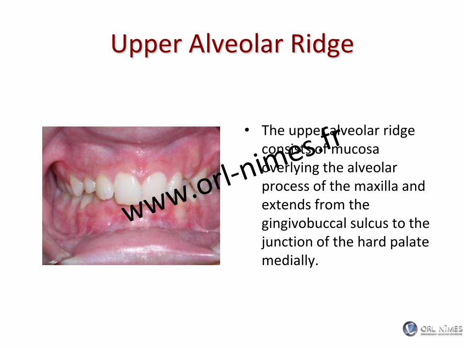
Upper Alveolar Ridge
• The upper alveolar ridge consists of mucosa overlying the alveolar process of the maxilla and extends from the gingivobuccal sulcus to the junction of the hard palate medially.
www.orl-nimes.fr

Inferior Alveolar Ridge
• The inferior alveolar ridge consists of mucosa overlying the alveolar process of the mandible and extends from the gingivobuccal sulcus to the junction of the floor of the mouth.
www.orl-nimes.fr

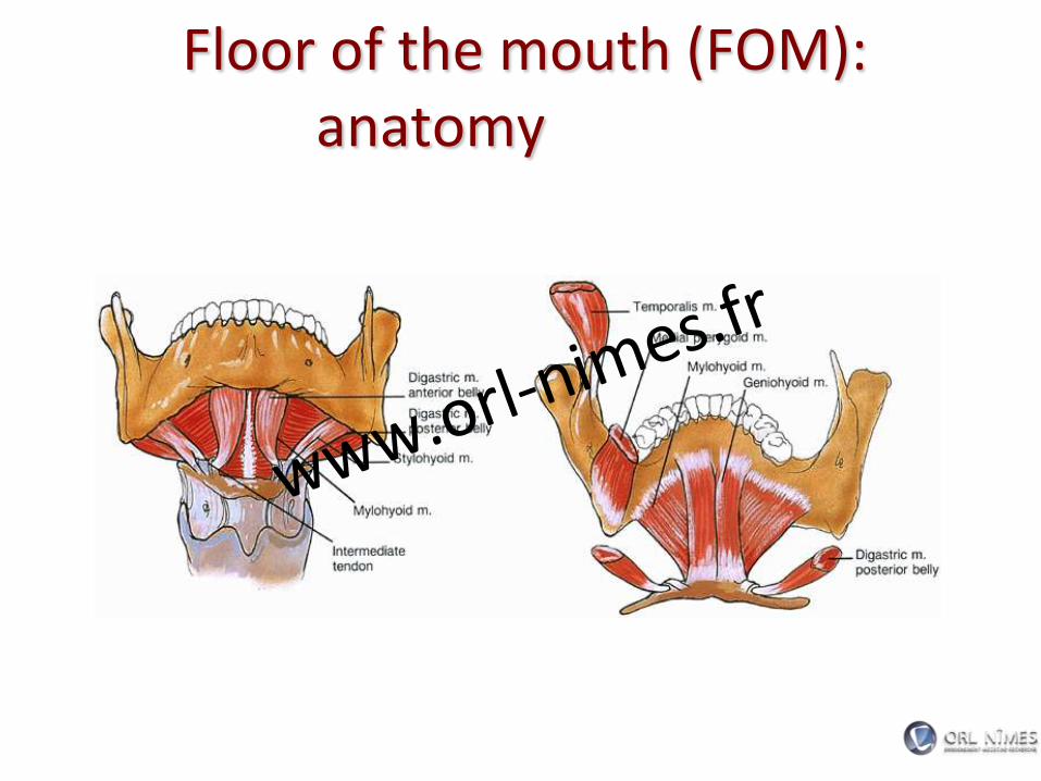
Floor of the mouth (FOM): anatomy
• FOM extends from the inferior alveolar ridge to the anterior tongue
www.orl-nimes.fr

Floor of the mouth (FOM): anatomy
www.orl-nimes.fr

Floor of the mouth (FOM): anatomy
www.orl-nimes.fr

Floor of the mouth (FOM): anatomy
www.orl-nimes.fr
Floor of the mouth (FOM): anatomy
www.orl-nimes.fr

Floor of the mouth (FOM): anatomy
www.orl-nimes.fr

Lips and buccal mucosa: anatomy
www.orl-nimes.fr

Lips and buccal mucosa: anatomy
www.orl-nimes.fr
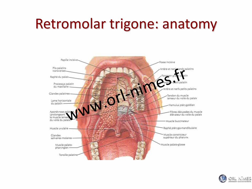
Retromolar trigone: anatomy
www.orl-nimes.fr
Head & Neck Surgery Course
Dr Pierfrancesco PELLICCIA
Pr Benjamin LALLEMANT
Service ORL et CMF
CHU de Nîmes
CH de Arles
Oral cavity: surgical options and technique
www.orl-nimes.fr

Surgical approaches to the oral cavity
(a) Peroral
(b) Mandibulotomy
(Mandibular swing): lower
lip-splitting incision or visor flap
(c) Lower cheek flap
(d) Visor flap (cervical
degloving)
(e) Upper cheek flap
www.orl-nimes.fr

a) Peroral
www.orl-nimes.fr

Peroral partial glossectomy
www.orl-nimes.fr

Peroral Pelvectomy
www.orl-nimes.fr

Peroral approach to the palate
www.orl-nimes.fr

Peroral resection of tumor of the upper alveolus
www.orl-nimes.fr

Marginal mandibulectomy-Pull Trough operation (peroral)
www.orl-nimes.fr

b) Mandibulotomy (Mandibular swing)
www.orl-nimes.fr

Transmandubular resection of oral/oropharyngeal cancer
• “Mandibular swing”
• Midline lip splitting or visor flap
• Mandibulotomy anteriorly, incise
along floor of mouth to anterior
tonsillar pillar
• Identify hypoglossal nerve and
lingual nerve
• Divide styloglossus and
stylopharyngeus muscle
• Need tracheotomy
www.orl-nimes.fr

Partial glossectomy via lip split mandibulotomy (Mandubular swing)
www.orl-nimes.fr

c) Lower cheek flap
www.orl-nimes.fr

Marginal mandibulectomy via lower cheek flap (Pull Through operation)
www.orl-nimes.fr

Marginal mandibulectomy via lower cheek flap (Pull Through operation)
www.orl-nimes.fr

Marginal mandibulectomy via lower cheek flap (Pull Through operation)
www.orl-nimes.fr

Segmental mandibulectomy – Commando operation via lower cheek
flap
www.orl-nimes.fr

Segmental mandibulectomy – Commando operation via lower cheek
flap
www.orl-nimes.fr

Segmental mandibulectomy – Commando operation via lower cheek
flap
www.orl-nimes.fr

d) Visor flap (cervical degloving)
www.orl-nimes.fr

Marginal mandibulectomy-Pull Trough operation (visor flap)
www.orl-nimes.fr

Anterior Segmental mandibulectomy via visor flap
www.orl-nimes.fr

Anterior Segmental mandibulectomy via visor flap
www.orl-nimes.fr

Partial glossectomy via visor flap (Mandubular swing)
www.orl-nimes.fr

e) Upper cheek flap
www.orl-nimes.fr
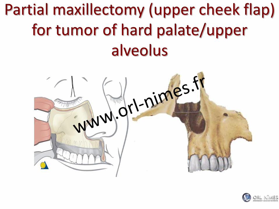
Partial maxillectomy (upper cheek flap) for tumor of hard palate/upper
alveolus
www.orl-nimes.fr

f) Other approaches
www.orl-nimes.fr

Midfacial degloving approach for tumor of hard palate/upper alveolus
www.orl-nimes.fr

Total glossectomy via mandibular lingual release (Pull Through)
www.orl-nimes.fr

Appendix: mandible management
www.orl-nimes.fr

Patterns of tumor invasion of the mandible
www.orl-nimes.fr

Patterns of tumor invasion of the mandible
www.orl-nimes.fr

• Patterns of tumor invasion of the mandible dictate mandible management
– Marginal mandibulectomy
– Segmental mandibulectomy
Mandible management
www.orl-nimes.fr

Marginal mandibulectomy
www.orl-nimes.fr

Marginal mandibulectomy
www.orl-nimes.fr

Segmental mandibulectomy
www.orl-nimes.fr


