1 An update on Evidence-Informed Assessment and Rehabilitation for Cervicogenic Headaches. Scott Euype PT, DPT, OCS Philip Toal PT, DPT, OCS, FAAOMPT Headaches and the Neck? Can the neck cause HA? Is this accepted across the medical community? Presentation Title l March 14, 2017 l 2 What is a Cervicogenic HA? “Referred pain perceived in any region of the head caused by a primary nociceptive source in the musculoskeletal tissues innervated by cervical nerves” North American Cervicogenic Headache Society Presentation Title l March 14, 2017 l 3
Transcript
1
An update on Evidence-Informed
Assessment and Rehabilitation for
Cervicogenic Headaches.
Scott Euype PT, DPT, OCS
Philip Toal PT, DPT, OCS, FAAOMPT
Headaches and the Neck? Can the neck cause HA?
Is this accepted across the
medical community?
Presentation Title l March 14,
2017 l 2
What is a Cervicogenic HA?
“Referred pain perceived in any region of the head caused by a
primary nociceptive source in the musculoskeletal tissues
innervated by cervical nerves”
North American Cervicogenic Headache Society
Presentation Title l March 14,
2017 l 3
2
PREVALENCE OF HEADACHES
16% of general population have headaches Kränzlin P, WälchliB. The concept of cervicogenic
headache. Annual postgraduate course of the association of Swiss chiropractors .1993:13
14% of population had HA Olesen Funct Neurol 1990;5;159-164
35% of both male and female university students reported headaches
Tumors of craniovertebral junction and upper cervical
spine
Meningiomas
Pagets Disease of skull with secondary basilar
invagination
Osteomyelitis of upper cervical vertebrae
Presentation Title l March
14, 2017 l 8
Generally Accepted Cervical Causes of
Headache
Cervical disk disease
Rheumatoid Arthritis or Ankylosing Spondylitis of the upper cervical spine
Traumatic subluxation
Retropharyngeal tendonitis
Craniocervical dystonias
4
Trauma to the Cervical Spine and Head
“Based on the obvious anatomical association of the head and neck it is reasonable to expect that with any blunt impact and/or acceleration / deceleration of the head will also result in some degree of inertial loading to the soft tissue and joints of the cervical spine”
Important factors from WAD studies:
As little as 4.5 g of neck acceleration can cause tissue damage
Signs and symptoms mimic those of a mild TBI
Marshall CM, Vernon H, Leddy JJ, Baldwin BA. Phys Sportsmed. 2015. 3:1-11.
Post Traumatic Headache Severity of trauma does not matter
IHS Criteria:
Loss of consciousness
Post traumatic amnesia > 10 minutes
At least 2 abnormalities
Skull X-ray
Neuroimaging
Evoked potentials
CSF
Vestibular function
Neuropsych testing
Headache onset < 14 days
Headache gone within 8-weeks
Presentation Title l March 14,
2017 l 11
Cephalgia.
1997;18:S43-110
Commonly Reported Symptoms in High
School and College Athletes Within 3 days
of Concussion (Lovell, Neurosurgery 2007)
Ranking Symptom % with symptom
1 Headaches 71
2 Feeling Slowed Down 58
3 Difficulty Concentrating 57
4 Dizziness 55
5 Fogginess 53
6 Fatigue 50
7 Blurred or Double Vision 49
8 Light Sensitivity 47
9 Memory Dysfunction 43
10 Balance Problems 43
5
CERVICOGENIC HEADACHES
MEDICAL DIAGNOSTIC CRITERIA
Clinical examination or by imaging of a cervical source
Complete relief of head pain following controled, local
anesthetic blocks of one or more cervical nerves, or
structures innervated by cervical nerves
Bogduk (1992)
INTERNATIONAL HEADACHE SOCIETY (1990)
Pain localized to the neck and occipital region
Pain increased with neck movements, or sustained neck postures
One of the following:
Resistance/limitation of active/passive neck movements of the upper cervical spine
Abnormal tenderness of neck muscles
Changes in muscle contour
Radiologic exam finds either a fracture, abnormal posture, or movement abnormalities
CERVICOGENIC HEADACHES DIAGNOSTIC
CRITERIA (International Headache Society)
Clinical, laboratory and/or imaging evidence of a disorder or
lesion within the cervical spine or soft tissues of the neck,
known to be able to cause headache
Evidence of causation demonstrated by at least two of the
following:
1. Headache has developed in temporal relation to the onset of the cervical
disorder or appearance of the lesion
2. Headache has significantly improved or resolved in parallel with
improvement in or resolution of the cervical disorder or lesion
Cephalalgia. 2013; 33: 629–808
Presentation Title l March 14,
2017 l 15
6
CERVICOGENIC HEADACHES DIAGNOSTIC
CRITERIA (International Headache Society)
3. Cervical range of motion is reduced and
headache is made significantly worse by
provocative movements
4. Headache is abolished following diagnostic
blockade of a cervical structure or its nerve supply
Not better accounted for by another ICHD-3
diagnosis
Presentation Title l March 14,
2017 l 16
ICHD-3 Diagnostic Headache Criteria
Doesn’t believe ICHD-3 criteria
answered all the questions
Comments made that it falls short in
distinguishing Cervicogenic
Headaches from migraines and
tension headache
Frederickson TA. J Headache Pain. 2015;16:6
Presentation Title l March
14, 2017 l 17
CERVICOGENIC HEADACHES
DIAGNOSTIC CRITERIA
HA precipitated by neck movements or by pressure against certain tender spots on the neck
There may be a ipsilateral shoulder, arm and hand pain, which may even be radiculopathic
There is stiffness and pain in the neck with crepitation on movements
There is reduced motility of the neck
Further evidence from local ipsilateral C2 and C3 anesthetic blocks -lateral approach
Syndrome or “reaction pattern”
Sjaastad, O, et al. Cephalalgia.1983;3: 249-56.
Presentation Title l March
14, 2017 l 18
7
CERVICOGENIC HEADACHES DIAGNOSTIC
CRITERIA.
1. Unilaterality without side shift
2. a. Pain triggered by neck movement and/or
sustained awkward posture
b. Pain with external pressure over
ipsilateral, upper posterior neck region or
occipital region
c. Decreased cervical spine range of motion
d. Ipsilateral non-radicular neck, shoulder, and arm pain
Sjaastad O, Fredrickson TA. Clin Exp Rheumatol. 2000;18:S3-6
C2-3 most painful joint Cooper, G et al 2007 Pain Medicine
Referrals - Facet
Healthy subjects
Contrast medium
injected into facet
capsule Dwyer, A et al 1990 Spine
Images from Mercer, S and Bogduk, N 1999
Cervical Disc
13
Referral Patterns- Disc
Cervical Discography:: Clinical Implications From 12 Years of Experience.Grubb, Stephen; Kelly, Carol
Spine. 25(11):1382-1389, June 1, 2000.
Figure 2 . Pattern of pain provoked by discography at each cervical level: C2-C3 (A), C3-C4 (B), C4-C5 (C), C5-C6 (D), and C6-C7 (E). For purposes of illustration only, pain is depicted as unilateral to the left at C4-C5 through C6-C7.
Upper Cervical Muscles
Cervical Muscles - Anterior
https://en.wikipedia.org/wi
ki/Longus_capitis_muscle
#/media/File:Longus_capiti
s.png
14
Referral Patterns - Muscles
Bodguk,N, Govind, J
2009 Lancet
Upper Cervical Ligaments
Vascular Anatomy
Vertebral Artery Off of subclavian artery
Enters C6 and travels cranially through transverse foramen
Areas of potential compromise C1-C2 T.P. during Rot.
C4-6 OA
15
Possible Referrals
Trigeminal
Nucleus
Convergence between cervical and trigeminal nucleus
Nociceptive afferents from the C1, C2, and C3
Spinal nerves converge onto second-order neurons that also receive afferents from adjacent cervical nerves and from the first division of the trigeminal nerve (V), via the trigeminal nerve spinal tract.
Bodguk,N, Govind, J 2009 Lancet
Convergence between
cervical afferents allows
for upper cervical pain to
be referred to regions of
the head innervated by
cervical nerves (occipital
and auricular regions).
Convergence with
trigeminal afferents
allows for referral into the
parietal, frontal, and
orbital regions.
Trigeminal
Nucleus
Bodguk,N, Govind, J 2009 Lancet
16
Evidence Informed Guide to
Cervical Spine Examination
EXAMINATION HISTORY
POSTURE
ROM
STRENGTH
OUTCOMES
SPECIAL TESTS Neuro Screen
Ligament Laxity
VBI?
PALPATION
PHYSICAL EXAMINATION Subjective History
Headaches origin
When started
Location of HA
Temporal, occipital or frontal
Unilat / Bilat
HA description
Throbbing, dull or pressure
Frequency
How long do they last
Nausea / Dizziness
Neck pain ? Stiffness?
Presentation Title l March 14,
2017 l 48
17
Medical Red Flags
Craniocervical junction abnormalities – Systems review and Imaging
Fusion of the atlas and occipital bone - anterior-posterior diameter of
the foramen magnum post to the odontoid process decreases to < 19
mm. Symptoms of cervical myelopathy
Platybasia - asymptomatic flattening of the skull base.
Basilar invagination - odontoid process protrudes into the foramen
magnum. Short neck develops and combinations of brain stem,
cerebellar, lower cranial nerve, and spinal cord signs.
Medical Red Flags
Klippel-Feil malformation – a fusion of cervical vertebrae. Often
asymptomatic except for neck deformity and limited range of motion.
Atlantoaxial subluxation or dislocation - displacement of the atlas
anteriorly in relation to the axis. Acute or chronic spinal cord
compression as a result.
Presentation Title l March
14, 2017 l 50
Use Caution
Red Flags looking for the following conditions:
Cervical myelopathy
Babinski, clonus, sensory disturbance of hands,
unsteady gait, bowel and bladder problems
Upper cervical ligamentous laxity
Occipital HA, severe limit of neck motion all
directions, post trauma, RA, Downs
Vertebral artery insufficiency
Drop attack, dizziness, dysarthria, diplopia, cranial
nerve signs, ataxia
18
EXAMINATION
Instability Tests Transverse Ligament Tests
- Sharp-Purser
Patient: Sitting
PT: Hand on forehead, and other hand Thumb on SP of C2
Patient flexes head, with while PT presses backward on forehead
(+) Test Reduction in HA symptoms
Clunk
Sensitivity of 69%, and a specificity of 96% for laxity >3mm
Uitvlugt G, Indenbaum S. Arthritis and Rheumatism 1988;31:918-922.
Bottom line: Studies have much bias, and much variability
Examination Neurological and Orthopaedic Testing
Sensation testing – dermatome patterns
Assess Trigeminal distribution
Myotomal testing
Cranial Nerve testing ?
Deep tendon reflexes were symmetrical and 2+ for biceps brachii, brachioradialis, and triceps brachiimuscles
Jaw Trigeminal Nerve: Potential Brain Stem
Scapulohumeral (Shimizu)
(C 0 to C 4) “Blind Zone”
20
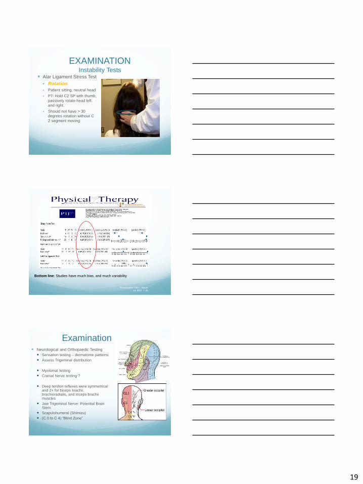
PT EXAMINATION
Posture Done in both sitting and standing
Look most specifically for:
Cervical spine rotation
Lateral head tilt
Thoracic / pelvic girdle rotation
Uneven weight-bearing
Lateral head tilt and correction giving patient the perception of “leaning to the right” supported by
concept of “Joint Position Error”(Treleaven et al 2003)
POSTURAL STRESSES
Sitting in Class
Carrying Backpacks
Doing Homework/Reading
Playing Video Games
Watching TV/Movies
Surfing the Net/ Emailing Friends
Texting / CellI-Pad / Kindle
Patient’s Dominant Posture Dominant versus Non
dominant postures
“Cross-over”
Presentation Title l March
14, 2017 l 60
Course notes, Susan Clinton,
Oct 1, 2016
21
EXAMINATION BREATHING
Presentation Title l March
14, 2017 l 61
Course notes, Susan Clinton, Oct 1, 2016
POSTURE AND CERVICAL FLEXOR
MUSCLE STRENGTH
Subjects with headaches had:
Greater amount of forward head,
Less isometric strength, and
Less endurance
When compared to non
headache group
Watson, Trott Cephalgia.1993
POSTURE AND CERVICAL FLEXOR
MUSCLE STRENGTH
Subjects with headaches had less: Cervical extension ROM,
Strength for both cervical flexors and extensors than non headache subjects
Forward head and shoulders was no different between groups
Placzek et al. J Man Manip Ther.1999
22
PT EXAMINATION
Neck Range of Motion
Cervical Range of Motion Device (CROM)
• Recorded on single trial, after 2 warm-up motions
ICC were found to be greater than 0.80 for intra and inter-tester reliability Youdas et al. Phys Ther. 1992;72:770-780.
• Clinical reliability study:
Test-retest reliability ICC .89-.98
– Found that change in motion >6.5 degrees in any direction is true change
Audette et al JOSPT 2010;40:318-323.
• Standard Errors of Measurement was found to be 4° for flexion, 3° for extension, 2° for lateral flexion, 3° for right rotation, and 2° for left rotation
Olsen et al JOSPT 2000;30:13-20.2000
• Universal Goniometer: ICC was > 0.80 Youdas et al. Phys Ther. 1991;71:96-97
PHYSICAL EXAMINATION
Passive Segmental Spinal Mobility
Inter-rater agreement of PIVM for the
cervical spine has been found to be poor
Huijbregts J Man Manip Ther2002)
Excellent intra-examiner reliability has been
found for segmental mobility assessment of
the cervical spine
Hanten et al . J Man Manip Ther 2002)
Flexion Rotation Test
Was found to have 100% inter-tester
reliability in assessing the specific mobility
of the C1-2 spinal segment
Hall T, Robinson K.. Man Ther. 2004;9:197-202.
UPPER CERVICAL SPINE and HEADACHES
Flexion Rotation test showed average unilateral rotation of 27.6 degrees for headache patients, and 44.7 degrees for non symptomatic controls
Flexion rotation test has Sensitivity of 91%, and Specificity of 90%
Severity of headache is not correlated to degree of ROM restriction
Side of C1 C2 restriction correlated with side of headache
Hall and Robinson .Man Ther. 2004 and 2010
Asymmetry of greater than 10 degrees or 17 degree loss B/L is a positive test
Sensitivity and specificity of the flexion–rotation test was 91% and 90%, respectively (P<.001), with an overall diagnostic accuracy of 91% (P<.001)
23
CERVICAL SPINE RANGE
OF MOTION LIMITATION
Subjects with cervicogenic headaches showed
significantly greater ROM limitation for
flexion/extension, rotation compared to migraine and
tension headache groups
Zwart JA. Headache.1997;37:6-11
EXAMINATION
OA FLEXION Patient
Neutral head position
Therapist
Knees slightly flexed
Fingertips between C1 TP and the tips of the mastoid process
By extending knees, rock patient’s head into forward flexion
Feel for separation between mastoid and C1 TPs
Axis of motion through the ears
Can also rotate 30 degrees to R and nod, 30 degrees to left and nod
INTERVENTION
OA LATERAL GLIDE
Patient:
Neutral head position
Therapist:
Knees slightly flexed
2nd Ray /finger on C1 Transverse Process
Rock hips to left
Should feel Left TP move into your finger
Axis of motion through the nose
C1 TP
24
Mobility testing Passive Physiologic Intervertebral Movement
Testing PPIVM) C2-7
Prone: P/A to each level assess / compare mobility
Supine: as a gentle side glide to each level, while
supported the head and neck, cradle head
*Can also be tested in seated position
*Technique does not matter as long as it ID’s
deficits
Examination
Muscle strength testing
Mid range isometric neck
Flexors
Lateral flexors
Consider Cervical Rotation strength testing in 3 positions
Shortened
Mid range
Lengthened (Joint?)
Deep Neck Flexors
Scapular strength
Abdominals
PT EXAMINATION
Strength Testing Cranio-cervical
Flexion Test On wedge, OA nodding
Jull: Pressure sensor under neck. Cervical spine retraction: flattening out lordosis, increasing pressure 10 mmHg
Activation Score is that which patient can hold increase of 10 mmHg for 10 seconds, for 10 repetitions Spine.2002;27:1835-1843
Cephalgia.1999;19:179-
O’Leary et al Specificity in Retraining Craniocervical
Flexor Muscle Performance. J Orthop Phys Ther.
2007;37:3-9.
25
Cervical Strength Testing
Neck Endurance Test
Maximal chin retraction and maintained
~ 2.5 cm (1 in) above the plinth while
keeping the chin retracted to the chest
Verbal commands (ie, “Tuck your chin”
or “Hold your head up”) are given when
either the line edges began to separate
or the subject's head touched the rater's
left hand
The test is terminated if the edges of
the lines no longer approximated each
other due to loss of chin tuck or the
subject's head touched the rater's hand
for more than 1 second
Presentation Title l March
14, 2017 l 73
PT EXAMINATION
Strength Testing: Deep Cervical Flexors
• Neck Endurance Test– Excellent reliability
ICC= 0.93
Standard error: 6.4 seconds
– Minimum change required to
represent change
17.8 seconds
Edmondston SJ et al. Reliability of isometric muscle
endurance tests in subjects with postural neck pain.
J Manip Physiol Thera. 2008;31:348-354.
Harris KD. Reliability of a
measurement of neck flexor muscle
endurance. Phys Ther. 2005;85:1349-
1355
PT EXAMINATION
Strength Testing: Deep Cervical Flexors
• Normative Values for
Deep Neck Flexor
Endurance Test
– Mean hold times
Men: 38.9 seconds
Women: 29.4 seconds
– Correlations were not
significant between age
hold times or in activity
levels and hold times Domenech MA et al. The deep neck
flexor endurance test: normative data
scores in healthy adults. Phys Med
Rehab. 2011;3:105-110.
26
CERVICAL MUSCLE
STRENGTH
Deep neck flexor
muscle strength
significantly inferior in
headache group
when compared to
controlJull (1999)
PALPATION
Palpation of soft tissue:
Trigger point referrals
Sternocleidomastoid
Proximal and distal
Upper traps
Scalenes,
Suboccipital region
Jaw contributions:
buccinator, temporalis, masseters, pterygoids
Pectoralis Minor
Scapulothoracic muscles
OUTCOME TOOLS
Numeric Rating Scale Has been shown to be a valid and reliable measurement, which
can be used easily in the clinical setting (Williamson 2005)
Has been shown that only a 2-point change in score is needed
to indicate a minimum clinically important difference (MCID) in
patients with low back pain (Childs et al 2005)
MCID of the NRS was found to be 1.3 in patients with neck pain
(Cleland et al 2008)
27
OUTCOME TOOLS
Numeric Rating Scale
Asked per symptoms
reported by patient
Light headedness
Dizziness
Headache
Neck pain / stiffness
No research found using
NRS for headaches
OUTCOME TOOLS
Neck Disability Index (NDI)
Developed from a modification of the Oswestry Low Back Pain
Index (Vernon and Mior 1991)
Test-retest reliability of 0.89
Minimal detectable change (MDC) of 4.2 percentage points
Several studies have looked at Minimal detectable change
(MDC) and MCID scores, with the best being a MDC of 10.2,
and MCID of 7.0 percentage points (Westaway et al 1998, Cleland et al
2006)
OUTCOME TOOLS
Headache Disability Inventory (HDI)
25-item scale
Determines impact of headache on daily living
Good test-retest reliability
Total score
Functional
Emotional
MDC: 29 points
Jacobson GP et al. The henry ford hospital headache disability inventory. Neuro.
1994;44:837-842.
28
Lab: Examination Tests of
The Cervical Spine
Manual joint palpation
Cranio-cervical flexion test
Cervical flexion rotation test
Cervical AROM
Head forward posture
Trigger point palpation
Muscle tests of shoulder
girdle
Passive physiologic
intervertebral movements
(PPIVM’s)
Reproduction and resolution
of HA symptoms
Screening of thoracic spine
Combined movement tests
Modified Sharp Purser
Uitvlugt, G & Indenbaum, S 1988
-patients head is slightly flexed and
assess symptoms
-examiner grasps the spinous process of
C2 using a pincer grasp
-gently apply a posterior translation force
with the palm of your other hand through
the patients forehead, while stabilizing
C2.
-positive test is reproduction of
myelopathic symptoms with forward
flexion or decrease in symptoms during
AP movement or excess displacement
during movement. (may or may not have
a clunk)
Specificity 96
+LR 17.3
29
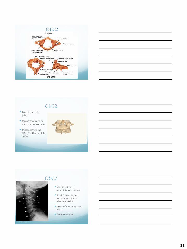
Alar Ligament Stress Test
-With the patient in sitting
-stabilize the spinous process and gently rotate the head
-there should be minimal rotation available
-increased rotation would indicate the Alar ligament has been
compromised
No data
for
sensitivity/
specificity
Alar Ligament Test
-the patient is in a sitting or supine position
-using a pincer grip, place thumb and index finger on the sides of the C2 spinous
process
-place the opposite hand on vertex of hand, then impart either sidebending or
rotation
-the examiner attempts to perceive movement of the C2 spinous process
-a positive finding is lack of movement palpated at C2 or a delay in movement,
indicating alar ligament compromise
No data for
sensitivity/spe
cificity
C0-1
Possible left C0-1
restriction
Bilateral Assessment
30
C1-2 Mobility
Mid Cervical Assessment
Assess translations in flexion to assess for opening
restrictions
Assess translations in extension/neutral for closing
restrictions.
Palpation Multifidus/deep rotatores
Dr. Junak
Spinous
process
Multifidus
31
Treatment Interventions for the
Cervical Spine and Dysfunction
todayilaughed.com
Treatment of CGH Manual Therapy
For identified segmental dysfunctions
For trigger points
Exercise prescription
Normalization of muscle length
Activation of deep neck flexors and
extensors
Manual Therapy
Physiotherapy and spinal manipulative therapy might
Results Thrust manipulation group found to have superior
outcomes (NPRS, NDI) compared to nonthrust
mobilization group and exercise
Non-thrust with exercise some improvement
Trigger Points Soft tissue techniques to:
Posterior cervical musculature
Sternocleidomastoid
Upper trapezius
Dry Needling
May be helpful in conjunction with other treatment
modalities (Man Ther, exercise) (France, S, et al 2014)
Weakly supports dry needling
34
Exercises – Deep Neck Flexors
tieroneeducation.com
Exercise - DNF advancedDNF lift with rotation
DNF with wall PU
Exercises - Deep Neck Extensors
35
Exercises – Deep Neck Extensors
Cervical Extensor Endurance Test (Sebastian, D. et al 2015)
Exercises – Deep Neck Extensors
Manual Therapy Techniques for the Upper
Cervical
Lab Time!
36
Prone C1-2 UPA
Ferna´ndez-de-las-
Pen˜ as, C et al
2016
Nonthrust C2-3 Opening Mobilization
Prone T1-2 CPA
37
LAB Specific Exercise
Training
Presentation Title l March
14, 2017 l 109
INTERVENTION
Posture
Passively elevating the
scapulae by supporting
the weight of the patient’s
arms and asking her to
adduct her scapulae
allowed the patient to
report a decrease in her
posterior neck tension
(Mc Donnell et al 2005)
INTERVENTION
Posture Sleeping
Pillow(s)
Neutral positioning
Side-lying / Supine
Not Prone
38
INTERVENTION
Home Exercise Program
Sitting Scapular Setting
/Humeral External
Rotation
Adduct Scapulae
Humeral ER
**Don’t extend spine
INTERVENTION
SOFT TISSUE
MOBILIZATION
Pectoralis Minor
Gentle Pressure
downward on anterior
shoulder
Fingers under 2nd Rib,
sustaining pressure
INTERVENTION
Cervico-thoracic
Extension
Passive
Active
39
INTERVENTION
SELF MOBILIZATION
Seated supported cervical
extension
INTERVENTION
SELF MOBILIZATION
Cervicothoracic
Sitting
Rolled towel along thoracic spine
Standing against the wall
Towel or foam roll
Supine on Ball
Exercise
Diaphragmatic Breathing
Reduce activation of accessory muscles for breathing
Scalenes, SCM’s
Muscles are tight due to weakness of DNF
Educate patients how to breathe while activating the
appropriate muscles
Support patient in a posture that facilitates Breathing
40
INTERVENTION
Home Exercise Program Pectoralis Major
Stretch
Clavicular Fibers
Sternal Fibers
“Step into Door”
Not leaning
15 second hold time
4 Reps, 2 times daily
INTERVENTION
Home Exercise Program
Cervical spine
retraction “ Chin
Tucks”
Subcranial “Elongation”
5 second hold time,
5-10 reps
2 times daily
Supine and sitting
If supine, done in
hooklying
INTERVENTION
SELF MOBILIZATION
C Spine Retraction
Can do in neutral or
rotation posture
Use both hands
behind on occipital
region
Retract C-spine, and
gently pull up with both
hands
41
INTERVENTION
SELF MOBILIZATION Cervical retraction
with OA flexion
May use opposite hand
on top of head for over-
pressure
Can also vary angle of
pull to right or left
INTERVENTION
Passive to Active Assisted C1,2
Mobilization
Mobilization
Rotational glide of C1 by contact on spinous process
Tips
Pain Free
No oscillation
Horizontal plane
Very light pressure
Can vary flex/ext/rot
Little finger on C2 spinous process
INTERVENTION
SELF MOBILIZATION OPPOSITE HAND SELF
CERVICAL SPINE ROTATION
STRETCH
Using opposite hand
Sustain cervical spine
retraction
Keep Eyes Level
Prevent opposite shoulder
from moving
Adduct Scapulae
Supine or sitting
Usually start in supine
42
INTERVENTION
SELF MOBILIZATION
Patient places finger(s)
anterior to right
transverse process of C1,
and then rotates head to
the Right, applying gentle
overpressure at end
range
NO PAIN
INTERVENTION
SELF MOBILIZATION ATLANTOAXIAL SELF
MOBILIZATION Hold towel with 2 fingers
Only perform 3 repetitions on first day of treatment
Prevent shoulder from moving
(Mulligan 1991)
INTERVENTION
Home Exercise Program
Patient places towel onto
C2 spinous process
Patient gently retracts head
against C2 (anterior glide of
C2)
Hold towel with 2 fingers
(“Gentle”)
Can use with hand as well Slight
Retraction
43
INTERVENTION
Home Exercise Program Scalenes
Anterior
Middle
Posterior
Secondary breathing muscles
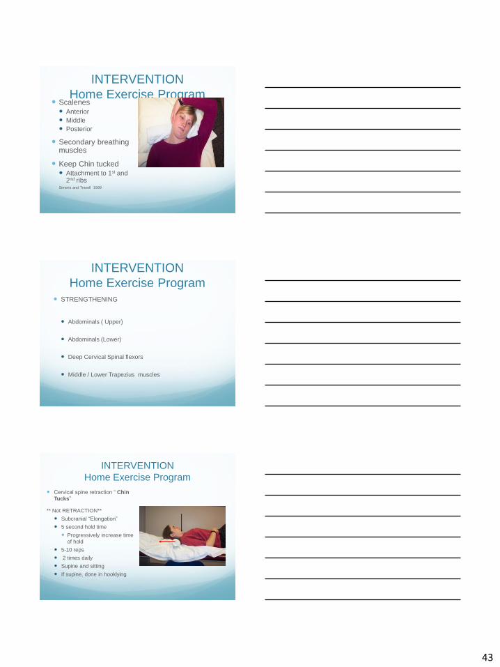
Keep Chin tucked
Attachment to 1st and 2nd ribs
Simons and Travell 1999
INTERVENTION
Home Exercise Program STRENGTHENING
Abdominals ( Upper)
Abdominals (Lower)
Deep Cervical Spinal flexors
Middle / Lower Trapezius muscles
INTERVENTION
Home Exercise Program
Cervical spine retraction “ Chin
Tucks”
** Not RETRACTION**
Subcranial “Elongation”
5 second hold time
Progressively increase time
of hold
5-10 reps
2 times daily
Supine and sitting
If supine, done in hooklying
44
INTERVENTION
STRENGTHENING Deep Cervical Flexors
On wedge, OA nodding
Jull: Pressure sensor under neck. Cervical spine retraction: flattening out lordosis, increasing pressure 10 mmHg
Activation Score is that which patient can hold increase of 10 mmHg for 10 seconds, for 10 repetitions Spine.2002;27:1835-1843
Cephalgia.1999;19:179-185
Cervical Flexion Strengthening
Craniocervical Flexion Test (CCFT)
Perform in supine, hooklying.
1. Inflate blood pressure cuff to 20 mmHg and place between the lordoticcurve and the surface of the table
2. Perform cranial cervical flexion in 5 increments (22, 24, 26, 28, and 30 mmHg)
3. Hold each position for 10 seconds with 10 seconds rest between (The cranial cervical flexion is performed by a head nod in the upper cervical spine)
4. Make sure the patient’s jaw and neck are relaxed
5. The test is ended when the pressure decreases >20% or when substitution occurs during the head nod.
Normal response is achieving 26-30 mmHg. > 10 second holds
Jull, T et al. BMC Musculoskelet Disord. 2013; 14:339.
STRENGTHENING
Eye Movement
Effective when HA / neck tenderness acute
Pain free directions
May be done with eyes open/closed
Self-Resistance
Mid range isometric
Flexion
Side Bending
Extension
Rotation
Presentation Title l March
14, 2017 l 132
45
Neck Flexor Endurance
Strengthening
Same as testing:
Chin retraction and maintained ~
2.5 cm (1 in)
Modify starting position
Standing, limit effect of gravity –
nods, nod with weighted head
strap
Inclined Supine
60 deg ->45 deg->30 deg
Then supine training
Sidelying, s/l with
UE PRE
Quadruped
Presentation Title l March
14, 2017 l 133
INTERVENTION
LOWER ABDOMINALS
Pull lower abdominals inward, without lifting your ribs. Your stomach will be flat or
“hollowed”
You may roll your pelvis so that your back flattens slightly
Hold 3-5 seconds
Keep your chin tucked
(Mc Donnell 2005)
Exercise - DNF advancedDNF lift with rotation
DNF with wall PU
46
Exercises - Deep Neck Extensors
Advanced training
Quadruped
Sustained
neutral Spine
Can add neck
movement
Arm lifts
Leg lifts
Advanced training
Quadruped
Small weight ~ 1
Pound
Can add
Eye movements
Cervical rotation
47
Exercises – Deep Neck Extensors
INTERVENTION
UPPER ABDOMINALS
Pull in your lower abdominals
Tuck your chin
Support back of lower neck with hands clasped
Keep elbows together
Roll head and shoulders up from bed without lifting chin away from chest
INTERVENTION
Scapulothoracic Strengthening
Middle and Lower
Trapezius Muscles
“Set” the Scapula
Externally rotate the
humerus
Increase reps
C spine neutral
Can do over bolster/
Swiss Ball
Progress to bilat
48
INTERVENTION
Home Exercise ProgramLOWER TRAPEZIUS
CERVICAL KINESTHESIA
Posture Direction Cm from
Target
↑
↓
→
←
Diagonal
PROPRIOCEPTION
Posture Direction # Times off
Line
↑
↓
→
←
Diagonal
SMOOTH PURSUIT
CERVICAL KINESTHESIA
Proprioception Testing
Tested in
Vertical
Horizontal
Diagonal
Patient moves to edge of board or pain free
One repetition
Score # cm off from center of target
If outside of target (10 cm)
49
CERVICAL KINESTHESIA
Smooth Pursuit Testing Tested in:
Vertical
Horizontal
Diagonal
Patient is to move the laser
along the line
Record # times dot leaves
the line
Speed of movement should
be 3-7 seconds per line
JPE Training http://www.skillworks.biz/Resources/Documents/JPE%20Target%20and%