Health ABC Operations Manual Vol. III Chapter 3A, page 1 ECG.OM1 Version 1.2 3/7/98 RESTING ECG St. Louis University Core ECG Laboratory Manual of Operations / Table of Contents A. Core Electrocardiographic Laboratory (CEL) ............................................... 2 A.1 Background and Rationale .................................................................................. 2 A.2 Core ECG Laboratory Purpose ........................................................................... 2 A.3 ECG Collection ...................................................................................................... 3 A.4 Training and Certification of ECG Technicians ............................................. 3 B. Electrocardiographic Equipment and Supply Specifications ................... 4 B.1 Equipment Information ....................................................................................... 4 B.2 Equipment Support and Troubleshooting Information ............................... 4 B.3 Supply Information .............................................................................................. 5 B.4 Quality Control Checklist: Equipment and Supplies................................... 5 C. Marquette Equipment Set-up Procedures .................................................... 6 C.1 Overview: MAC PC Keyboard ........................................................................... 6 C.2 MAC PC Equipment Setup ................................................................................. 7 C.2.1 Setting Date and Time ......................................................................................... 7 C.2.2 Setting Receiving Unit (Core ECG Lab) Phone Number .............................. 7 C.2.3 Equipment Setup Continued .............................................................................. 8 C.2.4 Report Format Setup ............................................................................................ 9 D. Resting ECG Acquisition .................................................................................. 11 D.1 Safety and Exclusions: ....................................................................................... 11 D.2 Pre Examination Procedures ............................................................................. 11 D.2.1 Body Position and Lead Placement ................................................................. 11 D.2.2 ECG Skin Prep..................................................................................................... 13 D.2.3 Electrode Attachment ......................................................................................... 13 D.2.4 Electrode Cable Attachment ............................................................................. 13 D.3 Preparing the Marquette.................................................................................... 14 D.4 Recording the 12 Lead ECG .............................................................................. 15 D.5 Post Recording Quality Check ......................................................................... 16 D.6 Post Recording Alert Check .............................................................................. 16 D.7 Record the ECG in the ECG Log ...................................................................... 17 D.8 Quality Control Checklist: ECG Acquisition ............................................... 18 E. ECG Transmission........................................................................................... 18 E.1 Telephone Transmission of ECGs ................................................................... 18 E.1.1 ECGs Unavailable for Telephone Transmission .......................................... 19 E.2 Procedure for Transmitting ECGs by Telephone ......................................... 19 E.2.1 Printing a Directory- Selection of ECGs for Transmitting ......................... 19 E.2.2 Deleting ECGs ..................................................................................................... 19 E.2.3 Transmitting an ECG ......................................................................................... 20 E.3 Quality Control Checklist: Transmitting and Deleting ECGs .................. 21 F. Confirmation of Data Receipt from the Core ECG Laboratory .............. 21 G. Core Electrocardiographic Laboratory Directory ...................................... 22 H. Quality Assurance ........................................................................................... 22 H.1 Training Requirements .................................................................................. 22 H.2 Certification Requirements............................................................................... 23 H.3 Quality Assurance Checklist ............................................................................ 23
Transcript
Health ABC Operations Manual Vol. III Chapter 3A, page 1
ECG.OM1
Version 1.2 3/7/98
RESTING ECG
St. Louis University Core ECG Laboratory Manual of Operations / Table of Contents
A. Core Electrocardiographic Laboratory (CEL) ............................................... 2 A.1 Background and Rationale.................................................................................. 2 A.2 Core ECG Laboratory Purpose ........................................................................... 2 A.3 ECG Collection...................................................................................................... 3 A.4 Training and Certification of ECG Technicians ............................................. 3 B. Electrocardiographic Equipment and Supply Specifications................... 4 B.1 Equipment Information....................................................................................... 4 B.2 Equipment Support and Troubleshooting Information ............................... 4 B.3 Supply Information.............................................................................................. 5 B.4 Quality Control Checklist: Equipment and Supplies................................... 5 C. Marquette Equipment Set-up Procedures .................................................... 6 C.1 Overview: MAC PC Keyboard ........................................................................... 6 C.2 MAC PC Equipment Setup ................................................................................. 7 C.2.1 Setting Date and Time ......................................................................................... 7 C.2.2 Setting Receiving Unit (Core ECG Lab) Phone Number .............................. 7 C.2.3 Equipment Setup Continued.............................................................................. 8 C.2.4 Report Format Setup ............................................................................................ 9 D. Resting ECG Acquisition .................................................................................. 11 D.1 Safety and Exclusions: ....................................................................................... 11 D.2 Pre Examination Procedures ............................................................................. 11 D.2.1 Body Position and Lead Placement ................................................................. 11 D.2.2 ECG Skin Prep..................................................................................................... 13 D.2.3 Electrode Attachment ......................................................................................... 13 D.2.4 Electrode Cable Attachment ............................................................................. 13 D.3 Preparing the Marquette.................................................................................... 14 D.4 Recording the 12 Lead ECG .............................................................................. 15 D.5 Post Recording Quality Check ......................................................................... 16 D.6 Post Recording Alert Check.............................................................................. 16 D.7 Record the ECG in the ECG Log ...................................................................... 17 D.8 Quality Control Checklist: ECG Acquisition............................................... 18 E. ECG Transmission........................................................................................... 18 E.1 Telephone Transmission of ECGs................................................................... 18 E.1.1 ECGs Unavailable for Telephone Transmission.......................................... 19 E.2 Procedure for Transmitting ECGs by Telephone ......................................... 19 E.2.1 Printing a Directory- Selection of ECGs for Transmitting ......................... 19 E.2.2 Deleting ECGs..................................................................................................... 19 E.2.3 Transmitting an ECG ......................................................................................... 20 E.3 Quality Control Checklist: Transmitting and Deleting ECGs.................. 21 F. Confirmation of Data Receipt from the Core ECG Laboratory .............. 21 G. Core Electrocardiographic Laboratory Directory ...................................... 22 H. Quality Assurance ........................................................................................... 22 H.1 Training Requirements .................................................................................. 22 H.2 Certification Requirements............................................................................... 23 H.3 Quality Assurance Checklist ............................................................................ 23
Resting ECG Health ABC Operations Manual Vol. III Chapter 3A, page 2
ECG.OM1
Version 1.2 3/7/98
I. Attachments Attachment A: Request for Certification of ECG Technician............................. 25 Attachment B: 12 Lead ECG Electrode Lead Placement....................................... 26 Attachment C: ECG Log ............................................................................................. 27 Attachment D: Unavailable ECG Form .................................................................... 28
A. Core Electrocardiographic Laboratory (CEL)
A.1 Background and Rationale Significance of Resting ECG: The purpose of the ECG is to characterize coronary disease prevalence and to determine the contribution of cardiovascular disease as assessed clinically and electrocardiographically to risk of accelerated sarcopenia. The ECG will serve five main purposes:
• Define prevalent myocardial infarction. • Identify cases of silent MI longitudinally. • Identify resting ECG characteristics including LVH, conduction defects, Q, ST
and T wave abnormalities at baseline and longitudinally. • Investigate the relationship of resting ECG characteristics and sarcopenia
longitudinally. • Evaluate resting 12-lead exclusion criteria for selected measurements.
It is well known that myocardial infarction and impaired LV function contribute to decline in function and may be associated with muscle-wasting. To understand the contribution of loss of lean mass to decline in function independent of heart disease, we need to characterize cardiovascular disease.
A.2 Core ECG Laboratory Purpose St. Louis University Core ECG Lab (CEL) will provide centralized evaluation and analysis of all ECGs obtained for the Health ABC study. Tasks of the Core ECG Laboratory include:
• Participate in protocol development • Provide quality assurance and quality control of ECG collection and coding • Provide technical and logistical support for issues pertaining to the Core ECG
Lab • Collect, analyze and store protocol ECGs • Transmit data analysis to Data Coordinating Center (DCC) at specified intervals • Participate in analyses and publication of results
The methods used to acquire data must be consistent to ensure uniform analysis at the Core ECG Lab. A standard technique for the acquisition and collection of ECGs will allow serial comparison analysis of data for myocardial infarction classification.
Resting ECG Health ABC Operations Manual Vol. III Chapter 3A, page 3
ECG.OM1
Version 1.2 3/7/98
A.3 ECG Collection Standard 12 lead ECGs will be collected at the following time points:
Exam 1 Exam 4 Exam 7
It is important to submit all required ECGs to allow sequential comparison of the ECGs for detection of significant changes suggestive of myocardial infarction. Missing electrocardiograms can jeopardize the diagnosis of myocardial infarction. Certification prior to study initiation and ongoing quality assessments via a monthly confirmation report will ensure high-quality data for the study duration. Certification is discussed below, and the certification transmittal form is included as Attachment A.
A.4 Training and Certification of ECG Technicians A representative of the Core ECG Laboratory will provide initial training at the centralized training session for the baseline exam. Following the training session, each person at the field centers responsible for acquiring 12 lead ECGs will be certified by the procedure described below. Any personnel added to the field centers with ECG acquisition responsibilities will need to be certified by the same procedure. New staff will be trained by the local QC Officer. The use of the Health ABC technician number will allow the Core ECG Laboratory to track quality control and identify problems. ECG Certification Sites are required to certify ECG technicians before they are involved in the collection of electrocardiographic data for the Health ABC study. The certification procedure is as follows:
1. Five ECGs will be acquired using the methods described in section D: Resting ECG Acquisition.
2. The five resting supine ECGs will be obtained consecutively. 3. The ECGs will be transmitted to the Core ECG Lab by the procedures discussed
in E: ECG submission. The certification form will be faxed to the core ECG Lab at the time of transmission.
4. Upon receipt of the ECGs and certification form, the Core ECG Lab will evaluate the ECGs for quality parameters and provide the sites and Coordinating Center with certification confirmation.
5. Certification requires successful acquisition of five high quality ECGs. Quality ratings are identical to the quality ratings during the course of the study as outlined in section D.5.
Resting ECG Health ABC Operations Manual Vol. III Chapter 3A, page 4
ECG.OM1
Version 1.2 3/7/98
Indicators of poor quality include:
a) Excessive baseline wander b) Motion artifact or loose electrode contact c) Excessive muscle noise d) Excessive 60 Hz noise
e) Valid calibration documentation f) Missing leads g) Check for lead reversal
The Request for Certification form is included as Attachment A. The field center should make several copies of this form and keep it on file. The Request for Certification form will accompany the certification ECGs. A technician number will be assigned by the data coordinating center to each person certified to perform Health ABC resting ECGs. This number will allow tracking of ECG quality for the study duration.
B. Electrocardiographic Equipment and Supply Specifications
B.1 Equipment Information
• Marquette Electronic MAC PC Resting ECG Analysis System (Part # MACPC-AAA-ABCA)
All data is acquired using Marquette equipment to allow electronic data transmission to the Core ECG Lab. The MAC PC is portable, weighing approximately 12 pounds, and is equipped with a validated 12SL ECG analysis program to provide the sites with a complete computerized analysis of the subjects ECG in both morphology and rhythm. The 12SL program processes and evaluates ECGs in the same manner as the larger central ECG systems and produces a hard copy tracing for the participant’s chart as well as an electronic copy for telephone transmission to the Core ECG Laboratory. This portable unit operates on rechargeable NiCad Batteries supplied with a line- operated charger. The unit may be operated with the AC adapter or battery pack. Refer to the manual for specific information regarding care of equipment and optimal use/charge of the battery for optimal performance.
B.2 Equipment Support and Troubleshooting Information The Marquette MAC PC Operators manual provides additional information and should be used in conjunction with this manual. It is an excellent resource and contains detailed information for troubleshooting equipment problems.
Resting ECG Health ABC Operations Manual Vol. III Chapter 3A, page 5
ECG.OM1
Version 1.2 3/7/98
B.3 Supply Information
Item specification
Part # Vendor Cost Quantity
Silver Mactrodes plus electrodes
9623-003P
Marquette Service and Supplies
$40/case
1,000/case
Mac PC Recording Paper
9402-023
Marquette Service and Supplies
$45/case
18 rolls/case
Alcohol/ pumice prep pads
4828-004
Marquette Service and Supplies
$10.80/ box
100/box
Prep Razors
3704-901
Marquette Service and Supplies
$18.90/ pack
100/pack
Replacement Lead Wire Set
900177-001
Marquette Service and Supplies
$130.50/ set
12 leadwires / set
Replacement Adaptors
900178-002
Marquette Service and Supplies
$54/set
set of 10
Felt Tip Pen
Supplied locally
B.4 Quality Control Checklist: Equipment and Supplies
Monthly Equipment and Supply Check—Every month do the following:
1. Drain and charge the battery according to the MAC PC operators manual 2. Visually inspect the MAC PC 3. Check the case and display screen for cracks 4. Inspect cords and cables for fraying or other damage 5. Inspect all plugs, cables, and connectors for bent prongs or pins 6. Verify all cords, socketed components, and connectors are securely seated
Resting ECG Health ABC Operations Manual Vol. III Chapter 3A, page 6
ECG.OM1
Version 1.2 3/7/98
7. Inspect keys and controls for proper operation, i.e., toggle keys should not stick in one position, and knobs should rotate fully in both directions.
8. Clean the exterior surface with a damp soft cloth (wring excess water from the cloth) Do not drip water or any liquid on the writer assembly and avoid contact with open vents, plugs, and connectors. Dry the surface with a clean cloth or paper towel
9. Check supplies (ECG paper, and prep supplies). Order if necessary
C. Marquette Equipment Set-up Procedures (system setup stored for study duration; only change in time will need to be modified) The MAC PC is operational at delivery. For the Health ABC study, you will need to modify the set up to the following specifications (Refer to the Marquette Operators manual chapter 11- SETUP.) Once these details are set, the system will retain them until they are manually modified.
C.1 Overview: MAC PC Keyboard The following diagram is from the Marquette MAC PC Operator’s Manual and provides a brief overview to the MAC PC keyboard.
Resting ECG Health ABC Operations Manual Vol. III Chapter 3A, page 7
ECG.OM1
Version 1.2 3/7/98
Note: Each of the 10 numerical keys on the keyboard has a number from 1-5 on it preceded by the letter F. These keys are dual purpose: They are used to either type in numerical data or as a function key to select an item from the LCD.
C.2 MAC PC Equipment Setup
C.2.1 Setting Date and Time To enter the setup menu:
• Press the STOP key to display the main menu. • Hold the shift key down while pressing the 2 key for the system functions menu. • Set up to enter the Cart Set up menu. Enter the date/time and phone numbers
from this menu. • Select Date/Time
Select Date: type the day, a dash, the month, a dash, and the year. (Press enter) Select Time: Type the hour, a dash, and the minute (Press enter)
• Press the STOP key to return to the main menu. Note: remember to edit when the time changes
C.2.2 Setting Receiving Unit (Core ECG Lab) Phone Number To enter the setup menu:
• press the STOP key to display the main menu • hold the shift key down while pressing the 2 key for the system functions menu • set up to enter the Cart Set up menu. Enter the date/time and phone numbers
from this menu. • Select Phone • The Core ECG Laboratory Marquette receiving unit phone number can be stored
in the clinical unit Marquette machine. Entering and storing the CEL phone number will save time because the number will not have to be entered for each transmission. Store the Core ECG Laboratory phone with the following specifications:
Phone Numbers: Select Number 1 Phone Number 1 description only allows 7 characters
Type in ABC SLU (press enter)
Resting ECG Health ABC Operations Manual Vol. III Chapter 3A, page 8
ECG.OM1
Version 1.2 3/7/98
Phone Number 1: The CEL Marquette receiving unit phone number is (314) 725-2907. Refer to the manual to enter the phone number; note special entry is required if you must dial 9 for an outside line
Press the STOP key to return to the main menu.
C.2.3 Equipment Setup Continued The remaining system setup items are entered as follows: Step 1: Press the STOP key to display the main menu Step 2: Hold the shift key down while pressing the 2 key for the system functions menu Step 3: Select set up to enter the 1st Cart Set up menu
Step LCD display Your action 4 Cart Setup
Date/Time Phone Ldgrps Reports More
Select More
5 Cart set up Modem Passwds Misc Defaults More
Select Misc
6 Line Frequency: 60 Hz 50 Hz
Select 60 Hz
7 Cart ID: 0-255
type 01 for Memphis Field Center 02 for Pittsburgh Field Center
8 Site ID: 1-255
type: 10 (CEL code for Health ABC ECG)
9 Default Location: 0-999
10 Institution Name: Up to 40 characters
Type your Institution Name and State
11 Number of Patient ID digits: 1-12
12 ID required to record an ECG: Yes No
Select Yes
13 Height/Weight: in.lb cm/kg
Select in/lbs to record height and weight in inches and pounds
14 Input Participant Age as: DOB Years
Select DOB to enter date of birth
Resting ECG Health ABC Operations Manual Vol. III Chapter 3A, page 9
ECG.OM1
Version 1.2 3/7/98
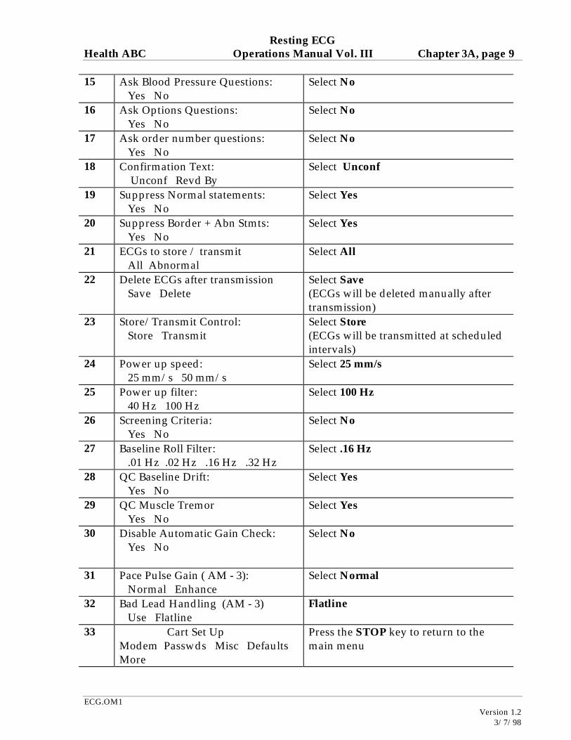
15 Ask Blood Pressure Questions: Yes No
Select No
16 Ask Options Questions: Yes No
Select No
17 Ask order number questions: Yes No
Select No
18 Confirmation Text: Unconf Revd By
Select Unconf
19 Suppress Normal statements: Yes No
Select Yes
20 Suppress Border + Abn Stmts: Yes No
Select Yes
21 ECGs to store / transmit All Abnormal
Select All
22 Delete ECGs after transmission Save Delete
Select Save (ECGs will be deleted manually after transmission)
23 Store/Transmit Control: Store Transmit
Select Store (ECGs will be transmitted at scheduled intervals)
Resting ECG Health ABC Operations Manual Vol. III Chapter 3A, page 10
ECG.OM1
Version 1.2 3/7/98
C.2.4 Report Format Setup In the Health ABC study, two separate 12-lead ECGs will be printed. The first 12-lead will have the 12 SL interpretation of the ECG. This is the ECG reviewed for alert conditions. After printing the first copy with the interpretation, the machine will prompt if you want an extra copy. The extra copy will be printed for the participant’s physician and will not include the interpretation. The following setup section describes how to program the Marquette Machine to automatically print the first report with interpretation and the second report without the interpretation. To enter the setup menu:
• Press the stop key to display the main menu • Hold the shift key down while pressing the 2 key for the systems function menu • Press setup to enter cart setup
In the setup menu do the following: Step LCD screen Your action 1 Cart setup
Dat/Time Phone LdGrps Reports More
select reports
2 Report formats for: Confirmed Unconf
select unconf
3 Ask for extra copies of plots: Yes No
select Yes
4 suppress orig Rpt Interpretation Yes No
select No
5 Suppress copy interpretation Yes No
select Yes
6 Suppress Text page Yes No
select Yes
7 Rhythm and Morphology report Yes No
select No
8 1 Complex/Lead Yes No
select No
9 1 Complex/Lead with abnormals Yes No
select No
10 Add tic to complexes Yes No
select No
11 Times 2 complexes Yes No
select No
12 Automatic rhythm (1 X 10) Yes No
select No
13 Automatic rhythm (1 X 10) with abnormals Yes No
select No
14 12 lead (4 X 2.5) Yes No
select Yes
Resting ECG Health ABC Operations Manual Vol. III Chapter 3A, page 11
ECG.OM1
Version 1.2 3/7/98
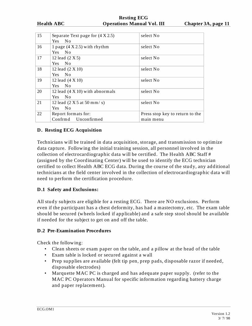
15 Separate Text page for (4 X 2.5) Yes No
select No
16 1 page (4 X 2.5) with rhythm Yes No
select No
17 12 lead (2 X 5) Yes No
select No
18 12 lead (2 X 10) Yes No
select No
19 12 lead (4 X 10) Yes No
select No
20 12 lead (4 X 10) with abnormals Yes No
select No
21 12 lead (2 X 5 at 50 mm/s) Yes No
select No
22 Report formats for: Confrmd Unconfirmed
Press stop key to return to the main menu
D. Resting ECG Acquisition Technicians will be trained in data acquisition, storage, and transmission to optimize data capture. Following the initial training session, all personnel involved in the collection of electrocardiographic data will be certified. The Health ABC Staff # (assigned by the Coordinating Center) will be used to identify the ECG technician certified to collect Health ABC ECG data. During the course of the study, any additional technicians at the field center involved in the collection of electrocardiographic data will need to perform the certification procedure.
D.1 Safety and Exclusions: All study subjects are eligible for a resting ECG. There are NO exclusions. Perform even if the participant has a chest deformity, has had a mastectomy, etc. The exam table should be secured (wheels locked if applicable) and a safe step stool should be available if needed for the subject to get on and off the table.
D.2 Pre-Examination Procedures Check the following:
• Clean sheets or exam paper on the table, and a pillow at the head of the table • Exam table is locked or secured against a wall • Prep supplies are available (felt tip pen, prep pads, disposable razor if needed,
disposable electrodes) • Marquette MAC PC is charged and has adequate paper supply. (refer to the
MAC PC Operators Manual for specific information regarding battery charge and paper replacement).
Resting ECG Health ABC Operations Manual Vol. III Chapter 3A, page 12
ECG.OM1
Version 1.2 3/7/98
Explain the procedure to the subject:
Script: “The resting ECG is a test used to detect heart disease. This procedure is not painful and should take less than 10 minutes. First I will need to prepare your skin for the test which requires marking and preparing the sites for attachment of the electrodes on your chest.” • Ask the subject to undress to the waist and put on a gown with the opening to
the front. • Position the subject. • Ask the participant to lie in the supine position with head on one pillow,
shoulders straight and arms relaxed at the sides. (If table has wheels, confirm the wheels are locked and it is safe for the subject to climb up on.)
D.2.1 Body Position and Lead Placement
To ensure comparability of data, the following uniform procedures for skin preparation, electrode placement, and quality control should be followed: The participant should be supine with one pillow; the upper body is exposed; shoulders are straight and the arms are relaxed. Ask the participant to avoid movements that may cause errors in marking the electrode locations, or artifact in the 12 lead ECG recording. Mark each electrode site with an X using a felt tip pen, anatomical landmarks are used to determine lead placement (see Attachment B): Lead RA: Inner right arm above the wrist Lead LA: Inner left arm above the wrist. Lead RL: Right lower leg above the inner ankle or as close as the electrode will reach. Lead LL: Left lower leg above the inner ankle or as close as the electrode will reach. If the participant has an amputated limb, place the electrode on the body part closest to that limb Chest Electrodes (diagram from MAC PC Operators Manual):
Resting ECG Health ABC Operations Manual Vol. III Chapter 3A, page 13
ECG.OM1
Version 1.2 3/7/98
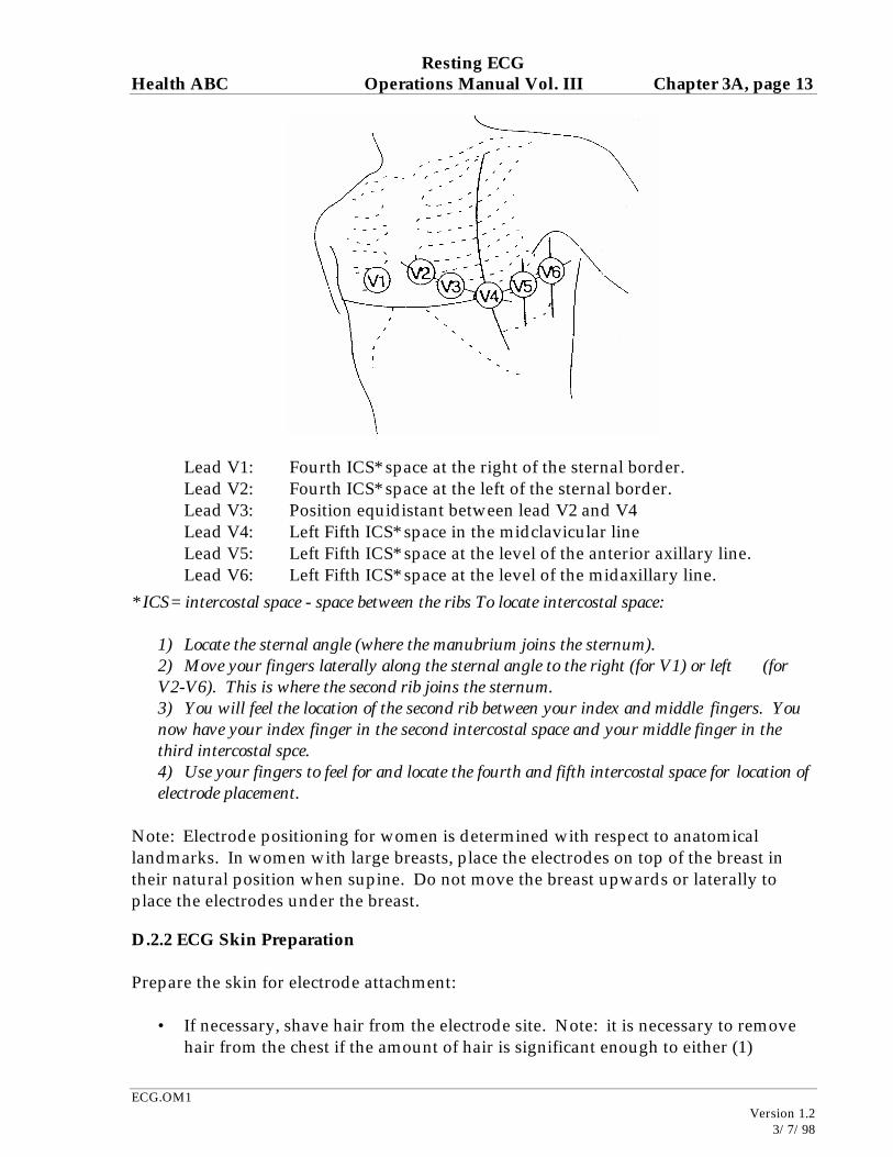
Lead V1: Fourth ICS* space at the right of the sternal border. Lead V2: Fourth ICS* space at the left of the sternal border. Lead V3: Position equidistant between lead V2 and V4 Lead V4: Left Fifth ICS* space in the midclavicular line Lead V5: Left Fifth ICS* space at the level of the anterior axillary line. Lead V6: Left Fifth ICS* space at the level of the midaxillary line. * ICS= intercostal space - space between the ribs To locate intercostal space:
1) Locate the sternal angle (where the manubrium joins the sternum). 2) Move your fingers laterally along the sternal angle to the right (for V1) or left (for V2-V6). This is where the second rib joins the sternum. 3) You will feel the location of the second rib between your index and middle fingers. You now have your index finger in the second intercostal space and your middle finger in the third intercostal spce. 4) Use your fingers to feel for and locate the fourth and fifth intercostal space for location of electrode placement.
Note: Electrode positioning for women is determined with respect to anatomical landmarks. In women with large breasts, place the electrodes on top of the breast in their natural position when supine. Do not move the breast upwards or laterally to place the electrodes under the breast.
D.2.2 ECG Skin Preparation Prepare the skin for electrode attachment:
• If necessary, shave hair from the electrode site. Note: it is necessary to remove hair from the chest if the amount of hair is significant enough to either (1)
Resting ECG Health ABC Operations Manual Vol. III Chapter 3A, page 14
ECG.OM1
Version 1.2 3/7/98
interfere with adhesion of the electrode or (2) result in discomfort to the participant due to the chest hair pulling at point of electrode removal. Inform the participant before you shave the electrode site that removing the hair from the electrode area will result in a better quality tracing (because of better adhesion) and easier removal of the electrode (because the chest hair will not be pulled during electrode removal).
• Rub each electrode site with alcohol pumice prep pad; Dry with gauze or buff with sand paper; work in a single direction to remove the skin oils and epidermal layer at each site; the skin should appear slightly red, this removes dead skin and oil to enhance conduction.
D.2.3 Electrode Attachment • Place an electrode at each prepared site. • Press on each electrode to promote good skin contact with gel. • Attach clamp only to metal part of electrodes. • Allow the gel to penetrate a minute before recording the 12 lead to enhance
conduction.
D.2.4 Electrode Cable Attachment By following a uniform sequence of electrode attachment, the risk of wrong connections (lead reversals) can be minimized. The following sequence of connecting electrode cables is recommended:
Right leg Right arm V1 V2 V3 V4 V5 V6 Left arm Left leg
Follow the electrode cable wires from the acquisition module to the electrode site prior to attaching electrodes to decrease the possibility of lead reversals. After attaching the lead wires, verify one by one that the connections to all electrode sites are correct.
D.3 Prepare the Marquette (information to enter for each 12 lead ECG obtained) Before acquiring the 12 lead ECG, enter participant information.
Resting ECG Health ABC Operations Manual Vol. III Chapter 3A, page 15
ECG.OM1
Version 1.2 3/7/98
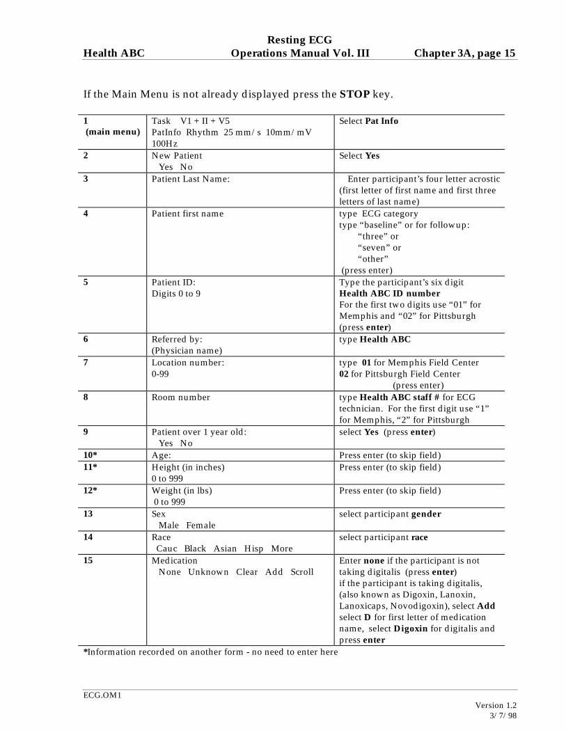
If the Main Menu is not already displayed press the STOP key. 1 (main menu)
3 Patient Last Name: Enter participant’s four letter acrostic (first letter of first name and first three letters of last name)
4 Patient first name type ECG category type “baseline” or for followup: “three” or “seven” or “other” (press enter)
5 Patient ID: Digits 0 to 9
Type the participant’s six digit Health ABC ID number For the first two digits use “01” for Memphis and “02” for Pittsburgh (press enter)
6 Referred by: (Physician name)
type Health ABC
7 Location number: 0-99
type 01 for Memphis Field Center 02 for Pittsburgh Field Center (press enter)
8 Room number
type Health ABC staff # for ECG technician. For the first digit use “1” for Memphis, “2” for Pittsburgh
9 Patient over 1 year old: Yes No
select Yes (press enter)
10* Age: Press enter (to skip field) 11* Height (in inches)
0 to 999 Press enter (to skip field)
12* Weight (in lbs) 0 to 999
Press enter (to skip field)
13 Sex Male Female
select participant gender
14 Race Cauc Black Asian Hisp More
select participant race
15 Medication None Unknown Clear Add Scroll
Enter none if the participant is not taking digitalis (press enter) if the participant is taking digitalis, (also known as Digoxin, Lanoxin, Lanoxicaps, Novodigoxin), select Add select D for first letter of medication name, select Digoxin for digitalis and press enter
*Information recorded on another form - no need to enter here
Resting ECG Health ABC Operations Manual Vol. III Chapter 3A, page 16
ECG.OM1
Version 1.2 3/7/98
After entering the participant information, the main menu will reappear. This completes the entry of participant information.
D.4 Recording the 12 Lead ECG Ask the participant to relax, breathe normally, and refrain from talking or moving while the 12 lead ECG is recorded. Record the 12 lead ECG, by pressing the record ECG key at the top right corner of the keyboard next to the numerical zero key. A display will appear in the LCD window 1st ** Acquiring Data **, followed by ** ECG Acquisition Complete** The Mac PC will process the data (LCD screen: ** Analyzing ECG **) and print reports (LCD screen: **Printing reports**)
• The first printout will have an interpretation printed on the ECG. • The LCD screen will prompt for Number of extra copies (0-9) • Select 1. This copy will not have an interpretation. • The first copy with interpretation will be retained at the field center. • The second copy without interpretation is for the participant or the participant’s
physician. The LCD screen will display ** Processing ECG ** then ** ECG Storage complete** Press enter to return to the main menu.
D.5 Post Recording Quality Check Inspect the ECG recording immediately for quality. If quality problems are detected, correct the source of artifact and repeat the recording. Check the following:
a) Excessive baseline wander: Baseline wander is defined as 1.0 mm difference between the PQ baseline in three consecutive ECG complexes. The source of this artifact is usually inadequate skin prep.
b) Motion artifact or loose electrode contact: These may cause sudden jumps in some ECG leads.
c) Excessive muscle noise: Random noise in excess of 5 mm. The source of this artifact may be inadequate skin prep or a participant that is shivering or trembling.
d) Excessive 60 Hz noise: 60 Hz noise is visible on the 12 lead ECG. This noise is usually associated with A-C interference from nearby machines. Other sources may include poor skin contact.
e) Valid calibration documentation: Calibration standard should be recorded on
Resting ECG Health ABC Operations Manual Vol. III Chapter 3A, page 17
ECG.OM1
Version 1.2 3/7/98
each resting ECG. Invalid calibration exceeds + 1.0 mm of the standard 10 mm pulse. Equipment should be calibrated if out of acceptance range. Please note, however, that in special cases when with standard calibration the ECG complexes run off the page, which interferes with ECG analysis, it is permissible to change the calibration. Whenever this is done, however, a memo should be faxed to Karen Stocke (314-725-2171) at the ECG Reading Center that indicates that the calibration was changed, and exactly how it was changed. Include the participant’s Health ABC Enrollment ID#, acrostic, date, and time of the ECG on this memo.
f) Missing leads: ensure all 12 leads are recorded, if a lead is missing, identify the missing lead, replace the lead, and obtain another ECG.
g) Check for lead reversal: The 12 lead ECG should be inspected for possible lead reversals. Look for normal progression of chest lead patterns from V1 to V6. Inspect lead AVR for negativity, and lead I for mainly positive P, QRS and T wave. If any condition is suggestive of lead reversal, re-check the electrode cable attachment.
Sources of artifact: The most common cause of artifact is inadequate skin preparation. Other sources of artifact include:
• Defective electrodes (check expiration dates; gel dries over time) • Fractured lead wires (connects the electrodes to the acquisition module) • Fractured wires in the acquisition cord interface (connects the acquisition module
to the electrocardiograph) • Participant not resting quietly and relaxed
D.6 Post Recording Alert Check ECG procedure would include printing one tracing with the Marquette machine reading and one tracing without the reading. Any abnormal result as listed below would be overread by a physician in the clinic before the participant leaves. From ECG Operations Manual Chapter, the following statements would require clinic physician review: Heart rate <40 (bradycardia) or >135 (tachycardia) Atrial fibrillation or atrial flutter (new onset) Wolff-Parkinson-White (WPW) or ventricular pre-excitation Idioventricular rhythm Ventricular tachycardia Third degree or complete A-V block
Any statement including reference to acute injury or ischemia, or marked T-wave abnormality
Resting ECG Health ABC Operations Manual Vol. III Chapter 3A, page 18
ECG.OM1
Version 1.2 3/7/98
For any of the above alerts, the clinic physician will determine if immediate notification of participant’s physician is required before the participant leaves the clinic. If necessary, ECG will be faxed to physician or sent with participant to his physician. Clinic physician will overread all other abnormal ECGs within one week of participant’s visit. The clinic physician will decide if participant and participant’s physician should receive a report of the abnormal ECG before the final report is ready for distribution. Cover letter similar to that used in CHS will be sent to the participant and the participant’s physician. Final report to participant will include a statement that a copy of the ECG has been sent to his physician, with no classification as normal/abnormal. Final report to participant’s physician will include a copy of the ECG without interpretation. If the participant does not have a physician, a copy of the ECG will be included in the participant report.
D.7 Record the ECG in the ECG Log Enter the ECG into the ECG Log (Attachment C). Each ECG with acceptable quality selected for transmission to the Core ECG Lab should be recorded in the ECG Log. The purpose of the ECG Log is to record each Health ABC ECG performed, transmitted, and received by the Core ECG Lab. Use this log as a guide to:
• Record ECGs to transmit (and which ECGs need to be deleted before
transmission) • Cross-check the directory of ECGs to be transmitted to the CORE ECG Lab
before transmission • Confirm successful transmission of the ECG from the confirmation of receipt
report sent form the CORE ECG Lab • Select ECGs to delete following verification of successful transmission from the
Core ECG Lab. • Record ECGs not available or sent in hard copy to the Core ECG Lab
D.8 Quality Control Checklist: ECG Acquisition Prior to participant arrival:
• Confirm battery is charged • Confirm adequate paper supply • Skin prep supplies are available • Exam table has clean sheets and gown is available for the participant; table is
secured against a wall or the wheels are locked.
Resting ECG Health ABC Operations Manual Vol. III Chapter 3A, page 19
ECG.OM1
Version 1.2 3/7/98
After participant arrival: • Explain procedure. • Request the participant disrobe to the waist and provide a clean (or disposable)
gown with the opening to the front. • Have the participant assume a semi-recumbent position on the exam table. with
head on one pillow and the arms relaxed and to the side. • Mark electrode positions with felt tip pen. • Skin prep the electrode sites. • Attach the electrodes. • Attach the lead wires to the electrodes. • Enter the participant information in the MAC PC. • Record 12 lead ECG. • Visually inspect the 12 lead for quality (including baseline wander, excess artifact
and lead reversal. • Repeat ECG if quality is poor. • Inspect the ECG for alert conditions ( if alert conditions present contact the study
physician). • Complete the ECG log with participant information and the ECG date and time
of the ECG to be transmitted. • Disconnect the leadwires. • Remove the electrodes. • Wipe marks and goop off skin.
E. ECG Submission
E.1 Telephone Transmission of ECGs ECGs will be transmitted to the Core ECG Laboratory via a phone line. Sites are required to retain a hard copy ECG for the participant’s permanent record. The Core Lab will accept a hard copy of the ECG if necessary. The submission of hard copy ECGs should be kept to a minimum.
E.1.1 ECGs Unavailable for Telephone Transmission In the rare event that an ECG is collected, but unavailable for transmission, the ECG can be submitted to the Core ECG Laboratory with the Unavailable ECG form included as Attachment D. Because the ECGs are routinely evaluated in an electronic format from telephone transmission, the submission of original ECGs is discouraged and should be kept to a minimum. In the very rare event that an ECG cannot be collected, the CEL should be notified in writing using the Unavailable ECG Form (Attachment D).
Resting ECG Health ABC Operations Manual Vol. III Chapter 3A, page 20
ECG.OM1
Version 1.2 3/7/98
E.2 Procedure for Transmitting ECGs by Telephone
E.2.1 Printing a directory- Selection of ECGs for Transmitting • Print a participant directory to determine which ECGs will be transmitted. • Press stop to display the main menu. • Hold down shift and press 2 to display the system functions menu. • Select storage. • Select directory.
A directory will be printed. Compare this directory to the handwritten log (Attachment C) to determine which ECGs to transmit. You must print a directory before transmission so you can delete ECGs which are recorded twice due to poor quality.
• Delete bad ECGs or ECGs not to be transmitted (see next section), and print a final directory of the ECGs to be transmitted.
• Initial the directory. The date will be stamped by the machine. • Attach the directory to your daily log for later verification of receipt by the Core
ECG Lab.
E.2.2 Deleting ECGs See Chapter 10- Deleting an ECG in the MAC PC Operators’ Manual. Routine deletion: ECGs are usually deleted if it is a duplicate record or when the ECG has been transmitted to the Core ECG Lab. If more than one ECG was acquired in an attempt to improve the quality of the tracing, the recording time will provide a clue as to which ECG to keep and which to delete. To delete one or more ECGs:
1. Press stop to get to the main menu. 2. Hold down shift and press the 2 key. 3. Select storage. 4. Select more. 5. Select delete. 6. “Select by Participant ID?“ Select “No.” 7. Next the participant data for each stored ECG will appear in the LCD display,
the following options are available: “Yes:” Selects this ECG for deletion, “No:” bypasses the ECG for deletion, “No...” bypasses this and all subsequent ECGs for deletion, “Yes...” Selects this and all subsequent ECGs for deletion. “Expand” provides additional information such as date and time of ECG which may be required in the decision to select the ECG for deletion.
Resting ECG Health ABC Operations Manual Vol. III Chapter 3A, page 21
ECG.OM1
Version 1.2 3/7/98
After the ECGs are selected for deletion, you have a chance to change your mind. The LCD will display the number of ECGs selected for deletion and you select “Yes” to perform the deletion and “No” to cancel the deletion.
E.2.3 Transmitting an ECG
Transmit records at the end of each day.
1. Connect a telephone cable from a telephone wall outlet to the telephone connector on the back of the system (cable PN 80159-014).
2. Press the stop key to get the main menu. 3. Hold down shift and press the 2 key to display the system functions menu. 4. Select storage. 5. Select transmit. 6. Select phone. 7. The machine was set up with one location for transmission. This number will
appear in the LCD. Press enter. 8. The LCD will display: “Select by Participant ID?” Select “NO.”
The participant data will be displayed for a single ECG with a LCD display such as
9. Select “Yes...” to include all stored ECGs in the file transmission (the ECGs were
selected in the previous section) The LCD will display ** Batch Transmission** After the last ECG is transmitted, a message will appear displaying the number
of ECGs transmitted (should equal the number of ECGs on the directory printed prior to transmission)
10. Press stop to return to the main menu. 11. Record the transmission date on the ECG Log (Attachment C).
Do not delete the ECGs from storage until a confirmation is received from the Core ECG Laboratory of data received.
E.3 Quality Control Checklist: Transmitting and Deleting ECGs
ECG Transmission • Print a directory. • Cross check the directory with the ECG log for selection of ECGs for
transmission. • Delete ECGs that do not need to be transmitted. • Transmit the ECGs.
Resting ECG Health ABC Operations Manual Vol. III Chapter 3A, page 22
ECG.OM1
Version 1.2 3/7/98
• Document the date transmitted on the ECG Log.
ECG Deletion • Print a directory. • Cross check the confirmation report from the Core ECG lab with the directory
print out. • Document the date the CEL confirmed receipt on the ECG Log. • Delete ECGs successfully transmitted to the Core ECG Lab. • Document the ECG deleted on the ECG Log.
F. Confirmation of Data Receipt from the Core ECG Laboratory ECGs collected at the clinical unit are sent in batches daily to the Core ECG Laboratory via transtelephonic transmission. ECGs are evaluated for quality and enter an inventory table in the Health ABC database. Daily confirmation reports are sent to the field center and made available to the coordinating center by request. Upon verification of receipt: Document the date the CEL confirmed receipt on the ECG Log (Attachment C) Delete the ECGs from storage (see section E.2.2). Note: All ECGs should be retained in hard copy in the participant’s study chart. Quarterly reports provide feedback to the sites itemizing data received for the period and detailing the quality of data received with site-specific as well as site-combined quality summaries.
Resting ECG Health ABC Operations Manual Vol. III Chapter 3A, page 23
ECG.OM1
Version 1.2 3/7/98
G. Core Electrocardiographic Laboratory Directory Mailing address, telephone and fax number for the Health ABC Study
St. Louis University Core ECG Laboratory 1034 South Brentwood, Suite 1550
St. Louis, Missouri 63117-1215
Phone: (314) 725-4668 Fax: (314) 725-2171
Phone transmission of ECGs- Marquette Cart: (314) 725-2907
Principal Investigator Bernard Chaitman, M.D. Phone: (314) 577-8890 St. Louis University Medical Center Fax: (314) 268-5172 3635 Vista Avenue at Grand Blvd. Page: (314) 275-9608 P.O. Box 15250 email: [email protected] St. Louis Missouri 63110-0250 Laboratory Supervisor Karen Stocke, B.S, M.B.A. email: [email protected] CEL Physician Ihor Gussak, M.D., PhD Research Assistant Mark Miller, B.A. email: [email protected] For questions regarding protocol requirements contact Karen Stocke or Mark Miller For questions regarding receipt of data and confirmation reports contact Mark Miller H. Quality Assurance H.1 Training Requirements Previous clinical experience in acquring ECGs is required to perform this assessment. Training should include:
• Read and study manual • Attend HABC training session on techniques (or observe administration by
experienced examiner) • Practice on volunteers
Resting ECG Health ABC Operations Manual Vol. III Chapter 3A, page 24
ECG.OM1
Version 1.2 3/7/98
• Discuss problems and questions with local expert or QC officer
H.2 Certification Requirements
• Complete training requirements • Recite alert diagnoses • Demonstrate monthly maintenance procedures • Explain and demonstrate transmission procedure • Explain and demonstrate ECG deletion procedure • Conduct exam on one subject while being observed by QC officer using QC
checklist • Submit five consecutive resting ECGs to CEL for certification
H.3 Quality Assurance checklist Before participant arrival:
� Confirm battery is charged � Confirm adequate paper supply � Skin prep supplies are available � Exam table has clean sheets and gown is available for the participant. � Table is secured against a wall or the wheels are locked.
After participant arrival: � Explain procedure � Request the participant disrobe to the waist and provide a clean (or
disposable) gown with the opening to the front � Have the participant assume a semi-recumbent position on the exam
table with head on one pillow and the arms relaxed and to the side � Mark electrode positions with felt tip pen � Correct prep procedure for the electrode sites � Correct lead placement � Attach the lead wires to the electrodes � Enter the participant information in the MAC PC � Record 12 lead ECG � Visual inspection of the 12 lead for quality (including baseline wander,
excess artifact and lead reversal. � Repeat ECG if quality is poor � Inspect the ECG for alert conditions and report as instructed � Complete the ECG log with participant information and the ECG date
and time of the ECG to be transmitted. � Disconnect the lead wires � Remove the electrodes
Resting ECG Health ABC Operations Manual Vol. III Chapter 3A, page 25
ECG.OM1
Version 1.2 3/7/98
� ECG log transmission dates up to date � ECG log CEL receipt dates up to date � Transmission-confirmed and unneeded ECGs deleted from directory
I. Attachments
Resting ECG Health ABC Operations Manual Vol. III Chapter 3A, page 26
ECG.OM1
Version 1.2 3/7/98
Attachment A: Request for Certification of ECG Technician This form is to be used by the study coordinators to request ECG technician certification. Each technician will receive a technician ID for the Health ABC study. Be certain that each rest ECG is obtained according to the protocol, i.e. the equipment should be set up to Health ABC specifications and the patient information should be entered as specified. All ECGs must be obtained serially. Transmit the rest ECGs to (314) 725-2907 and fax this form to: Mark Miller St. Louis University Core ECG Laboratory (314) 725-2171 Fax
Please complete the following information: 1. Institution: 2. Research Coordinator: 3. Certification of a ECG Technician is requested for: Name: 4. Date transmitted: The five consecutive ECGs submitted to the Core ECG lab for certification are: Patient ID ECG Date 6. Coordinator's Signature: Date This section to be completed by the CEL: 1. Date Request Received: 2. Action Recommended: Pass without comment: Pass with comment: Fail: resubmit 3. Comments: Assigned Health ABC technican number:
(314) 725-4668 Phone
Resting ECG Health ABC Operations Manual Vol. III Chapter 3A, page 27
ECG.OM1
Version 1.2 3/7/98
4. Signature of Certifier: Date
Resting ECG Health ABC Operations Manual Vol. III Chapter 3A, page 28
ECG.OM1
Version 1.2 3/7/98
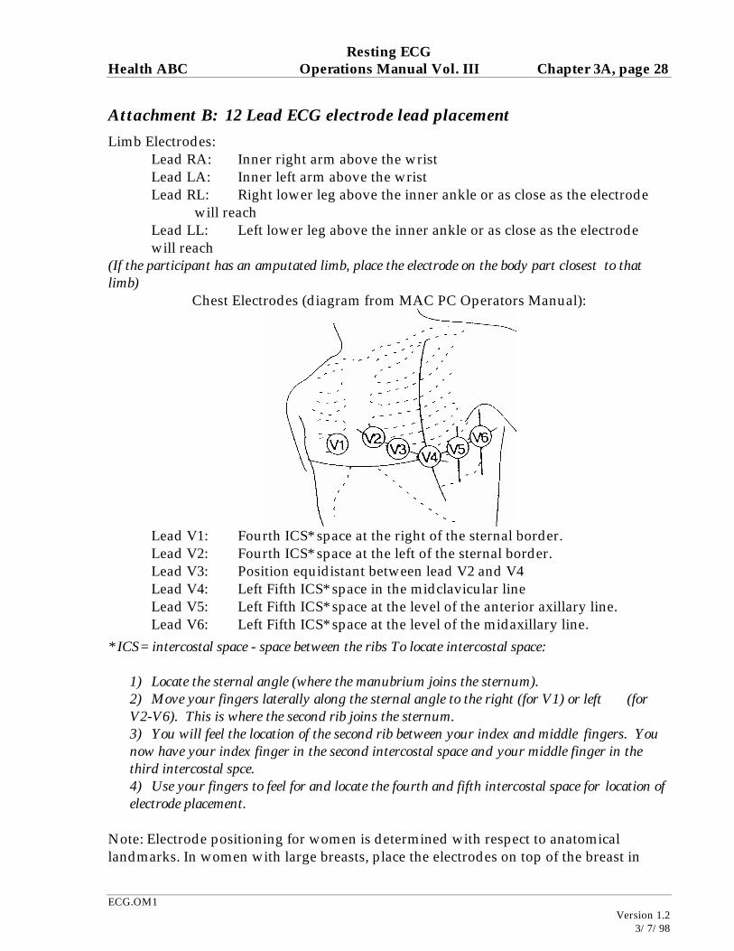
Attachment B: 12 Lead ECG electrode lead placement
Limb Electrodes: Lead RA: Inner right arm above the wrist
Lead LA: Inner left arm above the wrist Lead RL: Right lower leg above the inner ankle or as close as the electrode will reach Lead LL: Left lower leg above the inner ankle or as close as the electrode will reach (If the participant has an amputated limb, place the electrode on the body part closest to that limb)
Chest Electrodes (diagram from MAC PC Operators Manual):
Lead V1: Fourth ICS* space at the right of the sternal border. Lead V2: Fourth ICS* space at the left of the sternal border. Lead V3: Position equidistant between lead V2 and V4 Lead V4: Left Fifth ICS* space in the midclavicular line Lead V5: Left Fifth ICS* space at the level of the anterior axillary line. Lead V6: Left Fifth ICS* space at the level of the midaxillary line. * ICS= intercostal space - space between the ribs To locate intercostal space:
1) Locate the sternal angle (where the manubrium joins the sternum). 2) Move your fingers laterally along the sternal angle to the right (for V1) or left (for V2-V6). This is where the second rib joins the sternum. 3) You will feel the location of the second rib between your index and middle fingers. You now have your index finger in the second intercostal space and your middle finger in the third intercostal spce. 4) Use your fingers to feel for and locate the fourth and fifth intercostal space for location of electrode placement.
Note: Electrode positioning for women is determined with respect to anatomical landmarks. In women with large breasts, place the electrodes on top of the breast in
Resting ECG Health ABC Operations Manual Vol. III Chapter 3A, page 29
ECG.OM1
Version 1.2 3/7/98
their natural position when supine. Do not move the breast upwards or laterally to place the electrodes under the breast.
Resting ECG Health ABC Operations Manual Vol. III Chapter 3A, page 30
ECG.OM1
Version 1.2 3/7/98
Attachment C: ECG Log The purpose of the ECG Log is to record each Health ABC ECG performed, transmitted, and received by the Core ECG Lab. Use this log as a guide to:
• Select ECGs to transmit (and which ECGs need to be deleted before transmission)
• Crosscheck the directory of ECGs to be transmitted to the CORE ECG Lab before transmission
• Confirm successful transmission of the ECG from the confirmation of receipt report sent from the CORE ECG Lab
• Select ECGs to delete following verification of successful transmission from the Core ECG Lab.
• Record ECGs not available or sent in hardcopy to the Core ECG Lab
Date Time Patient Name code
Patient ID Number
ECG category
ECG available for transmission Y/N/Miss
Date ECG transmitted
Date CEL confirmed receipt
Delete ECG after verify receipt (Y/N)
Comments
Resting ECG Health ABC Operations Manual Vol. III Chapter 3A, page 31
ECG.OM1
Version 1.2 3/7/98
Attachment D: Unavailable ECG Form
Use this form to submit ECGs that are either:
Unavailable for telephone transmission or missing and cannot be obtained
Date
Field Center
Patient name code: Patient ID number
ECG category: (circle) Exam 1 Exam 4 Exam 7
Is the ECG available in hardcopy? (Circle) YES / NO
If the ECG is available in hardcopy, the ECG date is the ECG time is
digitalis: yes/ no
reason the ECG cannot be sent via telephone
Is the ECG Missing and can not be retrieved? (Circle) YES / NO