7-1 HEART AND CIRCULATORY SYSTEM II 7. Dr. Daphne T. Hsu Children’s Hospital of New York , Room 229 North [email protected]212-305-6575 RECOMMENDED READING: Larsen Human Embryology, 3rd Edition, pp. 169-187; 189- 191; 199-204, 222-226. SUMMARY: During this lecture we will continue to describe the development of the fetal heart. Topics will include septation of the ventricles, formation of the atrioventricular valves, and formation of the cardiac outflow tracts. The derivation of the great vessels leading out of the heart will also be described briefly. We will see the course of the fetal circulation, whose task is to pump and maintain the flow of blood from the placenta through the fetal body and back to the placenta where wastes can be exchanged for nutrients and oxygen. The structural and functional changes in the cardiovascular system that occur at birth when the placental circulation is abruptly interrupted and breathing begins will also be discussed. Finally, several congenital cardiac malformations will be discussed from the perspective of abnormal cardiac development. GLOSSARY: Aortic arches: paired arteries surrounding the pharynx; portions will contribute to formation of the great vessels Aorticopulmonary septum: the portion of the conotruncal septum that divides the truncus arteriosus into the ascending aorta and the pulmonary trunk Bulbar ridges: another name for the truncoconal ridges or conus swellings that ultimately separate the conotrucal outflow tract Conotruncal septum: the septum that divides the conus cordis into the outflow tracts (infundibulum of the right ventricle [conus arteriosus] and aortic vestibule) as well as the truncus arteriosus Ductus arteriosus: shunts blood from the left pulmonary artery to the descending aorta, by-passing the lungs Ductus venosus: shunts most of the blood in the umbilical vein into the inferior vena cava (IVC) (by-passes the liver sinusoids) Membranous interventricular septum: outgrowth of tissue from the inferior endocardial cushion, which fuses with the conus swellings and the muscular septum and closes the foramen between the right and left ventricles Semilunar valves: the aortic and pulmonary valves
Transcript
7-1
HEART AND CIRCULATORY SYSTEM II7.
Dr. Daphne T. HsuChildren’s Hospital of New York , Room 229 [email protected]
RECOMMENDED READING: Larsen Human Embryology, 3rd Edition, pp. 169-187; 189-191; 199-204, 222-226.
SUMMARY:
During this lecture we will continue to describe the development of the fetal heart. Topics willinclude septation of the ventricles, formation of the atrioventricular valves, and formation of thecardiac outflow tracts. The derivation of the great vessels leading out of the heart will also bedescribed briefly. We will see the course of the fetal circulation, whose task is to pump andmaintain the flow of blood from the placenta through the fetal body and back to the placenta wherewastes can be exchanged for nutrients and oxygen. The structural and functional changes in thecardiovascular system that occur at birth when the placental circulation is abruptly interrupted andbreathing begins will also be discussed. Finally, several congenital cardiac malformations will bediscussed from the perspective of abnormal cardiac development.
GLOSSARY:
Aortic arches: paired arteries surrounding the pharynx; portions will contribute to formation ofthe great vesselsAorticopulmonary septum: the portion of the conotruncal septum that divides the truncus arteriosus into theascending aorta and the pulmonary trunkBulbar ridges: another name for the truncoconal ridges or conus swellings that ultimately separate theconotrucal outflow tractConotruncal septum: the septum that divides the conus cordis into the outflow tracts (infundibulum of the rightventricle [conus arteriosus] and aortic vestibule) as well as the truncus arteriosusDuctus arteriosus: shunts blood from the left pulmonary artery to the descending aorta, by-passing the lungsDuctus venosus: shunts most of the blood in the umbilical vein into the inferior vena cava (IVC) (by-passesthe liver sinusoids)Membranous interventricular septum: outgrowth of tissue from the inferior endocardial cushion, whichfuses with the conus swellings and the muscular septum and closes the foramen between the right andleft ventriclesSemilunar valves: the aortic and pulmonary valves
7-2
LEARNING OBJECTIVES:
The student should be able to:1) Discuss the formation and remodeling of the primitive embryonic ventricles and their
respective outflow tracts.2) Know the derivation of the great vessels.3) Explain the structural and functional design of the fetal circulation.4) Discuss origin of some well known cardiac malformations.
REVIEW: (Figs. 7-1, 7-2) The single common atrium is incompletely divided into a right and leftatrium. The superior vena cava as well as the coronary sinus deliver blood to the right atrium andthe pulmonary veins enter the left atrium. Oxygenated blood from the placenta is transported in theinferior vena cava (IVC) to the right atrium. The primitive ventricle gives rise to the left ventricle;the proximal portion of the bulbus cordis gives rise to the right ventricle. Blood flows from thesinus venosus into the common atrium. It passes through the common atrioventricular (AV) canalinto the primitive ventricle, through the bulbus cordis (primitive right ventricle, conus cordis andtruncus arteriosus), into the aortic sac, and is then distributed to cranial structures and dorsal aortaevia the aortic arches. After the AV canal expands and shifts to the right, it communicates with bothventricles. When the AV endocardial cushions (EC) fuse, the left AV canal opens into the primitiveleft ventricle and the right AV canal opens into the primitive right ventricle. The muscularinterventricular (IV) septum incompletely separates the 2 ventricles. Blood flows from the left to
Fig. 7-1. Frontal section through the heart of a 6-mmembryo showing the primary interventricular foramen andthe entrance of the atrium into the primitive left ventricle.Note the bulboventricular flange. Arrows indicatedirection of blood flow.
Fig. 7-2. Fronted section through the heart of a 9-mm embryo.At this stage of development blood from the atrial cavity entersthe left primitive ventricle as well as the right primitive ventricle.Note the development of the cushions in the A-V canal. Theswellings in the truncus and conus are clearly visible. The ringindicates the primitive interventricular foramen. Arrows indicateblood flow.
7-3
the right ventricle through the primary IV foramen. The conus cordis and truncus arteriosus shift tothe left. Both ventricles now have access to the truncus arteriosus by way of the conus cordis (Fig 7-3).The following events will be described separately although they occur concurrently.
Fig. 7-3. A, B, Initial septation of the ventricles. The muscular interventricular septum enlarges in the region of theinterventricular sulcus between weeks 4 and 7 (Larsen p.174, Fig 7-18).
I. Formation of the muscular portion of the interventricular (IV) septum (Fig. 7-3). At theend of 4th week the two primitive ventricles begin to dilate. The myocardium grows on theoutside and diverticulates and trabeculates on the inside. The medial walls form an incompletemuscular (primary) interventricular septum. The ventricles communicate with each otherthrough the interventricular foramen.
II. Formation of the atrioventricular valves. The atrioventricular valves begin to formbetween the fifth and eighth week. Undermining of the myocardium surrounding the right andleft atrioventricular canals forms the leaflets or cusps (Fig. 7-4) . The free edge of each leafletis attached to the ventricular walls by thin sinews, the chordae tendineae, which insert intosmall hillocks of myocardium, the papillary muscles (Fig. 7-5).
7-4
III. Partitioning the truncus arteriosus and conus cordis. Formation of the outflow tractsfrom the right and left ventricles.
a) Septum formation in truncus arteriosus. During the 5th week, truncus swellingsappear on opposite walls of the truncus arteriosus (Fig. 7-2). The right truncus swellinggrows superiorly and to the left; the left truncus swelling grows superiorly and to the rightin the direction of the aortic sac. Fusion of the two swellings forms a helical truncusseptum (aorticopulmonary septum) (Fig. 7-6A) that twists 180o counterclockwise anddivides the truncus into an aortic and a pulmonary channel. The aortic channelcommunicates with the 3rd and 4th aortic arches. The pulmonary channel communicateswith the 6th aortic arches (Fig. 7-7B). The subsequent split in the aortico-pulmonaryseptum creates the ascending aorta and pulmonary trunk (Fig. 7-6B).
b) Septum formation in the conus cordis (outflow tracts from the ventricles) andformation of the conotruncal septum. Two conus swellings (Fig. 7-2), also referred to asbulbar ridges (Fig. 7-7B), appear on opposite walls of the conus cordis just inferior tothe truncus swellings. The conus swellings fuse as they grow inferiorly toward themuscular interventricular septum. The conus (bulbar) portion of the conotruncal septumdivides the conus into an anterolateral portion that descends into the right ventricle tobecome the conus arteriosus (infundibulum) (smooth walled portion) of the right
Figs. 7-4, 7-5. Development of the atrioventricular valves,including the papillary muscles, chordae tendineae, and cusps,are sculpted from the muscular walls of the ventricles. Thedefinitive tricuspid valve within the right ventricle is notcompletely formed until the development of a septal cusp inthe third month.
7-5
ventricle, and a posteromedialportion that descends into theleft ventricle forming theaortic vestibule (smoothwalled portion) of the leftventricle (Fig. 7-8).
c) The aortic and pulmonaryvalves (semilunar valves)develop in the inferior portionsof the truncus swellings. Eachvalve is derived from 3tubercles: 2 lateral tubercles thatdivide when the aorta andpulmonary trunk separate and athird tubercle that develops inthe wall in each valve (Fig. 7-9).
IV. Formation of themembranous portion of theinterventricular (IV) septum.At the end of the 4th week themedial walls form anincomplete muscular(primary) interventricularseptum. The ventriclescommunicate with each otherthrough the interventricularforamen. The two primitiveventricles dilate further as aresult of continuous growth ofthe myocardium on the outsideand diverticulation and
Fig. 7-6. (A) Diagram toshow the spiral shape ofthe aortico-pulmonaryseptum. (B) Position ofaorta and pulmonaryartery at 25-mm stage(eighth week).Note howthe aorta and pulmonaryartery twist around eachother.
Fig. 7-7.
Fig. 7-8. (A) Interior of the adult right ventricle. (B) Interior of the adultleft ventricle.
7-6
Fig. 7-10. Development of theconotruncal ridges (swellings) andclosure of the interventricularforamen. Proliferations of the rightand left conus swellings, combinedwith proliferation of the inferiorendocardial cushion, close theinterventricular foramen and formthe membranous portion of theinterventricular septum. A. 6 weeks(12 mm). B. Beginning of theseventh week (14.5 mm). C. End ofthe seventh week (20 mm).
trabecular formation on the inside (Fig. 7-7). The IV foramen, between the free rim of themuscular ventricular septum and the fused EC, permits communication between the 2ventricles. When the two conus swellings fuse this space is reduced (Fig. 7-7B and Fig. 7-10B). It is obliterated by an outgrowth of tissue from the inferior EC forming the membranousportion of the IV septum (Fig. 7-10B, C).
V. Development of the great arteries. Derivatives of the 3rd, 4th and 6th aortic arches (Figs. 7-11and 7-12). The 3rd arch participates in formation of the carotid arteries; the 4th forms the
Fig. 7-9. Formation of thesemilunar valves in the outflowtract of the heart. Neural crest cells(green areas) may contribute in partto the formation of the valvularleaflets.
7-7
aortic arch on the left side and a portion of the subclavian on right; the 6th arch forms theproximal parts of pulmonary arteries and the ductus arteriosus, a fetal vessel that shuntsblood from the pulmonary trunk to the descending aorta, bypassing the fetal lungs (Fig. 7-13).The ductus arteriosus closes after birth when the lungs become functional.
Figs. 7-11, 7-12. Development of the aortic system.
Fig. 7-13.
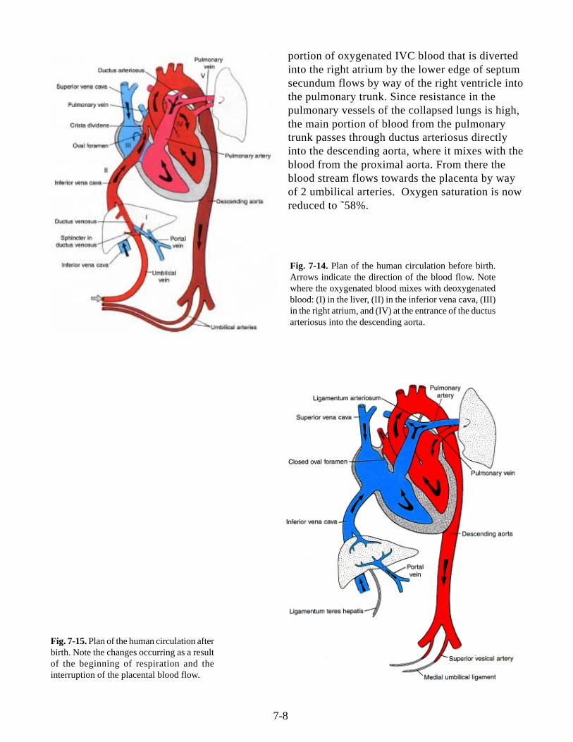
VI. Fetal circulation (Fig. 7-14).a) Oxygenated blood from placenta, (˜80%saturated), is delivered to the heart by way of theumbilical vein. The main portion of blood flowsthrough ductus venosus into the IVC. A smallerportion enters the liver sinusoids and mixes withblood from the portal circulation. A functionalsphincter (not observed anatomically) is thought toregulate blood flow through the liver sinusoids.b) In the IVC, the still highly oxygenated bloodmixes with a small amount of deoxygenated bloodfrom the lower limbs and enters the right atrium.Most of it passes from the right atrium, through theforamen ovale, to the left atrium where it mixeswith a small amount of blood returning from thelungs. From the left atrium, blood enters the leftventricle and ascending aorta. Since the coronaryand carotid arteries are the first branches of theascending aorta, the heart musculature and the brainreceive well oxygenated blood. Blood in thedescending aorta is delivered to the trunk and lowerlimbs.c) Desaturated blood from the SVC and a small
7-8
portion of oxygenated IVC blood that is divertedinto the right atrium by the lower edge of septumsecundum flows by way of the right ventricle intothe pulmonary trunk. Since resistance in thepulmonary vessels of the collapsed lungs is high,the main portion of blood from the pulmonarytrunk passes through ductus arteriosus directlyinto the descending aorta, where it mixes with theblood from the proximal aorta. From there theblood stream flows towards the placenta by wayof 2 umbilical arteries. Oxygen saturation is nowreduced to ˜58%.
Fig. 7-14. Plan of the human circulation before birth.Arrows indicate the direction of the blood flow. Notewhere the oxygenated blood mixes with deoxygenatedblood: (I) in the liver, (II) in the inferior vena cava, (III)in the right atrium, and (IV) at the entrance of the ductusarteriosus into the descending aorta.
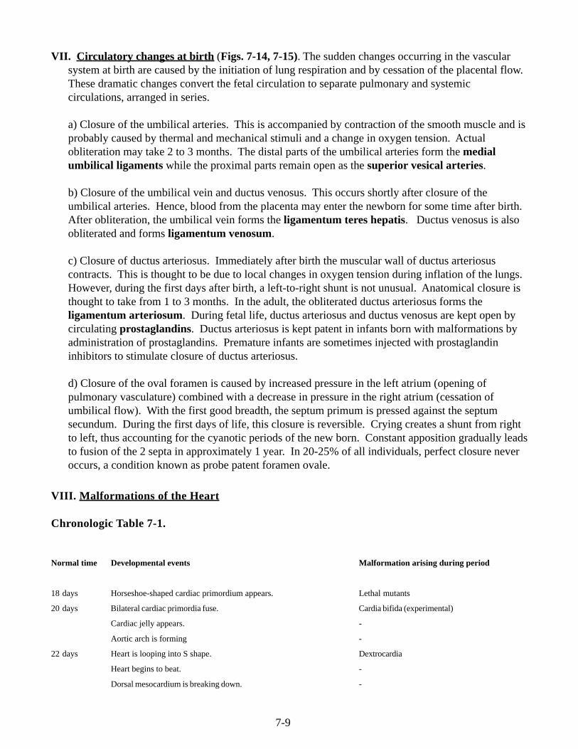
Fig. 7-15. Plan of the human circulation afterbirth. Note the changes occurring as a resultof the beginning of respiration and theinterruption of the placental blood flow.
7-9
VII. Circulatory changes at birth (Figs. 7-14, 7-15). The sudden changes occurring in the vascularsystem at birth are caused by the initiation of lung respiration and by cessation of the placental flow.These dramatic changes convert the fetal circulation to separate pulmonary and systemiccirculations, arranged in series.
a) Closure of the umbilical arteries. This is accompanied by contraction of the smooth muscle and isprobably caused by thermal and mechanical stimuli and a change in oxygen tension. Actualobliteration may take 2 to 3 months. The distal parts of the umbilical arteries form the medialumbilical ligaments while the proximal parts remain open as the superior vesical arteries.
b) Closure of the umbilical vein and ductus venosus. This occurs shortly after closure of theumbilical arteries. Hence, blood from the placenta may enter the newborn for some time after birth.After obliteration, the umbilical vein forms the ligamentum teres hepatis. Ductus venosus is alsoobliterated and forms ligamentum venosum.
c) Closure of ductus arteriosus. Immediately after birth the muscular wall of ductus arteriosuscontracts. This is thought to be due to local changes in oxygen tension during inflation of the lungs.However, during the first days after birth, a left-to-right shunt is not unusual. Anatomical closure isthought to take from 1 to 3 months. In the adult, the obliterated ductus arteriosus forms theligamentum arteriosum. During fetal life, ductus arteriosus and ductus venosus are kept open bycirculating prostaglandins. Ductus arteriosus is kept patent in infants born with malformations byadministration of prostaglandins. Premature infants are sometimes injected with prostaglandininhibitors to stimulate closure of ductus arteriosus.
d) Closure of the oval foramen is caused by increased pressure in the left atrium (opening ofpulmonary vasculature) combined with a decrease in pressure in the right atrium (cessation ofumbilical flow). With the first good breadth, the septum primum is pressed against the septumsecundum. During the first days of life, this closure is reversible. Crying creates a shunt from rightto left, thus accounting for the cyanotic periods of the new born. Constant apposition gradually leadsto fusion of the 2 septa in approximately 1 year. In 20-25% of all individuals, perfect closure neveroccurs, a condition known as probe patent foramen ovale.
VIII. Malformations of the Heart
Chronologic Table 7-1.
Normal time Developmental events Malformation arising during period
18 days Horseshoe-shaped cardiac primordium appears. Lethal mutants
20 days Bilateral cardiac primordia fuse. Cardia bifida (experimental)
Cardiac jelly appears. -
Aortic arch is forming -
22 days Heart is looping into S shape. Dextrocardia
Heart begins to beat. -
Dorsal mesocardium is breaking down. -
7-10
Aortic arches I and II are forming. -
24 days Atria are beginning to bulge. -
Right and left ventricles act like two pumps in series. -
Outflow tract is distinguishable from right ventricle. -
Late fourth Sinus venosus is becoming incorporated into right atrium. Venous inflow malformations
Early septum I appears between left and right atria. Common atrium
Muscular interventricular septum is forming. Common ventricle
Truncoconal ridges are forming. Persistent truncus arteriosus
Aortic arch I is regressing. -
Aortic arch III is formed. -
Aortic arch IV is forming. -
Early fifth Endocardial cushions are coming together, forming right and Persistent atrioventricular canal
week left atrioventricular canals.
Further growth of interatrial septum I and muscular interven- Muscular ventricular septal defects
tricular septum occurs.
Truncus arteriosus is dividing into aorta and pulmonary artery. Transposition of great vessels
Aortic and pulmonary stenosis or atresia
Atrioventricular bundle is forming; there is possible neurogenic -
control of heart beat.
Pulmonary veins are becoming incorporated into left atrium. Aberrant pulmonary drainage
Aortic arches I and II have regressed. -
Aotic arches III and IV have formed. -
Aortic arch VI is forming. -
Late fifth to Endocardial cushions fuse. -
early sixth Interatrial foramen II is forming. -
week Interatrial septum I is almost contacting endocardial cushion. Low atrial septal defects
Membranous part of interventricular septum starts to form. Membranous interventricular septal defects
Semilunar valves begin to form. Aortic and pulmonary valvular stenosis
Late sixth Interatrial foramen II is large. High atrial septal defects
week Interatrial septum II starts to form. -
Atrioventricular valves and papillary muscles are forming. Tricuspid or mitral valvular stenosis or atresia
Interventricular septum is almost complete. Membranous interventricular septal defects
Coronary circulation is becoming established. -
Eighth to Membranous part of interventricular septum is completed. Membranous interventricular septal defects
ninth week
a) Normal Heart: In the normal adult heart (Fig. 7-15) blood from the inferior vena cava(IVC) and superior vena cava (SVC) enters the right atrium, traverses the tricuspid valve,and is pumped from the right ventricle through the pulmonary trunk (regulated by thepulmonary valve) to the lungs. It then passes to the left atrium via the pulmonary veins,through the mitral valve to the left ventricle, and is pumped into the ascending aorta
7-11
Fig. 7-16. (A) Normal atrial septum formation. (B and C) Ostium secundum defect caused by excessive resorption of theseptum primum. (D and E) Similar defect caused by failure of development of the septum secundum. (F) Common atrium orcor trilocular biventriculare—complete failure of the septum primum and septum secundum to form.
(regulated by the aortic valve). Considering the complicated formation and critical timingfor the correct formation and alignment of multiple cardiac structures, it is not surprisingthat defects arise. A few examples will be discussed during the lecture. Thesemalformations are also described in Larsen.
b) Malformation of cardiac looping: Abnormalities in cardiac looping often result inincomplete formation of one of the ventricles. This leads to a functionally univentricularheart.
c) Malformations of atrial septation (Fig. 7-16). (i) Ostium secundum defect(ASD):excessive resorption of septum primum. (ii) Septum secundum fails to develop. (iii)Complete absence of the atrial septum; both septum primum and septum secundum fail todevelop.
d) Malformations of ventricular septation (Figs. 7-17 and 7-18). Ventricular septal defect(VSD): Isolated defect in membranous interventricular septum: Blood from the leftventricle flows to the right through an open ventricular foramen.
7-12
Interventricular defects.Fig. 7-18.
Fig. 7-19. Tetralogy of Fallot.
e) Malformations of conotruncal septation (Fig. 7-19). Tetralogy of Fallot (TOF): Unequaldivision of the conus caused by anterior displacement of the conotruncal septum results instenosis of the pulmonary trunk, hypertrophy of the right ventricle, overriding aorta,interventricular septal defect.
Fig. 7-17
7-13
Fig. 7-21. Persistent Truncus Arteriosus.
Fig. 7-22. Separation of the truncus arteriosus into thepulmonary artery and aorta. The truncoconal septa(between the aorta and the pulmonary trunk) forms fromthe cells of the cardiac neural crest. (A) Human cardiacneural crest cells migrate to pharyngeal arches 4 and 6during the fifth week of gestation and enter the truncusarteriosus to generate the septa. (B) Quail cardiac crestcells were transplanted into the analogous region of a chickembryo, and the embryos were allowed to develop. Thequail cardiac neural crest cells can be recognized by a quail-specific antibody, which stains them darkly. In the heart,these cells can be seen separating the truncus arteriosus(right) into the pulmonary artery and the aorta (left). (Aafter Kirby and Waldo 1990; B from Waldo et al. 1998,photographs courtesy of K. Waldo and M. L. Kirby.)
f) Failure of conotruncal septum to spiral (Fig.7-20). Transposition of the Great Arteries(TGA): Conotruncal septum fails to spiral and instead descends straight downward. Theaorta originates from the right ventricle and the pulmonary artery from the left.
g) Absence of the conotruncal septum (Fig. 7-21).Persistent Truncus Arteriosus: The pulmonaryartery originates from the common truncusarteriosus. Accompanied by a ventricular septaldefect, the undivided outflow tract overrides bothventricles. Neural crest cells participate information of the conotruncal septum (Fig. 7-22).The crest cells migrate through pharyngeal arches4, and 6, and invade the conotruncal swellings.Persistent truncus arteriosus can be experimentallyreproduced in chicks by ablating the cardiac neuralcrest.