98
HEAT SHOCK PROTEINS
| Date post: | 12-Apr-2017 |
| Category: |
Health & Medicine |
| Upload: | dr-shahar-bano-khanzoya-khan |
| View: | 819 times |
| Download: | 2 times |

HEAT SHOCK PROTEINS

What is Protein folding ?

Structural Levels of Proteins
Primary Secondary


Protein stability The situation becomes more complicated since this stability must be ascertained in a certain range of environmental conditions
ProteinpH Temp.
Salt Conc.
a certain range of environmental conditions.The native conformation of a protein is stable in a narrow range of temperature, pH, chemical composition of solvent, etc.

• Hydrogen Bonding• Vander Waals interactions • Ionic strengths• Disulfide bonds Hydrophobicity: the
dominant force in protein folding
Forces involved in Protein stabilisation

A stably folded proteins has…..
Hydrophobic side chains buried Charged side chains on the surface Cysteine’s form Covalent disulfide bonds
“All these features will contribute to Minimum” energy state
Pack as close together as possible Minimize contacts between hydrophobic groups and water

Mutations Premature termination of Translation Fault in post-translational modifications Strong Promoters High Inducer concentrations
Reasons for protein misfolding
Loss of conformation due to stress`

ALL CELLS

ALL ORGANISMS

Living in the World

Must cope with…

Stress !!!
What is stress?
In biology, stress is the driving force behind the process of adaptation and evolution.

Tempenviron
Tempcell
Folded Proteins
Unfolded
Proteins
Aggregates
Loss of ProteinFunction
Networkfailure
Death
Cell

Interesting storyF. Ritossa –1960 discovered the heat shock (HS) response while observing the salivary cells of Drosophila and named them HSP’s
My name is Chaperone

Cover and back image Conceptual image depicting Hsps as protein-folding chaperones in the mitochondria. As nascent mitochondrial peptides (light blue) emerge from the ribosome (purple), they are bound by the mitochondrial Hsp70 (orange) via its peptide binding domain (revealed schematic ribbon structure) to prevent mis folding and aggregation. Some proteins require further folding assistance by the Hsp60/Hsp10 (yellow) chaperonin complexes. A single Hsp60 subunit of the chaperonin complex is shown as a ribbon structure.
HEAT SHOCK PROTEINS
Heat shock proteins are a group of highly conserved proteins found in both eukaryotic and prokaryotic cells. They are involved in a wide range of cellular processes such as assisting protein folding and degradation of misfolded proteins, modulating signaling pathways and regulating immune responses.
The multi-functional nature of heat shock proteins enables them to play critical roles in the regulation of protein homeostasis and cell survival.
DEFINITION

How do HSPs work?One major function of chaperones is to prevent both newly
synthesised polypeptide chains and assembled subunits from aggregating into non functional structures
High temperatures and other stresses, such as altered pH and oxygen deprivation, make it more difficult for proteins to form their proper structures and cause some already structured proteins to unfold
Heat Shock Proteins are induced rapidly at high levels to deal with this problem

MASTER REGULATOR OF HEAT SHOCK PROTEIN EXPRESSION
INACTIVE STATE-HSF1 monomers are held in complex with Hsp 70/hsp 90
ACTIVE STATE-That is on the onset of stress, 5 MAJOR steps occur-
1.Hsf 1 is released from the complex2.It homo trimerizes3.Translocates to nucleus4.And activates the transcription of its downstream targets i.e Hsp 72 and Hsp 275.By binding to the HSE(heat shock elements) in the promoter regions of target proteins.
Heat shock factor 1


Different Types of Heat Shock Proteins
Heat Shock Proteins are classified by their molecular weight, size, structure, and function. They are divided into several families, namely - 1. HSP100 2. HSP90 3. HSP70 4. HSP60 (chaperonin) 5. Small Heat Shock Proteins/ (alpha)-crystalline proteins

Hsp 100
Hsp 90
Hsp 60
Hsp 70
SmallHsp’s
Hsp 40
Family Major Functions
Stress tolerance, Protein disaggregation, thermo tolerance
Regulatory interactions with signaling proteins, stabilization of misfolded proteins
Protein folding, membrane transport of proteins,Auto regulation in heat shock response, anti apoptotic
Protein folding (limited substrates in eukaryotic cytoplasm)
Protein folding, co-chaperone for Hsp70
Stabilization of misfolded proteins, thermotolerance,eye lens structural proteins
Functions of HSP families

Why Don't Heat Shock Proteins Denature?
Better Hydrogen BondsBetter Hydrophobic Internal PackingEnhanced Secondary StructureHelix Dipole Stabilization

All organisms exhibit homeostatic-like responses when subjected to rapid changes in their environment.
The ability of the organism to successfully adapt or acclimate to its new environment is critical to its survival, and likely represents an integral driving force in evolution.
One well studied response to sudden adverse environmental changes is the so-called heat shock or stress response.
INTRODUCTION

The resultant increase and accumulation of the Hsps now gives the stressed cell added protection, thereby allowing for continued cell survival. In addition to increased temperatures,
other insults also result in increased Hsp expression.
These include exposure of cells to various metals, amino acid analogues, hypoxia, and a large number of agents/treatments which result in reduced ATP levels. Because so many adverse conditions lead to increased Hsp expression, the heat shock response now is commonly referred to as the “stress response”.

HSP 90
stabilizes proteins prior to complete folding or activationforms stable complexes with inactive glucocorticoid receptor and other transcription factorsmost abundant non-ribosomal protein (cytosolic version)most abundant protein in endoplasmic reticulum (ER version) Represents almost 1% of total cellular proteins in unstressed cells.Bacterial homologue: HTpG family (typically non essential proteins) , A functional Hsp90 is required for viability under all conditions in eukaryotic cells.
Dimer ATP dependent HoP and p23

HSP90 interacts with HSP40, HSC70/HSP90 organizing protein(HOP), and co-chaperones to bind and stabilize newly synthesized substrate/client proteins. This ATPregulatedcycle of substrate binding is critical to the activation of many oncogenic signaling molecules.

Hsp 90a Cytosolic form, induced by elevated temperature; ATPase activity; three splice variants
Hsp 90 b Cytosolic form, constitutively expressed; ATPase activity; three splice variants
p23 Binds to telomerase and progesterone receptor; also functions as a cytosolic prostaglandin E2 synthase; phosphorylated at Ser113, 118, 148 and 151 and acetylated at Lys33
P50/cdc37 A chaperone that binds Hsp90 and is required for the activity of numerous protein kinases
TRAP1 Mitochondrial form; has ATPase activity that is inhibited by both geldanamycin and radicicol; highly conserved through evolution; phosphorylation by PINK1 prevents oxidative-stressinduced apoptosis; four splice variants
Grp 94 ER form; glucose regulated and induced by glucose starvation; participates in protein folding and assembly, protein secretion, protecting cells from apoptosis, and mediating immunogenicity; C-terminal sequence KDEL mediates retention in the ER; two splice variants
Members of hsp 90 family

Hsp90 is involved in the regulation of many of the “hallmarks of cancer”.
namely sustaining proliferative signalling, resisting cell death, evading growth suppressors, inducing angiogenesis, enabling replicative immortality, invasion and metastasis, and emerging hallmarks including deregulating cellular energetic and avoiding immune destruction .
In addition, high expression of Hsp90 is an independent prognostic marker in a number of cancers.
In breast cancer, it is associated with decreased survival.
in gastric cancer, high Hsp90 expression is linked to poor prognosis and tumour aggressiveness .
In CML, Hsp90 correlates with disease state and high levels are associated with resistance to therapy


Hsp90 proteins exist as homodimers of subunits consisting of an N-terminal ATPase domain, a C-terminal dimerisation / protein interaction domain, and a middle domain associated with client protein binding. ATPase activity is essential for the chaperoning activity of Hsp90. Following the addition of ATP, Hsp90 undergoes a conformational change, which induces an open to shut conformation shift , with transient dimerisation of the N-terminal domains and N-M domain association. The middle segment of Hsp90 has been identified as the binding site for protein kinase PKB/Akt and is implicated as the main site for client protein interactions . This segment can also interact with cochaperones and is required for N-terminal ATPase activity. The C-terminal domain is involved in dimerisation and contains a highly conserved EEVD sequence which is required for the binding of tetratricopeptide repeat (TPR) containing family of cofactors, such as HOP. .

Hsp90 along with one set of its co-chaperones (and the Hsp70/Hsp40 chaperone machinery) binds to and stabilizes steroid hormone receptors in their inactive state within the cytosol.
Upon subsequent binding to the appropriate steroid hormone ligand, the receptor undergoes a conformational change resulting in its acquisition of DNA binding and transcriptional activity.
In a similar scenario, Hsp90 along with another set of co-chaperones binds to and stabilizes the newly synthesized forms of various protein kinases, maintaining them in a folding-competent conformation.
Thus, via its utilization of numerous co-chaperones and ATP, the very abundant Hsp90 chaperone functions in unstressed cells to regulate client proteins important for growth and development.
Altering the levels of Hsp90 (via genetic means or manipulations with Hsp90 inhibitory drugs) leads to rapid alterations in cell signaling pathways and the adaptation of new cellular phenotypes.
Functions of hsp 90

Elevated levels of Hsp70 proteins have been linked with inhibition of apoptosis as well as the resistance of cells to various chemotherapeutic agents.
In addition, numerous studies continue to demonstrate that changes in the levels of the different Hsp70 family members may prove clinically useful for the diagnosis of many important human diseases.
Purified Hsp70 proteins are available for biochemical and immunological studies.

HSP100
Functions -solubilize protein aggregates thereby dissociating them -facilitates proteolysis -essential in yeast for acquired thermo tolerance -essential for yeast prion propagation
6-7 monomer ATP no co-chaperon is required
Members of the Hsp family participate in the early stages of protein synthesis, protein folding, and the transport of newly synthesized proteins from the cytoplasm into different intracellular compartments.
Under conditions of stress, where protein folding/assembly events may be compromised, the increased expression and accumulation of the stress proteins facilitates the ability of cells to both repair and synthesize new proteins to replace those that were damaged after the particular metabolic insult.

Various medical conditions, including fever, ischemia, hemodynamic overload or neurological injuries are well known activators of the stress response in vivo.
The ability of the affected tissue or organ to mount a robust stress response is thought to be important for its survival and recovery.
In infectious diseases, stress proteins present within different pathogens are known to be major targets of our immune system.

One of the largest stress protein families Its bacterial counter part Dna K Set of genes for both phage and host DNA
replication and therefore referred to as Dna J and Grp E.
Three above genes work together in facilitating the disassembly of large protein complex necessary for commencement of DNA replication.
Dna K function as a type of “MOLECULAR CROWBAR/DETERGENT”.
Hsp 70/40 chaperone machinery


Dna J co chaperones family facilitates subtrate binding as well as stimulate Dna K and ATP hydrolysis.
Grp E protein facilitates nucleotide exchange reaction needed to allow for new reaction cycle.
Thus through repeated cycles of binding and releasing the Dna K chaperone machinery helps prevent premature folding/ aggregation, therefore facilitating high fidelity protein maturation throughout the cell.
This cycling of DnaK chaperone between the open and closed states is regulated by the co-chaperones DnaJ (or Hsp40) and GrpE (or BAG-1).

1.cytosolic/nuclear Hsp 70 2.Hsc 70/hsp 72 3.Hsp 70/hsp 72 4.Grp 78/Bip –present in lumen of ER 5.Grp 75/MORTALIN – present in mitochondria Bag 1 Bag 2 hip
MEMBERS OF Hsp 70

1.Hsp 40 -co chaperone for Hsc/Hsp 70 2.cytosolic Dna J homologue- co chaperone
for Hsc/Hsp 70 3.Dna J homologue(ER)- Bip 4.Dna J like protein- mortalin/Grp 75 5.hip,hop,CHIP- also influence chaperone
machinery.
Co Chaperons-for Hsp 70 family

Hsp70 has been implicated as a potential autoantigen in MS. In IDDM, the preferential expression of Hsp70 by β cells, but not
α cells, in the islets of Langerhans might be important for the understanding of autoimmune destruction of β cells in this disease.
Autoantibodies to the constitutive form of Hsp70 (Hsc70) have been identified in a proportion of patients with primary biliary cirrhosis (45.7%) and patients with autoimmune hepatitis patients (52.9%), but not in patients with chronic hepatitis B or C infection.
Reactivity to Hsp70 has also been implicated in the induction of disease in toxin induced interstitial nephritis.
Role in diseases

The cytoplasmic Hsp70s regulate the apoptosis pathway at multiple levels, for example,
Hsp70s have been shown to protect Bcl-2 from proteasomal degradation, block Bax translocation to the mitochondria thereby preventing cytochrome c
release bind Apaf-1 and prevent the recruitment of caspase-9 to the apoptosome and to prevent AIF translocation to the nucleus to cause chromatin condensation
and DNA degradation. It is interesting to note that the function of Hsp70s do not always rely on their
ATPase activity, for instance it has been shown that Hsp72 inhibits JNK activation independently of its chaperoning activity.
The Hsp70s also play a protective role against senescence. Hsp72 knock down induces senescence in a variety of cancer cell lines , and Hsp72 controls Her-2-induced senescence by regulating p21 and survivin in a mouse breast tumour model
Evidence also suggests that Hsp70 supports autophagy by maintaining protein homeostasis and supporting cancer cell survival. Hsp70 localises at the autophagosome/ lysosomal membrane compartments and inhibits lysosomal permeabilisation. In addition, Hsp70 participates in chaperone mediated autophagy by delivering target proteins to the lysosome surface receptor LAMP-2A, where it enables their translocation into the lysosomal lumen
Role of hsp 70 in diseases

Evidence also suggests that Hsp70 supports autophagy by maintaining protein homeostasis and supporting cancer cell survival. Hsp70 localises at the autophagosome/ lysosomal membrane compartments and inhibits lysosomal permeabilisation. In addition, Hsp70 participates in chaperone mediated autophagy by delivering target proteins to the lysosome surface receptor LAMP-2A, where it enables their translocation into the lysosomal lumen
Chaperone mediated autophagy

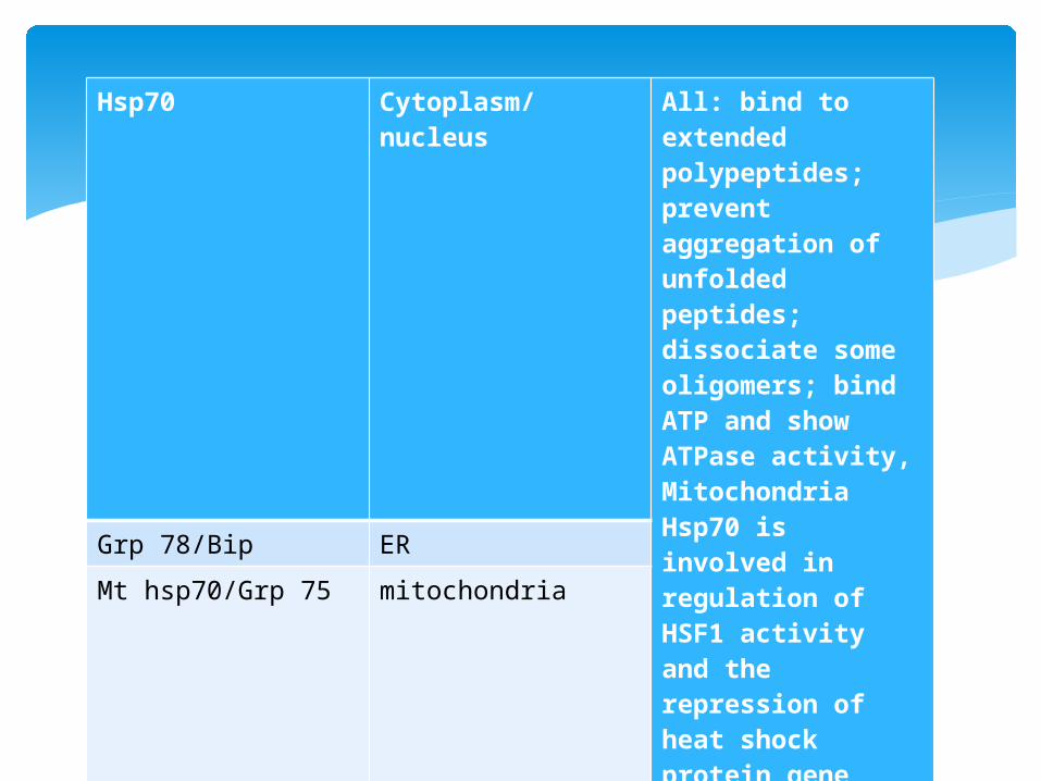
Hsp70 Cytoplasm/nucleus
All: bind to extended polypeptides; prevent aggregation of unfolded peptides; dissociate some oligomers; bind ATP and show ATPase activity, Mitochondria Hsp70 is involved in regulation of HSF1 activity and the repression of heat shock protein gene transcription
Grp 78/Bip ERMt hsp70/Grp 75 mitochondria

Distant relative to Hsp 70 FUNCTION- ACTS AS NUCLEOTIDE exchanger
factor for the cytosolic Hsc/Hsp 70 proteins. As an independent chaperone, unlike Hsp70,
Hsp110 cannot assist protein folding, but acts to prevent protein aggregation of denatured proteins with higher efficiency compared to Hsp70
It also exhibits differential substrate binding properties to Hsp70s with preference for substrates with aromatic residues, and this may account for the different chaperone activities of Hsp110 and Hsp70 .
Hsp 110
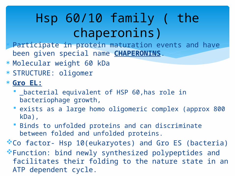
Participate in protein maturation events and have been given special name CHAPERONINS.
Molecular weight 60 kDa STRUCTURE: oligomer Gro EL:
bacterial equivalent of HSP 60,has role in bacteriophage growth,
exists as a large homo oligomeric complex (approx 800 kDa), Binds to unfolded proteins and can discriminate between
folded and unfolded proteins. Co factor- Hsp 10(eukaryotes) and Gro ES (bacteria)Function: bind newly synthesized polypeptides and
facilitates their folding to the nature state in an ATP dependent cycle.
Hsp 60/10 family ( the chaperonins)

Specifically, binding and sequestration of the substrate polypeptide occurs within the large central cavity of the chaperonin complex.
Thereby, reduce the probability of misfolding and aggregation of the target protein with other polypeptides.
GroEL proteins from different pathogens elicit strong humoral and cellular immune responses.
Finally, chaperonins are now proving useful as it leads to the in vitro folding of recombinant proteins important for clinical medicine and therapeutic purposes.
Functions

3 domains of hsp 60: 1. apical domain : 2. equatorial domain :binding site for ATP 3.intermediate domain : binds equatorial domain and
apical domain induces conformational change when ATP is bound allowing for an alternation between hydrophilic and hydrophobic substrate binding sites.In inactive state, the protein is in a hydrophobic state.When activated by ATP-intermediate domain undergoes conformational change and exposes the hydrophilic region.Hsp 10-acts like a lid or dome like cover on the ATP active form of HSP60.this causes the central cavity to enlarge and helps in protein folding.
Structure of hsp 60

Hsp60/Hsp10 and TRiC/CCT chaperonins. The group I (mitochondrial) and group II (cytosolic) chaperones are large oligomeric complexes, Hsp60/Hsp10 and TriC/ CCT, involved in ATP-dependent folding of client proteins such as aconitase and actin, respectively.

Most notable is the bacterial GroEL protein, the so called “common antigen,” which elicits both a strong humoral and cellular immune response whenever animals are infected with different microbes.
Oligomer formed by monomers. Monomers are arranged in 2 stacked
heptameric rings These rings form a barrel like cavity. In this cavity misfolded/unfolded substrate
proteins are folded. GroES(hsp 10),forms a single heptameric
ring,acts as a lid to the chamber and can bind to either end of double GroEL rings.
GroEL protein

A) Reconstruction of the GroEL structure with and without the GroES™lid∫ from cryoelectron microscopy pictures.B) Model of the GroEL chaperone cycle. Two misfolded proteins (greenand blue) are simultaneously folded in a phase-shifted manner. The red circlessymbolize the hydrophobic substrate binding sites of GroEL

ATP and polypeptide binds to one GroEL ring followed by GroES capping, resulting in the
encapsulation of polypeptide in a hydrophilic cavity which promotes protein folding conditions.
Once the substrate is inside the chamber, ATP is hydrolysed slowly, allowing time for the protein to fold.
The two rings of GroEL act in an alternate fashion, with ATP hydrolysis in one ring resulting in a structural transition in the opposite ring making it available for ATP binding.
Which in turn triggers the release of GroES and substrate protein from the original ring
A substrate protein may go through multiple binding and release cycles to reach its folded state.
How GroEL works……


Hsp 60 Mitochondrial protein, essential for folding and assembly of newly imported proteins; also a signaling molecule in the innate immune system; mutations associated with autosomal recessive spastic paraplegia
Hsp 10 Closely linked to the Hsp60 gene (HspD1); forms chaperonin 'cap' structure
Tcp 1 Member of the chaperonin containing TCP1 complex (CCT), also known as the TCP1 ring complex (TRiC), consisting of two identical stacked rings, each containing eight different proteins; the complex folds various proteins, including actin and tubulin, in an ATP-dependent manner; two alternative splice forms; unlike Hsp60, no known associated Hsp10/GroES cofactor
CCT2,CCT3,CCT4,CCT5,CCT6A,CCT6B,CCT7
Components of TRiC complex
Members of chaperonin family

Human Hsp60 activates CD45RA+RO− (naive) T cells, bacterial-specific peptides activate CD45RA−RO+ (memory) T cells and bacterial Hsp60 activates both CD45RA+RO− and CD45RA−RO+ T cells . The observation that both types of T-cell subset are activated by bacterial Hsp60 indicates that T cells can recognise and respond to conserved (self) epitopes on the whole bacterial molecule
Hsp 60 as immuno modulators and intercellular signaling molecules

Hsp60 is expressed in the synovial tissue of patients with rheumatoid arthritis (RA) and juvenile chronic arthritis , and T cells derived from the synovial fluid are activated by mycobacterial Hsp65 .
T-cell reactivity to self-Hsp60 has been reported in patients with RA immortalised B cells from the synovial tissue of RA patients show
specificity for bacterial Hsp60 and elevated levels of circulating antibodies to Hsp60 are present in children with juvenile chronic arthritis
. T-cell-mediated responses to mycobacterial Hsp65 have also been implicated in experimental models of arthritis, and disease can be initiated in rats by the transfer of T-cell clones specific for mycobacterial Hsp65
In addition, antibodies to Hsp65 are elevated in mice with pristane-induced arthritis
Role of hsp 60 in diseases1. rheumatoid arthiritis

Evidence of a role for Hsp60 in type 1 diabetes [insulin-dependent diabetes mellitus (IDDM)] is somewhat equivocal .
Supporting such a role is evidence that naive T cells from non-obese diabetic (NOD) mice can be activated by both self-Hsp60 and mycobacterial Hsp60 ,
that anti-Hsp60 T cells can mediate insulitis and hyperglycaemia in the NOD mouse ,
and that peripheral blood T cells from patients with IDDM demonstrate a heightened proliferative response to human Hsp60 and Hsp60 peptides.
However, in NOD mice, immunity to autoantigens other than heat shock proteins, such as glutamic acid decarboxylase 65 (GAD), appears much earlier than responsiveness to mycobacterial Hsp65 , thereby arguing against an essential role for heat shock proteins in disease induction in this model.
In addition, no evidence for serological immunity to islet cell heat shock proteins has been reported in IDDM .
2. Diabetes mellitus

Evidence of a role for Hsp60 in the pathogenesis of multiple sclerosis (MS) is less apparent.
Hsp60 expression has been identified in chronic MS plaques , and a humoral response to Hsp60 has been detected in the cerebrospinal fluid of patients with MS;
however, the latter is not specific for MS and is also present in a number of chronic degenerative conditions .
Nor is peripheral blood lymphocyte reactivity to Hsp60 altered in MS patients.
3. Multiple sclerosis

sHsp most widespreadMost conserved8-24 monomer
exhibit chaperone activity in vitro
thermo protection in vivo
produced at significant levels in cells experiencing heat stress
most are heat inducible, but some are synthesized in unstressed
conditions-such as for cell development.
Mol wt: 14-45 kDa with most in the 20 kDa range
While bacteria and single-cell eukaryotes express only one or two
members, Drosophila melanogaster expresses 16, humans 10, and
plants as many as 19

Denatured or unfolded substrates bind to the hydrophilic surface of small HSP complexes and prevent the substrate from aggregating.The substrate either stays sequestered or is released to be refolded or degraded.

The best characterized member of the family, a-crystallin is abundant in the lens where the overall protein concentration is quite high.
In such a crowded environment the a-crystallins (A and B) are thought to help prevent protein aggregation resulting from light damage and/or other metabolic insults.
Similarly, other members of the low molecular weight Hsps are now thought to function as ATP-independent molecular chaperones.
Via their large surface and potential to recognize and bind exposed hydrophobic patches, Hsp27 and its counterparts may act to bind unfolded proteins and then present their substrates to the other ATPdependent molecular chaperone machineries (e.g. Hsp60, Hsp70 or Hsp90) for subsequent re-folding
Members of small hsp family

Functions: to prevent protein aggregation by directly binding misfolded substrates,promoting protein refolding by interaction with the Hsp70 chaperone
complex. In addition, Hsp27 can directly prevent cell death by interfering
with key components of the apoptosis pathway, such as blocking the formation of the apoptosome by binding to cytochrome c released from the mitochondria and by interacting with Daxx, a mediator of Fas-induced apoptosis
Under stress conditions, Hsp27 is also directly involved in the ubiquitin-proteasome pathway by binding to the 26S proteasome and multi-ubiquitin chains, to facilitate the degradation of a selective range of target proteins. By doing so, Hsp27 can mediate its cytoprotective effect at multiple levels by facilitating the degradation of various apoptotic and cell cycle proteins. For example,
Hsp27 can enhance the anti-apoptotic activtity of the transcription factor NF-κB, as the presence of Hsp27 in the proteasome-protein substrate complex is required for the degradation of I-κBα, the inhibitor of NF-κB.
Hsp27 can also promote the degradation of the cell cycle inhibitor p27, thereby avoiding cell cycle arrest during stress.
Hsp 27
sequence homology with the a-crystalline proteins;
In the case of the human low molecular weight Hsp, collectively termed the Hsp27 family, the proteins are found in complexes of 400 to 500 k Da.
Phosphorylation of Hsp27, in response to different stimuli, may play a role in the oligomeric dynamics of the protein.

Crystallin alpha-A
Expression restricted to the lens
Crystallin alpha-B Broad tissue expression; elevated expression in many neurological diseases; a missense mutation associated with a desmin-related myopathy
Crystallin beta-A1 Mutation causes the autosomal dominant disease 'zonular cataract with sutural opacities'; member of the acidic group of b crystallins; b-crystallins form aggregates of different sizes and are able to self-associate to form dimers or to form heterodimers with other b-crystallins.
Crystallin, gamma- N
b-g hybrid crystallin; expressed in retina and lens nuclear fibers in rodents
Heat shock 22kDa protein 8
Charcot-Marie-Tooth disease type 2L; Hereditary motor neuropathy type II
Heat shock 27kDa protein 2
Associates with myotonic dystrophy protein kinase (DMPK)
Heat shock protein, a-crystallin-related, B6
Structural component of eye lens
Heat shock protein, a-crystallin-related, B9
Testes specific

ER-localized member of the serpin family of serine
protease inhibitors; expression induced by heat shock; binds collagen and thought to be a
chaperone involved in the maturation of collagen;
auto-antibodies found in patients with rheumatoid arthritis
Other stress proteins :Hsp 47

Protein Disulfide Isomerases (PDI) are numerous, with the different family members likely acting on both common and distinct protein targets. Finally, because many secreted or membrane localized proteins are modified by glycosylation, the ER contains a number of lectin-like chaperones including calnexin and calreticulin.
These latter chaperones recognize carbohydrate moieties and therefore ensure that proteins being read for secretion are properly glycosylated and folded prior to their transport out of the ER. It is believed that all of the ER localized chaperones together provide for a type of cellular “quality control.”
Protein Disulfide Isomerases (PDI)

Calcium-binding ER protein interacts transiently with newly synthesized N-
linked glycoproteins, facilitating protein folding and assembly; it may also play a central role in the quality
control of protein folding by retaining incorrectly folded protein subunits within the ER for degradation.
Transmembrane protein
calnexin

Calcium-binding ER protein; also found in the nucleus; can bind and inhibit nuclear hormone receptors. Non- transmembrane protein. Function:Calreticulin binds to misfolded proteins
and prevents them from being exported from ER to the Golgi apparatus.
calreticulin

HMOX1, Hsp Haeme oxygenase (decyclizing) 32Highly inducible by heavy metals, endotoxin, oxidizing
agents, UVA;cleaves heme ring at the a methene bridge to form
biliverdin Haeme oxygenase (decyclizing) 2, HMOX2Non-inducible; cleaves heme ring at the a methene bridge to form
biliverdin.
Haeme oxygenase(decyclizing)

Heat shock proteins might provide a link between infection and autoimmunity, either through recognition of conserved epitopes or via cross-reactivity/molecular mimicry .
Evidence for a link between heat shock protein reactivity and disease pathogenesis, particularly autoimmune disease, vascular disease and organ allograft rejection, has arisen from several studies.
Pathogenic Role of hsps in diseases

The histopathological hallmark of ND diseases is the accumulation of inclusions of disease causing proteins in residual neurons in targeted regions.
The inclusions combine with many components of molecular chaperone pathways and ubiquitin proteasome, raising the possibility that mis folding and altered degradation of mutant proteins may be involved in pathogenesis of ND diseases
Overexpression of Hsps have been reported to reduce the number and size of inclusions and accumulation of disease causing proteins.
With the use of pathophysiology of Hsps and using animal models has led to the development of disease modifying drugs, i.e. Hsp90 inhibitors and Hsp inducers which inhibit the pathogenic process of ND. Hsp90 inhibitors also exert therapeutic effects through selective proteasome degradation of its client protein.
Hsp role in neurodegenerative diseases-ALZHEIMER’S DISEASE

“Kakimura” found that extracellular hsps, such as Hsp90,Hsp70 and Hsp 32, facilitate Aβ clearance by activation of microglial phagocytosis and Aβ degradation.
High affinity Hsp-CHIP complex recognizes and selectively degrades phosphorylated tau proteins in AD. A CRITICAL MEDIATOR OF THIS MECHANISM IS CARBOXY TERMINUS OF HSP70 INTERACTING PROTEIN(CHIP) ,A TAU UBIQUITIN LIGASE.
Co chaperones were involved in Hsp90 mediated removal of p-tau,while the maure Hsp90 refolding complex prevented this effect.
THIS MAY PROVIDE A RATIONALE FOR DEVELOPMENT OF NOVEL HSP90 BASED THERAPEUTIC STRATEGIES.

Lewi bodies with aberrant misfolding and aggregation are common pathologic hallmark of PD, again due to abnormality in protein homeostasis.
It was found that Hsp70 could inhibit αSN (alpha synuclein) fibril formation through preferential binding to prefibrillar species to change the characteristics of toxic αSN aggregates
αSN plays a critical role in regulating synaptic vesicle size with particular relevance to dopamine storage.
αSN at nanomolecular concentration is able to increase Hsp70 protein level in cells, which can reduce αSN aggregation and toxicity.
PARKINSONS DISEASE

Heat shock proteins and reactivity to heat shock proteins have been associated with allograft rejection.
Heat shock proteins are induced during graft preservation, ischemia–reperfusion and surgery , and by the inflammatory process of the rejection response, including the localized production of cytokines by infiltrating leukocytes .
Studies In rats, Hsp70 gene and protein expression are increased in rejecting cardiac allografts, and graft-infiltrating lymphocytes proliferate in response to recombinant
mycobacterial Hsp65 and Hsp71 . Heat shock protein expression is also induced in the intestinal epithelium and
lamina propria after rat small-bowel transplantation and appears, in part at least, to be resistant to immunosuppression with tacrolimus
Studies In humans, heat shock protein expression is increased in rejecting lungs T cells from rejected renal grafts respond to Hsp72 and mycobacterial Hsp65-induced growth of graft-infiltrating lymphocytes from
endomyocardial biopsies correlates with cardiac graft rejection
Potential role of hsps reactivity in transplantation

These findings have led to the proposition that hsp expression in allograft tissue induces heat shock protein reactivity, thereby promoting the development of acute and chronic graft rejection.
However, heat shock proteins are cytoprotective molecules and their induction in the peri and immediate post-transplantation periods is likely to be a protective response targeted towards the maintenance of cell and tissue integrity.
This is supported by reports that heat shock proteins attenuate preservation and ischaemia–reperfusion injury and that they protect endothelial cells from neutrophil-mediated necrosis and a variety of cell types from oxidative injury.
In addition, lower levels of Hsp70 in pre-liver-transplant biopsies and organ perfusates are associated with early graft loss.
On the basis of above findings

The precise influence of heat shock proteins on allograft survival is currently unclear.
A direct involvement of Hsp60 in the rejection process has been suggested by the observations that skin from transgenic mice overexpressing Hsp60 transplanted into allogeneic recipients is rejected more rapidly than skin transplanted from wild-type donors.
By contrast, skin transplanted into Hsp60-transgenic mice, in which spontaneous autoimmunity to Hsp60 is reduced, is rejected more slowly than skin grafted into wild-type recipients.
Continued-

First, the intensity of heat shock protein expression positively correlates with the severity of atherosclerosis.
Second, there is a localized enrichment of γδ T cells in the lesion , and this is of particular interest given the capacity of γδ T cells to directly recognize and respond to autologous heat shock proteins.
Third, immunization with recombinant mycobacterial Hsp65 can induce atherosclerotic lesions in normo cholesterolaemic rabbits.
Role of hsp reactivity and vascular disease

The inflammatory response associated with atherosclerosis might in part be promoted by the activation of T cells reactive with the Hsp60 that is expressed on monocytes within the lesion or released locally.
Localized expression of heat shock proteins might also be influenced by hemodynamic factors, as raised blood pressure has direct effects on the vasculature and vessels subjected to greater mechanical and shear stress express heat shock proteins and are more prone to the development of atherosclerosis.
Continued-
Humoral responses to heat shock proteins have also been implicated in vascular disease. Elevated levels of circulating antibody to the mycobacterial 65 kDa heat shock protein have been reported in carotid atherosclerosis, coronary heart disease and borderline hypertension.
Levels of antibodies to human Hsp60 are also raised in peripheral vascular disease .
However, hsps have been shown to mediate endothelial cell cytotoxicity
Levels of anti-Hsp65 antibodies shown to have diagnostic value as titres have recently been shown to predict the 5-year mortality of patients with carotid atherosclerosis.

With increasing age there is progressive decline in hsp levels. Therefore, studies have suggested that an attenuated heat shock
protein response could contribute the increased susceptibility to environmental challenges and the more prevalent morbidity and mortality seen in aged individuals .
In vitro studies have shown that Hsp70 expression in heat-stressed lung cells, hepatocytes, myocardium and mononuclear cells is reduced with increasing age, as is the induction of Hsp70 expression in response to ischemia and mitogenic stimulation.
Hsp70 gene expression declines during normal aging in human retina, and heat shock-induced Hsp70 expression is decreased in senescent and late-passage cells, both of which suggest that the process of aging itself might be associated with reduced Hsp70 production to the increased susceptibility to environmental challenges and the more prevalent morbidity and mortality seen in aged individuals .
Hsp in aging

Patients with DM-II have decreased expression of Hsp 72, which leads to decreased insulin sensitivity.On heat therapy ,Hsp 72 activation leads to increased insulin sensitivity.Activation of several inflammatory signalling proteins such as c-jun amino terminal kinase(JNK), – of kappa B kinase and TNF-α, induce insulin resistance,BUT HSP72 CAN BLOCK THE INDUCTION OF THESE MOLECULES IN VITRO.THEREFORE, heat shock therapy was applied there was elevation of Hs
Hsp 72 protects against obesity induced insulin resistance

de Graeff-Meeder and co-workers observed in patients with juvenile chronic arthritis, in whom the disease follows a relapsing–remitting rather than progressive course,
the presence of circulating T cells responsive to human (self) Hsp60 was beneficial.
These T cells were of the regulatory T helper 2 (Th2) phenotype,
whereas T cells reactive with the 65 kDa mycobacterial antigen Hsp65 displayed the inflammatory Th1 phenotype and their presence correlated with disease severity .
The ability of Hsp60 peptides to modulate adjuvant arthritis appears to reside in the capacity of induced regulatory T cells to produce IL-10, as well as IL-4 and interferon γ (IFN-γ)n of T cells from the synovial fluid of RA patients with humans.
Autoimmune diseases


Two Hsp 70 genes are located within the MHC class III region between complement and TNF genes, similarly, Hsp 90 is linked to minor histocompatibility locus, therefore Hsps appear to play variety of important roles in TNF genes.
It has been found that immunization with antigens genetically fused with Hsp 70 elicits strong and long lived humoral and cellular immune responses in the absence of adjuvant.(in mice)
It has been seen that recombinant protein consisting of HIV-1 p24 Ag fused to amino terminus of mycobacterial Hsp 70 (producing fusion protein) elicits both humoral and cellular responses in mice.
These antibody responses were stronger and more persistant upto 68 wks.
Development of HSP as a vaccine vehicle

Splenocytes from mice immunized with fusion protein proliferated and produced IFN γ, IL-2 and IL-5 in response to in vitro stimulation.
Later they fused oval albumin(ova), a well characterized T cell Ag, to the amino terminus of mycobacterial Hsp 70
Mice were immunized twice with OVA produced OVA specific cytotoxic T cells (CTL) with lysed cells expressing the immuno dominant OVA MHC class 1 epitope; (SINFEKL)
THIS WAS AN UNEXPECTED RESULT, as immunization with soluble proteins especially in the absence of adjuvant,rarely elicit CTL responses which is CD4 independent.

Chaperone function of hsps delivers the fusion protein to intracellular compartments of APCs for processing into short peptides and loading onto MHC class 1.
CTLs were also induced when CD4 knock out mice was immunized with OVA mycobacterium Hsp 70
A 200 a.a domain of Hsp 70 is sufficient to elicit CTL Therefore,above mechanism suggests tht hsp 70 may
be useful vehicle for development of prophylaxis and therapy of HIV 1 and oppurtunistic infections.


Thank you…..
Dr. Shahar Bano KhanP.G.1 Dept of Pathology


![INTERNATIONAL JOURNAL OF SCIENTIFIC & TECHNOLOGY … · to as either ‗‗Heat-shock proteins‘‘, ‗‗Stress-induced proteins‘‘ or ‗‗Stress proteins‘‘ [2, 11]. Almost](https://static.documents.pub/doc/80x56/6051975d216f365a1c1d7c49/international-journal-of-scientific-technology-to-as-either-aaheat-shock.jpg)


