The Journal of Neuroscience, March 1994, 14(3): 1634-l 645 Hebbian Synapses in Visual Cortex Alfred0 Kirkwood and Mark F. Bear Brown University Department of Neuroscience and Institute for Brain and Neural Systems, Brown University, Providence, Rhode Island 02912 We discovered in slices of rat visual cortex that reliable long- term potentiation (LTP) of synaptic responses in layer Ill could be elicited by theta burst stimulation delivered to a site in the middle of the cortical thickness, corresponding mainly to layer IV. This synaptic plasticity was reflected in the extracellular field potentials and intracellular EPSPs in layer Ill, but was not observed in the intracellular responses of layer V neurons, suggesting a preferential involvement of synapses on layer Ill neurons. Tetanus-induced LTP in this preparation was input specific, and was blocked by appli- cation of an NMDA receptor antagonist (but not by an an- tagonist of nitric oxide synthase). In addition, LTP of layer IV-evoked responses could also be produced reliably by pairing low-frequency synaptic stimulation (- 100 pulses at 1 Hz) with strong intracellular depolarization of layer Ill neu- rons. Thus, LTP in this circuit satisfies the definition of a “Hebbian” modification. Tetanic stimulation of the white matter, in sharp contrast, consistently failed to elicit LTP in layer Ill unless a GABA, receptor antagonist was applied to the slice. Analysis indi- cated that the critical difference between layer IV and white matter stimulation was not the magnitude of the responses to single stimuli delivered to the two sites, but that it might lie in the postsynaptic response during high-frequency stim- ulation. Consistent with this idea, “associative” LTP could be elicited from white matter when converging but indepen- dent inputs from the white matter and layer IV simultaneously received tetanic conditioning stimulation. A hypothetical model is presented to account for the dif- ferences between layer IV and white matter stimulation. Ac- cording to this “plasticity gate hypothesis,” inhibitory cir- cuitry in layer IV normally acts as a sort of band-pass filter that constrains the types of activity patterns that can gain access to the modifiable synapses in layer Ill. By stimulating in layer IV, we have bypassed this filter and therefore do not need to block GABA, receptors to achieve the threshold for LTP in layer Ill. [Key words: synaptic plasticity, development, long-term potentiation, NMDA receptors, neocortex, learning] During the postnatal development of the visual cortex, the es- tablishment and maintenanceof binocular connectionsrequire that the patterns of activity arising at homotypic points in the Received May 17, 1993; revised Aug. 19, 1993; accepted Aug. 26, 1993. This work was supported by the U.S. Office of Naval Research, The National Eye Institute, and the Human Frontiers Science Program. Correspondence should be addressed to Mark F. Bear, Brown University, De- partment of Neuroscience, Box 1953, Providence, RI 029 12. Copyright 0 1994 Society for Neuroscience 0270-6474/94/l 4 1634 12$05,00/O two retinas be well correlated with one another (Wiesel, 1982). This has led to the idea that synapses at the sites of binocular convergence are modified according to Hebb’s rule (cf. Raus- checker and Singer, 1979; Stryker and Strickland, 1984; Bear et al., 1987;FrCgnac et al., 1988),which states that the effectiveness of an excitatory synapse will increase if the input activity to this synapse consistently correlateswith the activity of the postsyn- aptic neuron (Hebb, 1949). The challengeremains to identify the mechanisms that produce Hebbian synaptic plasticity in the visual cortex. Hebbian synaptic modifications have now been demonstrated directly in slices of hippocampus. It had been known for some time that brief high-frequency electrical activation of the Schaf- fer collateral pathway yields a long-term potentiation (LTP) of the stimulated synapses in CAl. However, in 1986 four labo- ratories reported independently that LTP could alsobe evoked usinglow-frequency stimulation when it waspaired with strong intracellular depolarization (Kelso et al., 1986; Malinow and Miller, 1986; Sastry et al., 1986; Wigstrom et al., 1986),a con- dition that fulfills the requirementsof a Hebbian modification. The biophysical basis for this form of LTP in hippocampus has now been worked out in considerable detail. Activation of NMDA receptors by presynaptic stimulation leads to a post- synaptic CaZ+flux when Mg’+ is displaced from the NMDA channel by strong postsynaptic depolarization (Gustafsson et al., 1987); an elevation of postsynaptic Ca*+ triggers the bio- chemical mechanisms that lead to lasting synaptic potentiation (Malenka et al., 1988). In hippocampus,high-frequency tetanic stimulation induces LTP because it activates an NMDA con- ductance via a combination of temporal summation of EPSPs and fatigue of inhibition (Collingridge and Davies, 1989). The successful elucidation of a Hebbian mechanismin hip- pocampus inspired a number of laboratories to use slices of neocortex to assess whether plasticity hereisgoverned by similar principles. The standard approach has been to stimulate corti- copetal fibers in the white matter and record responses in layer III. The reason for studying responses in layer III is partly con- ceptual and partly methodological. The conceptual reasons are that (1) in most species with binocular vision, this is the first site of significantconvergence of input from the two eyes (Hubel and Wiesel, 1962), and (2) this is where the highest density of NMDA receptors is found in neocortex (Monaghan and Cot- man, 1985). The practical reasons are that (1) a large extracel- lular field potential can be recorded in layer III that corresponds to an excitatory synaptic current sink (cf. Mitzdorf, 1985), and (2) intracellular recordings can be readily obtained due to the relatively large size and high packing density of the neuronsin this layer (cf. Gabbott and Stewart, 1987).The rationale behind stimulating the white matter is that this will activate ascending optic radiation fibers (amongother things).
Transcript
The Journal of Neuroscience, March 1994, 14(3): 1634-l 645
Hebbian Synapses in Visual Cortex
Alfred0 Kirkwood and Mark F. Bear
Brown University Department of Neuroscience and Institute for Brain and Neural Systems, Brown University, Providence, Rhode Island 02912
We discovered in slices of rat visual cortex that reliable long- term potentiation (LTP) of synaptic responses in layer Ill could be elicited by theta burst stimulation delivered to a site in the middle of the cortical thickness, corresponding mainly to layer IV. This synaptic plasticity was reflected in the extracellular field potentials and intracellular EPSPs in layer Ill, but was not observed in the intracellular responses of layer V neurons, suggesting a preferential involvement of synapses on layer Ill neurons. Tetanus-induced LTP in this preparation was input specific, and was blocked by appli- cation of an NMDA receptor antagonist (but not by an an- tagonist of nitric oxide synthase). In addition, LTP of layer IV-evoked responses could also be produced reliably by pairing low-frequency synaptic stimulation (- 100 pulses at 1 Hz) with strong intracellular depolarization of layer Ill neu- rons. Thus, LTP in this circuit satisfies the definition of a “Hebbian” modification.
Tetanic stimulation of the white matter, in sharp contrast, consistently failed to elicit LTP in layer Ill unless a GABA, receptor antagonist was applied to the slice. Analysis indi- cated that the critical difference between layer IV and white matter stimulation was not the magnitude of the responses to single stimuli delivered to the two sites, but that it might lie in the postsynaptic response during high-frequency stim- ulation. Consistent with this idea, “associative” LTP could be elicited from white matter when converging but indepen- dent inputs from the white matter and layer IV simultaneously received tetanic conditioning stimulation.
A hypothetical model is presented to account for the dif- ferences between layer IV and white matter stimulation. Ac- cording to this “plasticity gate hypothesis,” inhibitory cir- cuitry in layer IV normally acts as a sort of band-pass filter that constrains the types of activity patterns that can gain access to the modifiable synapses in layer Ill. By stimulating in layer IV, we have bypassed this filter and therefore do not need to block GABA, receptors to achieve the threshold for LTP in layer Ill.
During the postnatal development of the visual cortex, the es- tablishment and maintenance of binocular connections require that the patterns of activity arising at homotypic points in the
Received May 17, 1993; revised Aug. 19, 1993; accepted Aug. 26, 1993.
This work was supported by the U.S. Office of Naval Research, The National Eye Institute, and the Human Frontiers Science Program.
Correspondence should be addressed to Mark F. Bear, Brown University, De- partment of Neuroscience, Box 1953, Providence, RI 029 12. Copyright 0 1994 Society for Neuroscience 0270-6474/94/l 4 1634 12$05,00/O
two retinas be well correlated with one another (Wiesel, 1982). This has led to the idea that synapses at the sites of binocular convergence are modified according to Hebb’s rule (cf. Raus- checker and Singer, 1979; Stryker and Strickland, 1984; Bear et al., 1987; FrCgnac et al., 1988), which states that the effectiveness of an excitatory synapse will increase if the input activity to this synapse consistently correlates with the activity of the postsyn- aptic neuron (Hebb, 1949). The challenge remains to identify the mechanisms that produce Hebbian synaptic plasticity in the visual cortex.
Hebbian synaptic modifications have now been demonstrated directly in slices of hippocampus. It had been known for some time that brief high-frequency electrical activation of the Schaf- fer collateral pathway yields a long-term potentiation (LTP) of the stimulated synapses in CAl. However, in 1986 four labo- ratories reported independently that LTP could also be evoked using low-frequency stimulation when it was paired with strong intracellular depolarization (Kelso et al., 1986; Malinow and Miller, 1986; Sastry et al., 1986; Wigstrom et al., 1986), a con- dition that fulfills the requirements of a Hebbian modification. The biophysical basis for this form of LTP in hippocampus has now been worked out in considerable detail. Activation of NMDA receptors by presynaptic stimulation leads to a post- synaptic CaZ+ flux when Mg’+ is displaced from the NMDA channel by strong postsynaptic depolarization (Gustafsson et al., 1987); an elevation of postsynaptic Ca*+ triggers the bio- chemical mechanisms that lead to lasting synaptic potentiation (Malenka et al., 1988). In hippocampus, high-frequency tetanic stimulation induces LTP because it activates an NMDA con- ductance via a combination of temporal summation of EPSPs and fatigue of inhibition (Collingridge and Davies, 1989).
The successful elucidation of a Hebbian mechanism in hip- pocampus inspired a number of laboratories to use slices of neocortex to assess whether plasticity here is governed by similar principles. The standard approach has been to stimulate corti- copetal fibers in the white matter and record responses in layer III. The reason for studying responses in layer III is partly con- ceptual and partly methodological. The conceptual reasons are that (1) in most species with binocular vision, this is the first site of significant convergence of input from the two eyes (Hubel and Wiesel, 1962), and (2) this is where the highest density of NMDA receptors is found in neocortex (Monaghan and Cot- man, 1985). The practical reasons are that (1) a large extracel- lular field potential can be recorded in layer III that corresponds to an excitatory synaptic current sink (cf. Mitzdorf, 1985), and (2) intracellular recordings can be readily obtained due to the relatively large size and high packing density of the neurons in this layer (cf. Gabbott and Stewart, 1987). The rationale behind stimulating the white matter is that this will activate ascending optic radiation fibers (among other things).
The Journal of Neuroscience, March 1994, 14(3) 1635
This approach to study LTP in neocortex has yielded mixed results, however. While LTP has been induced in layer III of mature sensory neocortex, it has generally required strong and prolonged stimulation in the presence of the GABA, receptor antagonists such as bicuculline methiodide (BMI) (for review, see Bear and Kirkwood, 1993). One of the hazards of using BMI in the neocortical slice is that unwanted epileptiform discharges may follow electrical stimulation of the white matter (Chagnac- Amitai and Connors, 1989). This places an upper limit on the BMI concentrations that can be used and, unfortunately, below this limit the probability of eliciting LTP can be rather low (Bear and Kirkwood, 1993). The requisite use of GABA, receptor antagonists also complicates the interpretation of the result that the NMDA receptor antagonist 2-amino-5phosphonovaleric acid (AP5) can inhibit LTP in visual cortex (Kimura et al., 1989; Artola and Singer, 1990; Bear et al., 1992). AP5 reduces the extent to which polysynaptic circuits are recruited by stimula- tion in the presence of BMI (Chagnac-Amitai and Connors, 1989; Sutor and Hablitz, 1989; Hirsch and Gilbert, 1993). Thus, AP5 could be effective either because it directly blocks the bio- chemical trigger for LTP (i.e., Ca’+ entry through the NMDA receptor), or because it suppresses intracortical inputs to layer III that are necessary for induction of LTP. Indeed, direct ac- tivation of NMDA receptors by pairing low-frequency stimu- lation of the white matter with postsynaptic depolarization sel- dom has yielded LTP in the cortex in vitro (Bindman et al., 1988; Sah and Nicoll, 199 1; Komatsu and Iwakiri, 1992; FrCg- nac et al., 1993) leading to the question of whether NMDA receptors provide a mechanism for Hebbian plasticity in layer III (see also Komatsu et al., 199 1).
We have been experimenting with different approaches to increase the probability of LTP in neocortex with the long-term aim of clarifying the role of NMDA receptors in visual cortical plasticity. In 1992, it was reported that focal application of BMI facilitated induction of LTP of layer III field potentials following brief, patterned high-frequency stimulation of the white matter in slices of kitten striate cortex (Bear et al., 1992). The present study began as an attempt to investigate the intracellular cor- relates of LTP in the layer III field potential using slices of rat and kitten visual cortex. In the course of this investigation, we discovered a new method for the demonstration of LTP in the neocortex that does not require the use of BMI at all. We found that by stimulating cortical layer IV instead of the white matter we could reliably evoke LTP in layer III using precisely the same type of tetanus that is effective in hippocampal field CA1 . Fur- thermore, LTP in this circuit is Hebbian and appears to be triggered by NMDA receptor activation. Recruitment of this circuit may be required for induction of LTP from the white matter.
Some of these data have been presented previously in brief form (Kirkwood et al., 1993).
buffer and gently transferred to an interface slice chamber (Medical Systems Corp., Greenvale, NY). Here, the slices were maintained in an atmosphere of humidified 95% O2 and 5% CO,, and superfused with 35°C artificial cerebrospinal fluid (ACSF) at a rate of 1 ml/min. The ACSF was saturated with 95% O2 and 5% CO,, and had the same composition as the dissection buffer except that kynurenate was omitted. Kynurenate was included in the dissection buffer to prevent any toxicity caused by excitatory amino acid release from the tissue during cutting. Slices were left undisturbed for 2 1 hr before beginning an experiment; during this equilibration period in the slice chamber, any traces of kynurenate were washed away.
Microelectrodes were filled with 1 M NaCl or 1 M NaCl with 10 mM BMI (l-2 Ma) for extracellular recording, or 3 M K-acetate (So-120 MQ) for intracellular recording. Only cells with resting membrane po- tentials more negative than - 70 mV and input resistances greater than 20 MQ were studied. In experiments in which intracellular depolariza- tion by current injection was attempted, the electrodes were filled with Cs-acetate to block K conductances. Synaptic responses were evoked with 0.02 msec pulses of lo-200 FA amplitude delivered using a bipolar stimulating electrode (outside diameter, 200 Km; FHC #16-60-3). In every experiment, a full input-output curve was generated. Baseline responses were obtained at 0.07-0.03 Hz using a stimulation intensity that yielded a half-maximal field potential response or an intracellular response that was two-thirds what was necessary to evoke an ortho- dromic action potential. Stimulation was applied either at the border of white matter and layer VI, or at a site in the middle of the cortex (600-800 pm from the pia) that corresponds mainly to cortical layer IV and upper layer V. To induce LTP, two to five episodes of theta burst stimulation (TBS) were delivered at 0.1 Hz. TBS consists of lo-13 stimulus trains delivered at 5-7 Hz; each train consists of four pulses at 100 Hz. The total number of pulses delivered during conditioning ranged from 80 to 250.
Synaptic responses were digitized at 20 kHz and stored on a computer using EXPERIMENTER'S WORKBENCH (BrainWave Systems Inc., Boulder, CO). The initial slope and amplitude of the intracellular excitatory postsynaptic potential (EPSP), and the amplitude of the field potential were extracted as measures of the magnitude of the cellular and pop- ulation synaptic responses, respectively.
The only experiments used for analysis were those in which the initial baseline period showed no drift in the response magnitude. Averages were calculated as follows: (1) the response magnitude data for each experiment were expressed as percentages of the preconditioning base- line average, (2) the time scale in each experiment was converted to time from the onset of conditioning, and (3) the time-matched, nor- malized data were averaged across experiments and expressed as the means (*SEM). A t test was used for the statistical comparison of the average baseline response magnitude with the response magnitude mea- sured 20 min after conditioning stimulation. P 5 0.05 was considered significant.
Results Intracellular correlates of LTP in layer IIIjield potentials following white matter stimulation in the presence of BMI In a previous study Bear et al. (1992) used a method introduced by Steward et al. (1990) to apply BMI focally to the site of recording in layer III. This method entailed filling the extra- cellular recording pipette with concentrated BMI. The BMI leak- ing from the electrode tip reduces inhibition sufficiently at the site of recording to allow LTP of the layer III field potential following tetanic stimulation of the white matter. Using this method, Bear et al. (1992) reported that LTP occurred in kitten striate cortex in 14 of 17 attempts (82%).
The negative field potential evoked by white matter stimu- lation is maximal in layer III, and therefore, according to current source density theory, we can be confident that this reflects a current sink (Mitzdorf, 1985). Furthermore, based on its time course (Bode-Greuel et al., 1987) and its sensitivity to low Ca2+ (Lee, 1982; Kimura et al., 1989; Lee et al., 199 1) and excitatory amino acid receptor antagonists (Bear et al., 1992), it has been determined that this signal reflects the strength of excitatory
Materials and Methods The experiments described in this article were performed on slices pre- pared from the visual cortex of adult pigmented rats (2 150 gm). Each animal was given an overdose of sodium pentobarbital (-75 mg/kg, i.p.) and was decapitated soon after the disappearance of any cornea1 reflexes. The brain was rapidly removed and immersed in ice-cold dis- section buffer containing (in mM) NaCl, 124; KCl, 5; NaH,PO,, 1.25; MgSO,, 1; CaC12, 2; NaHCO,, 26; dextrose, 10; and kynurenate, 1. A block of visual cortex was removed and sectioned in the coronal plane into 0.4-mm-thick slices using a Microslicer (DTK 1000; Ted Pella, Inc., Redding, CA). These slices were collected in ice-cold dissection
1636 Kirkwood and Bear - LTP in Visual Cortex
Figure 1. Intracellular correlates of LTP in layer III field potentials follow- ing white matter stimulation in the presence of bicuculline methiodide @MI). A, Drawing of the stimulation- recording configuration. Electrical stimulation kYl was apnlied to the white matter/layer ?I border, and intracel- lular (,!XW) and extracellular (FP) re- sponses were recorded in layer III. The pipette used for extracellular recording was filled with 1 M NaCl and 10 mM BMI. B, Record of one representative experiment. Theta burst stimulation (T&Y) produced a lasting increase in the amplitude of the intracellular (solid cir- cles) and the extracellular (open circles) responses to test stimulation. C, Intra- and extracellular traces collected before (I) and after (2) TBS in the experiment illustrated in B. D, Average of 16 ex- periments performed identically to that illustrated in B. In all cases, LTP of the field potential correlated with LTP of the EPSP.
C
synaptic connections in layer III. However, it is also true that the field potential reflects synaptic currents in layer III dendrites whose parent cells can reside outside of layer III. Therefore, to confirm that LTP of the layer III field potential reflects, at least in part, a change in the EPSPs of layer III neurons, we recorded simultaneous intracellular and extracellular potentials in layer III of rat visual cortex (Fig. 1A).
Figure 1, Band C, illustrates the results of a typical experiment in which field potentials were recorded in layer III using a BMI- filled pipette and EPSPs were recorded simultaneously from a nearby layer III neuron. After a suitable baseline period, in- duction of LTP was attempted using TBS. TBS was used because of its reported success in eliciting LTP in hippocampus (Larson and Lynch, 1986). As shown in Figure I, both the field potential and EPSP.amplitudes increased to approximately the same de- gree and with the same time course. Figure 1 D shows the average results of 16 experiments; in every case LTP of the extracellular field potential was accompanied,by LTP in the layer III somatic EPSP.
Efects of removing the deep layers of cortex on the induction of LTP Despite our success demonstrating LTP using the BMI-filled pipette, the method is of limited usefulness due to the lack of control over the amount of BMI that is applied to the tissue. Clearly, bath application of the drug is preferable in the sense that the tissue concentration can then be known; however, for reasons stated earlier, this approach is also limited because of the development of unwanted epileptic activity. In hippocam- pus, BMI or picrotoxin can be used in high concentrations when area CA3, which appears to be epileptogenic, is cut away (Schwartzkroin and Prince, 1978). There is evidence suggesting that neocortical layer V similarly may be epileptogenic (Con- nors, 1984). Therefore, we performed experiments in which the
B 200 1
,
0 FP amplitude
t . EPSP amplkude
TBS 50 .,
-20 -15 -10 -5 0 5 (0 15 20 25 30
D Time after tetanus (min)
ten 1
Tss en _,.,.,.,.,.,...,.I
-20 -15 -10 -5 0 5 to 15 20 25 Time after tetanus (mln)
deep layers (V and VI) of cortex were cut away with the aim of again trying bath application of BMI (Fig. 2A).
This experimental series was begun with a control experiment designed to confirm that LTP could not be induced in the ab- sence of BMI in this reduced preparation, as has been reported for the intact slice of adult rat visual cortex (Kato et al., 199 1). To our surprise, however, we found that TBS applied near the border of the cut, just deep to the recording site in layer III, reliably produced LTP of the layer III field potential (n = 8) without Bii4Z (Fig. 2). This effect could be explained by the removal of the deep layers, or it could be explained by the site of stimulation, which was now in cortical layer IV instead of at the white matter/layer VI border. To distinguish these possi- bilities, an additional series of experiments was performed in which layer IV was stimulated in the intact visual cortical slice. Once again, we found that TBS of layer IV reliably produced LTP in the field potential recorded in layer III (Fig. 2D), con- firming that the critical variable was the site of stimulation. The combined results of this experiment suggest that layer IV stim- ulation is a particularly effective means to recruit the cortical circuits that support LTP in the superficial layers.
Intracellular analysis of LTP evoked by stimulation of layer IV To assess the intracellular correlates of LTP in the layer III field potential, a series of experiments was performed in which si- multaneous intra- and extracellular recordings were made in layer III as layer IV was stimulated (Fig. 3A). As we had found previously using the BMI-filled pipette and white matter stim- ulation, LTP of the field potential after layer IV stimulation was always accompanied by LTP in the EPSP recorded in layer III neurons. An example is shown in Figure 3, B and C, the average of seven different experiments is shown in Figure 30. These data indicate that the increased inward currents reflected by the
The Journal of Neuroscience, March 1994, 74(3) 1637
C
negative field potential in layer III are accounted for, at least in part, by increased excitatory synaptic currents in layer III neu- rons.
Many deep-layer cells extend their apical dendrites into layer III, where they receive excitatory synaptic contacts, and excit- atory synaptic currents in these dendrites also are likely to be reflected in the layer III field potential. To assess the extent that LTP of the layer III field potential reflects a change in the EPSPs of deep-layer cells, a series of experiments was performed in which field potentials were monitored in layer III as the activity of layer V neurons was recorded intracellularly (Fig. 3E). The average data (n = 4) shown in Figure 3F, indicate that an increase in the somatic EPSP in layer V did not accompany the increase in the layer III field potential amplitude when layer IV is stimulated. These data do not rule out changes in other deep- layer cells (i.e., layer VI pyramidal cells), but the combined evidence does suggest that the field potential modifications after TBS of layer IV primarily reflect changes in the strength of synaptic excitation of layer III neurons.
Input specificity of LTP following layer IV stimulation One of the key properties of a Hebbian synaptic modification (and of LTP in hippocampus) is “input specificity,” which is to say that only those synapses that have undergone conditioning stimulation are potentiated. Inputs onto the same postsynaptic neuron that are inactive during conditioning stimulation should fail to show LTP. This property is easily demonstrated in hip- pocampus, because the inputs to a given population of CA1 neurons can be easily segregated from one another. In neocortex, it is more difficult to isolate two inputs because of the extensive interconnections of cortical neurons.
We attempted to isolate separate inputs to layer III using two approaches. The first method was to make a radial cut in the slice that extended from the white matter through layer IV, and
Figure 2. Effects of removing the deep layers of cortex on the induction of LTP in layer III. A, Drawing of the stimu- lation-recording arrangement. Con- ventions are the same as for Figure 1. B, Record of one representative exper- iment in which TBS was given to layer IV, without any BMI, following re- moval of layers V and VI. C, Field po- tentials collected before (I) and after (2) LTP at the times indicated in B. D, Av- erage of eight experiments performed identically to that illustrate in B (solid circles) compared with the average of 44 experiments in which layer IV was stimulated without removing the deep layers (open circles).
to then stimulate layer IV on either side of the cut as the layer III field potential was monitored (Fig. 4A). Field potential re- sponses to stimulation of the two sites showed summation that is expected from independent inputs. Using this approach, we found that TBS of layer IV on one side of the cut produced potentiation of test stimuli only on that side of the cut. Average data from four experiments are presented in Figure 4B.
The second approach was to place a second stimulating elec- trode lateral to the recording site in layer III (n = 4). To ensure that stimulation of this site would not activate reentrant con- nections from layer IV, a horizontal cut was made in the slice at the layer III/IV border as shown in Figure 4C. Again, the responses from the two sites showed summation, confirming pathway independence, and the stimulation intensities were ad- justed so that they produced responses of similar size. This method similarly revealed that conditioning stimulation of layer IV potentiated the response only to test stimuli of layer IV (Fig. 40). From the combined data, we conclude that the LTP evoked in layer III by layer IV stimulation is input specific.
Mechanisms involved in LTP induction
In hippocampus, LTP induction using TBS is prevented by application of the NMDA receptor antagonist APS (Larson and Lynch, 1988). In visual cortex, AP5 inhibition of LTP remains controversial and appears to depend importantly on the prep- aration and the type of stimulation (Bear and Kirkwood, 1993). Therefore, it was of interest to assess whether LTP in our neo- cortical preparation was sensitive to NMDA receptor blockade.
Figure 54 illustrates the results of four experiments in which we attempted to evoke LTP with TBS of layer IV as 100 WM
D,L-APS was bath applied. In none of these experiments were we able to induce LTP, however, in all cases LTP could be elicited following washout of the drug (Fig. 5B).
There have been several recent reports that induction of LTP
1636 Kirkwood and Bear * LTP in Visual Cortex
A
Figure 3. Intracellular analysis of LTP evoked by stimulation of layer IV. A, Drawing of the stimulation-recording arrangement used in experiments illus- trated in B-D. Intra- and extracellular responses were monitored in layer III as layer IV was stimulated. B, Record of one representative experiment. The- ta burst stimulation (TBS) produced a lasting increase in the amplitude of the intracellular (solid circles) and the ex- tracellular (open circles) responses to test stimulation. C, Intra- and extracellular traces collected before (I) and after (2) LTP at the times indicated in B. D, Av- erage of seven experiments performed identically to that illustrated in B. In all cases, LTP of the field potential cor- related with LTP ofthe EPSP. E, Draw- ing of the stimulation-recording con- figuration used in the experiment illustrated in F. F, Average of four ex- periments showing that LTP of the lay- er III field potential failed to correlate with a change in the layer V EPSP.
FP A
C
0.2 mv
E FP
in CA1 is sensitive to inhibitors of nitric oxide synthase (Bohme et al., 199 1; O’Dell et al., 199 1; Schuman and Madison, 199 1). To address this possibility in our preparation of rat visual cortex, we applied the inhibitor N-nitro-L-arginine (100 WM), following the exact incubation protocol of Schuman and Madison (199 1). Unlike their findings in CAl, we found that LTP in neocortex was completely unaffected by this procedure (n = 4; Fig. 5C).
Induction of LTP by pairing layer IV stimulation with postsynaptic depolarization The pharmacological experiments suggest an involvement of NMDA receptors in cortical LTP. By analogy with LTP in hip- pocampal field CA 1, if the potentiation were triggered specifi- cally by NMDA receptor activation, we expected that it should be induced by pairing low-frequency stimulation of layer IV with strong intracellular depolarization of the neurons in layer III. The results of such an experiment are shown in Figure 6, A and B. After a suitable baseline period, during which both the
B 175
1 150.
I 8 2
e! 125.
l,oo: &
* 75 -
4 0 FP amplitude
TEE3 l EPSP amplitude
53, , , -20 -15 -10 -5 0 5 10 15 20 25 30 as
D Time alter tetanus (min)
F The atter tetanus (min)
TBS 80
-20 -15 -10 -5 0 5 to 15 20 25
Time after tetanus (min)
field potential and intracellular responses were monitored, the layer III cell was depolarized by direct current injection (0.8 nA) to a level 5-10 mV below the reversal potential for the EPSP. Then 90 stimulus pulses were delivered to layer IV at 1 Hz. Following the pairing protocol, the current injection was terminated and baseline measurements were resumed. This re- vealed a marked potentiation in the intracellular response, but no change in the extracellular field potential. The average of seven similar experiments is shown in Figure 6C. Depolariza- tion alone, or 100 pulses at 1 Hz alone, never caused a lasting increase in the evoked EPSP, confirming that the LTP was a result of the pairing.
Comparison of layer IV and white matter stimulation The traditional approach to the investigation ofneocortical LTP has been to stimulate the white matter and record in layer III. Although LTP has often been difficult to demonstrate in this preparation without the use of BMI or picrotoxin to reduce
The Journal of Neuroscience, March 1994, 14(3) 1639
FP
FP
Figure 4. Input specificity of LTP fol- lowing layer IV stimulation. A, Draw- ing of one stimulation-recording con- figuration used to assess input specificity. Layer IV was stimulated on either side of a radial cut that extended from the white matter through layer IV. B, Average of four experiments using the arrangement in A, showing that TBS on one side of the cut produced poten- tiation of test stimuli only on that side of the cut. C, Drawing of a second stim- ulation-recording configuration used to assess input specificity. In these cases, a horizontal input from adjacent layer III (S2) was monitored as LTP was at- tempted from layer IV (SI). D, Average of four experiments using the arrange- ment in C, showing that TBS of layer IV produced LTP only of responses to test stimuli delivered to layer IV. In all experiments designed to test for input specificity, responses from the two in- puts showed summation and were matched for size.
inhibition (for review, see Bear and Kirkwood, 1993), this re- quirement reportedly varies depending on other factors such as postnatal age or the type ofconditioning stimulation (Aroniadou and Teyler, 199 1; Kato et al., 199 1). Therefore, it was of interest to compare the effects of TBS applied to layer IV directly with the effects of white matter stimulation (in the absence of BMI).
Figure 7A illustrates the average effect of TBS on the layer III field potential when layer IV was stimulated (n = 44) and when the white matter was stimulated (n = 25). The average change 20 min after TBS of the white matter was only 7 + Y/o, which is not significantly greater than baseline control (P > 0.2) and is significantly less than that observed after stimulation of layer IV (33 * 6%; P < 0.001). Another way to assess the effectiveness of TBS is to determine the probability that LTP will be induced in any given attempt. Because of the large n of our sample, we are able to estimate this by plotting histograms showing the fraction of cases that yield a change of a certain range (expressed as percentage of baseline) measured 20 min after TBS of layer IV or the white matter (Fig. 7B). This analysis reveals that the probability of observing a response > 115% of initial baseline after white matter stimulation was only 0.2, about the same as the probability of observing a response ~85% of initial baseline. In contrast, the probability of LTP measuring > 115% of baseline in the layer IV group was almost 0.9. These analyses confirm that layer IV stimulation is advantageous for inducing LTP in layer III with TBS.
According to our standard protocol, baseline responses are collected at a stimulation intensity that yields a half-maximal field potential amplitude, and all conditiomng stimulation is given at this same intensity. Not surprisingly, the half-maximal response evoked in layer III from layer IV (683 f 62 pV) is significantly greater than that evoked from the white matter (408 + 57 pV). Thus, the difference between layer IV and white
matter stimulation could be explained simply by this difference in response magnitude. In Figure 7C, we examine this issue by plotting the change in response amplitude after TBS (as per- centage of baseline) against the magnitude of the baseline re- sponse. It can be seen that there is no clear relationship between these parameters in either experimental group. Layer IV stim- ulation is more effective in eliciting LTP regardless of the mag- nitude of the baseline response.
The possibility remains that stimulation of layer IV may evoke a different pattern or amount of activation during high-fre- quency conditioning. To address this possibility, the responses of layer III neurons were recorded intracellularly during TBS of (1) layer IV, (2) the white matter, and (3) the white matter as BMI was focally applied to layer III. In each case, a stimulation intensity was chosen that produced an EPSP to test stimulation of 0.66 x action potential threshold. Representative examples of the intracellular responses to a single 2 set episode of TBS under the different conditions are shown in Figure 8A. A con- sistent observation in the cases where layer IV was stimulated was the rapid development of a stable depolarization of lo-30 mV on which the individual burst responses would ride. This depolarizing shift in membrane potential during conditioning was not characteristic of white matter stimulation, unless BMI was also applied in the superficial layers. To quantify this im- pression, the area of the depolarization during the tetanus was measured in each case and these values were compared across groups and in relation to the development of LTP (Fig. SB). This analysis confirmed that the two experimental groups that showed LTP after TBS were also characterized by a significantly greater response during the conditioning. The combined data suggest that activity evoked by white matter stimulation is fil- tered at high frequencies by a mechanism that involves GABA, inhibition, and that this impedes the induction of LTP in layer
1640 Kirkwood and Bear * LTP in Visual Cortex
AI “’ 1 160
91
814, I
100 PM AP5
L
A2 “O-
160.
t TSS 80 .,.,.,.,.,.,.,.,.1
-20 -15 -10 -5 0 5 10 15 20 25
100 )IM Nitro Arg
,r, *, Tss
1 -15 -10 -5 0 5 10 15 20 25
Time after tetanus (min)
Mechanisms involved in LTP induction. A, Effects of the NMDA receptor antagonist 2-amino-5-phosphonovaleric acid (AP5; 100 PM) on induction of LTP by TBS. In the presence of the drug, TBS failed to produce any change in the field potential response to test stimulation (AI). Following washout of the drug, however, LTP was reliably produced (42; n = 4). In Al and A2, responses were normalized with respect to the average response during the initial baseline period prior to TBS in AP5. The time that elapsed from the last data point in Al and the first data point in A2 varied, but was not greater than 30 min. B, Effects of the nitric oxide synthase inhibitor N-nitro-r.-arginine (100 ,uM). Slices were incubated continuously in the drug for >2.5 hr prior to delivering TBS, which still consistently produced LTP (n = 4).
III. Stimulation of layer IV evidently circumvents this activity filter.
Associative LTP in visual cortex
The preceding analysis suggests that layer IV stimulation can provide a level or type of activation of layer III neurons that permits induction of input-specific LTP. If this IV-III activation was provided coincident with white matter stimulation, could we then observe LTP of the response evoked from white matter? To address this question, we turned again to a preparation in which a radial cut was made in the slice to isolate two inputs,
. . . . -20 -15 -10 -5 0 ; 10 15 32 25 30 Time after pairing (min)
Intracellular
I-Y
EPSP
75 J
Pairing -
FP
-20 -10 0 ii i 30
Time alter pairing (min)
Figure 6. Induction of LTP by pairing layer IV stimulation with post- synaptic depolarization. A, Record of one representative experiment. Baseline responses were collected at 0.07 Hz for both intracellular EPSPs (solid circles) and extracellular field potentials (open circles). At the time indicated with the horizontal line, the intracellularly recorded cell was depolarized with 0.9 nA of continuous current to a level - 5 mV below the reversal potential for the EPSP and 90 stimuli were delivered to layer IV at 1 Hz. Following this pairing protocol, the intracellular current was turned off and baseline measurements were resumed, revealing a marked potentiation of the intracellular response, but no change in the field potential. B, Intra- and extracellular traces collected before (I) and after (2) pairing at the times indicated in A. C, Average of seven ex- periments performed identically to that illustrated in A.
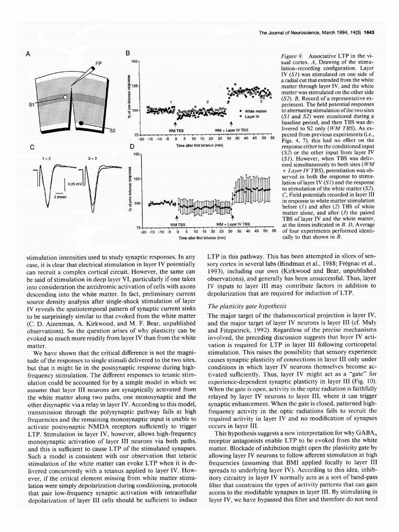
one from layer IV and the other from the white matter (Fig. 8A). The design of the experiment was first to give TBS to the white matter alone and then, after collecting additional baseline, to give TBS simultaneously to layer IV and the white matter. The results are illustrated in Figure 9. As expected, white matter stimulation alone failed to elicit a lasting change in the layer III field potential. However, when layer IV and white matter tetani were paired, a potentiated response was observed in response to test stimulation of both layer IV and the white matter. These data suggest that the postsynaptic response generated by tetanic activation of the strong layer IV input is sufficient to support LTP of other inputs that are active simultaneously.
The Journal of Neuroscience, March 1994, 14(3) 1641
Discussion
The unexpected outcome of this study was the discovery that conditioning stimulation of layer IV would reliably produce LTP of synaptic responses in layer III without any need for antagonists of GABA, receptors. This LTP could be evoked by as few as 80 pulses delivered in a theta burst pattern; it was input specific and was blocked by antagonists of the NMDA receptor. This form of LTP was also Hebbian; potentiation was a specific consequence of input activity that coincided with post- synaptic depolarization beyond some threshold level. In all these respects, this form of LTP in visual cortex resembles what has been documented previously in field CA1 of hippocampus.
Use of rat visual cortex as a model system
A central aim of our experiments was to understand the ele- mentary mechanisms of synaptic plasticity in the neocortex and how they might account for the experience-dependent modifi- cations of visual cortical circuitry during postnatal develop- ment. Although ideally these experiments might have been per- formed in kitten striate cortex, where modifications of binocular circuitry have been demonstrated in viva, we feel our use of rat visual cortex as a model system is justified. A recent side-by- side comparison of synaptic plasticity evoked in slices of adult rat and kitten visual cortex showed that both LTP and long- term depression can be evoked in the two tissues using precisely the same protocols and appear to exhibit identical properties (Kirkwood et al., 1993). As in rat visual cortex, LTP could not be evoked in kitten striate cortex from white matter using TBS (without GABA, receptor antagonists), but it could be elicited reliably from layer IV. Obviously rat and kitten striate cortex are not the same, but at this level of analysis they exhibit similar, if not identical, forms of synaptic plasticity in the superficial layers. Indeed, since rat and cat are phylogenetically quite dis- tant, it is possible that the elementary forms ofsynaptic plasticity described here apply universally to the superficial layers of sen- sory cortex in all mammals.
Field potentials versus intracellular recordings
In this study we have directly compared the plasticity ofsynaptic responses in layer III as measured using extracellular field po- tentials and intracellular EPSPs. Regardless of whether LTP is evoked by TBS of the white matter in the presence of BMI or by TBS oflayer IV without BMI, potentiation ofthe intracellular EPSP in layer III is always accompanied by an increase in the amplitude of the extracellular field potential. This perhaps is not surprising since current source density analyses and phar- macological studies had previously shown this field potential to reflect directly the amplitude of a synaptic current sink in layer III (Lee, 1982; Mitzdorf, 1985; Bode-Greuel et al., 1987; Ki- mura et al., 1989; Lee et al., 199 1; Bear et al., 1992). Indeed, direct comparisons showed that the peak of the field potential coincided with the maximum rate of rise of the simultaneously recorded EPSPs in layer III, supporting the idea that the am- plitude of this peak reflects a “population” excitatory synaptic current in layer III. Our results therefore provide further jus- tification for the use of field potential recordings for the inves- tigation of synaptic plasticity in layer III. This type of “popu- lation” recording provides an essential control for the interpretation of experiments in which intracellular manipula- tions of plasticity in individual cortical cells are attempted.
A 1 160
90
t TSS
80 , , ,
-20 -15 -10 -5 0 5 10 1.5 XI 25 Time from tetanus (min)
Figure 7. Comparison of layer IV and white matter stimulation. A, Effects of TBS on field potential responses evoked from white matter (open circles) and from layer IV (solid circles). Each point is the average (+SEM) response expressed as a percentage of the initial baseline re- sponse (n = 25 for white matter stimulation, n = 44 for layer IV stim- ulation). B, Histograms showing the fraction of cases in which a change of a given amplitude was observed 20 min after TBS of either layer IV (shaded bars) or the white matter (open bars). C, Plot of LTP amplitude as a function of initial field potential amplitude for layer IV stimulation (so/id circles) and for white matter stimulation (open circles). The border points of the distributions are connected by a shaded polygon for the layer IV group and by a solid line for white matter group. In neither group is there any correlation between the amplitude of the response collected for baseline and the magnitude of the LTP (linear regressions for the two groups are indicated by dashed lines).
IV to III LTP is Hebbian and requires NMDA receptor activation We have shown that LTP of the layer III EPSP results if low- frequency stimulation of layer IV is paired with strong intra- cellular depolarization. Simultaneous field potential recordings confirm that the population response is unaffected by the stim- ulation, and control experiments confirm that potentiation does
1642 Kirkwood and Bear * LTP in Visual Cortex
A
White Matter u
1 Layer IV 1 -201-
-9OmV 30 40 50 60 70 60 90 100 1 sec. mV-sec
Figure 8. Intracellular responses of layer III neurons to TBS of white matter and layer IV. A, Representative responses during TBS delivered under the indicated conditions. In each case, the stimulation amplitude was adjusted to yield an intracellular response to a single pulse that was two-thirds the action potential threshold. In the case of layer IV stimulation, there was marked summation of the four EPSPs within each burst, and a longer-lasting depolarizing potential that bridged the interval between bursts. B, Magnitude of the change in EPSP amplitude 20 min after TBS plotted as a function of the area of the depolarization (mV,sec; mean f SEM) during the TBS. Open circle, white matter stimulation (n = 9); solid circle, layer IV stimulation (n = 6); open square, white matter stimulation with BMI applied topically to the superficial layers (n = 6).
not result from depolarization alone. Thus, this LTP fulfills the requirements of a “Hebbian” modification, in which a lasting synaptic enhancement is the specific consequence of pre- and postsynaptic coactivation.
It appears very likely that this mechanism involves the ac- tivation of NMDA receptors on layer III neurons. There is evidence suggesting that LTP in layer III neurons requires an elevation of intracellular calcium (Yoshimura et al., 199 l), just as it does in hippocampal field CA1 (Malenka et al., 1988). Pairing presynaptic stimulation with continuous depolarization is a particularly effective means to activate CaZ+ entry through NMDA receptor channels (Mayer and Westbrook, 1987; Mal- enka et al., 1988) whereas there is ample evidence that voltage- gated calcium channels show substantial voltage- and Ca2+- dependent inactivation under these conditions (Eckert and Chad, 1984). The fact that AP5 blocks induction of tetanus-induced LTP in layer III further supports the notion that NMDA re- ceptor activation serves as a trigger for Hebbian plasticity in this location.
The assertion that NMDA receptor activation triggers LTP does not deny the existence of other factors that additionally may be necessary. For example, in CA1 there is evidence sug- gesting that metabotropic glutamate receptor activation and the subsequent release of intracellular Ca”+ may be a corequisite for LTP induction by NMDA receptor activation (Harvey and Collingridge, 1992). Given the apparent similarity of the Heb- bian synaptic plasticity we describe here for visual cortex with that shown previously in CAl, it will now be of interest to examine in neocortex the entire catalog of possible intracellular biochemical mechanisms that have been invoked to account for CA 1 potentiation.
Comparison with hippocampal LTP
We have already commented here and elsewhere (Kirkwood et al., 1993) on the similarities of LTP evoked in layer III by stimulation of layer IV with that evoked in CA1 by stimulation of the Schaffer collaterals. Both forms of LTP can be induced by a brief episode of TBS, both are input specific, both can be blocked by antagonists of NMDA receptors, and both can be induced simply by pairing low-frequency input activity with strong postsynaptic depolarization. However, one clear differ- ence concerns the early time course of the potentiation. In CA 1, synaptic potentiation is maximal just a few seconds after the
TBS and then decays to a stable value over a period of about 30 min. This decaying form of potentiation, called short-term potentiation (STP), is triggered by NMDA receptor activation and Ca*+ entry but may be mechanistically distinct from LTP (Anwyl et al., 1989; Kauer et al., 1989; Malinow et al., 1989; Malenka, 1991; Schuman and Madison, 1991). The data pre- sented here, however, are consistent with previous observations (Berry et al., 1989; Kimura et al., 1989; Artola and Singer, 1990; Bear et al., 1992) that STP does not occur in visual cortical layer III. In general, synaptic responses grow to a stable value over a time period of several minutes following conditioning stimulation in visual cortex. Neocortex therefore may prove to be advantageous for the investigation of LTP expression and maintenance. In CA 1, by comparison, the mechanisms under- lying STP and LTP overlap and may be easily confused (Stevens, 1993).
Another difference between induction of neocortical and CA 1 LTP might be a requirement for nitric oxide synthesis. Several groups have now reported that incubation of slices with com- petitive antagonists of nitric oxide synthase will block induction of LTP, but not of STP, in CA1 (Bohme et al., 199 1; O’Dell et al., 199 1; Schuman and Madison, 1991). Using precisely the same incubation procedures as Schuman and Madison (199 l), we were unable to detect any effect of N-nitro-L-arginine on LTP in visual cortex. However, since we have no independent confirmation that nitric oxide synthase was inhibited in our experiments, these data alone are not sufficient to reject the hypothesis that nitric oxide synthesis plays some role in neo- cortical LTP. On the other hand, we note that recent work has questioned the contribution of nitric oxide to LTP in CA1 when slices are maintained at temperatures of > 24°C (Li et al., 1992). All our studies were carried out at 35°C.
Comparison of plasticity evoked from layer IV and the white matter
It seems reasonable to assume that stimulation in the middle of the cortical thickness at the site we have referred to as “layer IV” activated direct projections of layer IV cells to layer III, and ascending intracortical and corticocortical axons that pass through layer IV en route to layer III. It is also possible that stimulation of layer IV activated recurrent collaterals of some layer III neurons, although intracellular recordings from layer III cells only rarely revealed antidromic action potentials at the
The Journal of Neuroscience, March 1994, 14(3) 1643
A B Figure 9. Associative LTP in the vi- FP 150- sual cortex. A, Drawing of the stimu-
lation-recording configuration. Layer P IV (SI) was stimulated on one side of
[ a radial cut that extended from the white
125- * matter through layer IV, and the white 8 matter was stimulated on the other side 5 5 (S2). B, Record of a representative ex- p! gioo-
periment. The field potential responses
$$g$g** 3 * White matter to alternating stimulation ofthe two sites z 0 Layer IV (SI and S2) were monitored during a
+ + baseline period, and then TBS was de- WM TSS WM + Layer IV TBS livered to S2 only (WM T&S’). As ex-
D Time alter first tetanus (min) response either to the conditioned input 150-
1+2 (S2) or the other input from layer IV (SI). However, when TBS was deliv-
1 v-
1 8 ered simultaneously to both sites (WM
+ Luyer I V TBS), potentiation was ob-
I
i$ 125. served in both the response to stimu- 0.25 mV 8 lation of layer IV (SI) and the response
I - 4 to stimulation ofthe white matter (S2).
5 msec
g loo- +-+vH ~~~~~~~~~~~~
c ~~~~1~~~
C, Field potentials recorded in layer III in response to white matter stimulation before (I) and after (2) TBS of white s
+’ + matter alone, and after (3) the paired TBS of layer IV and the white matter,
WM TBS 75,. I. a, I, I. I.. 0. - * - t. ‘. a””
WM + Layer IV TBS at the times indicated in B. D, Average -20 -15 -10 -5 0 5 IO 1s 20 25 so 35 40 45 50 5s of four experiments performed identi-
Time alter first tetanus (min) tally to that shown in B.
stimulation intensities used to study synaptic responses. In any LTP in this pathway. This has been attempted in slices of sen- case, it is clear that electrical stimulation in layer IV potentially sory cortex in several labs (Bindman et al., 1988; Fregnac et al., can recruit a complex cortical circuit. However, the same can 1993), including our own (Kirkwood and Bear, unpublished be said of stimulation in deep layer VI, particularly if one takes observations), and generally has been unsuccessful. Thus, layer into consideration the antidromic activation of cells with axons IV inputs to layer III may contribute factors in addition to descending into the white matter. In fact, preliminary current depolarization that are required for induction of LTP. source density analysis after single-shock stimulation of layer IV reveals the spatiotemporal pattern of synaptic current sinks The plasticity gate hypothesis
to be surprisingly similar to that evoked from the white matter The major target of the thalamocortical projection is layer IV, (C. D. Aizenman, A. Kirkwood, and M. F. Bear, unpublished and the major target of layer IV neurons is layer III (cf. Muly observations). So the question arises of why plasticity can be and Fitzpatrick, 1992). Regardless of the precise mechanisms evoked so much more readily from layer IV than from the white involved, the preceding discussion suggests that layer IV acti- matter. vation is required for LTP in layer III following corticopetal
We have shown that the critical difference is not the magni- stimulation. This raises the possibility that sensory experience tude of the responses to single stimuli delivered to the two sites, causes synaptic plasticity of connections in layer III only under but that it might lie in the postsynaptic response during high- conditions in which layer IV neurons themselves become ac- frequency stimulation. The different responses to tetanic stim- tivated sufficiently. Thus, layer IV might act as a “gate” for ulation could be accounted for by a simple model in which we experience-dependent synaptic plasticity in layer III (Fig. 10). assume that layer III neurons are synaptically activated from When the gate is open, activity in the optic radiation is faithfully the white matter along two paths, one monosynaptic and the relayed by layer IV neurons to layer III, where it can trigger other disynaptic via a relay in layer IV. According to this model, synaptic enhancement. When the gate is closed, patterned high- transmission through the polysynaptic pathway fails at high frequency activity in the optic radiations fails to recruit the frequencies and the remaining monosynaptic input is unable to required activity in layer IV and no modification of synapses activate postsynaptic NMDA receptors sufficiently to trigger occurs in layer III. LTP. Stimulation in layer IV, however, allows high-frequency This hypothesis suggests a new interpretation for why GABA, monosynaptic activation of layer III neurons via both paths, receptor antagonists enable LTP to be evoked from the white and this is sufficient to cause LTP of the stimulated synapses. matter. Blockade of inhibition might open the plasticity gate by Such a model is consistent with our observation that tetanic allowing layer IV neurons to follow afferent stimulation at high stimulation of the white matter can evoke LTP when it is de- frequencies (assuming that BMI applied focally to layer III livered concurrently with a tetanus applied to layer IV. How- spreads to underlying layer IV). According to this idea, inhib- ever, if the critical element missing from white matter stimu- itory circuitry in layer IV normally acts as a sort of band-pass lation were simply depolarization during conditioning, protocols filter that constrains the types of activity patterns that can gain that pair low-frequency synaptic activation with intracellular access to the modifiable synapses in layer III. By stimulating in depolarization of layer III cells should be sufficient to induce layer IV, we have bypassed this filter and therefore do not need
1644 Kirkwood and Bear - LTP in Visual Cortex
Plasticity Gate Hypothesis
Figure 10. The plasticity gate hypothesis. According to this simple model, layer III neurons receive direct inputs from fibers originating in the white matter, and indirect input via a relay with excitatory neurons in layer IV. Stimulation of the white matter at high frequencies causes a failure of the polysynaptic activity relayed through layer IV, partly as a consequence of the actions of inhibitory circuits in this layer. Without recruitment of layer IV inputs to layer III, no LTP can be elicited from the white matter (Gate Closed). However, under conditions in which inhibition is blocked, the layer IV cells are able to follow afferent stim- ulation at high frequencies, permitting induction of LTP (Gate Open). By stimulating in layer IV, we have bypassed this gating mechanism altogether, and do not need to block GABA receptors to achieve the threshold for LTP. Open circles represent excitatory neurons and solid circles represent inhibitory neurons. For simplicity, only feedback in- hibition in layer IV is represented.
to block GABA, receptors to achieve the threshold for LTP in layer III.
This concept of a “plasticity gate” controlled by inhibition in layer IV could, in principle, account for aspects of naturally occurring synaptic plasticity in the visual cortex during postnatal development. According to this idea, in early postnatal periods when the inhibitory circuits in visual cortex are not yet fully formed (BHhr and Wolff, 1985; Miller, 1986; Luhmann and Prince, 199 l), the gate is open for plasticity to be evoked by simple patterns of retinal activity, even spontaneous discharges. However, maturation of inhibition in layer IV during devel- opment places increasing constraints on the types of retinal activity that can trigger plasticity in the superficial layers and this, in a sense, closes the gate. Such a layer IV plasticity gate could also be a site of action for various neuromodulators, such as ACh and norepinephrine, that are believed to facilitate syn- aptic plasticity in visual cortex ofkittens (Bear and Singer, 1986; Gordon et al., 1990). According to this view, the end of the “critical period” for experience-dependent synaptic plasticity in the superficial layers of visual cortex may be determined less by changes in the elementary mechanisms of synaptic plasticity than by the maturation of intracortical circuits that function to limit the activity patterns that can gain access to the modifiable synapses in layer III.
This is not the first suggestion that the late maturation of inhibition could be responsible for a reduction of synaptic plas- ticity in the visual cortex (Komatsu, 1983). Indeed, the specific proposal was made that the development of inhibitory synapses on layer III neurons reduced plasticity by shunting excitatory postsynaptic currents, thus making it less likely that NMDA receptors would be recruited by stimulation (Artola and Singer, 1987; Kato et al., 199 1). However, in contrast to these previous
“membrane” models, which assumed that inhibition modulates synaptic plasticity at the level of the membrane conductance of layer III dendrites, ours is more of a “network” model in which it is assumed that inhibition modulates synaptic plasticity by filtering the patterns and amounts of afferent activity that reach the modifiable synapses in layer III. It will be of considerable interest to test experimentally the predictions of these two mod- els.
References Anwyl R, Mulkeen D, Rowan MJ (1989) The role of N-methyl-o-
aspartate receptors in the generation of short-term potentiation in the rat hippocampus. Brain Res 503: 148-l 5 1.
Aroniadou VA, Teyler TJ (1991) The role of NMDA receptors in long-term potentiation (LTP) and depression (LTD) in rat visual cor- tex. Brain Res 562: 136-143.
Artola A, Singer W (1987) Long-term potentiation and NMDA re- ceptors in rat visual cortex. Nature 330:649-652.
Artola A, Singer W (1990) The involvement of N-methyl-o-aspartate receptors in induction and maintenance of long-term potentiation in rat visual cortex. Eur J Neurosci 2:254-269.
Btihr S, Wolff JR (1985) Postnatal development of axosomatic syn- apses in the rat visual cortex: morphogenesis and quantitative eval- uation. J Comp Neurol 233:405-420.
Bear MF, Singer W (1986) Modulation of visual cortical plasticity by acetylchohne and noradrenaline. Nature 320: 172-l 76.
Bear MF, Cooper LN, Ebner FF (1987) A physiological basis for a theorv of svnantic modification. Science 237142-48.
Bear MF, Press WA, Connors BW (1992) Long-term potentiation in slices of kitten visual cortex and the effects of NMDA receptor block- ade. J Neurophysiol 67: l-l 1.
Berry RL, Teyler TJ, Taizhen H (1989) Induction of LTP in rat visual cortex: tetanus parameters. Brain Res 481:221-227.
Bindman LJ, Murphy KF’SJ, Pockett S (1988) Postsynaptic control of the induction of long-term changes in efficacy of transmission at neo- cortical synapses in slices of rat brain. J Neurophysiol60: 1053-1065.
Bode-Greuel KM, Singer W, Aldenhoff JB (1987) A current source density analysis of field potentials evoked in slices of visual cortex. Exp Brain Res 69:213-219.
Bohme GA, Bon C, Stutzmann JM, Doble A, Blanchard JC (199 1) Possible involvement of nitric oxide in long-term potentiation. Eur J Pharmacol 199:379-38 1.
Chagnac-Amitai Y, Connors BW (1989) Horizontal spread of syn- chronized activity in neocortex and its control by GABA-mediated inhibition. J Neurophysiol 61~747-758.
Collingridge GL, Davies SN (I 989) NMDA receptors and long-term potentiation in the hippocampus. In: The NMDA receptor (Watkins JC, Collingridge GL, eds), p 242. Oxford: Oxford UP,
Connors BW (1984) Initiation of synchronized neuronal bursting in neocortex. Nature 3 10:685-687.
Eckert R, Chad JE (1984) Inactivation of calcium channels. Prog Biophys Mol Biol 44:215-267.
FrCgnac Y, Shultz D, Thorpe S, Bienenstock E (1988) A cellular an- alogue of visual cortical plasticity. Nature 333:367-370.
Frtgnac Y, Smith D, Friedlander MJ (1993) Temporal covariance of post-synaptic membrane potential and synaptic input. Role in syn- aptic efficacy in visual cortex. Prog Brain Res 95:207-223.
Gabbott PLA, Stewart MG (1987) Distribution of neurons and glia in the visual cortex (area 17) of the adult albino rat: a quantitative description. Neuroscience 21:833-845.
Gordon B, Mitchell B, Mohatadi K, Roth E, Tseng Y, Turk F (1990) Lesions of non-visual inputs affect plasticity, norepinephrine content and acetylcholine content of visual cortex. J Neurophysiol64: 185 l- 1860.
Gustafsson B, Wigstriim H, Abraham WC, Huang Y-Y (1987) Long- term potentiation in the hippocampus using depolarizing potentials as the conditioning stimulus to single volley synaptic potentials. J Neurosci 7~774-780.
Harvey J, Collingridge GL (1992) Thapsigargin blocks the induction of long-term potentiation in rat hippocampal slices. Neurosci Lett 139:197-200.
The Journal of Neuroscience, March 1994, 14(3) 1645
Hebb DO (1949) The organization of behavior. New York: Wiley. Hirsch JA, Gilbert CD (1993) Long-term changes in synaptic strength
along specific intrinsic pathways in the cat visual cortex. J Physiol (Lond) 46 11241-262.
Hubel DH, Wiesel TN (1962) Receptive fields, binocular interaction and functional architecture in the cat’s visual cortex. J Physiol (Lond) 160:106-154.
Kato N, Artola A, Singer W (199 1) Developmental changes in the susceptibility to long-term potentiation of neurones in rat visual cor- tex slices. Dev Brain Res 60:43-50.
Kauer JA, Malenka RC, Nicoll RA (1989) NMDA application po- tentiates synaptic transmission in the hippocampus. Nature 334:250- 252.
Kelso SR, Ganong AH, Brown T (1986) Hebbian synapses in the hippocampus. Proc Nat1 Acad Sci USA 83:5326-5330.
Kimura F, Nishigori A, Shirokawa T, Tsumoto T (1989) Long-term potentiation and NMDA receptors in the visual cortex of young rats. J Physiol (Lond) 414:125-144.
Kirkwood A, Dudek SD, Gold JT, Aizenman CD, Bear MF (1993) Common forms of synaptic plasticity in hippocampus and neocortex in vitro. Science 260: 15 18-l 52 1.
Komatsu Y (1983) Development of cortical inhibition in kitten striate cortex investigated by a slice preparation. Dev Brain Res 8: 136-l 39.
Komatsu Y, Iwakiri M (1992) Low-threshold Ca” channels mediate induction of long-term potentiation in kitten visual cortex. J Neu- rophysiol 67:401-410.
Komatsu Y, Nakajima S, Toyama K (1991) Induction of long-term potentiation without the participation of N-methyl-D-aspartate re- ceptors in kitten visual cortex. J Neurophysiol 65:20-32.
Larson J, Lynch G (1986) Synaptic potentiation in hippocampus by patterned stimulation involves two events. Science 232:985-988.
Larson J, Lynch G (1988) Role of N-methyl-D-aspartate receptors in the induction of synaptic potentiation by burst stimulation patterned after the hippocampal theta rhythm. Brain Res 44 1: 11 l-l 18.
Lee KS (1982) Sustained enhancement of evoked potentials following brief, high-frequency stimulation of the cerebral cortex in vitro. Brain Res 2391617-623.
Lee SM, Weisskopf MG, Ebner FF (1991) Horizontal long-term po- tentiation of responses in rat somatosensory cortex. Brain Res 544: 303-3 10.
Li Y-G, Errington ML, Williams JH, Bliss TVP (1992) Temperature- dependent block of LTP by the NO synthase inhibitor L-NARG. Sot Neurosci Abstr 18:343.
Luhmann HJ, Prince DA (199 1) Postnatal maturation of the GA- BAergic system in rat neocortex. J Neurophysiol 65:247-263.
Malenka RC (199 1) Postsynaptic factors control the duration of syn- aptic enhancement in area CA1 of the hippocampus. Neuron 6:53- 60.
Malenka RC, Kauer JA, Zucker RS, Nicoll RA (1988) Postsynaptic calcium is sufficient for potentiation of hippocampal synaptic trans- mission. Science 242:8 l-84.
Malinow R, Miller JP (1986) Postsynaptic hyperpolarization during conditioning reversibly blocks induction of long-term potentiation. Nature 320:529-530.
Malinow R, Schulman H, Tsien RW (1989) Inhibition of postsynaptic
PKC or CaMKII blocks induction but not expression of LTP. Science 245:862-866.
Mayer ML, Westbrook GL (1987) The physiology of excitatory amino acids in the vertebrate central nervous system. Prog Neurobiol 28: 197-276.
Miller MW (1986) Maturation of rat visual cortex. III. Postnatal mor- phogenesis and synaptogenesis of local circuit neurons. Dev Brain Res 251271-285.
Mitzdorf U (1985) Current source-density method and application in cat cerebral cortex: investigation of evoked potentials and EEG phe- nomena. Physiol Rev 65:37-100.
Monaghan DT, Cotman CW (1985) Distribution of NMDA-sensitive L-‘H-glutamate binding sites in rat’s brain as determined by quan- titative autoradiography. J Neurosci 5:2909-29 19.
Muly EC, Fitzpatrick D (1992) The morphological basis for binocular and ON/OFF convergence in tree shrew striate cortex. J Neurosci 12: 1319-1334.
O’Dell TJ, Hawkins RD, Kandel ER, Arancio 0 (1991) Tests of the roles of two diffusible substances in long-term potentiation: evidence for nitric oxide as a possible early retrograde messenger. Proc Nat1 Acad Sci USA 88:11285-l 1289.
Rauschecker JP, Singer W (1979) Changes in the circuitry of the kitten visual cortex are gated by postsynaptic activity. Nature 280:58-60.
Sah P, Nicoll RA (199 1) Mechanisms underlying potentiation of syn- aptic transmission in rat anterior cingulate cortex in vitro. J Physiol (Lond) 433:615-630.
Sastry BR, Goh JW, Auyeung A (1986) Associative induction of post- tetanic and long-term potentiation in CA1 neurons of rat hippocam- pus. Science 232:988.
Schuman EM, Madison DV (199 1) A requirement for the intercellular messenger nitric oxide in long-term potentiation. Science 254: 1503- 1506.
Schwartzkroin PA, Prince DA (1978) Cellular and field potential prop- erties of epileptogenic hippocampal slices. Brain Res 147: 117-l 30.
Stevens CF (1993) Quanta1 release of neurotransmitter and long-term potentiation. Cell 72:55-63.
Steward 0, Tomasulo R, Ley WB (1990) Blockade of inhibition in a pathway with dual excitatory and inhibitory action unmasks a ca- pability for LTP that is otherwise not expressed. Brain Res 5 16:292- 300.
Stryker MP, Strickland SL (1984) Physiological segregation of ocular dominance columns depends on the pattern of afferent electrical ac- tivity. Invest Ophthalmol Vis Sci [Suppl] 25:28.
Sutor B. Hablitz JJ (1989) Long-term ootentiation in frontal cortex: role of NMDA-modulated polysynaptic excitatory pathways. Neu- rosci Lett 97:ll l-l 17.
Wiesel TN (1982) Postnatal development of the visual cortex and the influence of the environment. Nature 299:583-592.
Wigstrom H, Gustafsson B, Huang Y-Y, Abraham WC (1986) Hip- pocampal long-term potentiation is induced by pairing single afferent volleys with intracellularly injected depolarizing pulses. Acta Physiol Stand 126:317.
Yoshimura Y, Tsumoto T, Nishigori A (199 1) Input-specific induction of long-term depression in Ca’+ -chelated visual cortex neurons. Neu- roreport 2:393-396.