NEUROSCIENCE
RESEARCH ARTICLEL. Buczek et al. / Neuroscience 436 (2020) 34–45
Hedonic Eating: Sex Differences and Characterization of Orexin
Activation and SignalingLaura Buczek, Jennifer Migliaccio and Gorica D. Petrovich *
Department of Psychology, Boston College, Chestnut Hill, MA 02467, United States
Abstract—Palatable taste can stimulate appetite in the absence of hunger, and individual differences in hedoniceating may be critical to overeating. Women are more prone to obesity and binge eating than men, which warrantscomparisons of hedonic versus physiological consumption and the underlying neural substrates in both sexes.The current study examined palatable (high-sugar) food consumption in male and female rats under physiologicalhunger and satiety, and the role of the neuropeptide orexin/hypocretin (ORX). Across multiple tests, females con-sistently consumed similar amounts of palatable food regardless of whether they were hungry or sated prior totesting. In contrast, males typically adjusted their consumption according to their hunger/satiety state. This dif-ference was specific to palatable food consumption, as both sexes ate standard chow according to their hungerstate. ORX is important in food motivation and reward behaviors. Thus, to begin to determine the neuronal mech-anisms of hedonic eating, we examined activation and signaling of ORX neurons. We systematically characterizedFos induction patterns of ORX neurons across the entire rostrocaudal extent of the lateral hypothalamus andfound that they were activated by food and by fasting in both sexes. Then, we showed that systemic blockadeof ORX receptor 1 signaling with SB-334867 decreased palatable food consumption in hungry and sated rats ofboth sexes. These results demonstrate sex differences in hedonic eating; increased susceptibility in females toovereat palatable food regardless of hunger state, and that ORX is a critical neuropeptide mechanism of hedoniceating in both sexes. � 2020 IBRO. Published by Elsevier Ltd. All rights reserved.
Key words: hedonic, consumption, orexin, overeating, palatability, sex differences.
INTRODUCTION
Hunger and palatable taste of food both drive appetite and
consumption and could work in accord or independently.
Palatability can stimulate appetite through reward
mechanisms regardless of physiological hunger, and
this form of overeating contributes to obesity and binge
eating disorder (Cota et al., 2006; Berridge et al., 2010;
Stice et al., 2013). Many modern environments are satu-
rated with easily accessible high-calorie, palatable foods
that are innately liked across mammalian species
(Berridge, 2000). Cues for these foods can further amplify
the drive to eat in the absence of hunger (Weingarten,
1983; Boggiano et al., 2009; Berthoud, 2012; Petrovich,
2013; Kendig et al., 2018).
How individuals respond to sweet taste and other
appetite triggers when sated may be an important
difference between those who are vulnerable and those
who are resistant to overeating (Small, 2009; Reppucci
https://doi.org/10.1016/j.neuroscience.2020.04.0080306-4522/� 2020 IBRO. Published by Elsevier Ltd. All rights reserved.
*Corresponding author. Address: Department of Psychology, BostonCollege, 344 McGuinn Hall, 140 Commonwealth Avenue, ChestnutHill, MA 02467-3807, United States.
E-mail address: [email protected] (G. D. Petrovich).Abbreviations: KPBS, potassium phosphate-buffered saline; LHA,lateral hypothalamic area; NHS, normal horse serum; ORX, orexin.
34
and Petrovich, 2012; Sun et al., 2015). Women are more
susceptible than men to weight gain and obesity, as well
as binge eating and other disorders (Hudson et al.,
2007; Mitchell and Shaw, 2015). These differences war-
rant comparisons of the neural substrates mediating
hedonic versus physiological consumption in both sexes.
Here, we compared palatable food consumption in male
and female rats and examined the activation and signal-
ing of the neuropeptide orexin/hypocretin (ORX) (de
Lecea et al., 1998; Sakurai et al., 1998). ORX is important
in the motivation to eat without physiological incentive
(Choi et al., 2010; Mahler et al., 2014; Petrovich, 2019),
but whether ORX neurons are similarly driving hedonic
eating under sated and hungry states in males and
females has not been examined.
In order to characterize behavioral and neural
substrates in both sexes, in two studies we compared
consumption of palatable, sweet-tasting food (high-
sugar Test Diet pellets) under the physiological
conditions of hunger and satiety. In the first study, we
examined male and female rats that were either
deprived of food for 20 hours or had ad libitum access
to standard rat chow prior to consumption tests. Then
we characterized Fos induction in ORX neurons during
L. Buczek et al. / Neuroscience 436 (2020) 34–45 35
these tests in rats that had access to high-sugar pellets,
along with controls without access to food. We
systematically characterized patterns of Fos induction in
ORX neurons across the entire rostrocaudal extent of
the lateral hypothalamic area (LHA).
In the second study, we tested whether Fos induction
patterns in ORX neurons observed in the first experiment
indicate that ORX signaling is causal to hedonic eating.
We systemically blocked ORX signaling via receptor 1
with SB-334867 in male and female rats that were either
fasted or not fasted prior to tests with high-sugar pellets.
Additionally, to determine if observed sex differences
were due to palatability, rather than differences in
hunger/satiety, we tested consumption of another, less
palatable, familiar food (standard chow) in rats that were
hungry or sated. First, we tested rats in two separate
tests: one with high-sugar pellets and the other with
standard chow. Then, after completion of ORX signaling
blockade experiments we re-tested rats with both foods
in a within-subjects design across two tests, in order to
examine how individual rats that are hungry or sated
consume palatable food versus chow.
EXPERIMENTAL PROCEDURES
Subjects
Ninety-six, experimentally naıve, male and female
Sprague Dawley rats (48 male, 48 female) that were
obtained from Envigo at 250–275 g were used in these
experiments. There were two sets of experiments, with
n= 64 in the first (Palatable Food Consumption and
Fos Induction in Orexin Neurons) and n= 32 in the
second (Orexin Receptor 1 Signaling Blockade). Rats
were individually housed and maintained on a 12 h
light/dark cycle (lights on 06:00). Behavioral testing was
conducted during the light phase between 09:00 and
16:00. After arrival, rats were given 24 h to acclimate to
the colony room with ad libitum access to water and
standard laboratory chow (Purina Lab Diet Prolab RMH
3000; 3.47 kcal/g: 26% protein, 15% fat, 59%
carbohydrates (89% starch)) before any handling or
habituation procedures began. All housing and testing
procedures were in compliance with the National
Institute of Health’s Guidelines for Care and Use of
Laboratory Animals and were approved by the Boston
College Institution Animal Care and Use Committee.
Palatable food
The palatable food used here was Test Diet (TD) pellets
(5TUL 45 mg Scott Pharma: cat#1811155; 3.44 kcal/g;
20% protein, 13% fat, 67% carbohydrates—all from
sucrose). Previously, we compared rats’ preference for
TD pellets to other high-sugar/high-fat (Oreos, Nabisco),
high-sugar/low-fat (Lucky Charms, General Mills), and
low-sugar/ high-fat (Cheetos, Frito Lay) foods in adult,
sated male rats (Reppucci, 2010). Rats’ preferences were
evaluated based on their consumption during 30 min tests
(5 g of single food given, test order counterbalanced).
Rats preferred all high-sugar foods to the low-sugar/
high-fat food and the highest preference score was for
TD and the high-sugar/high-fat food (identical mean val-
ues), which was higher than the score for high-sugar /
low-fat food (but not statistically significant).
Habituation
After the 24 h acclimation period, all subjects were
handled daily for 5–6 days and were habituated to all
experimental conditions prior to testing. All subjects
received 1 g of TD pellets at the bottom of their home
cages. On a separate occasion, all subjects received
empty ceramic testing dishes for a 30 min exposure.
Next, all subjects received 10 g TD in ceramic dishes
and were allowed to consume it for 30 min (no chow
was given) after ad libitum access to chow, and after
20 h food deprivation (the order of these two habituation
sessions was counterbalanced). Subjects were also
habituated to being transported on a cart to and from
the procedure room on two separate occasions.
Palatable food consumption and Fos induction inOrexin neurons
Design. Following a full factorial experimental design,
male and female rats were assigned to either ad libitum or
20 h food deprivation eating schedules (n= 16 per sex).
Within each group, half of the rats (Food condition)
received 15 g TD in ceramic testing dishes during
testing, and half of the rats (NoFood condition) received
empty dishes during testing (n= 8 per group). Testing
occurred in home cages and consisted of a 100 min
exposure to the dish with food (Food condition) or
empty dish (NoFood condition) during which rats were
left undisturbed. Rats were perfused immediately after
the 100 min testing session to measure test-specific Fos
induction. All remaining food was collected and weighed
to determine consumption, including any food that was
moved from the dishes by rats during the testing
sessions. Cages were thoroughly checked by hand for
any pellets that may have been buried in the bedding, to
ensure correct consumption calculations.
Histological procedures. One hundred minutes after
the start of the testing session, rats were briefly
anaesthetized with isoflurane (Henry Schein, Pittsburgh,
PA, USA), and then intraperitoneally injected with
tribromoethanol (375 mg/kg; Sigma Aldrich, St. Louis,
MO, USA) and transcardially perfused with 0.9% saline
followed by 4% paraformaldehyde in 0.1 M borate
buffer. The brains were stored for 20–24 h at 4 �C in a
paraformaldehyde and 12% sucrose mixture and then
rapidly frozen in hexanes cooled with dry ice and stored
at �80 �C. Brains were cut into 30 lm coronal sections
using a microtome and were collected into four adjacent
series. One tissue series was processed with ORX
immunohistochemistry followed by Fos
immunohistochemistry, described below. Another series
was mounted and stained with thionin for identification
of cytoarchitectonic borders, and the remaining two
were stored in cryoprotectant in a �20 �C freezer.
36 L. Buczek et al. / Neuroscience 436 (2020) 34–45
ORX and Fos double-label immunohistochem-
istry. Free-floating sections were incubated for 1 h at
room temperature in a blocking solution (0.02 M
potassium phosphate-buffered saline [KPBS] containing
2% normal horse serum [NHS], 0.3% Triton X-100 and
10% milk), and then incubated with mouse monoclonal
antibody against ORX-A (1:500, MAB763; R&D
Biosystems, Minneapolis, MN, USA) in the blocking
solution for 72 h at 4 �C with gentle agitation. Sections
were rinsed with KPBS, 2% NHS and 10% milk,
incubated with biotinylated secondary antibody against
mouse (1:500, BA-2001; Vector Laboratories) in the
blocking solution, rinsed in KPBS, incubated in avidin
biotin complex (ABC, PK-6100; Vector Laboratories),
rinsed in KPBS and recycled through the secondary
antibody and ABC solutions with KPBS rinses in
between. Finally, the tissue was processed with 3,30-diaminobenzidine (SK-4100; Vector Laboratories) to
visualize ORX immunoreactivity. Brain sections were
then immediately processed again in a similar manner
but using anti-c-Fos antibody raised in rabbit (ABE457;
Millipore, 1:5000 lot:3059066) and biotinylated
secondary against rabbit (1:500, BA-1000; Vector
Laboratories). Nuclei for Fos were visualized as gray
after a two-minute incubation in nickel-intensified 3,30-diaminobenzidine (SK-4100; Vector Laboratories).
Sections were rinsed, mounted on SuperFrost slides
(Fisher Scientific), dried at 45 �C, dehydrated through
graded alcohols, cleared in xylenes, and coverslipped
with DPX Mountant (Electron Microscopy Services;
Hatfield, PA, USA).
Image acquisition and analysis. Processed tissue was
imaged using the 20� objective on an Olympus BX51
light microscope with attached Olympus DP74 camera,
using DP2-BSW software (Olympus America Inc.,
Center Valley, PA, USA). Images of the tissue were
taken bilaterally throughout the rostrocaudal extent of
the lateral hypothalamus containing ORX neurons
(Swanson et al., 2005); atlas levels 27–31 (Swanson,
2004). At each atlas level, four images were taken on
each side (total 8 images per level), one for each of 4
adjacent quadrants surrounding the fornix (with the fornix
placed in the dorsolateral corner of the ventromedial
image) as shown in Fig. 1A. The perifornical area (pf)was defined by the quadrant located dorsomedial to the
fornix, and the remaining quadrants were placed to cap-
ture areas located clockwise to the fornix, dorsolateral
(dl), ventrolateral (vl), and ventromedial (vm). Three types
of neurons were manually identified and counted in the
acquired images: single-labeled ORX-positive, single-
labeled Fos-positive, and double-labeled ORX and Fos-
positive neurons (Fig. 1B). ORX-positive neurons were
characterized by distinct cytoplasmic staining (brown)
and Fos protein-positive neurons were characterized by
distinct nuclear staining (gray). Double-labeled neurons
(ORX+ Fos) had both cytoplasmic (ORX) and nuclear
(Fos) labeling (Fig. 1B). An ORX-containing neuron was
counted as positive only if both the cell body and the
nucleus were clearly visible in the image. Fos-labeled
neurons were counted as positive if the Fos-labeling
intensity was clearly above background and contained
within the nucleus. Two experimenters conducted cell
identification and count analysis. They were blind to the
experimental condition of the tissue images observed
and images were counter-balanced across experimental
conditions and sex. Total counts from images of the left
and right hemispheres were pooled to calculate the total
number of Fos-positive neurons, the total number of
ORX-positive neurons, and the total number of double-
labeled (ORX+ Fos) neurons within each sampling
region (pf, dl, vl, vm) for each brain. To perform a more
accurate analysis of the degree of ORX neuron recruit-
ment the percentage of the total number of ORX-
specific neurons that were double-labeled with Fos were
then calculated for each sampling region. Due to tissue
damage or poor tissue quality, six brains were excluded
from all neural analyses (one from each of these groups:
Female Deprived NoFood, Male Deprived NoFood,
Female Ad Lib Food, Male Ad Lib Food, Female Deprived
Food, Female Ad Lib NoFood).
Orexin receptor 1 signaling blockade
Drugs. SB-334867 (SB; Tocris Bioscience;
Minneapolis, MN, USA) was suspended in a solution
consisting of 2% dimethylsulfoxide and 10% 2-
hydroxypropyl-b-cyclodextrin (Sigma-Aldrich, St. Louis,
MO, USA) in sterile water. Vehicle was 2%
dimethylsulfoxide and 10% 2-hydroxypropyl-b-
cyclodextrin (Sigma-Aldrich, St. Louis, MO, USA) in
sterile water. Both SB and vehicle were administered via
intraperitoneal injection at a volume of 4 ml/kg. SB dose
was 20 mg/kg and was injected at a concentration of
20 mg/4 ml. Injection was given 30 min prior to Test 3
and Test 4 (see below).
Design. Rats were first tested for consumption of
palatable food (TD) under sated and food deprived
states to replicate behavioral findings from the first
study (Test 1). Then they were tested with standard
chow under the same conditions of hunger and satiety
to establish whether differences between males and
females are due to food palatability (sweet taste) or
differences in physiological hunger state (Test 2). The
TD and chow have similar caloric densities (3.44 and
3.47 kcal/g, respectively), however, all carbohydrates in
TD are from sucrose and 89% of carbohydrates in chow
are from starch. Accordingly, TD tastes much sweeter
than chow. Next, rats were tested for palatable food
consumption (TD) after receiving SB or Vehicle across
two counterbalanced tests (Tests 3&4). Because the
order of Tests 1 and 2 was not counterbalanced, rats
were re-tested for chow and TD consumption at the end
of experiments (Tests 5&6).
Half of the male and female rats were food deprived
for 20 h (Deprived condition) while the other half had
ad libitum access to rat chow (Ad Lib condition)
(n= 8/group). All testing sessions consisted of a 60 min
exposure to either TD or standard rat chow in ceramic
dishes as indicated, after which food was weighed to
determine consumption. Experimenters were careful to
50µm
BA
Fig. 1. Image acquisition and analysis. (A) Representation of the sampling regions is shown on a modified rat brain atlas template (level 29,
Swanson, 2004). Images were taken in four sampling regions arranged in respect to the fornix (fx): perifornical area (pf), located dorsomedial to the
fornix, dorsolateral (dl), ventromedial (vm), and ventrolateral (vl). (B) Image shows representative types of labeled neurons. Arrows point to a
representative of each type of labeled neuron: single-labeled Fos (gray), single-labeled ORX (brown), and double-labeled ORX and Fos. The scale
bar (B)= 50 mm. (For interpretation of the references to colour in this figure legend, the reader is referred to the web version of this article.)
L. Buczek et al. / Neuroscience 436 (2020) 34–45 37
include in these calculations any food that remained in the
dish, as well as any food that was displaced to other areas
of the cage during testing. Cages were thoroughly
checked by hand after each test for any pellets that may
have fallen out of the dish or been buried in the bedding
by rats, to ensure correct consumption calculations.
Rats were provided with sufficiently large quantities of
food to ensure unrestricted eating (15–20 g, consistent
within each test). Only one type of food was given
during the tests. Thirty minutes prior to tests 3 and 4,
rats received either an SB or Vehicle intraperitoneal
injection in a within-subjects design (SB and Veh order
was counterbalanced across the two tests). For tests 5
and 6 rats were also tested in a within-subjects design
with TD and chow (order was counterbalanced across
tests). All testing sessions were conducted 2–4 days
apart.
Statistical analyses
Data were analyzed using analysis of variances
(ANOVAs) and t-tests. Main effects were followed by
post hoc Tukey HSD analyses. Statistical analyses were
performed using SPSS. Data are presented as mean
± SEM. In all cases, p � 0.05 was considered
significant. The data were tested for normal distributions
and equal variances. If these criteria were not met, log
and square root transformations were used to verify the
results. In one analysis, after square root transformation
the p value was 0.051 and that is reported in the
Results. In the Orexin Receptor 1 Signaling Blockade
study (Tests 3&4), the data were not normally
distributed due to an outlier. The outlier was identified
by a box plot (Male Deprived) and was removed from
the data (Fig. 6) and the statistical values are reported
in the Results for both the analysis with and without the
outlier.
RESULTS
Palatable food consumption and Fos induction inOrexin neurons
Consumption. Males and females exhibited different
patterns of TD consumption during the testing session
(Fig. 2A). Males ate more when they were in a
deprivation state than ad lib, while females ate similar,
large amounts regardless of their physiological state.
ANOVA of consumption with sex and deprivation state
(Ad Lib, Deprived) confirmed significant between-
subjects main effects of sex (F1,28 = 8.496, p= 0.007),
deprivation state (F1,28 = 25.254, p< 0.0001), and
interaction of sex with deprivation state (F1,28 = 6.245,
p= 0.019). Post hoc tests showed that Ad Lib males
ate significantly less than any of the other groups
(p< 0.005 in all cases). Ad Lib females ate similar
amounts as deprived females and deprived males
(p= 0.301 and p= 0.455, respectively).
The consumption patterns were similar when
calculated relative to body weight (2b). ANOVA of
consumption as a percentage of body weight with sex
and deprivation state (Ad Lib, Deprived) found
significant main effects of sex (F1,28 = 16.619,
p< 0.0005), deprivation state (F1,28 = 27.260,
p< 0.0001), and interaction of sex with deprivation
state (F1,28 = 5.156, p= 0.031). Post hoc tests
confirmed that Ad Lib males ate significantly less TD
than any of the other groups (p< 0.005 in all cases),
while Ad Lib females ate similar amounts as deprived
females and deprived males (p= 0.182 and p= 0.850,
respectively).
The average body weights for each group of rats prior
to testing are shown in Table 1. All subjects arrived at the
same weight range; however males gained weight much
0
5
10
Con
sum
ptio
n (g
)
*
Ad Lib DeprivedMale Female Male Female
0
1
2
3
4
Con
sum
ptio
n (%
Bod
y W
eigh
t)
*
Ad Lib DeprivedMale Female Male Female
A B
Fig. 2. Consumption during testing session. (A) Consumption of TD shown in grams (mean ± SEM).
(B) Consumption of TD expressed as a percentage of rat’s body weight (mean ± SEM). *Indicates
Male Ad Lib different (p< 0.005) from each other group (n= 8 per group).
38 L. Buczek et al. / Neuroscience 436 (2020) 34–45
more rapidly and thus had higher body weights than
females prior to testing (F1,55 = 74.635, p< 0.0001).
There were no body weight differences based on
deprivation state or food access conditions within each
sex (p> 0.05 for all main effects and interactions).
ORX neurons. Table 2 displays the total number of
ORX neurons (mean ± SEM) in each sampling region
for sex and condition. No main effects or interactions
were observed in the total number of ORX neurons in
the LHA. Similarly, there were no effects or interactions
when each sampling region was analyzed separately.
The pf region had the highest number of ORX neurons
compared to all sampling quadrants. ANOVA of total
ORX neurons with sex, deprivation state (Ad Lib,
Deprived), food access (Food, No Food), and region (pf,dl, vl, vm) found a main effect of region
(F3,200 = 209.309, p< 0.0001). Follow up tests found
that pf and dl differed from all other regions (p< 0.0001
in all cases), while vm and vl were similar (p= 0.401).
Fos induction in ORX neurons. Images in Fig. 3 show
Fos induction in ORX neurons in each group. The graphs
in Fig. 4 show that Fos induction in ORX neurons was the
Table 1. Body weights (mean ± SEM) prior to testing
Ad Lib D
Male Female M
No Food 285 ± 4 262 ± 5 2
Food 290 ± 2 265 ± 7 2
Table 2. Total numbers of ORX neurons observed in the LHA. Displayed are values for total number of O
quadrant for each group (mean ± SEM)
LHA
Region
Ad Lib Deprived
Male No Food Male
Food
Female No Food Female
Food
Male No Food Male
Food
All 117 ± 11 126 ± 14 136 ± 11 124 ± 11 123 ± 17 150 ±
pf 61 ± 8 63 ± 9 72 ± 7 65 ± 5 69 ± 11 76 ±
dl 30 ± 3 33 ± 4 34 ± 3 32 ± 5 27 ± 4 37 ±
vm 11 ± 2 13 ± 2 11 ± 1 11 ± 2 11 ± 2 20 ±
vl 14 ± 1 16 ± 2 19 ± 2 16 ± 3 15 ± 3 21 ±
lowest in Ad Lib groups without
food access, and that food
consumption and deprivation state
induced Fos in both sexes.
ANOVA of the percentage of ORX
neurons that were double-labeled
with Fos with sex, food access
(Food, No Food), and deprivation
condition (Ad Lib, Deprived) found
significant main effects of food
access (more Fos in the Food
compared to No Food groups;
F1,50 = 6.491, p= 0.014; after
square root transformation
F1,50 = 7.160, p= 0.010) and
deprivation state, (more Fos in the
Deprived compared to Ad Lib
groups; F1,50 = 4.116, p= 0.048; after square root
transformation F1,50 = 4.002, p= 0.051), but no
significant effect of sex (F1,50 = 0.497, p= 0.484; after
square root transformation F1,50 = 0.519, p= 0.475) or
interactions.
Table 3 displays the percent of ORX neurons that
were labeled with Fos (mean ± SEM) in each sampling
region for each sex and condition. Analyses of each
quadrant separately, found that pf exhibited main effects
of both deprivation state and food access (more Fos in
the Food compared to No Food group: F1,50 = 5.043,
p= 0.029 and more Fos in the Deprived compared to
Ad Lib groups: F1,50 = 6.113, p= 0.017) but no effects
of sex or interactions. There were main effects of food
access only for dl and vl regions (more Fos in the Food
compared to No Food groups in dl: F1,50 = 12.579,
p= 0.001 and vl: F1,50 = 4.617, p= 0.037) and no
effects for the vm region (p> 0.05 for all).
Total Fos induction. We analyzed the total number of
Fos-positive neurons in the LHA (sum across four
quadrants). ANOVA of total number of Fos neurons in
the LHA found the main effects of food access (more
eprived
ale Female
89 ± 2 261 ± 4
94 ± 3 266 ± 4
RX neurons counted within each sampling
Female No Food Female
Food
12 137 ± 14 148 ± 23
7 80 ± 9 79 ± 11
3 28 ± 3 39 ± 8
2 12 ± 2 13 ± 3
3 17 ± 3 17 ± 3
B
C D
E F
G H
A
100µm
Fig. 3. Images of Fos induction in ORX neurons. Images show induction of Fos (gray) in ORX
neurons (brown) in ad libitum-fed male groups that were either tested without food (A) or given food
(B), fasted males groups that were either tested without food (C) or given food (D), ad libitum-fed
females groups that were either tested without food (E) or given food (F), and fasted female groups
that were either tested without food (G) or given food (H). All images are from perifornical area. The
scale bar (G)= 100 mm.
L. Buczek et al. / Neuroscience 436 (2020) 34–45 39
Fos in the Food compared to No Food group;
F1,50 = 10.544, p= 0.002) and deprivation (more Fos in
the Deprived compared to Ad Lib groups; F1,50 = 7.252,
p= 0.010), but no effect of sex (F1,50 = 0.111,
p= 0.741) or interactions.
Table 4 displays the values of total number of Fos-
positive neurons (mean ± SEM) across LHA and in
each sampling region for each sex and condition.
Analyses of each quadrant separately, found that within
pf, vm and vl regions there were main effects of
deprivation state (more Fos in the Deprived compared
to Ad Lib groups; F1,50 = 4.982, p= 0.030 for pf,
F1,50 = 15.115, p< 0.0005 for vm, and F(1,50) = 4.751,
p= 0.034 for vl) and food access (more Fos in the
Food compared to No Food
group; F1,50 = 9.030, p= 0.004
for pf, F1,50 = 19.219, p< 0.0001
for vm, and F1,50 = 4.537,
p= 0.038 for vl). There was only
a main effect of food access
within dl region (more Fos in the
Food compared to No Food
group; F1,50 = 4.112, p= 0.048).
There were no main effects of sex
or interactions within any region.
Orexin receptor 1 signalingblockade
Consumption. In two separate
tests, we measured the
consumption of TD and chow
(Fig. 5). In the test with TD
(Fig. 5A) all rats ate similar
high amounts regardless of sex
or deprivation. This observation
was supported by ANOVA
which found no main effects of
sex (F1,28 = 1.136, p= 0.296),
deprivation state F1,28 = 3.866,
p= 0.059), or sex by deprivation
state interaction (F1,28 = 0.297,
p= 0.590).
In the test with chow (Fig. 5B),
both male and female rats in
the deprived condition ate more
than Ad Lib. There was a
main effect of deprivation state
(F1,28 = 86.839, p< 0.0001) and
no effect of sex (F1,28 = 4.053,
p= 0.054) or sex by deprivation
state interaction (F1,28 = 0.162,
p= 0.690).
ORX-R1 antagonist. In Tests 3
& 4, rats were tested for palatable
food intake after receiving SB or
Vehicle (order counterbalanced).
Rats in all groups consumed less
TD during the test following an
SB injection compared to their
consumption during the test
following a Veh injection (Fig. 6). In addition, males in
deprived state consumed more than males tested Ad
Lib, while females consumed similar amounts. A mixed
design repeated measures ANOVA (repeated: drug
treatment (SB, Veh) and between: sex and deprivation
groups) of consumption across tests 3 & 4 found a main
effect of treatment (F1,28 = 19.992, p< 0.0005) but no
effect of sex or interactions. In the same analysis, there
was a between-subject effect of deprivation state
(F1,28 = 14.793, p= 0.001), and a sex by deprivation
state interaction (F1,28 = 5.937, p= 0.021). To follow
up on the sex by deprivation interaction effect, a
repeated measures ANOVA was performed for each
0
10
20
30
40
Doub
le L
abel
ed O
RX N
euro
ns (%
) No Food
Food* *
Ad Lib DeprivedMale Female Male Female
Fig. 4. Fos induction in ORX neurons. Graphs show the percentage
of total ORX neurons that were labeled with Fos in male and female
Ad Lib and Deprived groups that either received food or no food
during testing (mean ± SEM). *Indicates a difference (p< 0.05)
between Ad Lib and Deprived and between Food and No Food
conditions (N= 7–8/group; N= 29 for each Ad Lib, Deprived, Food,
and NoFood conditions).
40 L. Buczek et al. / Neuroscience 436 (2020) 34–45
sex separately. Consistent with the findings of repeated
ANOVA with both sexes, there was an effect of
treatment in males (F1,13 = 8.544, p= 0.012; with an
outlier: F1,14 = 7.879, p= 0.014) and females
(F1,14 = 14.339, p= 0.002) but no interaction effects.
There was also a between subject effect of deprivation
state for males (F1,13 = 14.248, p= 0.002 ; with an
outlier: F1,14 = 13.401, p= 0.003), but not females
(F1,14 = 1.884, p= 0.191).
Consumption of TD and chow. As shown in Fig. 7,
male and female rats in the Ad Lib condition consumed
much less chow than rats in the Deprived condition but
all rats consumed similar, large amounts of TD. A mixed
design repeated measures ANOVA (repeated: food type
(TD, Chow) and between: sex and deprivation groups)
found within-subject main effects of food type
(F1,28 = 8.266, p= 0.008) and deprivation state by food
type interaction (F1,28 = 6.865, p= 0.014), as well as
between-subjects effects of deprivation (F1,28 = 64.443,
p< 0.0001) and sex by deprivation interaction
(F1,28 = 7.503, p= 0.011). To follow up on the sex by
Table 3. Fos induction in ORX neurons. The table shows the percentage of OR
and in each sampling region for each sex and condition. See text for statistica
Ad Lib
LHA Region Male No
Food
Male
Food
Female No
Food
Female
Food
All 14.7 ± 4.4 28.1 ± 6.1 12.3 ± 2.1 25.4 ±
pf 17.3 ± 4.8 33.6 ± 7.1 14.1 ± 3.0 31.6 ±
dl 9.7 ± 3.3 20.9 ± 6.1 8.5 ± 2.9 17.9 ±
vm 24.3 ± 10.1 37.2 ± 8.3 19.9 ± 5.5 24.9 ±
vl 8.0 ± 3.6 14.6 ± 2.4 5.0 ± 1.9 8.8 ± 4
deprivation interaction effect, a repeated measures
ANOVA was performed for each sex separately. In
females, there was both an effect of food type
(F1,14 = 7.533, p= 0.016) and a food type by
deprivation interaction (F1,14 = 15.958, p= 0.001), as
well as a between-subjects effect of deprivation
(F1,14 = 15.417, p= 0.002). In males, there were no
effects of food type (F1,14 = 3.231, p= 0.094) or food
type by deprivation interaction (F1,14 = 0.713,
p= 0.413), but there was a between-subjects effect of
deprivation (F1,14 = 53.033, p< 0.0001).
A paired samples t-test confirmed that both male and
female groups in the ad lib condition consumed more TD
than chow (t7 = 2.554, p= 0.038; t7 = 5.146,
p= 0.001, respectively), while groups in the deprived
condition consumed TD and chow similarly (females
t7 = �0.827, p= 0.436; males t7 = 0.557, p= 0.595).
A follow up ANOVA of TD consumption only found a
main effect of deprivation state (F1,28 = 7.801,
p= 0.009) and a sex by deprivation state interaction
(F1,28 = 5.022, p= 0.033). Pairwise comparisons
indicated that the male group in the deprived condition
consumed more TD than the male group in ad lib
condition (F1,28 = 12.671, p= 0.001), while females in
the deprived and ad lib conditions ate similar amounts
(F1,28 = 0.125, p= 0.699).
DISCUSSION
The current study examined hedonic eating and the role
of ORX in male and female rats. We compared
consumption of the same palatable food under different
physiological conditions of hunger and satiety in both
sexes. In each of four tests in two studies with different
animals, females consistently consumed similar
amounts of high-sugar (TD) pellets regardless of
whether they were fasted or not prior to testing. In
contrast, males typically adjusted consumption
according to their physiological hunger; rats that were
not fasted ate less than those that were fasted prior to
testing, except during one test in which both groups ate
similar amounts. Both males and females ate standard
chow according to their satiety/hunger state, indicating
that the observed sex differences were specific to
hedonic eating.
In order to replicate behavioral sex differences
observed in the first experiment, a new group of rats
underwent testing under the same conditions in the
second experiment. Consistent with the original
X neurons that were labeled with Fos (mean ± SEM) across LHA (all)
l differences
Deprived
Male No
Food
Male Food Female No
Food
Female
Food
6.8 19.2 ± 5.8 29.0 ± 5.1 30.2 ± 7.6 34.3 ± 5.6
7.7 25.6 ± 7.1 36.1 ± 5.9 37.5 ± 9.1 39.1 ± 6.4
6.0 5.1 ± 2.4 18.6 ± 3.1 14.1 ± 5.3 24.9 ± 5.5
8.1 23.5 ± 9.0 36.6 ± 7.8 50.5 ± 9.8 39.5 ± 8.3
.1 8.5 ± 3.8 12.3 ± 4.2 12.0 ± 5.6 23.4 ± 6.3
Table 4. Total number of Fos-positive neurons in the LHA. The table shows total numbers of Fos-positive neurons (mean ±SEM) across LHA (all) and
in each sampling region for each sex and condition
LHA
Region
Ad Lib Deprived
Male No
Food
Male Food Female No
Food
Female
Food
Male No
Food
Male Food Female No
Food
Female
Food
All 1286 ± 182 1572 ± 194 1133 ± 185 1777 ± 236 1472 ± 245 2185 ± 187 1718 ± 193 2070 ± 194
pf 555 ± 71 635 ± 86 490 ± 94 712 ± 94 569 ± 97 811 ± 68 688 ± 73 851 ± 84
dl 251 ± 32 275 ± 45 189 ± 30 298 ± 50 259 ± 57 342 ± 35 281 ± 29 293 ± 37
vm 296 ± 48 377 ± 45 234 ± 35 435 ± 55 366 ± 57 605 ± 67 409 ± 49 542 ± 55
vl 247 ± 45 284 ± 34 208 ± 37 332 ± 58 278 ± 53 357 ± 41 341 ± 59 383 ± 43
0
1
2
3
4
Con
sum
ptio
n (%
Bod
y W
eigh
t)
Deprived Ad LibFemaleMale FemaleMale
#
Vehicle*SB
Fig. 6. Consumption tests after ORX-R1 antagonist blockade. Graph
shows consumption after rats received SB or Veh in a within-subjects
design across two counterbalanced tests (Test 3 & 4). *Indicates a
difference (p< 0.005) between Vehicle and SB (n= 15–16 per
condition, n= 7–8 per sex), # indicates a difference between Ad Lib
and Deprived males (p= 0.002; n= 7–8 per group).
L. Buczek et al. / Neuroscience 436 (2020) 34–45 41
observation, female consumption was driven by
palatability, and was not adjusted according to their
satiety state. Both hungry and sated females ate similar
amounts of TD pellets. Males in sated condition in this
experiment consumed similar amounts as hungry males,
which was also similar to both female groups. Thus
males behaved differently across the first and second
experiments, as their consumption followed
physiological state in the first and palatability in the
second. In contrast, females consistently ate according
to food palatability in both experiments and during
additional tests, as discussed below.
In order to determine whether the observed sex
differences were due to palatability or differences in
hunger/satiety, we measured consumption of standard
rat chow under the same conditions as during the TD
tests. Both male and female rats adjusted their
consumption of standard chow according to their
hunger/satiety state. Rats of both sexes that were
fasted prior to testing ate much larger amounts of chow
than rats that had ad libitum access to food prior to
testing. Thus, we found sex differences in the
consumption of TD pellets but not in that of standard
chow. However, these tests were not counterbalanced
in order, and the test with standard chow occurred after
TD testing. Thus, to confirm these findings, we re-tested
these rats at the end of second study (after SB
treatment, discussed below), with TD and standard
chow across two counterbalanced tests, in a within-
subjects design. Sated rats of both sexes ate more TD
0
1
2
3
4
Con
sum
ptio
n (%
Bod
y W
eigh
t)
B
0
1
2
3
4
Con
sum
ptio
n (%
Bod
y W
eigh
t)
Ad Lib DeprivedMale Female Male Female
AChowTD
Ad LibMale Fem
Fig. 5. Consumption tests with TD (Test 1) and chow (Test 2). (A) Consumpti
percentage of rat’s body weight (mean ± SEM). (B) Consumption of regula
percentage of rat’s body weight (mean ± SEM). *Indicates a difference
Deprived and Ad lib (n= 16 per condition; n= 8 per sex).
than chow, while fasted rats ate similar amounts of TD
and chow. Furthermore, consistent with the patterns
*
Deprivedale Male Female
on of TD expressed as a
r chow expressed as a
(p< 0.0001) between
observed in the first experiment,
females in fasted and sated
groups ate similar amounts of TD,
while males in the sated condition
ate less TD than males in the
fasted condition.
The findings in the current
study are in agreement with prior
evidence, in Sprague Dawley rats,
that females may have enhanced
drive for palatable food compared
to males (Freund et al., 2015;
Reichelt et al., 2016). In a binge-
eating model, Sisk and colleagues
have demonstrated that females
are more likely to be classified as
binge eating prone than males
0
5
10
15
Cons
umpt
ion
(g)
TDChow
* *
Ad Lib DeprivedMale Female Male Female
#
Fig. 7. Consumption tests with TD and chow. Graph shows
consumption across two tests (Test 5 & 6) with TD and regular rat
chow in a within-subjects design (the order of tests was counterbal-
anced). *Indicates a difference between TD and Chow for Ad Lib
males (p= 0.038; n= 8) and females (p= 0.001; n= 8), #
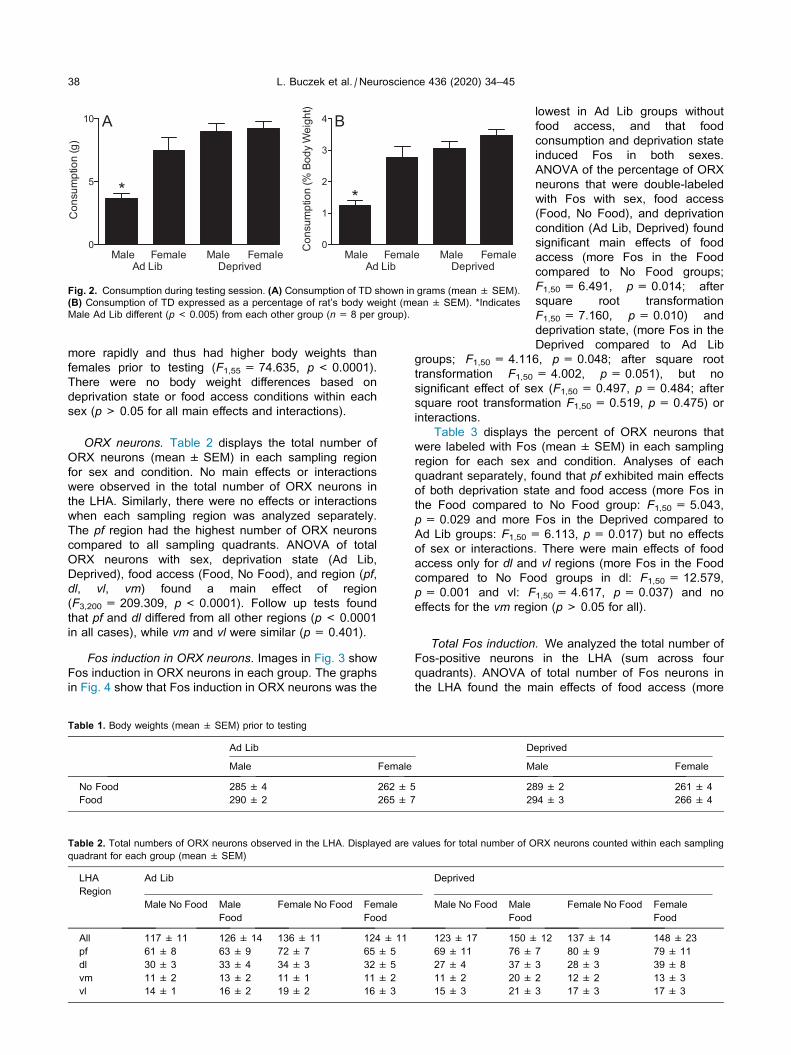
indicates a difference in TD consumption between Ad Lib and
Deprived males (p= 0.001; n= 8 per group).
42 L. Buczek et al. / Neuroscience 436 (2020) 34–45
(Klump et al., 2013). In another study with a conditioned
place preference task, they found that females had a
more pronounced shift in preference for the chamber that
was paired with palatable food during training compared
to males. Females also had longer feeding bouts and con-
sumed more food than males (standardized per body
weight) during training sessions when food was available
in the chamber (Sinclair et al., 2017). Another study
recently compared operant responding and consumption
of sucrose pellets in male and female rats after acute
(24 h) deprivation or ad libitum food access (Tapia
et al., 2019). That study found that female rats had higher
responding to obtain sucrose pellets in a progressive ratio
schedule of reinforcement and consumed more pellets
than males in fasted and sated states. Current results
add to that prior work by revealing that differences exist
between the sexes in hedonic eating before any training,
and that females differ from males in consumption of
palatable foods when sated. Together, prior and current
findings in animals are relevant to human eating behavior.
Collectively the findings suggest that female biological
vulnerability to hedonic eating may be casual to binge
and other forms of overeating and associated disorders.
ORX neurons: activation and signaling duringpalatable feeding tests
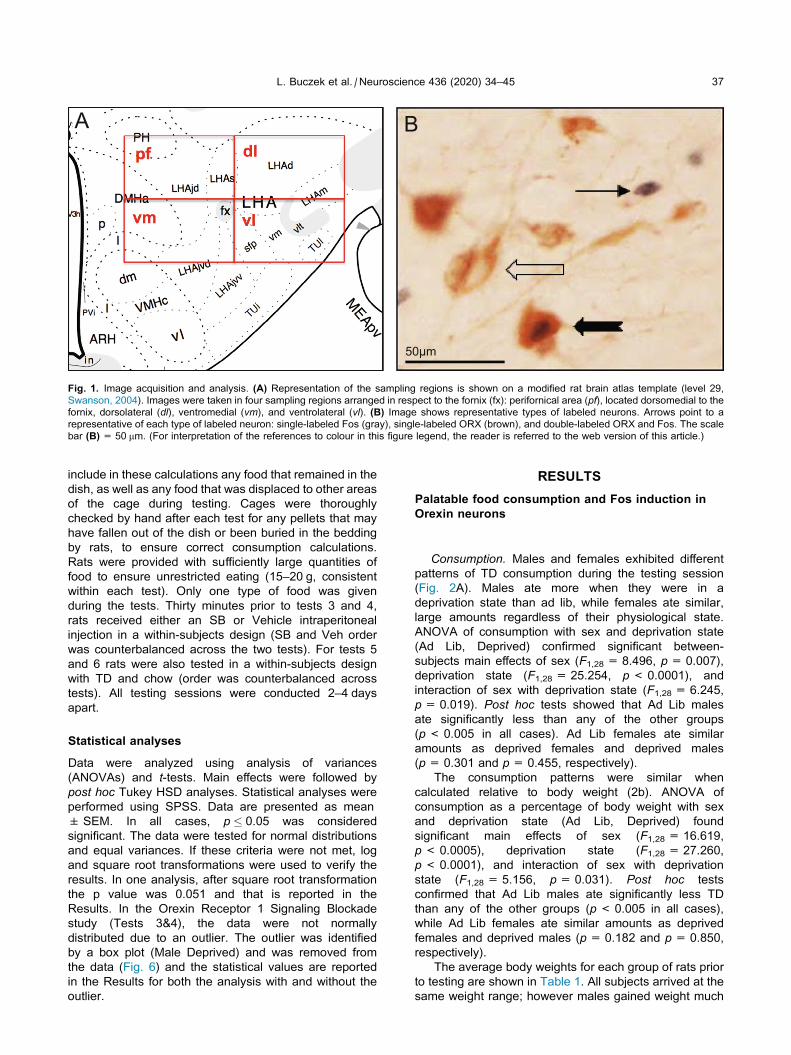
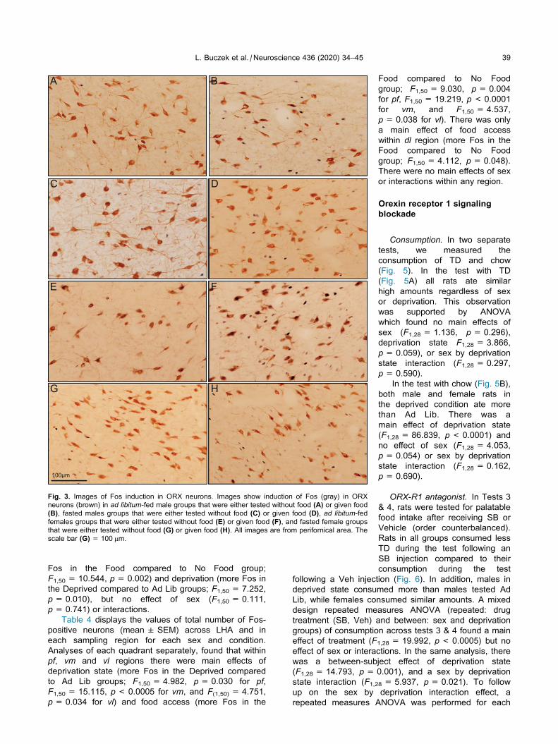
Fos induction in ORX neurons during palatable feedingtests. To begin to determine the neural substrates
underlying hedonic eating and sex differences, we
characterized Fos induction in ORX neurons during a
palatable feeding test in males and females. We
systematically counted ORX neurons across the entire
rostro-caudal extent of the LHA, within four adjacent
quadrants that were placed dorsomedially,
dorsolaterally, ventromedially and ventrolaterally in
respect to fornix. This parcellation was chosen in order
to reliably compare different groups of ORX neurons
across all conditions. In terms of density of ORX
neurons, each quadrant differed from the others. The pfquadrant was the densest, followed by dl, vl, and vmquadrants. These patterns generally match prior
observations (de Lecea et al., 1998; Sakurai et al.,
1998; Swanson et al., 2005; Yoshida et al., 2006).
We analyzed total number of ORX neurons across the
four quadrants together, as well as within each quadrant
separately. There were no differences in the number of
ORX neurons across the LHA, or in any of the four
quadrants analyzed, across any groups. Previous
research found higher ORX-A content and mRNA in the
hypothalamus in females compared to males, as well as
higher body weight in female ORX-knockouts (Taheri
et al., 1999; Ramanathan and Siegel, 2014; Grafe et al.,
2017). Here, we identified ORX neurons with the immuno-
histochemistry method, which does not linearly corre-
spond to the substrate quantities beyond the threshold
for labeling. Thus, we cannot rule out the possibility that
males and females produce different amounts of ORX.
Furthermore, levels of ORX and its receptors in the brain
tissue were shown to vary in female rats across estrous
cycle (Russell et al., 2001; Wang et al., 2003; Porkka-
Heiskanen et al., 2004); for a review see (Grafe and
Bhatnagar, 2018). We did not monitor estrous cycling, in
order to avoid potentially stressful effects of that proce-
dure that could impact food intake.
Food and fasting independently impacted Fos
induction in ORX neurons. Rats that had access to TD
pellets during testing had higher numbers of ORX
neurons with Fos compared to rats that were not given
food during testing. That recruitment of ORX neurons
could be due to anticipation of feeding (sight of food)
and associated induction of the drive to eat, as well as
the ingestive (palatability) and postingestive effects of
consumption (Cai et al., 1999; Gonzalez et al., 2016).
Nevertheless, these patterns suggest that ORX neurons
may be driving palatable intake in hungry and sated
states, which we tested in the second study.
Fasting also increased Fos induction in ORX neurons.
Rats that were fasted prior to testing had more Fos
induction in ORX neurons than rats that had ad libitumaccess to food. These findings are in agreement with
the original observations that fasting upregulates ORX
mRNA and that ORX mediates activity/arousal
associated with energy balance changes (Sakurai et al.,
1998; Cai et al., 1999; Yamanaka et al., 2003); for a
review see (Barson and Leibowitz, 2017). Previously,
Funabashi et al. (2009) found that fasting activated more
ORX neurons in females compared to males. The current
study found fasting-induced activation was similar in both
sexes. The discrepancy could be due to a longer fast in
Funabashi et al., study (48 h versus 20 h) or differences
in the marker of neural activity used (pCREB versus Fos).
Interestingly, we found different activation patterns
based on location of ORX neurons. ORX neurons that
were responsive to both food and fasting were located
L. Buczek et al. / Neuroscience 436 (2020) 34–45 43
within the pf (dorsomedial) quadrant. Food, but not
fasting, activated dorsolateral and ventrolateral
quadrants (dl, vl), while neither activated vm quadrant.
An important task for future research is to determine the
connectional targets within the reward system of these
different groups of ORX neurons, and how they are
engaged during hedonic eating (Harris et al., 2005;
Zheng et al., 2007; Ho and Berridge, 2013; Castro
et al., 2016; Ferrario et al., 2016).
In addition to the analysis of ORX neurons, we
counted total Fos induction in all neurons within the LHA
and found similar patterns. Groups that had food access
and groups that were fasted prior to testing had higher
numbers of neurons with Fos than corresponding
controls. The pf, vm and vl quadrants had more Fos
induction in the food and fasted conditions, while within
the dl region only food access impacted Fos induction.
The food and fasting associated Fos induction
patterns within the LHA in the current study are in
agreement with prior work. Most prior work was
exclusively in males but a study that compared males
and females found no sex differences in hypothalamic
activation, as in the current study (Sinclair et al., 2017).
However, another study found more Fos induction in
ORX neurons in females compared to males under con-
trol and repeated restraint stress conditions (Grafe
et al., 2017). Food anticipation in food-entrained rats
has also been shown to induce Fos within the LHA,
including in ORX neurons (Johnstone et al., 2006). In
the current study rats were not trained to expect feeding,
however, they were familiar with the pellets and Fos may
be due to some anticipatory activity in addition to
consumption.
Our findings are well matched to the patterns of Zseli
et al. study (Zseli et al., 2016) that examined satiety net-
work recruitment during refeeding (for 2 h) after fasting
(for 40 h) in male rats and found dense Fos induction
across the LHA. They found dense Fos in both fasted
and re-fed rats within the suprafornical (LHAs) and juxta-
dorsomedial (LHAjd) regions (within our pf quadrant), thedorsal zone (LHAjvd) (within our vm quadrant), and poste-
rior (LHAsfp) and medial (LHAvm) zones (within our vlquadrant), as well as the dorsal (LHAd) region (within
our dl quadrant). We found that in all quadrants with these
regions (pf, vl, vm), food access and fasting increased
Fos induction, except in dl (which includes LHAd) where
only food, but not fasting, increased the number of neu-
rons with Fos. However, Zseli et al., study did not have
a non-fasted condition, which precludes direct compar-
isons of fasting-induced activation across the two studies.
ORX signaling during palatable feeding tests. The first
study showed that ORX neurons were activated in both
fasted and sated food groups. To follow up on these
findings, we tested whether ORX receptor 1 signaling
mediates palatable food intake regardless of hunger/
satiety state. Rats of both sexes in fasted and sated
conditions consumed less TD after receiving SB prior to
testing, compared to the amounts they consumed after
receiving a vehicle. These data indicate that ORX
signaling via receptor 1 drives hedonic eating regardless
of physiological hunger in both sexes. This is in
agreement with prior work in males, and with the notion
that ORX is critical in driving non-homeostatic hunger
and the overconsumption of food and drugs (Rodgers
et al., 2001; Choi et al., 2010; Barson and Leibowitz,
2017).
Previously, Cason and Aston-Jones (2014) found that
SB decreased sucrose self-administration only in fasted
but not in ad libitum-fed females. In the current study
there was an overall effect of SB across fasted and ad libi-tum-fed rats of both sexes. A difference in the amount of
effort needed to obtain palatable food in the two studies—
free feeding versus lever-pressing—may be the reason
for different findings in the two studies.
Furthermore, in these tests, consumption patterns of
males and females were consistent with sex differences
we found in the first experiment. Fasted males
consumed more than sated males, while both fasted
and fed females consumed similar amounts. Thus,
across four tests in two studies with different animals,
females consistently consumed based on palatability,
while males consumed according to their physiological
need in three tests.
Sex differences
Interestingly, there were no sex differences in Fos
induction in ORX neurons during palatable feeding tests.
Furthermore, blockade of ORX signaling decreased
consumption in both sexes. These results indicate
that ORX is important for hedonic eating in both
sexes, and may not be key to sex differences.
However, there are methodological limitations that may
have precluded the detection of the role of ORX in
behavioral sex differences. As discussed above, the
immunohistochemistry methods used here to identify
ORX neurons and Fos induction are semi-quantitative
methods that do not linearly track the quantities of
substrates. Males and females may have different
receptor sensitivities and a lower dose of SB may be
needed to detect sex differences (Cason and Aston-
Jones, 2014; Barson, 2018). The dose used in the current
study was higher than the lowest dose (3 mg/kg) shown to
impact palatable intake (Barson, 2018) but it did not pro-
duce non-specific locomotor effects (Cole et al., 2015;
Keefer et al., 2016). Nevertheless, this dose could have
potentially activated orexin 2 receptors in addition to
ORXR1 and could have even affected other neurochemi-
cals (Scammell and Winrow, 2011; Barson, 2018). Simi-
larly, systemic pharmacological manipulations do not
address specific targets of action. Thus, different amounts
of ORX may be released in males and females from the
same number of neurons. Furthermore, differences in
ORX targets and distribution of ORX receptors may medi-
ate hedonic eating differently in males and females. Sup-
porting this idea, more Fos was found within the
infralimbic cortex and the dorsal shell of the nucleus
accumbens in females compared to males after con-
sumption of palatable food (Sinclair et al., 2017). These
areas contain ORX fibers and receptors (Peyron et al.,
1998; Marcus et al., 2001; Baldo et al., 2003) and ORX
infusions into the nucleus accumbens enhanced hedonic
44 L. Buczek et al. / Neuroscience 436 (2020) 34–45
responses to sucrose and consumption (Castro et al.,
2016). Furthermore, sex differences have been shown
in a prominent ORX target, the paraventricular nucleus
of the thalamus, during renewal of responding to cues
for the same palatable food used in the current study
(Anderson and Petrovich, 2017).
In conclusion, this comprehensive behavioral and
neural analysis in intact, adult males and females is a
first step in characterizing similarities and differences
between the sexes in hedonic eating (McCarthy et al.,
2012). These findings reveal important sex differences
in feeding behavior that warrant further investigation. Of
notable interest are potential targets of ORX signaling
as substrates underlying eating dysregulation in males
and females.
ACKNOWLEDGEMENTS
This work was supported by the National Institutes of
Health, NIDDK grant R01DK085721 to GDP. A portion
of the research reported here partially fulfilled the
requirements for the Senior Honors Thesis awarded to
JM by Boston College. We thank Dr. Ehri Ryu and Dr.
Amanda Madden for helpful advice regarding statistical
analyses.
REFERENCES
Anderson LC, Petrovich GD (2017) Sex specific recruitment of a
medial prefrontal cortex-hippocampal-thalamic system during
context-dependent renewal of responding to food cues in rats.
Neurobiol Learn Mem 139:11–21.
Baldo BA, Daniel RA, Berridge CW, Kelley AE (2003) Overlaping
distribution of orexin/hypocretin- and dopamine-b-hydroxylase
immunoreactive fibers in rat brain regions mediating arousal,
motivation, and stress. J Comp Neurol 464:220–237.
Barson JR, Leibowitz SF (2017) Orexin/hypocretin system: Role in
food and drug overconsumption. Int Rev Neurobiol 136:199–237.
Barson JR (2018) Orexin/hypocretin and dysregulated eating:
Promotion of foraging behavior. Brain Res (in press).
Berridge KC (2000) Measuring hedonic impact in animals and infants:
microstructure of affective taste reactivity patterns. Neurosci
Biobehav Rev 24:173–198.
Berridge KC, Ho CY, Richard JM, DiFeliceantonio AG (2010) The
tempted brain eats: pleasure and desire circuits in obesity and
eating disorders. Brain Research 1350:43–64.
Berthoud H-R (2012) The neurobiology of food intake in an
obesogenic environment. Proc Nutr Soc 71:478–487.
Boggiano MM, Dorsey JR, Thomas JM, Murdaugh DL (2009) The
Pavlovian power of palatable food: lessons for weight-loss
adherence from a new rodent model of cue-induced overeating.
Int J Obes 33:693–701.
Cai XJ, Widdowson PS, Harrold J, Wilson S, Buckingham RE, Arch
JRS, Tadayyon M, Clapham JC, Wilding J, Williams G (1999)
Hypothalamic orexin expression: modulation by blood glucose
and feeding. Diabetes 48:2132–2137.
Cason AM, Aston-Jones G (2014) Role of orexin/hypocretin in
conditioned sucrose-seeking in female rats. Neuropharmacology
86:97–102.
Castro DC, Terry RA, Berridge KC (2016) Orexin in rostral hotspot of
nucleus accumbens enhances sucrose ‘liking’ and intake but
scopolamine in caudal Shell shifts ‘liking’ toward ‘disgust’ and
‘fear’. Neuropharmacology 41:2101–2111.
Choi DL, Davis JF, Fitzerald ME, Benoit SC (2010) The role of orexin-
A in food motivation, reward-based feeding behavior and food-
induced neuronal activation in rats. Neuroscience 167:11–20.
Cole S, Mayer HS, Petrovich GD (2015) Orexin/hypocretin-1 receptor
antagonism selectively reduces cue-induced feeding in sated rats
and recruits medial prefrontal cortex and thalamus. Sci Rep
5:16143.
Cota D, Tschop MH, Horvath TL, Levine AS (2006) Cannabinoids,
opioids and eating behavior: the molecular face of hedonism?
Brain Res Rev 51:85–107.
de Lecea L, Kilduff TS, Peyron C, Gao X, Foye PE, Danielson PE,
Fukuhara C, Battenberg EL, Gautvik VT, Bartlett FS, Frankel WN,
van den Pol AN, Bloom FE, Gautvik KM, Sutcliffe JG (1998) The
hypocretins: hypothalamus-specific peptides with neuroexcitatory
activity. Proc Natl Acad Sci USA 95:322–327.
Ferrario CR, Labouebe G, Liu S, Nieh EH, Routh VH, Xu S, O’Connor
EC (2016) Homeostasis meets motivation in the battle to control
food intake. J Neurosci 36:11469–11481.
Freund N, Thompson BS, Norman KJ, Einhorn P, Andersen SL
(2015) Developmental emergence of an obsessive-compulsive
phenotype and binge behavior in rats. Psychopharmacology
232:3173–3181.
Funabashi T, Hagiwara H, Mogi K, Mitsushima D, Shinohara K,
Kimura F (2009) Sex differences in the responses of orexin
neurons in the lateral hypothalamic area and feeding behavior to
fasting. Neurosci Lett 463:31–34.
Gonzalez JA, Jensen LT, Iordanidou P, Strom M, Fugger L, Burdakov
D (2016) Inhibitory interplay between orexin neurons and eating.
Curr Biol 26:2486–2491.
Grafe LA, Bhatnagar S (2018) The contribution of orexins to sex
differences in the stress response. Brain Res (in press).
Grafe LA, Cornfeld A, Luz S, Valentino R, Bhatnagar S (2017)
Orexins mediate sex differences in the stress response and in
cognitive flexibility. Biol Psychiatry 81:683–692.
Harris GC, Wimmer M, Aston-Jones G (2005) A role for lateral
hypothalamic orexin neurons in reward seeking. Nature
437:556–559.
Ho CY, Berridge KC (2013) An orexin hotspot in ventral pallidum
amplifies hedonic ‘liking’ for sweetness.
Neuropsychopharmacology 38:1655–1664.
Hudson JI, Hiripi E, Pope HG, Kessler RC (2007) The prevalence and
correlates of eating disorders in the national comorbidity survey
replication. Biol Psychiatry 61:348–358.
Johnstone LE, Fong TM, Leng G (2006) Neuronal activation in the
hypothalamus and brainstem during feeding in rats. Cell Metab
4:313–321.
Keefer SE, Cole S, Petrovich GD (2016) Orexin/hypocretin receptor 1
signaling mediates Pavlovian cue-food conditioning and
extinction. Physiol Behav 162:27–36.
Kendig MD, Boakes RA, Corbit LH (2018) Variety overcomes the
specificity of cue-potentiated feeding in rats. J Exp Psychol
44:56–66.
Klump KL, Racine S, Hildebrandt B, Sisk CL (2013) Sex differences in
binge eating patterns in male and female adult rats. Int J Eat
Disord 46:729–736.
Mahler SV, Moorman DE, Smith RJ, James MH, Aston-Jones G
(2014) Motivational activation: a unifying hypothesis of orexin/
hypocretin function. Nat Neurosci 17:1298–1303.
Marcus JN, Aschkenasi CJ, Lee CE, Chemelli RM, Saper CB,
Yanagisawa M, Elmquist JK (2001) Differential expression of
orexin receptors 1 and 2 in the rat brain. J Comp Neurol
435:6–25.
McCarthy MM, Arnold AP, Ball GF, Blaustein JD, De Vries GJ (2012)
Sex differences in the brain: the not so inconvenient truth. J
Neurosci 32:2241–2247.
Mitchell S, Shaw D (2015) The worldwide epidemic of female obesity.
Best Pract Res Clin Obstet Gynaecol 29:289–299.
Petrovich GD (2013) Forebrain networks and the control of feeding by
environmental learned cues. Physiol Behav 121:10–18.
Petrovich GD (2019) Orexins and control of feeding by learned cues.
In: Fadel JR, Burk JA, editors. The orexins/hypocretins system:
functional roles and therapeutic potential. Academic Press. p.
85–98.
L. Buczek et al. / Neuroscience 436 (2020) 34–45 45
Peyron C, Tighe DK, van den Pol AN, de Lecea L, Heller HC, Sutcliffe
JG, Kilduff TS (1998) Neurons containing hypocretin (orexin)
project to multiple neuronal systems. J Neurosci 18:9996–10015.
Porkka-Heiskanen T, Kalinchuk A, Alanko L, Huhtaniemi I, Stenberg
D (2004) Orexin A and B levels in the hypothalamus of female
rats: the effects of the estrous cycle and age. Eur J Endocrinol
150:737–742.
Ramanathan L, Siegel JM (2014) Gender differences between
hypocretin/orexin knockout and wild type mice: age, body
weight, body composition, metabolic markers, leptin and insulin
resistance. J Neurochem 131:615–624.
Reichelt AC, Abbott KN, Westbrook RF, Morris MJ (2016) Differential
motivational profiles following adolescent sucrose access in male
and female rats. Physiol Behav 157:13–19.
Reppucci CJ (2010) Chronic stress, food consumption, and emotional
behavior in rats. Master’s thesis. Boston College.
Reppucci CJ, Petrovich GD (2012) Learned food-cue stimulates
persistent feeding in sated rats. Appetite 59:437–447.
Rodgers RJ, Halford JCG, Nunes de Souza RL, Canto de Souza AL,
Piper DC, Arch JRS, Upton N, Porter RA, Blundell JE (2001) SB-
334867, a selective orexin-1 receptor antagonist, enhances
behavioural satiety and blocks the hyperphagic effect of orexin-
A in rats. Eur J Neurosci 13:1444–1452.
Russell SH, Small CJ, Kennedy AR, Stanley SA, Seth A, Murphy KG,
Taheri S, Ghatei MA, Bloom SR (2001) Orexin A interactions in
the hypothalamo-pituitary gonadal axis. Endocrinology
142:5294–5302.
Sakurai T, Amemiya A, Ishii M, Matsuzaki I, Chemelli RM, Tanaka H,
Williams SC, Richarson JA, Kozlowski GP, Wilson S, Arch JRS,
Buckingham RE, Haynes AC, Carr SA, Annan RS, McNulty DE,
Liu WS, Terrett JA, Elshourbagy NA, Bergsma DJ, Yanagisawa M
(1998) Orexins and orexin receptors: a family of hypothalamic
neuropeptides and G protein-coupled receptors that regulate
feeding behavior. Cell 92:573–585.
Scammell TE, Winrow CJ (2011) Orexin receptors: pharmacology
and therapeutic opportunities. Annu Rev Pharmacol Toxicol
51:243–266.
Sinclair EB, Hildebrandt BA, Culbert KM, Klump KL, Sisk CL (2017)
Preliminary evidence of sex differences in behavioral and neural
responses to palatable food reward in rats. Physiol Behav
176:165–173.
Small DM (2009) Individual differences in the neurophysiology of
reward and the obesity epidemic. Int J Obesity 33:S44–S48.
Stice E, Figlewicz DP, Gosnell BA, Levine AS, Pratt WE (2013) The
contribution of brain reward circuits to the obesity epidemic.
Neurosci Biobehav Rev 37:2047–2058.
Sun X, Kroemer NB, Veldhuizen MG, Babbs AE, de Araujo IE,
Gitelman DR, Sherwin RS, Sinha R, Small DM (2015) Basolateral
amygdala response to food cues in the absence of hunger is
associated with weight gain susceptibility. J Neurosci
35:7964–7976.
Swanson LW (2004). In: Brain Maps: Structure of the Rat Brain. A
Laboratory Guide with Printed and Electronic Templates for Data,
Models and Schematics., 3rd revised Edition with CD-
ROM. Edition: Elsevier, Amsterdam.
Swanson LW, Sanchez-Watts G, Watts AG (2005) Comparison of
melanin-concentrating hormone and hypocretin/orexin mRNA
expression patterns in a new parceling scheme of the lateral
hypothalamic zone. Neurosci Lett 387:80–84.
Taheri S, Mahmoodi M, Opacka-Juffry J, Ghatei MA, Bloom SR
(1999) Distribution and quantification of immunoreactive orexin A
in rat tissues. FEBS Lett 457:157–161.
Tapia MA, Lee JR, Weise VN, Tamasi AM, Will MJ (2019) Sex
differences in hedonic and homeostatic aspects of palatable food
motivation. Behav Brain Res 359:396–400.
Wang JB, Murata T, Narita K, Honda K, Higuchi T (2003) Variation in
the expression of orexin and orexin receptors in the rat
hypothalamus during the estrous cycle, pregnancy, parturition,
and lactation. Endocrine 22:127–134.
Weingarten HP (1983) Conditioned cues elicit feeding in sated rats: a
role for learning in meal initiation. Science 220:431–433.
Yamanaka A, Beuckmann CT, Willie JT, Hara J, Tsujino N, Mieda M,
Yagami K, Sugiyama F, Goto K, Yanagisawa M, Sakurai T (2003)
Hypothalamic orexin neurons regulate arousal according to
energy balance in mice. Neuron 38:701–713.
Yoshida K, McCormack S, Espana RA, Crocker A, Scammell T
(2006) Afferents to the orexin neurons of the rat brain. J Comp
Neurol 494:845–861.
Zheng H, Patterson LM, Berthoud HR (2007) Orexin signaling in the
ventral tegmental area is required for high-fat appetite induced by
opioid stimulation of the nucleus accumbens. J Neurosci
27:11075–11082.
Zseli G, Vida B, Martinez A, Lechan RM, Khan AM, Fekete C (2016)
Elucidation of the anatomy of a satiety network: Focus on
connectivity of the parabrachial nucleus in the adult rat. J Comp
Neurol 524:2803–2827.
(Received 4 October 2019, Accepted 5 April 2020)(Available online 10 April 2020)