2/17/2015
1
©2014 National Pressure Ulcer Advisory Panel | www.npuap.org
Heel Pressure Ulcers: 2014 International Pressure Ulcer Prevention & Treatment Guidelines
Diane Langemo, PhD, RN, FAAN
Objectives
• Discuss heel anatomy & physiology as it
contributes to pressure ulcer development
• Discuss the NPUAP-EPUAP 2014 International
Pressure Ulcer Prevention & Treatment
Guidelines for heels
• Discuss evidence-based heel pressure ulcer
prevention & treatment interventions
©2014 National Pressure Ulcer Advisory Panel | www.npuap.org2
2/17/2015
2
Heel Pressure Ulcers
©2014 National Pressure Ulcer Advisory Panel | www.npuap.org3
#1
facility
acquired
PU
Debilitating
& painful
Time to
closure &
healing can
be long
#2
overall
PU
Common for surgery
pts, hip fx pts, TKA,
debilitated, sedated,
Etc
Heel Anatomy & Physiology
• Calcaneous is largest bone of the foot & is angular
• ↓fatty tissue: heel soft tissue pad = 18mm thick; Skin
0.64mm thick
• Dermis thins, dermal-epidermal junction flattens,
↑separation, ↑ risk blister formation (↓shock absorption)
• Sustained, high pressure over small contact area
• Skin thins also with diabetic neuropathy
(↓ shock absorption)
• Supine position yields highest risk
• Friction & shear when “sliding down” in bed
©2014 National Pressure Ulcer Advisory Panel | www.npuap.org4
2/17/2015
3
Heel Anatomy & Physiology
• Blood supply: End arterial
plexus from posterior tibial
artery & peroneal artery
• Blood flow impaired by peripheral vascular disease
• Impaired tolerance for ischemia due to lack of subcutaneous tissue & ↑ blood vessel degeneration
• Bottom Line: heel is not
well protected from
pressure, friction or shear
• Rapid damage can occur
©2011 National Pressure Ulcer Advisory Panel | www.npuap.org5
What Makes the Heel Vulnerable?
• ↓resting blood perfusion levels
• Calcaneus has small surface w/large wt bearing bony prominence
• Result: higher than normal pressures
• Study of 450,000 pts, heel accounted for 41% of all DTIs (Van Gilder et al, 2008)
©2011 National Pressure Ulcer Advisory Panel | www.npuap.org6
2/17/2015
4
Consider that….
• If the heel has little
subcutaneous tissue
or muscle; and IF the
muscle is MOST
vulnerable, is it not
highly likely pressure
or pressure + shear
will result in a PU
©2011 National Pressure Ulcer Advisory Panel | www.npuap.org7
Heel
Bed
What Places the Heel at Risk?
• Heavy weight of foot
• Sharp posterior calcaneus
• Thin soft tissue padding; little to no muscle
• Blood supply via peroneal artery
• Diabetes
• Edema
• Leg spasms, agitation
• Dementia
• Muscle very sensitive to pressure & ischemia
• Poor perfusion/vasopressors
• IMMOBILITY: ≈87% of the cases; #1 PU risk factor
©2014 National Pressure Ulcer Advisory Panel | www.npuap.org8
2/17/2015
5
Risk Related to Heel Position
©2014 National Pressure Ulcer Advisory Panel | www.npuap.org9
Risk Factors for Heel PUs
• Extrinsic Factors:
– Pressure
– Friction
– Shear
• Intrinsic Factors:– Age (↓cushioning)
– ↓mobility & activity
– Malnut/dehydration
– ↓Sensation, ↓LOC
– Vascular disease
– Fragile skin/edema
– Underlying comorbidities
– Contractures
– “Hammocking” of mattress
©2011 National Pressure Ulcer Advisory Panel | www.npuap.org10
2/17/2015
6
Additional Heel PU Risk Factors
• Agitated patients
• Diabetes mellitus
• Peripheral neuropathy
• Arterial insufficiency
• Malnutrition
• Poor tissue resilience
• Braden 16 or less
• Braden 17-18 + addtl
risk factor
• Paralysis/epidural
• Knee/hip
replacements
• Post open heart
surgery
• Push self up in bed
with heels
• Not removing
compression hose
• Leg spasms
• >50% of perioperative
PUs©2011 National Pressure Ulcer Advisory Panel | www.npuap.org11
Risk Reduction Pre-Admission
©2014 National Pressure Ulcer Advisory Panel | www.npuap.org12
The legal system believes that if you admitted the patient or resident you must feel you can provide care that meets the standard of care
Be certain you have the staff, equipment and training to handle the patient or resident
Problem of timely insurance approvals for some specializedproducts/devices
2/17/2015
7
Assessing the Heel
©2014 National Pressure Ulcer Advisory Panel | www.npuap.org13
Assessing Risk of Heel PUs
• Is the Braden Scale score the best predictor? Both total & individual subscale scores important.
• Are there other independent factors that increase risk such as medical problems?– Is a hip fracture different from immobility?
– Hip & knee joint replacements
– Does the Buck’s traction score as inactivity?
– Is DM neuropathy different from impaired cognition?
– How is the lack of blood flow to the feet scored?©2014 National Pressure Ulcer Advisory Panel | www.npuap.org14
2/17/2015
8
General Recommendations
• Inspect the skin of the heels regularly. (SOE=C;
SOR=
©2014 National Pressure Ulcer Advisory Panel | www.npuap.org15
Assessment
• Remove devices:
– Stockings
– Support hose
– Boots
• Reinspect once “device” removed
• Look up, look down, look all around!
• Feel: for warmth, bogginess, pulses
©2014 National Pressure Ulcer Advisory Panel | www.npuap.org16
2/17/2015
9
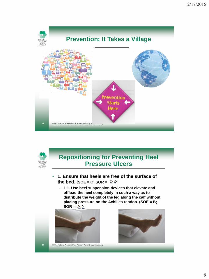
Prevention: It Takes a Village
©2014 National Pressure Ulcer Advisory Panel | www.npuap.org17
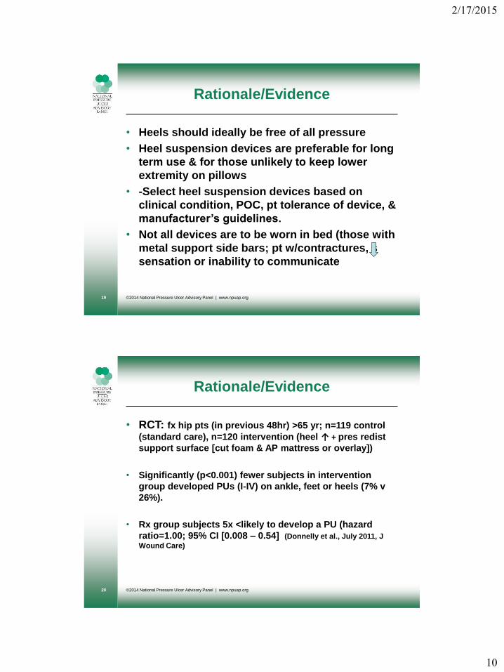
Repositioning for Preventing Heel Pressure Ulcers
• 1. Ensure that heels are free of the surface of
the bed. (SOE = C; SOR = 2+)
– 1.1. Use heel suspension devices that elevate and
offload the heel completely in such a way as to
distribute the weight of the leg along the calf without
placing pressure on the Achilles tendon. (SOE = B;
SOR = )
©2014 National Pressure Ulcer Advisory Panel | www.npuap.org18
2/17/2015
10
Rationale/Evidence
• Heels should ideally be free of all pressure
• Heel suspension devices are preferable for long
term use & for those unlikely to keep lower
extremity on pillows
• -Select heel suspension devices based on
clinical condition, POC, pt tolerance of device, &
manufacturer’s guidelines.
• Not all devices are to be worn in bed (those with
metal support side bars; pt w/contractures, s
sensation or inability to communicate
©2014 National Pressure Ulcer Advisory Panel | www.npuap.org19
Rationale/Evidence
• RCT: fx hip pts (in previous 48hr) >65 yr; n=119 control
(standard care), n=120 intervention (heel ↑ + pres redist
support surface [cut foam & AP mattress or overlay])
• Significantly (p<0.001) fewer subjects in intervention
group developed PUs (I-IV) on ankle, feet or heels (7% v
26%).
• Rx group subjects 5x <likely to develop a PU (hazard
ratio=1.00; 95% CI [0.008 – 0.54] (Donnelly et al., July 2011, J
Wound Care)
©2014 National Pressure Ulcer Advisory Panel | www.npuap.org20
2/17/2015
11
Heel Device TIPS
• The device should completely lift the heel
from the bed
• Do the paper or hand test
• Boots: foam, air, fiber, plastic, sheepskin,
synthetic sheepskin
• Alignment should be neutral
• Rigid Boots (AFO): for maintenance of
alignment in 90° neutral position
©2014 National Pressure Ulcer Advisory Panel | www.npuap.org21
Repositioning for Preventing Heel Pressure Ulcers
• 2. The knee should be in slight (5° to 10°)
flexion. (SOE = C; SOR = )
©2014 National Pressure Ulcer Advisory Panel | www.npuap.org22
2/17/2015
12
Rationale/Evidence
• Knee hyperextension can cause popliteal vein
obstruction, predisposing to DVT (Indirect
evidence)
• (Huber & Huber, Eur J Vascular & Endovascular Surgery, 2009)
©2014 National Pressure Ulcer Advisory Panel | www.npuap.org23
Repositioning for Preventing Heel Pressure Ulcers
• 3. Avoid areas of high pressure,
especially under the Achilles tendon. (SOE = C; SOR = ))
©2014 National Pressure Ulcer Advisory Panel | www.npuap.org24
2/17/2015
13
Repositioning for Preventing Heel Pressure Ulcers
– 3.1. Use a foam cushion under the full length of the
calves to elevate heels. (SOE = B; SOR = ))
©2014 National Pressure Ulcer Advisory Panel | www.npuap.org25
Rationale/Evidence
• RCT: ICU pts (n=70), foam cushion (n=35) compared to
no heel intervention (n=35). Fewer heel PU in foam group
(8.5% vs 54.2%), also longer heel PU-free time in foam
group (5.6d vs 2.8d) (Cadue et al., Presse Macdiacale, 2008)
• Pillows under full length of calf appropriate for short-
term use in alert & cooperative pts.
• Pillows or foam cushions should extend length of
calf to avoid pressure (eg, Achilles area).
• Flex knee slightly to avoid popliteal vein compression
& DVT risk.
©2014 National Pressure Ulcer Advisory Panel | www.npuap.org26
2/17/2015
14
Repositioning for Preventing Heel Pressure Ulcers
• 4. Apply heel suspension devices according
to the manufacturer’s instructions. (SOE = C;
SOR = ))
©2014 National Pressure Ulcer Advisory Panel | www.npuap.org27
Repositioning for Preventing Heel Pressure Ulcers
• 5. Remove the heel suspension device
periodically to assess skin integrity. (SOE
= C; SOR = )).
©2014 National Pressure Ulcer Advisory Panel | www.npuap.org28
2/17/2015
15
Rationale/Evidence
• Routinely assess skin under device for
pressure-related damage.
• Assess skin more frequently & loosen device in
individuals with, or likely to develop, lower
extremity edema & individuals with neuropathy
& PAD.
©2014 National Pressure Ulcer Advisory Panel | www.npuap.org29
Rationale/Evidence
• Apply heel suspension devices to avoid creation
of areas of increased pressure under the device.
• Ensure device is not too tight & doesn’t create
additional pressure damage.
©2014 National Pressure Ulcer Advisory Panel | www.npuap.org30
2/17/2015
16
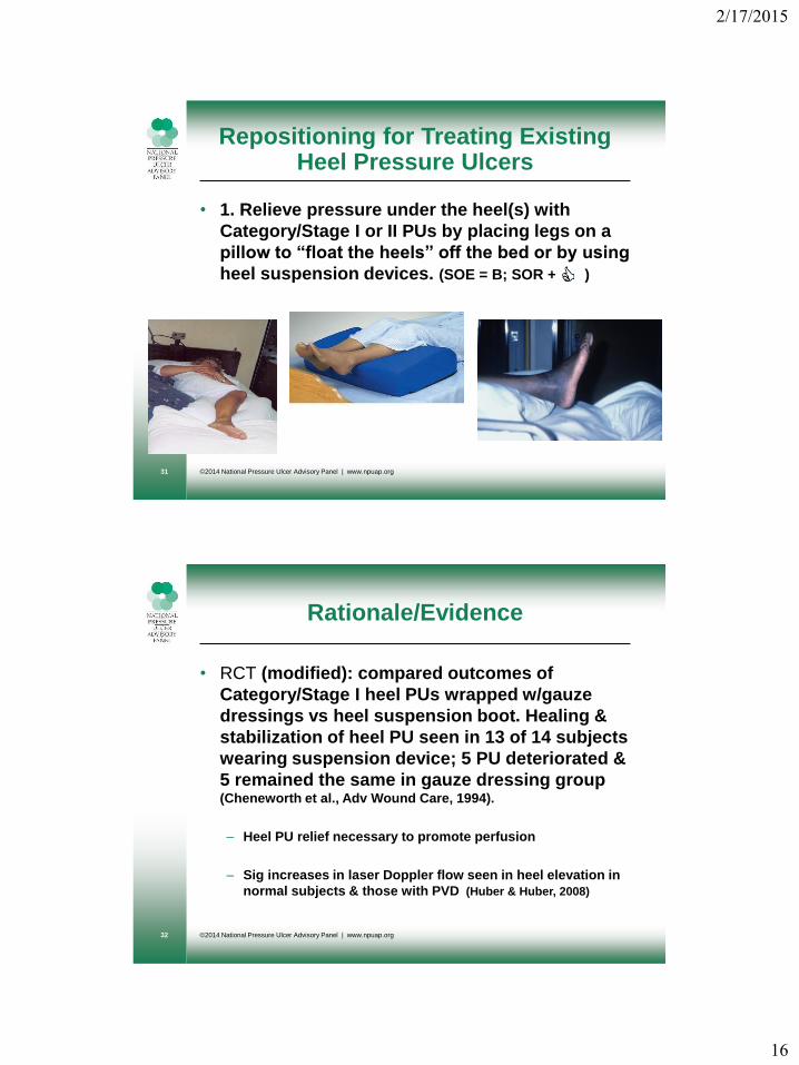
Repositioning for Treating Existing Heel Pressure Ulcers
• 1. Relieve pressure under the heel(s) with
Category/Stage I or II PUs by placing legs on a
pillow to “float the heels” off the bed or by using
heel suspension devices. (SOE = B; SOR + )
©2014 National Pressure Ulcer Advisory Panel | www.npuap.org31
Rationale/Evidence
• RCT (modified): compared outcomes of
Category/Stage I heel PUs wrapped w/gauze
dressings vs heel suspension boot. Healing &
stabilization of heel PU seen in 13 of 14 subjects
wearing suspension device; 5 PU deteriorated &
5 remained the same in gauze dressing group (Cheneworth et al., Adv Wound Care, 1994).
– Heel PU relief necessary to promote perfusion
– Sig increases in laser Doppler flow seen in heel elevation in
normal subjects & those with PVD (Huber & Huber, 2008)
©2014 National Pressure Ulcer Advisory Panel | www.npuap.org32
2/17/2015
17
Repositioning for Treating Existing Heel Pressure Ulcers
• 2. For Category/Stage III, IV and unstageable
PUs, place the leg in a device that elevates the
heel from the surface of the bed, completely
offloading the PU. Consider a device that
prevents footdrop. (SOE = C; SOR = )
©2014 National Pressure Ulcer Advisory Panel | www.npuap.org33
Rationale/Evidence
• Category/Stage III, IV or unstageable heel PUs
should be completely offloaded to the extent
possible.
• Elevating the heel on a pillow is usually
inadequate.
• Due to the time required to heal deeper ulcers, a
device that completely offloads the ulcer area &
prevents footdrop is preferred.
©2014 National Pressure Ulcer Advisory Panel | www.npuap.org34
2/17/2015
18
Always…Educate
• Educate patient on how to position heels
& rationale for need to do so
©2014 National Pressure Ulcer Advisory Panel | www.npuap.org35
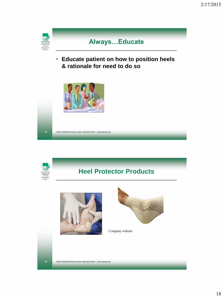
Heel Protector Products
©2014 National Pressure Ulcer Advisory Panel | www.npuap.org36
Company website
2/17/2015
19
Heel Protector Products
©2011 National Pressure Ulcer Advisory Panel | www.npuap.org37
Heel Offloading Devices
©2014 National Pressure Ulcer Advisory Panel | www.npuap.org38
2/17/2015
20
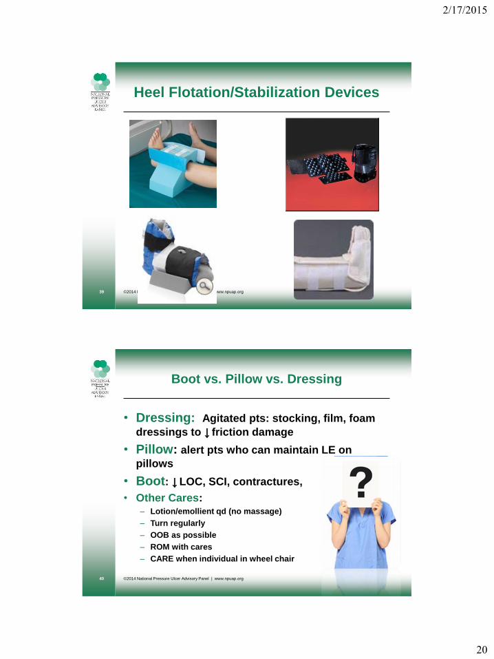
Heel Flotation/Stabilization Devices
©2014 National Pressure Ulcer Advisory Panel | www.npuap.org39
Boot vs. Pillow vs. Dressing
• Dressing: Agitated pts: stocking, film, foam
dressings to ↓ friction damage
• Pillow: alert pts who can maintain LE on
pillows
• Boot: ↓ LOC, SCI, contractures,
• Other Cares: – Lotion/emollient qd (no massage)
– Turn regularly
– OOB as possible
– ROM with cares
– CARE when individual in wheel chair
©2014 National Pressure Ulcer Advisory Panel | www.npuap.org40
2/17/2015
21
What we do know….
• “It is impossible to determine which heel
suspension device is most effective.”
• Offloading is effective!
©2014 National Pressure Ulcer Advisory Panel | www.npuap.org41
So That’s What’s New in…
©2014 National Pressure Ulcer Advisory Panel | www.npuap.org42
2/17/2015
22
Thank You & Now You Shouldn’t Be Left Hanging…..
©2014 National Pressure Ulcer Advisory Panel | www.npuap.org43