Wu et al. BMC Veterinary Research (2015) 11:16 DOI 10.1186/s12917-015-0336-6
RESEARCH ARTICLE Open Access
Hematological and histopathological effects ofswainsonine in mouseChenchen Wu, Xiaoxue Liu, Feng Ma and Baoyu Zhao*
Abstract
Background: Livestock that consume locoweed exhibit multiple neurological symptoms, including dispiritedbehavior, staggered gait, trembling, ataxia, impaired reproductive function and cellular vacuolar degeneration ofmultiple tissues due to toxicity from plant-derived alkaloids such as swainsonine.
Results: Swainsonine was administered to F0 and F1 mice by intraperitoneal injection before, during and afterpregnancy at the following doses: 0.525 mg/kg BW(I), 0.2625 mg/kg BW(II), 0.175 mg/kg BW(III) and 0 mg/kg BW(IV).Hemosiderin deposits were observed the lamina propria of endometrium in uterus and the red pulp of spleen.Ovary corpus lutea counts in F0 mice were higher in swainsonine-treated mice compared to control mice. Indirectbilirubin content and reticulocyte numbers were increased in swainsonine-treated F0 and F1 generation micecompared to control group (P < 0.05). Lactate dehydrogenase, alkaline phosphatase, aspartate aminotransferaseand alanine aminotransferase content in F0-I and F0-II mice were significantly increased compared with F0-IVgroup mice (P < 0.05). Red blood cells, hemoglobin and mean corpuscular hemoglobin levels were significantlydecreased in F0 and F1 mice compared with the control group (P < 0.05).
Conclusions: Swainsonine exerts effects on estrus period and reproductive ability, and offspring of damsdosed with swainsonine were affected in-utero or from nursing. Damage to liver, uterus and spleen, as wellas hematological changes, are observable before neurological symptoms present.
BackgroundLocoweeds are perennial herbaceous plants of theAstragalus spp and Oxytropis spp. containing the toxicindolizidine alkaloid swainsonine [1]. Locoism causessignificant economic losses to the livestock industry onwestern grasslands in China and the United States [2].Swainsonine, a trihydroxy indolizidine alkaloid, is theprimary toxin in locoweeds [1]. Astragalus and Oxytropisspecies that contain swainsonine are found on multiplecontinents, and have poisoned animals in South Americaand Asia [3,4]. Early studies demonstrated that naturalor experimental long-term ingestion of swainsonine-containing plants causes serious disorders in reproductivefunctions of livestock (cattle, sheep, horses and goat), in-cluding failure to conceive and early embryo loss or abor-tion, resulting in great economic losses to the livestockindustry [5-8]. Therefore, various animal models have
* Correspondence: [email protected] of Animal Veterinary Medicine, Northwest A & F University, Yangling712100, Shaanxi, People’s Republic of China
been used to study the toxic effects of swainsonine onreproduction and development, including goat, sheep andcattle. Locoweed poisoning is usually chronic, and thetoxic symptoms are observed after a few weeks of loco-weed feeding. Mice were fed a small quantity of locoweedfor four months, demonstrated that pathological and clin-ical damage to internal organs and neuronal processeswere reversible [9]. In this study, we then selected fourgroups of mice to treat with either vehicle control orswainsonine (10 each group, F0-I: 0.525 mg/kg BW; F0-II:0.2625 mg/kg BW; F0-III: 0.175 mg/kg BW and F0-IV:0 mg/kg BW). After treatment with swainsonine for twoweeks, female mice were mated to untreated male mice,and pups were kept with dams for one month. We sacri-ficed dams and offspring and observed swainsonine toxicityeffects on internal organs via histopathological analysis aswell as altered hematological and blood biochemical pa-rameters in both parent and offspring mice.
is is an Open Access article distributed under the terms of the Creativeommons.org/licenses/by/4.0), which permits unrestricted use, distribution, andiginal work is properly credited. The Creative Commons Public Domaing/publicdomain/zero/1.0/) applies to the data made available in this article,
Wu et al. BMC Veterinary Research (2015) 11:16 Page 2 of 8
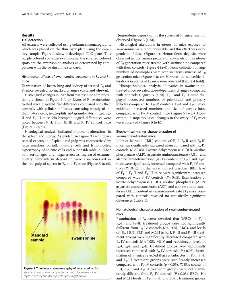
ResultsTLC detectionAll extracts were collected using column chromatography,which was placed on the thin layer plate using the capil-lary sample. Figure 1 show a developed TLC plate. Thispurple colored spots are swainsonine, the rose red coloredspots are the swainsonine analogs as determined by com-parison with the swainsonine standard.
Histological effects of swainsonine treatment in F0 and F1miceExamination of heart, lung and kidney of treated F0 andF1 mice revealed no marked changes (data not shown).Histological changes in liver from swainsonine administra-
tion are shown in Figure 2 (a-d). Livers of F0 swainsonine-treated mice displayed few differences compared with theircontrols, with cellular infiltrates consisting mostly of in-flammatory cells, neutrophils and granulocytes in F0-I, F0-II and F0-III mice. No histopathological differences werenoted between F1-I, F1-II, F1-III and F1-IV control mice(Figure 2 (e-h)).Histological analysis indicated important alterations in
the spleen and uterus. As evident in Figure 3 (a-h), dose-related expansion of splenic red pulp was characterized bylarge numbers of inflammatory cells and lymphocytes,hypertrophy of splenic cells and a considerable numberof macrophages and megakaryocytes. Increased extrame-dullary hemosiderin deposition were also observed inthe red pulp of spleen in F0 and F1 mice (Figure 3 (a-c)).
Figure 1 Thin-layer chromatography of swainsonine. Thestandard swainsonine sample (left arrow). The swainsonine isrepresented by the deep purple spots (right arrow).
Hemosiderin deposition in the spleen of F1 mice was notobserved (Figure 3 (e-h)).Histological alterations in uterus of mice exposed to
swainsonine were more noticeable, and this effect was inde-pendent of dose (Figure 4). Hemosiderin deposits wereobserved in the lamina propria of endometrium in uterusof F0 generation mice treated with swainsonine comparedwith their controls (Figure 4 (a-d)). Focal collection of largenumbers of neutrophils were seen in uterus mucosa of F0generation mice (Figure 4 (a-c)). However, no noticeable al-terations in uterus of F1 mice were observed (Figure 4 (e-h)).Histopathological analysis of ovaries in swainsonine-
treated mice revealed dose-dependent changes comparedwith controls (Figure 5 (a-d)). F0-I and F0-II mice dis-played decreased numbers of primordial and primaryfollicles compared to F0-IV controls. F0-I and F0-II miceexhibited increased numbers and size of corpus luteacompared with F0-IV control mice (Figure 5 (a-d)). How-ever, no histopathological changes in the ovary of F1 micewere observed (Figure 5 (e-h)).
Biochemical marker characterization ofswainsonine-treated miceIndirect bilirubin (IBIL) content of F0-I, F0-II and F0-IIImice was significantly increased when compared with F0-IVcontrols (P < 0.05). Lactate dehydrogenase (LDH), alkalinephosphatase (ALP), aspartate aminotransferase (AST) andalanine aminotransferase (ALT) content of F0-I and F0-IImice were significantly increased compared with F0-IV con-trols (P < 0.05). Furthermore, indirect bilirubin (IBIL) levelof F1-I, F1-II and F1-III mice were significantly increasedcompared with F1-IV controls (P < 0.05). Examination oflactate dehydrogenase (LDH), alkaline phosphatase (ALP),aspartate aminotransferase (AST) and alanine aminotrans-ferase (ALT) content in swainsonine-treated F1 mice com-pared with controls revealed no statistically significantdifferences (Table 1).
Hematological characterization of swainsonine-treatedmiceExamination of F0 dams revealed that WBCs in F0-I,F0-II and F0-III treatment groups were not significantlydifferent from F0-IV controls (P > 0.05). RBCs, and levelsof Hb, HCT, PLT, and MCH in F0-I, F0-II and F0-III treat-ment groups were significantly decreased compared withF0-IV controls (P < 0.05). MCV and reticulocyte levels inF0-I, F0-II and F0-III treatment groups were significantlyincreased compared with F0-IV controls (P < 0.05). Exam-ination of F1 mice revealed that reticulocytes in F1-I, F1-IIand F1-III treatment groups were significantly increasedcompared with F1-IV controls (p < 0.05). WBCs counts inF1-I, F1-II and F1-III treatment groups were not signifi-cantly different from F1-IV controls (P > 0.05). RBCs, Hband MCH levels in F1-I, F1-II and F1-III treatment groups
Figure 2 Histological changes in F0 and F1 mice after swainsonine treatment. a-d represent changes in the liver of F0-I, F0-II, F0-III and F0-IV(×400); e-h represent changes in the liver of F1-I, F1-II, F1-III and F1-IV (×400).
Wu et al. BMC Veterinary Research (2015) 11:16 Page 3 of 8
were significantly decreased compared with F1-IV controls(P < 0.05). HCT levels in F1-I group mice were signifi-cantly decreased compared with F1-IV control mice(P < 0.05), and MCV levels in F1-I and F1-II mice weresignificantly increased compared with F1-IV control mice(P < 0.05) (Table 2).
Figure 3 Histological changes in F0 and F1 mice after swainsonine treF0-IV (×400); e-h represent changes in the spleen of F1-I, F1-II, F1-III and F1-I
DiscussionSwainsonine, a trihydroxy indolizidine alkaloid, is themain toxin in locoweed. The structure of the swainso-nine cation is similar to the structure of mannose, andit has a higher affinity than mannose for mannosidase[10]. Swainsonine is a well-known inhibitor of lysosomal
atment. a-d represent changes in the spleen of F0-I, F0-II, F0-III andV (×400).
Figure 4 Histological changes in F0 and F1 mice after swainsonine treatment. a-d represent changes in the uterus of F0-I, F0-II, F0-III andF0-IV (×400); e-h represent changes in the uterus of F1-I, F1-II, F1-III and F1-IV (×400).
Wu et al. BMC Veterinary Research (2015) 11:16 Page 4 of 8
ɑ-mannosidase and Golgi ɑ-mannosidase II. Swainsonineinduces toxicity through inhibition of ɑ-mannosidaseand subsequent glycoprotein synthesis. This enzymaticdysfunction causes accumulation of complex oligosac-charides in lysosomes as well as the production of a mix-ture of mannose and asparagine polysaccharides, resulting
Figure 5 Histological changes in F0 and F1 mice after swainsonine tre(×100); e-h represent changes in the ovary of F1-I, F1-II, F1-III and F1-IV (×40
in vacuolar degeneration in multiple cells [11]. Clin-ical symptoms in livestock are characterized by neuro-logical and behavioral disorders, gait abnormalities,difficulty standing, abnormal posture, emaciation, re-productive disorders and cellular vacuolar degener-ation of multiple tissues by pathological observation
atment. a-d represent changes in the ovary of F0-I, F0-II, F0-III and F0-IV0).
Table 1 Serum marker assessment in swainsonine-treated mice
The values are the mean ± S.D.*Significantly different from the control group at same generation (P < 0.05).
Wu et al. BMC Veterinary Research (2015) 11:16 Page 5 of 8
[12,13]. However, we observed two generations of miceand show organ selective vacuolar degeneration by micegiven swainsonine via pathological observation. We foundhemosiderin deposition in spleen and enlargement ofspleen in F0 and F1 mice, and a large amount of hemosid-erin deposition in uterus in F0 mice. When animals arefed a dose of swainsonine arrive to a certain time, thevacuolar degeneration of pathological change will show inthe internal organs [9]. Therefore, we think that thisexperiment period did not arrive to a certain time thatvacuolar degeneration found in organ. However, in ourprevious experiment, we also found hemosiderin depos-ition in spleen of rat and goat using a different dose ofswainsonine [14]. We posited that some tissue bleedingoccurred after swainsonine administration and found thathemosiderin deposition leads to damage in some tissues.Whether the presence of hemosiderin deposition can beused as a pathological marker of swainsonine poisoningrequires further research.The experiment results showed that two ways were not
significantly different between irrigation and intraperito-neal injection by Liu Tianya [15]. Therefore, we selectedthe way of intraperitoneal injection for give mice to swain-sonine. In this study, we demonstrate that swainsonine ex-erts hepatotoxicity in F0 mice. Alterations in liver weightand histopathological changes in liver of swainsonine-
Table 2 Hematological assessment in swainsonine-treated mi
The values are the mean ± S.D.*Significantly different from the control group at same generation (P < 0.05).
treated mice were slight. Liver from swainsonine-treatedmice showed cellular infiltrates consisting mostly of in-flammatory cells and neutrophil granulocytes. Significantincrease of liver weight and significant alterations in levelsof AST, ALT and ALP in plasma may indicate hepatic in-jury in F0 mice given swainsonine. The elevations of ALT,AST and ALP observed in swainsonine-treated mice may,in part, be due to the hepatic hypertrophic effect of swain-sonine and/or may also represent borderline chronic livertoxicity [16,17]. Increased LDH activity levels have beenobserved in conditions of chemical stress when high levelsof energy are required in a short period of time [18]. Inthe present study, LDH was significantly increased in F0mice. However, no significant differences in biochemicalmarkers were found between treatment and control miceF1 mice. This is consistent with the lack of histopatho-logical changes in liver of F1 mice.The present study identifies important histological al-
terations in the spleen in F0 and F1 mice, namely expan-sion of red pulp with vascular congestion. Furthermore,the endometrium of the uterus displayed notable depos-ition of hemosiderin granules in a swainsonine-treateddose-dependent manner in F0 mice. The molecularweight of swainsonine is small enough to penetrate theplacental barrier and expose offspring in-utero. A majorfunction of the spleen is to remove aged and damaged
Wu et al. BMC Veterinary Research (2015) 11:16 Page 6 of 8
erythrocytes from the blood [19]. Excess hemosiderindeposition in spleen can result in the destruction ofmacrophages and the release of the contents such asiron, toxic compounds and/or its metabolites into spleen[20]. Toxic effects in both F1 and F0 mice include reduc-tion of RBCs, reduction in levels of Hb, HCT, PLT andMCH, as well as an increase in the number of reticulo-cytes, suggesting the development of anemia [21]. Sig-nificant increases in IBIL were observed in F0 and F1mice given swainsonine. The increase of IBIL further in-dicates that swainsonine could be damaging red bloodcells. When organs bleed, red blood cells are phagocy-tized by macrophages and degraded by lysosomes; Fe3+
of hemoglobin from lysed red blood cells can combinewith protein to form hemosiderin. Because we observeddecreased RBCs, and decreased levels of Hb, MCH andMCV as well as an increase in reticulocytes, we suspectthat our dose levels of swainsonine may lead to anemia.Swainsonine is water-soluble and rapidly distributed to
many parts of the body. In previous studies, swainsonineconcentrations varied widely in various tissues and or-gans of sheep that had ingested locoweed [22-24]. In thisstudy, uterus of swainsonine-treated F0 mice was heavilydamaged. This was characterized by the presence of he-mosiderin deposits in the lamina propria of endomet-rium in uterus of F0 mice in this study. In ovary, F0-Iand F0-II mice displayed decreased numbers of primor-dial and primary follicles compared to F0-IV controls. Inaddition, F0-I and F0-II mice displayed increased size andnumber of corpus lutea compared to F0-III and F0-IV.The lesions in ovary and uterus were dose-dependentlyobserved in F0-I, F0-II and F0-III treatment groups. How-ever, F1 did not display notable histopathological changesin the uterus and ovary. Swainsonine easily accumulatesin uterus at high concentrations, which may impair uterusand ovary function and cause toxicity. In the presentstudy, the uterus suffered noticeable damage, which led toa decline in the rate of conception, an increase in the rateof abortion and increases in stillborn births. It is suspectedthat significant early embryonic loss occurs in cattle andsheep grazing locoweed, and there are documented effectsof swainsonine on oocyte maturation, fertilization andsubsequent embryonic implantation and development[24]. Increased numbers of corpus lutea in ovary can leadto delayed or halted estrus. The pathological lesions weobserved, combined with altered hematological and serumbiochemical parameters in swainsonine-treated mice, sug-gest that exposure to swainsonine may lead to inhibitionof reproductive performance under certain doses.
ConclusionsBased on sub-chronic toxicity results, our data estab-lishes effects of swainsonine on reproductive toxicity ina mouse model. In addition, we found that swainsonine
can cause hematological changes and lesions in spleen,uterus, ovary and liver. Furthermore, we provide evi-dence of trans-generational swainsonine toxicity throughplacental barrier and milk. Spleen, heart, liver, lung, kid-ney, uterus and ovary were among the organs affected inoffspring of dams given swainsonine. Large amounts ofhemosiderin deposition in uterus and spleen were ob-served in the parent generation. We present evidence thathemosiderin deposition may preclude vacuolar degener-ation in some tissues of mice given swainsonine. Alter-ations in hematological and histopathological parameterssuggest a link to anemia and decreases in reproductionability. Our data suggest that anemia and organ-specifichemosiderin deposition followed by destruction of redblood cells are clinical features of swainsonine-treatedmice. However, further research is needed to elucidatespecific mechanisms of swainsonine toxicity.
MethodsEthical statementFemale Rattus norvegicus mice were supplied by theAnimal Center of the Fourth Military Medical Univer-sity. During the experiment, mice were housed individu-ally in polypropylene cages with laboratory grade pineshavings as bedding. Mice were maintained in a con-trolled environment with temperature maintained be-tween 19-25°C, relative humidity maintained between40-70%, >8 air changes/hour, and with a 12:12-h light: darkcycle. The experimental procedures were in accordancewith the Ethical Principles (Animal [Scientific Procedures]Act 2012) in Animal Research adopted by the ChinaCollege of Animal Experimentation and were approvedby the College of Veterinary Medicine- Northwest A&FUniversity.
Study designExtraction of swainsonine from locoweedThe aerial portion of Oxytropis kansuensis was collectedfrom the grassland in Tianzhu city, Gansu province inJuly 2011. The plants were then taxonomically identified byZhao Bao-Yu, College of Veterinary Medicine, NorthwestA and F University, China. The plants were subsequentlydried in the shade, finely ground and comminuted.The extraction and analysis method of swainsonine
from Oxytropis kansuensis was conducted as previouslydescribed [25].
Analysis of swainsonineThin-layer chromatography (TLC) detection was per-formed on silica gel G precoated plates with the developingsolvents chloroform:methanol:ammonia:water (70:26:2:2,V/V), chloroform:methanol:ammonia:water (70:26:10:10,V/V), and methanol: ethylacetate: ammonia (4:1:1,V/V)and modified potassium heptaiodobismuthate reagent or
Wu et al. BMC Veterinary Research (2015) 11:16 Page 7 of 8
H2O2/10% acetic anhydride in EtOH/Ehrlich’s reagent wasthe chromogenic agent.The extracts were dissolved in methanol, spotted onto
the GF254 silica gel G precoated plates. The plates weredeveloped with an ascendant run after saturation withthe mobile phase in a s glass chamber for 5–10 min. Theplates were dried when the mobile phase was 10 mm fromthe front edge of the plates. The plates were stained suc-cessively with a spray of H2O2 (heated for 10 min in anoven at 115°C), a spray of 10% acetic anhydride in dehy-drated alcohol (heated at the same temperature until thesmell of acetic anhydride disappeared) and finally a sprayof Ehrilich’s reagent (heated for 15 min at 120°C). Thecolor of the spots in each plate was recorded, and the Rf
was determined [25].
Animals to experimental groupsFemale mice (N = 40, six weeks old) were divided into fourequal groups of 10 mice (10 each group, F0-I: 0.525 mg/kgBW; F0-II: 0.2625 mg/kg BW; F0-III: 0.175 mg/kg BW andF0-IV: 0 mg/kg BW). All mice were administered swainso-nine by intraperitoneal injection 14 days before the matingperiod followed by re-administration every three days.After this pre-mating period, the treated mice were trans-ferred to the home cage of a male in the same group andcohabited on a 1:1 basis until achievement of successfulmating. During the mating period, mice were examineddaily for presence of vaginal plugs, and a vaginal plug wasconsidered evidence of successful mating. Pregnant damscontinued to receive swainsonine every three days viaintraperitoneal injection throughout parturition and thelactation. Upon weaning of four-week-old pups (F1), thedams (F0) were sacrificed, and the liver, kidney, heart,spleen, lung, uterus and ovary were collected. In total, F0mice were given swainsonine for six to eight weeks in thewhole experiment.Female offspring (F1) of treated dams were selected
from each of the four treatment groups (40 F1 mice intotal, 10 from each F0 treatment group). The F1 offspringwere not treated with swainsonine, however, the damscontinued to be dosed while nursing their F1 pups. The F1offspring were then sacrificed after approximately 1 monthof nursing. The liver, kidney, heart, spleen, lung, uterus,and ovary were collected.All F0 group mice received intraperitoneal injections
of swainsonine once every three days under aseptic con-ditions. Upon sacrifice, the liver, kidney, heart, spleen,lung, uterus and ovary were trimmed of extraneous fatand weighed immediately.
Histopathological preparationAll tissues were removed and fixed in 10% formaldehydeat room temperature. The tissue samples were then dehy-drated and embedded in paraffin according to standard
histological procedures. Serial cross-sections of 3 μm wereprepared from each organ. The sections were mountedand stained with hematoxylin-eosin.
Hematological assessmentWhite blood cells (WBCs), red blood corpuscles (RBCs),hemoglobin (Hb), hematocrit (HCT), mean corpuscularvolume (MCV), blood platelets (PLTs), mean corpuscularhemoglobin (MCH) and reticulocyte counts were deter-mined by automatic hematological analyzer, MEK-8222 K(TOA Medical Electronics, Kobe, Japan).
Blood biochemical analysisBlood was collected when mice were sacrificed. Lactatedehydrogenase (LDH), aspartate aminotransferase (AST),alanine aminotransferase (ALT), alkaline phosphatase(ALP) and indirect bilirubin (IBIL) were quantitated usingthe Beckman Synchron CX7 Delta Chemistry Analyzer(Beckman, USA).
Statistical methodsThe statistical software “Statistical Product and ServiceSolutions” (SPSS V11.3) was used to determine statisti-cally significant differences between treatment groups andthe control group. A one-way ANOVA was used to evalu-ate the homogeneity of the data, and a least squared dif-ferences model or Dunnett’s multiple comparison testwere then used. Values of p < 0.05 were considered signifi-cant. The data are presented as the group mean values ±SD (standard deviation).
Competing interestsThe authors declare that they have no competing interests.
Authors’ contributionsMF carried out the extration of swainsonine from Oxytropis kansuensis; ZBparticipated in the test design and drafted the manuscript; LX raised miceand performed the statistical analysis; WC participated in the design of thestudy and wrote the manuscript. All authors read and approved the finalmanuscript.
AcknowledgmentsThe authors thank Li Xiaoming the research assistant for makingpathological, Mr Han from translatinng the manuscript. This study wasfinanced by the grants from the National Natural Science Foundation(No. 31302153) and the origination fee of doctoral research (No. Z111021305),Postdoctoral program (No. K308021401) and the Special Scientific ResearchFund of Agriculture Public Welfare industry (No. 201203062).
Received: 30 August 2014 Accepted: 22 January 2015
References1. Molyneux RJ, James LF. Loco intoxication indolizidine alkaloids of spotted
locoweed (Astragalus lentiginosus). Science. 1982;216:190–1.2. Li JK. The present situation and prospect of the studies on locoweed In
China. Agric Sci China. 2003;36:1091–9.3. Molyneux RJ, Gomez-Sosa E. Presencia del alcaloide indolizidinico swainsonine
enAstragalus pehuenches(Leguminosae-Galegueae). Bol Soc Argent Bot.1991;27:59–64.
4. Molyneux RJ, James LF, Ralphs MH, Pfister JA, Panter KE, Nash RJ.Polyhydroxylated glycosidase inhibitors from poisonous plants of global
Wu et al. BMC Veterinary Research (2015) 11:16 Page 8 of 8
distribution: analysis and identification. In: Colegate SM, Dorling PR, editors.Plant-associated toxins, agricultural and phytochemical aspects. Wallingford,UK: CABI; 1994. p. 107–12.
5. Panter KE, James LF, Stegelmeier BL, Ralphs MH, Pfister JA. Locoweeds:effects on reproduction in livestock. J Nat Toxins. 1999;8:53–62.
6. Panter KE, James LF, Mayland HF. Reproductive response of ewes fed alfalfapellets containing sodium selenate orAstragalus bisulcatusas a seleniumsource. Vet Hum Toxicol. 1995;37:30–2.
7. Stegelmeier BL, James LF, Panter KE, Molyneux RJ. Serum swainsonineconcentration and alpha-mannosidase activity in cattle and sheep ingestingOxytropis sericea and Astragalus lentiginosus (locoweeds). Am J Vet Res.1995;56:149–54.
8. James LF, Panter KE, Nielsen DB, Molyneux RJ. The effect of natural toxinson reproduction in livestock. J Anim Sci. 1992;70:1573–9.
9. Chenchen W, Wenlong W, Xiaoxue L. Pathogenesis and preventivetreatment for animal disease due to locoweed poisoning. Environ ToxicolPhar. 2014;37:336–47.
10. Galyean ML, Ralphs MH, Reif MN. Effects ofprevious grazing treatment andconsumption of locoweed onliver mineral concentrations in beef steers.J Anim Sci. 1996;74:827.
11. Abraham DJ, Rsidebothom BG, Winchester PR. Swainsonine affects theprocessing of glycoproteins in vivo. FEBS Lett. 1983;163:110–3.
12. Das PC, Roberts JD, White SL. Activation of resident tissue-specific macrophagesby swainsonine. Oncol Res. 1995;7(9):425–33.
13. Jacob GS. Glycosylation inhibitors in biology and medicine. Curr Opin StructBiol. 1995;5(5):605–11.
14. Wang wenlong: Extracts of oxytropis kansuensis induced toxic damage ofrats and protective effects of “Jifang E”. Northwest A&F University Press2014; pp.57-59
15. Tianya L, Zongyuan H. Effects of irrigation and intraperitoneal injection onlocomotor activity of mice. Editorial Board Acta Academiae MedicinaeWannan. 2010;29(4):241–4.
16. Amacher DE. Serum transaminase elevations as indicators of hepatic injuryfollowing the administration of drugs. Regul Toxicol Pharmacol.1998;27:119–30.
17. Boone L, Meyer D, Cusick P, Ennulat D, Bolliger AP. Selection andinterpretation of clinical pathology indicators of hepatic injury in preclinicalstudies. Vet Clin Pathol. 2005;34:182–8.
18. Kamble N, Velhal V. Cytopathological assessment of uterine cells in rattusnorvegicus due to induced sodium fluoride. The bioscan. 2010;5:301–3.
19. Stefanski SA, Elwell MR, Stromberg PC, Boorman GA, Eustis SL, Elwell MR,editors. Pathology of the fischer Rat reference and atlas. San Diego:Academic Press; 1990. p. 369–93.
20. Fujitani T, Tada Y, Yoneyama M. Chlorpropham-induced splenotoxicity andits recovery in rats. Food Chem Toxicol. 2004;42:1469–77.
21. Hui A, Jinyi L, Lujun Y, Shengxue L, Yanhong Z, Huan Y, et al. Acute andsubchronic toxicity of hydroxylammonium nitrate in Wistar rats. J MedColleges PLA. 2008;23:137–47.
22. Tong D, Mu P, Dong Q, Zhao B, Liu W, Zhao J. Immunological evaluation ofSW-HSA conjugate on goats. Colloids Surf B Biointerfaces. 2007;58:61–7.
23. Stegelmeier BL, James LF, Panter KE, Molyneux RJ. Tissue and serumswainsonine concentrations in sheep ingesting Astragalus lentiginosus(locoweed). Vet Hum Toxicol. 1995;37:336–9.
24. Stegelmeier BL, James LF, Panter KE, Gardner DR, Ralphs MH, Pfister JA.Tissue swainsonine clearance in sheep chronically poisoned with locoweed(Oxytropis sericea). J Anim Sci. 1998;76:1140–4.
25. Pengbin G, Baoyu Z, Dewen T. Extraction and fractionation andidentification of swainsonine on structure from oxytropis glabra. Chin AgricSci Bull. 2003;19(1):1–5.
Submit your next manuscript to BioMed Central
and take full advantage of:
• Convenient online submission
• Thorough peer review
• No space constraints or color figure charges
• Immediate publication on acceptance
• Inclusion in PubMed, CAS, Scopus and Google Scholar
• Research which is freely available for redistribution
Submit your manuscript at www.biomedcentral.com/submit