Marc D. Castle, Bruce M. Christensen, and Tonic E. Rocke12
Department of Veterinary Science, 1655 Linden Drive, University of Wisconsin,
Madison, Wisconsin 53706, USA2 Present address: National Wildlife Health Center, 6006 Schroeder Road,Madison, Wisconsin 53711, USA
ABSTRACT: One hundred twenty-three of 300 blood samples (41%) taken from Rio Grande wildturkeys (Meleagris gallopavo intermedia) from three locations in southern Texas (Welder WildlifeRefuge, Chaparrosa Ranch, and Campo Alegre Ranch) and subinoculated into domestic broad-breasted white turkey poults were positive for a Plasmodium (Novyella) sp. Analysis of bloodfilms from 350 turkeys revealed Haemoproteus meleagridis in 76% of the birds. A significantlygreater mean parasite intensity was observed in birds from Welder Wildlife Refuge. Birds fromthe Campo Alegre Ranch exhibited a significantly higher prevalence of H. meleagridis than birdsfrom Chaparrosa. The Plasmodium sp. was infective for canaries (Serinus canaria), bobwhites(Colinus virginianus ), and ring-necked pheasants (Phaslanus coichicus ), but would not produceinfection in white leghorn chickens (Gallus gallus) or Coturnix quail (Coturnix coturnix). Attemptsto infect Culex tarsalis and C. pipiens pipiens were unsuccessful. Asexual erythrocytic synchronywas not observed when blood-induced infections were monitored in two domestic turkey poultsevery 4 hr for 72 hr. Exoerythrocytic stages were not found upon examination of impression
smears and tissue samples taken from brain, liver, spleen, kidney, lung, and bone marrow. ThePlasmodium sp. is most similar morphologically to three species in the subgenus Nov yella, P.hexamerium, P. vaughani, and P. kempi. The most striking similarities are to P. hexamerlum,
and involve mean merozoite number, erythrocytic schizont location, and vertebrate host suscep-tibility. It differs from P. vaughani in being able to infect turkeys and in type of parasitizederythrocytes. Differences to P. kempi include mean merozoite number, and ability to infectpheasants, and its inability to develop in C. pipiens and C. tarsalis.
Key words: Rio Grande wild turkeys, Meleagris gallopavo intermedia, Plasmodium (No-vyella) sp., Haeinoproteus meleagridis, hematozoan parasites, natural and experimental infections.
INTRODUCTION
Documentation of Plasmodium sp. from
wild turkeys in North America began only
in the mid-1970’s when Forrester et al.
(1974) isolated a Plasmodium sp. from wild
turkeys in Florida. Subsequently, Telford
and Forrester (1975) named this species P.
(Huff-ia) hermani. In this decade, Chris-
tensen et al. (1983) reported a second dis-
tinct species from eastern wild turkeys
(Meleagris gallopavo silvestris) in Iowa,
which they named P. (Nov yella) kempi.
Both of these isolations were made by sub-
inoculating collected blood into domestic
turkey poults, and not by examination of
peripheral blood smears.
Reports of other hematozoan parasites
infecting wild turkeys are numerous, but
are limited mostly to Haemo proteus mele-
agridis and/or Leucocytozoon srnithi
(Travis et al., 1939; Mosby and Handley,
1943; Kozicky, 1948; Love et al., 1953;
Byrd, 1959; Cook et al. , 1966; Goggans,
1966; Roslien and Haugen, 1970; Eve et
al., 1972; Stone et al., 1972; Forrester et
al., 1974; Noblet and Moore, 1975; Castle
and Christensen, 1984). However, a recent
report on the recovery of an intraerythro-
cytic rickettsia (Aegyptianella pullorum)
from 24 Rio Grande wild turkeys in Texas
raised some questions on the possibility of
this pathogen infecting domestic fowl as
well as indigenous wild birds (Castle and
Christensen, 1985).
Herein, we (1) report prevalence data
of H. meleagridis and a third Plasmodium
sp. from North American wild turkeys, (2)
provide biological information on the Plas-
modium sp., including vertebrate host
specificity, experimental vector studies,
CASTLE ET AL-BLOOD PARASITES OF WILD TURKEYS 89
and synchrony data, and (3) discuss the
possible taxonomic status of the Plasmo-
dium sp.
MATERIALS AND METHODS
Sample collections and parasite isolation
Using a heparinized syringe, blood was takenfrom the jugular vein of live-trapped Rio Grandewild turkeys from three locations in southern
Texas in January and February of 1983 and1984. Turkeys were captured either by cannon-
or drop-net from the Rob and Bessie WelderWildlife Refuge, San Patricio County; Chapar-
rosa Ranch, Zavala County; and, in 1984 only,
Campo Alegre Ranch, Willacy County. Thinblood smears were made and the remainder ofthe collected blood placed on wet ice and shippedto the Department of Veterinary Science (Uni-versity of Wisconsin, Madison, Wisconsin 53706,
USA) for subinoculation into domestic turkeypoults (Nicholas broad-breasted white turkeys:
obtained at 1 day of age from Jerome Foods,Barron, Wisconsin 54812, USA). Collected bloodwas inoculated into 5-22-day-old poults eitherintraperitoneally (i.p.) or intravenously (iv.) andranged in volume from 0.5 to 1.0 ml. Timesbetween collection of blood and subinoculationranged from 1 to 7 days. All recipient birds were
housed in the Charmany Farm semi-isolationfacilities maintained by the Department of Vet-erinary Science (University of Wisconsin, Mad-ison, Wisconsin 53706, USA) throughout thecourse of the investigation. These studies wereperformed in the winter months when acciden-tal feeding by indigenous mosquitoes would nottake place.
Beginning 1 wk postinoculation, blood smearswere made twice weekly for 6 wk. A minimumof 20,000 erythrocytes were examined per slideusing oil immersion optics (1,250x). Erythro-cyte numbers were estimated by counting theerythrocytes in a number of random fields, av-
eraging those counts, and then counting thenumber of fields until a minimum of 20,000
erythrocytes had been viewed. Counts of H.meleagridis were all based on 10,000 erythro-cytes. Blood smears were stained with Giemsa’sat pH 7.2-7.3 (1:10 dilution for 1 hr). Measure-ments were made using a calibrated ocular mi-crometer. All measurements are expressed injim. Representative specimens of the Plasmo-diurn sp. and H. meleagridis were deposited inthe International Reference Centre for AvianHaematozoa (Memorial University of New-foundland, St. Johns’s, Newfoundland, CanadaA1B 3X9, accession numbers 97607-97608) andin the U.S. National Parasite Collection (Belts-ville, Maryland 20705, USA, as USNM collectionnumbers 79484-79485).
Vertebrate host specificity
Bobwhites, canaries, Coturnix quail, ring-necked pheasants, and white leghorn chickenswere inoculated with blood from domestic tur-keys with an active parasitemia to determine
their susceptibility to the Plasmodium sp.Chickens, Coturnix quail, and pheasants wereobtained from the Department of Poultry Sci-ence (University of Wisconsin, Madison, Wis-consin 53706, USA); bobwhites from Thomp-son’s Quail Farm (Franksville, Wisconsin 53126,USA); and canaries from the laboratory of Dr.R. P. Hanson (Department of Veterinary Sci-ence, University of Wisconsin, Madison, Wis-consin 53706, USA).
Birds were inoculated either iv. or i.p. with0.2-0.65 ml of heparinized blood and main-
tamed in isolation facilities throughout the study.Beginning 5-7 days postinoculation, blood filmswere made 2 or 3 times per wk for 4-6 wk.Blood films were stained and examined as pre-viously described.
Laboratory vector studies
Two species of ornithophilic mosquitoes (Cu-lex pipiens pipiens and C. tarsalis) were ob-tamed from the laboratory of Dr. W. A. Rowley
(Department of Entomology, Iowa State Uni-versity, Ames, Iowa 50011, USA) and tested fortheir susceptibility to the Plasmodium sp. Mos-
quitoes were reared and maintained as de-scribed by Christensen et al. (1983). Sucrose wasremoved 24 hr prior to blood feeding. All mos-quitoes were 4-14 days old when exposed toturkey poults with an active parasitemia.
Blood films were made from infected poultsbefore exposure to mosquitoes, and parasitemia,percentage of gametocytes, and exposure index(parasitemia x percentage of gametocytes) weredetermined. The age of turkey poults rangedfrom 3 to 5 wk. Poults were restrained and placedin mosquito cages for 2-14 hr, depending onthe number of mosquitoes blood-feeding to re-pletion. Blood-fed mosquitoes were removed andplaced in 0.473-liter ice cream cartons with afine-mesh marquisette covering. Mosquitoeswere dissected in Aedes aegypti saline (Hayes,
1953) using a stereomicroscope. Midguts andsalivary glands were examined for oocysts andsporozoites using phase contrast optics.
Synchrony studies
To evaluate whether the Plasmodiurn sp. ex-hibited any synchrony in its asexual erythrocyticschizogony, two birds were inoculated iv. with1.0 ml of infected blood, and once peak para-sitemia was reached (approximately 2-3%), thebirds were bled every 4 hr beginning at 0900hr for the next 72 hr. Two blood smears were
90 JOURNAL OF WILDLIFE DISEASES, VOL. 24, NO. 1, JANUARY 1988
TABLE 1. Haernoproteus nieleagridis and a Plasmodium sp. from Rio Grande wild turkeys in southernTexas, 1983-1984.
made from each bird at each bleeding and werestained and examined as previously described.
Trophozoites, immature schizonts, matureschizonts, immature gametocytes, and maturegametocytes were then counted per 10,000erythrocytes for each blood film, and the dataanalyzed for any evidence of asexual synchrony.
Determination of exoerythrocytic schizogony
Although no controlled studies were per-formed, infected turkey poults of various agesand stages of infection (before, during, and after
active parasitemia) were necropsied periodi-cally and examined for possible sites of exo-erythrocytic schizogony. Impression smears were
made and tissue samples were taken from brain,liver, spleen, kidney, lung, and bone marrow.Impression smears were stained with Giemsa’sas described previously, and tissues were cut at5 �im and stained with haematoxylin and eosin.
Both were screened at 500 x and examined us-
ing oil immersion optics (1,250x).
Statistical analysis
Prevalence data for H. meleagridis and thePlasrnodium sp. were analyzed using chi-square
analysis of 2 x 2 contingency tables, intensitydata analyzed by Student’s t-test and ANOVA,and synchrony data by ANOVA. Prior to theseanalyses, intensity data were tested for normaldistribution by chi-square analysis. Consequent-ly, log transformations of the data were per-
formed and the data retested for normal distri-bution. All analyses were done from statisticalpackages on a Hewlett-Packard HP 86 micro-computer system (Hewlett-Packard Company,1010 N.E. Circle Boulevard, Corvallis, Oregon97330, USA). Differences were considered sig-nificant at P < 0.05.
Prevalence
RESULTS
Data on prevalence and intensity are
presented in Table 1. Seventy-six percent
of 350 turkeys were positive for H. mele-
agridis. Birds from Welder Wildlife Ref-
uge consistently exhibited a significantly
greater mean parasite intensity (number
of parasites/10,000 erythrocytes) than birds
from Chaparrosa or Campo Alegre Ranch-
es. Although prevalence of H. meleagridis
in birds from Campo Alegre was signifi-
cantly higher than those of Chaparrosa,
intensity of infection was not statistically
different.
Of the 123 samples positive for the Plas-
modium sp., 15 (12%) were identified by
observation of the parasite on direct blood
films. The majority of these (eight) were
CASTLE ET AL-BLOOD PARASITES OF WILD TURKEYS 91
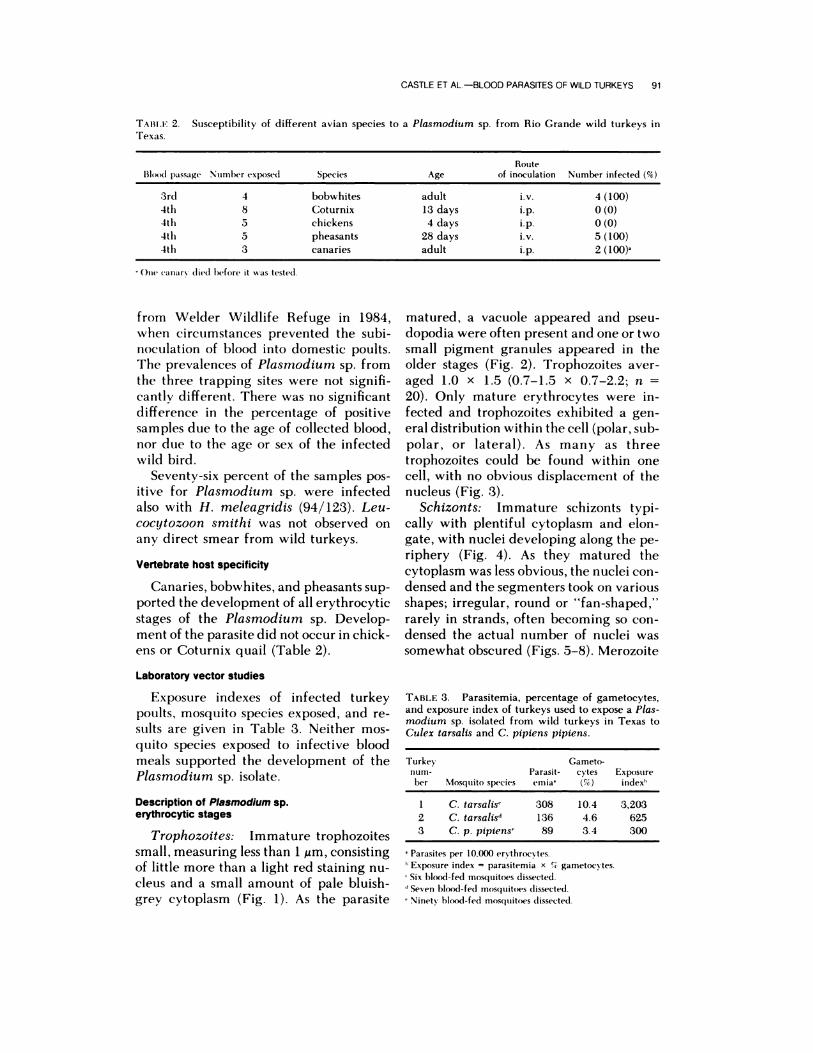
TAIII.F 2. Susceptibility of different avian species to a Plasmodium sp. from Rio Grande wild turkeys inTexas.
Route
Blood passage Number exposed Species Age of inoculation Number infected (%)
3rd 4 bobwhites adult iv. 4 (100)
4th 8 Coturnix 13 days i.p. 0 (0)
4th 5 chickens 4 days i.p. 0 (0)
4th 5 pheasants 28 days iv. 5 (100)
4th 3 canaries adult i.p. 2 (100)’
One canary (lied before it was tested.
from Welder Wildlife Refuge in 1984,
when circumstances prevented the subi-
noculation of blood into domestic poults.
The prevalences of Plasmodium sp. from
the three trapping sites were not signifi-
cantly different. There was no significant
difference in the percentage of positive
samples due to the age of collected blood,
nor due to the age or sex of the infected
wild bird.
Seventy-six percent of the samples pos-
itive for Plasmodium sp. were infected
also with H. meleagridis (94/123). Leu-
cocytozoon smithi was not observed on
any direct smear from wild turkeys.
Vertebrate host specificity
Canaries, bobwhites, and pheasants sup-
ported the development of all erythrocytic
stages of the Plasmodium sp. Develop-
ment of the parasite did not occur in chick-
ens or Coturnix quail (Table 2).
Laboratory vector studies
Exposure indexes of infected turkey
poults, mosquito species exposed, and re-
stilts are given in Table 3. Neither mos-
quito species exposed to infective blood
meals supported the development of the
Plasmodium sp. isolate.
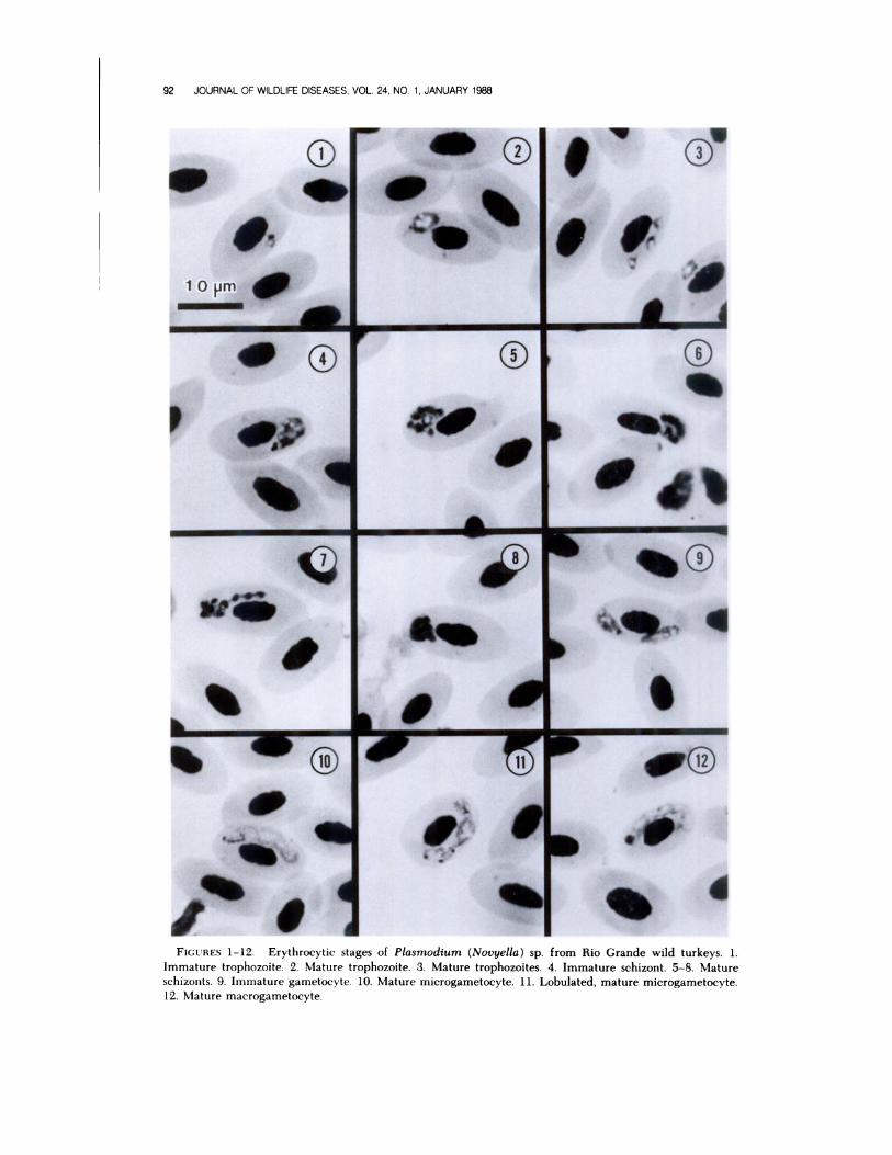
Description of Plasmodium sp.erythrocytic stages
Trophozoites: Im ure t rophozoites
small, measuring less than 1 jim, consisting
of little more than a light red staining nu-
cleus and a small amount of pale bluish-
grey cytoplasm (Fig. 1). As the parasite
matured, a vacuole appeared and pseu-
dopodia were often present and one or two
small pigment granules appeared in the
older stages (Fig. 2). Trophozoites aver-
aged 1.0 x 1.5 (0.7-1.5 X 0.7-2.2; n
20). Only mature erythrocytes were in-
fected and trophozoites exhibited a gen-
eral distribution within the cell (polar, sub-
polar, or lateral). As many as three
trophozoites could be found within one
cell, with no obvious displacement of the
nucleus (Fig. 3).
Schizonts: Immature schizonts typi-
cally with plentiful cytoplasm and elon-
gate, with nuclei developing along the pe-
riphery (Fig. 4). As they matured the
cytoplasm was less obvious, the nuclei con-
densed and the segmenters took on various
shapes; irregular, round or “fan-shaped,”
rarely in strands, often becoming so con-
densed the actual number of nuclei was
somewhat obscured (Figs. 5-8). Merozoite
TABLE 3. Parasitemia, percentage of gametocytes,and exposure index of turkeys used to expose a Plas-modium sp. isolated from wild turkeys in Texas to
Culex tarsalis and C. pipiens pipiens.
Turkey
num-ber Mosquito species
Parasit-
emia’
Gameto-cytes
(%)
Exposure
index5
1 C. tarsalls’ 308 10.4 3,203
2 C. tarsalis” 136 4.6 625
3 C. p. pipiens� 89 3.4 300
‘Parasites per 10.000 ervthrocytes.
Exposure index = parasiteniia x � gametocvtes.
Six blood-fed mosquitoes dissected.
d Seven blood-fed mosquitoes dissected.
Ninety blood-fed niosquitoes (lissecte(l.
0
‘S
92 JOURNAL OF WILDLIFE DISEASES, VOL. 24, NO. 1, JANUARY 1988
�p,.#{149}41
SS
- d
FIGURES 1-12. Erythrocytic stages of Plasmodium (Novyella) sp. from Rio Grande wild turkeys. 1.Immature trophozoite. 2. Mature trophozoite. 3. Mature trophozoites. 4. Immature schizont. 5-8. Mature
94 JOURNAL OF WILDLIFE DISEASES, VOL. 24, NO. 1, JANUARY 1988
that of the Texas isolate. Other differences
of this isolate with P. vaughani include the
number of granules found in the game-
tocytes and in the types of red blood cells
the parasites invades. The isolate from
Texas is found invariably in mature eryth-
rocytes whereas P. vaughani has a ten-
dency to invade erythroblasts and other
immature cells (Laird, 1962). Two to 24
pigment granules may be found in the ga-
metocytes of P. vaughani, depending on
the strain (Garnham, 1966), which is much
more than the maximum of 12 seen in our
isolate.
A third species, P. kempi, found in east-
em wild turkeys from Iowa (Christensen
et al. , 1983), also shows morphological sim-
ilarities to the species from wild turkeys
in Texas. The number of merozoites is sim-
ilar for both species (four to eight) al-
though the average for P. kempi is five,
compared to an average of six reported
here. However, mature schizonts of P.
kempi are found most often in a “fan-
shape,” which was seen only rarely in this
study. Both species also will infect canaries
and bobwhites, but will not infect chickens
or Coturnix quail. One difference is that
pheasants were infected readily by the
Texas isolate but were not infected by P.
kempi.
The one species in the subgenus Gio-
vannolaia, P. polare, that is similar mor-
phologically to our isolate, has never been
isolated before from wild turkeys (Bennett
et al., 1982). Other differences include the
higher number of merozoites in P. polare
(eight to 14) and the large amount of cy-
toplasm in mature schizonts (Manwell,
1935). One similarity was in the “strand-
ed” appearance of the merozoites, al-
though this was observed rarely. The taxo-
nomic position of P. polare was debated
by Corradetti and Scanga (1973) as to
whether it should even be placed in the
subgenus Giovannolaia, but because it
possesses more than eight merozoites and
because the mature schizont contains plen-
tiful cytoplasm it remains in Giovannolaia
(Garnham, 1966).
The characteristics of the parasite de-
scribed here (scant cytoplasm in the ma-
ture schizont, never with more than eight
merozoites, and elongate gametocytes)
clearly place this species in the subgenus
Novyella. Additionally, the absence of syn-
chrony in the asexual erythrocytic cycle is
common in this subgenus (Seed and Man-
well, 1977). However, difficulties arise in
attempting to assign this parasite to a par-
ticular species. The three species of No-
vyella mentioned already have morpho-
logical similarities and also share some
biological similarities. Recently, Bennett et
al. (1982) reported that they regard P. hex-
amerium as a synonym of P. vaughani.
Christensen et al. (1983), in naming P.
kempi as a previously unreported species,
used as their criteria the biological differ-
ences mentioned already as well as work
with experimental vectors. They reported
parasite development to both oocyst and
sporozoite stages in Culex pipiens pipiens,
C. restuans and C. tarsalis, with the latter
species functioning as a slightly better lab-
oratory vector. Studies with P. hexamer-
ium and P. vaughani were unsuccessful in
their attempts to infect the same mosquito
species with either species of Plasmodium
(Manwell, 1947; Huff, 1965), although
Janovy (1966) was able to demonstrate
parasite development of P. hexamerium
in C. tarsalis. Attempts to infect both mos-
quito species with the isolate from Texas
have been unsuccessful to date, although
the number of C. tarsalis used in our study
was very limited. An attempt was made
in August of 1986 to identify the natural
vector(s) of the Plasmodium sp., but due
to dry conditions no mosquitoes were
caught.
It is likely that the parasite reported here
is a strain or subspecies of P. vaughani or
P. hexamerium, assuming the latter is a
valid species. Corradetti et al. (1961) be-
lieve that P. vaughani is still undergoing
speciation to such a great degree that iden-
tification of the species is difficult, which
could account also for the minor biological
differences observed between the two
CASTLE ET AL-BLOOD PARAS1�ES OF WILD TURKEYS 95
species. If this is the case, P. kempi prob-
ably could be included in this species-com-
plex. Elucidation of the complete life cycle
of the parasite isolated from Texas, in-
cluding site of exoerythrocytic schizogony
and invertebrate host susceptibility is
needed before a more precise identifica-
tion is possible.
ACKNOWLEDGMENTS
The authors thank James G. Teer and theWelder Wildlife Foundation, the ChaparrosaRanch, and the Campo Alegre Ranch for al-lowing us to collect blood samples; Jerome Foodsfor donating turkey poults; and Becky Huff, JohnGustafson, Carolyn Tiller, Karen Harris andLinda Christensen for their able technical as-sistance. This study was supported in part by
the College of Agricultural and Life Sciences,University of Wisconsin-Madison, AnimalHealth Project No. 2676, and by a Grant-In-Aidfrom the National Wild Turkey Federation.
LITERATURE CITED
Bl:NNl�TT, G. F., NI. WIlrrEwAY, AND C. WooD-
WORTh- L N .‘tS. 1982. A host-parasite catalogue
of the avian haematozoa. Memorial Universityof Newfoundland Occasional Papers in Biology,
No. 5, 243 pp.
BI11, NI. A. 1959. Observations on Leucocytozoon
in pen-raised and free-ranging wild turkeys.Journal of Wildlife Management 23: 145-156.
C.�s’rl.I:, M. D., ANI) B. M. CHRISTENSEN. 1984.Blood and gastrointestinal parasites of easternwild turkeys from Kentucky and Tennessee.
Journal of Wildlife Diseases 20: 190-196..�NI) . 1985. Isolation and identifi-
cation of Aegyptianella pullorum (Rickettsiales,
Anaplasmataceae) in wild turkeys from North
America. Avian Diseases 29: 437-445.
Ctllus’i’l-:\sIN, B. NI., H. J. BARNES, AND W. A.
Rowi.i:i. 1983. Vertebrate host specificity andexperimental vectors of Plasmodium (Novyella)kempi sp. n. from the eastern wild turkey in Iowa.
Journal of Wildlife Diseases 19: 204-213.CooK, R. S., D. 0. TuAINI-:lt, ANI) W. C. GLAZENF:R.
1966. Haemoproteus in wild turkeys from thecoastal bend of south Texas. Journal of Proto-
zoology 13: 588-590.CORRAI)ETTI, A., P. C. C. GARNIIAM, ANt) M. LAIRD.
1963. New classification of the avian malarial
parasites. Parassitologia 5: 1-4.I. NEIII, C. PALNIIEIII, F. VEROLINI, V. GIL-
l.IANI, AN!) NI. Sc..�Nc.\. 1961. Note su Plas-nsodiuns vaughani e su un plasmodio con ciclo
schizgonico endoemoblastico di tipo elongatumrinvenuti in Turd us merula. Parassitologia 3: 97-
Blood parasitism in wild turkeys in the south-eastern United States. Journal of the AmericanVeterinary Medical Association 161 : 638-640.
FoIuIEsTEII, D. J., L. T. HON. L. E. WI[.1.IANIS, JR.,AND D. H. AUSTIN. 1974. Blood protozoa of
wild turkeys in Florida. Journal of Protozoology21: 494-497.
GABALDON, A., AND G. ULLOA. 1977. Plasmodium(Haemamoeba ) tejarai sp. n. del pavo domestico
(Meleagris gallopavo) de Venezuela. Boletin dela Direccion de Malariologia y Saneamiento Am-
biental 17: 255-273.
GAnNIIANI, P. C. C. 1966. Malaria parasites andother haemosporidia. Blackwell Scientific Pub-lications, Oxford, England, 1 1 14 pp.
GocG..�Ns, J. C. 1966. A survey of blood parasitesin the wild turkey of Alabama. MS. Thesis. Uni-versity of Auburn, Auburn, Alabama, 46 pp.
HAYES, R. 0. 1953. Determination of a physiolog-
ical saline for Aedes aegypti (L.). Journal of Eco-
nomic Entomology 46: 624-627.
HERMAN, C. M. 1941. Plasmodium durae, a newspecies of malaria parasites from the common
turkey. American Journal of Hygiene 34: 22-26.
HUFF, C. G. 1935. Plasmodium hexamerium, n.sp. from the bluebird, inoculable to canaries.American Journal of Hygiene 22: 274-277.
- 1965. Susceptibility of mosquitoes to avianmalaria. Experimental Parasitology 16: 107-132.
JANovY, J., JR. 1966. Epidemiology of Plasmodium
hexamerium Huff, 1935, in meadowlarks and
starlings of the Cheyenne Bottoms, Barton Coun-ty, Kansas. Journal of Parasitology 52: 573-578.
Koz1:KY, E. L. 1948. Some protozoan parasites of
the eastern wild turkey in Pennsylvania. Journalof Wildlife Management 27: 261-271.
LAIRD, M. 1962. Malayan protozoa, 5. Two avianmalaria parasites. Journal of Protozoology 9: 21-
26.
LOVE, C. J., S. A. WILKIN, ANI) M. H. GOODWIN.
1953. Incidence of blood parasites in birds col-lected in southwestern Georgia. Journal of Par-
asitology 39: 53-57.
MAN\V ELI., R. D. 1935. Plasmodium vaughani
(Novy and MacNeal). American Journal of Hy-
giene 21: 180-187.
1947. Failure of Aedes aegypti and Culex
pipiens to transmit Plasmodium vaughani. Jour-nal of Parasitology 33: 167-169.
1952. Turkeys and ducks as experimentalhosts for Plasmodium hexamerium and P.
vaughani. Experimental Parasitology 1: 274-282.
M0SBY, H. S., AN!) C. 0. HAN D1.EY. 1943. The wild
turkey in Virginia: Its status, life history, and
management. Virginia Commonwealth Game
96 JOURNAL OF WILDLIFE DISEASES, VOL. 24, NO. 1, JANUARY 1988
and Inland Fisheries, Richmond, Virginia, 281
pp.NOBLET, R., AND H. S. MooRE. 1975. Prevalence
and distribution of Leucocytozoon smithi andHaemoproteus meleagridis in wild turkeys in
South Carolina. Journal of Wildlife Diseases 11:516-518.
ROSLIEN, D. J., AND A. 0. HAUGEN. 1970. Someblood parasite and disease antibody findings inwild Rio Grande turkeys stocked in Iowa. Pro-
ceedings of the Iowa Academy of Science 77: 93-
96.SEED, T. M., AND R. D. MANWELL. 1977. Plasmodia
of birds. In Parasitic protozoa, Vol. III, J. P. Kreier
(ed). Academic Press, New York, New York, pp.31 1-357.
STONE, W. B., L. W. DEGRAFF, S. W. EATON, AND
B. L. WEBER. 1972. Blood parasites of wildturkeys in New York. New York Fish and Game
Journal 19: 116-122.
TELFORD, S. R., AND D. J. FORRESTER. 1975. Plas-
modium (Huffia) herrnani sp. n. from wild tur-keys (Meleagris gallopavo) in Florida. Journal of
Protozoology 22: 324-328.
TRAVIS, B. V., M. H. GOODWIN, JR., AND E. CAM-
BRELL. 1939. Preliminary note on the occur-rence of Leucocytozoon smithi Laveran and Lu-cet (1905) in turkeys in the southeastern UnitedStates. Journal of Parasitology 25: 278.