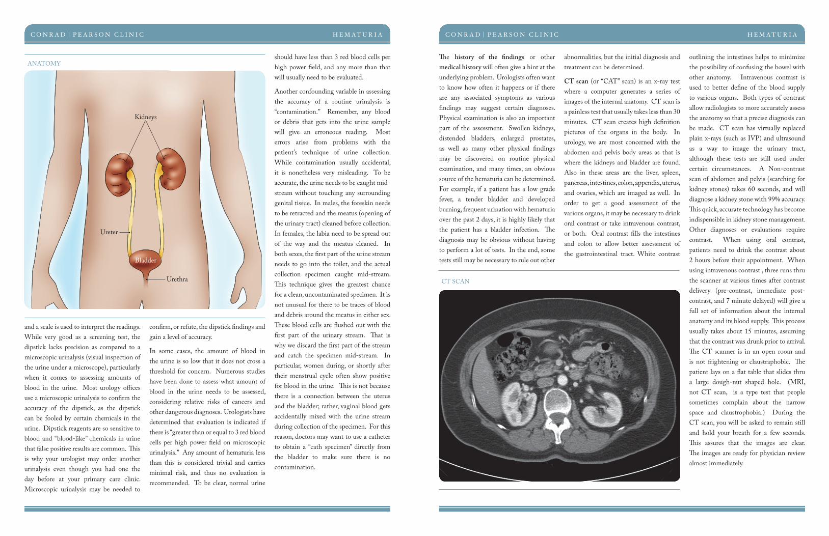
+ All Categories
Home > Documents > Hematuria CONRAD PEARSON CLINIC UROLOGY CENTER OF THE SOUTH Introduction Hematuria, or blood in the...

Hematuria CONRAD PEARSON CLINIC UROLOGY CENTER OF THE SOUTH Introduction Hematuria, or blood in the...

Date post: 11-May-2019
Category:
Upload: vothien
View: 215 times
Download: 0 times
Share this document with a friend
2
C ONRAD P EARSON T HE C LINIC UROLOGY CENTER OF THE SOUTH Introduction Hematuria, or blood in the urine, is one of the most common conditions that a urologist will evaluate. Blood in the urine is never normal; however, it is very common. Whether it involves grossly bloody urine or just a microscopic trace, the need for evaluation is the same. A simple “Google” search of “blood in the urine” will give a list of many “not-so-pleasant-to-think-about” diagnoses; however, the vast majority of cases do not involve dangerous or life threatening diagnoses. By far the most common cause of hematuria is a simple urinary tract infection, but it is also very important to remember that traces of blood in the urine may be the only early sign of more complicated underlying diseases, even cancers. Don’t try to convince yourself that the bloody urine is not important. Even if it is painless, and even if it goes away on its own, it is still a significant event and your doctor needs to sort it out. Many things that cause blood in the urine do so by irritating the lining of the urinary tract. Urologic disorders like urinary infections and stones aggravate or scratch the lining of the urinary tract and make it bleed. Sometimes this bleeding is associated with pain or other symptoms, but often it has no associated symptoms whatsoever. Whether there are, or are not any associated symptoms, the need for evaluation remains the same. In addition to urologic causes, certain medical diseases of the kidney make it prone to bleeding or weeping blood cells into the urine. In general, the function of the kidney is to filter waste products out of the bloodstream. e kidney’s filters are fine enough to keep blood cells in the blood stream but allow wastes to pass thru the filter mechanism into the urine to be expelled. If the filter mechanism is diseased, the filter may allow blood cells to pass thru the filter into the urine. Diseases such as glomerulonephritis, Berger’s disease, and Lupus (to name a few) may affect the kidney filter and present with hematuria. ese conditions are less common than the urologic causes and usually will have other pertinent findings to aid in making the diagnosis. Typically a nephrologist (doctor that manages medical kidney diseases) rather than a urologist (doctor that manages surgical kidney diseases) will treat these types of problems. Evaluation In most instances of hematuria, your physician will recommend some tests to fully evaluate the urinary tract. In assessing the anatomy, it is important to check the entire urinary system- the upper urinary tract (both of the kidneys and ureters) and the lower urinary tract (the bladder and urethra.) In decades past, such an evaluation was complicated and required a hospital visit. Presently, the full evaluation can be performed in the office or outpatient setting. Today’s state-of- the-art evaluation for hematuria involves urinalysis, history with physical exam, CT scan, and cystoscopy. In some cases, further tests may be ordered, such as a urine cytology or biopsy. Additionally, when tests are inconclusive, repeat tests or more sophisticated tests may be recommended. Urinalysis is the first part of the testing process. As a screening test, “dipstick” urinalysis is used in many clinics. e dipstick is a paper strip with imbedded reagents that react with components in the urine. e reagents change color depending on the concentrations of various chemicals in the urine (acid levels, blood, protein, etc), HEMATURIA Hematuria By Robert S. Hollabaugh, Jr. MD HEMATURIA CONRAD | PEARSON CLINIC Cystoscopy is a procedure designed to look at the lining of the urethra and bladder. It is easily done in the office or surgery center under local anesthesia (numbing jelly in the urethra). e flexible cystoscope is a small caliber telescope that easily bends around curves in the urinary tract. It is introduced into the opening of the urethra and carefully guided to the bladder where the entire lining of the bladder can be seen. Often, urologist’s offices will have a video camera attachment so that patients can watch alongside the doctor as the procedure is being performed. Cystoscopy is necessary to evaluate the urethra and bladder even when CT scan is also performed. While CT is very accurate for certain organs, it is not reliable for bladder pathology. CT scan is done to evaluate the upper urinary tract (kidneys and ureters) and cystoscopy is done to evaluate the lower urinary tract (bladder and urethra). Together these tests can fully evaluate the entire urinary tract. Cystoscopy is a simple office or surgery center procedure. ere is minimal discomfort. Patients are naturally anxious, but the thought of it is more worrisome than any part of the actual procedure. No preparation is necessary and the whole procedure usually takes less than 5 minutes. While the actual scope is inside the body for less than 60 seconds, the findings provide extremely valuable information about the health of the urinary tract. Recurrent Hematuria Some patients undergo a full evaluation for hematuria and nothing is discovered. Obviously, the patient is relieved to know that nothing serious is wrong. However, they are perplexed as to what the exact cause may have been. Often times, the physician will suspect that low grade infection was the culprit, even though no conclusive evidence was uncovered. In certain cases, patients may just have urologic anatomy that is prone to slight bleeding as a chronic condition. ese patients may continue to have detectable hematuria every time the urine is tested. ey often ask how often an evaluation will be needed. Most urologists suggest a complete evaluation every 2-3 years in the face of ongoing, unexplained hematuria. is is similar to recommendations for repeat colonoscopy in the face of persisitent blood in the bowel movements. ese cases are concerning because detecting blood in the urine is usually the only early warning we get for cancers and more worrisome urologic diseases. If we come to accept blood in the urine as normal in an individual, then we sacrifice all the early warning systems for that patient. If over the years a cancer were to develop in that patient, physicians would have no means of knowing when to perform an evaluation. As such, in cases where blood in the urine is a constant finding, we recommend a CT and cystoscopy every 2-3 years as a means of surveillance on the urinary tract to make sure nothing new is developing that would otherwise go undetected. Special Considerations Anticoagulation at normal therapeutic levels does not predispose to hematuria. Patients who develop hematuria, irrespective of anticoagulation, need to be evaluated. In other words, simply being on blood thinners does not cause the urinary tract to bleed. Wolf River Office and Surgery Center 1325 Wolf Park Drive, Suite 102 Germantown, TN 38138 Southaven Office 125 Guthrie Drive Southaven, MS 38671 Methodist North Office 3950 New Covington Pike, Suite 340 Memphis, TN 38128 West Memphis Office 228 West Tyler, Suite 202 West Memphis, AR 72301 phone: 901.252.3400 fax:901.763.4305 Please visit our website at www.conradpearson.com John R. Adams, Jr., M.D., FACS Ravi D. Chauhan, M.D., FACS Lynn W. Conrad, M.D., FACS Paul R. Eber, M.D. Howard B. Hasen, Jr., M.D. H. David Hickey, Jr., M.D., FACS Robert S. Hollabaugh, Jr., M.D., FACS Perry J. Larimer, M.D., FACS H. Benjamin Maddux, Jr., M.D., FACS H. Michael McSwain, M.D., FACS omas B. Shelton, M.D., FACS Carla Dirmann, FNP Differential Diagnosis » Urinary Tract Infection (“cystitis,” or bladder infection) » Pyelonephritis (kidney infection) » Renal Medical Diseases (glomerulonephropathy, Lupus, Berger’s Disease, etc) » Prostatitis » Urethritis » Benign Prostatic Hyperplasia (BPH) » Kidney Stones » Bladder Stones » Urethral Stricture » Trauma to kidney or bladder » Exercise-induced hematuria » Bladder Cancer » Kidney Cancer » others
Transcript

CONRAD PEARSONTHE CLINICUROLOGY CENTER OF THE SOUTH

IntroductionHematuria, or blood in the urine, is one of the most common conditions that a urologist will evaluate. Blood in the urine is never normal; however, it is very common. Whether it involves grossly bloody urine or just a microscopic trace, the need for evaluation is the same. A simple “Google” search of “blood in the urine” will give a list of many “not-so-pleasant-to-think-about” diagnoses; however, the vast majority of cases do not involve dangerous or life threatening diagnoses. By far the most common cause of hematuria is a simple urinary tract infection, but it is also very important to remember that traces of blood in the urine may be the only early sign of more complicated underlying diseases, even cancers. Don’t try to convince yourself that the bloody urine is not important. Even if it is painless, and even if it goes away on its own, it is still a signi� cant event and your doctor needs to sort it out.

Many things that cause blood in the urine do so by irritating the lining of the urinary tract. Urologic disorders like urinary infections and stones aggravate or scratch the lining of the urinary tract and

make it bleed. Sometimes this bleeding is associated with pain or other symptoms, but often it has no associated symptoms whatsoever. Whether there are, or are not any associated symptoms, the need for evaluation remains the same.

In addition to urologic causes, certain medical diseases of the kidney make it prone to bleeding or weeping blood cells into the urine. In general, the function of the kidney is to � lter waste products out of the bloodstream. � e kidney’s � lters are � ne enough to keep blood cells in the blood stream but allow wastes to pass thru the � lter mechanism into the urine to be expelled. If the � lter mechanism is diseased, the � lter may allow blood cells to pass thru the � lter into the urine. Diseases such as glomerulonephritis, Berger’s disease, and Lupus (to name a few) may a� ect the kidney � lter and present with hematuria. � ese conditions are less common than the urologic causes and usually will have other pertinent � ndings to aid in making the diagnosis. Typically a nephrologist (doctor that manages medical kidney diseases) rather than a urologist (doctor that manages surgical kidney diseases) will treat these types of problems.

EvaluationIn most instances of hematuria, your physician will recommend some tests to fully evaluate the urinary tract. In assessing the anatomy, it is important to check the entire urinary system- the upper urinary tract (both of the kidneys and ureters) and the lower urinary tract (the bladder and urethra.) In decades past, such an evaluation was complicated and required a hospital visit. Presently, the full evaluation can be performed in the o� ce or outpatient setting. Today’s state-of-the-art evaluation for hematuria involves urinalysis, history with physical exam, CT scan, and cystoscopy. In some cases, further tests may be ordered, such as a urine cytology or biopsy. Additionally, when tests are inconclusive, repeat tests or more sophisticated tests may be recommended.

Urinalysis is the � rst part of the testing process. As a screening test, “dipstick” urinalysis is used in many clinics. � e dipstick is a paper strip with imbedded reagents that react with components in the urine. � e reagents change color depending on the concentrations of various chemicals in the urine (acid levels, blood, protein, etc),

H E M AT U R I A

Hematuria By Rober t S . Hol labaugh, Jr. MD

H E M AT U R I AC O N R A D | P E A R S O N C L I N I C

Cystoscopy is a procedure designed to look at the lining of the urethra and bladder. It is easily done in the o� ce or surgery center under local anesthesia (numbing jelly in the urethra). � e � exible cystoscope is a small caliber telescope that easily bends around curves in the urinary tract. It is introduced into the opening of the urethra and carefully guided to the bladder where the entire lining of the bladder can be seen. Often, urologist’s o� ces will have a video camera attachment so that patients can watch alongside the doctor as the procedure is being performed. Cystoscopy is necessary to evaluate the urethra and bladder even when CT scan is also performed. While CT is very accurate for certain organs, it is not reliable for bladder pathology. CT scan is done to evaluate the upper urinary tract (kidneys and ureters) and cystoscopy is done to evaluate the lower urinary tract (bladder and urethra). Together these tests can fully evaluate the entire urinary tract. Cystoscopy is a simple o� ce or surgery center procedure. � ere is minimal discomfort. Patients are naturally anxious, but the thought of it is more worrisome than any part of the actual procedure. No preparation is necessary and the whole procedure usually takes less than 5 minutes. While the actual scope is inside the body for less than 60 seconds, the � ndings provide extremely valuable information about the health of the urinary tract.

Recurrent HematuriaSome patients undergo a full evaluation for hematuria and nothing is discovered. Obviously, the patient is relieved to know that nothing serious is wrong. However, they are perplexed as to what the exact cause may have been. Often times, the physician will suspect that low grade infection was the culprit, even though no conclusive evidence was uncovered.

In certain cases, patients may just have urologic anatomy that is prone to slight bleeding as a chronic condition. � ese patients may continue to have detectable hematuria every time the urine is tested. � ey often ask how often an evaluation will be needed. Most urologists suggest a complete evaluation every 2-3 years in the face of ongoing, unexplained hematuria. � is is similar to recommendations for repeat colonoscopy in the face of persisitent blood in the bowel movements. � ese cases are concerning because detecting blood in the urine is usually the only early warning we get for cancers and more worrisome urologic diseases. If we come to accept blood in the urine as normal in an individual, then we sacri� ce all the early warning systems for that patient. If over the years a cancer were to develop in that patient, physicians would have no means of knowing when to perform an evaluation. As such, in cases where blood in the urine is a constant � nding, we recommend a CT and cystoscopy every 2-3 years as a means of surveillance on the urinary tract to make sure nothing new is developing that would otherwise go undetected.

Special ConsiderationsAnticoagulation at normal therapeutic levels does not predispose to hematuria. Patients who develop hematuria, irrespective of anticoagulation, need to be evaluated. In other words, simply being on blood thinners does not cause the urinary tract to bleed.

Wolf River O� ce and Surgery Center1325 Wolf Park Drive, Suite 102

Germantown, TN 38138

Southaven O� ce125 Guthrie Drive

Southaven, MS 38671

Methodist North O� ce3950 New Covington Pike, Suite 340

Memphis, TN 38128

West Memphis O� ce228 West Tyler, Suite 202

West Memphis, AR 72301

phone: 901.252.3400fax:901.763.4305

Please visit our website atwww.conradpearson.com

John R. Adams, Jr., M.D., FACSRavi D. Chauhan, M.D., FACSLynn W. Conrad, M.D., FACSPaul R. Eber, M.D.Howard B. Hasen, Jr., M.D.H. David Hickey, Jr., M.D., FACS

Robert S. Hollabaugh, Jr., M.D., FACSPerry J. Larimer, M.D., FACSH. Benjamin Maddux, Jr., M.D., FACSH. Michael McSwain, M.D., FACS� omas B. Shelton, M.D., FACSCarla Dirmann, FNP

Di� erential Diagnosis

» Urinary Tract Infection (“cystitis,” or bladder infection)

» Pyelonephritis (kidney infection)

» Renal Medical Diseases (glomerulonephropathy, Lupus, Berger’s Disease, etc)

» Prostatitis

» Urethritis

» Benign Prostatic Hyperplasia (BPH)

» Kidney Stones

» Bladder Stones

» Urethral Stricture

» Trauma to kidney or bladder

» Exercise-induced hematuria

» Bladder Cancer

» Kidney Cancer

» others

and a scale is used to interpret the readings. While very good as a screening test, the dipstick lacks precision as compared to a microscopic urinalysis (visual inspection of the urine under a microscope), particularly when it comes to assessing amounts of blood in the urine. Most urology o� ces use a microscopic urinalysis to con� rm the accuracy of the dipstick, as the dipstick can be fooled by certain chemicals in the urine. Dipstick reagents are so sensitive to blood and “blood-like” chemicals in urine that false positive results are common. � is is why your urologist may order another urinalysis even though you had one the day before at your primary care clinic. Microscopic urinalysis may be needed to

con� rm, or refute, the dipstick � ndings and gain a level of accuracy.

In some cases, the amount of blood in the urine is so low that it does not cross a threshold for concern. Numerous studies have been done to assess what amount of blood in the urine needs to be assessed, considering relative risks of cancers and other dangerous diagnoses. Urologists have determined that evaluation is indicated if there is “greater than or equal to 3 red blood cells per high power � eld on microscopic urinalysis.” Any amount of hematuria less than this is considered trivial and carries minimal risk, and thus no evaluation is recommended. To be clear, normal urine

should have less than 3 red blood cells per high power � eld, and any more than that will usually need to be evaluated.

Another confounding variable in assessing the accuracy of a routine urinalysis is “contamination.” Remember, any blood or debris that gets into the urine sample will give an erroneous reading. Most errors arise from problems with the patient’s technique of urine collection. While contamination usually accidental, it is nonetheless very misleading. To be accurate, the urine needs to be caught mid-stream without touching any surrounding genital tissue. In males, the foreskin needs to be retracted and the meatus (opening of the urinary tract) cleaned before collection. In females, the labia need to be spread out of the way and the meatus cleaned. In both sexes, the � rst part of the urine stream needs to go into the toilet, and the actual collection specimen caught mid-stream. � is technique gives the greatest chance for a clean, uncontaminated specimen. It is not unusual for there to be traces of blood and debris around the meatus in either sex. � ese blood cells are � ushed out with the � rst part of the urinary stream. � at is why we discard the � rst part of the stream and catch the specimen mid-stream. In particular, women during, or shortly after their menstrual cycle often show positive for blood in the urine. � is is not because there is a connection between the uterus and the bladder; rather, vaginal blood gets accidentally mixed with the urine stream during collection of the specimen. For this reason, doctors may want to use a catheter to obtain a “cath specimen” directly from the bladder to make sure there is no contamination.

� e history of the � ndings or other medical history will often give a hint at the underlying problem. Urologists often want to know how often it happens or if there are any associated symptoms as various � ndings may suggest certain diagnoses. Physical examination is also an important part of the assessment. Swollen kidneys, distended bladders, enlarged prostates, as well as many other physical � ndings may be discovered on routine physical examination, and many times, an obvious source of the hematuria can be determined. For example, if a patient has a low grade fever, a tender bladder and developed burning, frequent urination with hematuria over the past 2 days, it is highly likely that the patient has a bladder infection. � e diagnosis may be obvious without having to perform a lot of tests. In the end, some tests still may be necessary to rule out other

abnormalities, but the initial diagnosis and treatment can be determined.

CT scan (or “CAT” scan) is an x-ray test where a computer generates a series of images of the internal anatomy. CT scan is a painless test that usually takes less than 30 minutes. CT scan creates high de� nition pictures of the organs in the body. In urology, we are most concerned with the abdomen and pelvis body areas as that is where the kidneys and bladder are found. Also in these areas are the liver, spleen, pancreas, intestines, colon, appendix, uterus, and ovaries, which are imaged as well. In order to get a good assessment of the various organs, it may be necessary to drink oral contrast or take intravenous contrast, or both. Oral contrast � lls the intestines and colon to allow better assessment of the gastrointestinal tract. White contrast

outlining the intestines helps to minimize the possibility of confusing the bowel with other anatomy. Intravenous contrast is used to better de� ne of the blood supply to various organs. Both types of contrast allow radiologists to more accurately assess the anatomy so that a precise diagnosis can be made. CT scan has virtually replaced plain x-rays (such as IVP) and ultrasound as a way to image the urinary tract, although these tests are still used under certain circumstances. A Non-contrast scan of abdomen and pelvis (searching for kidney stones) takes 60 seconds, and will diagnose a kidney stone with 99% accuracy. � is quick, accurate technology has become indispensible in kidney stone management. Other diagnoses or evaluations require contrast. When using oral contrast, patients need to drink the contrast about 2 hours before their appointment. When using intravenous contrast , three runs thru the scanner at various times after contrast delivery (pre-contrast, immediate post-contrast, and 7 minute delayed) will give a full set of information about the internal anatomy and its blood supply. � is process usually takes about 15 minutes, assuming that the contrast was drunk prior to arrival. � e CT scanner is in an open room and is not frightening or claustraphobic. � e patient lays on a � at table that slides thru a large dough-nut shaped hole. (MRI, not CT scan, is a type test that people sometimes complain about the narrow space and claustrophobia.) During the CT scan, you will be asked to remain still and hold your breath for a few seconds. � is assures that the images are clear. � e images are ready for physician review almost immediately.

H E M AT U R I AC O N R A D | P E A R S O N C L I N I C

ANATOMY

H E M AT U R I AC O N R A D | P E A R S O N C L I N I C

CT SCAN

Kidneys

Bladder

Ureter

Urethra


Recommended