INTERNATIONAL JOURNAL OF QUANTUM CHEMISTRY. VOL. XVI. 311-329 (1979)
Heme Proteins and Metalloporphyrins: Redox Chemistry and Oxygen Binding
DAVID DOLPHIN, ANTHONY W. ADDISON, MAX CAIRNS, ROBERT K. DINELLO, NICHOLAS P. FARRELL, BRIAN R. JAMES,
DONALD R. PAULSON, AND CURTIS WELBORN Department of Chemistry, The University of British Columbia, Vancouver, B.C., Canada V6T 1 W5
Abstract
Metalloporphyrins perform a variety of functions in nature from the storage and transport of electrons and molecular oxygen to the decomposition of hydrogen peroxide and the activation of oxygen. The chemistry of both the centrally coordinated metal and the porphyrin macrocyde play important roles in these reactions. The use of model systems and metalloporphyrins, other than iron porphyrins, is described for the elucidation of the mechanism of action of the natural systems.
Iron protoporphyrin (1), the prosthetic group of heme proteins, and the closely related photosynthetic pigments chlorophyll (2) and bacteriochlorophyll (3) are widespread in nature. While these pigments, in concert with their apoproteins, play many varied biochemical roles, their chemistry can be generalized as that principally related to redox reactions and the transport storage and activation of molecular oxygen.
PROTOHEME
t·1 : H:
( C~Me° C~Phytol
CHLOROPHYLL a 2
BACTERIOCHLOROPHYLL c :3
Thus hemoglobin and myoglobin serve, in mammals, as the means of transportation and storage of oxygen, while the cytochromes of the respiratory chain serve to transport and store electrons. More recently the cytochromes P-450 have been shown to couple both of these phenomena in order to activate molecular oxygen and generate hydroxylating agents. In addition, the enzymes peroxidase and catalase are both oxidized by hydrogen peroxide, but while the peroxidases
use this oxidizing power to bring about one-electron oxidations of organic substrates (usually phenols and aromatic amines), catalase, in one of the fastest enzymatic reactions known, catalyzes the decomposition of hydrogen peroxide to molecular oxygen and water. The photosynthetic pigments couple the photonic energy of the sun to the production of chemical energy, and during the course of these reactions A TP is made during oxidative phosphorylation, while CO2 is reduced to sugars and water oxidized to molecular oxygen.
We shall attempt here to show how these apparently diverse reactions are related to each other and to show how both the organic porphyrin macrocyde and the centrally coordinated metal play important roles in these biochemical systems.
When cobalt(II) octaethylporphyrin Co(II)OEP (4) was oxidized by bromine in methylene dichloride, two one-electron oxidations were observed [1] and the
4
changes in the electronic spectra of the system are shown in Figure 1. When the same system was oxidized electrochemically, using tetra-n-propylammonium perchlorate as electrolyte in methylene dichloride, two one-electron oxidations were again observed, but the changes in electronic spectra Figure 2 were now
<t I 0
'" '"
.---.---,,---.----r----,----.---~----.---,
2.5
2.0
1.5
1.0
0.5
0 300
,, . , ..
I I
._-_.---- ..... \ ' .. ~
\':::.:-"
400 500 wavelength.nm
, l\ " ........ :'
\ ........ . \-"
.'\ \ \ , '"
600
-""
700
'" I
Q ><
'"
Figure 1. Optical absorption spectra (in CH2Ch) of Co(II)OEP (--); [Co(III)OEPtBr- (- - - -); and [Co(IlI)OEP]2+' 2Br- ( ..... ).
Figure 2. Optical absorption spectra (in CH2Ch) of Co(II)OEP (--); [Co(III)OEPrCl04 - (---); and [Co(III)OEPf+' 2Cl04 -(----).
313
quite different. However, we had observed similar electronic spectra for other oxidized metalloporphyrins. Thus the one-electron oxidation of magnesium(II) octaethylporphyrin gave [2] a 1T-cation radical having an optical spectrum (Fig. 3) showing strong absorption around 670 and 600 nm. The radical nature of this species was confirmed by its EPR spectrum, which showed [Fig. 4(a)] a five-line
Figure 4. EPR spectra of metalloporphyrins. (a) [Mg(II)OEPr', and (b) [Mg(II)OEP-d4r-, in CH30H at -50·C; (c) [Zn(II)TPPt", and (d)
[Zn(II)TPP-d28r-, in CHCl3 at 25°C.
signal which arose from the hyperfine coupling of the four equivalent peripheral hydrogen atoms. When these four hydrogen atoms were replaced by deuterium the five-line signal, as expected, collapsed to a single line [Fig. 4(b)]. Electrochemical oxidation of zinc meso-tetraphenylporphyrin Zn(II)TPP (5) by one
electron also generated a 1T-cation radical [2] but the optical spectrum of this species (Fig. 3) showed strong absorption over the whole visible region. Moreover, the ESR spectrum [Fig. 4(c)] showed a nine-line signal. When all 28 hydrogen atoms of 5 were replaced by deuterium the nine-line signal [Fig. 4(d)] still remained [3]. Thus the nine lines must result from the hyperfine coupling of the four equivalent nitrogen atoms. These observations immediately raise the question as to why these two 1T-cation radicals exhibit different electronic spectra, and why their EPR spectra are different with one of the radicals exhibiting coupling
HEME PROTEINS AND METALLOPORPHYRINS 315
to peripheral substituents and no coupling with the inner nitrogens, while the other radical shows coupling principally to the nitrogen atoms. These questions were answered by SCF-MO calculations. Gouterman [4] has shown that the two highest filled bonding molecular orbitals are nearly degenerate with a2u and al"
symmetries, and Felton [2] has shown that the 1T-cation radicals which result from the one-electron oxidations of metalloporphyrins result from the loss of an electron from one or the other of these orbitals such that their electronic configurations are either a 2 A 2u or a 2 A I" state. The calculations explain the observed EPR spectra and show that the 1T-cation radical derived from magnesium OEP viz. Mg(II) [OEPt'CIO; is characteristic of the 2A 1u ground state, which has low electron density at nitrogen, while that derived from zinc TPP viz. Zn(II)[TPPt'ClO; is characteristic of the 2 A 2u ground state with medium electron density at nitrogen and consequent hyperfine coupling of the unpaired electron with the nitrogen atoms (Fig. 5).
3 I \ I
2 I
EI
EI
ground stote symmetry
eleclron densily
\ I I \ \ \ , ... , ... _-
500
EI
EI
zA 1U
N
meso
fl
600 800 1000
wavelength, nm
EI B
EI
RJ £J
EI
EI RJ
2Azu
LON MEDIUM
LOW HIGH
HIGH LOW
Figure 5. Comparison of the optical spectra and electron densities of the 2A Iu and 2 A 2u ground-state symmetries of metalloporphyrin 1T-cation radicals.
316 DOLPHIN ET AL.
A comparison of the optical spectra of these two doublet ground states (Fig. 5) with those of the two different forms of the oxidized cobalt octaethylporphyrins Figures 1 and 2 suggests that the system oxidized by bromine gives the 7T-cation radical [Co(III}OEPf+·2Br- (6) with a 2A1u ground state, while the electrochemically generated 7T-cation radical Co(III}[OEP]2+' 2CIO; (7) has a 2 A 2u
ground state. Further evidence for this two-ground-state theory was evident when we found that treatment of the dibromide 6 with silver perchlorate gave the diperchlorate salt 7 with a concomitant change in the electronic spectrum from that of the 2A1u state (Fig. 6). This suggests that the difference in ground state of a
(a)
100 10
7 ~ ~/ '.
Q b x or---------------------------------------~O ~ w w
Figure 6. Comparison of the optical absorption spectra of (a) [Co(III)OEP]2+ 2Br(---) and [Co(III)OEP]2+' 2Cl04 - ( •••• ); (bJ catalase compound 1 (---J and
HRP compound I ( .... J.
specific metalloporphyrin 7T-cation radical can be determined by the nature of the axial ligands, and indeed theoretical calculations [2] confirm that the difference in energy between these two states is in the region of 2000 cm -1 (i.e., about 5 kcal/mol).
Of special significance was the observation that optical spectra of the enzymatically active forms of horse radish peroxidase and catalase parallel those of the two different 7T-cation radicals of cobaltic octaethylporphyrin (Fig. 6); this led us to infer that these two heme proteins function enzymatically via their porphyrin 7T-cation radicals [1]. Both of these enzymes in their resting state contain ferric protoporphyrin, and both undergo a two-electron oxidation by hydrogen peroxide to the green primary compounds (compounds I). While various electronic configurations have been suggested for these two-electron oxidation products [5J
HEME PROTEINS AND METALLOPORPHYRINS 317
it is now apparent that they both contain porphyrin '7T-cation radicals. The loss of one electron from the porphyrin ring still leaves one electron unaccounted for during the oxidation to the compounds I. However, the Mossbauer spectra [6] of both enzymes showed that the first oxidation was of the iron; hence the twoelectron oxidation of the resting ferric heme proteins, to their primary compounds, involve both a one-electron oxidation of the metal and of the porphyrin macrocycle such that their final electronic configuration can be represented as the '7T-cation radical Fe(lV) [protoporphyrinf+·. The optical spectra of these oxidized enzymes (Fig. 6) suggests that the primary compound of catalase has a 2A1u ground-state symmetry, while that of horseradish peroxidase has a 2 A 2u ground-state symmetry. Since both of these enzymes contain the same iron porphyrin, the different electronic configurations between oxidized catalase and horseradish peroxidase must arise from perturbations provided by the protein, where we might anticipate that the dominant effect would be the axial ligation.
Since one cannot yet replace specific amino acids in an enzyme, we have looked at the reconstitution of horseradish peroxidase with iron porphyrins other than protoheme. The same experiments cannot be performed with catalase since it has not yet been successfully reconstituted. The SCF-MO calculations described above suggested that the two ground states available to metalloporphyrin '7Tcation radicals differ in energy by only a few kcal/mol, and indeed changing the axial ligation can change the ground state. We further expected that changing the peripheral substituents of an iron porphyrin and reconstitution with apo horseradish peroxidase might change the ground state of the oxidized enzyme to that possessed by catalase.
In order to stabilize and better observe the compounds I of the heminsubstituted peroxidases their oxidations were performed, and electronic spectra were obtained, in 30% 3.3mM potassium phosphate/70% N,N-dimethylacetamide at -42°C. These low-temperature spectra were almost identical to those previously recorded at room temperature [7]. The initial reconstitution experiments were with hemins bearing strongly electron-withdrawing groups at their periphery. 2-Formyl-4-vinyldeutero-hemin (8, R = -CHO, R2 = -CH=CH2), 2-vinyl-4-formyl deuteroporphyrin (8, Rl = -CH=CH2, R2 = -CHO), and diacetyldeuteroporphyrin (8, Rl = R2 = -COCH3 ); however, all
8
318 DOLPHIN ET AL.
exhibited compound I spectra similar to that of the native protohemin enzyme and thus the same e A 2u ) ground state.
Nonetheless compound I spectrum of de utero hemin (8, Rl = R2 = H) reconstituted peroxidase (Fig. 7) was found to be characteristic of a 2 Al u ground state,
60.-----------------------------~
400
.. /'\. .................... ..........
sao 600
Wavelength, nm
. ................... .
700
12
10
B
'" I
6 Q >< W
4
2
Figure 7. Low-temperature optical absorption spectra of deuterohemin reconstituted HRPI (- - - -) and room temperature spectrum of catalase I (--) [5].
i.e., like that of catalase. This observation adds further support to the two-groundstate theory of metalloporphyrin TT- cation radicals and to the hypothesis that both catalase and the peroxidases perform their enzymatic functions via such radical intermediates. Although the deuterohemin reconstituted horseradish peroxidase compound I exhibits a 2 A lu (catalase-like) TT-cation radical, its activity toward hydrogen donors is identical to that of the native enzyme [8], which leads us to conclude that the enzymatic action of these systems is a function not only of the electronic configuration of the iron porphyrins; but of the structure of the proteins as well.
The observations that catalase and horseradish peroxidase function via porphyrin TT-cation radicals have prompted the search for the intermediacy of other such radicals in biochemical processes. One such area is photosynthesis, where it has been shown [9] that the initial photochemical events result in the formation of chlorophyll or bacteriochlorophyll TT-cation radicals. At this time, however, we wish to turn our attention to the question of how the cytochromes, in the respiratory chain, transport electrons. It is apparent that cytochrome c, for instance, functions via an Fe(II)!:::; Fe(III) couple. The various routes by which electron transfers may occur between the cytochromes have been discussed in some detail [10]. X-ray structural information shows that in both oxidized and reduced cytochrome c [11] the iron atom is inaccessible to cytochrome c oxidase and reductase, which are themselves macromolecules. An especially striking feature of the crystal structures of cytochromes c is the partially exposed edge of the heme porphyrin ring (a similar structural feature is noted with cytochrome bs [12]). If electron transfer occurs here via the porphyrin periphery then the redox
HEME PROTEINS AND MET ALLOPORPHYRINS 319
chemistry of cytochrome c can be described as shown in Scheme 1 whereby the initial oxidation of the ferro heme protein oxidizes the porphyrin ring, and not the iron, to give the ferroheme 1T-cation radical, which is followed by an internal electron transfer, with the hole moving from the porphyrin to the iron atom, to give the ferric heme protein. Intermediates of this type have not, as yet, been
Fe(ll) [cytochrome c J ~ Fe(fI)[cytochrome c J+a :o=:~~'nl~e.;=:,,,~' =;:==" feUlU[cyfochrome] - 1-e- t - electron trOf'sfer
1]'-cafion rodiool -e- • ' +. Internal
NIII) [tetra~enylporphyrtnlTe':"'Ni(lI) [tetraphenyiporphyrtn] electron tronsfe. Nil .,[telraphenylporphyrin]
Scheme I.
observed for heme proteins. Nonetheless these electronic configurations and internal electron transfers have been mimicked with other metalloporphyrins [10).
Oxidation of Ni(Il) tetraphenylporphyrin at 1.24 V (vs. Agj Ag +) in methylene dichloride and a tetra-n-butylammonium salt (CIO; or PF6) gives the wellcharacterized 1T-cation radical [Ni(II)TPPt·. At room temperature this radical gives a green solution with an optical spectrum (Fig. 8) characteristic of a
A
;-\ I \
/ \ I \
I \ I \
400
, , , "
500 600 700 Wave length, nm
Figure 8. Optical absorption spectra, in CH2CI 2 , of [Ni(II)TPPt'PF6 - at room temperature (---), the same sample at 77°K, i.e., [Ni(III)TPPtPF6 - (--l,
and Ni(II)TPP at 77°K (- - - -).
320 DOLPHIN ET AL.
porphyrin 1T-cation radical. In addition the EPR spectrum of the green solution exhibited a single signal at g = 2.0041 with a peak-to-peak width of 47.2 G (characteristic once again of a 1T-cation radical). When the green solution was frozen to 77°K an orange solid was formed and these color changes were reflected in a dramatic change of the optical spectrum, which for the frozen solid resembles that of a normal metalloporphyrin (Fig. 8). In addition, the EPR spectrum changes upon cooling and the orange solid showed signals at gJ.. = 2.286 and gil = 2.086, both signals being consistent with a low-spin d 7 Ni(III) electronic configuration [13]. When the sample was warmed to room temperature the spectral and color changes were reversed, and thus the Ni(II) tetraphenylporphyrin 1T-cation radical, upon cooling, undergoes a reversible internal electron transfer to Ni(III) tetraphenylporphyrin (Scheme 1) in a manner analogous to that proposed for the redox chemistry of the cytochromes.
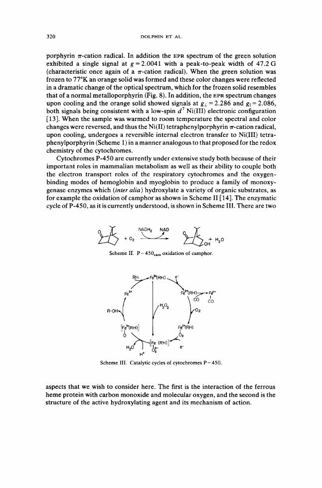
Cytochromes P-450 are currently under extensive study both because of their important roles in mammalian metabolism as well as their ability to couple both the electron transport roles of the respiratory cytochromes and the oxygenbinding modes of hemoglobin and myoglobin to produce a family of monoxygenase enzymes which (inter alia) hydroxylate a variety of organic substrates, as for example the oxidation of camphor as shown in Scheme II [14]. The enzymatic cycle of P-450, as it is currently understood, is shown in Scheme III. There are two
Scheme II. P-4S0cam oxidation of camphor.
Scheme III. Catalytic cycles of cytochromes P-4S0.
aspects that we wish to consider here. The first is the interaction of the ferrous heme protein with carbon monoxide and molecular oxygen, and the second is the structure of the active hydroxylating agent and its mechanism of action.
HEME PROTEINS AND METALLOPORPHYRINS 321
Cytochromes P-450 when reduced to the ferrous state and coordinated to dioxygen exhibit optical spectra similar to those of oxyhemoglobin and oxymyoglobin. When, however, P-450 binds carbon monoxide its optical spectrum (which has an intense absorption at -450 nm, from which its name is derived [15]) is unlike that of any other known hemoprotein.
We initially turned our attention to mimicking this unusual optical spectrum by using model compounds since it was expected that this would give a better understanding of the enzymes themselves. An early attempt on such model studies reported [16] that a partial P-450 spectrum could be obtained from the combination of reduced heme, carbon monoxide, a thiol, and strong base under special mixing conditions. We [17] and others [18] expanded this initial observation and showed that the 450-nm absorption of the enzyme could be obtained when a ferrous porphyrin was coordinated by both CO and a thiolate anion. This work also showed that another intense absorption band centred around 370 nm always accompanied the 450-nm band (Fig. 9) when carbonmonoxy ferrous
100
350 400 450 50) 5:Kl 6CO 6:Kl 700
Wavelength, nm
Figure 9. Formation of the protoheme-mercaptide-CO complex in dimethylacetamide at 24°C. CO partial pressures are 0, 4.7, 30, and 760 torr, respectively.
porphyrins were coordinated by a sixth thiolate ligand. A reexamination of the P-450 enzymes [19] showed that both the 370- and 450-nm bands are also characteristic of the enzymes. Gouterman [20] has classified metalloporphyrins with such double Soret bands as "hyper" type and their electronic spectra have been interpreted as resulting from a charge transfer of the thiolate p-electrons to the porphyrin eg (1T*) orbital coupled to the normal 1T ~ 1T* Soret transition. Many examples of hyper-type metalloporphyrin spectra have now been recorded
322 DOLPHIN ET AL.
[20]. At the present time the cytochromes P-450 are unique amongst the heme proteins in having a thiolate anion as a sixth axial ligand. From both SCF-MO
calculations [19] and experimental observations [21] it would appear that the oxygenated form of P-450 is coordinated by a thiol rather than a thiolate. Since carbon monoxide is a powerful electron acceptor, it would be expected to lower the electron density at iron, which in turn would increase the acidity of a coordinated thiol ligand. We suggest that the active site of P-450 has a base capable of accepting this proton (cf. Scheme IV) and that the protonationdeprotonation of the thiolate ligand will prove to be an integral part of the enzymatic reaction (but which at the present time is only observed under the nonphysiologic conditions of CO binding).
All substrates hydroxylated by cytochromes P-450 are hydrophobic and one can assume that the active site of the enzymes will also be hydrophobic in character, and it is suggested that during the activation of oxygen and substrate hydroxylation the protonation-deprotonation of the thiolate ligand can be used to maintain electrical neutrality.
As indicated in Scheme III the enzymatic cycle of P-450, which normally utilizes molecular oxygen and two electrons, can be bypassed by treating the ferric heme protein with hydrogen peroxide. The resultant intermediate (c, Scheme IV) upon losing hydroxide (or water) can be formulated in a variety of electronic configurations: as the ferric oxene complex (d, Scheme IV), or as a state analogous to the compound I of catalase or peroxide, i.e., and Fe(IV) porphyrin 1r-cation radical (e, Scheme IV). At first sight the oxene formulation seems very attractive since many of the hydroxylating reactions carried by P-450 can be formulated in a manner similar to the chemistry of carbenes and nitrenes, i.e., the direct insertion of an oxene into a C-H bond of the substrate to give the hydroxylated product. However, the recent elegant work of Groves [22] shows that in at least some cases the hydroxylation proceeds via a substrate intermediate, and cannot be direct insertion of oxygen. An attractive possibility is that the hydroxylating intermediate (c, Scheme IV) abstracts a hydrogen atom from the substrate to make a free radical and a formally Fe(IV) porphyrin coordinated by hydroxide (this being isoelectronic to a ferric porphyrin and a hydroxyl free radical). Transfer of a hydroxyl radical to the substrate radical would give the hydroxylated product and the resting ferric heme protein.
The abstraction of a hydrogen atom by P-450 would parallel the chemistry of the primary complex of horseradish peroxidase, which can also function by hydrogen atom abstraction from substrate.
Evidence for such a radical pathway can be deduced from a somewhat unexpected source, namely, the metabolism by rats of the potent insecticide dieldrin (9). Thus P-450-mediated metabolism of dieldrin by rat liver microsomes gives as a major product the ketone 11. The mechanism by which the ketone is formed is as yet unknown, but clearly it cannot be generated by a direct insertion of oxygen. Rather the mechanism presented in Scheme V whereby P-450 functions both to abstract a hydrogen atom and deliver a hydroxyl radical via the intermediacy of the substrate radical 10, seems most likely. These observations
HEME PROTEINS AND METALLOPORPHYRINS 323
a (450nm)
d c
e
Scheme IV. Hypothetical mechanisms of action of P-4S0.
324 DOLPHIN ET AL.
CI
~I H HO CIU_I
CI~O CI P-450 CI CI -H"'·"o""b-='SI=rO':rCI!=:,on=--
CI 9 CI 10
o J ~C~ H P-450 ~I • H
CI CI HO" delivery CI I CI CI
CO L O~HfiO CI~
CI 11
Scheme V. Proposed mechanism for P - 450-mediated oxidation of dieldrin. Based upon scheme proposed by C. T .. Bedford in Foreign Compound Metabolism in
Mammals (The Chemical Society, London, 1975), Vol. 3, p. 402.
suggest that in at least some instances P-450 can hydroxylate substrates via a radical mechanism.
One major problem in studies associated with oxygen binding to hemes is the ease with which the extracellular heme proteins and enzymes undergo autoxidation, a problem which is magnified when dealing with simple ferrous porphyrins. A major route to autoxidation occurs through the formation of ferric p.-peroxo complexes which can collapse by a number of possible routes to the ferric p.-oxo dimer (Scheme VI). Formation of these wcomplexes can be prevented by
02 Felnl) FeUU) o I FOIl) I I
Fe(ll)~Felt))-02 -0-I I
)2 Feltll) Fell))
Scheme VI. Autoxidation of ferrous hemes.
sterically hindering the iron atoms so that they cannot be bridged by the small dioxygen molecule. Nature achieves this purpose using the protein, and similar steric constraints in model systems have been achieved by several groups [23, 24]. A second method for the study of oxygen binding is to stabilize the 1 : 1 complex (12) at low temperatures [25]. A third method which we have developed [26] makes use of the "inertness" toward ligand exchange of ruthenium compared to iron. We anticipated the 1: 1 ruthenium porphyrin dioxygen complex would be more stable than the corresponding iron complexes (12) and would not so readily suffer autoxidation via p.-peroxo formation.
HEME PROTEINS AND METALLOPORPHYRINS 325
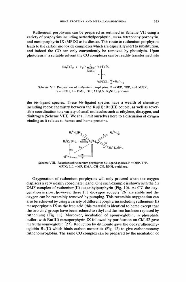
Ruthenium porphyrins can be prepared as outlined in Scheme VII using a variety of porphyrins including octaethylporphyrin, meso-tetraphenylporphyrin, and mesoporphyrin IX (MPIX) as its diester. This route to ruthenium porphyrins leads to the carbon monoxide complexes which are especially inert to substitution, and indeed the CO can only conveniently be removed by photolysis. Upon photolysis in a suitable solvent the CO complexes can be readily transformed into
RU3(CO~2 + H2P TO~NE' Rup(Cols ~~~:lnS) 1
+L -5
RuP<COlL ~RuPL2
Scheme VII. Preparation of ruthenium porphyrins. P = OEP, TPP, and MPIX; S = EtOH; L = DMF, THF, eH3eN, R2NH, pyridines.
the his-ligand species. These his-ligated species have a wealth of chemistry including redox chemistry between the Ru(II): Ru(III) couple, as well as reversible coordination to a variety of small molecules such as ethylene, dioxygen, and dinitrogen (Scheme VIII). We shall limit ourselves here to a discussion of oxygen binding as it relates to hemes and heme proteins.
Scheme VIII. Reactions of ruthenium porphyrins bis-ligand species. P = OEP, TPP, MPIX; L,L' = MF, DMA, eH3eN, RNH, pyridines.
Oxygenation of ruthenium porphyrins will only proceed when the oxygen displaces a very weakly coordinate ligand. One such example is shown with the his DMF complex of ruthenium(II) octaethylporphyrin (Fig. 10). At O°C the oxygenation is slow; however, these 1: 1 dioxygen adducts [26] are stable and the oxygen can be reversibly removed by pumping. This reversible oxygenation can also be achieved by using a variety of different porphyrins including ruthenium(II) mesoporphyrin IX as the free acid (this material is identical to heme except that the two vinyl groups have been reduced to ethyl and the iron has been replaced by ruthenium) (Fig. 11). Moreover, incubation of apomyoglobin, in phosphate buffer, with Ru(III) meso porphyrin IX followed by purification on CM-52 gave metruthenomyoglobin [27]. Reduction by dithionite gave the deoxyruthenomyoglobin Ru(II) which binds carbon monoxide (Fig. 12) to give carbonmonoxy ruthenomyoglobin. The same CO complex can be prepared by the incubation of
326 DOLPHIN ET AL.
2.0 40 -4min
~ ~ Q ,-16min Q
" If - 30min x
U) U)
",- 60 min I 2.0
i- . 300 600 WCl\'elength, nm
Figure 10. Oxygenation of Ru(II)OEP· 2DMF (in DMF) to give RU(II)02' DMF Reaction carried out at O·C, 760 torr O2 •
carbonmonoxy Ru(II) mesoporphyrin with apomyoglobin [27, 28]. Removal of the CO, even by photolysis, leads to protein denaturation; hence it is important, if one wishes to study the chemistry of ruthenomyoglobin, that it be prepared as the met- or deoxy-form.
From our earlier observations on the stability of the ruthenium porphyrin~ oxygen adduct we had anticipated that oxyruthenomyoglobin would be more stable toward auto-oxidation than oxymyoglobin itself. Nonetheless when Ru(II) deoxyruthenomyoglobin was treated with oxygen it immediately oxidized to the met- [Ru(HI)] form which, of course, no longer bound either oxygen or carbon monoxide. Similarly, incubation of the Ru(II) dioxygen complex of mesoporphyrin IX with apomyoglobin gave only the metruthenomyoglobin (Fig. 13). It is
500 600 Wavelength. nm
Figure 11. Oxygenation of Ru(II) mesoporphyrin IX·2DMF to give Ru(II)MPIX O2, DMF. Reaction carried in DMF at () C, 760 torr O 2,
HEME PROTEINS AND METALLOPORPHYRINS
20 \ 02 \
A I-RuCftilMb A
\ \ \ \ \ RuCII) (CO) \ 165min _ .
~ 285mtn -\ RU(1Il - 37mtn
1.0 \ - 21 min 0.1 \ \ , -
Wavelength ,nm
Figure 12. Metruthenomyoglobin (- - -) in phosphate buffer, reduced by S20~in the presence of CO (1 torr) at room temperature.
327
apparent that when coordinated to apomyoglobin auto-oxidation of the dioxygen adduct is rapid. The mechanism by which oxidation occurs is unknown, but presumably it cannot, in the protein, occur through #L-peroxo formation (Scheme VI). The optical spectra of oxyruthenium(II) porphyrins and ruthenium(III) porphyrins are similar, suggesting the Ru(III)-superoxide formulation 14 may
A
I 10
400 500 Wavelength, nm
Figure 13. Incubation of oxygenated Ru(II)MPIX with apomyoglobin (t1/2 = 90 min at room temperature).
328 DOLPHIN ET AL.
play a major contribution to electronic formulation (13 ~ 14) of the oxygen adduct. If this is the case then loss of superoxide ion from oxyruthenomyoglobin might account for its rapid autoxidation .
.. .. -; O· .. /' ..
O. O·
°O~ 0°
~ S 13 14
Acknowledgments
This work is a contribution from the Bioinorganic Chemistry Group and was supported by the United States National Institutes of Health (AM 17989) and by operating and negotiated development grants from the National Research Council of Canada. A special note of gratitude goes to our collaborators in the earlier aspects of this work, Dr. D. C. Borg, Dr. C. K. Chang, Dr. J. Fajer, and Dr. R. H. Felton.
Bibliography
[1] D. Dolphin, A. Forman, D. C. Borg, 1. Fajer, and R. H. Felton, Proc. Natl. Acad. Sci. 68, 614 (1971).
[2] J. Fajer, D. C. Borg, A. Forman, D. Dolphin, and R. H. Felton, 1. Am. Chem. Soc. 92, 3451 (1970).
[3] 1. Fajer, D. C. Borg, A. Forman, R. H. Felton, L. Vegh, and D. H. Dolphin, Ann. N.Y. Acad. Sci. 206,349 (1973).
[4] C. Weiss, H. Kobayashi, and M. Gouterman, 1. Mol. Spectrosc. 16,415 (1965). [5] G. R. Schonbaum and B. Chance, in The Enzymes, 3rd ed., P. D. Boyer, Ed. (Academic, New
York, 1976), Vol. XIII, p. 363. [6] T. H. Moss, A. Ehrenberg, and A. 1. Bearden, Biochemistry 8, 4159 (1969). [7] G. R. Schonbaum and S. Lo, 1. BioI. Chem. 247, 3353 (1972). [8] R. K. DiNello and D. Dolphin, Biochem. Biophys. Res. Commun. 86,190 (1979). [9] D. C. Borg, 1. Fajer, R. H. Felton, and D. Dolphin, Proc. Natl. Acad. Sci. 67, 813 (1970).
[10] E. C.lohnson, T. Niem, and D. Dolphin, Can. 1. Chem. 56,1381 (1978). [11] R. E. Dickerson and R. TimkovIich, in The Enzymes, P. D. Boyer, Ed. (Academic, New York,
1975), Vol. XI, p. 397. [12] F. S. Mathews, M. Levin, and P. Argos, 1. Mol. BioI. 64, 449 (1972). [13] N. E. Tokel, K. Farmery, L. Anderson, F. V. Lovechio, E. S. Gore, and D. H. Busch, 1. Am.
Chem. Soc. 96, 731 (1974). [14] B. W. Griffin, 1. A. Peterson, and R. W. Estabrook, in The Porphyrins, D. Dolphin, Ed.
(Academic, New York, 1979), Vol. VII. [15] T. Omura and R. Sato, 1. BioI. Chem. 239, 2370 (1964). [16] 1. O. Stern and 1. Peisach, 1. BioI. Chem. 249, 7495 (1974). [17] C. K. Chang and D. Dolphin, 1. Am. Chem. Soc. 97, 5948 (1975). [18] 1. P. Collman and T. N. Sorrell, 1. Am. Chem. Soc. 97, 4133 (1975).
HEME PROTEINS AND METALLOPORPHYRINS 329
[19] L. K. Hanson, W. A. Eaton, S. G. Sligar, L C. Gunsalus, M. Gouterman, and C. R. Connell, J. Am. Chern. Soc. 98, 2672 (1976).
[20] M. Gouterman, in The Porphyrins, D. Dolphin, Ed. (Academic, New York, 1978), Vol. VIII, p. 1. [21] c. K. Chang and D. Dolphin, J. Am. Chern. Soc. 98, 1607 (1976). [22] J. T. Groves, G. A. McClusky, R. E. White, and M. J. Coon, Biochem. Biophys. Res. Commun.
81, 154 (1978). [23] J. Almog, J. E. Baldwin, and J. Huff, J. Am. Chern. Soc. 97, 227 (1975). [24] J. P. Collman, Acc. Chern. Res. 10,265 (1977). [25] c. K. Chang, D. Dowell, and T. G. Traylor, Croat. Chern. Acta 49,295 (1977). [26] N. P. Farrell, D. Dolphin, and B. R. James, J. Am. Chern. Soc. 100,324 (l978). [27] D. R. Paulson, A. W. Addison, D. Dolphin, and B. R. James, unpublished results. [28] T. S. Srivastava, Biochem. Biophys. Acta 491,599 (1977).
Received October 23, 1978 Accepted for publication January 29, 1979