61
Heme/onc Board Review Taylor Wofford June 8, 2010
| Date post: | 20-Dec-2015 |
| Category: |
Documents |
| View: | 216 times |
| Download: | 3 times |

Heme/onc Board Review
Taylor Wofford
June 8, 2010

Board review topics
• “Benign” heme– Anemia– Transfusion medicine– Hemostasis– Platelet disorders– Thrombotic disorders– Heme issues in pregnancy

Board review topics
• Malignant– ALL, AML, CLL, CML, MDS, MPD, marrow
failure– Multiple myeloma, MGUS, amyloid,
Waldenstrom’s, POEMS– Solid tumors: risk, dx, staging, rx, monitoring– Onc emergencies– Toxicities of cancer therapies

Anemia - MKSAP 23
• 27 yo F in ED with 2 day hx of diffuse h/a, fatigue, and gingival bleeding while brushing her teeth. She is otherwise healthy. Only med: OCP. FHx: unremarkable.
• PE: a&o with h/a. fundoscopy normal. There are a few scleral hemorrhages and mild icterus noted. Petichiae that had gone unnoticed by the pt are visible on LE’s. Cardiac and pulm exams are normal.

• Hg 8 g/dL
• Plt 34,000/uL
• Reticulocyte count 12% of erythrocytes
• INR 1.1, aPTT 32 s, Thrombin time 16s
• LD 2000 U/L, D dimer negative
• Serum creatinine 0.8 mg/dL
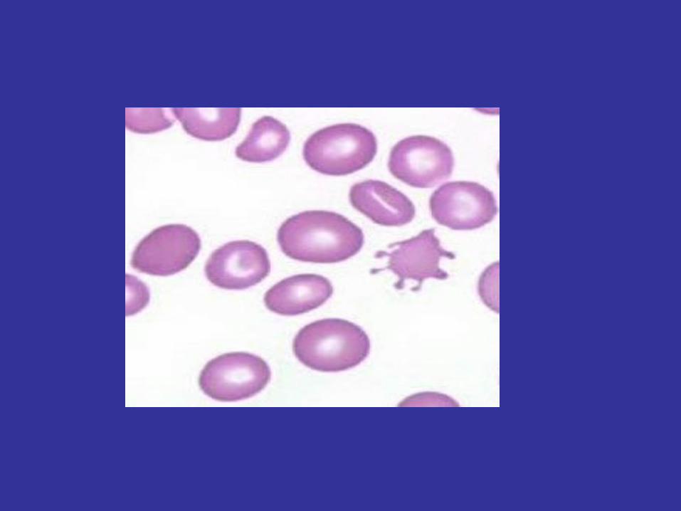
• Peripheral smear: schistocytes

Which of the following is the most appropriate next step in management:
• 1) aPTT mixing study
• 2) direct antiglobulin (Coombs) test
• 3) IVIG
• 4) Plasma exchange
• 5) Platelet antibody test

Thrombotic thrombocytopenic purpura
• Microangiopathic hemolytic anemia
• Thrombocytopenia with normal coags
• CNS sx
• Rx: plasma exchange. Reverses plt consumption that is responsible for thrombus formation causing characteristic sx

Transfusion medicine
• Acute hemolytic transfusion rxn:– Fever, chills, flank pain, abd pain, ATN, tachy– 2/2 ABO mismatch– + Coombs (detects IgG or complement on erythrocyte
surface)– Stop tx and maintain UOP
• Delayed hemolytic transfusion rxn– Unexplained drop in hg, ↑ bili, LDH, retic,
↓ haptoglobin– A new alloantibody

Transfusion medicine
• Post transfusion purpura: IVIG• Febrile non-hemolytic transfusion rxn: from
donor leukocyte cytokines or recipient antibodies against donor leukocyte antigens.
• TRALI: donor antileukocyte antibodies reacting with recipient leukocytes, causing leukocyte agglutination in the pulmonary capillary bed
• Allergic reactions/anaphylaxis• Volume overload
Hemostasis – MKSAP 5
• 18 yo M evaluated 5 hrs after a routine tooth extraction in which he experienced excessive bleeding that the dentist was able to control. The patient is healthy and takes no medications, including aspirin or NSAIDs. Medical hx includes easy bruisability and occasional nose bleeds that are easily controllable. The patient was circumcised at birth and recalls his mother saying that he had more bleeding than expected from the circumcision site. His father also has easy bruisability.
• PE: unremarkable, no petichiae, ecchymoses, or abnormal vasculature.

• Lab studies:• Hg 14.2 g/dL• Platelets 195,000/uL• INR 1.1• aPTT 41 s• aPTT mixing study:
corrects to normal• Thrombin time 16 s
(control 15 s)
• Fibrinogen 266 mg/dL• D-dimer assay
Negative• Bleeding time 10 min• Factor VIII activity
60% (normal 65-120%)

• Which of the following is the most likely diagnosis:
• A) Factor XI deficiency
• B) Hemophilia A (factor VIII deficiency)
• C) Presence of a lupus inhibitor
• D) Vitamin K deficiency
• E) von Willebrand disease

von Willebrand’s Disease
• Hx
• Labs with qualitative platelet defect AND mild coagulopathy

Hemostasis clues
• Petechiae, mucosal: TCP, vWF, vascular dz• Palpable purpura: vasculitis• Joint destruction: hemophilia• Soft tissue hematoma: hemophilia or
coagulopathy• Periorbital purpura: amyloidosis• Perifollicular and soft tissue hemorrhage:
scurvy

Platelet disorders – MKSAP 42
• 67 yo F admitted to hospital from ED for new dx of LLE DVT, confirmed by duplex u/s. PMHx significant for a 2 day hospitalization 4 wks ago for a NSTEMI for which she underwent cardiac cath and was given LMW heparin. Her current meds include ASA, clopidogrel, pravastatin, and lisinopril.
• PE: L thigh swollen and tender. Labs: cbc, lytes, and liver chemistry tests are unremarkable except for a platelet count of 102,000/uL.
• Unfractionated heparin is administered. Twelve hours later the pt’s plt count is 27,000/uL.

• Which of the following is the most appropriate next step in treatment?– A) Continue heparin and administer a platelet
transfusion– B) Continue heparin and initiate high-dose
corticosteroid therapy– C) Stop heparin and initiate argatroban– D) Stop heparin and initiate warfarin

HIT
• Thrombocytopenia and thrombosis
• Antibodies directed against a complex of heparin and platelet factor 4 (PF4)
• Leads to platelet activation and release of prothrombotic platelet microparticles.

Thrombosis pearls
• Venous and arterial clots: APLA (long aPTT and DRVVT and +anticardiolipin Ab) or homocysteine (MTHFR gene)
• Old pt: think malignancy
• Most common inherited thrombophilia: Factor V Leiden mutation (amino acid substitution, factor V made more resistant to cleavage by activated protein C)

Thrombosis – MKSAP 35
• A 33 yo F who has been trying to become pregnant for 8 years is evaluated after receiving positive pregnancy test results. Medical history is significant for 3 miscarriages (6 years ago, 3 years ago, and 1 year ago), each of which occurred early in her pregnancies. Following her last miscarriage, laboratory studies indicated the presence of a lupus inhibitor and antiphospholipid antibodies. She has no history of venous thromboembolism. Her LMP was 5 weeks ago.
• PE including vital signs and abdominal examination is normal.

• Which of the following is the most appropriate anticoagulation regimen for the duration of this patient’s pregnancy?
a) Full-dose unfractionated heparin
b) Low-dose aspirin
c) Prophylactic-dose low-molecular-weight heparin plus aspirin
d) Prophylactic-dose LMWH
e) Warfarin

Pregnancy and Platelets
• Gestational: 2-3rd trimester, incidental• ITP: prior or early in pregnancy. Prednisone
if plt <50k.• TTP: increased risk during pregnancy,
plasmapherese.• HELLP: plt <100k, AST>70 U/L, maha, tbili
>1.2mg/dL, LD > 600 U/L. Deliver.• Preeclampsia: tcp, HTN, proteinuria, deliver.


Topics in malignant heme
• Malignant– ALL, AML, CLL, CML, MDS, MPD, marrow
failure– Multiple myeloma, MGUS, amyloid,
waldenstrom’s, POEMS– Solid tumor risk, dx, staging, rx, monitoring– Onc emergencies– AE’s of cancer/cancer therapies


Heme malignancies -MKSAP 91
• 75 yo F c CLL who was previously asymptomatic on no therapy undergoes f/u eval for CAP for which she was hospitalized for 5 days and d/c’ed 14 days ago. The pt completed a course of abx therapy and currently feels well. She reports no fevers, chills, night sweats, weigh loss, abdominal pain, or new LAD, and her pulm sx have resolved.
• PMHx: prior PNA for which she was hospitalized within the last year.

• PE: temp 36.7 C, bp 130/78, p 72, rr 14, bmi 22. No palpable LAD. Cardiopulm exam is normal. Abd exam shows splenomegaly.
• Labs: Hg 11 g/dL, Leukocyte count 24,000/uL with 80% mature lymphocytes, plt 120,000/uL, ALC 20,000/uL, IgG 500mg/dL
• AP/lat CXR during hospitalization: resolving RLL infiltrate.

• Which of the following is the most appropriate next step in management?
• A) IVIG
• B) Prophylactic tmp-smx
• C) Splenectomy
• D) Repeat blood counts in 1 month

Chronic Lymphocytic Leukemia
• Accumulation of mature lymphocytes
• Immunophenotype: CD5+, CD20+, CD23+ B cells
• Pancytopenia
• ITP, autoimmune hemolytic anemia
• Hypogammaglobulinemia and recurrent infections: manage with IVIG

MKSAP 60
• 64 yo M c 3-year hx of stage II, grade 1 follicular lymphoma is evaluated because of cervical and axillary lymph nodes that have been gradually enlarging since diagnosis. He is unable to wear a tie, cannot fasten the top button of his shirt, has trouble shaving, and is concerned about the appearance of the cervical lymph nodes. The enlarged axiallary LN are also uncomfortable. The pt feels otherwise well and has not had night sweats, weight loss, or fevers.

• PE: t 37.1, bp 124/64, p 64, rr 12, BMI 24. Bilateral cervical LAD (largest aggregate LN measuring 6 cm) and bilateral axillary LAD (largest aggregate LN measuring 5-6 cm) are present. There is no palpable inguinal LAD or organomegaly.
• Lab: hg 12.0 g/dL, leukocyte count 9000/uL, plt 165,000/uL
• Contrast-enhanced CT-neck, c/a/p: progressive axillary, mediastinal, and cervical LAD. No measurable nodes below the diaphragm.

• Which of the following is the most appropriate next step in managing this patient?– A) Cyclophosphamide, vincristine, and prednisone
with rituxumab– B) High-dose chemotherapy followed by stem cell
transplantation– C) Fine-needle aspiration biopsy of a cervical lymph
node– D) Cyclophosphamide, vincristine, prednisone, and
radiation therapy– E) Observation with follow-up in 3 months

Follicular lymphoma
• Indolent
• CD20+, CD10+
• Offer palliative therapy for symptoms, threat to vital organs, concerns for appearance, and pt preference
• Debulking with chemo + rituxumab improves outcomes

MKSAP 21
• 42 yo F who is scheduled for hysterectomy for endometrial carcinoma comes in for preop eval. At the time of initial eval, she was discovered to have thrombocytosis. In addition to menorrhagia, the pt recently developed epistaxis and easy bruising. Her only meds are OCP and fe supplement.
• PE: temp normal, bp 110/70 mmHg, pulse 68, and rr 16. Exam is wnl. No petichiae, echhymoses, or splenomegaly is noted.

• Repeat labs:– Hg 11.3 g/dL– Leukocyte count 4600/uL– Platelet count 1,500,000/uL– Plt function studies: abnormal platelet
aggregation with decreased response to adenosine diphosphate and collagen.
– Cytogenetics: + JAK 2 mutation– BMBx: hypercellular marrow with increased
megakaryocytes in clusters.

• Which of the following is the most appropriate treatment:– A) Anagrelide beginning 2 days preop– B) Hydroxyurea beginning the night after
surgery– C) Low dose aspirin postoperatively– D) Platelet apheresis preoperatively and
hydroxyurea postoperatively

Essential thrombocythemia
• Bleeding and clotting complications
• Prior to surgery, goal <1million

MKSAP 34
• An asymptomatic 35 yo M comes for a routine annual examination. PMHx and FHx are unremarkable. Only med: MVI.
• PE: temp nl, bp 120/70, p 64, rr 14. No abnormal findings.
• Labs: hg 9.1 g/dL, leukocyte 2100/uL, plt 135,000/uL, LD 890 U/L, uric acid 11.6 mg/dL.
• Peripheral smear: circulating blasts and promyelocytes. BM: hypercellular marrow with 80% myeloblasts and promyelocytes. Cytogenetics: t(15:17).

• In addition to hydration and allopurinol, which of the following is the most appropriate management at this time?– A) Broad spectrum antibiotics– B) Chemotherapy– C) Chemotherapy plus all-trans-retinoic acid– D) HLA typing

Acute promyelocytic leukemia
• M3 subtype, 10% of AML
• t(15;17) = PML/RARα gene
• Present with pancytopenia, may be asymptomatic
• ATRA differentiates (also arsenic trioxide)
• ATRA syndrome

Solid tumors
• Breast: BRCA 1&2 screening– Prophylactic mastectomy– Oophorectomy– SERM therapy
• Tamoxifen - premenopausal and postmenopausal• Raloxifene – postmenopausal
• Ovarian: – No screening test, CA-125 nonspecific.– Staging at initial surgery.– Chemotherapy with platinum-based agent + paclitaxel– Advanced dz: survival advantage but greater toxicity if
intraperitoneal chemo added to systemic chemo.

Solid tumors, cont.
• Colorectal:– AJCC TNM staging– LN + disease (stage III) adjuvant chemo– Several monoclonal antibody therapies
• SCLC:– Staging limited or extensive– Prophylactic cranial irradiation– Offer chemo even if extensive and poor performance
status• NSCLC:
– Stages I, II, III, IV– PET-FDG superior to CT scan for mediastinal LN
detection

Solid tumors, cont.
• Thyroid:– RET proto-oncogene leads to medullary thyroid
cancer in MEN-2A, MEN-2B, and familial non-MEN medullary thyroid cancer.
– If RET mutation is discovered offer prophylactic thyroidectomy
– Check 24hr urine catecholamine levels to r/o pheochromocytoma
• Testicular:– Seminoma– Nonseminoma (embryonal cell, choriocarcinomas,
yolk sac tumors, teratomas). Any elevated AFP treat as nonseminoma.
Onc emergencies
• SVC syndrome: rx with radiation, chemo, consider anticoagulation and angioplasty/stenting
• Brain mets/increased ICP: rx with steroids, mannitol, NSG
• Spinal cord compression• Tumor lysis syndrome: rx with IVF’s,
allopurinol, rasburicase, HD. • Hypercalcemia

Chemotherapy Toxicities

References
• MKSAP 14 and 15
• Virginia Health Systems online atlas
• Chieh Lin Fu, MD
• Soham Puvvada, MD


