Hemodynamic, hemostatic and inflammatory effects of cardiotomy suction blood in cardiac surgery Jakob Gäbel Department of Molecular and Clinical Medicine Institute of Medicine Sahlgrenska Academy at University of Gothenburg Gothenburg 2013
Transcript
Hemodynamic, hemostatic and inflammatory effects of cardiotomy
suction blood in cardiac surgery
Jakob Gäbel
Department of Molecular and Clinical Medicine Institute of Medicine
Hemodynamic, hemostatic and inflammatory effects of cardiotomy suction blood in cardiac surgery
Jakob Gäbel
ABSTRACT Background: Cardiac surgery with cardiopulmonary bypass induces an inflammatory response and a coagulopathy, which may contribute to perioperative complications. Retransfusion of unprocessed cardiotomy suction blood may contribute to the inflammatory response and coagulopathy. Aims: To investigate the hemodynamic, hemostatic and inflammatory effects of retransfusion of unwashed cardiotomy suction blood and their potential associations, and to study the effect of cell saver processing of cardiotomy suction blood before retransfusion. Materials and methods: Four studies were performed. I: 25 patients were randomized to cell saver processing of cardiotomy suction blood or not before retransfusion. Hemodynamic effects and inflammatory markers were assessed. II: Pro- and anti-inflammatory markers were analyzed in cardiotomy suction blood and the systemic circulation in 25 patients randomized to cell saver processing of cardiotomy suction blood or not. III: Hemostatic markers were assessed in cardiotomy suction blood and in the systemic circulation in 30 patients randomized to retransfusion of unwashed cardiotomy suction blood or not. In an ex vivo study on 13 patients hemostasis was assessed when increasing amounts of unprocessed cardiotomy suction blood was added to systemic blood. IV: In an ex vivo study on 10 patients hemostasis was investigated when increasing amounts of cell saver processed or unprocessed cardiotomy suction blood was added to systemic blood. Results: I: Retransfusion of unprocessed cardiotomy suction blood causes a transient reduction in systemic vascular resistance that is attenuated if the cardiotomy suction blood is cell saver processed before retransfusion. II: Cardiotomy suction blood has an unfavorable balance between pro- and anti-inflammatory cytokines. Cell saver processing of cardiotomy suction blood before retransfusion improves the postoperative balance. III: Cardiotomy suction blood has poor hemostatic properties and ex vivo addition of cardiotomy suction blood to systemic blood impairs clot formation and platelet function dose-dependently. Retransfusion of small amounts of cardiotomy suction blood in vivo does not affect systemic markers for hemostasis. IV: Cell saver processing of cardiotomy suction blood ameliorates the negative effects on platelet function but does not influence the negative effects on clot formation. Conclusions: Cardiotomy suction blood is inflammatory activated and has poor hemostatic properties. Retransfusion of unwashed cardiotomy suction blood impairs the balance between pro- and anti-inflammatory cytokines, impairs hemostasis and reduces vascular resistance. These negative effects can be reduced if cardiotomy suction blood is cell saver processed before retransfusion. Alternatively, small to moderate amounts of unwashed suction blood may be discarded. Keywords: Cardiopulmonary bypass, inflammatory activation, coagulation, hemostasis ISBN: 978-91-628-8789-6 Gothenburg 2013
i
LIST OF PAPERS This thesis is based on the following studies, referred to in the text by their Roman numerals.
I. Westerberg M, Gäbel J, Bengtsson A, Sellgren J, Eidem O, Jeppsson A. Hemodynamic effects of cardiotomy suction blood. J Thorac Cardiovasc Surg. 2006 Jun;131(6):1352-7
II. Gäbel J, Westerberg M, Bengtsson A, Jeppsson A. Cell salvage of cardiotomy suction blood improves the balance between pro- and anti-inflammatory cytokines after cardiac surgery. Eur J Cardiothorac Surg. 2013 Sep;44(3):506-11
III. Gäbel J, Shams Hakimi C, Westerberg M, Radulovic V, Jeppsson A. Retransfusion of cardiotomy suction blood impairs haemostasis: Ex vivo and in vivo studies. Scand Cardiovasc J. 2013 (Epub ahead of print)
IV. Gäbel J, Radulovic V, Shams Hakimi C, Westerberg M, Jeppsson A. Cell saver processing ameliorates negative effects of cardiotomy suction blood on systemic platelet function: An ex vivo study. (Manuscript)
ii
CONTENT ABBREVIATIONS .............................................................................................. V 1 INTRODUCTION ........................................................................................... 1
1.1 Cardiopulmonary bypass ....................................................................... 1 1.2 Cardiotomy suction blood ..................................................................... 1 1.3 Cardiac surgery and inflammatory activation ....................................... 2 1.4 Cardiac surgery and hemostasis ............................................................ 3 1.5 Coagulation and platelets ...................................................................... 4 1.6 Fibrinolysis ............................................................................................ 6 1.7 Cardiac surgery and cell saver processing ............................................ 6 1.8 Study objectives .................................................................................... 7
2 AIMS OF THE STUDY ................................................................................. 10 3 PATIENTS AND METHODS ......................................................................... 11
3.1 Patients ................................................................................................ 11 3.1.1 Paper I and II ............................................................................... 11 3.1.2 Paper III ....................................................................................... 12 3.1.3 Paper IV ....................................................................................... 13
3.2 Clinical management ........................................................................... 13 3.2.1 Paper I-IV .................................................................................... 13
3.3 Study design ........................................................................................ 14 3.3.1 Paper I .......................................................................................... 14 3.3.2 Paper II ........................................................................................ 14 3.3.3 Paper III ....................................................................................... 15 3.3.4 Paper IV ....................................................................................... 15
3.4 Analyses .............................................................................................. 16 3.4.1 Paper I .......................................................................................... 16 3.4.2 Paper II ........................................................................................ 17 3.4.3 Paper III ....................................................................................... 17 3.4.4 Paper IV ....................................................................................... 21
iii
3.5 Statistics ............................................................................................... 21 3.5.1 Paper I .......................................................................................... 21 3.5.2 Paper II ......................................................................................... 21 3.5.3 Paper III ....................................................................................... 22 3.5.4 Paper IV ....................................................................................... 22
4 RESULTS .................................................................................................... 23 4.1 Paper I .................................................................................................. 23 4.2 Paper II ................................................................................................ 25 4.3 Paper III ............................................................................................... 27
4.3.1 Ex vivo study ................................................................................ 27 4.3.2 In vivo study ................................................................................. 28
4.4 Paper IV ............................................................................................... 32 5 DISCUSSION .............................................................................................. 35
5.1 Paper I .................................................................................................. 35 5.2 Paper II ................................................................................................ 36 5.3 Paper III ............................................................................................... 37 5.4 Paper IV ............................................................................................... 38
6 SUMMARY ................................................................................................. 41 7 ACKNOWLEDGEMENTS ............................................................................. 42 8 REFERENCES ............................................................................................. 43 9 SAMMANFATTNING PÅ SVENSKA .............................................................. 53
iv
v
ABBREVIATIONS ACT activated clotting time
ADP adenosine diphosphate
ALAT alanine-aminotransferase
ANOVA analysis of variance
aPTT activated partial thromboplastin time
ASAT aspartate-aminotransferase
AU arbitrary unit
AUC area under the curve
AVR aortic valve replacement
CABG coronary artery bypass grafting
CAT calibrated automated thrombogram
CFT clot formation time
CPB cardiopulmonary bypass
CT clotting time
CTS cardiotomy suction
ECC extra-corporeal circulation
EDTA ethylenediaminetetraacetic acid
EIA enzyme immunoassay
ELISA enzyme-linked immunosorbent assay
ETP endodgenous thrombin potential
vi
IL interleukin
LVEF left ventricular ejection fraction
MAP mean arterial pressure
MCF maximum clot firmness
NSAID non-steroidal anti-inflammatory drug
PAR-1 protease-activated receptor 1
PT prothrombin time
Ra receptor antagonist
RBC red blood cells
SVR systemic vascular resistance
TAT thrombin-antithrombin complex
TF tissue factor
TNF tumor necrosis factor
TRAP thrombin receptor activating peptide
t-PA tissue plasminogen activator
vii
Jakob Gäbel
1
1 INTRODUCTION Cardiac surgery is today a standard procedure to correct congenital or acquired heart disease. Most cardiac surgery procedures are performed with cardiopulmonary bypass (CPB) using a heart-lung machine. The first heart-lung machine was used in the 1950s by Gibbon. The heart-lung machine has since then constantly been evolved. However, even though cardiac surgery with CPB today has excellent results there is always a risk of major complications such as stroke, myocardial infarction, heart failure, renal failure, respiratory failure, bleeding and infections. The use of a heart-lung machine and its different components may contribute to the risk of complications.
1.1 Cardiopulmonary bypass During cardiac surgery it is often necessary to arrest the heart. To achieve this, the circulation and oxygenation of the blood is temporarily accomplished by cardiopulmonary bypass (CPB) using a heart-lung machine. Deoxygenated venous blood is drained from the right atrium or the caval veins to a reservoir and then pumped via a heat exchanger to an oxygenator where the gas exchange takes place. The oxygenated blood is then pumped back to the patient through a cannula in the ascending aorta or another major artery. To avoid the blood from clotting in the CPB circuit the patient is anticoagulated with heparin. Heparin facilitates the action of antithrombin III and thereby inhibits generation of thrombin that is a key enzyme in the coagulation process.
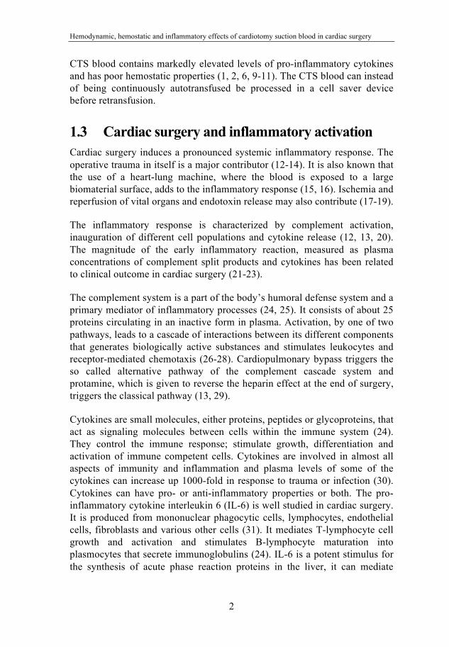
1.2 Cardiotomy suction blood During cardiac surgery wound blood is accumulated in the pericardium or sometimes also in an open pleural cavity. The total wound blood volume can be substantial and may reach 1- 1.5 liters (1-4). Normally this wound blood, often referred to as cardiotomy suction (CTS) blood, is continuously suctioned back to the systemic circulation via the heart-lung machine to preserve an appropriate hemoglobin concentration and to reduce the need for allogeneic blood transfusions. The CTS blood is filtered to reduce the amount of blood clots, fat particles and bone fragments before it is recirculated to the venous reservoir. It is well known that there is a pronounced activation of coagulation, fibrinolysis and inflammation in CTS blood and its retransfusion is suggested to contribute to the systemic inflammatory response (1, 5-8).
Hemodynamic, hemostatic and inflammatory effects of cardiotomy suction blood in cardiac surgery
2
CTS blood contains markedly elevated levels of pro-inflammatory cytokines and has poor hemostatic properties (1, 2, 6, 9-11). The CTS blood can instead of being continuously autotransfused be processed in a cell saver device before retransfusion.
1.3 Cardiac surgery and inflammatory activation Cardiac surgery induces a pronounced systemic inflammatory response. The operative trauma in itself is a major contributor (12-14). It is also known that the use of a heart-lung machine, where the blood is exposed to a large biomaterial surface, adds to the inflammatory response (15, 16). Ischemia and reperfusion of vital organs and endotoxin release may also contribute (17-19).
The inflammatory response is characterized by complement activation, inauguration of different cell populations and cytokine release (12, 13, 20). The magnitude of the early inflammatory reaction, measured as plasma concentrations of complement split products and cytokines has been related to clinical outcome in cardiac surgery (21-23).
The complement system is a part of the body’s humoral defense system and a primary mediator of inflammatory processes (24, 25). It consists of about 25 proteins circulating in an inactive form in plasma. Activation, by one of two pathways, leads to a cascade of interactions between its different components that generates biologically active substances and stimulates leukocytes and receptor-mediated chemotaxis (26-28). Cardiopulmonary bypass triggers the so called alternative pathway of the complement cascade system and protamine, which is given to reverse the heparin effect at the end of surgery, triggers the classical pathway (13, 29).
Cytokines are small molecules, either proteins, peptides or glycoproteins, that act as signaling molecules between cells within the immune system (24). They control the immune response; stimulate growth, differentiation and activation of immune competent cells. Cytokines are involved in almost all aspects of immunity and inflammation and plasma levels of some of the cytokines can increase up 1000-fold in response to trauma or infection (30). Cytokines can have pro- or anti-inflammatory properties or both. The pro-inflammatory cytokine interleukin 6 (IL-6) is well studied in cardiac surgery. It is produced from mononuclear phagocytic cells, lymphocytes, endothelial cells, fibroblasts and various other cells (31). It mediates T-lymphocyte cell growth and activation and stimulates B-lymphocyte maturation into plasmocytes that secrete immunoglobulins (24). IL-6 is a potent stimulus for the synthesis of acute phase reaction proteins in the liver, it can mediate
Jakob Gäbel
3
vasodilation and it can impair cardiac function (13, 21, 32). TNF-α is another pro-inflammatory cytokine that is secreted from mononuclear phagocytes, neutrophils, natural killer cells, endothelial cells or mastcells in response to activation (12, 24, 33). TNF-α can activate neutrophils, mediates adherence, degranulation and chemotaxis. It has also been shown that TNF-α induces vascular leakage and that it can depress human cardiac function (24, 34, 35).
Anti-inflammatory cytokines such as interleukin 10 (IL-10), interleukin 1 receptor antagonist (IL-1Ra) and interleukin 4 (IL-4) are known to counteract pro-inflammatory activation. IL-1Ra is a pleiotropic cytokine produced by macrophages and monocytes and it inhibits IL-1α and IL-1β-mediated cellular activation (36). Since IL-1 is a prominent pro-inflammatory cytokine involved in a number of inflammatory diseases, IL-1Ra therapy has been suggested to be a potential therapeutic option in systemic inflammatory disease (36). IL-1Ra levels have been shown to increase in systemic circulation after cardiac surgery (37, 38). IL-4 is a multifunctional anti-inflammatory cytokine that plays an important role in the regulation of immune response and plasma levels increase in sepsis (39, 40). IL-10 is the most studied anti-inflammatory cytokine in cardiac surgery. It is a potent anti-inflammatory cytokine secreted from T cells and macrophages (41, 42). These cells are important in controlling inflammation initiated by pro-inflammatory mediators such as IL-6, IL-8 and TNF-α. IL-10 inhibits the secretion of TNF-α, IL-1β and IL-6 and suppresses the propagation of downstream effects (41, 42). Preoperative treatment with corticosteroids has been shown to increase IL-10 levels in blood and to reduce the pro-inflammatory response after CPB (43). Since anti-inflammatory cytokines counteract pro-inflammatory activation they may protect tissues and organs during and after cardiac surgery. Recent data from patients with different inflammatory conditions suggest that the balance between pro- and anti-inflammatory cytokines is at least as important as the absolute levels of cytokines (37, 43-47). The balance is often presented as the IL-6 to IL-10 ratio.
1.4 Cardiac surgery and hemostasis Severe bleeding during and after cardiac surgery is a serious complication associated with increased morbidity and mortality (48, 49). About 5% of cardiac surgery patients need to be re-explored due to excessive bleeding or tamponade and about 50% of the patients receive blood transfusion (50, 51). Re-exploration due to bleeding is associated with sepsis, multiple organ failure, prolonged mechanical ventilation and mortality (50). It has also been
Hemodynamic, hemostatic and inflammatory effects of cardiotomy suction blood in cardiac surgery
4
suggested that blood transfusion is an independent risk factor for postoperative complications and mortality (52-54).
Normal hemostasis is the termination of bleeding by a complex coagulation process of the body that consists of vasoconstriction, platelet aggregation and thrombin and fibrin synthesis (55). This complex system is affected by cardiac surgery with CPB through a number of different mechanisms such as hemodilution, hypothermia and the surgical trauma (48). Despite anti-coagulation with heparin, the interaction of blood with artificial surfaces in the heart-lung machine and non-endothelialized surfaces in the surgical wound results in activation of coagulation which consumes coagulation factors and platelets (48). This activation increases with the length of CPB and can cause impaired hemostasis that may result in bleeding complications (48). The activation of platelets during CPB causes platelet dysfunction due to loss of important receptors and degranulation (49, 56-58). During CPB there is also an activation of fibrinolysis that can affect hemostasis (59, 60). Cardiac surgery patients are also often exposed to anticoagulant and/or antiplatelet therapy that may impair postoperative hemostasis.
1.5 Coagulation and platelets Coagulation is a process in blood where the end product is a clot formed by platelets and cross-linked fibrin to stop bleeding. Platelets are 2-3 µm small circulating disc shaped cell fragments derived from fragmentation of megacaryocytes (61). Platelets play a very important part in coagulation since primary hemostasis begins immediately when a vessel is damaged and platelets are exposed to tissue factor (TF) (55). This activates the platelets and they form a plug. Secondary hemostasis is a simultaneous process where plasma proteins, called coagulation factors (denoted by Roman numerals), respond in a complex cascade to create cross-linked fibrin to strengthen the plug (62). This coagulation cascade can be initiated in two ways, the contact activation pathway (or intrinsic pathway) and the tissue factor pathway (or extrinsic pathway). The two pathways are joined to a final common pathway. The pathways are a series of reactions where the different coagulation factors act as enzymes that activate the next step in the cascade finally resulting in cross-linked fibrin. The two pathways converge with the formation of thrombin from prothrombin, Fig. 1. Thrombin is an important factor in coagulation as it cleaves fibrinogen to fibrin and activates platelets, factor XI, and cofactors V and VIII (62). Thrombin also activates control mechanisms such as the inhibitor protein C and the fibrinolytic enzyme plasmin. The small amount of thrombin produced thus far is not sufficient to produce a
Jakob Gäbel
5
fibrin clot. Amplification of thrombin production is achieved by accelerating enzyme reactions on the platelet surface. Reduced thrombin generating capacity using Calibrated Automated Thrombogram (CAT) methodology has been reported after cardiac surgery (63, 64). Platelets are activated and localized via receptors for substances such as collagen and thrombin (65). They release procoagulant factors and change shape, exposing negatively charged membrane phospholipid. This starts a chain of reactions involving a number of factors that in the end produces an explosive increase in thrombin production sufficient to produce fibrin. Platelets become linked together in this platelet-fibrin clot via their fibrin-receptor glycoprotein IIbIIIa (GpIIbIIIa) (65).
Figure 1. The Coagulation Cascade. From Wikipedia.org
Hemodynamic, hemostatic and inflammatory effects of cardiotomy suction blood in cardiac surgery
6
Under physiologic conditions the primary pathway for the initiation of the coagulation cascade is the tissue factor pathway (66). However, contact activation may play an important role for coagulation during CPB (67-69). Different parts of the coagulation can be measured with different methods.
Coagulation overlaps with inflammatory pathways; for example, activated platelets release inflammatory cytokines and thrombin activates monocytes (65). Coagulation can activate the inflammatory system and vice versa. This becomes relevant with extreme activation of either system, such as in systemic inflammation (65).
1.6 Fibrinolysis The fibrinolysis breaks down a fibrin clot to prevent excessive clot formation. Inactive plasminogen is incorporated into the clot when it is formed. The plasminogen is converted to plasmin, the major fibrinolytic enzyme, by tissue plasminogen activator (t-PA) and urokinase (70). t-PA is slowly released into the blood by damaged endothelium of the damaged blood vessel and the clot is broken down. Under normal physiologic conditions both coagulation and fibrinolysis are precisely regulated by numerous substrates, activators, inhibitors, cofactors and receptors. Links between these systems permit timely and localized removal of fibrin clots to ensure blood flow while preventing blood loss (70, 71). However numerous studies indicate an enhanced fibrinolysis during CPB that may affect hemostasis during and after cardiac surgery (59, 60). It is also known that the fibrinolytic system is closely linked to control of inflammation and plays a role in disease states associated with inflammation (65). Plasmin can in addition to lysing fibrin clots, also cleave complement C3 (72).
1.7 Cardiac surgery and cell saver processing Autotransfusion is a process where the patients receive their own blood for transfusion instead of allogenic blood products from the blood bank. This autologous blood can either be pre-donated or collected during surgery using an intraoperative blood salvage device, a so-called cell saver. The first modern cell saver device was first used in 1976 (73). The collected blood is washed in a flow centrifuge process that returns only red blood cells suspended in saline solution. This means that it washes out for example platelets, plasma, leukocytes, heparin, free hemoglobin and activated clotting factors. The use of a cell saver device has the potential advantage of reducing the need for allogeneic blood. It may also wash out products such as activated
Jakob Gäbel
7
clotting factors and activated platelets that can cause coagulopathy (74). A disadvantage, especially if large amounts of blood are collected, is the depletion of plasma and platelets that may have to be replaced. In cardiac surgery a cell saver device can be used to reduce the content of potentially harmful substances in CTS blood. It can also be used to recover and concentrate the blood from the CPB circuit when it is disconnected.
1.8 Study objectives Cardiac surgery with cardiopulmonary bypass carries a risk of serious complications. Some of these complications can be attributed to a systemic inflammatory response. In its most severe form this inflammatory response (74) can lead to peripheral vasodilatation and hypotension that may result in insufficient end-organ perfusion and ischemic complications (12, 13, 32, 75-79). Unprocessed CTS blood is activated and contains elevated levels of inflammatory mediators such as complement split products and cytokines (2, 18). Some of these inflammatory mediators have vasoactive properties, at least in vitro, (78, 80, 81) and retransfusion of unprocessed CTS blood might therefore affect systemic vascular resistance. We sought to investigate potential hemodynamic effects of retransfusion of CTS blood in vivo. In addition, we wanted to study if these potential effects could be related to concentrations of inflammatory mediators in CTS blood and whether the effects are reduced if the CTS blood is cell saver processed before retransfusion. We therefore designed a prospective randomized study in patients undergoing coronary artery bypass grafting (CABG) with cardiopulmonary bypass to address these questions (Paper I).
As previously mentioned, cardiac surgery induces a massive inflammatory response that includes a profound release of pro- and anti-inflammatory cytokines (12, 13). Unwashed CTS blood is known to be highly activated and contains high levels of pro-inflammatory cytokines. Little is known about anti-inflammatory cytokines in CTS blood and it is not known whether the balance between pro- and anti-inflammatory differs between CTS blood and the systemic circulation during cardiac surgery with CPB. The use of a cell saver reduces the content of pro-inflammatory cytokines in CTS blood and retransfusion of cell saver processed CTS blood reduces the systemic levels of pro-inflammatory cytokines after cardiac surgery (1, 11, 82, 83). However, it is not known whether cell saver processing of CTS blood before retransfusion influences the levels of anti-inflammatory cytokines or affects the balance between pro- and anti-inflammatory cytokines. A prospective
Hemodynamic, hemostatic and inflammatory effects of cardiotomy suction blood in cardiac surgery
8
randomized trial in CABG patients was designed to address these issues (Paper II).
Severe bleeding during and after cardiac surgery is a serious complication associated with increased morbidity and mortality. Retransfusion of unprocessed CTS blood is suggested to contribute to postoperative bleeding. One option to avoid negative effects of CTS blood in patients with limited intraoperative bleeding is to completely refrain from retransfusion and discard the wound blood. This strategy has been shown to reduce the postoperative inflammatory response without reducing hemoglobin levels (10). There is reason to believe that the poor quality of unprocessed CTS blood adds to coagulopathy and platelet dysfunction after cardiac surgery with CPB. We sought to investigate the hemostatic properties of CTS blood and whether retransfusion of unwashed CTS blood influences postoperative hemostasis. For this purpose we designed an ex vivo study where increasing doses of CTS blood was added to blood samples from the systemic blood circulation as well as an in vivo prospective randomized pilot study in CABG patients (Paper III).
Unwashed CTS blood has poor quality and cell saver processing of CTS blood has proven to reduce the content of pro-inflammatory cytokines in CTS blood and to reduce the systemic concentrations of these cytokines after cardiac surgery (8, 9, 84, 85). We wanted to find out if it is possible that cell saver processing of CTS blood before retransfusion can also reduce the negative effect on hemostasis. We therefore designed an ex vivo study where increasing doses of unprocessed or processed CTS blood was added to blood from the systemic circulation in patients undergoing cardiac surgery with CPB (Paper IV).
Jakob Gäbel
9
Hemodynamic, hemostatic and inflammatory effects of cardiotomy suction blood in cardiac surgery
10
2 AIMS OF THE STUDY 1. To investigate if CTS blood has hemodynamic effects in vivo
and if potential hemodynamic effects correlate to concentrations of inflammatory mediators in CTS blood (Paper I).
2. To investigate if cell saver processing of CTS blood before retransfusion reduces potential hemodynamic effects (Paper I).
3. To describe the balance between pro- and anti-inflammatory cytokines in CTS blood and in the systemic circulation during and after cardiac surgery (Paper II).
4. To investigate if cell saver processing of CTS blood before retransfusion influences the balance between pro- and anti-inflammatory cytokines in systemic circulation (Paper II).
5. To investigate if addition of unwashed CTS blood ex vivo to systemic blood influences coagulation and platelet function (Paper III).
6. To investigate if addition of unwashed CTS blood in vivo affects systemic indices of hemostasis (Paper III).
7. To investigate if cell saver processing of CTS blood before retransfusion reduces potential negative effects on hemostasis (Paper IV).
Jakob Gäbel
11
3 PATIENTS AND METHODS
3.1 Patients The regional Research Ethics Committee approved all the study protocols. All patients were included after written informed consent. The studies were performed at the Department of Cardiothoracic Surgery at Sahlgrenska University Hospital. Patient characteristics for the four studies are presented in Table 1.
Table 1. Preoperative and perioperative variables in paper I-IV. Mean and standard deviation or number (%).
3.1.1 Paper I and II The study group consisted of 25 patients with a mean age of 68 ± 8 years and 80% were men. Inclusion criteria were: age 40-80 years, two or three vessel coronary disease with angina pectoris and appropriate coronary anatomy for CABG, left ventricular ejection fraction >40%, and no other significant disorders. Exclusion criteria were: preoperative use of steroids or NSAIDs, intraoperative administration of vasoactive drugs or signs of significant peripheral arterial disease. The patients were randomized into two groups, cell saver processing of CTS blood or not (Table 1 and 2).
Hemodynamic, hemostatic and inflammatory effects of cardiotomy suction blood in cardiac surgery
12
Table 2. Paper I and II. Pre- and perioperative variables in the cell saver processed and cell saver unprocessed group. Mean and standard deviation or number.
Processed Unprocessed P-value n 13 12 Age (years) 67 ± 2 69 ± 2 0.54 Female gender 3/13 2 /12 0.54 LVEF (%) 53 ± 9 52 ± 14 0.81 Aortic clamp time (min) 45 ± 17 41 ± 14 0.47 ECC time 90 ± 28 80 ± 22 0.36 Number of grafts 3.5 ± 1.0 3.1 ± 0.9 0.26
3.1.2 Paper III
Ex vivo study Thirteen patients undergoing elective CABG (n=10), aortic valve replacement (AVR) (n=2) or AVR+CABG (n=1) with cardiopulmonary bypass were included (Table 1). Exclusion criteria were known liver disease, kidney disease, known bleeding disorder and treatment with a P2Y12 receptor-blocking agent less than seven days before surgery. All patients were on on-going treatment with acetylsalicylic acid (75 mg daily).
In vivo study Patients were eligible if operated with elective CABG for stable angina. Exclusion criteria were known liver disease, kidney disease, known bleeding disorder and treatment with P2Y12 receptor-blocking agent less than seven days before surgery. Patients were also excluded if the amount of CTS blood was less than 100 ml or more than 600 ml. Thirty-four patients admitted for elective CABG at Sahlgrenska University Hospital between January 2009 and October 2010 were originally included in the study. Four patients were excluded from the analysis; one had severe pericardial adherences, one was treated with P2Y12 receptor –blocking agent overseen at inclusion, one had less than 100 ml of CTS blood and in one patient there was a technical error when the extra reservoir was connected. No patient was excluded due to more than 600 ml of CTS blood. The study is based on the remaining 30 patients (Table 1). The patients were randomized into two groups, retransfusion or no-retransfusion, in a one to one fashion (Table 3).
Jakob Gäbel
13
Table 3. Paper III in vivo study. Pre- and perioperative variables in the retransfusion and no-retransfusion group. Mean and standard deviation or number.
3.1.3 Paper IV Ten patients (8 men, 2 women, mean age 64±14 years) undergoing CABG (n=7), AVR (n=2) or mitral valve surgery (n=1) with cardiopulmonary bypass were included. Patient characteristics are given in Table 1. Exclusion criteria were known liver disease, kidney disease, known bleeding disorder and treatment with P2Y12 receptor-blocking agent less than seven days before surgery. Seven of the patients were on on-going treatment with acetylsalicylic acid (75 mg daily).
Hemodynamic, hemostatic and inflammatory effects of cardiotomy suction blood in cardiac surgery
14
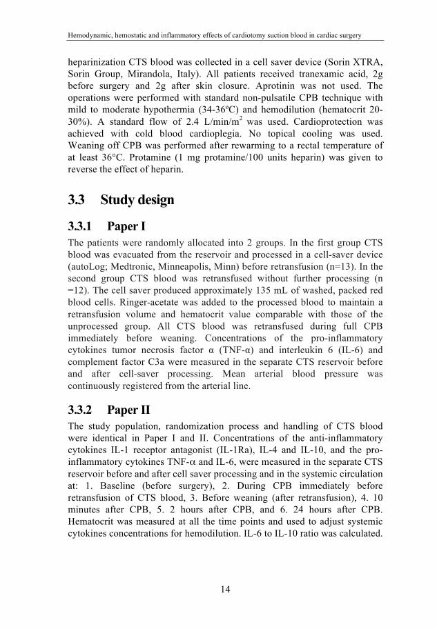
heparinization CTS blood was collected in a cell saver device (Sorin XTRA, Sorin Group, Mirandola, Italy). All patients received tranexamic acid, 2g before surgery and 2g after skin closure. Aprotinin was not used. The operations were performed with standard non-pulsatile CPB technique with mild to moderate hypothermia (34-36ºC) and hemodilution (hematocrit 20-30%). A standard flow of 2.4 L/min/m2 was used. Cardioprotection was achieved with cold blood cardioplegia. No topical cooling was used. Weaning off CPB was performed after rewarming to a rectal temperature of at least 36°C. Protamine (1 mg protamine/100 units heparin) was given to reverse the effect of heparin.
3.3 Study design
3.3.1 Paper I The patients were randomly allocated into 2 groups. In the first group CTS blood was evacuated from the reservoir and processed in a cell-saver device (autoLog; Medtronic, Minneapolis, Minn) before retransfusion (n=13). In the second group CTS blood was retransfused without further processing (n =12). The cell saver produced approximately 135 mL of washed, packed red blood cells. Ringer-acetate was added to the processed blood to maintain a retransfusion volume and hematocrit value comparable with those of the unprocessed group. All CTS blood was retransfused during full CPB immediately before weaning. Concentrations of the pro-inflammatory cytokines tumor necrosis factor α (TNF-α) and interleukin 6 (IL-6) and complement factor C3a were measured in the separate CTS reservoir before and after cell-saver processing. Mean arterial blood pressure was continuously registered from the arterial line.
3.3.2 Paper II The study population, randomization process and handling of CTS blood were identical in Paper I and II. Concentrations of the anti-inflammatory cytokines IL-1 receptor antagonist (IL-1Ra), IL-4 and IL-10, and the pro-inflammatory cytokines TNF-α and IL-6, were measured in the separate CTS reservoir before and after cell saver processing and in the systemic circulation at: 1. Baseline (before surgery), 2. During CPB immediately before retransfusion of CTS blood, 3. Before weaning (after retransfusion), 4. 10 minutes after CPB, 5. 2 hours after CPB, and 6. 24 hours after CPB. Hematocrit was measured at all the time points and used to adjust systemic cytokines concentrations for hemodilution. IL-6 to IL-10 ratio was calculated.
Jakob Gäbel
15
3.3.3 Paper III
Ex vivo study Immediately before weaning from bypass 3 ml of blood was collected from the pericardial cavity. At the same time 15 ml systemic blood was collected from an arterial line. After baseline measurements increasing doses of CTS blood was added to the samples from the systemic circulation, corresponding to 5, 10 and 20% of the systemic blood volume. Clot formation was analyzed with modified rotational thromboelastometry with addition of heparinase (HEPTEM) due to systemic heparinization during cardiopulmonary bypass. Platelet aggregability was analyzed with multiple electrode aggregometry.
In vivo study The study was a prospective randomized blinded single center pilot study. Patients were randomized, in a one to one ratio, to retransfusion or no retransfusion of CTS blood from the separate reservoir just prior to weaning from CPB. The primary endpoint was intergroup difference in INTEM clotting time two hours post cardiopulmonary bypass as assessed by thromboelastometry. Secondary endpoints were assessment of clot stability, platelet aggregability, thrombin generation, and fibrinolysis. All biochemical analyses were also performed in CTS blood. Blood samples were collected after induction of anesthesia before surgery, and 10 minutes, 2h and 24h post CPB.
3.3.4 Paper IV After systemic heparinization CTS blood was collected in a cell saver device (Sorin XTRA, Sorin Group, Mirandola, Italy). Immediately before weaning bypass, or after 400 ml of blood had been collected in the reservoir of the cell saver, 3 ml of unprocessed blood was collected from the reservoir in a tube without additives. The volume in the cell saver reservoir was then processed at 5600 rpm and 3 ml collected from the processed blood in a test tube without additives. When the processing was accomplished 25 ml systemic blood was collected from an arterial line in 3 test tubes with citrate (0.129 M, á 2.7 mL) and in 4 tubes with no additives.
After baseline measurements in the systemic blood samples increasing doses of unprocessed CTS blood was added ex vivo to the samples from the systemic circulation corresponding to 10 and 20% of the systemic blood volume. These doses were chosen since the total retransfused CTS blood volume in most studies corresponds to 10-20% of the total blood volume (1-4). In parallel cell saver processed CTS blood was added to systemic blood
Hemodynamic, hemostatic and inflammatory effects of cardiotomy suction blood in cardiac surgery
16
samples. Since the cell saver processing reduces the total volume, the retransfused processed volume was adjusted to the corresponding volume of unprocessed CTS blood. The following formula was used to calculate the individual volume of processed blood that was added to the systemic samples: Added volume of unprocessed blood = (blood volume after processing / initially collected blood volume) × volume of added unprocessed blood. This was done to achieve the same volume ratio in the test tube as if the patient had been retransfused with processed blood. Clot formation and platelet aggregability was analyzed in the unprocessed and processed samples.
3.4 Analyses
3.4.1 Paper I
Laboratory analyses Samples for cytokine and complement analyses were collected into tubes with ethylenediamine tetraacetic acid (EDTA) and placed immediately on ice. The aliquots were centrifuged immediately, and the resultant plasma was stored at -70°C until analysis. TNF-α, IL-6, and C3a levels were determined with commercially available enzyme-linked immunosorbent assay kits by using double-antibody enzyme-linked immunosorbent assays, according to the manufacturer’s instructions. The following assays were used: TNF-α and IL-6, R&D systems; C3a, Quidel (San Diego, Calif).
Measurement of hemodynamics Mean arterial blood pressure was continuously registered from the arterial line and stored with 200 samples per second by using a computer technique (Acknowledge; BioPac Systems, Santa Barbara, Calif). An investigator blinded to group affiliation analyzed the files.
Calculations Systemic vascular resistance (SVR) was calculated according to the formula SVR = 80 (MAP⁄cardiac output), where cardiac output equals the CPB flow rate. Plasma concentrations of TNF-α, IL-6, and C3a in suction blood were corrected for hematocrit by relating measurements to a standard hematocrit value of 40% according to the following formula:
Laboratory analyses The cytokines were assayed with a commercially available enzyme immunoassay (EIA) system (Endogen, Pierce, IL, USA). The detection limits for the different inflammatory variables were as follows: IL-Ra, 2 pg/ml; IL-4, 2 pg/ml; IL-10, 2 pg/ml; IL-6, 1 pg/ml and TNF-α, 2 pg/ml.
Calculations Plasma concentrations of the cytokines were adjusted for changes in hematocrit as in paper I. The pro-/anti-inflammatory cytokine balance was calculated by dividing the concentration of the pro-inflammatory cytokine IL-6 by the concentration of anti-inflammatory cytokine IL-10. It was not possible to calculate the IL-6 to IL-10 ratio correctly in CTS blood after cell-saver processing since IL-10 was completely abolished after processing in some of the plasma samples (division by 0).
3.4.3 Paper III
Laboratory analyses Platelet count, activated partial thromboplastin time (aPTT), prothrombin time (PT), aspartate-aminotransferase (ASAT), alanine-aminotransferase (ALAT), creatinine, hemoglobin concentration and hematocrit were analyzed with clinical standard methods at the laboratory at Sahlgrenska University Hospital. In the ex vivo study thromboelastometry and multiple electrode aggregometry was performed on point of care instruments at the operation theatre. The samples intended for platelet analysis were collected in tubes without additives, and the samples for clot formation analysis were collected in citrated tubes (0.129 M, á 2.7 mL).
Clot formation was assessed with thromboelastometry (ROTEM® Pentapharm, Munich, Germany) (86). ROTEM®
analyses were performed on
citrated blood in parallel on four channels (INTEM, EXTEM, FIBTEM and HEPTEM). INTEM analyzes the internal pathway of coagulation pathway and EXTEM analyzes the external pathway with tissue factor as activator. HEPTEM analyzes the internal pathway of the coagulation cascade with ellagic acid as activator in the presence of heparinase. In the FIBTEM analysis the fibrinogen component (concentration and function in clot polymerization) of coagulation is evaluated after the contribution of platelets is inhibited by addition of cytochalasin D. In each of the channels, the following variables were assessed: clotting time (CT), clot formation time (CFT) and maximum clot firmness (MCF), Fig. 2.
Hemodynamic, hemostatic and inflammatory effects of cardiotomy suction blood in cardiac surgery
18
Figure 2. Representative thromboelastometric tracing (ROTEM®) showing the initiation of clotting (CT), the propagation of coagulation (CFT) and maximum clot firmness (MCF). From www.practical-hemostasis.com
Platelet aggregability was analyzed by whole blood impedance aggregometry (Multiplate® Roche Diagnostics, Basel, Switzerland) (87). In the test cell of the aggregometer, 300 µl of the blood sample was added to 300 µl of saline pre-heated to 37ºC. Test kits used were ADP High Sensitivity kit (adenosine diphosphate as initiator) in combination with prostaglandin E1 for high-sensitivity detection of P2Y12-dependent aggregation, ASPI test (arachidonic acid as initiator) to assess cyclooxygenase-dependent platelet aggregation and TRAP test (thrombin receptor-activating peptide-6 as initiator) that detects PAR-1 receptor-dependent platelet aggregation. The change in impedance when platelets aggregate at electrodes in the test cell is expressed as a graph where the area under the curve (AUC) is a quantification of platelet aggregability reported in arbitrary aggregation units (AU×min), Fig. 3. According to the manufacturer’s instruction patients need to have a platelet count >150x109/L for reliable results. Aggregometry results from patients with platelet count <150x109/L was therefore excluded from analysis.
Jakob Gäbel
19
Figure 3. Representative Multiplate tracing and a drawing of the test cell. Two tests are run simultaneously. From www.Multiplate.net
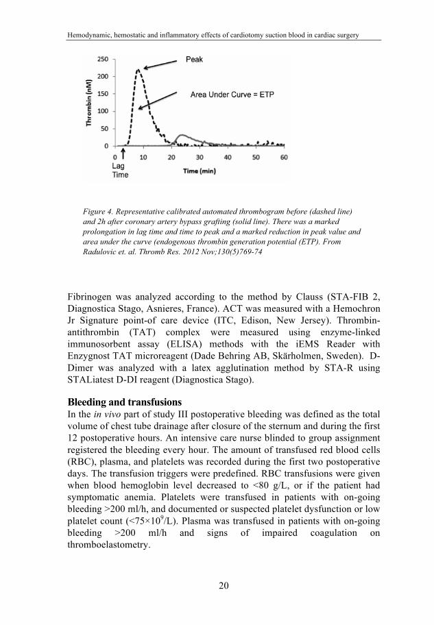
Thrombin generation capacity was analyzed in plasma with Calibrated Automated Thrombogram (CAT) with tissue factor as activator (88, 89). Thrombin has a half-life of less than one minute, and is rapidly cleared from the circulation by antithrombin (90). The CAT assay was performed in round-bottom 96-well plates. Citrated plasma samples (80 µl) and trigger solution (20 µl) containing 1 pM tissue factor (TF) were mixed in sample wells. In parallel, a calibrator was analyzed by mixing 20 µl of calibrator and 80 µl of pooled citrated normal human plasma in wells coupled to the sample wells. The plate was then moved to a fluorometer and 20 µl of FluCa solution containing fluorogenic substrate and CaCl2 was dispensed by the instrument. The fluorogenic signal was measured at λex 390 nm, λem 460 nm during 60 min. The 96-well plate was kept on a 37°C heating block during additions of the reagents. Start of thrombin activity (lag time), time to peak thrombin activity, peak thrombin activity, and total endogenous thrombin potential (ETP) were calculated using the software Thrombinoscope (version 3.0.0.29) from Thrombinoscope BV (Maastricht, Netherlands), Fig. 4.
Hemodynamic, hemostatic and inflammatory effects of cardiotomy suction blood in cardiac surgery
20
Figure 4. Representative calibrated automated thrombogram before (dashed line) and 2h after coronary artery bypass grafting (solid line). There was a marked prolongation in lag time and time to peak and a marked reduction in peak value and area under the curve (endogenous thrombin generation potential (ETP). From Radulovic et. al. Thromb Res. 2012 Nov;130(5)769-74
Fibrinogen was analyzed according to the method by Clauss (STA-FIB 2, Diagnostica Stago, Asnieres, France). ACT was measured with a Hemochron Jr Signature point-of care device (ITC, Edison, New Jersey). Thrombin-antithrombin (TAT) complex were measured using enzyme-linked immunosorbent assay (ELISA) methods with the iEMS Reader with Enzygnost TAT microreagent (Dade Behring AB, Skärholmen, Sweden). D-Dimer was analyzed with a latex agglutination method by STA-R using STALiatest D-DI reagent (Diagnostica Stago).
Bleeding and transfusions In the in vivo part of study III postoperative bleeding was defined as the total volume of chest tube drainage after closure of the sternum and during the first 12 postoperative hours. An intensive care nurse blinded to group assignment registered the bleeding every hour. The amount of transfused red blood cells (RBC), plasma, and platelets was recorded during the first two postoperative days. The transfusion triggers were predefined. RBC transfusions were given when blood hemoglobin level decreased to <80 g/L, or if the patient had symptomatic anemia. Platelets were transfused in patients with on-going bleeding >200 ml/h, and documented or suspected platelet dysfunction or low platelet count (<75#109/L). Plasma was transfused in patients with on-going bleeding >200 ml/h and signs of impaired coagulation on thromboelastometry.
Jakob Gäbel
21
3.4.4 Paper IV
Laboratory analyses Since all patients were heparinized no anticoagulant was used in the cell saver. Thromboelastometry and multiple electrode aggregometry were performed on point of care instruments at the operation theatre. Clot formation was assessed with modified rotational thromboelastometry (ROTEM® Pentapharm, Munich, Germany) as described for paper III. In this study only HEPTEM and FIBTEM analysis were performed due to systemic heparinization during CPB. HEPTEM clotting time (CT), clot formation time (CFT) and maximum clot formation (MCF) were assessed. FIBTEM is not affected by heparin. In the present study only FIBTEM-MCF was assessed.
Platelet aggregability was analyzed by whole blood impedance aggregometry (Multiplate® Roche Diagnostics, Basel, Switzerland). As previously described for paper II the test kits used were ADP High Sensitivity kit, ASPI test and TRAP test. In this study we also used COL test (final concentration of collagen equates to an activity of 3.2 µg/ml).
3.5 Statistics
3.5.1 Paper I The nonparametric Mann-Whitney U test (continuous non-normally distributed variables) and the Fisher exact test (categorical variables) were used to compare the groups. Differences within a group were compared with the paired nonparametric Wilcoxon test. Comparisons were made both with percentage change from baseline and with absolute values. Correlation was analyzed with the Spearman rank sum test. All the results are expressed as the mean ± standard error of the mean or mean ± standard deviation.
3.5.2 Paper II The non-parametric Mann–Whitney U-test (continuous non-normally distributed variables), the parametric unpaired t-test (continuous normally distributed variables) and Fisher’s exact test (categorical variables) were used to compare the groups. Differences within a group were compared with either the paired non-parametric Wilcoxon test or the paired t-test when appropriate. Systemic levels of cytokines were compared between the groups with analysis of variance for repeated measurements followed by the t-test at the fixed time points if group analysis or the interaction between group and time analysis indicated a significant difference (P < 0.05). No power analysis for
Hemodynamic, hemostatic and inflammatory effects of cardiotomy suction blood in cardiac surgery
22
the secondary group comparison was possible since data essential for the analysis (IL-6 to IL-10 ratio) were unavailable at the start of the study.
3.5.3 Paper III In the ex vivo study the non-parametric Friedman’s test was used to assess the effects of adding CTS blood to the systemic blood. In addition, changes from baseline were analyzed with the non-parametric Wilcoxon Signed Rank Test. In the in vivo study a power analysis based on an own previous study with thromboelastometry 2h after surgery was performed (91). It was calculated that 30 patients were necessary to be able to demonstrate a 25% difference in INTEM-CT 2h after surgery (with 80% power and a significance level of 0.05). Since most the variables were not normally distributed, group comparisons analyzed at one occasion were made with the non-parametric Mann-Whitney-test (continuous variables). Fisher’s exact test was used to compare categorical variables. Changes from baseline within a group were analyzed with the non-parametric Wilcoxon Signed Rank Test. For group comparisons of variables analyzed at more than one occasion, ANOVA for repeated measurements was used followed by Student’s t-test if the ANOVA P-value (group or interaction between group and time) was <0.05. All data are presented as mean ± standard deviation (normally distributed data) or median and range.
3.5.4 Paper IV A non-parametric paired test, Wilcoxon’s test, was used to assess the effects of adding CTS blood to the systemic blood compared to baseline and to compare processed and non-processed additives. A p-value of <0.05 was considered statistically significant. All data are presented as mean ± standard deviation (normally distributed data) or median and range.
Jakob Gäbel
23
4 RESULTS
4.1 Paper I Cardiotomy suction blood and systemic vascular resistance The mean volume of collected CTS blood during the operation was 477 ± 36 mL. The mean volume of retransfused CTS blood was 403 ± 17 mL, and the mean hematocrit value was 15% ± 2%. The mean arterial pressure before retransfusion was 67 ± 3 mm Hg. Rapid retransfusion of CTS blood induced a transient reduction in SVR immediately after retransfusion in all patients, Fig. 5.
Figure 5. Representative sample of mean arterial pressure during retransfusion of unprocessed CTS blood.
Inflammatory Mediators and SVR Plasma concentrations of TNF-α, IL-6, and C3a were significantly increased in unprocessed CTS blood compared with systemic plasma concentrations at the same time. There were statistically significant correlations between TNF-α concentrations in retransfused CTS blood and Δ% SVR (Fig. 6) and between C3a and Δ% SVR. No correlation between IL-6 and Δ% SVR was detected.
Hemodynamic, hemostatic and inflammatory effects of cardiotomy suction blood in cardiac surgery
24
Figure 6. Correlation between plasma levels of TNF-α in retransfused CTS blood and relative changes in systemic vascular resistance.
Effects of cell saver processing on inflammatory activation in cardiotomy suction blood Cell saver processing significantly reduced concentrations of TNF-α and C3a in CTS blood. IL-6 levels tended to decrease (from 633 ± 315 to 229 ± 81 pg/mL), but did not reach statistical significance.
Effects of cell saver processing on hemodynamic effects of cardiotomy suction blood Baseline SVR (immediately before retransfusion of CTS blood) was comparable between the cell saver group and the unprocessed group. During retransfusion of CTS blood, the vasodilation was less pronounced in the group receiving cell saver processed blood compared with that seen in the group receiving unprocessed blood (ΔSVR =-140 ± 34 vs. -326 ± 50 dynes · s1 · cm-5, P = 0.006). The relative reduction in ΔSVR is depicted in Fig. 7.
Jakob Gäbel
25
Figure 7. Relative changes in systemic vascular resistance (SVR) during retransfusion of cell saver processed or cell saver unprocessed CTS blood (mean ± standard error of the mean) There was a significant difference between the two groups (p<0.001).
4.2 Paper II Pro- and anti- inflammatory cytokines in CTS blood Plasma concentrations of IL-1Ra, IL-6 and TNF-α were significantly elevated in unprocessed CTS blood compared with systemic plasma concentrations at the same time point, while IL-10 and IL-4 levels were not significantly different (Table 4). The IL-6 to IL-10 ratio was markedly higher in CTS blood than in systemic plasma (Table 4). Cell saver processing of CTS blood significantly reduced the plasma concentrations of IL-1Ra, IL-10 and TNF-α (Table 5).
Table 4. Cytokines in CTS blood and in systemic circulation at the same time point (n=25). The values represent median and range.
Systemic release of pro- and anti-inflammatory cytokines TNF-α and IL-6 increased during and after surgery, with peak levels 2 h after surgery. IL-6 levels remained significantly elevated 24 h after surgery, while TNF-α approached baseline levels at this time point. Systemic plasma levels of IL-1Ra and IL-10 increased after the start of the operation and were significantly higher than at baseline in all intraoperative and postoperative measurements. IL-4 levels were absent or minimal at all time points. The IL-6 to IL-10 ratio was elevated at all intraoperative and postoperative measurements relative to the preoperative measurement, Fig. 8.
Figure 8. The mean IL-6 to IL-10 ratio in patients where CTS blood was either cell saver processed or not before retransfusion. *P<0.05. The error bars represent the standard deviation.
Jakob Gäbel
27
Effects of cell-saver processing on the systemic balance of pro- and anti-inflammatory cytokines Patients who had cell-saver processing of CTS blood tended to have lower levels of IL-1Ra, IL-10, TNF-α and IL-6 postoperatively, but the differences did not reach statistical significance. The IL-6-to-IL10 ratio was significantly lower in the processed group 24 h after surgery, Fig. 8.
4.3 Paper III
4.3.1 Ex vivo study
Clot formation and platelet aggregability Addition of CTS blood to the systemic samples influenced both clot formation and platelet aggregability in a dose dependent arrangement. With the highest dose of added CTS blood (20%) HEPTEM-CT was shortened, HEPTEM-CFT was prolonged (Fig. 9) and both HEPTEM-MCF and FIBTEM-MCF were reduced. ADP-induced and TRAP-induced platelet aggregability were both markedly reduced with the highest dose CTS blood added (Fig. 9) while AA-induced aggregability was not influenced.
Figure 9. Thromboelastometry HEPTEM-CT (A), HEPTEM-CFT (B), ADP-induced platelet aggregability (C) and TRAP-induced platelet aggregability (D) at baseline and after addition of low (5%) medium (10%) or high dose (20%) CTS blood to the systemic samples. Mean and standard deviation. *=p<0.05 vs. baseline, **=p<0.01 vs. baseline, **=p<0.001 vs. baseline.
100
120
140
160
180
200
220
Baseline 5% 10% 20%
sec
HEPTEM-CTA
5060708090100110
Baseline 5% 10% 20%
sec
HEPTEM-CFT
** ***
B
0
10
20
30
40
50
Baseline 5% 10% 20%
AU*m
in
ADP-testC
0
20
40
60
80
100
120
Baseline 5% 10% 20%
AU*m
in
TRAP-testD
Thromboelastometry Impedance aggregometry
*** ***
** ****
* **
Hemodynamic, hemostatic and inflammatory effects of cardiotomy suction blood in cardiac surgery
28
4.3.2 In vivo study
Cardiotomy suction blood Mean CTS blood volume was 280±140 ml in the retransfusion group (range 100-600 ml) and 250±130 ml in the no-retransfusion group (range 100-500 ml), p=0.53. D-dimer concentration was slightly and TAT concentration markedly increased in CTS blood compared to preoperative systemic levels. Thromboelastometry demonstrated mainly preserved initiation of clotting in CTS blood but markedly reduced clot stability as indicated by longer clot formation time and lower maximum clot firmness. Platelet count, hemoglobin, hematocrit and fibrinogen levels were markedly lower in CTS blood than in the systemic circulation before surgery, Table 6.
Table 6. Selected hemostatic variables in systemic circulation at baseline and in CTS blood. HEPTEM was used in the CTS blood samples. Median and range.
Systemic circulation (n=30)
Cardiotomy suction blood (n=30)
p-value
Clot formation INTEM-CT (s) INTEM-CFT (s) INTEM-MCF (mm) FIBTEM-MCF (mm)
Hemostatic variables in the systemic circulation Thromboelastometry demonstrated impaired clot formation with longer clotting time and less clot stability compared to baseline in both groups ten minutes and two hours after CPB, Fig. 10. The clot formation had largely normalized 24h after the operation. There were no statistically significant differences in thromboelastometric variables between the retransfusion and no-retransfusion groups at any time point, Fig. 10.
Jakob Gäbel
29
Figure 10. Thromboelastometry INTEM-CT (A), INTEM-CFT (B), INTEM-MCF (C) and FIBTEM-MCF (D) results (medians) in the retransfusion group (dotted line) and the no-retransfusion group (solid line). The p-value represents the group comparison in the ANOVA for repeated measurements analyses.
There were no statistically significant differences in platelet aggregability after CPB compared to baseline or between the retransfusion and no-retransfusion groups at any time point, Fig. 11. The CAT variables peak levels and ETP decreased markedly in both groups 10 minutes and 2h after surgery compared to the baseline values, Fig. 11. Twenty-four hours after surgery had both peak level and ETP returned to baseline levels and there were no statistically significant intergroup differences at any time point. CAT variables time to peak and lag time showed similar patterns without significant intergroup differences. Plasma concentration of TAT was higher in the retransfusion group 10 min after CPB, Fig. 12. There were no statistically significant intergroup differences in fibrinogen, D-dimer, aPTT, PT, platelet count or hemoglobin concentration at any time point, Fig. 12.
Hemodynamic, hemostatic and inflammatory effects of cardiotomy suction blood in cardiac surgery
30
Figure 11. ADP-induced platelet aggregability (A), TRAP-induced platelet aggregability (B), peak thrombin generation capacity (C) and total endogenous thrombin potential (D) results (medians) in the retransfusion group (dotted line) and the no-retransfusion group (solid line). The p-value represents the group comparison in the ANOVA for repeated measurements analysis.
Jakob Gäbel
31
Figure 12. Plasma concentration (median) of fibrinogen (A), D-dimer (B), thrombin-antithrombin concentration (C) and platelet count (D) in the retransfusion group (dotted line) and the no-retransfusion group (solid line). The p-value represents the group comparison in the ANOVA for repeated measurements analysis. ***=p<0.001 between the groups.
Bleeding and transfusions There was a significant difference in median postoperative drain loss between the retransfusion and no-retransfusion group (520 (300-1470) ml/12h vs. 390 (range 250-660) ml/12h, p=0.038. Two patients (13%) in the no-retransfusion group and six patients (40%) in the retransfusion group received transfusions of blood products (p=0.096). Mean red blood cell transfusion was 1.3 ± 1.9 in the retransfusion group and 0.1 ± 0.4 units in the no-retransfusion group, p=0.032. Median red blood cell transfusion was 0 (0-5) vs. 0 (0-1), p=0.066.
Hemodynamic, hemostatic and inflammatory effects of cardiotomy suction blood in cardiac surgery
32
4.4 Paper IV Effects of cell saver processing Cell saver processing reduced the CTS blood volume by 61 % and increased hematocrit two-fold.
Clot formation Addition of 10% of unprocessed or corresponding volumes of cell saver processed CTS blood had no significant effect on HEPTEM-CT, HEPTEM-CFT, HEPTEM-MCF or FIBTEM-MCF, in comparison to baseline, Fig. 13. Addition of 20% of unprocessed or corresponding volumes of processed CTS blood prolonged HEPTEM-CFT, and reduced HEPTEM-MCF and FIBTEM-MCF, while HEPTEM-CT was not influenced. When the different additives were compared there were no significant differences between processed and unprocessed CTS blood in any of the analyses neither at 10% nor at 20% addition, Fig. 13.
Figure 13. Clot formation HEPTEM-CT (A), HEPTEM-CFT (B), HEPTEM-MCF (C), FIBTEM-MCF (D). Results (mean and standard deviation) in the cell saver unprocessed group (solid line) and processed group (dotted line) at different concentrations added. No significant differences between the groups at any concentration.
Jakob Gäbel
33
Platelet aggregability Addition of 10 % unprocessed CTS blood significantly reduced ADP- AA-, and collagen-induced platelet aggregability while addition of 20% reduced ADP-, AA-, TRAP- and collagen-induced aggregability, Fig. 14. Addition of corresponding volumes of processed CTS blood had no significant effect with any of the agonists compared to at baseline. In a direct comparison there were significant difference between the different additives both at 10% and at 20%, Fig. 14.
Figure 14. ADP-induced platelet aggregability (A), AA-induced platelet aggregability (B), TRAP-induced platelet aggregability (C) and COL-induced platelet aggregability (D). Results (mean and standard deviation) in the cell saver unprocessed group (solid line) and processed group (dotted line) at different concentrations added. *=p<0.05, **=p<0.01, ***=p<0.001 between the groups
Hemodynamic, hemostatic and inflammatory effects of cardiotomy suction blood in cardiac surgery
34
Jakob Gäbel
35
5 DISCUSSION
5.1 Paper I CTS blood contains high levels of cytokines and complement split factors which all have vasoactive properties (78, 80, 81). Theoretically, CTS blood can thus contribute to perioperative vasodilation, but to our knowledge, this issue has not previously been investigated. We hypothesized that CTS blood is vasoactive and established an in vivo method to investigate the effect in patients undergoing CABG with CPB. CTS blood was collected in a separate reservoir during the operation and retransfused immediately before weaning of CPB during continuous blood pressure registration. The heart was completely unloaded, and thus a potential reduction in mean arterial pressure can be regarded as an effect caused by vasodilation. From the measured arterial pressure, the change in SVR was calculated. Retransfusion of unprocessed CTS blood in this model caused prompt reduction in systemic resistance (-28%), which thus demonstrates the vasoactive properties of CTS blood. It is conceivable that this is an effect of the inflammatory activity in CTS blood because the magnitude of the reduction in SVR correlated to the concentrations of TNF-α in CTS blood (Fig. 6) and, to a lesser degree, to C3a.
Cell saver processing reduces the amount of inflammatory mediators in CTS blood significantly (8, 84, 85). However, depending on the device, 5% to 20% of cytokines remain after processing (84, 85). In the present investigation cell saver processing decreased TNF-α and C3a concentrations in CTS blood and reduced the systemic vasodilation by approximately 60% compared with unwashed blood (12% vs. 28%). The subtotal reduction of inflammatory substances might explain why the reduction in SVR in the present study was not entirely abolished in the cell saver group. Alternatively, other properties of the retransfused blood volume (viscosity and temperature) that are not influenced by cell saver processing might have an effect on vascular resistance.
CTS blood is often suctioned directly into the venous reservoir and retransfused continuously, diluted in the circulating blood volume. Vascular effects of CTS blood might thus be insignificant or even undetectable in routine cardiac surgery. However, in high-risk patients or in patients undergoing long or complicated operations with extensive intraoperative bleeding, the observed effect of CTS blood on SVR might be of greater importance. Accordingly, the results of the present study do not indicate that
Hemodynamic, hemostatic and inflammatory effects of cardiotomy suction blood in cardiac surgery
36
the present handling of CTS blood should be revised but propose that the importance of retransfusion of CTS blood is investigated in studies with clinical endpoints. The observation that processing CTS blood with a cell saver device significantly reduced the effect on SVR supports the use of cell saver devices perioperatively, at least when considerable intraoperative bleeding can be expected. However, cell saver processing might also have potential harmful effects on the blood, such as bleeding diathesis related to loss of platelets and coagulation proteins, which might counteract beneficial effects.
5.2 Paper II The pro-inflammatory activation in cardiac surgery is well characterized, while the anti-inflammatory response is less well understood. The anti-inflammatory cytokine IL-1Ra levels increased in the present study in parallel with the pro-inflammatory cytokines, both in CTS blood and in the systemic circulation while plasma concentrations of IL-4 were minimal at all time points, both in the systemic circulation and in CTS blood, which confirms the results of previous studies in cardiac surgery patients (92, 93).
IL-10 levels increased in the systemic circulation during and after surgery. However, in contrast to IL-1r, IL-6 and TNF-α levels, IL-10 levels did not increase in CTS blood. This is a new and potentially important finding. The lack of any increase in IL-10 in CTS blood in combination with increased IL-6 levels also resulted in a markedly increased IL-6 to IL-10 ratio. Recent studies have suggested that this disturbed balance between pro- and anti-inflammatory activation might be more important than the increase in individual pro-inflammatory cytokines (37, 43-47). Our results demonstrate further that cell saver processing of the CTS blood before retransfusion improves the unfavorable balance between pro- and anti-inflammatory cytokines in the systemic circulation after cardiac surgery, Table 5 and Fig. 8.
CTS blood may be either retransfused without preceding cell saver processing or retransfused after processing, which is recommended in the current guidelines to reduce the need for autologous red blood cell transfusions (94). A recent study by Damgaard et al. (83) indicated that cell saver processing of CTS blood before retransfusion reduces the levels of both pro- and anti-inflammatory cytokines, but the authors did not report on the balance between pro- and anti-inflammatory responses. A third option in patients with limited intraoperative bleeding is to completely refrain from retransfusion and to discard the highly activated and fat-contaminated suction blood. There are data to suggest that this third alternative reduces the
Jakob Gäbel
37
inflammatory response and preserves the hemostatic properties in the systemic circulation in comparison with retransfusion of unprocessed blood (2, 10). However, there have been no studies directly comparing retransfusion of processed CTS blood with a no-retransfusion policy.
5.3 Paper III In this paper we investigated the hemostatic properties of CTS blood and whether retransfusion of unwashed CTS blood influences postoperative hemostasis and if so, by which mechanism. We found in accordance with previous studies that CTS blood has poor quality (1, 6, 10, 11). The CTS blood had low hematocrit, low hemoglobin and fibrinogen concentrations and low platelet count, Table 6. Furthermore, the functional tests showed that CTS blood had significantly reduced clot stability while clotting time was maintained. These results were confirmed in the ex vivo part of the study where addition of CTS blood shortened the clotting time and reduced measures of clot stability and platelet function. The shorter clotting time probably reflects the profound activation in CTS blood.
Our ex vivo results suggest that retransfusion of unwashed CTS blood impairs systemic hemostasis but the laboratory analyses in the in vivo pilot study could not confirm the ex vivo results. Contrary to our hypothesis there was no statistically significant difference in clot formation between the two groups. In addition, neither platelet aggregability nor thrombin generation capacity differed. The lack of difference in systemic hemostatic markers may be explained by the small volume of retransfused CTS blood in the present study (median 280 ml), which is only approximately 5% of the total blood volume. This is markedly less than previously reported (mean 617 to 1563 ml, or 12-20% of the total blood volume) (1, 6, 10, 11) and the small volume may thus attenuate a true effect of retransfusion. We observed a statistically significant difference in postoperative bleeding and a tendency towards differences in transfusions between the groups. It should be noted that the study lacks statistical power in clinical endpoints and these results should therefore be interpreted very cautiously in this small pilot study. However, our results are in accordance with a previous study from the de Haan and co-workers (2), where postoperative bleeding was reduced when retransfusion of larger amount of unwashed CTS blood was avoided.
An alternative approach to discarding CTS blood, especially in patients with larger wound blood volumes, is to process the CTS blood in a cell saver before retransfusion. This strategy reduces the postoperative inflammatory response (10, 83) and improves the balance between pro- and anti-
Hemodynamic, hemostatic and inflammatory effects of cardiotomy suction blood in cardiac surgery
38
inflammatory cytokines as shown in paper II (9), but its effect on hemostasis is less clear. It is possible that the cell saver not only washes out inflammatory cytokines but also factors that favor coagulation. In a large randomized study there was in fact increased postoperative bleeding when CTS blood was retransfused after cell saver processing compared to retransfusion of unwashed blood (95).
The only laboratory analysis in the present study that differed significantly between the two groups was systemic TAT level which was higher in the retransfusion group compared to the no-retransfusion group 10 minutes after CPB but not at any other time point. TAT is an indirect marker of thrombin generation and increases during and after cardiac surgery. The increase in TAT early after retransfusion of CTS blood could be caused by an increased systemic thrombin generation triggered by the retransfusion of CTS blood with high levels of tissue factor. Alternatively, and more likely in our opinion, the systemic increase is caused by the direct addition of TAT from the CTS blood. The concentration of TAT was almost 50-fold higher in CTS blood than in the normal blood (Table 6) and the addition of also small amounts may markedly increase systemic levels immediately after retransfusion. The half-life of TAT is only 15 minutes (96) which explains why there is no increase at later sampling points.
Both the ex vivo and the in vivo study has important limitations. The ex vivo approach lacks important factors compared to in vivo assessment of hemostasis and platelet function, such as the contribution of the endothelium and the blood flow. On the other hand the model is reproducible, contains all blood elements and study conditions can be standardized to a higher degree than in vivo. The in vivo study has also limitations. The study population was limited which implies a large risk for a statistical type II errors and the volume of retransfused CTS blood was small. On the other hand, the study population was homogenous; all patients had stable angina and no patient were on-going treatment with platelet inhibitors other than acetylsalicylic acid.
5.4 Paper IV Retransfusion of unwashed CTS blood has been questioned since the suction blood is highly activated and contains high levels of inflammatory factors and fat that may contribute to postoperative complications (1, 6, 10, 11). CTS blood has also poor hemostatic properties with low hemoglobin and fibrinogen levels and low platelet count (2, 7). As shown in paper III the addition of 5-20% of CTS blood ex vivo to systemic blood samples impairs
Jakob Gäbel
39
clot stability and platelet function in a dose dependent way (97). On the other hand in the in vivo part of Paper III where we compared systemic hemostatic biomarkers when CTS blood was either retransfused unprocessed or discarded there was no difference between the groups (97). This could, at least in part be explained by the limited retransfusion volume in the clinical study (approximately 5%).
As previously mentioned an alternative approach to discard CTS blood is to process it in a cell saver before retransfusion. Our results from paper II shows that this reduces the postoperative systemic inflammatory response and improves the systemic balance between pro-and anti-inflammatory cytokines (9). The effect of cell saver processing on hemostasis is less clear. There is a possibility that the cell saver washes out plasma factors that favor coagulation resulting in impaired hemostasis after retransfusion. Accordingly, large studies comparing retransfusion of cell saver processed or unprocessed CTS blood report conflicting results regarding bleeding and transfusion requirements (74, 95).
In accordance with paper III, addition of 20 % unprocessed CTS blood impaired clot formation. However, even if the differences were statistically significant the absolute effects were marginal, Fig. 13. This may explain why cell saver processing of CTS blood before retransfusion did not improve clot formation, which was contrary to our hypothesis. If unwashed CTS blood do not influence clot formation it is conceivable that cell saver processing before retransfusion has no effect.
In contrast to the clot formation results, cell saver processing of CTS blood before retransfusion ameliorated markedly the negative effects of unwashed CTS blood on platelet aggregability, Fig. 14. Cell saver processing of CTS blood restored the platelet function to baseline levels at both 10 and 20 % mixture for all tests performed despite that cell saver processing of CTS blood decreases the number of platelets (85). There are at least three possible explanations for the positive effects of cell saver processing. Firstly, the positive effects may be caused by less hemodilution when a smaller volume was retransfused (cell saver processing reduced the retransfused volume with 61%). However, the lack of effect of cell saver processing on clot formation argues against this theory. Secondly, cell saver processing removes activated and dysfunctional platelets that may have contributed to the negative effects of unwashed CTS blood. Finally, the improvement in platelet function after cell saver processing could be explained by the washout of unknown factors in the plasma that impairs platelet function. The present study cannot differentiate between these theories.
Hemodynamic, hemostatic and inflammatory effects of cardiotomy suction blood in cardiac surgery
40
Also this paper has important limitations. The ex vivo approach lacks important factors compared to in vivo assessment hemostasis, such as the contribution of the endothelium and the blood flow. On the other hand the model is reproducible, contains all blood elements and study conditions can be standardized to a higher degree than in vivo. Other limitations are the small study population, which implies a risk for a statistical type II error, and the heterogeneous study population. On the other hand, in our setting each patient is its own control that limits the risk for systematic errors.
Jakob Gäbel
41
6 SUMMARY
1. Rapid retransfusion of CTS blood induces a transient reduction in systemic vascular resistance. The observed vasodilation is proportional to the inflammatory activation in CTS blood (Paper I).
2. The hemodynamic effects can be reduced if the CTS blood is processed in a cell saver before retransfusion (Paper I).
3. The balance of pro- and anti-inflammatory cytokines in CTS blood and systemic circulation after cardiac surgery is unfavorable (Paper II).
4. Cell saver processing of CTS blood reduces the absolute concentrations of both pro- and anti-inflammatory cytokines and improves the postoperative balance (Paper II).
5. Addition of unwashed CTS blood ex vivo to blood samples from the systemic circulation impairs dose-dependently clot stability and platelet function (Paper III).
6. Retransfusion of small volumes of unwashed CTS blood in vivo does not
impact laboratory indices of hemostasis (Paper III).
7. Cell saver processing of CTS blood before retransfusion ameliorates the negative effects on platelet function but does not influence clot formation (Paper IV).
Hemodynamic, hemostatic and inflammatory effects of cardiotomy suction blood in cardiac surgery
42
7 ACKNOWLEDGEMENTS I wish to express my appreciation and gratitude to:
My supervisor, Professor Anders Jeppsson, for absolutely everything. He made all of this possible.
My co-supervisor, Martin Westerberg, for excellent support.
My co-authors, Caroline Shams Hakimi and Vladimir Radulovic, for fruitful discussions.
My other co-authors for valuable help.
Research secretary, Caroline Ivarsson, for much needed support.
Research assistants, Linda Thimour-Bergström, Christine Roman-Emanuel, Pernilla Lyckner, Erika Backlund, Åsa Israelsson-Skogsberg, and Elisabeth Schyum, for invaluable help.
The perfusionists for help and encouragement.
The staff at the Coagulation Laboratory for help with the analyses.
All my colleagues and friends at work, none mentioned, none forgotten.
My parents, Marianne and Håkan, for love and support.
And most of all my family, Maria, Ebba, Frida and Klara, for everything that matters.
GI, et al. Limitation of thrombin generation, platelet activation, and inflammation by elimination of cardiotomy suction in patients undergoing coronary artery bypass grafting treated with heparin-bonded circuits. J Thorac Cardiovasc Surg. 2002;123(4):742-55.
2. de Haan J, Boonstra PW, Monnink SH, Ebels T, van Oeveren W.
Retransfusion of suctioned blood during cardiopulmonary bypass impairs hemostasis. Ann Thorac Surg. 1995;59(4):901-7.
3. Nakahira A, Sasaki Y, Hirai H, Matsuo M, Morisaki A, Suehiro S, et al.
Cardiotomy suction, but not open venous reservoirs, activates coagulofibrinolysis in coronary artery surgery. J Thorac Cardiovasc Surg. 2011;141(5):1289-97.
4. Weerwind PW, Lindhout T, Caberg NE, De Jong DS. Thrombin
generation during cardiopulmonary bypass: the possible role of retransfusion of blood aspirated from the surgical field. Thromb J. 2003;1(1):3.
5. de Haan J, Boonstra PW, Tabuchi N, van Oeveren W, Ebels T.
Retransfusion of thoracic wound blood during heart surgery obscures biocompatibility of the extracorporeal circuit. J Thorac Cardiovasc Surg. 1996;111(1):272-5.
Oystese R. Autotransfusion in coronary artery bypass grafting: disparity in laboratory tests and clinical performance. J Thorac Cardiovasc Surg. 1999;118(4):610-7.
7. Johnell M, Elgue G, Larsson R, Larsson A, Thelin S, Siegbahn A.
Coagulation, fibrinolysis, and cell activation in patients and shed mediastinal blood during coronary artery bypass grafting with a new heparin-coated surface. J Thorac Cardiovasc Surg. 2002;124(2):321-32.
8. Svenmarker S, Engstrom KG. The inflammatory response to recycled
pericardial suction blood and the influence of cell-saving. Scand Cardiovasc J. 2003;37(3):158-64.
Hemodynamic, hemostatic and inflammatory effects of cardiotomy suction blood in cardiac surgery
44
9. Gabel J, Westerberg M, Bengtsson A, Jeppsson A. Cell salvage of cardiotomy suction blood improves the balance between pro- and anti-inflammatory cytokines after cardiac surgery. Eur J Cardiothorac Surg. 2013;44(3):506-11.
10. Westerberg M, Bengtsson A, Jeppsson A. Coronary surgery without
cardiotomy suction and autotransfusion reduces the postoperative systemic inflammatory response. Ann Thorac Surg. 2004;78(1):54-9.
11. Westerberg M, Gabel J, Bengtsson A, Sellgren J, Eidem O, Jeppsson A.
12. Paparella D, Yau TM, Young E. Cardiopulmonary bypass induced
inflammation: pathophysiology and treatment. An update. Eur J Cardiothorac Surg. 2002;21(2):232-44.
13. Wan S, LeClerc JL, Vincent JL. Inflammatory response to
cardiopulmonary bypass: mechanisms involved and possible therapeutic strategies. Chest. 1997;112(3):676-92.
14. Gu YJ, Mariani MA, Boonstra PW, Grandjean JG, van Oeveren W.
Complement activation in coronary artery bypass grafting patients without cardiopulmonary bypass: the role of tissue injury by surgical incision. Chest. 1999;116(4):892-8.
15. Gibbon JH, Jr. The development of the heart-lung apparatus. Am J Surg.
cardiac inflammatory responses to postischemic reperfusion during cardiopulmonary bypass. Cardiovasc Res. 1999;41(3):722-30.
21. Cremer J, Martin M, Redl H, Bahrami S, Abraham C, Graeter T, et al.
Systemic inflammatory response syndrome after cardiac operations. Ann Thorac Surg. 1996;61(6):1714-20.
22. Deng MC, Dasch B, Erren M, Mollhoff T, Scheld HH. Impact of left
ventricular dysfunction on cytokines, hemodynamics, and outcome in bypass grafting. Ann Thorac Surg. 1996;62(1):184-90.
23. Jensen E, Bengtsson A, Berggren H, Ekroth R, Andreasson S. Clinical
variables and pro-inflammatory activation in paediatric heart surgery. Scand Cardiovasc J. 2001;35(3):201-6.
24. Borish LC, Steinke JW. 2. Cytokines and chemokines. J Allergy Clin
Immunol. 2003;111(2 Suppl):S460-75. 25. Monsinjon T, Richard V, Fontaine M. Complement and its implications
in cardiac ischemia/reperfusion: strategies to inhibit complement. Fundam Clin Pharmacol. 2001;15(5):293-306.
26. Chakraborti T, Mandal A, Mandal M, Das S, Chakraborti S.
Complement activation in heart diseases. Role of oxidants. Cell Signal. 2000;12(9-10):607-17.
27. Chenoweth DE, Cooper SW, Hugli TE, Stewart RW, Blackstone EH,
Kirklin JW. Complement activation during cardiopulmonary bypass: evidence for generation of C3a and C5a anaphylatoxins. N Eng J Med. 1981;304(9):497-503.
28. Utley JR. Pathophysiology of cardiopulmonary bypass: current issues. J
Bitran DD, et al. Effects of protamine administration after cardiopulmonary bypass on complement, blood elements, and the hemodynamic state. Ann Thorac Surg. 1986;41(2):193-9.
30. Makhija R, Kingsnorth AN. Cytokine storm in acute pancreatitis. J
Hepatobilliary Pancreat Surg. 2002;9(4):401-10.
Hemodynamic, hemostatic and inflammatory effects of cardiotomy suction blood in cardiac surgery
46
31. Akira S, Taga T, Kishimoto T. Interleukin-6 in biology and medicine. Adv Immunol. 1993;54:1-78.
32. Sablotzki A, Friedrich I, Muhling J, Dehne MG, Spillner J, Silber RE, et
al. The systemic inflammatory response syndrome following cardiac surgery: different expression of proinflammatory cytokines and procalcitonin in patients with and without multiorgan dysfunctions. Perfusion. 2002;17(2):103-9.
33. Beutler B. The complex regulation and biology of TNF (cachectin). Crit
Rev Oncog. 1990;2(1):9-18. 34. Cain BS, Meldrum DR, Dinarello CA, Meng X, Joo KS, Banerjee A, et
al. Tumor necrosis factor-alpha and interleukin-1beta synergistically depress human myocardial function. Crit Care Med. 1999;27(7):1309-18.
TB, Jr., et al. TNF-alpha causes reversible in vivo systemic vascular barrier dysfunction via NO-dependent and -independent mechanisms. Am J Physiol. 1997;273(6 Pt 2):H2565-74.
36. Gabay C, Lamacchia C, Palmer G. IL-1 pathways in inflammation and
human diseases. Nat Rev Rheumatol. 2010;6(4):232-41. 37. Castellheim A, Hoel TN, Videm V, Fosse E, Pharo A, Svennevig JL, et
al. Biomarker profile in off-pump and on-pump coronary artery bypass grafting surgery in low-risk patients. Ann Thorac Surg. 2008;85(6):1994-2002.
38. Cesari F, Caporale R, Marcucci R, Caciolli S, Stefano PL, Capalbo A, et
al. NT-proBNP and the anti-inflammatory cytokines are correlated with endothelial progenitor cells' response to cardiac surgery. Atherosclerosis. 2008;199(1):138-46.
modulate the cytokine-induced expression of IL-6 and IL-8 in human endothelial cells. Cytokine. 1996;8(1):58-65.
40. Das UN. Current advances in sepsis and septic shock with particular
emphasis on the role of insulin. Med Sci Monit. 2003;9(8):Ra181-92. 41. Manukyan MC, Alvernaz CH, Poynter JA, Wang Y, Brewster BD, Weil
BR, et al. Interleukin-10 protects the ischemic heart from reperfusion injury via the STAT3 pathway. Surgery. 2011;150(2):231-9.
Jakob Gäbel
47
42. Markewitz A, Lante W, Franke A, Marohl K, Kuhlmann WD, Weinhold
C. Alterations of cell-mediated immunity following cardiac operations: clinical implications and open questions. Shock. 2001;16 Suppl 1:10-5.
43. Weis F, Beiras-Fernandez A, Schelling G, Briegel J, Lang P, Hauer D, et
al. Stress doses of hydrocortisone in high-risk patients undergoing cardiac surgery: effects on interleukin-6 to interleukin-10 ratio and early outcome. Crit Care Med. 2009;37(5):1685-90.
SL, et al. Washing red blood cells and platelets transfused in cardiac surgery reduces postoperative inflammation and number of transfusions: results of a prospective, randomized, controlled clinical trial. Pediatr Crit Care Med. 2012;13(3):290-9.
Effect of cardiopulmonary bypass on cytokine network and myocardial cytokine production. Clin Cardiol. 2006;29(7):311-5.
46. Taniguchi T, Koido Y, Aiboshi J, Yamashita T, Suzaki S, Kurokawa A.
Change in the ratio of interleukin-6 to interleukin-10 predicts a poor outcome in patients with systemic inflammatory response syndrome. Crit Care Med. 1999;27(7):1262-4.
47. Taniguchi T, Koido Y, Aiboshi J, Yamashita T, Suzaki S, Kurokawa A.
The ratio of interleukin-6 to interleukin-10 correlates with severity in patients with chest and abdominal trauma. Am J Emerg Med. 1999;17(6):548-51.
48. Despotis GJ, Avidan MS, Hogue CW, Jr. Mechanisms and attenuation
of hemostatic activation during extracorporeal circulation. Ann Thorac Surg. 2001;72(5):S1821-31.
49. Paparella D, Brister SJ, Buchanan MR. Coagulation disorders of
cardiopulmonary bypass: a review. Intensive Care Med. 2004;30(10):1873-81.
GD. Increased mortality, postoperative morbidity, and cost after red blood cell transfusion in patients having cardiac surgery. Circulation. 2007;116(22):2544-52.
53. Koch CG, Li L, Duncan AI, Mihaljevic T, Loop FD, Starr NJ, et al.
Transfusion in coronary artery bypass grafting is associated with reduced long-term survival. Ann Thorac Surg. 2006;81(5):1650-7.
DS, et al. The association of perioperative red blood cell transfusions and decreased long-term survival after cardiac surgery. Anesth Analg. 2009;108(6):1741-6.
Reductions in platelet contractile force correlate with duration of cardiopulmonary bypass and blood loss in patients undergoing cardiac surgery. Thromb Res. 2002;105(6):523-9.
AC, et al. Correlation between thrombin potential and bleeding after cardiac surgery in adults. Blood Coagul Fibrinolysis. 2003;14(2):175-9.
64. Schols SE, Lance MD, Feijge MA, Damoiseaux J, Marcus MA,
Hamulyak K, et al. Impaired thrombin generation and fibrin clot formation in patients with dilutional coagulopathy during major surgery. Thromb Haemost. 2010;103(2):318-28.
65. O'Carroll-Kuehn BU, Meeran H. Management of coagulation during
coated bypass circuits (Carmeda) suppress the release of tissue plasminogen activator during normothermic coronary artery bypass graft surgery. J Cardiothorac Vasc Anesth. 1998;12(3):299-304.
69. Steinbrueckner BE, Steigerwald U, Keller F, Neukam K, Elert O, Babin-
Ebell J. Centrifugal and roller pumps--are there differences in coagulation and fibrinolysis during and after cardiopulmonary bypass? Heart Vessels. 1995;10(1):46-53.
70. Cesarman-Maus G, Hajjar KA. Molecular mechanisms of fibrinolysis.
Br J Haematol. 2005;129(3):307-21. 71. Nesheim M. Thrombin and fibrinolysis. Chest. 2003;124(3 Suppl):33S-
9S. 72. Barthel D, Schindler S, Zipfel PF. Plasminogen is a complement
inhibitor. J Biol Chem. 2012;287(22):18831-42.
Hemodynamic, hemostatic and inflammatory effects of cardiotomy suction blood in cardiac surgery
50
73. Healey TG. AANA journal course: new technologies in anesthesia: update for nurse anesthetists--intraoperative blood salvage. AANA J. 1989;57(5):429-34.
74. Wang G, Bainbridge D, Martin J, Cheng D. The efficacy of an
intraoperative cell saver during cardiac surgery: a meta-analysis of randomized trials. Anesth Analg. 2009;109(2):320-30.
75. Gomes WJ, Carvalho AC, Palma JH, Teles CA, Branco JN, Silas MG, et
al. Vasoplegic syndrome after open heart surgery. J Cardiovasc Surg. 1998;39(5):619-23.
Carvalho AC, et al. Vasoplegic syndrome after off-pump coronary artery bypass surgery. Eur J Cardiothorac Surg. 2003;23(2):165-9.
77. Levin RL, Degrange MA, Bruno GF, Del Mazo CD, Taborda DJ, Griotti
JJ, et al. Methylene blue reduces mortality and morbidity in vasoplegic patients after cardiac surgery. Ann Thorac Surg. 2004;77(2):496-9.
78. Menasche P, Haydar S, Peynet J, Du Buit C, Merval R, Bloch G, et al. A
potential mechanism of vasodilation after warm heart surgery. The temperature-dependent release of cytokines. J Thorac Cardiovasc Surg. 1994;107(1):293-9.
79. Wan S, LeClerc JL, Vincent JL. Cytokine responses to cardiopulmonary
bypass: lessons learned from cardiac transplantation. Ann Thorac Surg. 1997;63(1):269-76.
80. Doguet F, Litzler PY, Tamion F, Richard V, Hellot MF, Thuillez C, et
al. Changes in mesenteric vascular reactivity and inflammatory response after cardiopulmonary bypass in a rat model. Ann Thorac Surg. 2004;77(6):2130-7.
81. Minghini A, Britt LD, Hill MA. Interleukin-1 and interleukin-6
mediated skeletal muscle arteriolar vasodilation: in vitro versus in vivo studies. Shock. 1998;9(3):210-5.
Levels of inflammatory markers in the blood processed by autotransfusion devices during cardiac surgery associated with cardiopulmonary bypass circuit. Perfusion. 2002;17(2):117-23.
85. Reents W, Babin-Ebell J, Misoph MR, Schwarzkopf A, Elert O.
Influence of different autotransfusion devices on the quality of salvaged blood. Ann Thorac Surg. 1999;68(1):58-62.
86. Spiel AO, Mayr FB, Firbas C, Quehenberger P, Jilma B. Validation of
rotation thrombelastography in a model of systemic activation of fibrinolysis and coagulation in humans. J Thromb Haemost. 2006;4(2):411-6.
87. Toth O, Calatzis A, Penz S, Losonczy H, Siess W. Multiple electrode
aggregometry: a new device to measure platelet aggregation in whole blood. Thromb Haemost. 2006;96(6):781-8.
88. Hemker HC, Giesen P, AlDieri R, Regnault V, de Smed E, Wagenvoord
R, et al. The calibrated automated thrombogram (CAT): a universal routine test for hyper- and hypocoagulability. Pathophysiol Haemost Thromb. 2002;32(5-6):249-53.
89. Hemker HC, Giesen P, Al Dieri R, Regnault V, de Smedt E,
Wagenvoord R, et al. Calibrated automated thrombin generation measurement in clotting plasma. Pathophysiol Haemost Thromb. 2003;33(1):4-15.
90. Colman R. Thrombin. Hemostasis and thrombosis. Fourth ed: Lippincott
Williams and Wilkins; 2001. 91. Karlsson M, Ternstrom L, Hyllner M, Baghaei F, Flinck A, Skrtic S, et
al. Prophylactic fibrinogen infusion reduces bleeding after coronary artery bypass surgery. A prospective randomised pilot study. Thromb Haemost. 2009;102(1):137-44.
92. Kawamura T, Wakusawa R, Inada K. Interleukin-10 and interleukin-1
receptor antagonists increase during cardiac surgery. Can J Anesth. 1997;44(1):38-42.
Hemodynamic, hemostatic and inflammatory effects of cardiotomy suction blood in cardiac surgery
52
93. Nathan N, Preux PM, Feiss P, Denizot Y. Plasma interleukin-4, interleukin-10, and interleukin-13 concentrations and complications after coronary artery bypass graft surgery. J Cardiothorac Vasc Anesth. 2000;14(2):156-60.
94. Ferraris VA, Brown JR, Despotis GJ, Hammon JW, Reece TB, Saha SP,
et al. 2011 update to the Society of Thoracic Surgeons and the Society of Cardiovascular Anesthesiologists blood conservation clinical practice guidelines. Ann Thorac Surg. 2011;91(3):944-82.
The cardiotomy trial: a randomized, double-blind study to assess the effect of processing of shed blood during cardiopulmonary bypass on transfusion and neurocognitive function. Circulation. 2007;116(11 Suppl):I89-97.
96. Gold HK, Torres FW, Garabedian HD, Werner W, Jang IK, Khan A, et
al. Evidence for a rebound coagulation phenomenon after cessation of a 4-hour infusion of a specific thrombin inhibitor in patients with unstable angina pectoris. J Am Coll Cardiol. 1993;21(5):1039-47.
97. Gäbel J, Westerberg M, Radulovic V, Jeppsson A. Retransfusion of
cardiotomy suction blood impairs haemostasis: Ex vivo and in vivo studies. Scand Cardiovasc J. 2013 (Epub ahead of print).
Jakob Gäbel
53
9 SAMMANFATTNING PÅ SVENSKA Bakgrund
Komplikationer efter hjärtkirurgi är fortfarande vanliga. Det finns bl.a. en risk för lågt blodtryck och därmed försämrad genomblödning av vitala organ. Den kraftiga inflammatoriska reaktion som induceras av ingreppet kan bidra till komplikationer. Det finns även risk för stor blödning. Blödningen kan ha kirurgiska orsaker men också bero på en försämrad hemostas. Under operationen samlas sårblod runt hjärtat. Detta blod, ofta benämnt cardiotomisugblod, sugs normalt upp och ges tillbaka till patientens cirkulation utan ytterligare åtgärd. Cardiotomisugblod är aktiverat och innehåller höga koncentrationer av inflammatoriska faktorer såsom cytokiner och komplement. Det finns studier som tyder på att återtransfusion av otvättat cardiotomisugblod har betydelse för den postoperativa hemostasen.
Frågeställning
I avhandlingsarbetet undersöks hur cardiotomisugblod påverkar inflammatorisk aktivering, hemodynamik och hemostas under och efter hjärtkirurgi och om ett modifierat handhavande av cardiotomisugblodet kan påverka dessa faktorer.
Metodik
Patienter som genomgått hjärtkirurgi på Sahlgrenska har inkluderats i fyra prospektiva studier, varav tre randomiserade och en ex vivo studie där varje patient utgjorde sin egen kontroll. Detaljerad kartläggning av perioperativ hemodynamik, hemostas och inflammatorisk aktivering genomfördes och effekten av olika interventioner analyserades.
Resultat
I det första delarbetet visades att cardiotomisugblodet påverkar den perifera kärlresistensen och att den vasoaktiva effekten minskade om cardiotomisugblod tvättades med en cell saver före återtransfusion. I det andra delarbetet visades att cardiotomisugblod har en ofördelaktig balans mellan pro- och antiinflammatoriska cytokiner. Tvättning av sugblodet före återtransfusion minskade den totala mängden cytokiner i sugblodet och förbättrade den systemiska balansen mellan pro- och antiinflammatoriska cytokiner postoperativt. I delarbete tre visades att cardiotomisugblod har