Hemodynamic Response to Repeated Noxious Cold Pressor Tests Measured by Functional Near Infrared Spectroscopy on Forehead ZEINAB BARATI, 1 PATRICIA A. SHEWOKIS, 1,2 MELTEM IZZETOGLU, 1 ROBI POLIKAR, 3 GEORGE MYCHASKIW, 4 and KAMBIZ POURREZAEI 1 1 School of Biomedical Engineering, Science and Health Systems, Drexel University, 3141 Chestnut St., Bossone Research, Suite 718, Philadelphia, PA 19104, USA; 2 College of Nursing and Health Professions, Drexel University, Philadelphia, PA 19102, USA; 3 Department of Electrical and Computer Engineering, Rowan University, Glassboro, NJ 08028, USA; and 4 Department of Anesthesia, Nemours Children’s Hospital, Orlando, FL 32827, USA (Received 3 February 2012; accepted 11 August 2012) Associate Editor Jane Grande-Allen oversaw the review of this article. Abstract—The objective of this research was to assess the utility of a simple near infrared spectroscopy (NIRS) technology for objective assessment of the hemodynamic response to acute pain. For this exploration, we used functional near infrared spectroscopy (fNIRS) to measure the hemodynamic response on the forehead during three trials of a cold pressor test (CPT) in 20 adults. To measure hemodynamic changes at the superficial tissues as well as the intracranial tissues, two configurations of ‘far’ and ‘near’ source-detector separations were used. We identified two features that were found to be fairly consistent across all subjects. The first feature was the change of total hemoglobin (THb) concentration in a given condition divided by the duration of that condition THb 0 . Statistical analyses revealed that during the first CPT trial THb 0 significantly changed from its baseline value in all channels. Also, adaptation to repeated CPTs was observed in both THb 0 parameter and the reported post-stimulus pain rating scores. The second feature was the difference between the maximum and the minimum of the evoked changes in the THb concentration (DTHb). A significant correlation was observed between the post-stim- ulus pain rating score and DTHb at all channels. An asymmetrical activity was observed only at the ‘far’ channels. These results suggest that fNIRS can potentially be used as a reliable technique for the assessment of the hemodynamic response to tonic pain induced by the CPT. Keywords—Numerical rating scale, Pain, Sympathetic nervous system. ABBREVIATIONS CI Confidence interval CPT Cold pressor test CSF Cerebrospinal fluid FDR False discovery rate LED Light emitting diode fDA Functional data analysis fNIRS Functional near infrared spectroscopy Hb Deoxy-hemoglobin HbO 2 Oxy-hemoglobin LQ Laterality quotient NRS Numerical rating scale THb Total hemoglobin INTRODUCTION Cold pressor test (CPT) is a conventional test widely used in research involving psychological, cardiovascu- lar, and neurological disorders. It was first used by Hines and Brown in 1932 to experimentally raise blood pres- sure for the study of hypertension. 21 The application of CPT for inducing experimental pain in healthy adults was initially introduced by Wolf and Hardy in 1941. 44 Since then, a large number of research studies have employed CPT for two main purposes: to evoke gen- eralized sympathetic activation and to induce tonic pain. The effect of sympathetic activation evoked by CPT on the cerebral hemodynamics has been investigated by using transcranial laser Doppler sonography for the assessment of the cerebral blood flow velocity in the middle cerebral arteries. 27,30 Sympathetically mediated changes in capillary blood flow and skin microcircula- Address correspondence to Kambiz Pourrezaei, School of Bio- medical Engineering, Science and Health Systems, Drexel University, 3141 Chestnut St., Bossone Research, Suite 718, Philadelphia, PA 19104, USA. Electronic mail: [email protected], kambiz. [email protected]Annals of Biomedical Engineering (Ó 2012) DOI: 10.1007/s10439-012-0642-0 Ó 2012 Biomedical Engineering Society
Transcript
Hemodynamic Response to Repeated Noxious Cold Pressor Tests
Measured by Functional Near Infrared Spectroscopy on Forehead
ZEINAB BARATI,1 PATRICIA A. SHEWOKIS,1,2 MELTEM IZZETOGLU,1 ROBI POLIKAR,3 GEORGE MYCHASKIW,4
and KAMBIZ POURREZAEI1
1School of Biomedical Engineering, Science and Health Systems, Drexel University, 3141 Chestnut St., Bossone Research,Suite 718, Philadelphia, PA 19104, USA; 2College of Nursing and Health Professions, Drexel University, Philadelphia,
PA 19102, USA; 3Department of Electrical and Computer Engineering, Rowan University, Glassboro, NJ 08028, USA; and4Department of Anesthesia, Nemours Children’s Hospital, Orlando, FL 32827, USA
(Received 3 February 2012; accepted 11 August 2012)
Associate Editor Jane Grande-Allen oversaw the review of this article.
Abstract—The objective of this research was to assess theutility of a simple near infrared spectroscopy (NIRS)technology for objective assessment of the hemodynamicresponse to acute pain. For this exploration, we usedfunctional near infrared spectroscopy (fNIRS) to measurethe hemodynamic response on the forehead during threetrials of a cold pressor test (CPT) in 20 adults. To measurehemodynamic changes at the superficial tissues as well as theintracranial tissues, two configurations of ‘far’ and ‘near’source-detector separations were used. We identified twofeatures that were found to be fairly consistent across allsubjects. The first feature was the change of total hemoglobin(THb) concentration in a given condition divided by theduration of that condition THb0. Statistical analyses revealedthat during the first CPT trial THb0 significantly changedfrom its baseline value in all channels. Also, adaptation torepeated CPTs was observed in both THb0 parameter and thereported post-stimulus pain rating scores. The second featurewas the difference between the maximum and the minimumof the evoked changes in the THb concentration (DTHb). Asignificant correlation was observed between the post-stim-ulus pain rating score and DTHb at all channels. Anasymmetrical activity was observed only at the ‘far’ channels.These results suggest that fNIRS can potentially be used as areliable technique for the assessment of the hemodynamicresponse to tonic pain induced by the CPT.
CI Confidence intervalCPT Cold pressor testCSF Cerebrospinal fluidFDR False discovery rateLED Light emitting diodefDA Functional data analysisfNIRS Functional near infrared spectroscopyHb Deoxy-hemoglobinHbO2 Oxy-hemoglobinLQ Laterality quotientNRS Numerical rating scaleTHb Total hemoglobin
INTRODUCTION
Cold pressor test (CPT) is a conventional test widelyused in research involving psychological, cardiovascu-lar, and neurological disorders. It was first used byHinesand Brown in 1932 to experimentally raise blood pres-sure for the study of hypertension.21 The application ofCPT for inducing experimental pain in healthy adultswas initially introduced by Wolf and Hardy in 1941.44
Since then, a large number of research studies haveemployed CPT for two main purposes: to evoke gen-eralized sympathetic activation and to induce tonic pain.
The effect of sympathetic activation evoked by CPTon the cerebral hemodynamics has been investigated byusing transcranial laser Doppler sonography for theassessment of the cerebral blood flow velocity in themiddle cerebral arteries.27,30 Sympathetically mediatedchanges in capillary blood flow and skin microcircula-
Address correspondence to Kambiz Pourrezaei, School of Bio-
medical Engineering, Science and Health Systems, Drexel University,
3141 Chestnut St., Bossone Research, Suite 718, Philadelphia,
tion during local cooling were studied using laserDoppler flowmetry.24 Laser Doppler utilizes low powerlasers that can interrogate the outermost 0.5–1 mmdepth of the skin. Single point laser Doppler systemsdeliver good temporal but poor spatial resolution andthe measured change in blood flow greatly depends onthe position of the probe relative to the location of theaffected arterioles and venules.33 Another shortcomingof single point laser Doppler is its sensitivity to motionartifacts.33,37 Recently, laser Doppler imaging has beenintroduced which offers much greater spatial resolutioncompared to single point laser Doppler flowmetry.However, its temporal resolution is limited and the costof the laser Doppler imaging equipment limits its rou-tine application in clinical settings.20
The cortical processing of the tonic pain induced byCPT has been investigated in several neuroimagingstudies. Some brain regions have been identified to beinvolved in the processing of noxious cold stimuli. DiPiero et al.17 used aXenon-133 inhalation single-photonemission tomography (SPET) to assess the cerebralblood flow in response to CPT performed on the lefthand. They observed increased activation in regionalblood flow in the contralateral frontal lobe and bilateraltemporal regions as well as in the contralateral primarysensorimotor cortex in the cortical region representingthe hand. In a positron emission tomography (PET)neuroimaging study, Casey et al.12 found increasedregional cerebral blood flow (rCBF) in response to CPTin the lateral prefrontal, anterior cingulate and insular/precentral opercular cortices ipsilaterally and in thesensorimotor cortex contralaterally.
Despite advances in imaging technology that signifi-cantly help basic science, there remains an unmet clinicalneed for a practical, inexpensive tool for the reliable andobjective assessment of human response to pain.Recently, a few research studies suggested the use offunctional near-infrared spectroscopy (fNIRS) formonitoring cortical activation in response to noxiousstimuli in new-born infants,2 healthy adults,3,4 patientsundergoing cardiac surgery,19 and individuals sufferingfrom migraines.43 fNIRS is an emerging technologywhich enables real time measurement of tissue oxygen-ation and hemodynamics noninvasively.31 fNIRS can beportable and has low equipment and maintenance costs.It is relatively robust to motion artifacts and therefore,no movement restriction is required during measure-ment, unlike the constraints imposed by other functionalimaging techniques. Using the commonly used source-detector (S-D) separation distance of 2.5–4 cm, fNIRScan measure changes in blood oxygenation parameterswithin different layers of the head, i.e. the scalp and greymatter up to a depth of 1.25–2 cm, respectively.25
The main goal of the present research was to explorethe potential of fNIRS for objective assessment of
pain. Currently, there is no practical method availablefor an objective assessment of pain and clinicians arebasically relying on the subjective self-report measuresusing limited scales. Beside the subjective nature ofpain scales, their applicability for explaining differenttypes and origins of pain is questionable. We aimed toinvestigate whether the hemodynamic parametersmeasured by fNIRS can be used as a biomarker of thetonic pain induced by CPT in healthy adults. We useda CPT as an experimental model of tonic pain becauseit is a conventional test that is easy to implement inexperimental and clinical settings. Furthermore, heal-thy adults are typically familiar with the inducedstimulus and the evoked response to the CPT inhumans is well documented.23,36,41
MATERIALS AND METHODS
fNIRS Principles and Instrumentation
The fNIRS exploits the fact that in the near infraredrange (700–900 nm), water, the main ingredient oftissues in vivo, has the lowest light absorption, whereasdeoxy-hemoglobin (Hb) and oxy-hemoglobin (HbO2)chromophores are the main absorbers with distinctiveabsorption characteristics.13,22,39 By choosing twowavelengths in the near infrared spectrum and mea-suring the attenuation change at two different timepoints, the relative change in the concentration of Hband HbO2 molecules can be calculated using themodified Beer-Lambert law14
DODk ¼ logIbIt
� �¼ eHb
k DcHbdDPFk þ eHbO2
k DcHbO2dDPFk
where, DODk is termed optical density and is thechange in optical intensity for the wavelength k, Ib isthe light intensity measured during baseline, It is thelight intensity detected during or after a given task, eHb
kand eHbO2
k are the absorption coefficients of Hb andHbO2 molecules at the wavelength k, DcHb and DcHbO2
are the concentration changes of Hb and HbO2 mol-ecules due to the task, d is the physical distancebetween the light source and the photodetector, andDPFk is the differential pathlength factor adjusted forthe increased pathlength between the light source andthe photodetector due to scattering at the wavelengthk. When measured at two wavelengths k1 and k2, thisequation can be solved for the change in the concen-tration of Hb and HbO2 molecules.
In this study, fNIRS data were collected using thecontinuous wave fNIRS system first described byChance et al.13 and further developed in our laboratoryat Drexel University. The fNIRS system is composedof three subsystems: (1) fNIRS sensors that consist of
BARATI et al.
one light source and three photodetectors. The lightsource is a multi-wavelength light emitting diode(LED) manufactured by Epitex Inc. type L4*730/4*850-40Q96-I. The LED comes in a STEM TO-5package at 730 nm and 850 nm wavelengths with anoutput power of 5–15 mW. The photodetectors aremanufactured by Burr-Brown Corporation typeOPT101 and come in an 8-pin DIP package. (2) Acontrol box for operating the LEDs and photodetec-tors. (3) A computer running the COBI Studio soft-ware1 developed in our laboratory for data acquisitionand real-time data visualization. The fNIRS systemwas calibrated in our laboratory using solid and liquidphantoms with known optical absorption and scatter-ing parameters. The detailed specification of Drexel’sfNIRS system including safety assessment and signalto noise estimation are described elsewhere.8,9,11
The fNIRS probes used in this study utilized twoconfigurations of S-D separation in order to test thespecificity of the hemodynamic response to a CPT.Using a multi-distance probe, while the ‘far’ detectorssampled a superimposed hemodynamic change over alarger banana shape pathway reaching deeper layerswithin the head, the ‘near’ detector monitored theabsorption changes in a shorter pathway throughsuperficial layers, including the skin (Fig. 1). Therehave been several theoretical and experimental studiesto detect depth-dependent changes in absorption usingdifferent S-D separations.15,25,32
The choice of the S-D distance in our research wasmade based on previous phantom experiments in ourlaboratory and Monte Carlo simulations by othergroups. Okada et al.25 reported that for an S-D sepa-ration of 15 mm and less, the mean optical path lengthat the deep layers is small and thus, the tissue volumebeing interrogated is confined to the surface layer.They also described that for an S-D spacing of 30 mm,the near infrared light penetrates into the grey matter.
In our approach in using two S-D separations toinvestigate the hemodynamic response at multiplelayers, two detectors were placed at 2.8 cm distancefrom the light source (far channels), and one detectorwas located at 1 cm from the light source (near chan-nel). This selection leads to a nominal penetrationdepth of up to 0.5 cm at the ‘near channel’ and up to1.4 cm at the ‘far channels’ for measuring the hemo-dynamic changes within superficial extracranial tissuesand deep intracranial layers, respectively.34
Subjects
Twenty healthy, right-handed, as judged by theEdinburgh Handedness Inventory26 (Laterality Quo-tient (LQ): 83.03 ± 22.03), individuals (10 females)with no history of neurological, psychological, orpsychiatric disorders who were analgesic-free wererecruited from the Drexel University community.All participants signed the informed consent formapproved by the Institutional Review Board (IRB) atDrexel University. At least 1 day prior to any experi-mental session, subjects were invited to participate inan orientation session in which they would perform aCPT in the real experimental setting to demonstratethe characteristics of the stimulus and to minimizeanxiety associated with the initial exposure to coldwater. Subjects were instructed to refrain from smok-ing and drinking any caffeinated or alcoholic beveragesfor at least 3 h before the experiment.
Protocol
Two fNIRS sensors of the same configuration aspreviously described were positioned symmetrically onthe left and right sides of a subject’s forehead proximateto the anterior median line (Fig. 2) and were securedusing amedical band aid and aVelcro strap.Rawopticalintensity measurements were collected at a samplingfrequency of 2 Hz in a dimly lit room with an ambienttemperature of ~23 �C. The effect of background lighton fNIRS data was negligible and in fact, since thefNIRS measures the relative changes in Hb and HbO2
concentrations with respect to a baseline condition, thiseffect was washed out in the calculations. Subjects wereseated comfortably in an armchair, facing away from theexperimenter to minimize any distraction.
Each experiment consisted of a baseline recording atrest followed by the immersion of the right hand up tothe wrist into a bucket of circulating tepid water keptat room temperature (~23 �C) for 2 min for adapta-tion. Then, subjects performed three serial trials of aCPT in the ice water (~0 �C), each trial lasting 45 sfollowed by 2 min post-stimulus hand immersion in
FIGURE 1. A schematic of the fNIRS probe configuration.Photons travel from a light source to a photodetector througha banana shape pathway with a penetration depth of half thesource-detector distance. Measures are approximate.
Hemodyn. Response to Repeated Noxiuos CPTs Measured by fNIRS on Forehead
the tepid water for the hemodynamic recovery. A blockdiagram of the protocol is shown in Fig. 3.
Both water containers were equipped with com-mercial aquarium pumps for water circulation tominimize heat buildup around the immersed hand.40
The cold water container had a separate compartmentfor ice cubes in order to prevent any direct contact ofthe subject’s skin with ice. Subjects received an audi-tory command from the experimenter when to switchtheir hand from the tepid water into the cold water andvice versa. At the end of each CPT and upon immer-sion of the hand back into the tepid water, subjectswere requested to report the maximum intensity of thepain experienced during the CPT on a numerical ratingscale from 0 to 10 (NRS-11), where ‘0’ indicates nopain and ‘10’ indicates the worst imaginable pain.18
fNIR Data Processing
To eliminate high frequency noise, respiration andheart pulsation artifacts, raw intensity measurementswere first filtered by a finite impulse response low passfilter with a cut-off frequency set to 0.14 Hz. The cut-off frequency of 0.14 Hz was determined based on ourprevious near infrared spectroscopy (NIRS) studies.16
Changes in the concentrations of Hb and HbO2
were calculated relative to the mean value of theoptical intensity during the first 15 s of the pre-stimu-lus baseline recording. The Hb and HbO2 data werethen smoothed using a bspline basis expansion byimposing a penalty on the roughness of the secondderivative of the data with a lambda of 300.28 Totalhemoglobin (THb) concentration was obtained from:
THb ¼ HbþHbO2
All signal processing calculations were performed inMATLAB (R2011a, MathWorks, Natwick, MA) andthe smoothing was performed using the functionaldata analysis (fDA) package for MATLAB.28 Statis-tical analyses were conducted using the IBM SPSSStatistics 19. The significance criterion was a < 0.05for all analyses. In the case of departure fromsphericity, the degree of freedom associated with thecorresponding F-ratio was corrected using the Green-house–Geisser (G–G) correction value. For subjectivepain ratings, a non-parametric Friedman’s ANOVA byranks with Wilcoxon Signed Ranks post hoc analyseswere calculated. To control the type I error introducedby simultaneous testing of the experimental-wise errorrate, we applied the Benjamini and Hochberg FalseDiscovery Rate (FDR) procedure5 to the omnibusANOVAs and the post hoc multiple comparison tests.
RESULTS
The average hemodynamic response across 20 sub-jects is shown in Figs. 4a–4b. Here, we report the
FIGURE 2. A demonstration of the placement of fNIRSprobes on a subject’s forehead. The probes are shownreversed to illustrate the location of the light source andphotodetectors.
FIGURE 3. Block diagram of the protocol.
BARATI et al.
results of the analysis of THb concentration calculatedas the sum of Hb and HbO2 concentrations.
The analyses of the THb concentration were per-formed to assess the: (1) effect of a noxious cold waterstimulus on the hemodynamic response measured byfNIRS; (2) adaptation to repeated CPTs observable inTHb concentrations as well as in subjective painscores; (3) correlation between THb concentrationsand subjective pain scores within subjects; (4) speci-ficity of the hemodynamic response to the CPTs at thesuperficial layers versus deep tissues; and finally (5)laterality of the hemodynamic activity measured onboth sides of the forehead during the CPTs. In thefollowing sections, we have assessed the measures andreported on our findings.
The Effect of a CPT on the THb Concentration
To assess the effect of a noxious cold water stimuluson the hemodynamic response, we compared fourconditions: the initial baseline recording condition(30 s at rest), the pre-stimulus condition (the initial2 min hand immersion in the tepid water), the firststimulus condition (the first CPT trial for 45 s) and thefirst post-stimulus condition (the 2 min hand immer-sion in the tepid water following the first CPT trial)(Fig. 5). We hypothesized that a noxious cold stimulus(ice water) would increase the blood flow to the headand consequently, the THb concentration would alsoincrease. However, non-painful cold water (tepidwater) should not change the THb concentrationsubstantially. The dependent variable THb0 wasdefined as the change in THb concentration (lmoles)per a time period (in seconds) in a given condition, andwas calculated by the following equation:
THb0 ¼ THbe � THbsð Þjite � tsð Þji
¼ DTHbiDti
where, THbs and THbe are the values of THb con-centration at the beginning and ending of a givencondition, and ts and te correspond to the time pointswhere the condition begins and ends. Hence, Dt, theduration of a condition, could be 30 s, 45 s, or 2 min.
Repeated measures ANOVAs showed that THb0
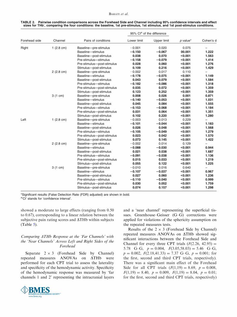
significantly changed across the four conditions in allchannels (Table 1). Post hoc FDR adjusted multiplecomparisons revealed that THb0 did not significantlychange from its baseline value due to hand immersionin tepid water, except for the ‘near channel’ on theright side of the forehead (Table 2). However, handimmersion in ice water caused a significant change inTHb0 as compared to its values at both the baselineand pre-stimulus conditions and it showed a largeeffect (d> 0.944). Also, THb0 at the pre-stimulus and
stimulus conditions were significantly different fromtheir values at the post-stimulus condition which alsoshowed large effects (d> 1.219).
Adaptation to Repeated CPTs Observable in SubjectivePain Ratings and THb
The adaptation to repeated trials of CPT observablein both subjective pain ratings (Fig. 6) and THb0 at thestimulus condition (Fig. 7) was further explored.
Post-stimulus pain rating scores reported based on theNRS-11 (in which ‘0’ means no pain and ‘10’ means theworst pain imaginable) was tested with Friedman’sANOVA by Ranks. There was a significant difference inthe median value of the subjects’ pain rating scores withrespect toCPT trials (v2(2) = 14.70, p = 0.001).Post hocWilcoxon signed ranks tests yielded that the intensity ofthe reported pain rating scores decreased significantlyacross CPT trials. It was revealed that the pain scorereported after the first trial of CPT was significantlyhigher than the pain scores given after the second andthird trials (Z = 2.29, p = 0.02 andZ = 2.92, p = 0.004,respectively). Also, the pain score reported after the sec-ond trial of CPT was significantly higher than the painscore given after the third trial (Z = 2.59, p = 0.01).
To assess the hemodynamic response across threeCPT trials, repeated measures ANOVAs were per-formed on THb0 for all channels separately on theleft and right sides of the forehead (Table 3). Therewere significant main effects of CPT trials in THb0 inall channels. FDR adjusted post hoc multiple com-parisons showed that THb0 at the first CPT trial wassignificantly different from the THb0 at the secondand third CPT trials in all channels on both sides ofthe forehead with large effects (d> 0.886) (Table 4,Fig. 8). However, except for the ‘near channel’ onthe left side, THb0 values at the second and thirdCPT trials were not significantly different. This sig-nificant difference represented a moderate effect(d = 0.573).
We experimentally identified another feature whichmay be a better representation of the individualizedhemodynamic response to CPTs. This variable, whichis denoted as DTHb, is expected to account for the in-ter-subject variability in the latency of the hemody-namic response and is defined as the change in the THbconcentration induced by each CPT. DTHb is calcu-lated as the difference between the minimum value ofTHb that is obtained within 20 s after hand immersionin ice water and the maximum value of THb within a35 s window starting 15 s before removing the handfrom the ice water and ending 20 s after it (Figs. 9a–9c).The minimum value of THb was searched within a re-gion when the effect of CPT is expected to be minimal.The maximum value of THb was searched within a
Hemodyn. Response to Repeated Noxiuos CPTs Measured by fNIRS on Forehead
FIGURE 4. (a–b) The hemodynamic response to three trials of cold pressor test (CPT) averaged across 20 subjects on the right (a)and left (b) sides of the forehead. The black vertical lines represent time points at which subjects switched their hand from the tepidwater to the ice water and vice versa. The pink shaded field represents the baseline period, the orange shaded fields represent theimmersion in tepid water condition, and the blue shaded fields represent the CPT conditions. Hb, HbO2 and THb are abbreviationsfor deoxy-hemoglobin, oxy-hemoglobin and total hemoglobin, respectively. ‘S-D’ symbol stands for ‘source-detector’.
BARATI et al.
window in which the cumulative effect of CPT wasexpected to reach its maximum with enough time forthe hemodynamics to fully evolve. Although weacknowledge that within the selected time windowssubjects may be cognitively involved in anticipating thenext stimulus or reporting their pain, the effect of thesecognitive tasks on the hemodynamics is considered tobe much lower than the observed effect of a cold waterstimulus and thus, it can be disregarded.
Correlation Analysis Between DTHb and SubjectivePain Scores
A secondary interest was to determine whether adecrease in the reported pain scores across the threeCPT trials within a subject was associated with adecrease in the measured hemodynamic parameter (i.e.DTHb). To answer this question, within-subjects cor-relation coefficients6 were calculated. All the correla-tion values were significant at the 0.05 level and
FIGURE 5. Line graphs of THb0 during four experimental conditions: baseline, pre-stimulus, stimulus, and post-stimulus con-ditions (for the definition of THb0, please see ‘‘The Effect of a CPT on the THb Concentration’’ section). Error bars represent the 95%confidence intervals (CI).
TABLE 1. Results of repeated measures ANOVA on THb0 during the four experimental conditions: baseline, pre-stimulus, stim-ulus, and post-stimulus conditions.
* Experiment-wise error rates are False Detection Rate (FDR) adjusted (p values).a‘SD’ stands for ‘standard deviation’.bDue to departure from sphericity, the degrees of freedom were adjusted using the Greenhouse–Geisser (G–G) correction value.
Hemodyn. Response to Repeated Noxiuos CPTs Measured by fNIRS on Forehead
showed a moderate to large effects (ranging from 0.50to 0.67), corresponding to a linear relation between thesubjective pain rating scores and DTHb within subjects(Table 5).
Comparing DTHb Response at the ‘Far Channels’ withthe ‘Near Channels’ Across Left and Right Sides of the
Forehead
Separate 2 9 3 (Forehead Side by Channel)repeated measures ANOVAs on DTHb wereperformed for each CPT trial to assess the lateralityand specificity of the hemodynamic activity. Specificityof the hemodynamic response was measured by ‘farchannels 1 and 2’ representing the intracranial layers
and a ‘near channel’ representing the superficial tis-sues. Greenhouse–Geisser (G–G) corrections wereapplied for violations of the sphericity assumption onthe repeated measures tests.
Results of the 2 9 3 (Forehead Side by Channel)repeated measures ANOVAs on DTHb showed sig-nificant interactions between the Forehead Side andChannel for every three CPT trials (F(2.26, 42.95) =
5.78 G–G, p = 0.004, F(3.05,58.03) = 5.46 G–G,p = 0.002, F(2.18,41.33) = 7.37 G–G, p = 0.001; forthe first, second and third CPT trials, respectively).There was a significant main effect of the ForeheadSide for all CPT trials (F(1,19) = 8.69, p = 0.008,F(1,19) = 8.40, p = 0.009, F(1,19) = 8.04, p = 0.01;for the first, second and third CPT trials, respectively)
TABLE 2. Pairwise condition comparisons across the Forehead Side and Channel including 95% confidence intervals and effectsizes for THb0, comparing the four conditions: the baseline, 1st pre-stimulus, 1st stimulus, and 1st post-stimulus conditions.
Forehead side Channel Pairs of conditions
95% CIa of the difference
p value* Cohen’s dLower limit Upper limit
Right 1 (2.8 cm) Baseline—pre-stimulus 20.001 0.020 0.075 –
*Significant results (False Detection Rate (FDR) adjusted) are shown in bold.a‘CI’ stands for ‘confidence interval’.
BARATI et al.
and a significant main effect of the Channel for thesecond and third CPT trials (F(2,38) = 4.70 G–G,p = 0.02, F(2,38) = 10.75, p< 0.001) (Fig. 10).
Post hoc multiple comparisons after controlling theFDR showed that there is no significant differencebetween the ‘near channel’ and ‘far channels’ on eitherside of the forehead.
Significant asymmetrical activity was revealed bythe FDR adjusted post hoc tests on the DTHb responsemeasured by the ‘far channels’, indicating moderate-
to-large effects (0.697 < d < 0.902). However, therewas no laterality in the DTHb for the superficial tissuesas measured by the ‘near channels’ (Table 6).
DISCUSSION
Despite the technological advancement in medicaldevice development, effective assessment and manage-ment of pain is poorly addressed. One possible reasoncould be the subjectivity of the pain experience. It iswell know that the pain experience including its per-ception and expression is highly individualized.Therefore, there is a need for a more objective assess-ment of pain.
In the conventional pain practice, vital signs such asheart rate, blood pressure, respiratory rate, galvanicskin response and/or cutaneous blood flow measuredby laser Doppler flowmetry have been used to monitorthe physiological parameters in response to a noxiousstimulus. However, the clinical experience has proventhat these physiological signs are not practically reli-able and should be interpreted cautiously. Forinstance, physiological parameters change due to manyfactors other than pain such as distress, medications,and illness. Laser Doppler flowmetry data are also verynoisy and the smallest motion causing any change inthe contact between the probe tip and the skin creates adrastic change in the signal. Further, since the typicaldepth of penetration for a laser Doppler flowmeter is
FIGURE 6. Boxplots of post-stimulus pain rating scores of20 subjects across three trials of cold pressor test (CPT). Twooutliers were identified for pain scores reported after 3rd trialof CPT and are represented as circles.
FIGURE 7. Line graphs of THb0 across cold pressor test (CPT) trials (for the definition of THb0, please see ‘‘The Effect of a CPT onthe THb Concentration’’ section).
Hemodyn. Response to Repeated Noxiuos CPTs Measured by fNIRS on Forehead
less than 1 mm, the results are very sensitive to the typeof the skin and the location where the probing isconducted.33,37
Over the past 5 years, we have used fNIRS formonitoring hemodynamic response to noxious stimuli.We aimed to find an association between a noxiousstimulus and the evoked hemodynamic response and todemonstrate that induced noxious stimuli elicit areproducible and consistent hemodynamic responsewhich can be reliably detected by fNIRS. During this
period, we have explored different protocols bychanging the setting of the stimulus such as type,intensity and duration as well as fNIRS probe config-urations in an effort to optimize the reproducibilityand strength of the response. The results presented inthis manuscript demonstrate only a piece of our questto identify reliable and robust stimulus–responseparameters. The present study aimed to investigate thehemodynamic changes during repeated CPTsemployed as an experimental model of the tonic pain
TABLE 3. Main effect of cold pressor test (CPT) trials repeated measures ANOVAs on THb0.
*Experiment-wise error rates are False Detection Rate (FDR) adjusted (p values).a‘SD’ stands for ‘standard deviation’.bDue to departure from sphericity, the degrees of freedom were adjusted using the Greenhouse–Geisser (G–G) correction value.
TABLE 4. Pairwise cold pressor test (CPT) trials comparisons across the Forehead Side and Channel including 95% confidenceintervals and effect sizes for THb0.
*Significant results (after controlling the False Detection Rate (FDR)) are shown in bold.a‘CI’ stands for ‘confidence interval’.
BARATI et al.
with the capability of evoking a general acute sympa-thetic activation. We established that a simple NIRSdevice can effectively monitor the hemodynamicresponse to a CPT. Since the signal penetrates deeperand the light source and detector are directly in contactwith the skin, there is a negligible noise due to themovement of components with respect to the skin.
According to the literature, cold sensation, atfreezing temperatures, becomes painful within the ini-tial 10 s and progressively increases until it reaches itsmaximum at approximately 60 s.41 Based on our pilotexperiments, we observed that 45 s was a reasonableduration of a CPT to induce an acute pain response innormal subjects and also to allow a detectable hemo-dynamic response to evolve. Moreover, the 2 min post-stimulus immersion in the tepid water was sufficienttime for the hemodynamic response to get close to asteady state. In retrospect, we realized that we couldhave allowed more time between CPTs to let thehemodynamics return to its initial baseline level.However, we tried to keep the duration of the experi-ment as brief as possible to avoid our subjectsbecoming bored and other potential confounds.
It is suggested that for a stimulus that lasts for 1 s,the hemodynamic response evolves over 10–12 s withsome components having longer recovery time.10 Forlonger events, the hemodynamic response adds up
roughly linearly over time.7 The selected search win-dows for calculating the DTHb were determinedexperimentally using our small sample size. We noticedthat some subjects reached their maximum THb valueas soon as 30 s after starting a CPT. Consequently, wechose to extend the search window for the maximumTHb response to 15 s before the termination of theCPT and 20 s after it in all trials.
The tonic pain induced by noxious cold water ismassively confounded and regulated by sympatheticactivity. Thus, it is plausible to conclude that theobserved hemodynamic change in response to CPT ismainly dominated by the sympathetic nervous systemin terms of global increase in cerebral blood flow as aconsequence of the increase in cardiac output follow-ing painful stimuli. The observed asymmetric change inDTHb at the intracranial tissues as measured by the‘far channels’ and no laterality in the superficial tissuesas measured by the ‘near channels’ could be indicatorsof the presence of simultaneous activations of thesystemic sympathetic system as well as higher cerebralregulatory systems. While the results of this study areconsistent with the previously reported cerebralasymmetry in regulations of the autonomic nervoussystem,38 further investigations are required prior tomaking any conclusion on the hemisphere specificity inresponse to a CPT.
FIGURE 8. Line graphs of THb0 for separate trials of cold pressor test (CPT) (for the definition of THb0, please see ‘‘The Effect of aCPT on the THb Concentration’’ section). Significant interactions between the Forehead Side and Channel were found for the 1stand 2nd CPT trials.
Hemodyn. Response to Repeated Noxiuos CPTs Measured by fNIRS on Forehead
FIGURE 9. (a–c) Three sample total hemodynamic responses to the first cold pressor test (CPT) measured at the ‘far channel 1¢on the right side of the forehead. The first black vertical line represents the time point at which subjects immersed their hand intothe ice water and the second black vertical line shows the time when subjects immersed their hand back into the tepid water.Dashed blue vertical lines represent the detected time points for DTHb calculation (see ‘‘Correlation Analysis between DTHb andSubjective Pain Scores’’ section).
BARATI et al.
Our experimental results did not provide any strongevidence to link the observed lateralized activation inthe prefrontal region to any specific pain-inducedcortical activity; partly due to the type of the stimulusapplied and limited data analyses techniques used.However, a NIRS study suggests that a stress-inducingmental task induces asymmetrical activities in theprefrontal cortex.38 Thus, the lateralized activation inthe prefrontal region could be related in part to thestress induced by a CPT or may reflect a generalarousal due to a cold water stimulus.
Repeated CPTs have been previously used toinvestigate cardiovascular habituation and to activate
endogenous analgesic systems.35,42 In the presentstudy, three trials of CPT were performed to test thereliability of our measurements as well as to study theadaptation in our sample regarding subjective painscores and the objective hemodynamic measurements.We observed that THb0 responses had significantlydifferent values in the second and third CPT trials ascompared to the first CPT trial in all channels withlarge effect sizes (d> 0.886). However, no significantdifference was found between the THb0 in the secondand third CPT trials, except for one channel (the ‘nearchannel’ on the left side of the forehead). Thisobservation suggests that the hemodynamic activityadapts to repeated trials of CPT. Furthermore, themedian of pain rating scores, which is a subjectivemeasure, also decreased across the CPT trials as amanifestation of the subjects’ adaptation to the pain.The correlation analysis showed a moderate to largeeffect size corresponding to a strong associationbetween the DTHb and pain rating scores withinsubjects. However, a future study that benefits from asignificantly larger sample size needs to be conductedto further validate this association. Since self-report-ing is very subjective in nature, an absolute correlationbetween fNIRS parameters and self-reported pain
FIGURE 10. Line graphs of DTHb for separate trials of cold pressor test (CPT) (for the definition of DTHb, please see Figs. 9a–9c).For all three trials of CPT, significantly different activation on the left and right sides of the forehead was observed in DTHb asmeasured by the ‘far channels’, while no laterality was observed in DTHb as measured by the ‘near channels’.
TABLE 5. Within-subjects correlation values between self--reported pain rating scores and DTHb.
Forehead Side Channel Within-subjects correlation*
Right Far channel 1 0.609
Far channel 2 0.577
Near channel 0.541
Left Far channel 1 0.556
Far channel 2 0.495
Near channel 0.670
*All the correlation coefficients were significant at a 0.05 level.
Hemodyn. Response to Repeated Noxiuos CPTs Measured by fNIRS on Forehead
scores is not expected. We suggest that a combinationof our measurement and patients’ self-report providesbetter information to the clinicians. In particular, ourmethod is most useful when in various conditionssubjects cannot provide reliable self-report, such as theelderly with dementia and impaired cognition, veryyoung children, or critically ill patients.29 We hope thatour objective measurement provides complimentaryinformation to the self-report for the clinicians.
Undoubtedly, there is a critical need for a repro-ducible, brief and simple measurement that can cor-relate subjective pain experience to objective andquantitative parameters for both clinical and researchpurposes. Reliable biomarkers of pain can help inevidence-based, personalized management of pain. Inthe present research, we showed that the evokedhemodynamic response to noxious cold water stimulican be measured using a non-invasive, portable device.Further refinement of the utilized method in this studymay enable the measurement of the hemodynamicresponse at the cortex and the extracranial tissues.These refinements include incorporating advancedsignal processing techniques and using other noxiousstimuli that would evoke a less generalized systemicresponse such as hot plates and pressure algometry.Then, fNIRS can be used as a powerful clinical tech-nique to assist clinicians in the assessment of pain withor without subjective self-reports.
ACKNOWLEDGMENTS
We would like to thank Mr. Frank Kepic for hisinvaluable technical support and advice. Authors alsoacknowledge Mr. Troy Carlson for designing andconstructing the system for cold pressor tests.
REFERENCES
1Ayaz, H., P. A. Shewokis, A. Curtin, and M. Izzetoglu.Using MazeSuite and functional near infrared spectros-copy to study learning in spatial navigation. J. Vis. Exp.56(e3443). doi:10.3791/3443, 2011.2Bartocci, M., L. L. Bergqvist, H. Lagercrantz, andK. Anand. Pain activates cortical areas in the pretermnewborn brain. Pain 122:109–117, 2006.3Becerra, L., W. Harris, M. Grant, E. George, D. Boas, andD. Borsook. Diffuse optical tomography activation in thesomatosensory cortex: specific activation by painful vs.non-painful thermal stimuli. PLoS One 4(11):e8016, 2009.4Becerra, L., W. Harris, D. Joseph, T. Huppert, D. A. Boas,and D. Borsook. Diffuse optical tomography of pain andtactile stimulation: activation in cortical sensory andemotional systems. NeuroImage 41:252–259, 2008.5Benjamini, Y., and Y. Hochberg. Controlling the false ratediscovery: a practical and powerful approach to multipletesting. J. R. Stat. Soc. B 57(1):289–300, 1995.6Bland, J. M., and D. G. Altman. Calculating correlationcoefficients with repeated observations: part 1—correlationwithin subjects. BMJ 310(6977):446, 1995.7Boynton, G. M., S. A. Engel, G. H. Glover, and D. J.Heeger. Linear systems analysis of functional magneticresonance imaging in human V1. J. Neurosci. 16(13):4207–4221, 1991.8Bozkurt, A., A. Rosen, H. Rosen, and B. Onaral. A por-table near infrared spectroscopy system for bedside moni-toring of newborn brain. Biomed. Eng. Online 4(1):29,2005. doi: 10.1186/1475-925X-4-29.9.Bozkurt, A., and B. Onaral. Safety assessment of nearinfrared light emitting diodes for diffuse optical measure-ments. Biomed. Eng. Online. 3(9), 2004. doi: 10.1186/1475-925X-3-9.
10Buckner, R. L. Event-related fMRI and the hemodynamicresponse. Hum. Brain Mapp. 6:373–377, 1998.
11Bunce, S. C., M. Izzetoglu, K. Izzetoglu, B. Onaral, andK. Pourrezaei. Functional near-infrared spectroscopy anemerging neuroimaging modality. IEEE Eng. Med. Biol.25(4):54–62, 2006.
12Casey, K. L., S. Minoshima, T. J. Morrow, and R. A.Koeppe. Comparison of human cerebral activation
TABLE 6. Pairwise channel pair comparisons across cold pressor test (CPT) trials including 95% confidence intervals and effectsizes for DTHb.
CPT trial Pair of channels
95% CIa of the difference
p-value* Cohen’s dLower limit Upper limit
1st Right far channel 1—left far channel 1 0.817 4.151 0.006 0.697
Right Far channel 2—left far channel 2 1.513 5.225 0.001 0.849
Right near channel—left near channel 20.793 3.652 0.194 –
2nd Right far channel 1—left far channel 1 0.595 2.025 0.001 0.857
Right far channel 2—left far channel 2 0.619 2.483 0.002 0.779
Right near channel—left near channel 20.866 1.818 0.467 –
3rd Right far channel 1—left far channel 1 0.614 2.214 0.002 0.827
Right far channel 2—left far channel 2 0.713 2.251 0.001 0.902
Right near channel—left near channel 20.642 2.444 0.237 –
*Significant results (after controlling the False Detection Rate (FDR)) are shown in bold.a‘CI’ stands for ‘confidence interval’.
patterns during cutaneous warmth, heat pain, and deepcold pain. J. Neurophysiol 76(1):571–581, 1996.
13Chance, B., E. Anday, S. Nioka, S. Zhou, L. Hong,K. Worden, C. Li, T. Murray, Y. Ovetsky, D. Pidikiti, andR. Thomas. A novel method for fast imaging of brainfunction, non-invasively, with light. Opt. Express 2(10):411–423, 1998.
14Cope, M., D. T. Delpy, E. O. Reynolds, J. Wray, andP. vanderZee.Methods of quantitating cerebral near infraredspectroscopy data. Adv. Exp. Med. Biol. 222:183–189, 1988.
15Dehghani, H., and D. T. Delpy. Near-infrared spectros-copy of the adult head: effect of scattering and absorbingobstructions in the cerebrospinal fluid layer on light dis-tribution in the tissue. Appl. Opt. 39(25):4721–4729, 2000.
16Devaraj, A. Signal Processing for Functional Near InfraredNeuroimaging. Master’s Thesis, Drexel University. 2005.
17Di Piero, V., S. Ferracuti, U. Sabatini, P. Pantano,G. Cruccu, and G. L. Lenzi. A cerebral blood flow study ontonic pain activation in man. Pain 56:167–173, 1994.
18Downie, W. W., P. A. Leatham, V. M. Rhind, V. Wright,J. A. Branco, and J. A. Anderson. Studies with pain ratingscales. Ann. Rheum. Dis. 37:378–381, 1978.
19Gelinas, C., M. Choiniere, M. Ranger, A. Denault,A. Deschamps, and C. Johnston. Toward a new approachfor the detection of pain in adult patients undergoing car-diac surgery: near-infrared spectroscopy—a pilot study.Heart Lung 39(6):485–493, 2010.
20Grothusen, J. R., and R. J. Schwartzman. Laser Dopplerimaging: usefulness in chronic pain medicine. Pain Phys.15(5):491–498, 2011.
21Hines, E. A., and G. E. Brown. A standard stimulus formeasuring vasomotor reactions: its application in the studyof hypertension. Mayo Clin. Proc. 7:332–335, 1932.
22Izzetoglu, M., K. Izzetoglu, S. C. Bunce, H. Ayaz,A. Devaraj, B. Onaral, and K. Pourrezaei. Functional near-infrared neuroimaging. IEEE Trans. Neural Syst. Rehabil.Eng. 13(2):153–159, 2005.
23Lovallo, W. The cold pressor test and autonomic function: areview and integration. Psychophysiology 12(3):268–282,1975.
24Music, M., Z. Finderle, and K. Cankar. Cold perception andcutaneousmicrovascular response to local cooling at differentcooling temperatures.Microvasc. Res. 81:319–324, 2011.
25Okada, E., M. Firbank, M. Schweiger, S. R. Arridge,M. Cope, and D. T. Delpy. Theoretical and experimentalinvestigation of near-infrared light propagation in a modelof the adult head. Appl. Opt. 36(1):21–31, 1997.
26Oldfield, R. The assessment and analysis of handedness: theEdinburgh inventory. Neuropsychologia 9(1):97–113, 1971.
27Panerai, R. B., S. L. Dawson, P. J. Eames, and J. F. Potter.Cerebral blood flow velocity response to induced and spon-taneous sudden changes in arterial blood pressure. Am.J. Physiol. Heart Circ. Physiol 280:H2162–H2174, 2001.
28Ramsay, J. O., G. Hooker, and S. Graves. Functional DataAnalysis with R and MATLAB. Springer, 2009.
29Ranger, M., and C. Gelinas. Innovating in pain assessmentof the critically ill: exploring cerebral near-infraredspectroscopy as a bedside approach. Pain Manag. Nurs.doi:http://dx.doi.org/10.1016/j.pmn.2012.03.005.
30Roatta, S., G. Micieli, D. Bosone, G. Losano, R. Bini,A. Cavallini, and P. Magda. Effect of generalised sympa-thetic activation by cold pressor test on cerebral haemo-dynamics in healthy humans. J. Auton. Nerv. Syst. 71:159–166, 1998.
31Rolfe, P. In vivo near infrared spectroscopy. Annu. Rev.Biomed. Eng. 2:715–754, 2000.
32Saager, R., and A. Berger. Measurement of layer-likehemodynamic trends in scalp and cortex: implications forphysiological baseline suppression in functional near-infrared spectroscopy. J. Biomed. Opt. 13(3):034017-1–034017-10, 2008.
33Seifalian, A. M., G. Stansby, A. Jackson, K. Howell, andG. Hamilton. Comparison of laser Doppler perfusionimaging, laser Doppler flowmetry, and thermographicimaging for assessment of blood flow in human skin. Eur.J. Vasc. Surg. 8(1):65–69, 1994.
34Sevick, E. M., C. L. Burch, and B. Chance. Near-infraredoptical imaging of tissue phantoms with measurement inthe change of optical path lengths. Adv. Exp. Med. Biol.345:815–823, 1994.
35Stancak, Jr., A., A. Yamamotova, I. P. Kulis, and I. V.Sekyra. Cardiovascular adjustments and pain duringrepeated cold pressor test. Clin. Auton. Res. 6:83–89,1996.
36Streff, A., L. K. Kuehl, G. Michaux, and F. Anton. Dif-ferential physiological effects during tonic painful handimmersion tests using hot and ice water. Eur. J. Pain14:266–272, 2010.
37Swain, I. D., and L. J. Grant. Methods of measuring skinblood flow. Phys. Med. Biol 34(2):151–175, 1989.
38Tanida, M., K. Sakatani, R. Takano, and K. Tagai.Relation between asymmetry of prefrontal cortex activitiesand the autonomic nervous system during a mental arith-metic task: near infrared spectroscopy study. Neurosci.Lett. 369:69–74, 2004.
39Villringer, A., and B. Chance. Non-invasive optical spec-troscopy and imaging of human brain function. TrendsNeurosci. 20:435–442, 1997.
40von Baeyer, C. L., T. Piira, C. T. Chambers, M. Trapan-otto, and L. K. Zeltzer. Guidelines for the cold pressor taskas an experimental pain stimulus for use with children.J. Pain 6(4):218–227, 2005.
41Walsh, N. E., L. Schoenfeld, S. Ramamurthy, andJ. Hoffman. Normative model for cold pressor test. Am.J. Phys. Med. 68(1):6–11, 1989.
42Washington, L. L., S. J. Gibson, and R. D. Helme. Age-related differences in the endogenous analgesic response torepeated cold water immersion in human volunteers. Pain89:89–96, 2000.
43Watanabe, Y., H. Tanaka, I. Dan, K. Sakurai, K. Kimoto,R. Takashima, and K. Hirata. Monitoring cortical hemo-dynamic changes after sumatriptan injection duringmigraine attack by near-infrared spectroscopy. Neurosci.Res. (Shannon, Irel) 69:60–66, 2011.
44Wolf, S., and J. D. Hardy. Studies on pain. Observationson pain due to local cooling and on factors involved inthe ‘‘cold pressor’’ effect. J. Clin. Invest. 20(5):521–533,1941.
Hemodyn. Response to Repeated Noxiuos CPTs Measured by fNIRS on Forehead