Hemodynamics in coronary arteries with overlapping stents Farhad Rikhtegar a , Christophe Wyss b , Kathryn S. Stok c , Dimos Poulikakos a , Ralph Müller c , Vartan Kurtcuoglu a,d,n a Laboratory of Thermodynamics in Emerging Technologies, Department of Mechanical and Process Engineering, ETH Zurich, Zurich, Switzerland b Clinic of Cardiology, University Hospital Zurich, Zurich, Switzerland c Institute for Biomechanics, Department of Health Sciences and Technology, ETH Zurich, Zurich, Switzerland d The Interface Group, Institute of Physiology, University of Zurich, Winterthurerstrasse 190, Y23 J8, 8057 Zurich, Switzerland article info Article history: Accepted 26 October 2013 Keywords: Stent overlap Thrombosis Hemodynamics Wall shear stress Restenosis abstract Coronary artery stenosis is commonly treated by stent placement via percutaneous intervention, at times requiring multiple stents that may overlap. Stent overlap is associated with increased risk of adverse clinical outcome. While changes in local blood flow are suspected to play a role therein, hemodynamics in arteries with overlapping stents remain poorly understood. In this study we analyzed six cases of partially overlapping stents, placed ex vivo in porcine left coronary arteries and compared them to five cases with two non-overlapping stents. The stented vessel geometries were obtained by micro-computed tomography of corrosion casts. Flow and shear stress distribution were calculated using computational fluid dynamics. We observed a significant increase in the relative area exposed to low wall shear stress (WSS o0.5 Pa) in the overlapping stent segments compared both to areas without overlap in the same samples, as well as to non-overlapping stents. We further observed that the configuration of the overlapping stent struts relative to each other influenced the size of the low WSS area: positioning of the struts in the same axial location led to larger areas of low WSS compared to alternating struts. Our results indicate that the overlap geometry is by itself sufficient to cause unfavorable flow conditions that may worsen clinical outcome. While stent overlap cannot always be avoided, improved deployment strategies or stent designs could reduce the low WSS burden. & 2013 Elsevier Ltd. All rights reserved. 1. Introduction About 30% of patients undergoing percutaneous coronary intervention (PCI) with stent placement are treated with over- lapping stents (Holmes et al., 2004; Räber et al., 2010). Stent overlap is associated with increased risk of adverse clinical out- come for both bare metal stents (BMS) and drug eluting stents (DES) (Ellis et al., 1992; Räber et al., 2010). Various studies have investigated clinical results and biological aspects of stent overlap, but the hemodynamics inside arteries with overlapping stents and the associated wall shear stress (WSS) parameters have received much less attention (Balakrishnan et al., 2005; Charonko et al., 2010; Peacock et al., 1995). This can be in part attributed to the lack of suitable methods for acquiring the geometry of arteries containing overlapping stents with sufficient accuracy. Stent overlap is associated with increased in-stent restenosis and lumen loss due to delayed healing and increased inflamma- tion regardless of stent type (Räber et al., 2010; Wang et al., 2000). While DES may reduce neointimal hyperplasia and restenosis in single stent cases (Moses et al., 2003; Tsagalou et al., 2005), their performance (Finn et al., 2005; Matsumoto et al., 2007) and safety (Moreno et al., 2005) in regions of overlap are a case of debate. Overlapping BMS are associated with worse clinical outcome compared to single BMS (Kastrati et al., 1999; Kereiakes et al., 2006; Serruys et al., 2002). This is attributed primarily to more pronounced arterial injury caused by the expansion of two stents at the same location, leading to increased inflammation. However, the poorer outcome may also be related to severe hemodynamic disturbances introduced by stent malapposition (Charonko et al., 2010) that are inherent in stent overlap but occur infrequently in single stents (Matsumoto et al., 2007). It is generally accepted that hemodynamics influences vascular health and pathogenesis. WSS as one of the manifestations of blood flow has been shown to be an important factor in ather- ogenesis (Chatzizisis et al., 2007; Cheng et al., 2006) and in the pathobiology of neointimal hyperplasia, thrombosis and in-stent restenosis (Papafaklis et al., 2010; Wentzel et al., 2008). These latter processes are a concern in percutaneous vascular interven- tion in general and in stent placement in particular. For example, stent malapposition has been shown to increase thrombogenicity. It is hypothesized that hemodynamics plays a role therein, as Contents lists available at ScienceDirect journal homepage: www.elsevier.com/locate/jbiomech www.JBiomech.com Journal of Biomechanics 0021-9290/$ - see front matter & 2013 Elsevier Ltd. All rights reserved. http://dx.doi.org/10.1016/j.jbiomech.2013.10.048 n Corresponding author at: The Interface Group, Institute of Physiology, Uni- versity of Zurich, Winterthurerstrasse 190, Y23 J8, 8057 Zurich, Switzerland. Tel.: þ41 44 635 50 55; fax: þ41 44 635 68 14. E-mail address: [email protected] (V. Kurtcuoglu). Journal of Biomechanics 47 (2014) 505–511
Transcript
Hemodynamics in coronary arteries with overlapping stents
Farhad Rikhtegar a, Christophe Wyss b, Kathryn S. Stok c, Dimos Poulikakos a, Ralph Müller c,Vartan Kurtcuoglu a,d,n
a Laboratory of Thermodynamics in Emerging Technologies, Department of Mechanical and Process Engineering, ETH Zurich, Zurich, Switzerlandb Clinic of Cardiology, University Hospital Zurich, Zurich, Switzerlandc Institute for Biomechanics, Department of Health Sciences and Technology, ETH Zurich, Zurich, Switzerlandd The Interface Group, Institute of Physiology, University of Zurich, Winterthurerstrasse 190, Y23 J8, 8057 Zurich, Switzerland
Coronary artery stenosis is commonly treated by stent placement via percutaneous intervention, at timesrequiring multiple stents that may overlap. Stent overlap is associated with increased risk of adverseclinical outcome. While changes in local blood flow are suspected to play a role therein, hemodynamicsin arteries with overlapping stents remain poorly understood. In this study we analyzed six cases ofpartially overlapping stents, placed ex vivo in porcine left coronary arteries and compared them to fivecases with two non-overlapping stents. The stented vessel geometries were obtained by micro-computedtomography of corrosion casts. Flow and shear stress distribution were calculated using computationalfluid dynamics. We observed a significant increase in the relative area exposed to low wall shear stress(WSSo0.5 Pa) in the overlapping stent segments compared both to areas without overlap in the samesamples, as well as to non-overlapping stents. We further observed that the configuration of theoverlapping stent struts relative to each other influenced the size of the low WSS area: positioning of thestruts in the same axial location led to larger areas of lowWSS compared to alternating struts. Our resultsindicate that the overlap geometry is by itself sufficient to cause unfavorable flow conditions that mayworsen clinical outcome. While stent overlap cannot always be avoided, improved deployment strategiesor stent designs could reduce the low WSS burden.
& 2013 Elsevier Ltd. All rights reserved.
1. Introduction
About 30% of patients undergoing percutaneous coronaryintervention (PCI) with stent placement are treated with over-lapping stents (Holmes et al., 2004; Räber et al., 2010). Stentoverlap is associated with increased risk of adverse clinical out-come for both bare metal stents (BMS) and drug eluting stents(DES) (Ellis et al., 1992; Räber et al., 2010). Various studies haveinvestigated clinical results and biological aspects of stent overlap,but the hemodynamics inside arteries with overlapping stents andthe associated wall shear stress (WSS) parameters have receivedmuch less attention (Balakrishnan et al., 2005; Charonko et al.,2010; Peacock et al., 1995). This can be in part attributed to thelack of suitable methods for acquiring the geometry of arteriescontaining overlapping stents with sufficient accuracy.
Stent overlap is associated with increased in-stent restenosisand lumen loss due to delayed healing and increased inflamma-tion regardless of stent type (Räber et al., 2010; Wang et al., 2000).
While DES may reduce neointimal hyperplasia and restenosis insingle stent cases (Moses et al., 2003; Tsagalou et al., 2005), theirperformance (Finn et al., 2005; Matsumoto et al., 2007) and safety(Moreno et al., 2005) in regions of overlap are a case of debate.Overlapping BMS are associated with worse clinical outcomecompared to single BMS (Kastrati et al., 1999; Kereiakes et al.,2006; Serruys et al., 2002). This is attributed primarily to morepronounced arterial injury caused by the expansion of two stentsat the same location, leading to increased inflammation. However,the poorer outcome may also be related to severe hemodynamicdisturbances introduced by stent malapposition (Charonko et al.,2010) that are inherent in stent overlap but occur infrequently insingle stents (Matsumoto et al., 2007).
It is generally accepted that hemodynamics influences vascularhealth and pathogenesis. WSS as one of the manifestations ofblood flow has been shown to be an important factor in ather-ogenesis (Chatzizisis et al., 2007; Cheng et al., 2006) and in thepathobiology of neointimal hyperplasia, thrombosis and in-stentrestenosis (Papafaklis et al., 2010; Wentzel et al., 2008). Theselatter processes are a concern in percutaneous vascular interven-tion in general and in stent placement in particular. For example,stent malapposition has been shown to increase thrombogenicity.It is hypothesized that hemodynamics plays a role therein, as
adjacent high shear stress areas and recirculation zones caused bymalapposed stents may activate platelets and increase local resi-dence times of these thrombocytes (Hathcock, 2006; Kolandaiveluet al., 2011; Peacock et al., 1995). Kolandaivelu and co-workersshowed in vitro and in a 2D computational model with idealizeddomain geometry that flow recirculation between malapposed andoverlapping stent struts may modulate stent thrombogenicity(Kolandaivelu et al., 2011).
Computational fluid dynamics (CFD) is the method of choice forassessing shear stress and local hemodynamics in stented arteries. Theprecise acquisition of the stent struts and arterial geometry isa prerequisite for accurate CFD analysis, but no clinical imagingmodality exists that could yield such data with sufficient resolution.Several approaches have been reported in the literature to circumventthis limitation: simulations may be conducted on idealized geometriesbased on stent CAD data (Gundert et al., 2011), on hybrid domainswhere the stent free geometry is obtained by CT, digital angiographyor MRI and a stent is virtually implanted (De Santis et al., 2010; LaDisaet al., 2006), on ex vivo micro-computed tomography (mCT) data ofexplanted, stented arteries (Morlacchi et al., 2011) or mCT images ofstented in vitro artery models (Benndorf et al., 2009; Connolley et al.,2007). While these methods have their undisputed respectivestrengths, they have either limited geometric accuracy, limited trea-table vascular domain size or incomplete representation of themechanical interaction between stent and arterial wall.
We have recently introduced a method that allows for preciseex vivo acquisition of arteries stented in vivo or ex vivo, yielding boththe macroscopic arterial tree geometry as well as the configurationand morphology of individual stent struts (Rikhtegar et al., 2013). Herewemake use of this method to investigate the shear stress distributionand hemodynamics of porcine coronary arteries with overlappingstents. Our goal is to evaluate flow disturbances and consequent shearstress alterations introduced by stent overlap which may contribute tothe reported clinical problems associated with overlapping stents.
2. Methods
A concise description of the utilized methods is given here. We refer the readerto the Supplemental material and Rikhtegar et al. (2013) for a more detailedexplanation of the individual process steps.
2.1. Heart preparation, stenting and vascular corrosion casting
After cannulation, two absorbable metal scaffolds of 10 mm length and 3 mmdiameter (Biotronik AG, Switzerland) were implanted in the left coronary artery of11 ex vivo porcine hearts under angiographic guidance by an interventionalcardiologist. The scaffolds had overlap in 6 arteries. The remaining 5 arteriesserved as control (no overlap).
A radio-opaque casting material was prepared as a mixture of Biodur E20 resin(Biodur Products GmbH, Germany) and iodine-saturated methyl ethyl ketonesolvent. The casting material was injected into the stented arteries under physio-logical pressure of 90 mm Hg. The hearts were left at room temperature for 36 h,and then macerated at 55 1C in a 7.5% w/v solution of potassium hydroxide. Thefinal products were rinsed with water to remove remaining tissue.
2.2. μCT imaging of stented casts and image processing
Micro-computed tomography (μCT80, Scanco Medical AG, Switzerland) wasused with an isotropic voxel size of 74 μm (energy 70 kVp, integration time 300 ms,tube current 114 μA, and two times frame averaging) to capture the overallgeometry of the arterial tree. The stented vessel segments were dissected fromthe remainder of the arterial tree and re-scanned at higher resolution (μCT40,Scanco) with an isotropic voxel size of 6 μm (energy 70 kVp, integration time300 ms, tube current 114 μA, and two times frame averaging).
To partly suppress noise in the raw μCT volumes, a constrained 3D Gauss filterwas used (s¼1.2 and support¼1). Both μCT datasets of low and high resolutionwere independently segmented using a semi-automatic, intensity-based approachin Avizo 6.2 (Visualization Sciences Group SAS, France) to obtain the lumengeometry. The resulting 3D geometries were exported to Geomagic Studio 12(Geomagic, Inc., USA) to register and merge the high resolution geometry of thestented segment and the low resolution remainder of the arterial tree.
2.3. CFD calculations
ANSYS ICEM CFD (ANSYS, Inc., USA) was used to generate a computational gridconsisting of approximately 50 million tetrahedral elements in the merged geo-metry. Steady-state CFD analysis was carried out with the finite volume codeANSYS CFX to determine hemodynamics and WSS distribution. Blood was modeledas a non-Newtonian fluid with constant density of 1050 kg/m3 and shear depen-dent dynamic viscosity according to the Carreau model (Chien et al., 1966). Inflowrate was set to 0.95 mL/s at the coronary ostium (Berne and Levy, 1986) and no slipwas prescribed at the stent surfaces and vessel wall. Murray's law was applied atthe outlets to where the largest diameter branch had zero relative pressure.Outflow rates at the remaining outlets were determined according to their cross-sectional area (Murray, 1926; Rikhtegar et al., 2013). Residual reduction to 10�8 ofthe initial value was set as the convergence criterion. Grid independence studiesconfirmed that the chosen grid was sufficient to capture WSS with a relative errorof o4% compared to a grid with 100% higher cell density.
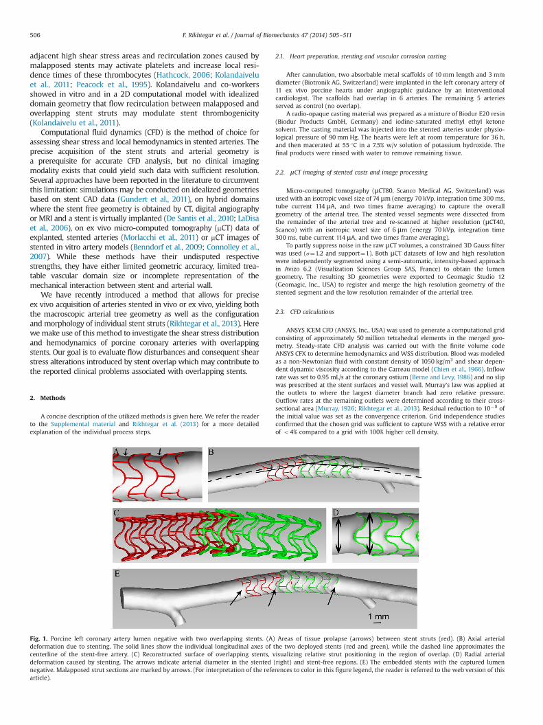
Fig. 1. Porcine left coronary artery lumen negative with two overlapping stents. (A) Areas of tissue prolapse (arrows) between stent struts (red). (B) Axial arterialdeformation due to stenting. The solid lines show the individual longitudinal axes of the two deployed stents (red and green), while the dashed line approximates thecenterline of the stent-free artery. (C) Reconstructed surface of overlapping stents, visualizing relative strut positioning in the region of overlap. (D) Radial arterialdeformation caused by stenting. The arrows indicate arterial diameter in the stented (right) and stent-free regions. (E) The embedded stents with the captured lumennegative. Malapposed strut sections are marked by arrows. (For interpretation of the references to color in this figure legend, the reader is referred to the web version of thisarticle).
F. Rikhtegar et al. / Journal of Biomechanics 47 (2014) 505–511506
3. Results
It has been shown previously that the acquisition method usedhere can capture single stented arteries accurately at high resolu-tion (Rikhtegar et al., 2013). Fig. 1 illustrates that this method alsoensures anatomic fidelity in the case of overlapping stents, show-ing that it captures regions of prolapse between stent struts (PanelA), axial arterial deformation caused by the stent implantation(Panel B), strut overlap and relative positioning (Panel C), radialarterial deformation (Panel D) and strut malapposition (Panel E).
WSS distribution in one of the porcine left coronary arterieswith overlapping stents is shown in Fig. 2. Here as in subsequentfigures, reported WSS values are normalized with respect to themaximum WSS along the arterial tree, which ranges between9.5 and 11 Pa depending on the investigated case. Absolute valuesare given in the Supplemental material. Shear stress below 5% ofthe maximum WSS is shown in dark blue. This corresponds toapproximately 0.5 Pa, here referred to as low and atheroproneaccording to Garasic et al. (2000). Such low WSS is present aroundindividual stent struts and at bifurcations. It is clearly visible in theinset that a larger wall area is exposed to low shear stress in theoverlapped region compared to the adjacent areas covered by asingle stent. This is not an observation that is limited to onesample: all six overlapping stents demonstrate the same qualita-tive behavior (Fig. 3). Differences between the six cases are mostlydue to varying stent overlap length: samples I–III have clearlyshorter overlap regions compared to the other cases, resulting insmaller areas of low WSS.
To understand why larger wall areas are exposed to lowWSS inthe overlapping stent segments, local hemodynamics must beconsidered. Fig. 4 shows streamlines projected onto an axial cutplane in the vicinity of stent struts. Panel A illustrates the velocitypattern in an overlap segment where blood is tunneled betweentwo alternating struts. Recirculation zones are visible adjacent tothe outer stent struts. A large recirculation zone between twostruts in close vicinity is shown in Panel B. This configuration canoccur in stent overlap zones, yielding increased local bloodresidence times and decreased WSS. Such recirculation zonesinherently yield low WSS values.
The struts of overlapping stents can either be aligned (the innerstent's struts are located immediately on top of the outer struts),or positioned in an alternating fashion (the struts of the stentsare axially offset). We refer to these as congruent and incongruentstrut configurations, respectively. Differences in the flow field
between congruent and incongruent configurations are presentedin Fig. 5. Velocity magnitudes are higher near the wall in theincongruent case, where blood can flow between the alternatingstruts (compare Fig. 6). In contrast, the congruent strut configura-tion produces larger obstacles to flow, increasing recirculationzone sizes. The relative positioning of the overlapping-stent strutsinfluences the location of the stagnation points and alters theextent of recirculation zones compared to struts without overlap.
The effect of strut alignment on WSS distribution is shown inFig. 6, where a longitudinal cut of stented segment is overlaid byWSS contour. Congruent strut regions (black arrows) feature lowerWSS compared to areas with alternating struts (white arrows),where the blood can flow at higher velocity near the vessel wall.
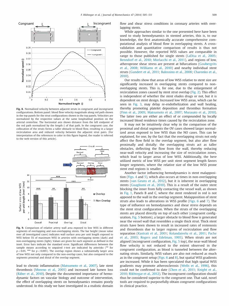
Fig. 7 shows normalized WSS along two axial paths past a set ofrepresentative congruent and incongruent struts, respectively.With the exception of the actual strut wall, WSS is lower in thecongruent configuration. The higher WSS at the strut wall iscaused by tunneling of blood between the closely-spaced con-gruent struts at increased speed. Normalized flow speeds betweenpairs of adjacent stent struts in congruent and incongruent casesare plotted in Fig. 8. Lower speeds are observed between pairs ofcongruent struts due to the formation of larger recirculation zones.It should be noted that this finding cannot be generalized to allstrut pairs. The three-dimensional nature of the flow field maycounteract the formation of large recirculation zones in thecongruent strut configuration or promote formation of such inthe incongruent configuration.
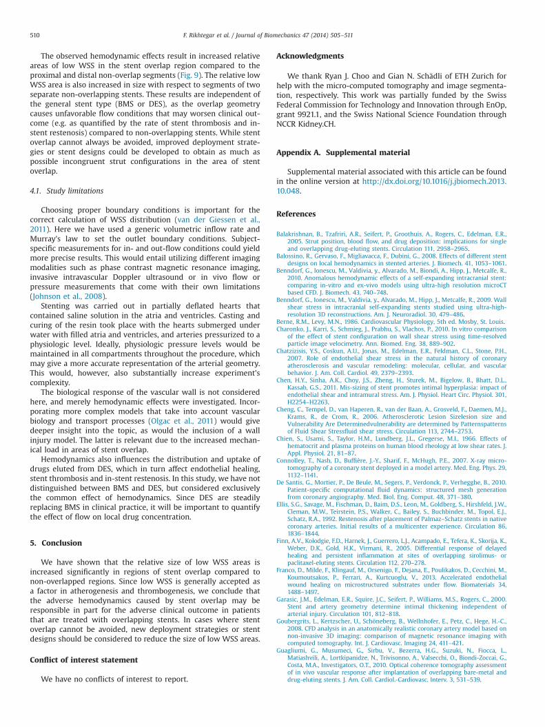
To further quantify the observed phenomena, we compared sixcases of arteries with overlapping stents to five arteries with twonon-overlapping stents. We chose low WSS (o5% of maximumWSS) as the relevant parameter, since it plays a critical role in thebiological response of arteries to stent deployment. In the caseswith overlap (OV), the overlapping segments as well as thesegments proximal and distal to the overlap were evaluated
Fig. 2. Normalized wall shear stress distribution in a porcine left coronary arterywith two overlapping stents. Magnified views of the stented segment and abifurcation are shown in the insets. Wall shear stress (WSS) below 5% of maximumWSS occurs mainly in the vicinity of stent struts and at bifurcations, which are sitesknown to be prone to intimal thickening. A large area of lowWSS is observed in theregion of stent overlap.
Fig. 3. Normalized wall shear stress distribution in the stented segment of sixarteries with overlapping stents. Flow direction is from left to right. A large area oflowWSS (o5% of maximum WSS) is clearly visible in all samples. The extent of thelow WSS region depends on the number of overlapping struts. The first three caseshave shorter length of overlap (two to three struts) compared to the remainingcases (four to five struts). The approximate length of overlap for the first threesamples is indicated by the dashed lines, while the approximate borders of theremaining samples are shown with solid lines.
F. Rikhtegar et al. / Journal of Biomechanics 47 (2014) 505–511 507
(Fig. 9). In the non-overlap cases (NO), the proximal and distalstents were considered. Since the segment lengths are not allequal, the total area of low WSS was normalized by the respectivesegment's length.
Over the whole stented artery length, the OV cases showa normalized area of low WSS equal to 7.7 mm2/mm, which is57% higher than in the combined segments of the NO cases.Qualitatively similar results were obtained when comparing thearea exposed to lowWSS in proximal and distal NO stent segmentsto the proximal and distal segments of the OV cases, respectively:the OV cases showed on average approximately 35% higher valuesof normalized area exposed to low WSS. Comparison of theoverlap segment alone to the combined NO segments and tothe combined remaining non-overlap segments of the OV cases
showed approximately 102% and 29% higher normalized area oflow WSS values, respectively.
4. Discussion
It is known that hemodynamics influences atherogenesis(Chatzizisis et al., 2007; Samady et al., 2011), thrombogenesis(Hathcock, 2006), vascular remodeling (Stone et al., 2003), neointi-mal hyperplasia (Wentzel et al., 2001) and endothelial healing(Franco et al., 2013). It is further known that stent overlap, comparedto single stents, increases thrombogenicity (Kolandaivelu et al., 2011;Rogers and Edelman, 1995), may delay the re-endothelization andenhance platelet deposition and thrombus formation (Finn et al.,2005; Murasato et al., 2010). Poor re-endothelization, in turn, can
Fig. 4. Projection of streamlines to cut plane in the vicinity of stent struts. (A) Streamlines in overlapping segment with alternating struts. Recirculation zones are clearlyvisible near the stent struts. (B) Large area of recirculation between two struts in close proximity.
Fig. 5. Vector plots of projected velocity for two different stent overlap configura-tions. Top panel: alternating (incongruent) struts. Bottom panel: aligned (con-gruent) struts. The latter configuration is associated with lower near-wall velocities(bottom inset) and decreased WSS (compare Fig. 6), while in the former relativelyhigh velocities are maintained in proximity of the wall (top inset).
Fig. 6. The effect of strut alignment on wall shear stress and velocity distribution.Top panel: longitudinal cut through stented artery section. Color map showsnormalized shear stress distribution on stent surfaces and artery wall. Low WSSis seen in areas adjacent to congruent struts (black arrows), while higher WSS isobserved near incongruent struts (white arrows). Bottom panels: vector plot ofvelocities projected onto axial cut plane in the vicinity of congruent (left) andincongruent struts (right).
Fig. 7. Comparison of normalized wall shear stress near congruent and incon-gruent struts. Top left panel: rendering of the studied arterial segment. Shear stressvalues plotted in the bottom panel are obtained in the region of length L betweenthe dashed vertical lines. Longitudinal sections shown in the top right panel areobtained along the two solid black lines. Top right panel: longitudinal cross-sections at congruent and incongruent struts locations according to the renderingin the top left panel. Bottom panel: normalized WSS plotted versus normalizedaxial distance from the left to the right dashed vertical lines shown in the toppanels. WSS is generally lower for the congruent case due to lower velocity nearthe wall, with the exception of the actual strut pair location. There, flow is tunneledbetween the closely spaced struts, leading to high WSS.
F. Rikhtegar et al. / Journal of Biomechanics 47 (2014) 505–511508
lead to chronic inflammation (Matsumoto et al., 2007), late stentthrombosis (Moreno et al., 2005) and increased late lumen loss(Räber et al., 2010). Despite the documented importance of hemo-dynamic factors on vascular biology and outcome of intervention,the effect of overlapping stents on hemodynamics remains poorlyunderstood. In this study we have investigated in a realistic domain
flow and shear stress conditions in coronary arteries with over-lapping stents.
While approaches similar to the one presented here have beenused to study hemodynamics in stented arteries, this is, to ourknowledge, the first anatomically accurate comprehensive com-putational analysis of blood flow in overlapping stents. A cross-validation and quantitative comparison of results is thus notpossible. However, the reported WSS values are comparable inrange to those published for single stents (LaDisa et al., 2005;Benndorf et al., 2010; Morlacchi et al., 2011), and regions of low,atheroprone shear stress are present at bifurcations (Goubergritset al., 2008; Williams et al., 2010) and nearby individual stentstruts (Gundert et al., 2011; Balossino et al., 2008; Charonko et al.,2010).
Our results show that areas of low WSS relative to stent size aresignificantly increased in overlapping stents compared to non-overlapping stents. This is, for one, due to the enlargement ofrecirculation zones caused by stent strut overlap (Fig. 2). This effectis independent of whether the stent eludes drugs or not, but it isdependent on stent design. Increased low WSS areas, which can beseen in Fig. 3, may delay re-endothelization and wall healing,thereby promoting platelet deposition and thrombus formation(Finn et al., 2005; Matsumoto et al., 2007; Murasato et al., 2010).The latter two are either an effect of or compounded by locallyincreased blood residence times caused by the recirculation zone.
It may not be intuitively clear why in the comparison of theproximal and distal segments the OV cases showed larger normal-ized areas exposed to low WSS than the NO cases. This can beexplained, for one, by the fact that the overlapping struts not onlyaffect the flow field in the overlap segment, but also alter flowproximally and distally: the overlapping struts act as tallerobstacles, deflecting the flow from the wall, thereby reducingnear-wall velocity and increasing the size of recirculation zones,which lead to larger areas of low WSS. Additionally, the hereutilized metric of low WSS per unit stent segment length favorslonger segments, where the relative size of the low WSS pronestent end regions is smaller.
Another factor influencing hemodynamics is stent malapposi-tion (Figs. 4 and 5), which also occurs at times in non-overlappingstents (van Geuns et al., 2012), but it is inherent in overlappingstents (Guagliumi et al., 2010). This is a result of the outer stentblocking the inner from fully contacting the vessel wall, as shownin Fig. 1, Panels B and C, where the stent rendered in red is notapposed to the wall in the overlap segment. Malapposition of stentstruts also leads to alterations in WSS profile (Figs. 6 and 7). Thetype of influence on hemodynamics and shear stress depends onthe stent strut configuration. When the struts of the overlappingstents are placed directly on top of each other (congruent config-uration, Fig. 5 bottom), a larger obstacle to blood flow is generatednear the vessel wall that resembles a single thick strut. Thick stentstruts have been shown to result in increased rates of restenosisand thrombosis due to larger regions of recirculation and flowseparation (Kastrati et al., 2001; Kolandaivelu et al., 2011; Pacheet al., 2003; Rogers and Edelman, 1995). When struts are notaligned (incongruent configuration, Fig. 5 top), the near-wall bloodflow velocity is not reduced to the extent observed in thecongruent configuration, as blood is tunneled between the alter-nating struts. Similarly, WSS values are also not reduced as muchas in the congruent setup (Figs. 6 and 8), but spatial WSS gradientsare increased. While it has been speculated that high spatial WSSgradients may promote atherosclerosis (Wells et al., 1996), thiscould not be confirmed to date (Chen et al., 2011; Knight et al.,2010; Rikhtegar et al., 2012). The incongruent configuration shouldthus be considered superior to the congruent one. That said, noveltools are required to purposefully obtain congruent configurationsin clinical practice.
Fig. 8. Normalized velocity between adjacent struts in congruent and incongruentconfigurations. Bottom panel: blood flow velocity magnitude along red path shownin the top panels for the strut configurations shown in the top panels. Velocities arenormalized by the respective values at the same longitudinal position on thearterial centerline. The horizontal axis shows distance from the left endpoint ofthe red path normalized by the length L of that path. In the congruent case, thecollocation of the struts forms a taller obstacle to blood flow, resulting in a largerrecirculation area and reduced velocity between the adjacent strut pairs. (Forinterpretation of the references to color in this figure legend, the reader is referredto the web version of this article).
Fig. 9. Comparison of relative artery wall area exposed to low WSS in differentsegments of overlapping and non-overlapping stents. The bar height (mean valueover all investigated cases) indicates wall surface area per unit length exposed toWSS below 5% of maximum WSS in arteries with overlapping stents (dark) andnon-overlapping stents (light). Values are given for each segment as defined in theinset. Error bars indicate the standard error. Significant differences between thesample means according to unpaired t-test are indicated by asterisks (nn forpo0.01, nnn for po0.001). The overlap region shows a substantially larger areaof low WSS not only compared to the non-overlap cases, but also compared to thesegments proximal and distal of the overlap segment.
F. Rikhtegar et al. / Journal of Biomechanics 47 (2014) 505–511 509
The observed hemodynamic effects result in increased relativeareas of low WSS in the stent overlap region compared to theproximal and distal non-overlap segments (Fig. 9). The relative lowWSS area is also increased in size with respect to segments of twoseparate non-overlapping stents. These results are independent ofthe general stent type (BMS or DES), as the overlap geometrycauses unfavorable flow conditions that may worsen clinical out-come (e.g. as quantified by the rate of stent thrombosis and in-stent restenosis) compared to non-overlapping stents. While stentoverlap cannot always be avoided, improved deployment strate-gies or stent designs could be developed to obtain as much aspossible incongruent strut configurations in the area of stentoverlap.
4.1. Study limitations
Choosing proper boundary conditions is important for thecorrect calculation of WSS distribution (van der Giessen et al.,2011). Here we have used a generic volumetric inflow rate andMurray's law to set the outlet boundary conditions. Subject-specific measurements for in- and out-flow conditions could yieldmore precise results. This would entail utilizing different imagingmodalities such as phase contrast magnetic resonance imaging,invasive intravascular Doppler ultrasound or in vivo flow orpressure measurements that come with their own limitations(Johnson et al., 2008).
Stenting was carried out in partially deflated hearts thatcontained saline solution in the atria and ventricles. Casting andcuring of the resin took place with the hearts submerged underwater with filled atria and ventricles, and arteries pressurized to aphysiologic level. Ideally, physiologic pressure levels would bemaintained in all compartments throughout the procedure, whichmay give a more accurate representation of the arterial geometry.This would, however, also substantially increase experiment'scomplexity.
The biological response of the vascular wall is not consideredhere, and merely hemodynamic effects were investigated. Incor-porating more complex models that take into account vascularbiology and transport processes (Olgac et al., 2011) would givedeeper insight into the topic, as would the inclusion of a wallinjury model. The latter is relevant due to the increased mechan-ical load in areas of stent overlap.
Hemodynamics also influences the distribution and uptake ofdrugs eluted from DES, which in turn affect endothelial healing,stent thrombosis and in-stent restenosis. In this study, we have notdistinguished between BMS and DES, but considered exclusivelythe common effect of hemodynamics. Since DES are steadilyreplacing BMS in clinical practice, it will be important to quantifythe effect of flow on local drug concentration.
5. Conclusion
We have shown that the relative size of low WSS areas isincreased significantly in regions of stent overlap compared tonon-overlapped regions. Since low WSS is generally accepted asa factor in atherogenesis and thrombogenesis, we conclude thatthe adverse hemodynamics caused by stent overlap may beresponsible in part for the adverse clinical outcome in patientsthat are treated with overlapping stents. In cases where stentoverlap cannot be avoided, new deployment strategies or stentdesigns should be considered to reduce the size of low WSS areas.
Conflict of interest statement
We have no conflicts of interest to report.
Acknowledgments
We thank Ryan J. Choo and Gian N. Schädli of ETH Zurich forhelp with the micro-computed tomography and image segmenta-tion, respectively. This work was partially funded by the SwissFederal Commission for Technology and Innovation through EnOp,grant 9921.1, and the Swiss National Science Foundation throughNCCR Kidney.CH.
Appendix A. Supplemental material
Supplemental material associated with this article can be foundin the online version at http://dx.doi.org/10.1016/j.jbiomech.2013.10.048.
References
Balakrishnan, B., Tzafriri, A.R., Seifert, P., Groothuis, A., Rogers, C., Edelman, E.R.,2005. Strut position, blood flow, and drug deposition: implications for singleand overlapping drug-eluting stents. Circulation 111, 2958–2965.
Balossino, R., Gervaso, F., Migliavacca, F., Dubini, G., 2008. Effects of different stentdesigns on local hemodynamics in stented arteries. J. Biomech. 41, 1053–1061.
Benndorf, G., Ionescu, M., Valdivia, y., Alvarado, M., Biondi, A., Hipp, J., Metcalfe, R.,2010. Anomalous hemodynamic effects of a self-expanding intracranial stent:comparing in-vitro and ex-vivo models using ultra-high resolution microCTbased CFD. J. Biomech. 43, 740–748.
Benndorf, G., Ionescu, M., Valdivia, y., Alvarado, M., Hipp, J., Metcalfe, R., 2009. Wallshear stress in intracranial self-expanding stents studied using ultra-high-resolution 3D reconstructions. Am. J. Neuroradiol. 30, 479–486.
of the effect of stent configuration on wall shear stress using time-resolvedparticle image velocimetry. Ann. Biomed. Eng. 38, 889–902.
Chatzizisis, Y.S., Coskun, A.U., Jonas, M., Edelman, E.R., Feldman, C.L., Stone, P.H.,2007. Role of endothelial shear stress in the natural history of coronaryatherosclerosis and vascular remodeling: molecular, cellular, and vascularbehavior. J. Am. Coll. Cardiol. 49, 2379–2393.
Chen, H.Y., Sinha, A.K., Choy, J.S., Zheng, H., Sturek, M., Bigelow, B., Bhatt, D.L.,Kassab, G.S., 2011. Mis-sizing of stent promotes intimal hyperplasia: impact ofendothelial shear and intramural stress. Am. J. Physiol. Heart Circ. Physiol. 301,H2254–H2263.
Cheng, C., Tempel, D., van Haperen, R., van der Baan, A., Grosveld, F., Daemen, M.J.,Krams, R., de Crom, R., 2006. Atherosclerotic Lesion Sizelesion size andVulnerability Are Determinedvulnerability are determined by Patternspatternsof Fluid Shear Stressfluid shear stress. Circulation 113, 2744–2753.
Chien, S., Usami, S., Taylor, H.M., Lundberg, J.L., Gregerse, M.I., 1966. Effects ofhematocrit and plasma proteins on human blood rheology at low shear rates. J.Appl. Physiol. 21, 81–87.
Connolley, T., Nash, D., Buffière, J.-Y., Sharif, F., McHugh, P.E., 2007. X-ray micro-tomography of a coronary stent deployed in a model artery. Med. Eng. Phys. 29,1132–1141.
De Santis, G., Mortier, P., De Beule, M., Segers, P., Verdonck, P., Verhegghe, B., 2010.Patient-specific computational fluid dynamics: structured mesh generationfrom coronary angiography. Med. Biol. Eng. Comput. 48, 371–380.
Ellis, S.G., Savage, M., Fischman, D., Baim, D.S., Leon, M., Goldberg, S., Hirshfeld, J.W.,Cleman, M.W., Teirstein, P.S., Walker, C., Bailey, S., Buchbinder, M., Topol, E.J.,Schatz, R.A., 1992. Restenosis after placement of Palmaz–Schatz stents in nativecoronary arteries. Initial results of a multicenter experience. Circulation 86,1836–1844.
Finn, A.V., Kolodgie, F.D., Harnek, J., Guerrero, L.J., Acampado, E., Tefera, K., Skorija, K.,Weber, D.K., Gold, H.K., Virmani, R., 2005. Differential response of delayedhealing and persistent inflammation at sites of overlapping sirolimus- orpaclitaxel-eluting stents. Circulation 112, 270–278.
Franco, D., Milde, F., Klingauf, M., Orsenigo, F., Dejana, E., Poulikakos, D., Cecchini, M.,Koumoutsakos, P., Ferrari, A., Kurtcuoglu, V., 2013. Accelerated endothelialwound healing on microstructured substrates under flow. Biomaterials 34,1488–1497.
Goubergrits, L., Kertzscher, U., Schöneberg, B., Wellnhofer, E., Petz, C., Hege, H.-C.,2008. CFD analysis in an anatomically realistic coronary artery model based onnon-invasive 3D imaging: comparison of magnetic resonance imaging withcomputed tomography. Int. J. Cardiovasc. Imaging 24, 411–421.
Guagliumi, G., Musumeci, G., Sirbu, V., Bezerra, H.G., Suzuki, N., Fiocca, L.,Matiashvili, A., Lortkipanidze, N., Trivisonno, A., Valsecchi, O., Biondi-Zoccai, G.,Costa, M.A., Investigators, O.T., 2010. Optical coherence tomography assessmentof in vivo vascular response after implantation of overlapping bare-metal anddrug-eluting stents. J. Am. Coll. Cardiol.-Cardiovasc. Interv. 3, 531–539.
F. Rikhtegar et al. / Journal of Biomechanics 47 (2014) 505–511510
Gundert, T., Shadden, S., Williams, A., Koo, B.-K., Feinstein, J., LaDisa, J., 2011. A rapidand computationally inexpensive method to virtually implant current andnext-generation stents into subject-specific computational fluid dynamicsmodels. Ann. Biomed. Eng. 39, 1423–1437.
Hathcock, J.J., 2006. Flow effects on coagulation and thrombosis. Arterioscler.Thromb. Vasc. Biol. 26, 1729–1737.
Holmes, D.R., Leon, M.B., Moses, J.W., Popma, J.J., Cutlip, D., Fitzgerald, P.J., Brown, C.,Fischell, T., Wong, S.C., Midei, M., Snead, D., Kuntz, R.E., 2004. Analysis of 1-yearclinical outcomes in the SIRIUS trial: a randomized trial of a sirolimus-elutingstent versus a standard stent in patients at high risk for coronary restenosis.Circulation 109, 634–640.
Johnson, K., Sharma, P., Oshinski, J., 2008. Coronary artery flow measurement usingnavigator echo gated phase contrast magnetic resonance velocity mapping at3.0 T. J. Biomech. 41, 595–602.
Kastrati, A., Elezi, S., Dirschinger, J., Hadamitzky, M., Neumann, F.J., Schomig, A.,1999. Influence of lesion length on restenosis after coronary stent placement.Am. J. Cardiol. 83, 1617–1622.
Kastrati, A., Mehilli, J., Dirschinger, J., Dotzer, F., Schuhlen, H., Neumann, F.J.,Fleckenstein, M., Pfafferott, C., Seyfarth, M., Schomig, A., 2001. Intracoronarystenting and angiographic results – strut thickness effect on restenosis outcome(ISAR-STEREO) trial. Circulation 103, 2816–2821.
Kereiakes, D.J., Wang, H., Popma, J.J., Kuntz, R.E., Donohoe, D.J., Schofer, J.,Schampaert, E., Meier, B., Leon, M.B., Moses, J.W., 2006. Periprocedural andlate consequences of overlapping cypher sirolimus-eluting stents: pooledanalysis of five clinical trials. J. Am. Coll. Cardiol. 48, 21–31.
Knight, J., Olgac, U., Saur, S.C., Poulikakos, D., Marshall Jr., W., Cattin, P.C., Alkadhi, H.,Kurtcuoglu, V., 2010. Choosing the optimal wall shear parameter for theprediction of plaque location—a patient-specific computational study in humanright coronary arteries. Atherosclerosis 211, 445–450.
Kolandaivelu, K., Swaminathan, R., Gibson, W.J., Kolachalama, V.B., Nguyen-Ehrenreich, K.-L., Giddings, V.L., Coleman, L., Wong, G.K., Edelman, E.R., 2011.Stent thrombogenicity early in high-risk interventional settings is driven bystent design and deployment and protected by polymer–drug coatings.Circulation 123, 1400–1409.
LaDisa Jr., J.F., Olson, L., Douglas, H., Warltier, D., Kersten, J., Pagel, P.S., 2006.Alterations in regional vascular geometry produced by theoretical stentimplantation influence distributions of wall shear stress: analysis of a curvedcoronary artery using 3D computational fluid dynamics modeling. Biomed. Eng.Online 5, 40.
LaDisa Jr., J.F., Olson, L.E., Guler, I., Hettrick, D.A., Kersten, J.R., Warltier, D.C., Pagel, P.S.,2005. Circumferential vascular deformation after stent implantation alters wallshear stress evaluated with time-dependent 3D computational fluid dynamicsmodels. J. Appl. Physiol. 98, 947–957.
Matsumoto, D., Shite, J., Shinke, T., Otake, H., Tanino, Y., Ogasawara, D., Sawada, T.,Paredes, O.L., Hirata, K.-i., Yokoyama, M., 2007. Neointimal coverage ofsirolimus-eluting stents at 6-month follow-up: evaluated by optical coherencetomography. Eur. Heart J. 28, 961–967.
Moreno, R., Fernandez, C., Hernandez, R., Alfonso, F., Angiolillo, D.J., Sabate, M.,Escaned, J., Banuelos, C., Fernandez-Ortiz, A., Macaya, C., 2005. Drug-elutingstent thrombosis – results from a pooled analysis including 10 randomizedstudies. J. Am. Coll. Cardiol. 45, 954–959.
Morlacchi, S., Keller, B., Arcangeli, P., Balzan, M., Migliavacca, F., Dubini, G., Gunn, J.,Arnold, N., Narracott, A., Evans, D., Lawford, P., 2011. Hemodynamics and in-stent restenosis: micro-CT images, histology, and computer simulations. Ann.Biomed. Eng. 39, 2615–2626.
Moses, J.W., Leon, M.B., Popma, J.J., Fitzgerald, P.J., Holmes, D.R., O'Shaughnessy, C.,Caputo, R.P., Kereiakes, D.J., Williams, D.O., Teirstein, P.S., Jaeger, J.L., Kuntz, R.E.,2003. Sirolimus-eluting stents versus standard stents in patients with stenosisin a native coronary artery. N. Engl. J. Med. 349, 1315–1323.
Murasato, Y., Hikichi, Y., Nakamura, S., Kajiya, F., Iwasaki, K., Kinoshita, Y.,Yamawaki, M., Shinke, T., Yamada, S., Yamashita, T., Choo, G.-H., Nam, C.-W.,Kim, Y.-H., Jepson, N., Ferenc, M., 2010. Recent perspective on coronarybifurcation intervention: statement of the “Bifurcation Club in KOKURA”.J. Interv. Cardiol. 23, 295–304.
Murray, C.D., 1926. The physiological principle of minimum work. I. The vascularsystem and the cost of blood volume. Proc. Natl. Acad. Sci. USA 12, 207–214.
Olgac, U., Knight, J., Poulikakos, D., Saur, S.C., Alkadhi, H., Desbiolles, L.M., Cattin, P.C.,Kurtcuoglu, V., 2011. Computed high concentrations of low-density lipoproteincorrelate with plaque locations in human coronary arteries. J. Biomech. 44,2466–2471.
Pache, J., Kastrati, A., Mehilli, J., Schuhlen, H., Dotzer, F., Hausleiter, J., Fleckenstein, M.,Neumann, F.J., Sattelberger, U., Schmitt, C., Muller, M., Dirschinger, J., Schomig, A.,
2003. Intracoronary stenting and angiographic results: strut thickness effect onrestenosis outcome (ISAR-STEREO-2) trial. J. Am. Coll. Cardiol. 41, 1283–1288.
Papafaklis, M.I., Bourantas, C.V., Theodorakis, P.E., Katsouras, C.S., Naka, K.K.,Fotiadis, D.I., Michalis, L.K., 2010. The effect of shear stress on neointimalresponse following sirolimus- and paclitaxel-eluting stent implantation com-pared with bare-metal stents in humans. J. Am. Coll. Cardiol. Cardiovasc. Interv.3, 1181–1189.
Peacock, J., Hankins, S., Jones, T., Lutz, R., 1995. Flow instabilities induced bycoronary artery stents: assessment with an in vitro pulse duplicator. J. Biomech.28, 17–26.
Räber, L., Juni, P., Loffel, L., Wandel, S., Cook, S., Wenaweser, P., Togni, M., Vogel, R.,Seiler, C., Eberli, F., Luscher, T., Meier, B., Windecker, S., 2010. Impact of stentoverlap on angiographic and long-term clinical outcome in patients undergoingdrug-eluting stent implantation. J. Am. Coll. Cardiol. 55, 1178–1188.
Rikhtegar, F., Knight, J.A., Olgac, U., Saur, S.C., Poulikakos, D., Marshall, W., Cattin, P.C.,Alkadhi, H., Kurtcuoglu, V., 2012. Choosing the optimal wall shear parameter forthe prediction of plaque location—a patient-specific computational study inhuman left coronary arteries. Atherosclerosis 221, 432–437.
Rikhtegar, F., Pacheco, F., Wyss, C., Stok, K.S., Ge, H., Choo, R.J., Ferrari, A., Poulikakos,D., Müller, R., Kurtcuoglu, V., 2013. Compound ex vivo and in silico method forhemodynamic analysis of stented arteries. PLoS ONE (8), e58147.
Samady, H., Eshtehardi, P., McDaniel, M.C., Suo, J., Dhawan, S.S., Maynard, C.,Timmins, L.H., Quyyumi, A.A., Giddens, D.P., 2011. Coronary artery wall shearstress is associated with progression and transformation of atheroscleroticplaque and arterial remodeling in patients with coronary artery disease.Circulation 124, 779–788.
Serruys, P.W., Foley, D.P., Suttorp, M.J., Rensing, B.J., Suryapranata, H., Materne, P.,van den Bos, A., Benit, E., Anzuini, A., Rutsch, W., Legrand, V., Dawkins, K.,Cobaugh, M., Bressers, M., Backx, B., Wijns, W., Colombo, A., 2002. A rando-mized comparison of the value of additional stenting after optimal balloonangioplasty for long coronary lesions: final results of the additional value of NIRstents for treatment of long coronary lesions (ADVANCE) study. J. Am. Coll.Cardiol. 39, 393–399.
Stone, P.H., Coskun, A.U., Kinlay, S., Clark, M.E., Sonka, M., Wahle, A., Ilegbusi, O.J.,Yeghiazarians, Y., Popma, J.J., Orav, J., Kuntz, R.E., Feldman, C.L., 2003. Effect ofendothelial shear stress on the progression of coronary artery disease, vascularremodeling, and in-stent restenosis in humans: in vivo 6-month follow-upstudy. Circulation 108, 438–444.
Tsagalou, E., Chieffo, A., Iakovou, I., Ge, L., Sangiorgi, G.M., Corvaja, N., Airoldi, F.,Montorfano, M., Michev, I., Colombo, A., 2005. Multiple overlapping drug-eluting stents to treat diffuse disease of the left anterior descending coronaryartery. J. Am. Coll. Cardiol. 45, 1570–1573.
van der Giessen, A.G., Groen, H.C., Doriot, P.-A., de Feyter, P.J., van der Steen, A.F.W.,van de Vosse, F.N., Wentzel, J.J., Gijsen, F.J.H., 2011. The influence of boundaryconditions on wall shear stress distribution in patients specific coronary trees. J.Biomech. 44, 1089–1095.
van Geuns, R.-J., Tamburino, C., Fajadet, J., Vrolix, M., Witzenbichler, B., Eeckhout, E.,Spaulding, C., Reczuch, K., La Manna, A., Spaargaren, R., García-García, H.M.,Regar, E., Capodanno, D., Van Langenhove, G., Verheye, S., 2012. Self-expandingversus balloon-expandable stents in acute myocardial infarction: results fromthe APPOSITION II study: self-expanding stents in ST-segment elevationmyocardial infarction. J. Am. Coll. Cardiol. Cardiovasc. Interv. 5, 1209–1219.
Wang, K., Zhou, X.R., Verbeken, E., Ping, Q.B., Huang, Y.M., Huang, J.H., Van deWerf, F.,De Scheerder, I., 2000. Overlapping coronary stents result in an increasedneointimal hyperplasia: insight from a porcine coronary stent model. J. Interv.Cardiol. 13, 173–177.
Wells, D.R., Archie Jr., J.P., Kleinstreuer, C., 1996. Effect of carotid artery geometry onthe magnitude and distribution of wall shear stress gradients. J. Vasc. Surg. 23,667–678.
Wentzel, J.J., Gijsen, F.J.H., Schuurbiers, J.C.H., van der Steen, A., Serruys, P.W., 2008.The influence of shear stress on in-stent restenosis and thrombosis. EuroInter-vention 4, C27–C32.
Wentzel, J.J., Krams, R., Schuurbiers, J.C., Oomen, J.A., Kloet, J., van Der Giessen, W.J.,Serruys, P.W., Slager, C.J., 2001. Relationship between neointimal thickness andshear stress after Wallstent implantation in human coronary arteries. Circula-tion 103, 1740–1745.
Williams, A.R., Koo, B.K., Gundert, T.J., Fitzgerald, P.J., LaDisa, J.F., 2010. Localhemodynamic changes caused by main branch stent implantation and sub-sequent virtual side branch balloon angioplasty in a representative coronarybifurcation. J. Appl. Physiol. 109, 532–540.
F. Rikhtegar et al. / Journal of Biomechanics 47 (2014) 505–511 511