PROGESTERONE, ESTRADIOL AND THEIR RESPECTIVE RECEPTORS IN LEIOMYOMA AND ADJACENT NORMAL MYOMETRIA UNIVERSITY OF NAIROBI library p. O. Box 30197 NAIROBI OF BLACK KENYAN WOMEN BY HENRYK TABlfm, MB.BS, A THESIS SUBMITTED IN PARTIAL FULFILMENT FOR THE DEGREE OF MASTER OF SCIENCE IN REPRODUCTIVE BIOLOGY UNIVERSITY OF NAIROBI 1996 University of NAIROBI Library
Transcript
PROGESTERONE, ESTRADIOL AND THEIR RESPECTIVE RECEPTORS
IN LEIOMYOMA AND ADJACENT NORMAL MYOMETRIAUNIVERSITY OF NAIROBI
library p. O. Box 30197
NAIROBIOF BLACK KENYAN WOMEN
BY
HENRYK TABlfm, MB.BS,
A THESIS SUBMITTED IN PARTIAL FULFILMENT FOR THE DEGREEOF
MASTER OF SCIENCE IN REPRODUCTIVE BIOLOGY
UNIVERSITY OF NAIROBI
1996
University of NAIROBI Library
(ii)ACKNOWLEDGMENTS
I would like to express my sincere gratitude to Dr L.
Muchiri, Associate Prof. D.W. Makawiti, Associate Prof. E.O. Wango
and Associate Prof. C. Kigondu, my supervisors, for the support,
supervision and encouragement they gave me.
My sincere appreciation to Mr. Hesbon Odongo whose technical
assistance made this project a success. I also wish to thank Ms
Esther Nagawa and Tina Ntulo for their assistance during the
typing of the work. A note of thanks to Ms Adeline Kapella and Ms
Pauline Kasara for their prayers and encouragement.
An inevitable word of acknowledgement goes to my wife - Ms
Tabifor Miranda for her invaluable contribution in reading through
the entire work both in manuscript and proof-form.
Finally, I am particularly indebted to all my lecturers in
the College of Biological and Physical Sciences, University of
Nairobi; the Department of Obstetrics and Gynaecology, Kenyatta
National Hospital, and the Institute of Primates Research.
This work would not have been possible without the financial
support from the World Health Organization's Special Programme for
Research, Development and Research Training in Human Reproduction,
through the University of Nairobi, to which I extend my sincere
appreciation.
(iii)
DEDICATION
To my wife Miranda
my children Camille, Lionel and Faith
for their love, patience and support.
(iv)
DECLARATION
This thesis is my original work and has not been presented for
This thesis has been submitted for examination with our
approval as University supervisors.
Dr. L. Muchiri MB.CHB; M.Med (Path)
Fellow in Cytopath.Sc Medical Anthropology. Lecturer Department of Human Pathology
Kenyatta National Hospital Nairobi
Associate Prof. D.W. Makawiti, Ph.D Professor of Biochemistry & Chairman
Department of Biochemistry University of Nairobi
a degree in any other University.
HENRY NKWETI TABIFOR, MB.BS.
Associate Prof. E.O. Wango, Ph.D Co-ordinator Reproductive Biology Unit
Snr. Lecturer Department of Animal Physiology University of Nairobi
Associate Prof. C.B.S. Kigondu, Ph.D Associate Professor of Clinical Biochemistry
Department of Obstetrics and Gynaecology Kenyatta National Hospital Nairobi
(Vi)
TABLE OF CONTENTS
a c k n o w l e d g e m e n t s ...................................
d e d i c a t i o n .......................................
and progesterone receptors (PR) in the leiomyomata and the adjacent
normal myometria; and comparing these levels in the leiomyomata
against those in the adjacent myometria.
2.2 STUDY AREA
This study was carried out in the Department of Obstetrics and
Gynaecology and the Department of Human Pathology in the Kenyatta
National Hospital Nairobi; and the Reproductive Biology Unit in the
College of Biological and Physical Sciences, University of Nairobi Kenya.
41
2.2.1 Kenyatta National HospitalKenyatta National Hospital (KNH) is the national referral and
teaching hospital of the Republic of Kenya. It has a 3,000 bed
capacity and receives referrals from all the Provinces of the
country and neighbouring countries for specialized care. It also
serves a population of the city of Nairobi and environs which has
approximately 2 million inhabitants.
Kenyatta National Hospital serves as the teaching hospital for
the college of Health Sciences, University of Nairobi, training
medical professionals at under-graduate and post-graduate levels.
It also trains para-medical staff in liaison with the college of
Health Professionals (that is, Medical Training Centre). The
hospital comprises of various departments, Obstetrics and
Gynaecology being one of them.
The Department of Obstetrics and Gynaecology.
This department provides both inpatient and outpatient
obstetrics and gynaecologic services. The out-patient services are
provided at the Casualty Department, Antenatal, Postnatal and
Gynaecology Consultant Clinics, a Family Planning Clinic and
facility which provides diagnostic laparoscopic and surgical
sterilization services.
In-patient services occupy the ground and first floors of the
tower complex and comprises of a labour ward, six lying-in wards
and a newborn unit. Ward 44 is located in the Old Hospital Wing for gynaecology Oncology Cases.
42
The Gynaecology Unit:
This comprises of an Out-patient Consultation Clinic, Wards 4,
5 and 6 on the first floor of the tower block and Oncology Ward 44
in the Old Hospital Wing. Each of the wards has a 32 bed capacity.
The Gynaecology Clinic:
This is located in Consultant Clinic No. 18 and is held on
Tuesday, Wednesday and Thursday afternoons. Monday is reserved for
special infertility and adolescent antenatal clinic. Each firm has
one clinic day per week. The commonest cases seen are those of
infertility, accounting to 2/3 of the patients, uterine fibroids
and abnormal uterine bleeding. Patients are fully investigated
before admission for an operative procedure in ward 4 and 5 to
shorten Hospital stay and hence improve utilization.
Admission:
Emergency gynaecological admission - Ward 6 - are made usually
through the Casualty Department.
Non-Emergency admissions - Ward 4 and 5 - are made through the
out-patient consultation. The available beds are divided among the
three firms for non-emergency surgery; for example vesico-vaginal
fistulae repairs, hysterectomy and tuboplasty.Synaerology Operations:
Gynaecology operations are done in the hospital main theater
0n Mondays and Fridays. Pre-operatively, haemograms, urea and
®lectrolytes, HIV screening and group and cross matching are done
n addition to specific investigations dictated by the pathology of
43
each case e.g fibroids - ultrasound and pap smears.
Routine theater lists for "cold" wards are prepared on the
firm basis. Most of the major operations such as vesico-vaginal
fistula repair (V.V.F), total abdominal hysterectomy (T.A.H),
radical vulvectomy etc are carried out in the routine theater
lists.
2.2.2 Department of Human PathologyThe Department of Human Pathology is one of the departments
in the Kenyatta National Hospital - Nairobi. It offers training to
Medical (under-graduate and post-graduate) and Para-medical
students. It also offers service to other departments of the
hospital and faculties of the College of Health Sciences in the
areas of Surgical pathology (Histopathology), Cytology, Morbid
anatomy (Autopsies for KNH) and Immunology.
Research in this department is being carried out by staff
members and post-graduate students. Research areas include cervical
cancer, schistosomiasis, HIV/AIDS and placenta pathology in HIV.
Support for teaching, research and staff training has been from
CIDA and Belgium in the recent past.
^*2.3 Reproductive Biology UnitThe Reproductive Biology Unit (RBU) is an informal research
Sroup of scientists at Chiromo Campus, College of Biological and
Physica]_ Sciences, University of Nairobi, who are involved in basic
re8earches in mammalian reproduction so as to gain better
44
understanding of the reproductive processes on which better means
of regulation of fertility can be based.
The Unit i.e RBU evolved from research of individual
scientists but has been consolidated into a functional, cohesive
unit, on provision of institutional strengthening funds by WHO-HRP
and other donors like IAEA, IDRC. NCST and the University of Nairobi itself.
The unit comprises a core group of scientists and technicians
from the Departments of Animal Physiology, Veterinary Anatomy,
Biochemistry, Zoology, Reproduction and Obstetrics and the Kenya
Medical Research Institute. It carries out physiological,
biochemical, and anatomical research in domestic, laboratory and
wild animals which may provide suitable models for human and
veterinary reproduction in the areas of:
(a) Sperm maturation and epididymal physiology;
(b) Implantation and early pregnancy;
(c) Infertility problems caused by parasitic diseases and/or
environmental factors;and
(d) Methodological developments for endocrine investigations and
managements.
Techniques for RIA, RRA, EIA and chemiluminescence have been
developed, as has in vitro culture system for corpus luteum and Leydig cells.
45
2.3 SELECTION OF PATIENTS FOR THE STUDY
This study was conducted among women undergoing elective
hysterectomy for uterine fibroids at the Kenyatta National Hospital
(Obstetrical and Gynaecological ward and theater) Nairobi during
the period August to December 1995. The Research Project Proposal
for this study had been dully submitted and approved by the ethical
committee in the same hospital. In this hospital gynaecological
cases for elective surgery are usually operated upon on Mondays and
Thursdays. These patients are admitted into the ward at least 24
hours prior to the day of operation.
From the list of booked patients, those diagnosed with uterine
fibroids and scheduled for hysterectomy were identified. On
average they were usually three of such cases each week. Hospital
files and physical examinations were used to assess the medical
status of these patients and the findings recorded in a proforma
form (Appendix 1). Socio-demographic and reproductive data
included: name, age, date of operation, last menstrual period,
menstrual patterns in the last 3 months, parity, drug treatment and
uterine size.
2*3.1 Characteristics of Patients Selected
Cases used in this study were selected among women diagnosed
0r uterine fibroid and admitted for hysterectomy. Only women of
reProductive age (20-45 years) were included in the study. Women
46
with fibroids co-existing with cancers diagnosed before or during
surgery were excluded. Also excluded were women on hormonal therapy
e.g patients on estrogen and progesterone.
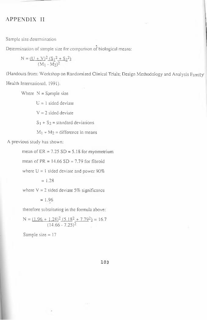
2.4 SAMPLE SIZE DETERMINATION
The sample si?e of twenty (Appendix II) was attained by
recruiting all women who fulfilled the inclusion criteria as above.
This took from August to December 1995.
2.5 MATERIALS
2.5.1 Tissue Samples
Specimens of uterine leiomyomata and the adjacent normal
myometria were collected from twenty patients undergoing
hysterectomy in the gynaecology theater. While in the theater, as
soon as the uterus was removed the fibroid tissues was separated rom the adjacent normal myometrium by trimming with a scalpel blade and forceps.
Paired specimens (fibroid and myometrium) weighing aPPtoximately 20-30 g were collected and immediately transferred, r°zen into ice block containers, and transported to the laboratory
47
where multiple tissue samples were removed for histological
examination and the rest frozen at -20°c and later processed for
the following:
(a) Reproductive hormones i.e estradiol and progesterone and
(b) Receptor contents i.e estrogen and progesterone receptors.
2.5.2 Solvents and Reagents
The list and source of chemicals used in these studies is given
below:
Dithiotheitol, Triton x, Bovine serum albumin (BSA), Dextran
activated charcoal, Diestrine and Formasaline were obtained from
Sigma. Antibody (Antiserum to estradiol-17B), (2,4,6,7-3H)estradiol
and 17a-Hydroxyl(1,2,6,7-3H)Progesterone were from the WHO RIA
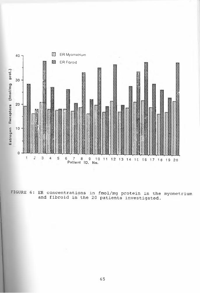
FIGURE 6: ER concentrations in fmol/mg protein in the and fibroid in the 20 patients investigated.
19 20
myometrium
65
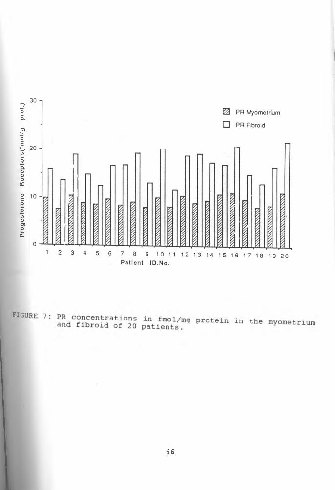
Pro
ge
ste
ron
e
Re
ce
pto
rs(f
mo
l/g
p
rot.
)
66
i
Me
an
±
S.E
.M
(fm
ol/
mg
p
rot.
)U N IV E R S IT Y O F N A IR O B I L IB R A R Y
FIGURE 8
ERF30 -i
20 -
10 -
0-20 1 -20 1 -20 1 - 20
Mean ±SEM . of ER and PR in the myometria and fibroids in fmol/gm protein. (n=20).
ERM - Estrogen receptor concentration in the myometria.
ERF = Estrogen receptor concentration in the fibroids.PRM = Progesterone
myometria.receptor concentration in the
PRF = Progesterone fibroids.
receptor concentration in the
67
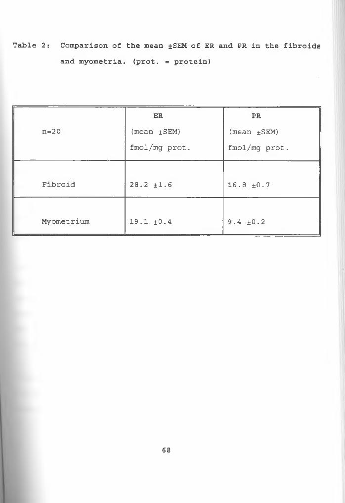
Table 2 : Comparison of the mean ±SEM of ER and PR in the fibroidsand myometria. (prot. = protein)
ER PRn=2 0 (mean ±SEM) (mean ±SEM)
fmol/mg prot. fmol/mg prot.
Fibroid 28.2 ±1.6 16.8 ±0.7
Myometrium 19.1 ±0.4 9.4 ±0.2
68
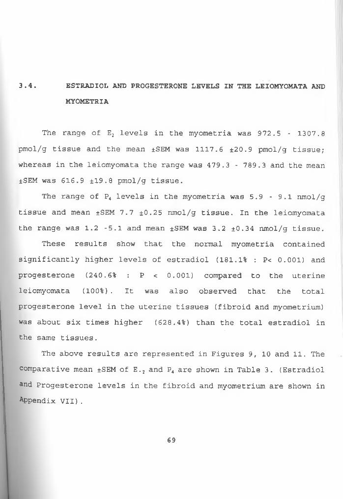
3.4. ESTRADIOL AND PROGESTERONE LEVELS IN THE LEIOMYOMATA ANDMYOMETRIA
The range of E2 levels in the myometria was 972.5 - 1307.8
pmol/g tissue and the mean ±SEM was 1117.6 ±20.9 pmol/g tissue;
whereas in the leiomyomata the range was 479.3 - 789.3 and the mean
±SEM was 616.9 ±19.8 pmol/g tissue.
The range of P4 levels in the myometria was 5.9 - 9.1 nmol/g
tissue and mean ±SEM 7.7 ±0.25 nmol/g tissue. In the leiomyomata
the range was 1.2 -5.1 and mean ±SEM was 3.2 ±0.34 nmol/g tissue.
These results show that the normal myometria contained
significantly higher levels of estradiol (181.1% : P< 0.001) and
progesterone (240.6% : P < 0.001) compared to the uterine
leiomyomata (100%) . It was also observed that the total
progesterone level in the uterine tissues (fibroid and myometrium)
was about six times higher (628.4%) than the total estradiol in
the same tissues.
The above results are represented in Figures 9, 10 and 11. The
comparative mean ±SEM of E.2 and P4 are shown in Table 3. (Estradiol
and Progesterone levels in the fibroid and myometrium are shown in
Appendix VII).
69
Es
tra
dio
l (p
mo
les
/g
tis
su
e)
0 E2 Myometrium
IH E2 Fibroid
Patien t ID No.
FIGURE 9: Estradiol levels fibroids. in pmol/mg tissue in the myometria and
70
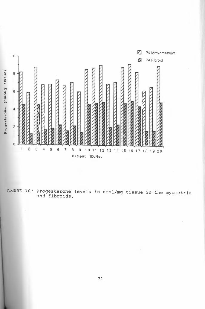
Prog
este
rone
(n
mol/g
tis
sue)
10 -i0 P4 Mmyometrium
£3 P4 Fibroid
11 12 13 14
P a t ien t ID .No.
15 16 17 18 1920
FIGURE 10: Progesterone levels in nmol/mg tissue in the myometria and fibroids.
71
Me
an
±
S.E
.M
(pm
ol/
gm
ti
ss
ue
)
P4 M
FIGURE 11: Mean +SEM of E2 and P4 in the myometria and fibroidsin pmol/mg tissue. (n=20).
E2M = Estradiol concentration in the myometria.E2F = Estradiol concentration in the fibroids.P4M = Progesterone concentration in the myometria.P«F = Progesterone concentration in the fibroids.
72
Table 3s Comparison of the Mean ±S.E.M. of E2 and P4 in the fibroids compared to the myometria.
E, (estradiol) P4 (progesterone)
d II to o mean ±SEM mean ±SEM
pmol/g tissue nmol/g tissue
Fibroid 616.9 ±19.8 3.2 ±0.34
Myometrium 1117.6 ±20.9 7.7 ±0.25
73
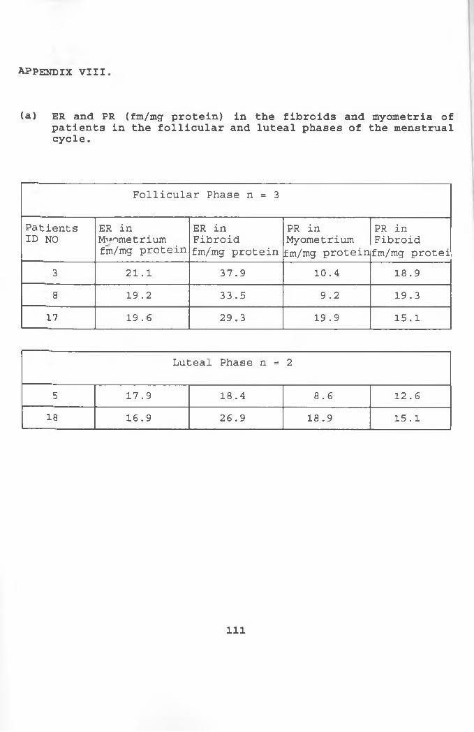
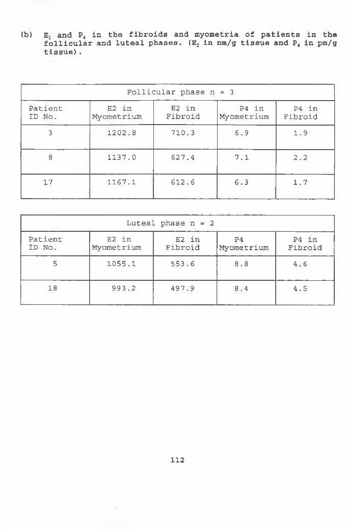
3.5 RECEPTOR AND HORMONAL PROFILE OF PATIENTS IN THEFOLLICULAR AND LUTEAL PHASES OF THE MENSTRUAL CYCLE
From the results obtained for patients investigated in the
follicular and luteal phases of the menstrual cycle the ER and PR
were as usual higher in the fibroids compared to the normal
myometria. Also the E2 and P4 were lower in the fibroid compaed to
the myometria. However, the total ER and PR in the uterine tissue
(fibroid and myometrium) were higher in the follicular phase
compared to the luteal. The total E2 was also higher in the
follicular phase. On the contrary, the total P4 was lower (Table 4) .
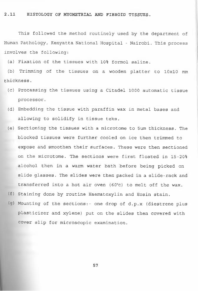
3.6 RESULTS OF HISTOLOGY OF MYOMETRIAL AND FIBROID TISSUES.
The histology of the uterine tissues (myometrium and fibroid)
was done in order to exclude patients with fibroids co-existing
with cancers and degenerative changes. The myometrial specimens of
the 20 patients showed bundles of smooth muscle in normal
arrangement. Likewise, the fibroid specimens showed whorling bands
of spindle shaped smooth muscle cells in normal fashion. The
histology of these specimens are shown in Plates I (normal
myometrium) , II (circumscribed fibroid nodole) , III (whorling bands
of spindle shaped smooth muscle cells in fibroid), IV (fascicles of
smooth muscle cells in fibroids.
77
PLATE I: Normal myometrium showing bundles cf smooth musclenormal arrangement.
PLATE Hi A well circumscribed nodule of closely packed bundles cf smooth muscle in a fibroid. Notice how the noraa! myometrium has been pushed to the side (lower left).
4.1 ESTROGEN AND PROGESTERONE RECEPTOR CONCENTRATIONS IN THELEIOMYOMATA AND NORMAL MYOMETRIA.
In the quantitative estimation of receptor concentrations in
steroid dependent tissues, physiological and analytical factors
have been found to affect such measurements. Such factors include
the race (Sadan et al. 1988), age (Roth, 1979) and menstrual status
(Soules and McCarty, 1982) of the patient as well as exposure to
hormonal agents. Hence in this study the receptor concentrations in
the uterine leiomyomata were described in comparison to
corresponding paired concentrations in the non-myomatous myometria
from the same patient.
However, a careful patient selection was still done to ensure
better estimates of the receptor levels. The patients used were
pre-menopausal black females and age group 31-42 years.
Difficulties were experienced in obtaining large sample sizes for
patients in the proliferative and secretory phases of the menstrual
cycles because most of the patients booked for hysterectomy had
menstrual irregularity. Of the 20 patients studied 75 percent had
Menstrual irregularity, 15 percent were in the proliferative phase
82
while 10 percent in the secretory phase. All the patients had not
been exposed to hormonal agents prior to surgery.
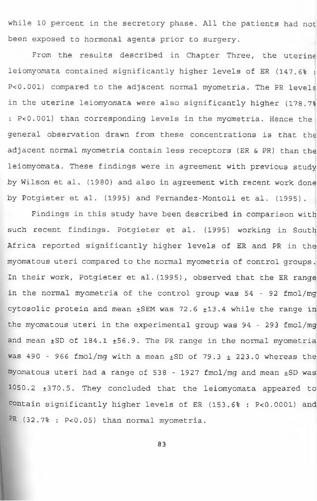
From the results described in Chapter Three, the uterine
leiomyomata contained significantly higher levels of ER (147.6% :
PcO.OOl) compared to the adjacent normal myometria. The PR levels
in the uterine leiomyomata were also significantly higher (178.7%
: PcO.OOl) than corresponding levels in the myometria. Hence the
general observation drawn from these concentrations is that the
adjacent normal myometria contain less receptors (ER & PR) than the
leiomyomata. These findings were in agreement with previous study
by Wilson et al. (1980) and also in agreement with recent work done
by Potgieter et al. (1995) and Fernandez-Montoli et al. (1995).
Findings in this study have been described in comparison with
such recent findings. Potgieter et al. (1995) working in South
Africa reported significantly higher levels of ER and PR in the
myomatous uteri compared to the normal myometria of control groups.
In their work, Potgieter et al.(1995), observed that the ER range
in the normal myometria of the control group was 54 - 92 fmol/mg
cytosolic protein and mean ±SEM was 72.6 ±13.4 while the range in
the myomatous uteri in the experimental group was 94 - 293 fmol/mg
and mean ±SD of 184.1 ±56.9. The PR range in the normal myometria
was 490 - 966 fmol/mg with a mean ±SD of 79.3 ± 223.0 whereas the
myomatous uteri had a range of 538 - 1927 fmol/mg and mean ±SD was
1050.2 ±370.5. They concluded that the leiomyomata appeared to
contain significantly higher levels of ER (153.6% : PcO.0001) and
PR (32.7% : P<0.05) than normal myometria.
83
Fernandez-Montoli et al. (1995) in a study in Spain showed a
mean ±SD value 7.25 ±5.18 fmol/mg protein of ER in the myometria as
compared to 14.66 ±7.79 fmol/mg protein in the leiomyomata. The
mean ±SD value of PR was 68.38 ±62.55 fmol/mg protein in the
myometria and 216.90 ± 206.67 fmol/mg protein in the leiomyomata.
They also showed that ER and PR levels were higher in the myomatous
uteri compared to the normal myometrium. Hence the general and
popular opinion is that ER and PR levels are higher in the
leiomyomata compared to normal myometrium.
In the present study though the ER and PR concentrations
(compared within the same patients) were higher in the leiomyomata
than in the myometrium, the ER levels in the myometrium and
leiomyomata were higher than corresponding levels of PR in the same
tissues. But the PR levels in the leiomyomata were raised to the
same extend as the corresponding ER levels. This was contrary to
results obtained by Potgieter et al. (1995), who reported higher
levels of PR in the myometia and leiomyomata than corresponding ER
in similar tissues and their PR levels in the leiomyomata were not
raised to the same extend as corresponding ER levels. Also the
results from Fernandez-Montoli et al. (1995) showed that though PR
levels were higher than ER levels in corresponding myometria and
leiomyomata, some individuals had much lower PRs than ERs. However,
the differences in the levels of ER and PR in these three studies
c°uld arise from the difference in sample size, methodology
(fibroid and myometrium from different or same patients), and from
t-he fact that the phase of menstual cycles were not controlled.
84
The higher levels of ER and PR in the fibroids could explain
the occurrence of these tumours during the childbearing life and
their increase in size during pregnancy, since these are
physiological states in which circulating levels of sex steroid
hormones are relatively high. This also explains the decrease in
size of these tumours with GnRH-a therapy, whereby circulatory
levels of estradiol are reduced to postmenopausal levels.
The high levels of receptors in the fibroids determine the
capacity of the tumours to retain the hormones and could indicate
a degree to which the same i.e fibroids are sensitive to the
hormone action, and hence can synthesize specific proteins with
enzyme activity in the target cells. This is in agreement with the
speculation made by Potgierter et al. (1995) that the regulation of
the physiological action of both estrogen and progesterone is
dependent on the relative concentrations of both ER and PR
receptors.
4.2 ESTRADIOL AND PROGESTERONE LEVELS IN THE LEIOMYOMATA ANDNORMAL MYOMETRIA
The results described in Chapter Three show that the adjacent
normal myometria contained significantly higher levels of E2 (18.1%
• P<0.001) compared to the leiomyomata. The P4 levels were also
significantly higher (240.6% : P<0.001) in the adjacent normal
Myometria compared to the leiomyomata. Figures 9, 10 and 11 show
85
the comparative levels and means ±SEM of E2 and P4 in the myometr
and leiomyomata. The observations drawn from these levels is t
the adjacent normal myometria contain more of the free hormones
& P4) than the leiomyomata. This is in agreement with findings
Farber, (1972) who had found out that the specific high affinity
steroid hormone in a tissue determines its capacity to retain i
hormone and indicate a degree to which the target tissue
sensitive to the hormone action. Hence the fibroid tissues b:
more of the steroid hormone than does the adjacent nom
myometrium. Jorge et al. (1990) also reached a similar conclusd
when they showed that estrogen concentrations were higher in t
leiomyomata than the myometrium.
Observations have shown that although the levels of estrog
and progesterone are different in the leiomyomata and myometriu
the circulating levels of these hormones are normal in patien
with leiomyomata compared to control groups (Potgieter et al. 199
Most scientific report have tended to support the involvement
steroid hormones in the uterine fibroids, but there is litt
scientific evidence to the fact that these tumours are steroit
hormone dependent. Work done to demonstrate this dependency h;
been criticized for reasons that fibroids in the human were foui
to be different from those in other animals, like the pig and tl
cow (Vollenhoven, 1990). With the lack of animal models, treatmei
modalities are being used to demonstrate the involvement of steroi
hormones in fibroids. Treatment of leiomyomata with GnRH-agonist
e-g leoprolide acetate reduces serum E2 concentrations to pos
86
menopausal levels, and results in the shrinkage of the leiomyomata.
Graigner et al. (1993) had concluded that the suppression of
leiomyomata growth by GnRH-agonist was mediated by a decrease in
estrogen receptors. Treatment with anti-progesterone RU486 (Murphy
et al. 1993) had shown a decrease in fibroid size and the tissue
examined after hysterectomy showed a significantly decreased PR
content.
The above workers i.e Graigner et al. 1993 and Murphy et al.
1993, are in agreement with similar conclusions reached by Friedman
et al.(1989) when they showed that a decrease in uterine size is
dependent on the hypoestrogenic state which follow down regulation
of the pituitary GnRH receptors and desensitization of gonatropes.
Therefore the involvement of steroid hormones (E2 & P4) and steroid
hormone receptors (ER & PR) in the etiopathogenesis has been
scientifically established.
4.3 RECEPTOR AND HORMONAL PROFILE OF PATIENTS INVESTIGATED DURING THE FOLLICULAR AND LUTEAL PHASES OF THE MENSTRUAL CYCLE.
From the results represented in Table 4 in Chapter Three,
higher levels of ER and PR were observed in the fibroids compared
to the normal myometria in patients investigated during the
follicular and luteal phases of the menstrual cycle. However, the
total ER and total PR (i.e in the fibroid and myometrium) were
higher in the follicular phase compared to the luteal. The total E2
ln the follicular phase was higher than the same in the luteal
87
phase. On the contrary the total P4 was higher in the luteal
compared to the follicular phase.
It has been stated that a definite biochemical and
physiological link exists between ER and PR; whereby PR levels
appear to be stimulated by estrogens, while the ER and PR levels
are down regulated by progesterone (Potgierter et al. 1995). This
could explain the low levels of total ER and total PR during the
luteal phase wherein the P4 levels were high, but fails to explain
the high levels of both ER and PR during the follicular phase
wherein the E2 levels were high. A probable postulate that can be
made from the observation of hormone and receptor concentrations
among patients investigated in the two phases of the menstrual
cycle is that the regulation of the physiological action of both
estrogen and progesterone is dependent on the relative
concentrations of both ER and PR receptors. Such a postulation
would be limited by the fact that the sample sizes were small and
the menstrual status of these patients were determined only by
recording their menstrual histories.
5. CONCLUSIONS
Findings in this study show that the concentration of the sex
steroid receptors (ER and PR) were significantly higher in the
leiomyomata compared to the adjacent normal myometria of black
Kenyan women. On the contrary the levels of the sex steroid
88
hormones (P4 and E2) were lower in the leiomyomata compared to the
myometria in the same population. The total ER in the uterine
tissues (fibroid and myometria) were higher than the total PR in
same tissues, and the total P4 levels were higher than total E2. For
patients investigated during the different phases of the menstrual
cycle the total ER and total PR were higher during the follicular
phase compared to the luteal phase. The total E2 was also higher
during the follicular phase as opposed to the total P4 which was
lower in the same but higher in the luteal. However, observations
during the different phases of the menstrual cycle were limited by
the sample size.
From the above conclusions it is postulated that the relative
proportions of E2, P4, ER and PR in the individual patients uterine
tissue may be important in the pathogenesis of fibroids in the
black population in Kenya. It is further suggested that the
treatment and management of the problem should involve
manipulations of sex steroids and their receptors. Also research
should continue in order to identify a nonsurgical means of
treating this disease. This would be an important public health
initiative especially in the black (negroid) population where
fibroids occur at a higher incidence, surgical complications
commoner, and childbearing the center of matrimonial harmony. In
the clinical management of this disease the importance of early
diagnosis and detection of high risk groups should be emphasized
since current treatment modality i.e chemotherapy only reduces
uterine size.
89
It is hoped that, besides the above recommendations, this
study would help in resolving some of the controversies surrounding
the levels of sex steroid hormones and their respective receptors
in the leiomyomata and the normal myometria and provide information
regarding this disease in the black population. This should also
enable clinicians working in this population to appreciate more the
use of chemotherapy in the management of uterine fibroids.
90
REFERENCES
Allan, G.F., X.Leng, S.X.Ysai, N.L.Weigel, D.P.Edwards, M.J.Tsai and B.W.O'Malley, (1992). Hormone and antihormone induced distinct conformational changes which are central to steroid receptor
activation. Journal of Biological Chemistry, 267: 19513-19520.
Auber, G. (1990). Acta European Fertility, 21: 185-189.
Babaknia A., Rock J.A., Jones,H.W. (1978). Pregancy success
following endometrial myomectomy for infertility. Fertility and
sterility, 30: 644-653.
Bauer, M.A.D. and Gorell, T.A. (1980). Analysis of progesterone
receptor binding in the ovine uterus. Steroids, 36: 581-591.
Blaustein, A. (1977). Benign lesions of the myometrium: In
Pathology of the Female Genital Tract; 1st edition, Springer- Verlag, New York, pg 299-321.
Brewer, J.I., Decosta, E.J. (1967). Uterine myomas. Textbook of
Gynaecology, 4th edition Williams and Wilkins publication Waverly
Press, Inc. pg 650-651.
91
Buchi, K.A., Keller, P.J. (1983). Cytoplasmic progestin receptors in myoma1 and myometrial tissues. Acta. Obstetrics and Gynaecology.
symptomatology and management. Fertility and Sterility, 36: 433-445
Collins, J.R., Levin,J., and Savage,N. (1980). Racial differences
in estrogen receptor and peroxidase status of human breast cancer
tissue. South African Medical Journal, 57: 444-446.
Dawood, M.Y., Khan-Dawood, F.S. (1994). Plasma insulin-like growth factor-I, CA-125, estrogen, progesterone in women with leiomyomas.
Fertility and Sterility, 61: 617-621.
Evans, L.H. and R. Hahnel. (1971). Oestrogen receptors in human
uterine tissue. J. Endocrinol. 50: 209-229.
Evans, R.M. (1988). The steroid and thyroid hormone receptor
superfamily. Science, 240: 889-895.
Farber, M. (1972) . Estradiol binding by fibroid tumours and normal myometrium. Department of Obstetrics and Gynaecology, University of
Washington School of Medicine, Seattle, Washington 98195- Obstetric
Gynaecology (NY), 40/41:479-486.
92
Ferandez-Montoli, M.E., Diez-Gibert, 0., Samanniego, J.M., Balaguero, L. and Navaro, M.A. (1995). Total and unbound cytosolic estrogen and progesterone receptors in myometrium and fibroid after
gonadotropin-releasing hormone agonist treatment. Fertility and Sterility 63: 522-527.
Friedman, A., Beribieri, R.L., Bencerraf, B.C., Schiff, 1.(1987).Treatment of leiomyomata with intranasal or subcutaneous
Lenprolide, a GnRH-a. Fertility and sterility 48:560-566.
Friedman, A.J., Beribieri, R.L., Doubilet, P.M., Fine, C., Schiff, I. (1988). Arandomised double of a gonadotropin releasing-hormone agonist (leuprolide) with or without medroxyprogesterone acetate in
the treatment of leiomyomata uteri. Fertility and Sterility 49:
404-409.
Friedman, A.J., Rein,M.S., Harrison-Atlas, Garfield, J.M.Donbit, P.M. (1989) . A randomized, placebo-controlled double blind study
evaluating Leuprolide acetate depot treatment before myomectomy.
Fertility and Sterility 51: 251-305.
GOLAN, A., Bukovsky, I., Schneider, D., Ron-El, R., Herman, A., Capsi, E. (1989). D-Trp-6-luteinizing hormone-releasing hormone
microcapsules in the treatment of uterine leiomyomas. Fertility and Sterility 52: 406-411
93
Grainger, D.A., Carol, R.S., Harbison, L.A., Imakawak, Cho., Webster, B.W. (1993). Estrogen and progesterone receptor mRNA in
leiomyomas versus adjacent myometrium. American Fertility Society 1993 Abstracts S213.
Green, S. (1990). Steroid receptors and new anti-steroidal agents. Modulation of oestrogen receptor activity by estrogens and anti-
estrogens. Journal Steroids, Biochemistry and Molecular Biology.
37: 747-745.
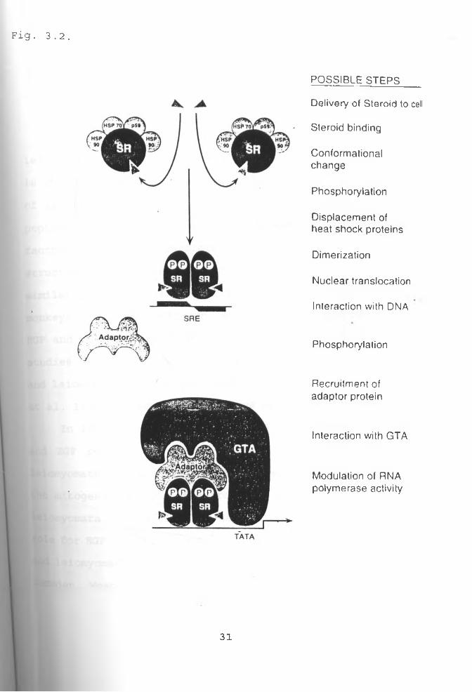
Henderson,D., D.Philibert, A.K. Roy & G. Tentsh (1995). Steriod
receptors and antihormones. Annals of the New York Academy of
Science. 781: 123-125.
Harrison-Woolrych, B.M., Robinson, R. (1995). Fibroid growth in
response to high-dose progestogen. Fertility and Sterility. 64: 191-192 .
Healy, D.L., Shekleton ,P., Downing, G.B., Philips, S., O'Grady, C., Barrgah, S.H. (1989). LH-RH and its analogues, their use in
Hunt, J.E., Walleach, E.E., (1974). Uterine factors in
infertility-an overview. Clinical Obstetrics and Gynaecology. 17: 974-981.
94
Hofmann, G.E., Rao, C.V., Barrows, G.H., Schultz, G.S., Sanfilippo, J. (1984). Binding sites for epidermal growth factor
in human uterine tissues and leiomyomas. Journal of Clinical
Endocrinology and Metabolism 58: 880-883.
Huet-Hudson, Y.M., Chakraborty, C., De, S.K., Suzuki, Y., Andrews, G.K., Dey, S.K. (1990). Estrogen regulates the synthesis of
epidermal growth factor in mouse uterine epidermal cells. Molecular
Endocrinology 4: 507-513.
<Ingersol, P.M., (1963). Fertility following myomectomy. Fertility
and Sterility 14: 596-598.
Jenson, E.V. (1995). Steroid hormone antagonists (summary and
future challenges) Annals of the New York Academy of Sciences. 761:
1-3 .
Jones, H.W., and Jones G.S. (1981). Uterine fibroids. In: Jones HW, Jones GS,editors. Novak's textbook of gynaecology. 10th edition.
Baltimore: Williams & Wilkins Company, pg 230-244.
Jorge, R.P., Edgrd, C.,Jacques, G., Christine, V., Bernard, S., Albert, N. (1990). Effect of Decapeptyl, an agonist analog of GnRH on estrogens, estrogen sulfates, and progesterone receptors in
leiomyoma and myometrium. Fertility and Sterility 53: 1012-1015.
95
Langdon Parsons and Sheldon C-Sommers (19 63) . Fibromymomata of the uterus. In Gynaecology 2nd edition. Lea and Febriger Publication pg
and gynaecology in the tropics and developing countries. 1st
edition E.L.B.S.and Edward Arnold Ltd publications, pg 385-386.
Lipschutz, A. (1942). Experimental Fibriods and antifibromatogenic of steroid hormones. Journal of American Medical Record
Association. 120: 171-174.
Lowry, O.H., Rosebrough, N.J., Farr, A.L. and Randall, R.J. (1951).Protein measurement with folin phenol reagent. Journal Biology and
Chemistry. 198: 265-272.
Lumsden, M.A., West, C.P., Bramley, T., Rumgay, L., Baird, D.T. (1988). The binding of epidermal growth factor to the human uterus and leiomyomata in women rendered hypo-estrogenic by continuous
administration of an LHRH agonist. British Journal of Obstetrics
and Gynaecology 95: 1299-1301.
MacDonnel, D.P., D.J. Mangelsdorf, J.W. Pike, M.R. Hanssler, and B.w. O'Malley (1987). Molecular cloning of complementary DNA
encoding the aviation receptor for vitamin D. Science 235:1214-
96
j
1217.
Mettler L., Steinmuller H., Schachner-Sunschmann E. (1991). Human Repoduction; 6 : 694-698.
Murphy, A.A., Kettel, L.M., Morales, A.J.,. Roberts, V.J., Yen, S.S.C. (1993) . Regeneration of uterine leiomyomata in response to
the antiprogestrone RU 486. Journal of Clinical Endocrinology and
Metabolism. 76: 513-517.
Nelson, O.W., (1937). Endometrial and myometrial changes
fibromyomatous nodules induced in guinea pig by estrogen. Anatomy
Records. 68: 99-105.
Norman, A., R.G. Schrot, P. Balch, D.E. Borron, R. Erk, (1970).Interaction of experiments and ideas: Statistical evaluation of
Pollow, K., Sinneck, G., Boqueri, E., Pollow, B. (1978). In vitro
conversions of estradiol-17-beta into estrone in normal human
myometrium and leiomyoma. Journal of Clinical Chemistry and
Clinical Biochemistry. 16: 503-511.
Potgieter, H.C., Magagane, F., Bester, M.J. (1995). Oestrogen and progesterone receptor status and PgR/ER ratios in normal and
97
myomatous human myometrium. East African Medical Journal. 72:
510-514.
Pukka, M.J., Kontula, K.K., Kanpila A.J.I., Janne, D.A. Vihko, R.K. (1976). Oestrogen receptor in human myoma tissue. Molecular cell
endocrinology 6: 35-64.
Ran, N., Bieber, E., Wood,M., Peping, P. (1988). Treatment of
leiomyomata with GnRh-a prior to myomectomy. Fertility and
Sterility. 6: 306-309.
Rein, M.S., Freidman, A.J., Start, J.m., McLanghlin, D.T. (1990).Fibriods and myometrial steroids receptors in women treated with
GnRH-a L .A. Fertility and Sterility 90: 1018-1023.
Roth, G.S. (1979). Hormone receptor changes during adulthood and
senescence: significance for aging research. Fed. Proc. 1979; 35: 1910-1914.
Russell D. (1977). Neoplasms. Textbook of Gynaecology. Lea and
Febiger. Henry Kimptom publishers pg 253-259.
Sadan, 0., Van Iddekinge, B. Savage, N. Robinson, M and Zakut H. (1980). Ethnic variation in oestrogen and progesterone receptor and peroxidase status of human breast cancer tissue. S. afr. Med. J.
98
5 7: 444 - 446.
Sadan, O., Van Iddekinge, B., Van Gelderen (1987). Oestrogen and
progesterone receptor concentrations in leiomyomata and normal
myometrium. Annals of Clinical Biochemistry 24: 263-267.
Sadan, 0., Van Iddekinge B., Savage, N., Van der Walt, L.A. and Zakut, H. (1990) . Endocrine profiles associated with oestrogen and progesterone in leiomyoma and normal myometrium. Gynec.
Endoccrinol. 4: 33-42.
Scialli, A. R. and Jestila, K. J. (1995). Sustained benefits of
leuprolide with or without subseqent medroxyprogesterone acetate in
the nonsurgical management of leiomyomata uteri. Fertility and
Sterility 64: 313-320.
Segaloff, A., Weed, J.C., Sternberg, W.H., Parson, W. (1948). Theprogesterone therapy of human uterine leiomyomas. Journal of
Clinical Endocrinology and Metabolism 9:12-15.
Soules, M.R. and McCarty, K.S. (1982). Leiomyomas: steroid receptor content. Variation within normal menstrual cycles. American Journal
of Obstetrics and Gynaecology 143: 6-7.
Smith, D.F., & D.O. Toft. (1993). Steroid and their associated
99
proteins. Molecular Endocrinology 7:4-11.
Stewart, T. (1969). Benign conditions of the uterus. Essentials of Gynaecology 4th edition. Lea and Febiger Publication pg 171 - 180.
Steroids from Steraloids Inc. (1985) . 9th Edition by Steraloids
Inc. Wilton N.H., U.S.A. pg 89-150
Sufi, S.B., Donaldson and Jeffcoate S.L. (1993). WHO Matched
Reagent Program Method Manual, pg 52-73.
Tamaya,T., Motayama, T., Ohomo, Y, Ide, N., Tsurusaki, T., Okada, H. (1979). Estradiol 17-beta progesterone and 5-alpha-
dihydrtestosterone receptors of the uterine myometrium and myoma in
human subject. Journal of Steroid and Biochemistry 107: 615-622.
Tindall, V.R. (1987). Tumours of the corpus uteri. Jeffcoate's
Principles of gynaecology, 5th edition; Butterworths publications pg 418-432.
Townsend, D.E., Sparkes, R.S., Baluda, M.C., McLelland (1970).Unicellular histogenesis of uterine leiomyomata as determined by
electrophoresis of glucose-6-phosphate dehydrogenase. American
Journal of Obstetrics and Gynaecology 107: 1168-1171.
100
Van Leusden H.A.I.M. (1986). Rapid reduction of uterine myomas
after short-term treatment with micro encapsulated D-Trp6-LHRH.
Lancet 2: 1212-1217.
Venturini P.L., Fasce V., Constantinis, (1990). In Shaw RW (ED).
Advances in reproductive endocrinology; vol. 1, Endometriosis.
APPENDIX III.Levels of statistical significance of results of ER and PR in the fibroids compared to the myometria
This was done using the student-t-test formula described in statistical procedures in Chapter 2:
x, - X2
t = .... -----
a/sj2 + s22
Applying the above formula for ER in myometrium and fibroid as in table 5 where
Xj = 2 8 . 2
X2 = 19.1
n = 2 0 (n, = n2)
S.E.Mj = 1.6
S.E.M2 = 0.4
But SD = S . E .M x Vn
104
= > SD2 « (S.E.M x Vn)2
= > SD2 = S.E.M2 x n
Where SD = standard deviation
n = Sample size
= > Sj2 = 51.2
Sc s 2 = 3.2
Substituting these values in the formula above t = 5.515.
From the tables of "Distribution of t Probability" abridged from
Table III of Fisher and Yates by biological science curriculum
study (Norman, Schrot, Balch et al. 1970), a P-value of 0.05 or
less is taken as showing significant difference. Hence for the t of
5.515 calculated above the P-value is less than 0.001, expressed
mathematically as P< 0.001. Therefore, a significant difference
exist between the ER levels in the myometrium and fibroid.
Applying the same formula for PR as per table 5 :
=> t = 10.137
From the table of "Distribution of t Probability the P-value is
105
less than 1.001 i.e . p<
difference between the two0*001. Hence
PR levels.there is significant
106
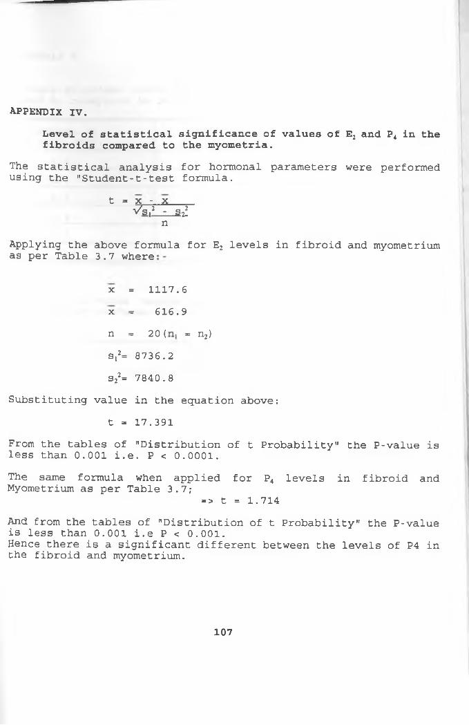
a p p e n d i x IV.Level of statistical significance of values of E2 and P4 in the fibroids compared to the myometria.
The statistical analysis for hormonal parameters were performed using the "Student-t-test formula.
t = x - xv'sjlJL-S
n
Applying the above formula for E2 levels in fibroid and myometrium as per Table 3.7 where
x = 1117.6
x = 616.9
n = 2 0 ( = n2)
S j2= 8736.2
s 22= 7840.8
Substituting value in the equation above:
t = 17.391
From the tables of "Distribution of t Probability" the P-value is less than 0.001 i.e. P < 0.0001.
The same formula when applied for P4 levels in fibroid and Myometrium as per Table 3.7;
=> t = 1.714
And from the tables of "Distribution of t Probability" the P-value is less than 0.001 i.e P < 0.001.Hence there is a significant different between the levels of P4 in the fibroid and myometrium.
107
APPENDIX V:
Cytosol dilutions (with TEDG buffer) versus percentage of tracer bound to receptors in the myometria and fibroids.