Hepatitis C Virus Structural Proteins Assemble intoViruslike Particles in Insect Cells
THOMAS F. BAUMERT,1 SUSUMU ITO,2 DAVID T. WONG,3 AND T. JAKE LIANG1*
Liver Diseases Section, National Institute of Diabetes and Digestive and Kidney Diseases, National Institutes of Health,Bethesda, Maryland 208921; Department of Neurobiology and Department of Cell Biology, Harvard Medical School,
Boston, Massachusetts 021152; and Division of Gastroenterology, Department of Medicine,Stanford University School of Medicine, Palo Alto, California 943043
Received 30 December 1997/Accepted 20 January 1998
Hepatitis C virus (HCV) is a leading cause of chronic hepatitis in the world. The study of HCV has beenhampered by the low level of viral particles in infected individuals, the inability to propagate efficiently thevirus in cultured cells, and the lack of a convenient animal model. Due to these obstacles, neither the structureof the virus nor the prerequisites for its assembly have been clearly defined. In this report, we describe a modelfor the production and purification of HCV-like particles in insect cells using a recombinant baculoviruscontaining the cDNA of the HCV structural proteins. In insect cells, expressed HCV structural proteinsassembled into enveloped viruslike particles (40 to 60 nm in diameter) in large cytoplasmic cisternae,presumably derived from the endoplasmic reticulum. Biophysical characterization of viruslike particles byCsCl and sucrose gradient centrifugation revealed biophysical properties similar to those of putative virionsisolated from infected humans. The results suggested that HCV core and envelope proteins without p7 weresufficient for viral particle formation. Analysis of particle-associated nucleic acids demonstrated that HCVRNAs were selectively incorporated into the particles over non-HCV transcripts. The synthesis of HCV-likeparticles in insect cells may provide an important tool to determine the structural requirements for HCVparticle assembly as well as to study viral genome encapsidation and virus-host interactions. The describedsystem may also represent a potential approach toward vaccine development.
Hepatitis C virus (HCV) is a major causative agent of post-transfusion and community-acquired hepatitis in the world (2,23, 26). The majority of HCV-infected individuals developchronic hepatitis progressing eventually to liver cirrhosis andhepatocellular carcinoma (48). Neither an effective treatmentfor chronic HCV infection nor a vaccine to prevent HCVinfection is available at the present time (19, 28).
HCV is a member of the Flaviviridae family (44). The virioncontains a positive-stranded RNA genome of 9.5 kb. The ge-nome consists of a highly conserved 59 noncoding region (35)followed by a long open reading frame of 9,030 to 9,099 nu-cleotides (nt) that is translated into a single polyprotein of3,010 to 3,030 amino acids (16, 35). Initiation of translationoccurs by a mechanism of internal ribosomal entry requiringthe 59 untranslated region (UTR) and a short stretch of HCVcoding sequences (43). Processing of the polyprotein occurswith a combination of host and viral proteases. The HCVstructural proteins comprise the nucleocapsid or core protein(C) and the two envelope glycoproteins, E1 and E2 (for areview, see reference 39). The cleavage of structural proteinsfrom the polyprotein is catalyzed by a host signal peptidase (16,30), whereas polyprotein cleavage in the nonstructural regionrequires HCV-encoded proteases (11). An additional cleavageproduct in the coding region of the structural proteins wasrecently identified as p7 (30, 41). Although the characteriza-tion of the viral genome organization has been described indetail (35), analysis of the structural features of HCV has beenhampered by the inability to propagate the virus efficiently incultured cells. The levels of viral particles present in infected
patient plasma or liver tissues are very low, making it difficultto visualize the virus. In analogy to other members of theFlaviviridae, the genome organization of HCV suggests a viralstructure consisting of a nucleocapsid or core protein and aviral genome coated by a lipid envelope containing envelopeglycoproteins E1 and E2. Transmission studies with chimpan-zees, the only reliable animal model for HCV, have providedevidence that HCV is inactivated by chloroform, indicatingthat it contains lipids and therefore is probably enveloped (9).Filtration studies have estimated the virion particle size at adiameter of 30 to 60 nm (15).
The baculovirus-insect cell expression system has been ap-plied successfully for the synthesis of viral capsids for viruses ofvarious families (10, 24, 49) but not for the enveloped RNAviruses of the Flaviviridae family. The baculovirus-insect cellexpression system has two features which make it attractive forHCV protein expression. First, eukaryotic insect cells areknown to carry out a number of co- or posttranslational mod-ifications, including fatty acid acetylation and glycosylation,similar to mammalian cells (33). Second, in contrast to manymammalian cell expression systems, the baculovirus expressionsystem allows high-level synthesis of heterologous proteins(33). We therefore rationalized that the baculovirus systemmay be able to direct the synthesis of HCV-like particles ininsect cells.
(This work was presented in part at the 47th Annual Meet-ing of the American Association for the Study of Liver Dis-eases, 8 to 12 November 1996, Chicago, Ill., and the 4th Inter-national Meeting on Hepatitis C Virus and Related Viruses, 6to 10 March 1997, Kyoto, Japan.)
MATERIALS AND METHODS
Baculovirus constructs and insect cell cultures. For the construction of re-combinant baculoviruses, a recently described baculovirus expression system was
* Corresponding author. Mailing address: Liver Diseases Section,NIDDK, National Institutes of Health, 10 Center Dr., Rm. 9B16,Bethesda, MD 20892-1800. Phone: (301) 496-1721. Fax: (301) 402-0491. E-mail: [email protected].
3827
Dow
nloa
ded
from
http
s://j
ourn
als.
asm
.org
/jour
nal/j
vi o
n 27
Oct
ober
202
1 by
117
.28.
32.1
57.
applied (Bac-to-Bac; Gibco BRL, Gaithersburg, Md.) (32). The cDNA for theHCV structural proteins, cloned from a Japanese patient with chronic hepatitis(HCV-J strain, genotype 1b), was used to generate the recombinant baculovirusBVHCV.S. pFastBacHCV.S was generated by subcloning an EcoRI-Tth111Ifragment (nt 259 to 2819) of pCMV980 (23) into the EcoRI and SpeI sites ofpFastBac. The Tth111I and SpeI sites were blunt ended with the Klenow frag-ment before ligation. An in-frame stop codon is present in the vector sequenceclose to the 39 end of the subcloned cDNA. pFastBacHCV.Sp72 was generatedby PCR with the following primers: 59 GAGACAGACGTGCCTGCTACTTAGCAACACGCG 39 (sense nt 1918 to 1951) and 59 TCGAAAGCTTAGGCCTCAGCCTGGGCTATCAGC 39 (antisense nt 2567 to 2543). A stop codon and aHindIII site were introduced at the 39 end of the p7 protein coding region. TheNotI-HindIII digestion product of the PCR fragment was subcloned intoNotI-HindIII site (multiple cloning site) of pFastBacHCV.S. pFastBacHIVgp160containing the cDNA for the human immunodeficiency virus (HIV) glycoproteinprecursor gp160 was generated by subcloning a HindIII-NotI fragment (nt 1 to2481) of pSyngp160 (kindly provided by Brian Seed, Department of Genetics,Massachusetts General Hospital, Boston) into the StuI and NotI sites of pFastBac. The HindIII site was blunt ended with the Klenow fragment before ligation.The correct sequences of the pFastBacHCV.S, pFastBacHCV.Sp72, and pFastBacHIVgp160 constructs were confirmed by restriction enzyme digestions andDNA sequencing. pFastBacGUS (Gibco BRL) containing the coding sequenceof the enzyme b-glucuronidase (GUS) was used to generate the control baculo-virus BVGUS.
Recombinant baculoviruses were generated as described previously (32), iden-tified by immunofluorescence and immunoblotting of transfected Spodopterafrugiperda Sf9 insect cells with specific antibodies, and amplified by subsequentrounds of Sf9 cell infection until a final titer of 5 3 107 PFU/ml was achieved. Sf9insect cells were maintained in spinner or monolayer cultures at 28°C in Sf-900II serum-free medium (Gibco BRL). For all protein expression experiments, Sf9cells in mid-log growth in monolayer cultures were infected with a multiplicity ofinfection (MOI) of 1 to 10. Infection of insect cells with BVGUS served as anegative control in all experiments.
Anti-HCV antibodies. The monoclonal anti-core, anti-E1, and anti-E2(G/H)mouse antibodies (7) as well as the polyclonal anti-E2(9284) rabbit antibody (29,42) were described previously. Additional monoclonal anti-E1 and anti-E2 an-tibodies were obtained from Johnson Lau (University of Florida, Gainesville).Human serum containing antibodies against HCV was obtained from two pa-tients with chronic hepatitis C and high-titer anti-HCV antibodies. The patientswere serologically negative for hepatitis B virus (HBV), hepatitis A virus, andHIV.
Immunofluorescence of HCV proteins. At 96 h postinfection, BVHCV.S- andBVGUS-infected Sf9 cells were fixed in 7% paraformaldehyde (fixative B, de-scribed below), dehydrated, embedded, and sectioned as described below (elec-tron microscopy). Semithin sections (0.5 to 1 mm) were incubated with anti-HCV(serum from HCV-infected individuals as described above; diluted 1:200 in 1%bovine serum albumin [BSA]–phosphate-buffered saline [PBS]), anti-core (dilut-ed 1:200 in 1% BSA–PBS), anti-E1 (diluted 1:100 in 1% BSA–PBS), or anti-E2(9284) (diluted 1:200 in 1% BSA–PBS) antibody or 1% BSA in PBS followedby fluorescein isothiocyanate-conjugated anti-human (for serum), anti-mouse(for anti-core and anti-E1), or anti-rabbit (for anti-E2) antibody (all from Jack-son Laboratories, West Grove, Pa; diluted 1:500 in 1% BSA–PBS) each for 30min at room temperature. Between steps, plates were rinsed three times withPBS.
Immunoblotting and immunoprecipitation of HCV proteins. For immunoblotanalysis, Sf9 cells infected with BVHCV.S, BVHCV.Sp72, and BVGUS werelysed with a buffer containing 1% sodium deoxycholate, 0.1% sodium dodecylsulfate (SDS), 1% Triton X-100, 10 mM Tris, and 140 mM NaCl (pH 8.0),whereas for immunoprecipitation analysis, the lysis buffer consisted of 0.1%Nonidet P-40 (NP-40), 50 mM Tris, 50 mM NaCl, and 5 mM EDTA (pH 7.5). Alllysis buffers contained 1 mM phenylmethylsulfonyl fluoride (PMSF), 2 mg ofaprotinin per ml, and 2 mg of leupeptin per ml. The cell lysate was cleared of celldebris and nuclei by low-speed centrifugation (15 min at 15,000 3 g and 4°C). Forimmunoblotting, a fraction of the supernatant (containing 50 mg of protein) wassubjected to 12% polyacrylamide gel electrophoresis (PAGE). For immunopre-cipitation, 400 ml of the cleared supernatant of either BVHCV.S- or BVGUS-infected cells was incubated with 1 ml of anti-E2(9284) antibody for 16 h at 4°Cand then with 50 ml of protein A-Sepharose 4B-Cl beads (Pharmacia BiotechInc., San Francisco, Calif.) for 1 h at room temperature with mixing. The beadswere washed repeatedly, and the bound proteins were released and denatured byheating for 5 min at 95°C in SDS sample buffer (13). The immunoprecipitatedproteins were analyzed by electrophoresis on a 12% polyacrylamide gel. After geltransfer to polyvinylidene difluoride membranes (Westran; Schleicher & Schuell,Keene, N.H.), the blots were probed with anti-core (diluted 1:2,000), anti-E1(diluted 1:2,000), or anti-E2(G/H) (diluted 1:1,000) antibody followed by horse-radish peroxidase-conjugated anti-mouse immunoglobulin G antibody (diluted1:4,000; Amersham Corp., Arlington Heights, Ill.), with subsequent chemilumi-nescence detection (ECL; Amersham). For analysis of the expression of theHCV structural proteins in mammalian cells, BSC-1 cells (grown in a monolayerin modified Eagle medium–2% fetal calf serum) were infected at an MOI of 10with wild-type vaccinia or a vaccinia virus (vvHCV.S) containing the same HCVstructural cDNA as the baculovirus BVHCV.S (provided by E. V. Schmidt,
Massachusetts General Hospital, Boston). At 8 h postinfection, expression of theHCV structural proteins was analyzed as described above.
Electron microscopy. Sf9 cells infected with BVHCV.S, BVHCV.Sp72, andBVGUS were washed with PBS and fixed in various solutions. For morphologicalstudies, the fixative buffer consisted of 1.25% paraformaldehyde, 2.5% glutaral-dehyde in 0.03% picric acid, and 0.05 M cacodylate buffer at pH 7.4 (fixative A)(21). For immunodecoration, the cells were fixed in 7% paraformaldehyde–0.25M sucrose in 0.03% picric acid–0.05 M cacodylate buffer at pH 7.4 (fixative B).The cells were scraped from the cell culture dishes with a razor blade, pelletedin an Eppendorf desktop centrifuge for 10 min at 18,000 3 g, and postfixed with1% osmium tetroxide in 0.05 M cacodylate buffer for 15 min. The pellets werewashed in 0.1 M maleate buffer (pH 5.0), treated with 1% uranyl acetate (pH 5.0)for 30 min, washed with maleate buffer, dehydrated in a graded series of ethanolsolutions followed by propylene oxide, embedded in a mixture of Epon 812 andAraldite, and polymerized at 40°C for 3 days. Thin sections were stained withsaturated uranyl acetate diluted to 50% with acetone and then with lead citratefor electron microscopy examination. Prior to immunogold labeling of thin sec-tions, plastic-embedded cells in semithin sections (0.5 to 1 mm) were mounted onglass slides and stained with anti-HCV-positive human serum, anti-E1, or anti-E2(9284) antibody as described above. For immunogold labeling, ultrathin sec-tions collected on nickel grids were etched with saturated NaIO4. After beingwashed with PBS, the grids were incubated with 3% BSA in PBS for 30 min. Thegrids were then incubated for 1 h with either anti-HCV (HCV patient serum;diluted 1:100 in 1% BSA–PBS), anti-E1 (diluted 1:50 in 1% BSA–PBS), oranti-E2 ([polyclonal anti-E2(9284) rabbit; diluted 1:50 in 1% BSA–PBS] anti-body or 1% BSA–PBS only. After five washes with PBS, samples were incubatedwith protein A coupled to 10-nm gold particles (Jan Schlott Laboratories,Utrecht, The Netherlands) in PBS (dilution 1:200) and rinsed five times withPBS. After counterstaining with uranyl acetate and lead citrate, samples wereexamined with a transmission electron microscope (JEOL 1200 EX) operated at80 kV. For electron microscopy of partially purified viruslike particles, sucrosegradient fractions were pooled, diluted 1:10 with PBS, and subjected to ultra-centrifugation (Beckman SW55 rotor; 40,000 rpm, 2 h, 4°C). The pellet was fixedin fixative A or B and subjected to the same processing as that described above.
Purification of HCV-like particles. At 96 h postinfection, insect cells infectedwith BVHCV.S or BVHCV.Sp72 (approximately 5 3 107 cells, grown in sus-pension) were lysed in 50 mM Tris–50 mM NaCl–0.5 mM EDTA (pH 7.5) with1 mM PMSF, 2 mg of aprotinin per ml, and 2 mg of leupeptin per ml. In someexperiments, 0.1% NP-40 was added to the lysis buffer, and sonication of thelysate was performed. The lysate was homogenized and subjected to low-speedcentrifugation (15 min at 4°C and 15,000 3 g), and the supernatant was pelletedover a 30% (wt/vol) sucrose (in 20 mM Tris–150 mM NaCl [pH 7.4]) cushion (6h at 4°C and 150,000 3 g). The pellet, containing the HCV-like particles, wasresuspended in 50 mM Tris–100 mM NaCl (pH 7.4) or PBS, homogenized, andsubjected to a second sucrose or CsCl equilibrium gradient centrifugation. Forsucrose equilibrium gradient centrifugation, the resuspended pellet was layeredonto a 20 to 60% (wt/wt) sucrose (in 50 mM Tris–100 mM NaCl [pH 7.4])gradient and centrifuged for 22 h at 4°C and 150,000 3 g (17). Ten 0.5-mlfractions were collected from the top and analyzed by SDS–12% PAGE. ForCsCl equilibrium gradient centrifugation, 0.5 ml of the resuspended pellet wasmixed with 4.5 ml of PBS containing 0.5 mM PMSF and 1.67 g of CsCl (33%
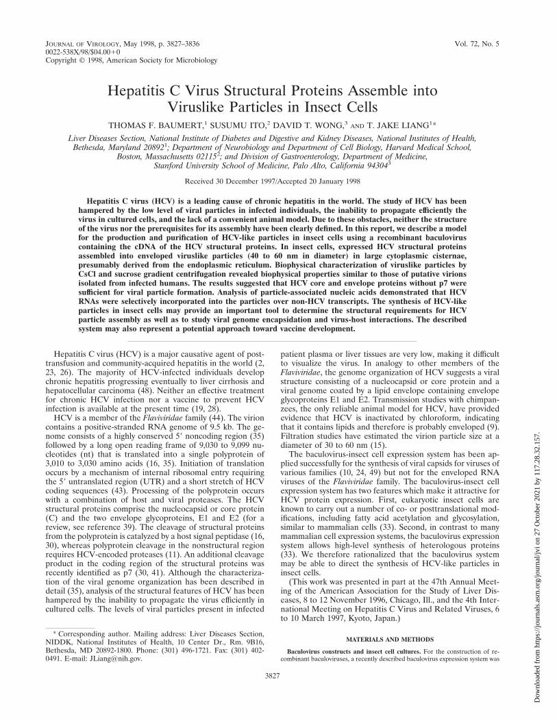
FIG. 1. Map depicting segments of the HCV genome in the recombinantbaculoviruses expressing HCV structural proteins. The BVHCV.S construct con-tains a short stretch of the 59 UTR as well as the coding sequences for the core,E1, E2, and p7 proteins and 21 amino acids of the NS2 protein. The polyproteinopen reading frame begins at nt 330, and the junction of E2-p7 and NS2 is at nt2756. BVHCV.Sp72 contains the same 59 UTR, core, E1, and E2 coding se-quences as BVHCV.S but not p7 and the amino-terminal part of the NS2protein. pPolh, baculovirus polyhedrin promoter; SV40, simian virus 40.
3828 BAUMERT ET AL. J. VIROL.
Dow
nloa
ded
from
http
s://j
ourn
als.
asm
.org
/jour
nal/j
vi o
n 27
Oct
ober
202
1 by
117
.28.
32.1
57.
[wt/wt] in PBS) and centrifuged for 72 h at 4°C and 300,000 3 g (14). Aftercentrifugation, 10 0.5-ml fractions were collected from the top, extensively dia-lyzed against PBS at 4°C, and analyzed by SDS–12% PAGE. After gel transfer,the blots were probed with anti-core, anti-E1, or anti-E2(G/H) antibody andhorseradish peroxidase-labeled anti-mouse antibody as described above. Forsucrose sedimentation velocity centrifugation, insect cell lysates were layeredonto a 10 to 60% (wt/wt) sucrose (in 50 mM Tris–100 mM NaCl [pH 7.4])gradient and centrifuged for 2.5 h at 4°C and 200,000 3 g. Ten fractions werecollected from the top and analyzed for HCV structural proteins as describedabove. Empty HBV surface particles and HBV core particles were subjected tothe same sucrose gradient velocity centrifugation and used as sedimentationmarkers.
HBV surface and core particles were isolated from the cell culture mediumand cytosol of hepatoma cell lines transfected with a replication-competent HBVDNA construct (1, 3, 14). Sedimentation of empty HBV surface particles wasanalyzed by hepatitis B surface antigen detection in sucrose gradient fractionswith a hepatitis B surface antigen-specific radioimmunoassay (Ausria II; AbbottLaboratories, Abbott Park, Ill.). Sedimentation of HBV core particles was ana-lyzed by immunoblotting of sucrose gradient fractions with an anti-HBV core-specific antibody (Dako Corp., Carpinteria, Calif.).
Analysis of HCV-like particle-associated nucleic acids. Insect cells grown inmonolayers (5 3 106 cells) were infected with various combinations of BVGUS,BVHCV.S, and BVHIVgp160. At 96 h postinfection, total RNA was purified bythe guanidium isothiocyanate-acid-phenol method (5), and a total of 20 mg ofRNA was subjected to Northern blot analysis as described below. To analyzeHCV particle-incorporated RNA, HCV-like particles were isolated either byimmunoprecipitation with a polyclonal rabbit anti-E2(9284) antibody or by pel-leting of insect cell lysates over a 30% sucrose cushion and subsequent immu-noprecipitation of the resuspended pellet with a polyclonal rabbit anti-E2(9284)antibody (immunoprecipitation buffer: 50 mM Tris, 100 mM NaCl, 0.5% digito-nin [pH 7.4]) as described above. After digestion of the precipitated particleswith Staphylococcus aureus nuclease (Boehringer Mannheim Biochemicals, In-dianapolis, Ind.) at a concentration of 24 mg/ml and RNase A (Sigma, St. Louis,
Mo.) at a concentration of 50 mg/ml for 2 h at 37°C (in 20 mM Tris [pH 8.8], 5mM CaCl2, 2 mM NaCl) to eliminate any nonincorporated nucleic acids (3, 14),particle-associated RNA was isolated by the guanidium isothiocyanate-acid-phe-nol method (5) and subjected to Northern blot analysis with [a-32P]dCTP-labeled HCV (nt 259 to 2819)-, GUS-, or HIV gp160-specific cDNA probes(equal cDNA counts per minute per nanogram in each probe).
RESULTSExpression and interaction of HCV structural proteins in
insect cells. Recombinant baculovirus BVHCV.S, containingthe coding sequences for the core, E1, E2, and p7 proteins and21 amino acids of the NS2 protein (HCV nt 259 to 2819; Fig.1), directed the production of HCV structural proteins in in-sect cells, as demonstrated by immunofluorescence analysis ofinfected insect cells with anti-HCV antibodies (Fig. 2) andimmunoblotting of insect cell lysates (Fig. 3). Immunofluores-cence analysis of semithin sections of BVHCV.S-infected in-sect cells with anti-HCV antibodies demonstrated that theexpression of HCV structural proteins was confined to thecytoplasm (Fig. 2A to C). Cytoplasmic staining with clusters ofimmunoreactivity was observed with serum from an HCV-infected individual with high-titer anti-HCV antibodies (Fig.2A) and specific antibodies against E1 (Fig. 2B), E2 (Fig. 2C),or the core (data not shown). The anti-HCV antibodies used inthis study did not display any cross-reactivity against insect cellor baculovirus proteins (Fig. 2D).
Analysis of insect cell lysates by SDS-PAGE and immuno-
FIG. 2. Immunofluorescence of HCV structural proteins expressed in insect cells. (A, B, and C) Insect cells were infected with the recombinant baculovirusBVHCV.S containing the cDNA for the HCV structural proteins at an MOI of 10. At 96 h postinfection, the cells were fixed and processed as described in Materialsand Methods. Protein expression was analyzed with serum obtained from an HCV-infected human (A), anti-E1 antibody (B), or anti-E2 antibody (C) as described inMaterials and Methods. The insets show a higher magnification of the stained insect cells. Immunostaining was confined to the cytosol, whereas the nucleus (N)remained unstained. (D) Insect cells infected with the control baculovirus BVGUS containing the cDNA for GUS were subjected to staining with a mixture of the sameserum and antibodies as those shown in panels A, B, and C.
VOL. 72, 1998 HCV VIRUSLIKE PARTICLES IN INSECT CELLS 3829
Dow
nloa
ded
from
http
s://j
ourn
als.
asm
.org
/jour
nal/j
vi o
n 27
Oct
ober
202
1 by
117
.28.
32.1
57.
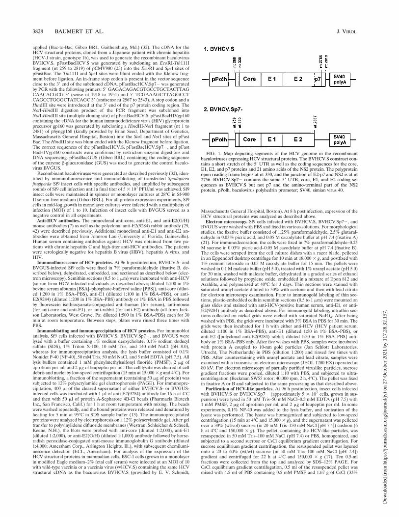
blotting with monoclonal antibodies against the core, E1, andE2 proteins revealed appropriate posttranslational processingof the HCV structural proteins. As shown in Fig. 3A, the coreprotein had an apparent molecular mass of 22 kDa, the E1protein was present in various glycosylated forms and had amolecular mass of 30 to 35 kDa, and the E2 protein had amolecular mass of approximately 66 kDa. The size of the coreprotein was identical to that of the core protein expressed in amammalian tissue culture system (Fig. 3A), whereas the sizesof the E1 and E2 proteins were similar to the sizes of envelopeproteins expressed in mammalian cells (E1, 34 to 36 kDa; E2,68 kDa; Fig. 3A). These data suggested similar posttransla-tional processing of HCV structural proteins in insect andmammalian expression systems (39). The variations in the sizesof the envelope proteins probably reflected differences in gly-cosylation between the two protein expression systems. Thisfinding has been noted by other investigators (34) and is notunexpected, since glycosylation in insect and mammalian cells
has been shown to be distinctly different (33). The difference inglycosylation may have resulted in different signal intensitieson the immunoblot, thereby explaining the apparently higherlevels of E1 and E2 relative to the core in mammalian cellsthan in insect cells (Fig. 3A). On the other hand, it is possiblethat aberrant translational termination or accelerated degra-dation of a particular protein species may have been respon-sible for this difference between mammalian and insect cells.As shown in Fig. 3B, the E2 protein was coimmunoprecipitatedwith the E1 and core proteins. The interaction of the E2protein with the E1 and core proteins was also confirmed bymetabolic labeling and coimmunoprecipitation (data notshown). These results extend the findings of recent studieswhich demonstrated interactions of the E1 and E2 proteins (7)as well as the core and E1 proteins (31).
HCV-like particle assembly occurs in cytoplasmic vesicles.Transmission electron microscopy of BVHCV.S-infected cellsrevealed abundant viruslike particles in cytoplasmic vesicles or
FIG. 3. (A) Expression of HCV structural proteins in insect and mammalian cells. Sf9 insect cells were infected either with control baculovirus BVGUS (lane 3)or with the HCV expression baculoviruses BVHCV.S (lane 4) and BVHCV.Sp72 (lane 5). At 96 h postinfection, the cells were lysed as described in Materials andMethods. To compare the expression of HCV structural proteins between insect and mammalian cells, BSC-1 cells were infected with a wild-type vaccinia virus (vvWT)(lane 1) or a vaccinia virus (vvHCV.S) containing the same cDNA for the HCV structural proteins as baculovirus BVHCV.S (lane 2). At 8 h postinfection, the cellswere lysed as described in Materials and Methods. Sf9 and BSC-1 cell lysates were then subjected to SDS-PAGE and immunoblotting (IB) with anti-core (left panel),anti-E1 (middle panel), or anti-E2 (right panel) antibodies. Molecular masses (in kilodaltons) of protein molecular weight (MW) markers are indicated on the left;HCV-specific proteins are indicated on the right. (B) Coimmunoprecipitation of HCV structural proteins in insect cells. Insect cells were infected with BVGUS (lane6) or BVHCV.S (lane 7). At 96 h postinfection, the cells were lysed and subjected to immunoprecipitation (IP) with an anti-E2 antibody or nonimmune serum asindicated. The immunoprecipitated proteins were subjected to SDS-PAGE and immunoblotting (IB) with anti-core (C), anti-E1, or anti-E2 antibodies as indicated; thecontrol immunoprecipitate with nonimmune serum was probed with a mixture of antibodies to core, E1, and E2 proteins. The exposure times for the probed blot wereadjusted to visualize the proteins and do not represent a quantitation of the precipitated proteins.
3830 BAUMERT ET AL. J. VIROL.
Dow
nloa
ded
from
http
s://j
ourn
als.
asm
.org
/jour
nal/j
vi o
n 27
Oct
ober
202
1 by
117
.28.
32.1
57.
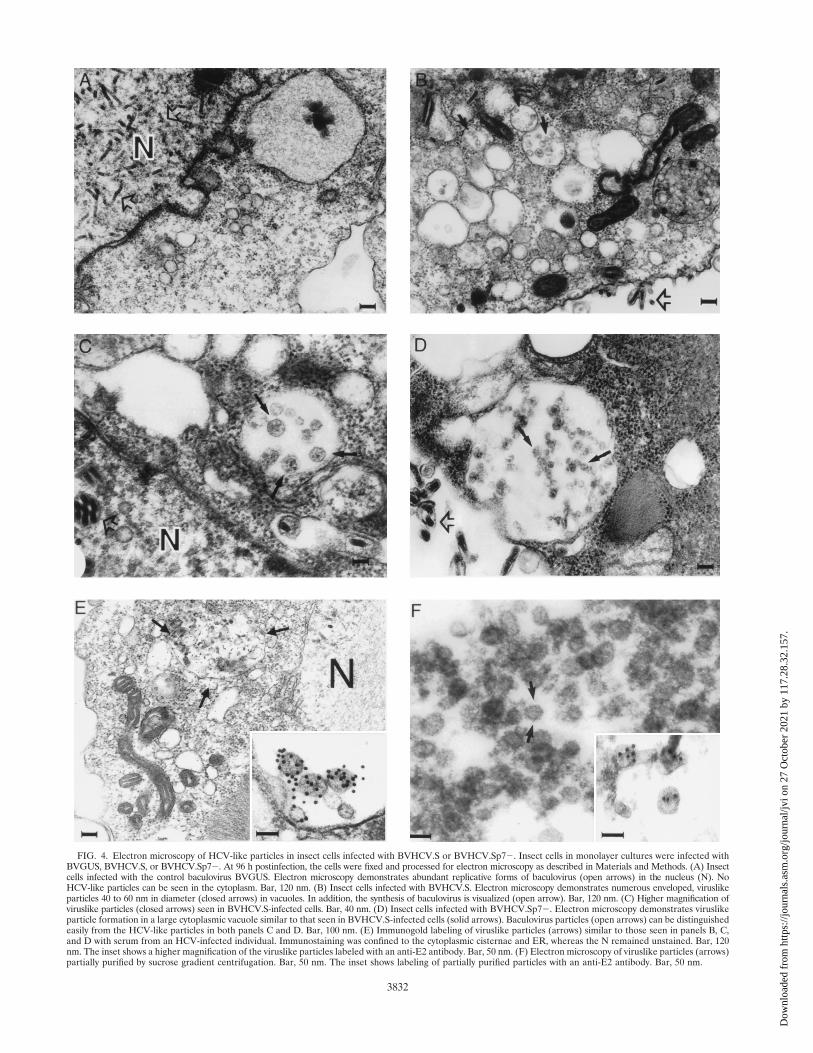
vacuoles, presumably derived from the endoplasmic reticulum(ER) of the insect cells (Fig. 4B, C, and E). These particles, 40to 60 nm in diameter, were polymorphic in appearance andhad an envelope consisting of a membrane (Fig. 4B, C, and E);many of them had unevenly distributed electron-dense struc-tures suggestive of possible nucleocapsids. Visualization ofthese particles was only possible after osmium treatment,which stains membranes. These features resemble the previ-ously described structure and morphology of pestiviruses ininfected cells (4, 12, 44). The particles formed predominantlyin cytoplasmic vesicles, giving the appearance of virion trans-port through the ER secretory pathway of the cells. Theseparticle-containing structures were not observed in insect cellsinfected with a control baculovirus expressing GUS (BVGUS;Fig. 4A) or uninfected insect cells (data not shown), suggestingthat they were not related to baculovirus protein expressionand replication. In addition to these vesicles containing virus-like particles, floccular membranous materials with irregularstructures of 20 to 100 nm were clustered in large vacuoles inthe cytoplasm (Fig. 4B). These diversely polymorphic struc-tures also contained membranous envelopes, but most of themdemonstrated no viruslike structures.
The viruslike particles were immunostained specifically withhuman anti-HCV (Fig. 4E), anti-E2 (Fig. 4E, inset), and an-ti-E1 (data not shown) antibodies, suggesting that the viruslikeparticles were derived from HCV structural proteins. In addi-tion to strong immunostaining of the particles, labeling of theER and the vesicular membranous complexes mentionedabove was also present. No nuclear staining was observed withany of the antibodies. The immunolabeling was highly specificfor the indicated structures. No labeling of any cellular orbaculovirus structures was seen in BVGUS-infected (Fig. 4A)or noninfected insect cells, and staining was not present insamples not incubated with primary antibodies (data notshown). Since examination of the spent medium at varioustimes was negative for HCV-like particles but abundantly pos-itive for baculoviruses, the viruslike particles appeared not tobe released or secreted into the culture medium.
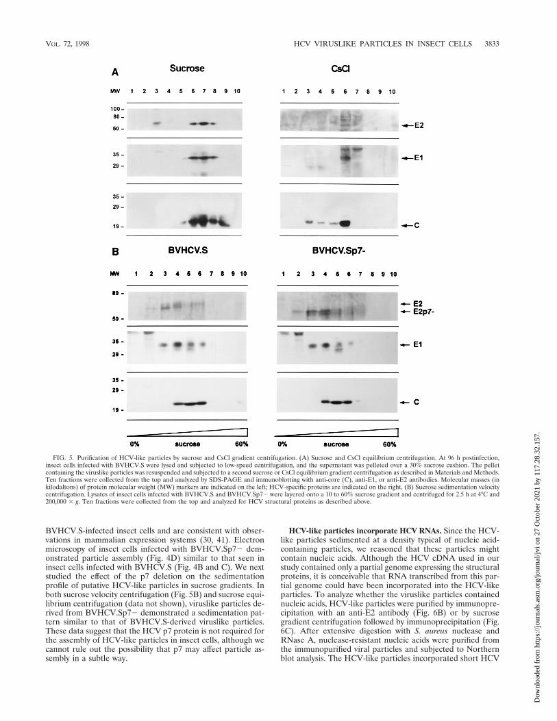
Purification of HCV-like particles by sucrose and CsCl gra-dient centrifugation. In order to purify the viruslike particles,lysates of baculovirus-infected insect cells were subjected tosucrose or CsCl equilibrium gradient centrifugation. In bothsucrose and CsCl equilibrium gradients, the HCV-like particlesbanded in specific fractions (Fig. 5A), confirming the assemblyof viruslike particles. The densities of the fractions demonstrat-ing immunoreactivity for HCV-like particles were 1.14 to 1.18g/cm3 in sucrose equilibrium gradients (fractions 6 to 8 in Fig.5A) and 1.14 to 1.16 g/cm3 in CsCl equilibrium gradients (frac-tion 6 in Fig. 5A). Colocalization of the HCV structural pro-teins in specific sucrose fractions was confirmed by coimmu-noprecipitation of the HCV structural proteins. To confirmthat the colocalization of the HCV structural proteins in thegradient fractions did not represent randomly assembled pro-tein aggregates, sucrose gradient fractions showing immunore-activity for HCV structural proteins were examined with trans-mission electron microscopy. Like the structures seen withinthe BVHCV.S-infected insect cells, 40- to 60-nm envelopedviruslike particles were present (Fig. 4F) and were labeledspecifically with an anti-E2 antibody (Fig. 4F, inset).
To further analyze the biophysical properties of the viruslikeparticles, insect cell lysates were subjected to sucrose sedimen-tation velocity centrifugation (Fig. 5B). Immunoblotting of thesucrose fractions revealed colocalization of the HCV structuralproteins in high-sedimentation fractions (fractions 4 to 6), in-dicating the presence of viruslike particles. Fractions 2 and 3contained E1 and E2 only, probably representing E1 and E2
complexes as assembly intermediates prior to interaction withthe core. To estimate the sedimentation coefficient of theHCV-like particles, we determined the percentage of sucrosein the particle-containing sucrose gradient fractions and ap-plied the approximation tables of McEwen (37). The sedimen-tation coefficient S20,V can be approximated by the Svedbergequation, including the parameters of the applied angular ve-locity V, centrifugation time and temperature, percentage ordensity of sucrose in which the particle is sedimenting, and thedensity of the sedimenting particle (37). The validity of thisapproximation under the experimental conditions was con-firmed by sedimentation analysis of HBV surface and coreparticles as standards. Empty HBV surface particles with asedimentation coefficient of 39S to 54S and a particle density of1.16 g/cm3 (18) sedimented to fraction 3 of the sucrose velocitygradient, and HBV core particles with a sedimentation coeffi-cient of 124S and a particle density of 1.30 g/cm3 (18) sedi-mented to fractions 5 and 6 of the sucrose velocity gradient.Using the approximation of McEwen (37) and an assumedHCV-like particle density of 1.14 to 1.18 g/cm3 (from sucroseand CsCl equilibrium gradients; Fig. 5A), we estimated thesedimentation coefficient S20,w of HCV-like particles to bebetween 100S and 230S (peak at approximately 160S). Thesedimentation coefficient S20,w of HCV-like particles was dif-ferent from that of similarly sedimenting HBV core particlesbecause of the difference in particle densities. The S value forHCV-like particles is within the range of reported sedimenta-tion coefficients for other members of the Flaviviridae family(44).
Since the visualized viral particles were present predomi-nantly in membrane-enclosed cytoplasmic vesicles, various celllysis conditions were studied to increase the yield of partiallypurified particles. Although the qualitative findings did notchange, the quantity of purified particles was enhanced signif-icantly by the addition of a low concentration of a mild non-ionic detergent (0.1% NP-40 or 0.5% digitonin) to the lysisbuffer. The well-known detergent effect of the de-envelopmentof viruses has been shown to be a time- and concentration-dependent process. Since the majority of the detergent in thecell lysate was bound in micellar aggregates with cellular pro-teins and lipids, the effective detergent concentration, beingmuch lower than the original concentration, resulted in therelease of viruslike particles from the cytoplasmic vesicles with-out affecting its envelope. In contrast, higher concentrations ofdetergent in the lysis buffer (.0.5% NP-40) resulted in thedisruption of the core-envelope interactions, as indicated by alack of their coimmunoprecipitation (data not shown). On theother hand, the sucrose gradient-purified particles were highlysensitive to detergent treatment: incubation of partially puri-fied particles with 0.1% NP-40 disrupted the cosedimentationof HCV structural proteins in the sucrose gradient and elimi-nated particular viruslike structures, as seen by electron mi-croscopy in Fig. 4F.
HCV protein p7 is not required for viral assembly in insectcells. In order to study whether HCV protein p7 is required forviral assembly, a recombinant baculovirus containing a dele-tion of the p7 protein (BVHCV.Sp72) was generated. Thisconstruct contained the same 59 UTR, core, E1, and E2 cod-ing sequences as BVHCV.S but not p7 (Fig. 1). Infection ofinsect cells with BVHCV.Sp72 resulted in levels of core andE1 protein expression similar to those seen in BVHCV.S-infected cells. In contrast, the molecular mass of the E2 pro-tein was approximately 7 kDa lower in insect cells infected withthe construct BVHCV.Sp72 than in insect cells infected withthe construct BVHCV.S (Fig. 3A). These data indicate thatp7 is not cleaved efficiently from the E2-p7 polyprotein in
VOL. 72, 1998 HCV VIRUSLIKE PARTICLES IN INSECT CELLS 3831
Dow
nloa
ded
from
http
s://j
ourn
als.
asm
.org
/jour
nal/j
vi o
n 27
Oct
ober
202
1 by
117
.28.
32.1
57.
FIG. 4. Electron microscopy of HCV-like particles in insect cells infected with BVHCV.S or BVHCV.Sp72. Insect cells in monolayer cultures were infected withBVGUS, BVHCV.S, or BVHCV.Sp72. At 96 h postinfection, the cells were fixed and processed for electron microscopy as described in Materials and Methods. (A) Insectcells infected with the control baculovirus BVGUS. Electron microscopy demonstrates abundant replicative forms of baculovirus (open arrows) in the nucleus (N). NoHCV-like particles can be seen in the cytoplasm. Bar, 120 nm. (B) Insect cells infected with BVHCV.S. Electron microscopy demonstrates numerous enveloped, viruslikeparticles 40 to 60 nm in diameter (closed arrows) in vacuoles. In addition, the synthesis of baculovirus is visualized (open arrow). Bar, 120 nm. (C) Higher magnification ofviruslike particles (closed arrows) seen in BVHCV.S-infected cells. Bar, 40 nm. (D) Insect cells infected with BVHCV.Sp72. Electron microscopy demonstrates viruslikeparticle formation in a large cytoplasmic vacuole similar to that seen in BVHCV.S-infected cells (solid arrows). Baculovirus particles (open arrows) can be distinguishedeasily from the HCV-like particles in both panels C and D. Bar, 100 nm. (E) Immunogold labeling of viruslike particles (arrows) similar to those seen in panels B, C,and D with serum from an HCV-infected individual. Immunostaining was confined to the cytoplasmic cisternae and ER, whereas the N remained unstained. Bar, 120nm. The inset shows a higher magnification of the viruslike particles labeled with an anti-E2 antibody. Bar, 50 nm. (F) Electron microscopy of viruslike particles (arrows)partially purified by sucrose gradient centrifugation. Bar, 50 nm. The inset shows labeling of partially purified particles with an anti-E2 antibody. Bar, 50 nm.
3832
Dow
nloa
ded
from
http
s://j
ourn
als.
asm
.org
/jour
nal/j
vi o
n 27
Oct
ober
202
1 by
117
.28.
32.1
57.
BVHCV.S-infected insect cells and are consistent with obser-vations in mammalian expression systems (30, 41). Electronmicroscopy of insect cells infected with BVHCV.Sp72 dem-onstrated particle assembly (Fig. 4D) similar to that seen ininsect cells infected with BVHCV.S (Fig. 4B and C). We nextstudied the effect of the p7 deletion on the sedimentationprofile of putative HCV-like particles in sucrose gradients. Inboth sucrose velocity centrifugation (Fig. 5B) and sucrose equi-librium centrifugation (data not shown), viruslike particles de-rived from BVHCV.Sp72 demonstrated a sedimentation pat-tern similar to that of BVHCV.S-derived viruslike particles.These data suggest that the HCV p7 protein is not required forthe assembly of HCV-like particles in insect cells, although wecannot rule out the possibility that p7 may affect particle as-sembly in a subtle way.
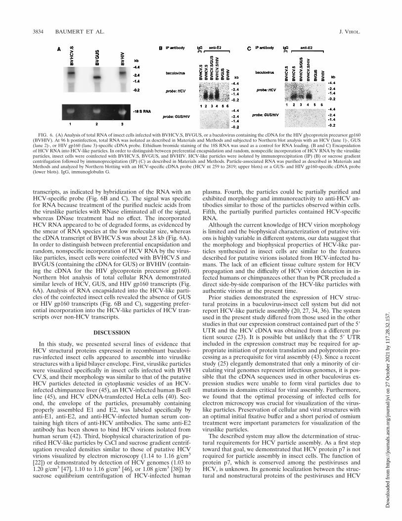
HCV-like particles incorporate HCV RNAs. Since the HCV-like particles sedimented at a density typical of nucleic acid-containing particles, we reasoned that these particles mightcontain nucleic acids. Although the HCV cDNA used in ourstudy contained only a partial genome expressing the structuralproteins, it is conceivable that RNA transcribed from this par-tial genome could have been incorporated into the HCV-likeparticles. To analyze whether the viruslike particles containednucleic acids, HCV-like particles were purified by immunopre-cipitation with an anti-E2 antibody (Fig. 6B) or by sucrosegradient centrifugation followed by immunoprecipitation (Fig.6C). After extensive digestion with S. aureus nuclease andRNase A, nuclease-resistant nucleic acids were purified fromthe immunopurified viral particles and subjected to Northernblot analysis. The HCV-like particles incorporated short HCV
FIG. 5. Purification of HCV-like particles by sucrose and CsCl gradient centrifugation. (A) Sucrose and CsCl equilibrium centrifugation. At 96 h postinfection,insect cells infected with BVHCV.S were lysed and subjected to low-speed centrifugation, and the supernatant was pelleted over a 30% sucrose cushion. The pelletcontaining the viruslike particles was resuspended and subjected to a second sucrose or CsCl equilibrium gradient centrifugation as described in Materials and Methods.Ten fractions were collected from the top and analyzed by SDS-PAGE and immunoblotting with anti-core (C), anti-E1, or anti-E2 antibodies. Molecular masses (inkilodaltons) of protein molecular weight (MW) markers are indicated on the left; HCV-specific proteins are indicated on the right. (B) Sucrose sedimentation velocitycentrifugation. Lysates of insect cells infected with BVHCV.S and BVHCV.Sp72 were layered onto a 10 to 60% sucrose gradient and centrifuged for 2.5 h at 4°C and200,000 3 g. Ten fractions were collected from the top and analyzed for HCV structural proteins as described above.
VOL. 72, 1998 HCV VIRUSLIKE PARTICLES IN INSECT CELLS 3833
Dow
nloa
ded
from
http
s://j
ourn
als.
asm
.org
/jour
nal/j
vi o
n 27
Oct
ober
202
1 by
117
.28.
32.1
57.
transcripts, as indicated by hybridization of the RNA with anHCV-specific probe (Fig. 6B and C). The signal was specificfor RNA because treatment of the purified nucleic acids fromthe viruslike particles with RNase eliminated all of the signal,whereas DNase treatment had no effect. The incorporatedHCV RNA appeared to be of degraded forms, as evidenced bythe smear of RNA species at the low molecular size, whereasthe cDNA transcript of BVHCV.S was about 2.8 kb (Fig. 6A).In order to distinguish between preferential encapsidation andrandom, nonspecific incorporation of HCV RNA by the virus-like particles, insect cells were coinfected with BVHCV.S andBVGUS (containing the cDNA for GUS) or BVHIV (contain-ing the cDNA for the HIV glycoprotein precursor gp160).Northern blot analysis of total cellular RNA demonstratedsimilar levels of HCV, GUS, and HIV gp160 transcripts (Fig.6A). Analysis of RNA encapsidated into the HCV-like parti-cles of the coinfected insect cells revealed the absence of GUSor HIV gp160 transcripts (Fig. 6B and C), suggesting prefer-ential incorporation into the HCV-like particles of HCV tran-scripts over non-HCV transcripts.
DISCUSSION
In this study, we presented several lines of evidence thatHCV structural proteins expressed in recombinant baculovi-rus-infected insect cells appeared to assemble into viruslikestructures with a lipid bilayer envelope. First, viruslike particleswere visualized specifically in insect cells infected with BVHCV.S, and their morphology was similar to that of the putativeHCV particles detected in cytoplasmic vesicles of an HCV-infected chimpanzee liver (45), an HCV-infected human B-cellline (45), and HCV cDNA-transfected HeLa cells (40). Sec-ond, the envelope of the particles, presumably containingproperly assembled E1 and E2, was labeled specifically byanti-E1, anti-E2, and anti-HCV-infected human serum con-taining high titers of anti-HCV antibodies. The same anti-E2antibody has been shown to bind HCV virions isolated fromhuman serum (42). Third, biophysical characterization of pu-rified HCV-like particles by CsCl and sucrose gradient centrif-ugation revealed densities similar to those of putative HCVvirions visualized by electron microscopy (1.14 to 1.16 g/cm3
[22]) or demonstrated by detection of HCV genomes (1.03 to1.20 g/cm3 [47], 1.10 to 1.16 g/cm3 [46], or 1.08 g/cm3 [38]) bysucrose equilibrium centrifugation of HCV-infected human
plasma. Fourth, the particles could be partially purified andexhibited morphology and immunoreactivity to anti-HCV an-tibodies similar to those of the particles observed within cells.Fifth, the partially purified particles contained HCV-specificRNA.
Although the current knowledge of HCV virion morphologyis limited and the biophysical characterization of putative viri-ons is highly variable in different systems, our data suggest thatthe morphology and biophysical properties of HCV-like par-ticles synthesized in insect cells are similar to the featuresdescribed for putative virions isolated from HCV-infected hu-mans. The lack of an efficient tissue culture system for HCVpropagation and the difficulty of HCV virion detection in in-fected humans or chimpanzees other than by PCR precluded adirect side-by-side comparison of the HCV-like particles withauthentic virions at the present time.
Prior studies demonstrated the expression of HCV struc-tural proteins in a baculovirus-insect cell system but did notreport HCV-like particle assembly (20, 27, 34, 36). The systemused in the present study differed from those used in the otherstudies in that our expression construct contained part of the 59UTR and the HCV cDNA was obtained from a different pa-tient source (23). It is possible but unlikely that the 59 UTRincluded in the expression construct may be required for ap-propriate initiation of protein translation and polyprotein pro-cessing as a prerequisite for viral assembly (43). Since a recentstudy (25) elegantly demonstrated that only a minority of cir-culating viral genomes represent infectious genomes, it is pos-sible that the cDNA sequences used in other baculovirus ex-pression studies were unable to form viral particles due tomutations in domains critical for viral assembly. Furthermore,we found that the optimal processing of infected cells forelectron microscopy was crucial for visualization of the virus-like particles. Preservation of cellular and viral structures withan optimal initial fixative buffer and a short period of osmiumtreatment were important parameters for visualization of theviruslike particles.
The described system may allow the determination of struc-tural requirements for HCV particle assembly. As a first steptoward that goal, we demonstrated that HCV protein p7 is notrequired for particle assembly in insect cells. The function ofprotein p7, which is conserved among the pestiviruses andHCV, is unknown. Its genomic localization between the struc-tural and nonstructural proteins of the pestiviruses and HCV
FIG. 6. (A) Analysis of total RNA of insect cells infected with BVHCV.S, BVGUS, or a baculovirus containing the cDNA for the HIV glycoprotein precursor gp160(BVHIV). At 96 h postinfection, total RNA was isolated as described in Materials and Methods and subjected to Northern blot analysis with an HCV (lane 1)-, GUS(lane 2)-, or HIV gp160 (lane 3)-specific cDNA probe. Ethidium bromide staining of the 18S RNA was used as a control for RNA loading. (B and C) Encapsidationof HCV RNA into HCV-like particles. In order to distinguish between preferential encapsidation and random, nonspecific incorporation of HCV RNA by the viruslikeparticles, insect cells were coinfected with BVHCV.S, BVGUS, and BVHIV. HCV-like particles were isolated by immunoprecipitation (IP) (B) or sucrose gradientcentrifugation followed by immunoprecipitation (IP) (C) as described in Materials and Methods. Particle-associated RNA was purified as described in Materials andMethods and analyzed by Northern blotting with an HCV-specific cDNA probe (HCV nt 259 to 2819; upper blots) or a GUS- and HIV gp160-specific cDNA probe(lower blots). IgG, immunoglobulin G.
3834 BAUMERT ET AL. J. VIROL.
Dow
nloa
ded
from
http
s://j
ourn
als.
asm
.org
/jour
nal/j
vi o
n 27
Oct
ober
202
1 by
117
.28.
32.1
57.
and the presence of two species of E2 proteins, with andwithout p7, have led to the suggestion that p7 may play a rolein glycoprotein maturation or virus morphogenesis (8, 30, 41).However, no functional data have been presented so far tosupport this hypothesis. For pestiviruses, it has been shownthat the p7 protein is not a major structural component of thevirion (8). Our functional analysis demonstrates that the p7protein does not seem to be necessary for viral assembly ininsect cells, although we cannot completely exclude the possi-bility of p7 affecting viral assembly at the ultrastructural level.
Analysis of particle-associated nucleic acids demonstratedthat HCV-like particles contained short HCV RNAs and thatthese RNAs were selectively incorporated into the particlesover non-HCV transcripts. The HCV RNA transcripts gener-ated in this study may contain sufficient cis-acting information(encapsidation signal) interacting specifically with viral struc-tural proteins for encapsidation. However, since the incorpo-rated transcripts were substantially degraded compared to the2.8-kb transcript of the HCV cDNA, the incorporation morelikely represents a cis-dominant, nonspecific effect of the tran-scripts interacting with the structural proteins; i.e., the physicalproximity of the transcripts with their translated proteins con-ferred a preferential interaction. During such a process, cyto-solic RNases may have access to the transcripts, resulting inpartial degradation. Further studies are under way to elucidatethe mechanism of the observed RNA encapsidation.
The synthesis of HCV-like particles in insect cells is poten-tially an important tool for studying viral assembly and virus-cell interactions and for the development of an HCV vaccine.In the latter case, efforts to generate recombinant HCV sub-unit vaccines have been directed at the expression of portionsof individual structural proteins in soluble form (6). This ap-proach has met with marginal success, partly as a result of thenonnative forms of the expressed viral proteins. In contrast tothe proteins in recombinant subunit vaccines, the HCV pro-teins in HCV-like particles presumably are presented in anative, virionlike conformation and may therefore be superiorin eliciting protective humoral and cellular immune responses.Since HCV-like particles synthesized in insect cells are derivedfrom partial viral genomes without the nonstructural genesrequired for viral replication, they are noninfectious and there-fore represent excellent candidates for an HCV vaccine. Stud-ies are under way to determine the immunogenicity of theHCV-like particles as a potential vaccine.
ACKNOWLEDGMENTS
We thank Kunitada Shimotohno for generously providing constructpCVM980, Brian Seed for kindly providing plasmid pSyngp160, Rich-ard Lesniewski for the gift of polyclonal rabbit anti-E2(9284) anti-body, Johnson Lau for monoclonal mouse anti-E1 and anti-E2 anti-bodies, Emmett V. Schmidt for providing recombinant vaccinia virusvvHCV.S, and Bernard Moss for the gift of wild-type vaccinia virusvvWT. We also thank Harry B. Greenberg, Stephen M. Feinstone, andJay H. Hoofnagle for helpful discussions. Excellent technical assistanceby Hucheng Bei, John Vergalla, Louise Trakimus, and Maria Ericssonis gratefully appreciated.
This work was supported in part by a postdoctoral fellowship grantfrom the Deutsche Forschungsgemeinschaft (Ba 1417/1-1) to T.F.B.and by grants from the National Institutes of Health to T.F.B. (VF-DK-14361) and T.J.L. (DK-01952 and CA-54525). S.I. was supportedby the Harvard Digestive Disease Center. D.T.W. was a recipient of aGlaxo Institute for Digestive Health scientific research award and wassupported by NIH grants T30DK38707 and AI95-012.
REFERENCES
1. Acs, G., M. A. Sells, R. H. Purcell, P. Price, R. Engle, M. Shapiro, and H.Popper. 1987. Hepatitis B virus produced by transfected HepG2 cells causes
hepatitis in chimpanzees. Proc. Natl. Acad. Sci. USA 84:4641–4644.2. Alter, H. J., R. H. Purcell, J. W. Shih, J. C. Melpolder, M. Houghton, Q.-L.
Choo, and G. Kuo. 1989. Detection of antibody to hepatitis C virus inprospectively followed transfusion recipients with acute and chronic non-A,non-B hepatitis. N. Engl. J. Med. 321:1494–1500.
3. Baumert, T. F., S. A. Rogers, K. Hasegawa, and T. J. Liang. 1996. Two corepromoter mutations identified in a hepatitis B virus strain associated withfulminant hepatitis result in enhanced viral replication. J. Clin. Invest. 98:2268–2276.
4. Bielefeldt Ohmann, H., and B. Bloch. 1981. Electron microscopic studies ofbovine viral diarrhea virus in tissues of diseased calves and in cell cultures.Arch. Virol. 71:57–74.
5. Chomczynski, P. N., and N. Sacchi. 1987. Single-step method of RNA iso-lation by acid guanidinium thiocyanate-phenol-chloroform extraction. Anal.Biochem. 162:156–159.
6. Choo, Q.-L., G. Kuo, R. Ralston, A. J. Weiner, D. Chien, G. Van Nest, J. Han,K. Berger, K. Thudium, C. Kuo, J. Kansopon, J. McFarland, A. Tabrizi, K.Ching, B. Moss, L. B. Cummins, M. Houghton, and E. Muchmore. 1994.Vaccination of chimpanzees against infection by the hepatitis C virus. Proc.Natl. Acad. Sci. USA 91:1294–1298.
7. Dubuisson, J., H. H. Hsu, R. C. Cheung, H. B. Greenberg, D. R. Russell, andC. M. Rice. 1994. Formation and intracellular localization of hepatitis C virusenvelope glycoprotein complexes expressed by recombinant vaccinia andsindbis viruses. J. Virol. 68:6147–6160.
8. Elbers, K., N. Tautz, P. Becher, D. Stoll, T. Rumenapf, and H.-J. Thiel. 1996.Processing of the pestivirus E2-NS2 region: identification of proteins p7 andE2p7. J. Virol. 70:4131–4135.
9. Feinstone, S. M., K. B. Mihalik, T. Kamimura, H. J. Alter, W. T. London,and R. H. Purcell. 1983. Inactivation of hepatitis B viruses and non-A, non-Bhepatitis by chloroform. Infect. Immun. 41:816–821.
10. Gheysen, D., E. Jacobs, F. De Foresta, C. Thiriart, M. Francotte, D. Thines,and M. De Wilde. 1989. Assembly and release of HIV-1 precursor pr55gagvirus-like particles from recombinant baculovirus-infected insect cells. Cell59:103–112.
11. Grakoui, A., C. Wychowski, C. Lin, S. M. Feinstone, and C. M. Rice. 1993.Expression and identification of hepatitis C virus polyprotein cleavage prod-ucts. J. Virol. 67:1385–1395.
12. Gray, E. W., and P. F. Nettleton. 1987. The ultrastructure of cell culturesinfected with border disease and bovine virus diarrhoea viruses. J. Gen.Virol. 68:2339–2346.
13. Harlow, E., and D. Lane. 1988. Antibodies. A laboratory manual. ColdSpring Harbor Laboratory, Cold Spring Harbor, N.Y.
14. Hasegawa, K., J. Huang, S. A. Rogers, H. E. Blum, and T. J. Liang. 1994.Enhanced replication of a hepatitis B virus mutant associated with an epi-demic of fulminant hepatitis. J. Virol. 68:1651–1659.
15. He, L. E., D. Alling, D. Popkin, M. Shapiro, H. J. Alter, and R. H. Purcell.1987. Determining the size of non-A, non-B hepatitis virus by filtration.J. Infect. Dis. 156:636–640.
16. Hijikata, M., N. Kato, Y. Ootsuyama, M. Nakagawa, and K. Shimotohno.1991. Gene mapping of the putative structural region of the hepatitis C virusgenome by in vitro processing analysis. Proc. Natl. Acad. Sci. USA 88:5547–5551.
17. Hijikata, M., Y. K. Shimuzu, H. Kato, A. Iwamoto, J. W. Shih, H. J. Alter,R. H. Purcell, and H. Yoshikura. 1993. Equilibrium centrifugation studies ofhepatitis C virus: evidence for circulating immune complexes. J. Virol. 67:1953–1958.
18. Hollinger, F. B. 1996. Hepatitis B virus, p. 2738–2808. In B. N. Fields, D. M.Knipe, and P. M. Howley (ed.), Fields virology, 3rd ed. Lippincott-Raven,Philadelphia, Pa.
19. Hoofnagle, J. H., and A. M. Di Bisceglie. 1997. The treatment of chronic viralhepatitis. N. Engl. J. Med. 336:347–356.
20. Hsu, H., M. Donets, H. B. Greenberg, and S. M. Feinstone. 1993. Charac-terization of hepatitis C virus structural proteins with a recombinant bacu-lovirus expression system. Hepatology 17:763–771.
21. Ito, S., and M. J. Karnovsky. 1968. Formaldehyde-glutaraldehyde fixativescontaining trinitro compounds. J. Cell Biol. 39:168A.
22. Kaito, M., S. Watanabe, K. Tsukiyama-Kohara, K. Yamaguchi, Y. Koba-yashi, M. Konishi, M. Yokoi, S. Ishida, S. Suzuki, and M. Kohara. 1994.Hepatitis C virus particle detected by immunoelectron microscopic study.J. Gen. Virol. 75:1755–1760.
23. Kato, N., M. Hijikata, Y. Ootsuyama, M. Nakagawa, S. Ohkoshi, T. Sug-imura, and K. Shimotohno. 1990. Molecular cloning of the human hepatitisC virus genome from Japanese patients with non-A, non-B hepatitis. Proc.Natl. Acad. Sci. USA 87:9524–9528.
24. Kirnbauer, R., F. Booy, N. Cheng, D. R. Lowy, and J. T. Schiller. 1992.Papillomavirus L1 major capsid protein self-assembles into virus-like parti-cles that are highly immunogenic. Proc. Natl. Acad. Sci. USA 89:12180–12184.
25. Kolykhalov, A. A., E. V. Agapov, K. J. Blight, K. Mihalik, S. M. Feinstone,and C. M. Rice. 1997. Transmission of hepatitis C by intrahepatic inoculationwith transcribed RNA. Science 277:570–574.
26. Kuo, G., Q.-L. Choo, H. J. Alter, G. L. Gitnick, A. G. Redeker, R. H. Purcell,
VOL. 72, 1998 HCV VIRUSLIKE PARTICLES IN INSECT CELLS 3835
Dow
nloa
ded
from
http
s://j
ourn
als.
asm
.org
/jour
nal/j
vi o
n 27
Oct
ober
202
1 by
117
.28.
32.1
57.
T. Miyamura, J. L. Dienstag, M. J. Alter, C. E. Stevens, G. E. Tegtmeier, F.Bonino, M. Colombo, W.-S. Lee, C. Kuo, K. Berger, R. J. Shuster, L. R.Overby, D. W. Bradley, and M. Houghton. 1989. An assay for circulatingantibodies to a major etiologic virus of human non-A, non-B hepatitis.Science 244:362–364.
27. Lanford, R. E., L. Notvall, D. Chavez, R. White, G. Frenzel, C. Simonsen,and J. Kim. 1993. Analysis of hepatitis C virus capsid, E1, and E2/NS1proteins expressed in insect cells. Virology 197:225–235.
28. Lemon, S. M., and D. L. Thomas. 1997. Vaccines to prevent viral hepatitis.N. Engl. J. Med. 336:197–203.
29. Lesniewski, R., G. Okasinski, R. Carrick, C. Van Sant, S. Desai, R. Johnson,J. Scheffel, B. Moore, and I. Mushahwar. 1995. Antibody to hepatitis C virussecond envelope (HCV-E2) glycoprotein: a new marker of HCV infectionclosely associated with viremia. J. Med. Virol. 45:415–422.
30. Lin, C., B. D. Lindenbach, B. Pragai, D. W. McCourt, and C. M. Rice. 1994.Processing of the hepatitis C E2-NS2 region: identification of p7 and twodistinct E2-specific products with different C termini. J. Virol. 68:5063–5073.
31. Lo, S.-Y., M. S. Selby, and J.-H. Ou. 1996. Interaction between hepatitis Cvirus core protein and E1 envelope protein. J. Virol. 70:5177–5182.
32. Luckow, V. A., S. C. Lee, G. F. Barry, and P. O. Olins. 1993. Efficientgeneration of infectious recombinant baculoviruses by site-specific transpo-son-mediated insertions of foreign genes into a baculovirus genome propa-gated in Escherichia coli. J. Virol. 67:4566–4579.
33. Luckow, V. A., and M. D. Summers. 1988. Trends in the development ofbaculovirus expression vectors. Bio/Technology 6:47–55.
34. Matsuura, Y., S. Harada, R. Susuki, Y. Watanabe, Y. Inoue, I. Saito, and T.Miyamura. 1992. Expression of processed envelope protein of hepatitis Cvirus in mammalian and insect cells. J. Virol. 66:1425–1431.
35. Matsuura, Y., and T. Miyamura. 1993. The molecular biology of hepatitis Cvirus. Semin. Virol. 4:297–304.
36. Matsuura, Y., T. Suzuki, R. Suzuki, M. Sato, H. Aizaki, I. Saito, and T.Miyamura. 1994. Processing of E1 and E2 glycoproteins of hepatitis C virusexpressed in mammalian and insect cells. Virology 205:141–150.
37. McEwen, C. R. 1967. Tables for estimating sedimentation through linearconcentration gradients of sucrose solution. Anal. Biochem. 20:114–149.
38. Miyamoto, H., H. Okamoto, K. Sato, T. Tanaka, and S. Mishiro. 1992.Extraordinarily low density of hepatitis C virus estimated by sucrose gradient
centrifugation and the polymerase chain reaction. J. Gen. Virol. 73:715–718.39. Miyamura, T., and Y. Matsuura. 1993. Structural proteins of hepatitis C
virus. Trends Microbiol. 1:229–231.40. Mizuno, M., G. Yamada, T. Tanaka, K. Shimotohno, M. Takatani, and T.
Tsuji. 1995. Virion-like structures in HeLa G cells transfected with thefull-length sequence of the hepatitis C virus genome. Gastroenterology 109:1933–1940.
41. Mizushima, H., M. Hijikata, S.-I. Asabe, M. Hirota, K. Kimura, and K.Shimotohno. 1994. Two hepatitis C virus glycoprotein E2 products withdifferent C termini. J. Virol. 68:6215–6222.
42. Prince, A. M., T. Huima-Byron, T. S. Parker, and D. M. Levine. 1996.Visualization of hepatitis C virions and putative defective interfering parti-cles isolated from low-density lipoproteins. J. Viral Hepatitis 3:11–17.
43. Reynolds, J. E., A. Kaminski, H. J. Kettinen, K. Grace, B. E. Clarke, A. R.Carrol, D. J. Rowlands, and R. J. Jackson. 1995. Unique features of internalinitation of hepatitis C virus RNA translation. EMBO J. 14:6010–6020.
44. Rice, C. M. 1996. Flaviviridae: the viruses and their replication, p. 931–959.In B. N. Fields, D. M. Knipe, and P. M. Howley (ed.), Fields virology, 3rd ed.Lippincott-Raven, Philadelphia, Pa.
45. Shimuzu, Y. K., S. M. Feinstone, M. Kohara, R. Purcell, and H. Yoshikura.1996. Hepatitis C virus: detection of intracellular virus particles by electronmicroscopy. Hepatology 23:205–209.
46. Shindo, M., A. M. Di Bisceglie, T. Akatsuka, T.-L. Fong, L. Scaglione, M.Donets, J. H. Hoofnagle, and S. M. Feinstone. 1994. The physical state of thenegative strand of hepatitis C virus RNA in serum of patients with chronichepatitis C. Proc. Natl. Acad. Sci. USA 91:8719–8723.
47. Thomssen, R., S. Bonk, C. Propfe, K.-H. Heermann, H. G. Koechel, and A.Uy. 1992. Association of hepatitis C virus in human sera with beta-lipopro-tein. Med. Microbiol. Immunol. 181:293–300.
48. Tong, M. J., N. S. El-Farra, A. R. Reikes, and R. L. Co. 1995. Clinicaloutcomes after transfusion-associated hepatitis C. N. Engl. J. Med. 332:1463–1466.
49. Zeng, C. Q.-Y., M. J. Wentz, J. Cohen, M. K. Estes, and R. F. Ramig. 1996.Characterization and replicase activity of double-layered and single-layeredrotavirus-like particles expressed from baculovirus recombinants. J. Virol.70:2736–2742.