+ All Categories
Home > Documents > Hepatitis E - Virus (HEV) · Zanetti et al., 1999). These variants were related most closely to the...

Hepatitis E - Virus (HEV) · Zanetti et al., 1999). These variants were related most closely to the...

Date post: 03-Dec-2018
Category:
Upload: nguyennhu
View: 213 times
Download: 0 times
Share this document with a friend
84
Aus dem Zentrum der Hygiene der Johann Wolfgang Goethe-Universität Frankfurt am Main Institut für Medizinische Virologie Direktor: Prof. Dr. Hans Wilhelm Doerr Hepatitis E - Virus (HEV): Comparative evaluation of IgG antibody assays in a low-endemicity setting INAUGURAL - DISSERTATION zur Erlangung des Doktorgrades der Medizin des Fachbereiches Medizin der Johann Wolfgang Goethe-Universität Frankfurt am Main vorgelegt von Mark Asante aus Accra/Ghana Frankfurt am Main 2003
Transcript

Aus dem Zentrum der Hygieneder Johann Wolfgang Goethe-Universität

Frankfurt am Main

Institut für Medizinische VirologieDirektor: Prof. Dr. Hans Wilhelm Doerr

Hepatitis E - Virus (HEV):Comparative evaluation of IgG antibody assays

in a low-endemicity setting

INAUGURAL - DISSERTATION

zur Erlangung des Doktorgrades der Medizindes Fachbereiches Medizin

der Johann Wolfgang Goethe-UniversitätFrankfurt am Main

vorgelegt von Mark Asanteaus Accra/Ghana

Frankfurt am Main 2003

Dekan: Prof. Dr. med. J. PfeilschifterReferent: Prof. Dr. med. H. W. DoerrKorreferent: Prof. Dr. med. S. ZielenTag der mündlichen Prüfung: 11. February 2004

Now to God be the glory, for all the things he has done!

Blessed are those who dwell in your house, oh God: for they will ever praiseYou. Blessed is the man whose strength is you. (Psalm 84:4-5)

Hepatitis E - Virus (HEV):Comparative evaluation of IgG antibody assays in a low-endemicity setting

Contents Page

1 Introduction and Background .......................................................................................................7

1.1 Historical Notes ............................................................................................................................7

1.2 The Genome of the Hepatitis E Virus (HEV)..............................................................................11

1.3 Genetic Heterogeniety................................................................................................................14

1.4 Cell Culture.................................................................................................................................16

1.5 Stages of Replication .................................................................................................................17

1.6 Pathogenesis and Pathology .....................................................................................................20

1.7 Epidemiology..............................................................................................................................22

1.8 Transmission, Routes of Spread and Reservoirs .....................................................................24

1.9 Hepatitis E in Pregnancy............................................................................................................28

1.10 Diagnosis and Seroprevelance Data .........................................................................................29

1.11 Therapeutic Approaches to Hepatitis E Infection.......................................................................31

1.12 Immunoprophylaxis ....................................................................................................................33

1.13 Prevention ..................................................................................................................................35

1.14 Objective ....................................................................................................................................36

2 Methods and Materials ...............................................................................................................37

2.1 Methods......................................................................................................................................37

Hepatitis E - Virus (HEV):Comparative evaluation of IgG antibody assays in a low-endemicity setting

2.2 Materials .....................................................................................................................................38

2.3 Test Principles: ..........................................................................................................................38

2.3.1 Diagnostics Systems Laboratries ELISA...................................................................................38

2.4 Genelabs ELISA.........................................................................................................................40

2.4.1 Chemical and Biological Principles of the Procedure................................................................40

2.4.2 Assay Procedure........................................................................................................................40

2.5 Mikrogen Recomblot ..................................................................................................................41

2.5.1 Test Procedure ..........................................................................................................................42

2.5.2 General Aspects.........................................................................................................................42

2.6 Assay Procedure........................................................................................................................44

2.7 Test Interpretation ......................................................................................................................46

3 Results and Discussions ...........................................................................................................56

3.1 Abbott EIA Assay........................................................................................................................56

3.2 Genelabs ELISA.........................................................................................................................57

3.3 Mikrogen RecomBlot:.................................................................................................................59

3.4 Diagnostics Systems Laboratories ( DSL) ELISA.....................................................................61

3.5 Concordance Between Assays .................................................................................................62

3.5.1 Evaluation Of Abbott EIA Versus Genelabs ELISA....................................................................62

3.5.2 Evaluation of Abbot EIA Versus Mikrogen RecomBlot..............................................................64

Hepatitis E - Virus (HEV):Comparative evaluation of IgG antibody assays in a low-endemicity setting

3.5.3 Evaluation of Genelabs ELISA Versus Mikrogen RecomBlot ....................................................65

3.5.4 Evaluation of Abbott EIA Versus DSL ELISA............................................................................67

4 Discrepancies ............................................................................................................................68

4.1 Problems and Conclusions........................................................................................................70

5 Summary....................................................................................................................................72

6 Literature ....................................................................................................................................74

7 Danksagung...............................................................................................................................79

8 Zusammenfassung ....................................................................................................................80

9 Lebenslauf..................................................................................................................................81

10 Ehrenwörtliche Erklärung...........................................................................................................82

Hepatitis E - Virus (HEV):Comparative evaluation of IgG antibody assays in a low-endemicity setting

Page 7

1 Introduction and Background

1.1 Historical Notes

Hepatitis E (HEV), the causative agent of what has been referred to as enterically transmitted

non-A, non-B hepatitis or ´waterborne hepatitis´, is a major cause of epidemic and acute

sporadic hepatitis in developing countries (Khuroo et al., 1980; Bradley et al, 1990;

Mushawar et al., 1997).The disease was first recognized as distinct, clinical entity in the

1980´s when the sera from persons affected during a large waterborne epidemic of viral

hepatitis during 1956-56 in New Delhi, India and another epidemics in Kashmir were found to

lack serological markers of acute hepatitis A and B .(Viswanathan et al., 1957;Khuroo et al.,

1980). The occurrence of the first recorded epidemic of hepatitis E as late as 1955 and the

frequency of this disease suggest that hepatitis E is a new, emerging infectious disease. The

first proof of the existence of a new viral hepatitis agent was obtained in 1983, when virus-like

particles were detected by immune electron microscopy in the faeces collected from a

volunteer who was infected with faecal material from patients with suspected enterically

transmitted non-A, non-Bhepatitis. (Balayan et al., 1983). Outbreaks of HEV infections have

occurred in a number of countries in Africa, Asia as well as in Mexico, and are usually

associated with warm weather and poor sanitation leading to faecal contamination of drinking

water. Sporadic cases can also occur in areas where HEV is endemic, usually during periods

between major outbreaks. Sporadic cases have been also reported in areas not considered

to be endemic for HEV. Many of these can be associated with travellers returning from visits

to areas where HEV is endemic. There have been reports, however, of HEV-associated

hepatitis among individuals with no history of travel to regions endemic for HEV ( Zaijer et al.,

1993; Zannetti et al., 1994). It was generally believed that such occurrences are rare in

developed nations.

The genome of the agent was originally identified and characterized using cynomoglus

macaques infected experimentally with human faeces obtained from patients during an

outbreak in Burma (Reyes et al.,1990). A second isolate was similarly identified from an

outbreak that had occurred in Mexico. These first two prototype isolates of HEV were similar

in overall genomic organization but still distinct from each other having 76% identity over the

Hepatitis E - Virus (HEV):Comparative evaluation of IgG antibody assays in a low-endemicity setting

Page 8

entire genome comprised of three discontinuous open- reading frames (ORF´s) (Huang et al.,

1992). In the years immediately after the identification of the Burmese and Mexican prototype

viruses, a number of additional isolates were identified from Pakistan, India and China..

Interestingly, all of these isolates were related very closely to the original isolate from Burma

having a greater than 93% nucleotide identity across the genomes. At that time, the genotypic

distribution of HEV consisted of a group of Burmese-like and the lone Mexican isolate.

The first indication that additional genetic diversity may occur in regions non-endemic for HEV

was the identification of a distinct endogenous variant identified in an acute hepatitis patient

from United States (US) who reported no recent travel history to endemic regions (Schlauder

et al., 1996; Kwo et al.,1997). Additional sequencing of the full-length genome of this and the

isolate confirmed that a new group of HEV did exist that was distinct from the Burmese-like

and Mexican isolates (Schlauder et al., 1998; Erker et al., 1999).

The discovery of the human US strains of HEV led to the identification of a number of

additional isolates from other non-endemic regions using primers based on the sequence

from the first human US isolate. Patients from Italy and Greece were identified who were

infected with variants of HEV that were quite distinct from the original isolates from Burma

and Mexico, as well as the those from US (Psichogiou et al., 1995; Schlauder et al., 1999;

Zanetti et al., 1999). These variants were related most closely to the US isolates, however,

they were not subtypes of the US group. In fact, they were found to represent three new groups

of HEV.

Although there appeared to be a geographical distribution of genotypes up to this time (fig.1),

the identification of the two distinct isolates from Greece indicated that there could be

significant diversity between strains from the same region. Recently, isolates from patients

from Argentina and Austria have been identified and are distinct from each other as well as

all previously identified isolates (Worms et al., 1998; Schlauder et al., 2000). Most recently ,

two isolates from patients from Spain have been identified and were reported to be related

most closely to one of the Greek isolates (Pina et al., 2000). Extensive diversity has also be

reported between HEV strains from a number of patients from China and Taiwan that are

distinct from the original Chinese isolates, which are closely related to the Burmese isolates

(Wang et al. 2002; Norder et al., 2002).

Hepatitis E - Virus (HEV):Comparative evaluation of IgG antibody assays in a low-endemicity setting

Page 9

The possibility of zoonotic infections from pigs to humans was initially indicated after the

experimental infection of pigs with human strain of HEV (Balayan et al.,1990) The

identification of antibodies to HEV in domestics farm animals in Thailand, and the

identification of HEV RNA and HEV-like sequences in swine from China, New Zealand,

Thailand, and the US, support this possibility (Clayson et al., 1996; Meng et al., 1997;

Garkawenko et al., 2000; Haqshenas et al., 2002). In addition, HEV RNA very similar to the

isolates identified in patients from Spain was also detected in swine slaughterhouse sewage

(Pina et al., 2000). The observation that the Chinese and US isolates are almost closely

related to the human viruses from those regions suggests that human and swine HEV may co-

exist in several different geographical regions. These results were highly suggestive that HEV

infection could be a zoonotic, although no direct evidence has yet been demonstrated

(Haqshenas et al., 2002; Norder et al., 2002).

Hepatitis E - Virus (HEV):Comparative evaluation of IgG antibody assays in a low-endemicity setting

Page 10

Figure 1. Phylogentic relationships between hepatitis E virus isolates Nucleotide sequences

encoding the structural regions (Open Reading Frame ORF2 and ORF3 of selected hepatitis

E virus (HEV) isolates from different geographic locales were subjected to phlylogenetic

analysis by S. Jameel Expert Reviews in Molecular Medicine 1999; Cambridge University

Press.

Hepatitis E - Virus (HEV):Comparative evaluation of IgG antibody assays in a low-endemicity setting

Page 11

1.2 The Genome of the Hepatitis E Virus (HEV)

After successful transmission of Virus like particles (VLP´s) to non human primates and by

using bile (Bradley et al., 1994) as source of VLP´s for cloning of the viral genome, the virus

was finally characterized and named hepatitis E virus. The cloning of HEV was followed by the

sequencing of its genome. Sequencing of four distinct isolates of HEV including those from

Burma, Mexico, Pakistan and China (Yarbough et al., 1991) has already been completed. A

partial HEV genome sequence has been identified in a strain from Kirgizstan and Uzbekistan

(Yarbough et al., 1991).

Molecular analysis of the HEV genome has shown that it is a positive single-stranded RNA of

a about 7.5kb in length with short 5´- and 3´-noncoding regions which contain three separate

open reading frames (ORF´s) and very small variations among isolates (Purdy et al., 1993).

Minor sequence divergence was noted between Asian and Mexican isolates. The main

features of the HEV genome are as follow;

The putative non structural genes at end of the genome.ORF1 is located at the 5´end and the

structural genes are located at the 3´end of the genome (Fig 2). ORF1 is located at the 5´end

(5kb), it begins 28 nt away from the 5´end, extends 5.079 bp , ends at the 5,107 nt and

encodes for non structural proteins. The ORF2 is located at the 3´end, begins at 5,147 nt,

extends 1,980 bp, terminates 65 bp upstream of the poly (A) tail and encodes for structural

proteins. The ORF3, the smallest one, is located at the end of ORF1, encompasses 369 bp

overlaps the first ORF1 at its 5´end by 1 nt, significantly overlaps ORF2 and encodes a

protein of an unknown function(s).

The 5´untranslated region (28bp) possibly plays a role in transcription of the virus. Analysis of

the nucleotide sequence of the Burmese isolate reveals the presence of consensus

sequences in ORF1 (putative non structural gene) encoding;

Hepatitis E - Virus (HEV):Comparative evaluation of IgG antibody assays in a low-endemicity setting

Page 12

a) An RNA-dependent RNA polymerase located at the extreme carboxy-terminal portion

of the ORF1 polyprotein.

b) An RNA helicase (seen in all other geographically distinct isolates).

c) A methyl transferase, located at the amino-terminal of the polyproteins, suggesting that

HEV is capped.

d) An Ý ´domain of unknown function.

e) A putative papain-like cystein protease domain.

f) An ´X´ domain of unknown function, acting as a flexible hinge

(Bradley et al., 1991).

Figure 2: The genetic structure of HEV Virus (Yarbough et al, 1999)

Hepatitis E - Virus (HEV):Comparative evaluation of IgG antibody assays in a low-endemicity setting

Page 13

ORF2 encodes major structural proteins comprising a glycosylated protein (660aa) used in

transport, major immunological epitopes and a polypeptide with a high percentage of basic

amino acids ( Jamell et al., 1996). ORF3 encodes a protein (123aa) with an immunoreactive

epitope at the carboxy –terminus and a transmembrane sequence at the amino-terminus.

HEV appears to be substantially different from picorna viruses including HAV (Bradley et al.,

1991). Based on hybridisation experiments, IEM and immuneflourescence blocking studies, it

was indicated that a single major agent is responsible of ET-NANBH cases worldwide

(Bradley et al., 1991). A comparison of parts of the sequences of the ORF2 regions of HEV

cDNA demonstrated that the nucleotide sequence homology is 95.7% among the four

Chinese strains of HEV. The amino acid sequence identity ranged from 95.4 to 98.5% The

homology of the nucleotide and the amino acid sequence of the ORF2 regions between the

four Chinese HEV strains and the Burma strain (92.3 to 94.5% and 96.2 to 97.7%,

respectively) was much higher than the Mexico strain (79.3 to 80.2% and 88.8 to 93.9%,

respectively). Thus, all four Chinese strains showed a higher degree of nucleotide sequence

homology compared to the Burma and Mexico strain, suggesting that HEV subtypes may be

associated with different geographical location (Yarbough et al., 1999).

Hepatitis E - Virus (HEV):Comparative evaluation of IgG antibody assays in a low-endemicity setting

Page 14

1.3 Genetic Heterogeniety

The genome sequence of HEV seems to relatively stable. The nucleotide identity of strains

isolated in a single outbreak is high and serial passages in cynomolgus monkeys do not

results in a genetic drift. Substantial genetic diversity one isolate in comparison to others from

the same outbreak probably represents the sporadic cases of hepatitis E (Arankalle et al,

1999).

The genome of strains isolated from geographically distinct locations is generally more

diverse. The full-length overall nucleic acid identity of two prototype strains Myanmar and

Mexico. However, the deduced amino acid identity is 84%, 93% and 87% for ORF1, ORF 2

and ORF 3, respectively (Takahahi et al, 2003). Most of the nucleotide substations are

conservative and only hypervariable sequences of ORF1 result in substantial aminoacids

diversity. No evidence has been found that serological heterogeneity results from the genetic

diversity, thus HEV seems to exist as a single serotype.

As present, no consensus exists on genotype classification of HEV and the same holds true

for antigenic classification. Currently, the detected HEV strains are genetically characterized

in laboratories on the basis of ORF1 and 2 as well as ORF 3 regions. In addition, some

research workers started to antibodies produced by recombinant-expressed capsid proteins

(Wang et al., 2001). Genetically distinct strains may turn out to have serologically surface-

exposed epitopes. Strains isolated in endemic regions can be classified into genotype (

strain from Asia and Africa). Genotype II would be the Mexican-type group. Besides strains

from Mexico, this group from Nigeria (Schlauder et al, 2001). Strains detected in cases of

acute HEV infection in the US, in parts of Europe (Italy, Greece, Spain) and Argentina show a

relatively high degree of diversity (> 20%) to strains from endemic areas, and would genotype

III strains. Genotype IV consist of recently detected Chinese isolates. Sequences of ORF 1

fragments indicates that Taiwanese isolates may be a part of this group (Shlauder et al,

2001).

Hepatitis E - Virus (HEV):Comparative evaluation of IgG antibody assays in a low-endemicity setting

Page 15

In addition, novel strains have been isolated in pigs (Purcell et al, 2000). Some swine HEV

isolated showed a close genetic relatedness between the genotype III humans strains and

swine strains isolated in the same area (Hsieh et al, 1999). Both the human and the swine

isolates , can cross species barriers. This was demonstrated by infecting primates, rats and

swine with human HEV isolates and primates with human isolates (Takahashi et al 2003,

Meng et al, 1998).

Hepatitis E - Virus (HEV):Comparative evaluation of IgG antibody assays in a low-endemicity setting

Page 16

1.4 Cell Culture

The replication strategy of the HEV is poorly understood. Several strategies for experimental

propagation and production of HEV to study the molecular biology and facilitate vaccine

development have been published, but their reproducibility and feasibility need confirmation

(Clemente-Casares et al, 2003).

A recent in vivo cell culture experiment used in vivo infected highly differentiated primate liver

cells for the in vitro replication of HEV in a serum-free medium supplemented with growth

factors and hormones. A strand-specific RT-PCR technique was used for monitoring.(Worm

et al , 2002) Both positive strand and negative-strand HEV RNA were detected in cellular

RNA of the culture cells and the positive-strand HEV RNA was detected in the culture medium

(indicating shedding of virus-like particles into the culture medium).No cytopathic effects were

observed. Thereafter, using the identical culture system, primary hepatocytes were infected

with non-inactivated tissue culture-derived viruses and replication of REV RNA was

demonstrated in this model. A neutralizing anti-HEV antibody directed against the ORF-2

encoded putative capsid protein blocked the infection of the liver cells (Yarbough et al, 1999).

New Chinese strains of HEV have been isolated and cultivated in an in vitro cell culture using

continuous cell lines derived from the lung, kidney, or liver. Recently, a Chinese HEV isolate

was successfully cultivated in a A549 cells (human lung carcinoma cells) under the conditions

of a relatively high concentration of MgCl2 (30 mM), a pH of 7.2, and a short (< 6 months)

preservation time of propagated strains. Cytopathic effects (cell rounding and mono layer

destruction) were visible at day 2 post-inoculation and could be neutralized by specific acute-

phase antibodies to HEV (Huang et al, 1999).

Hepatitis E - Virus (HEV):Comparative evaluation of IgG antibody assays in a low-endemicity setting

Page 17

1.5 Stages of Replication

Because evidence for propagation of HEV in cell culture is quite limited(Huang et al .

1992), and because HEV is not closely related to any other well- characterized virus, little

is known about its strategy for replication. The mechanism of attachment, entry, and

uncoating of HEV is unknown but assumed that the virus attaches to receptor sites on the

hepatocytes and possibly cells in the intestine. After uncoating, the 7,5-kb genome is

probably translated via cellular mechanisms that recognize capped RNA. Cleavage of

translated ORF-1 is likely achieved by cellular proteases. The motif of a papain-like

protease has been detected in the sequence ORF-1 (Koonin et al., 1993), but it is not

known whether it is functional. Replicative intermediate negative-stranded RNA is

probably synthesized by the viral polymerase, as are subsequent strands of positive sense

full-length (genomic and messenger) RNA, as well as at least two subgenomic and co-

terminal messenger RNAs, 3,7 and 2,0-kb in size, respectively(Tam et al.,1991). Nothing

is known about assembly and transport. The gene products of both ORF-2 and ORF-3

contain hydrophobic signal-like sequences at their 5 ´ends but it is not known whether

they have a function (Reyes GR et al.,1991). Nothing is known about release of the virus

from infected cells.

Hepatitis E - Virus (HEV):Comparative evaluation of IgG antibody assays in a low-endemicity setting

Page 18

Figure 4. A proposed model of hepatitis E virus replication.

This model is based on domain homologies between hepatitis E virus (HEV) and other

positive-stranded RNA viruses. (a) Following attachment to an as-yet-uncharacterised

receptor on the surface of hepatocytes, HEV is internalised and uncoated in the cytoplasm

by unknown mechanism. (b) The genomic positive-strand RNA is translated into nsP, the

Hepatitis E - Virus (HEV):Comparative evaluation of IgG antibody assays in a low-endemicity setting

Page 19

non structural polyprotein encoded by ORF1, which can be processed into individual

functional units that might include methyltransferase, protease, helicase and replicase

activities. (c) The replicase so generated can use the positive-strand RNA as templates to

synthesise the negative strand replicative intermediates. Because of homology to

alphaviral junction sequence (shown as boxed region on the negative-strand RNA), it is

proposed the two classes of positive-strand RNA species, genomic (d) and

subgenomic(e), are synthesised from the negative-strand RNA intermediates. (f) The

genomic RNAs are translated into pORF2 viral structural proteins encoded by ORF2 (and

possibly pORF3 encoded by ORF3 ).

Hepatitis E - Virus (HEV): Comparative evaluation of IgG antibody assays in a low-endemicity setting

Page 20

1.6 Pathogenesis and Pathology

Because serologic and molecular tests for HEV were developed only recently, the

pathogenesis of hepatitis E is poorly understood. However, some speculative conclusions

can be drawn. Entry of the virus into the host is believed to be primarily by the oral route

via contaminated water. The site of primary replication has not been identified but is

presumed to be intestinal tract. It is not clear how the virus reaches the liver but it is

presumably via the portal vein. It replicates in the cytoplasm of hepatocytes (Bradley et al

1991) and is released into the bile and the blood by mechanisms that are not understood.

Based upon limited studies of oral infection in a volunteer, viremia was first detected by

PCR 22 days after exposure and over a week before onset of disease on 30 days

(Chauhan et al., 1992). Virus particles were found by IEM in the faeces by 34 days after

exposure .Liver enzyme values peaked on day 46. Anti-HEV was first detected on day 41

and was still detectable 2 years later. Similarly, in another, earlier volunteer study, HEV

was detected in faeces by IEM on the day 28, and liver enzyme levels peaked on a

approximately day 4 .(Balayan et al 1983). The individual volunteers in each of the studies

recovered completely. As in hepatitis A, specific IgM and IgG immune responses occur

early in the disease, usually by onset of clinical illness. IgM anti-HEV disappears after

several months; IgG anti-HEV persists , but at a relatively rapidly decreasing levels shortly

after infection ( half-life of IgG anti-HEV;< 6 months) (Bryan et al.,1994). Later, the decay

of anti-HEV is less rapid but it is still considerably more rapid than that of anti-HAV.

Nevertheless, anti-HEV can be detected for as long as 14 years after infection (Khuroo et

al., 1993). It is not known whether such antibody eventually disappears or remains at low

but detectable levels. Anti-HEV of the IgA class has also been detected in the serum of

naturally infected individuals (Dawson GJ et al 1990). The significant of such antibody is

not known. Virus found in the faeces of the individuals is presumed to be primary source of

infectious virus in the environment. Because large quantities of HEV have been found in

the bile of experimentally infected primates, it is assumed the bulk of virus in the intestinal

tract originates in the liver (Reyes et al., 1990). In this respect, hepatitis E is similar to

hepatitis A. The severity of HEV infections is, on average somewhat greater than the

severity of HAV infections. Mortality of hepatitis E has varied in different reports bit has

been as high as 1%, compared to 0,2% for hepatitis A (Purcell et al., 1989). More

Hepatitis E - Virus (HEV): Comparative evaluation of IgG antibody assays in a low-endemicity setting

Page 21

important, however, is the of hepatitis E in pregnancy increases with each succeeding

trimester and my reach 20%. In contrast, none of the other four recognized hepatitis

viruses causes such severe hepatitis in pregnancy (Mishra et. al 1992). The reason for the

excessive mortality of hepatitis E in pregnancy is unknown. Fulminant NANB hepatitis

occurring in non-endemic areas not to be caused by HEV (Kuwada et al.,1994).

Histological changes in the liver of patients with hepatitis are thought by some to be

characteristic (Purcell et al.,1982). The changes include focal necrosis with minimal

infiltration and no localization to a particular zone of the lobule. Modest inflammation

consisting predominantly of Kupffer cells and the polymorphnuclear leukocytes is seen and

the focal lesions resembles drug-associated toxic hepatitis. Cholestatic hepatitis is often

present is often, characterized by ballooning hepatocytes, cytoplasmic cholestasis , and

the focal cytolytic necrosis. An unusual “pseudoglandular” alteration of the hepatocyte

plates has been recognized in some epidemics. The pathogenesis of fulminant hepatitis E

of pregnancy was though by the late Hans Popper to be a manifestation of endotoxin –

mediate release of toxic cytokines as the result of damage to Kupffer cells, the main

protection of hepatocytes from toxins generated in the intestinal tract (Purcell et al.,1982).

The discrepancy between the time of appearance of viral replication in the liver and

histopathologic and biological evidence of hepatitis suggest that HEV is not cytopathic

and that the pathogenesis of hepatitis E is immunologically mediated, but there is no

direct evidence for or against this hypothesis(Soe et al.1989) . It is not known whether

HEV causes sequelae or extrahepatic manifestation of infections. None has been

recognized , except for excess fetal wastage, which has been reported in some (Malkani

et.al.l1992), but not all , studies of pregnant females with fulminant hepatitis E. Premature

deliveries with a high infant mortality (33%) have also been reported (Song D-Y et al.,

1991).

Hepatitis E - Virus (HEV): Comparative evaluation of IgG antibody assays in a low-endemicity setting

Page 22

1.7 Epidemiology

Figure: 3 Geographic distribution of the Hepatitis E Virus

Hepatitis E virus infection is endemic in Southeast and Central Asia. Several outbreaks of

hepatitis E have been observed in the Middle East, northern and western parts of Africa

and North America (Mexico), Fig: 3) (Krawczynski K. Hepatology 1993;17 923-41).

In other parts of the world, HEV infection is infrequent and restricted predominately to

persons who have travelled to disease-endemic areas. Hepatitis E outbreaks are large,

affect several hundred to several thousand persons, and vary from short-lived, single-

peaked outbreaks to prolonged, multimodal epidemics lasting for more than a year

.(Vishwanathan R. Indian J. Med. 1957;45:1-29). During these outbreaks, overall attack

rates range fro 1 to 15%, being higher among adults (3 to 30%) than among children (0,2

to 10%) (Khuroo et al,. 1980). The lower attack of children may reflect a higher frequency

of anicteric and / or subclinical HEV infection in this age group. Males are usually more

frequently affected. The outbreaks are characterized by a particularly high rate and

mortality(as high as 25%) among pregnant women (Naik SR Bull. WHO 1992; 70;597-

604) In areas where hepatitis E outbreaks occur, HEV account for a substantial proportion

of acute sporadic hepatitis in both children and adults. In India, HEV infection accounts for

50 to 70% of all sporadic viral hepatitis (Khuroo et al., 1993;) Demographic and clinical

features of patterns with sporadic hepatitis E resembles of those patients with epidemic

hepatitis E Sporadic hepatitis E has been observed in several countries where outbreaks

Hepatitis E - Virus (HEV): Comparative evaluation of IgG antibody assays in a low-endemicity setting

Page 23

have been not reported, including Egypt, Hong Kong, Senegal and Turkey. In non-

endemic regions, the disease accounts for fewer than 1% of reported cases of acute viral

hepatitis; these hepatitis E cases are almost associated with travel to endemic-regions

(CDC Weekly Rep.1993;42;1-4), although some cases have been reported among

persons with no history of such travel (Dawson et al.,1994). In the United States, all cases

of HEV infection have been related to disease-endemic countries until recently, when HEV

infection was reported in a patient without such a travel history (Dawson GJ et al., 1998).

Molecular study have shown that the patient was infected with the novel HEV isolate (HEV

US-1).

Hepatitis E - Virus (HEV): Comparative evaluation of IgG antibody assays in a low-endemicity setting

Page 24

1.8 Transmission, Routes of Spread and Reservoirs

The faecal-oral route is the predominant mode of transmission of epidemic HEV infection.

Most reported epidemics have been shown to be related to consumption of faecally

contaminated drinking water. (Khuroo et al., 1980). The outbreaks frequently follow heavy

rains and floods, when water sources have become contaminated. (Bradley et al.,1984 ).

Some epidemics have occurred in hot summer months , when the reduction of water flow

in rivers and streams may contribute to an high risk of infection (Corwin A et al.,1995). In

Indian subcontinent, China Indonesia and Central Asia republics of the former Soviet

Union, a pattern of recurrent epidemics has been observed, this is probably related to

continuous existence of conditions that that allow faecal contamination of water. During

hepatitis E outbreaks, person to person transmission of HEV appears to be distinctly

uncommon (Aggrawal et al.,1994;). Secondary attack rates among household members of

hepatitis E cases are only 0,7-2,2%; in contrast, 50-75%, of susceptible household

contact of hepatitis A cases known to become infected (Anderson et al., 1982). Even

when such multiple cases occur among members of a family, such occurrence is related to

exposure to a common source of contaminated water rather than person to person spread

(Naik et al., 1994). The mode of transmission responsible for sporadic HEV infection is

unclear. Water contamination appears to be responsible for most cases in this setting.It is

however, plausible that food and fomites, and even person to person may play a role.

Nosocomial spread of HEV was presumed to be responsible for acute hepatitis in three

health- care workers in South Africa who had treated a patient with fulminant hepatic

failure due to HEV infection (Robson SC et al.,1992).

The reason of transmission patterns of hepatitis A and HEV are presently unclear but may

relate to difference in viral titres in stools of infected persons, number of viral particles

required to cause disease, or viability of these viruses in the environment. Vertical

transmission of HEV infection from mother to infants is known to occur. One study, six of

eight babies born to mothers who had either acute uncomplicated hepatitis or fulminant

hepatic failure from HEV infection in the third trimester of pregnancy were found to have

evidence of HEV infection (Khuroo et al 1.,995). Of these six babies, five had HEV-RNA

in their cord blood ; another one baby had serological evidence of HEV infection acquired

Hepatitis E - Virus (HEV): Comparative evaluation of IgG antibody assays in a low-endemicity setting

Page 25

before birth. There is no evidence of sexual transmission of HEV, although in a report from

Italy , 20% of homosexual men had anti-HEV antibodies as compared with only 3% of

intravenous drug users (Psichogiou et al.,1996), thus suggesting faecal-oral transmission

of HEV. There is no evidence serological for transmission of HEV by transfusion of blood

or blood products, however, serological tests used by Thomas and colleagues (Thomas et

al, 1997) have reported that anti-HEV is detected in about 21% of blood donors and 23%

of injection drug users from Baltimore, Maryland. In Northern California, about 1.2 to 1.4%

of blood donors are found to be anti-HEV positive. Similar results have been reported in

other industrialized countries including the Netherlands, Italy Sweden, Germany, Greece,

England, Finland, Spain and Taiwan (Peng, CT et al, 2002). However, it should be

remembered that the specificity and the sensitivity of these ´´ in house´´ anti-HEV

serological assays are unknown, though some anti-HEV assays validated with serial

serum samples from animals experimentally infected with different HEV strains (Xiang-Jin

et al 2002). Furthermore, anti-HEV antibody prevalence rates among patients with

haemophilia and thalassaemia and intravenous drug users are similar to those for general

population (Schulman S JMV 1995;46;153-6). Presumably, an environment reservoir of

HEV exist in disease-endemic areas that is responsible for recurrent epidemics.

Laboratory investigations have shown, however, that HEV is labile virus when exposed to

high concentration of salt, freeze-thawing and pelleting (Bradleyet al.,.1992). Another

potential reservoir for persistence of HEV during interepidemic periods in disease-

endemic areas may be in the form of serial transmission among susceptible individuals

who have sporadic or subclinical hepatitis E. Recent data suggest that the hepatitis E may

be zoonotic disease. Hepatitis E virus- RNA has been detected in faeces of domestic

swine in Nepal and anti-HEV antibodies have been detected in sera of pigs, cattle, sheep

and rodent in disease-endemic areas .(Clayson et al.,.1995). Pigs and lambs have been

shown to develop transaminasaemia, histological changes in the liver and viral excretion

in faeces after experimental HEV infection (Usmanov et al., .1994) In addition, a novel of

strain of HEV, a swine HEV has recently been identified and shown to be ubiquitous in the

USA. Antibodies to swine HEV cross- reacted with human capsid proteins from human

strains of HEV. (Pilar, C et al 2003). Swine in USA become infected by swine HEV at

about two to three months of age. Although the infection is subclinical, microscopic

evidence of hepatitis has been found in naturally infected swine (Xian-Jin et al, 2002).HEV

Hepatitis E - Virus (HEV): Comparative evaluation of IgG antibody assays in a low-endemicity setting

Page 26

infection in swine is likely to be common worldwide. Recently the prevalence of anti-HEV

IgG in swine was assessed in two countries where HEV is endemic (China and Taiwan)

and the two countries where it is non-endemic (Canada and Korea) (Tsutomu, N et al

2003). It was found that swine herds in all four countries contain many swine that are

seropositive for HEV, suggesting that HEV is enzootic in swine regardless of whether

HEV is endemic in the respective human population (Masaharu, T et al, 2003). As refer to

above, a US isolate of HEV and swine HEV have been shown to be phylogenetically

related and to posses cross species infectively, leading to further support to the zoonotic

hypothesis (Meng et al, 1998).

Hepatitis E - Virus (HEV): Comparative evaluation of IgG antibody assays in a low-endemicity setting

Page 27

Figure 4. Time of course of hepatitis E virus infection.

Biochemical markers (e.g serum ALT levels) and symptomatic markers (e.g. jaundice) of

viral hepatitis are correlated with detection of HEV RNA by TR_PCR in the bloodstream,

or shedding of virus in stools, and the immune response is measured as anti-HEV IgM or

IgG levels, detected by enzyme immunoassay on serum samples. Four to eight weeks

after exposure to HEV, there is a rise in ALT and the appearance of jaundice..

Immediately prior to the onset of clinical symptoms, HEV can be detected in the

bloodstream, but continues to be shed in stools for 3-4 weeks. At the onset of clinical

symptoms, HEV is lost from bloodstream, but continues to be shed in stools. Anti-HEV

IgM and IgG titires continue to increase in the asymptomatic phase.

The anti-HEV IgM titire peaks during the symptomatic phase and declines thereafter to

baseline within 3-6 months of symptomatic disease. The anti-HEV IgG remains detectable

for 2-13 years as determined in various studies (Modified from the viral hepatitis slide set

published by the US Centres for Control, Atlanta, GA, USA).

Hepatitis E - Virus (HEV): Comparative evaluation of IgG antibody assays in a low-endemicity setting

Page 28

1.9 Hepatitis E in Pregnancy

One distinctive clinical feature of hepatitis E, compared with other forms of viral hepatitis,

is its increased incident and severity in pregnant women (Khuroo MS et al1994), which

results in up to 20% mortality. By contrast, none of the other hepatitis viruses causes such

severe hepatitis in pregnancy. Though the mechanism(s) is not known, a hypothesis has

been put forward to explain the pathogenesis of fulminant hepatitis E in pregnancy

(Purcell RH et al Allan Liss Press N.Y USA). This suggests that the liver sinusoidal cells,

particularly the Kupffer cells, are damaged by HEV, which diminishes the ability of these

cells to protect hepatocytes against endotoxins that originate from Gram-negative bacteria

found in the intestinal tract. Hepatocytes can be injured directly by endotoxins or indirectly

intravasal coagulation affecting the liver and kidneys. In pregnant women, a high incidence

of disseminated intravascular coagulation associated with hepatitis E is well recognised.

However, in experimental HEV infection of pregnant monkeys, no increased mortality has

been observed, casting doubt on whether this is a good model for this aspect of human

hepatitis E. Liver histology of patients with hepatitis E reveals portal triaditis, chlolestasis,

lobular inflammation and degeneration of the liver to varying degrees, which are

suggestive of acute viral hepatitis. However, nearly half of the patients have distinctive

morphological changes designated as cholestatic viral hepatitis. The discrepancy

between the time of appearance of viral replication in the liver with the histopathological

and biochemical changes suggest that HEV might not be directly cytopathic and its

pathogenesis might be immunologically mediated. However, there is no direct evidence

for, or against, this hypothesis (Purcell Rh et al 1998). It is not known whether HEV causes

other sequelae or extrahepatic manifestations. None has been recognised apart from the

increased incidence of miscarriage, which has been reported in some, but not all, studies

on fulminant hepatitis E during pregnancy.

Hepatitis E - Virus (HEV): Comparative evaluation of IgG antibody assays in a low-endemicity setting

Page 29

1.10 Diagnosis and Seroprevelance Data

The prevalence of anti-HEV in healthy subjects has been studied in various population

worldwide to measure the extent of exposure to HEV. It has been found that anti-HEV

antibodies are present in persons living in all geographical areas. In disease-endemic

areas of Asia and Africa, the prevalence rates among healthy populations are much higher

than those in non-endemic areas. In most disease-endemic areas, anti-HEV has been

detected in as many as 5% of children less than 10 years of age, and this ratio increases

to 10-40% among adults older than 25 years of age (Moeckli et al., 1992). These findings

suggest that HEV infection, unlike that of enterically transmitted agents, is not frequent

among young children in developing countries. However, in a recent report from India anti-

HEV were detected in more than 60% of children below the age of 5 years (Aggarwal et

al.,1997). The differences between disease-endemic may be related to varying

epidemiological condition in different geographical areas, differences in diagnostic

techniques used, or both. In developed countries 1-5% of population have anti-HEV

antibodies (Yarbough et al.,1992). This range appears to be relatively high compared with

the low rate of clinically evident hepatitis E disease in these areas. However, in a recent

study that used two different serological test to estimate the prevalence of anti-HEV,

concordance between the two test was 27% (Favorov MO et al. 1997). Enzyme

immunoassays currently used in various laboratories were directed compared using a

panel of coded samples. Seroprevalence studies indicates that HEV EIA´s based on

large antigens expressed from ORF2 or capsid-like particles are superior to those based

on short sequences of ORF2 or antigenic epitopes of the ORF3 in detecting convalescent-

phase anti-HEV (Worm et al 2002). It should be noted however, that the overall

concordance of different tests for assessing the seroprevalence of anti-HEV in a non-

diseased population is low. Pair-wise comparison of twelve test set show a concordance

in blood donor sera raging from 41% to 94% (mean 68%), and a concordance among

reactive sera from 0% to 89% (mean, 32%) (Mast et al, 1998). EIAs based on synthetic

peptides could be not detected the convalescent-phase anti-HEV reliably. They are used

mainly to confirm positive test results from EIA based on recombinant proteins and

exclude non-specific anti-HEV in a acute-phase serum (Takashi et al, 2003). The

specificity and sensitivity of these tests for detecting convalescence-phase IgG have not

Hepatitis E - Virus (HEV): Comparative evaluation of IgG antibody assays in a low-endemicity setting

Page 30

been precisely established, which limits the reliability of the results from

seroepidemiological studies. Our study showed that their sensitivity rates varied widely

(from 17-100%), and concordance rates among reactive serum specimens range from

none to 89% (median 32%) (Purcell et al Hepatology 1998). The assays compared in this

study used recombinant HEV proteins, which differ in length, part of the genome with

which they correlate, and the geographical strain of HEV to which they correspond. Thus, it

remains unclear whether the anti-HEV seroreactivity in non-endemic areas reflects

subclinical and /or anicteric HEV infection, serological test , or a combination of all these

factors.

Hepatitis E - Virus (HEV): Comparative evaluation of IgG antibody assays in a low-endemicity setting

Page 31

1.11 Therapeutic Approaches to Hepatitis E Infection

No therapeutic compounds against hepatitis E are currently available; the only treatments

are supportive in nature. Possible drug targets include the HEV Pr and RdRP enzymes on

which even the basic biochemical information is not yet available. Such information will be

critical for developing assays to screen libraries of natural or synthetic molecules to search

for compounds with anti-HEV activity. The HEV RNA 5´and 3´ ends appear to interact with

viral and cellular proteins and are crucial for its replication; strategies designed to block

these interactions, for example with anti-sense oligonucleotides, ribozymes or small

molecules, might be of therapeutic value. No information is available as to whether any of

these approaches are currently being employed. HEV, or ET-NANBH virus as it was

called then, was recognised as a distinct entity in the early 1980s. It took almost 10 years

to develop suitable animal models and obtain epidemiological information. This directly

led to cloning of viral genome in 1990 (Reyes et al., 1990) and designation as HEV. Along

with the molecular cloning of the HCV genome a year earlier, this heralded a new age in

molecular virology, in which the genomes were cloned without first isolating or propagating

the virus in culture .The initial cloning of the genome of the Burma isolate of HEV (Reyes

et.al.,1990) led to the subsequent cloning of genomes from other geographically distinct

isolates and development of a diagnostic test. These developments have further

increased our knowledge of the epidemiology of hepatitis E. However, the pace of

research on hepatitis E has been slow for a number of reasons. Hepatitis E is not a

significant health problem in countries have technological capabilities and funds to carry

out front-line biological research. Furthermore, HEV causes an acute, self-limiting

infection with no associated chronicity, unlike HBV and HCV. However, new findings that

hepatitis E might have zoonotic reservoir and indigenous pockets of infection in

industrialised nation, and the identification of risk groups of displaying high mortality such

as pregnant women, are likely to provide impetus to hepatitis E research.

Hepatitis E - Virus (HEV): Comparative evaluation of IgG antibody assays in a low-endemicity setting

Page 32

This would be especially welcome in the direction of anti-HEV vaccines and therapeutics.

Limited information available on the molecular biology of HEV shows to be interesting,

virus that might serve as a good model to study virus- host interactions at the molecular

level (Shahid et.al.,1999). Perhaps this will also stimulate research.

Hepatitis E - Virus (HEV): Comparative evaluation of IgG antibody assays in a low-endemicity setting

Page 33

1.12 Immunoprophylaxis

No products are currently available to prevent hepatitis E. Passive immunization using

immuneglobulins prepared from plasma collected from HEV-infected persons in non-

HEV-endemic areas is not effective in preventing clinical disease during hepatitis E

outbreaks, and the efficacy of the immuneglobulins prepared from plasma collected in

HEV-endemic areas is also unclear (Shahid et al., 1999).

In studies with prototype anti-HEV vaccines in animals, vaccine-induced antibody could

attenuate HEV infection but did not prevent virus excretion in the stools of infected

immunised animals. For viral pathogens that are difficult to culture and therefore not easily

amenable to the development of live attenuated strains, a promising approach is to

develop subunit vaccines. A subunit vaccine consist of a part of the virus, typically a

protein capable of generating a protective immune response in immunised persons.

Recombinant DNA technology is now routinely used to generate large amounts of purified

viral proteins to be used as subunit vaccines. For HEV, the most promising subunit

vaccine candidate so far appears to be the ORF-2-encoded protein when expressed in

insect cells using recombinant baculoviruses. Two of such candidates were developed

simultaneously at the National Institutes of Health(Bethesda, MD USA) at the Genlabs

Technologies (Redwood City, CA, USA) (Zhang Y et al..1997).

After a pilot-scale production at the Smith Kline Beecham (Belgium), both vaccine

undergone feasibility testing in experimental animals and have shown promise (Fricker .et.

al.1999). The NIH vaccine candidate has also been subjected to a Phase I trial in US

volunteers and has been shown to be safe and immunogenic. A similar trial of this

candidate vaccine in Nepal, an area endemic for hepatitis E has been started in last year

(results unpublish). Immunogenicity and efficacy data should be available sometime in the

near future and will help answer many question still remaining. One question yet

unanswered concerns the number of epitopes on the Virion. Only neutralisation epitope

has been thus identified thus far (Emerson et al.,2000 ; Meng et al, 2001): it maps

between amino acids 578 and 607 0f the capsid protein (Emerson et al, 2003). Virus

mixed in vitro with either of two monoclonal antibodies that recognised this epitope was

unable subsequently to infect macaques. This epitope should be present in each of the

Hepatitis E - Virus (HEV): Comparative evaluation of IgG antibody assays in a low-endemicity setting

Page 34

baculovirus-expressed candidate vaccine. However, this epitope should not be in the

shorter protein that forms VLPs but includes only amino acids 112-578. Interestingly,

vaccination with this shorter protein did not prevent rhesus macaques from hepatitis

following a high dose challenge although the virus load appeared to be reduced (Emerson

et al, 2002). At this time, there is no good explanation as why vaccination with either the

112-607 or 112-578 protein reduced virus shedding to a similar extent whereas only the

larger protein afforded protection from hepatitis.(Emerson et al, 2003). Another

unanswered question has a more practical aspect. It concerns the duration of immunity

induced vaccination. Recently, the products of N-terminally truncated ORF-2 were shown

to form empty virus-like particles (VPLs) (Li TC et al 1997). These VLPs retain native virus

epitopes and appear to be good vaccine candidate(He J. et al !997). Alternative

strategies for developing anti-HEV vaccines are also being tried in research laboratories.

A naked DNA immunisation approach in which ORF-2 was injected as an expression

plasmid directly into muscles resulted in moderate anti-ORF-2 titres in mice (He J et al

1997;) within days of ORF-2 plasmid DNA injection, the subsequent injection of genes

encoding either of the immunomodulatory cytokines interleukin 2(IL-2) or granulocyte-

monocyte colony-stimulating factor GM-CSF) resulted ain higher anti-pORF-2 titres in

mice (Shahid et.al.,1999).

Hepatitis E - Virus (HEV): Comparative evaluation of IgG antibody assays in a low-endemicity setting

Page 35

1.13 Prevention

Any improvements in water quality and sanitation will be helpful in preventing transmission

of HEV since most of the outbreaks described can be attributed to contaminated water

supplies. Improvement should be wherever possible. Although passive protection has

been demonstrated in animals (Emerson et al, 2003), when administration of

immuneglobuline has been tried in humans it has met with disappointed results. Such

preparations are particularly of little value if they are produced in countries where HEV is

not endemic and there is little immunity. Arankalle et al, 1998 and Khuroo et al, 1996

looked at the efficacy on an Indian preparation of immune serum globulin.

Hepatitis E - Virus (HEV): Comparative evaluation of IgG antibody assays in a low-endemicity setting

Page 36

1.14 Objective

To evaluate the suitability of currently available HEV IgG antibody assays for use in low-

endemicity areas. Sera referred for routine antibody screening, mostly from long-term

expatriates ( development aid workers and family members). Samples reactive by Abbott

HEV antibody EIA since April 1998 were included in the panel.

Paradoxon: HEV antibody seroprevalence rates

1) in endemic areas lower than expected ( compare with Hepatitis A)

2) but in non-endemic areas higher than expected, in the absence of a consisitence

clinical or travel history ( e.g in blood donors).

Hepatitis E - Virus (HEV): Comparative evaluation of IgG antibody assays in a low-endemicity setting

Page 37

2 Methods and Materials

2.1 Methods

Sera were selected on the basis of al least borderline reactivity in the routinely used Abbot

EIA. Most were tested as part of routine screening of long-term expatriates in endemic

countries. The following assays (recombinant anitigens) were used : Abbott EIA,

Genelabs ELISA, Mikrogen recomBlot and a Prototype DSL ELISA.

HEV ANTIGENS EMPLOYED

Abbot EIA:

SG-3 327aa): C-terminus of ORF2

8-5 (123aa): full ORF3

Burma strain

GENELABS ELISA:

Type-common epitopes of ORF2 and ORF3

Mexico and Burma strains

MIKROGEN RECOMBLOT

N-terminal, C-terminal, middle portion of ORF2, full ORF3

Madras isolate

DIAGNOSTIC SYSTEMS LABORATRIES ELISA

ORF2

ORF3

Burma strain

Hepatitis E - Virus (HEV): Comparative evaluation of IgG antibody assays in a low-endemicity setting

Page 38

2.2 Materials

ABBOTT EIA

Biological Principle of the procedure.

In the Abbott HEV EIA, human serum or plasma is diluted in a spicemen diluent and

incubated with a polystrene bead coated with recombinant HEV proteins representing

sequences from Open Reading Frame (ORF2) and ORF3 of the Burmese strain of HEV. If

antibody is present in the sample, immunoglobulins in patient sample are fixed to the

coated bead. After removing the unbound materials and washing of the bead, human

immunoglobins remaining bound to the solid phase are detected by incubating the bead-

antigen-antibody complex with a solution containing horseradish peroxidase labeled goat

antibodies directed against human immunoglobulins

Unbound enzyme conjugate is then removed and the beads are washed. Next, o-

Phenylendiamine (OPD) solution containing hydrogen peroxide is added to the bead and,

after incubation, a yellow-orange colour develops in proportion to the amount of anti-HEV

which bound to the bead.

Specimens with absorbance values less than the Cut-off Value are considered initially

reactive by the ABBOT HEV EIA and then repeated in duplicate .Specimens with

absorbance values within ± 10% of the Cut-off Value are considered to be in the grey zone

and should be retested in duplicate to confirm the initial results.

2.3 Test Principles:

2.3.1 Diagnostics Systems Laboratries ELISA

Hepatitis E kit is based on the ELISA technique (Enzyme linked Immunosorbent Assay). In

the assay, Negative, Positive Controls and unknowns are incubated in microtitration wells

coated with recombinant derived HEV antigen of ORF2 and ORF3. After incubation and

washing, the wells are treated with the conjugate, composed of anti-human IgG-

monoclonal antibodies labelled with peroxidase. After a second incubation and washing

step, the wells are incubated with the substrate tetramethylbenzidine (TMB). An acidic

Hepatitis E - Virus (HEV): Comparative evaluation of IgG antibody assays in a low-endemicity setting

Page 39

stopping solution is then added and the degree of enzymatic turnover of the substrate is

determined by wavelength absorbance measurement at 450 nm. The absorbance

measured is directly proportional to concentration to the anti-HEV antibodies present.

ASSAY PROCEDURE

All specimens are reagents must be allowed to reach room temperature (25°C) before

use. Serum Samples, Control Positive and Control Negative should be assayed in

duplicate.

Mark the microtitration strips to be used. Dilute the serum samples 1:101 distributing 10 µl

of serum into 1 ml of Assay Buffer. Pipette 100 µl of each of each diluted serum sample

ready to use positive control, negative control serum to the appropriate wells. Leave one

for the blank, and performed using 100 µl of the substrate mixture. Cover the wells with

protective film and incubate for 45 minutes at 37 °C. Aspirate and wash each well four

times for 30 seconds with Washing Solution using an automatic microplate washer or

manually using dispenser (e.g. Multipette Ependorf).

Blot and dry by inverting plate on absorbent material. Add 100 µl of Enzyme Labelled 2nd.

Antibodies into each well. And cover the wells with protective film and incubate for 45

minutes at 7°C. Aspirate and wash each well four times for 30 seconds with Washing

Solution using an automatic microplate or wash manually using dispenser (e.g. Multipette

Eppendorf). Blot and dry by inverting plate on the absorbent material. Add 100 µl of TMB

Chromogen .

Solution to each well using a dispenser And then incubate for 10 minutes at room

temperature and avoid exposure to direct sunlight. Add 100 µl of Stopping Solution to

each well using a dispenser. Read the absorbance of the solution in the wells within 30

minutes, using a microplates reader set to 450nm. If wavelength correction is available,

set the instrument to dual wavelength measurement at 450 nm with background

wavelength correction set at 600 or 620 nm.

Hepatitis E - Virus (HEV): Comparative evaluation of IgG antibody assays in a low-endemicity setting

Page 40

2.4 Genelabs ELISA

2.4.1 Chemical and Biological Principles of the Procedure

The wells of the polystrene microplate strips are coated with three recombinant HEV

antigens which correspond to the structural regions of the Hepatitis E Virus. Humans

serum or plasma, diluted in diluent buffer, are incubated in these coated wells. HEV

specific antibodies , if present, will bind to the solid phase HEV antigens. The wells are

thoroughly washed to remove unbound materials and a mouse monoclonal anti-human

IgM labelled with horseradish peroxidase is added to the wells . this labelled antibody will

bind to any antigen-antibody complexes previously formed and excess unbound labelled

antibodies are removed by washing a substrate solution containing hydrogen peroxide

and o-Phenylenediamine Dihydrochloride (OPD.2HCL) is then added to each well.

The presence of specific antibodies is indicated bye the presence of a yellow-orange

colour after substrate addition. Reaction is terminated bye addition of sulpheric acid. The

intensity of the colour measured spectrophotometrically at 492nm and is proportional to

the amount of antibodies present in the specimen.

2.4.2 Assay Procedure

Remove microplate from the aluminium bag and shake specimen and control

vials before use.. Then fill a reagent reservoir with Diluent. .By using a multi- channel

pipettor, add 200ul of Diluent to all wells. Wells A1 and B1 are ´BLANKS´. Add an

additional 10ul of diluent to these wells.

Then add 10ul of specimen to the assigned well, starting at well H1. This will give a final

specimen dilution of 1:21. Add 10ul of Non-reactive Control to wells C1, D1 and E1.Add

10ul to wells F1 andG1 and mix thoroughly by tapping gently on all sides of microplate,

taking care to keep the plate flat on the bench-top. Carefully cover microplate with a plate

cover provided to prevent evaporation during incubation. Incubate for 30 minutes at

37°C.Prepare Working Conjugate as describe in the Preparation of Reagents prior to

washing to washing the microplates. Then wash the microplate with dilutet buffer. Blot dry

Hepatitis E - Virus (HEV): Comparative evaluation of IgG antibody assays in a low-endemicity setting

Page 41

by inverting the microplate and tapping firmly onto absorbent paper. Add all residual

plate wash buffer should be blotted dry. Colour formation can be inhibited during the

substrate incubation by residual plate buffer. Fill a reagent reservoir with the Working

Conjugate using a multi channel pippetor , add 100ul of Working Conjugate to all wells and

apply another cover. Incubate the microplate for 30 minutes at 37°C. Prepare Working

Substrate Solution as according to instructions in the Preparation of Reagents. Shield the

substrate from light.

Excess solution should be discarded after use. After that, remove and discard the plate

cover. Fill reagent reservoir with Working Substrate Solution. Using a multi channel

pippettor, add ^00ul of Working Substrate Solution to each well. Incubate for 15 minutes

in the dark at room temperature. Then using a multi channel pipettor, add 50ul of Stop

Solution to each well. and mix gently by tapping the plate.

Determine and Absorbance for each well at 294 nm . If a dual filter instrument is used, the

reference wavelength should be 620 nm.

2.5 Mikrogen Recomblot

TEST PRINCIPLE:

The purified, recombinant antigens used are separated according to their molecular

weight by using SDS polyacrylamide gel eletrophoresis. The HEV proteins are then

transferred electrophoretically to a nitrocellulose membrane (Western Blotting). The

membrane is subsequently incubated with a protein solution to block free binding sites,

washed, cut into strips, and then into tubes.

To detect, HEV-specific antibodies, the strips are incubated with the diluted serum

sample. During incubation, the antibodies bind to the antigens fixed on the strips are

incubated (2nd) with anti- human-IgG or anti-human-IgM coupled to horseradisch.

Specifically bound antibodies are detected by a peroxidase- catalyzed colour reaction.

Dark bands appear on the test strip, showing the presence of reactivity against one of the

HEV proteins. As the reaction control, a band of anti-human immunoglobulin, which must

show a reaction with every serum, is applied at the top end of the strip below the number.

Hepatitis E - Virus (HEV): Comparative evaluation of IgG antibody assays in a low-endemicity setting

Page 42

2.5.1 Test Procedure

2.5.2 General Aspects.

The reproducibility of Western blot results depends to a great extent on the uniform

washing of the strips. Therefore, the washing frequencies described.

First Incubation:

Before use, all reagents must be brought to room temperature (18-25°C) for about 30

minutes. The test is also carried out at room temperature. Spin reagents and control sera

in the small reaction tubes prior to use.. One well in an incubation tray is required for each

test. 2 ml of ready-to-use wash/dilution buffer are pipetted into each wells. With the help of

plastic tweezers, one test strip is carefully dipped into each of the wells. filled with wash

buffer. The number of the strip should point upwards.

Addition of Sample:

IgG test procedure: in each incubation batch 33 µl of an undiluted sample (human serum

or plasma) or the corresponding weak positive control are pipetted into the designated

well (1:60).

IgM test procedure: in each incubation batch 33 µl of an undiluted sample (human serum

or plasma) are pipetted into the designated well (dilution 1:60). (weak positive control not

available)

In each experimental run, the corresponding weak positive control should be included,

irrespective of the number of sera to be tested.

The incubation tray is covered with the plastic lid and incubated with gentle

shaking for 2 hours at room temperature. The incubation temperature should be between

18 and 25°C. It is very important that no contamination occurs between neighbouring

wells, possible giving an in correct positive results.

Hepatitis E - Virus (HEV): Comparative evaluation of IgG antibody assays in a low-endemicity setting

Page 43

Washing:

After incubation, the plastic lids are carefully removed from the incubation trays. The

diluted serum is carefully withdrawn from each well, preferably with a withdrawal device

that has an attached desinfection trap.2 ml of ready-to-use wash buffer are subsequently

added to each well and incubated on the shaker with gentle shaking for 5 minutes. The

wash buffer is withdrawn after incubation. The washing steps is repeated altogether four

times.

Second Incubation:

After washing the strips, 2 ml of the correspondingly prepared conjugate are added to

each well and incubated with gentle shaking for one hour at room temperature. The

incubation tray should be covered during this process. The conjugate solution are

withdrawn from wells and the strips are washed and again.

Third Incubation:

2 ml of ready-to -use substrate solution are added to each wells and incubated with gentle

shaking for 10-15 minutes at the room temperature.

Stop the reaction:

After withdrawal of the substrate solution, the strips are washed three times with deionised

water. Using tweezers, the strips are carefully taken out of the water and between

absorbent paper for 2 hours to dry. Subsequently, the strips can be stuck onto the

attached evaluation sheet and the result recorded.

Hepatitis E - Virus (HEV): Comparative evaluation of IgG antibody assays in a low-endemicity setting

Page 44

2.6 Assay Procedure

Dilution of specimen

A dispense of 10ul of each control or specimen into the bottom of an individual test tube,

and a dispense of 200ul of specimen diluent to each test tube. containing sample. and

then ensure adequate mixing by a gently tapping..10ul of each diluted control or specimen

will be transferred into appropriate well of reaction tray. Add a dispense 200ul of

specimen diluent to appropriate tray containing control or specimen and after that a

adequate mixture. by tapping.

Hepatitis E - Virus (HEV): Comparative evaluation of IgG antibody assays in a low-endemicity setting

Page 45

First Incubation:

Carefully add one bead to each well containing diluted control or specimen and apply

cover seal. After that, gently tap the tray to cover beads and remove any trapped air

bubbles. Then incubate at 40°C for 65minutes in a Water Bath.. After that, remove and

discard the cover seal. Aspirate the liquid and wash each bead three times for a total

rinse volume of 12 to 18 ml of distilled or deionised water.

Second Incubation:

Pipette 200ul of diluted conjugate into each well containing a beads. Apply new cover seal

and gently tap the tray to cover the beads and remove any trapped air bubbles. Then

incubate it at 40°C for 65 minutes in a Water Bath.

Remove and discard cover seal and aspirate the liquid and wash each bead as first

incubation.

Colour Development

Then immediate transfer of the beads to properly identified assay tube. Pipette 300ul of

freshly prepared OPD substrate solution into two empty tubes (substrate blanks) and then

into each tube containing a bead. Cover and

incubate at room temperature for 30 minutes and add 1ml of 1N Sulfuric Acid in each

tube.

Reading:

Blank spectrophotometry with a substrate blank at 429nm. Determine absorbance of

controls and specimens at 492nm within two hours after addition of acid.

Hepatitis E - Virus (HEV): Comparative evaluation of IgG antibody assays in a low-endemicity setting

Page 46

2.7 Test Interpretation ABBOTT EIA: Cut-off = mean NC + (0.45* mean PC) Grey zone = cut-off +- 10% If sample O.D. > cut-off……. Repeat in duplicate GENELABS ELISA: Cut-off = mean NC + 0.5 Grey zone not define If sample O.D. > cut-off……. Repeat in duplicate DIAGNOSTIC SYSTEM LABORATRIES ELISA: Cut-off = mean NC +0.1 Sample O.D.> cut-off……. Positive Sample O.D.< cut-off but > cut-off -10%…… borderline Sample O.D. < cut-off - 10%…….negative MIKROGEN RECOMBLOT:

reaction control O 2 - N O 2 - C O 2 - M O 3

Hepatitis E - Virus (HEV): Comparative evaluation of IgG antibody assays in a low-endemicity setting

Page 47

´´Scoring´´ system: uppermost band of triple band O2-C with weak- positive control serum : +;

no band visible - 0 point very weak band +- 1 point weak band + 2 points strong band ++ 3 points maximum score : 12 points

Interpretation : 4-12 points : positive 3 points ´´questionable´´ 0-2 points negative

H

epatitis E

- Viru

s (HE

V):

Com

parative evaluation of IgG antibody assays in a low

-endemicity setting

___________________________________________________________

Page 48

Hepatitis E - Virus (HEV): Comparative evaluation of IgG antibody assays in a low-endemicity setting

Page 49

RESULTS.

Table 1: Results Sample I

Assay positive borderline negative total Abbott 59 22 0* 81 Genelabs 22 0 45 67 Mikrogen 36 5 8 49 dsl 43 4 14 61

Table 2: Samples tested by all 4 assays

Assay positive Borderline negative total Abbott 32 12 0* 44 Genelabs 20 0** 24 44 Mikrogen 31 5 8 44 dsl 33 2 9 44 * selection criterion ** not defined

Our experience in Frankfurt am Main over the past four years (starting 1 Jan 1998)

all male female unkn % positive total tested 1184 716 423 45 100 positive 57 28 29 0 4.8 borderline 40 25 13 2 3.4 negative 1087 663 381 43 91.8

•Concordance between assays (pairwise; categories: positive, borderline, negative) : Ø Abbott versus Genelabs: 36.4 % Ø Abbott versus Mikrogen: 56.8 % Ø Abbott versus dsl: 54.6 % Ø Genelabs versus Mikrogen: 61.4 %

•Disagreement between assays (pairwise; categories: positive/negative or vice versa):

Hepatitis E - Virus (HEV): Comparative evaluation of IgG antibody assays in a low-endemicity setting

Page 50

Ø Abbott versus Genelabs: 50.0 % Ø Abbott versus Mikrogen: 9.1 % Ø Abbott versus dsl: 13.6 % Ø Genelabs versus Mikrogen: 27.3 %

Figure : 6 Frequency distribution: Genelabs EIA

0

5

10

15

20

25

30

0 0,6 1,2 1,8 2,4 3,0 3,6 4,2 4,8

normal ised O.D .

no

. of

sam

ple

s

cut-off (normalised O.D. = 1) Genelabs EIA ΣΣ = 67

0 1.0 2.0 3.0 4.0

H

epatitis E

- Viru

s (HE

V):

Com

parative evaluation of IgG antibody assays in a low

-endemicity setting

___________________________________________________________

Page 51

Frequency distribution: Mikrogen Western blot. Figure:7

0

2

4

6

8

10

12

0123456789101112

score

no. o

f sam

ples

≤≤ 2 points: negative

Mikrogen Western blot ΣΣ

≥≥ 4 points: 3 points:

"questionable"

H

epatitis E

- Viru

s (HE

V):

Com

parative evaluation of IgG antibody assays in a low

-endemicity setting

___________________________________________________________

Page 52

Frequency distribution: Abbott EIA

0

1

2

3

4

5

6

7

8

9

10

11

12

0 1,0 2,0 3,0 4,0 5,0 6,0

normalised O.D.

nu

mb

er o

f sam

ple

s

cut-off (normalised O.D. = 1)

Abbott EIAΣ = 80samples

0 1.0 2.0 3.0 4.0 5.0 6.0

Hep

atitis E - V

irus (H

EV

):C

omparative evaluation of IgG

antibody assays in a low-endem

icity setting___________________________________________________________

Page 53

Frequency distribution: Diagnostic Systems Laboratories EIA

0

1

2

3

4

5

6

0 1,0 2,0 3,0 4,0 5,0 6,0

normalised O.D.

nu

mb

er o

f sam

ple

s

Hep

atitis E - V

irus (H

EV

):C

omparative evaluation of IgG

antibody assays in a low-endem

icity setting___________________________________________________________

Page 54

HEV tests and test results according to age group

0

50

100

150

200

250

300

350

400

< 1 1-4 5-9 10-19 20-29 30-39 40-49 50-59 > 60age group / years

nu

mb

er

0

5

10

15

20

%

number positive total number tested % positive

Hep

atitis E - V

irus (H

EV

):C

omparative evaluation of IgG

antibody assays in a low-endem

icity setting___________________________________________________________

Page 55

Hepatitis E - Virus (HEV): Comparative evaluation of IgG antibody assays in a low-endemicity setting

Page 56

3 Results and Discussions

3.1 Abbott EIA Assay

. Eighty-one patients who had been determined to be positive for HEV RNA were selected

in this study. The serum samples were stored at –80°C until tested for anti-HEV IgM and

anti-HEV IgG using an enzyme immunoassay (Abbot GmbH Diagnostics, Wiesbaden

Germany) This assay is based on two recombinant antigens (ORF2 and ORF3) derived from

different open reading frame of the Burmese of HEV expressed as a CMP-2-keto-3-

deoxyoctulosonic acid synthetase fusion protein in Escherischia-coli .These samples were

tested in accordance with the manufacturer´s instructions, and those with absorbance less

than the Cut-off (CO) value were considered negative. Samples with the absorbance(S)

greater than or equal to the CO value were tentatively considered reactive and then retested

in duplicate to confirm the result. Results were recorded as S/CO ratios to allow comparison

of the intensity reactions of individual samples. The samples were considered reactive when

S/CO was higher than 1.0.

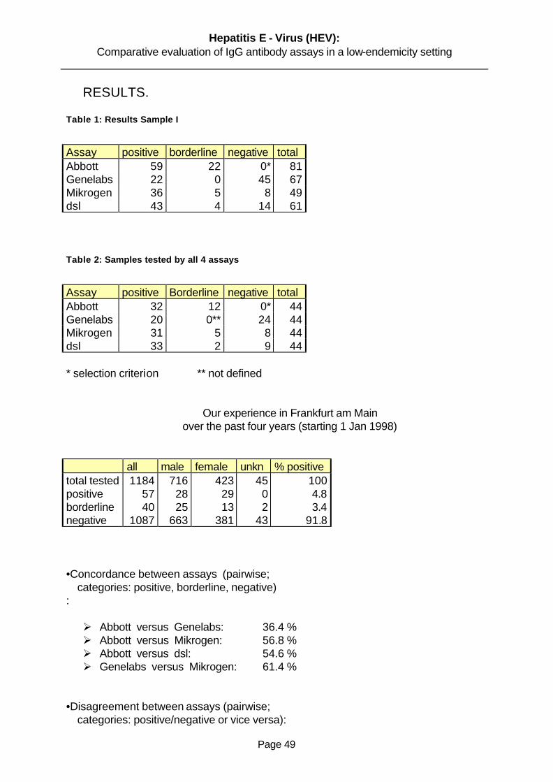
Fifty-nine turned to be positive and twenty-two reactive. This study confirms the high

prevalence of anti-HEV in countries where clinical hepatitis E is not endemic. The reason for

this study could reflect a mixed source of infection. A proportion of human HEV infections

probably occur through exposure in the endemic regions since some of these patients were

workers in these regions. But secondary spread of HEV infections between humans is not

common (2% secondary cases compared to 15% for HAV) (Skidmore et al 1995), and the

majority of the study population had no travel exposure , so this could explain a minority of

cases (Christensen et al., 2002). We had access to serial specimen collected over months

from 81 hepatitis E patients. This specimens demonstrated that IgM antibody levels were

very high soon after illness onset, declined little over several weeks, and then declined rapidly

to low levels over next 4 to 6 months. This is typical of IgM responses to other acute, self-

limited, systemic viral infections (Seriwatana et al.,2002).

The weeks-long duration of markedly elevated IgM levels after disease onset means that the

diagnosis using relatively sensitive IgM detections methods should be successful, even if

patients come to medical attention late. We began this study anticipating that an HEV IgM

Hepatitis E - Virus (HEV): Comparative evaluation of IgG antibody assays in a low-endemicity setting

Page 57

test would improve serological diagnosis on hepatitis E, then based on detection of HEV-

specific IgG. We confirmed that the detection of HEV IgM is the best serological test for

diagnosis of hepatitis E. Yet the most interesting aspect of our work was the observation that

in some cases of hepatitis E, there was a weak or absent IgM response. By combining HEV

IgM and IgG test, we identified primary immune responses among the hepatitis E patients in

areas of HEV IgM to- total IgG ratio.

This is an important observation because previously, some authorities have speculated that

waning immunity explained why most cases of hepatitis E among adults. The fact that more

than 90% of hepatitis E cases in areas of HEV endemicity occur in patients who have a

primary response refutes this speculation of waning immunity, since previously exposed

persons should mount an anamnestic response upon reexposure (Seriwatana et al 2002).

3.2 Genelabs ELISA

Detection of antibodies in sera of patients with Hepatitis E by use of Genelab assay.

Sixty-seven patients with clinical symptoms of acute hepatitis were diagnosed as having

hepatitis E on the basis of detection of antibodies by the Genelabs assay. These 67 samples

were tested by EIA based on the recombinant polypeptides from HEV genotypes

encoded by ORF2 and ORF3 of HEV genome (Genelabs Diagnostics, Singapore). The

assay was performed according to the instruction of the manufacturer. The results showed

that twenty-two of this samples were positive and forty-five were negative. These tests were

conducted at least three times. HEV antibodies were determined by ELISA of distinct

antigenic specificity. In this study we observed a wide range of sensitivity and specificity.

This information implies that this assay might be unreliable for the diagnosis of HEV infection

in areas where hepatitis is not endemic. However, most anti-HEV assays have not been

correlated with the HEV RNA determined by reverse transcription (Lin et al., 2000). In this

study, we evaluated the diagnostic value for acute hepatitis E patients and in long-term

expatriates of commercial anti-HEV IgG and IgM enzyme linked immunosorbent assays

(ELISA) relative to RNA detection. The prevalence of anti-HEV among the general population

in Frankfurt was also re-evaluated with this assay. We found a fairly good sensitivity (70%) of

the IgG anti-HEV for the diagnosis acute hepatitis E verified by HEV RNA. However, the

Hepatitis E - Virus (HEV): Comparative evaluation of IgG antibody assays in a low-endemicity setting

Page 58

sensitivity (50%) of the IgM anti-HEV appeared to be less satisfactory. In previous reports,

anti-HEV detection had a wide range of sensitivity and poor concordance among different

assays (Mast et a.,1998).

The sensitivity of IgG anti-HEV in this study was comparable to that of previous reports

(Favorov et al.,.1998) and (Meng et al ., 1999 ).

However, the sensitivity of IgM was relatively poor. Our study might underestimate the

sensitivity of these assays, since about five of the IgG anti-HEV-negative serum samples has

tested positive for IgG anti-HEV with the same kit in a previous test. These discrepant results

might be due to low-titer antibodies having been destroyed by repeated freezing and thawing

in the later test. There are three possibilities for low sensitivity of IgM anti-HEV in this study.

The first, delayed sampling, might account for negative IgM anti-HEV in some patients.

Although both HEV viremia and serum IgM anti-HEV were short-live (Koshy et al.. 1996 ),

protracted viremia has been reported for as long as one to four months in some patients

(Nanda et al.,1995) IgM anti-HEV might have declined to an undetectable level before

disappearance of HEV RNA. The second possible explanation is sequence variations among

different HEV genotypes. It was reported that IgM anti-HEV were detectable in a patient with

HEV strain US-1 using an assay based on Burmese and Mexican strain (Ferguson et al.,

2002). It is likely that IgM anti-HEV have been undetectable in some of patients infected with a

same genotype HEV using the same assay based on different genotypes (Tian-Chang et

al.,2000). Finally, a poor host immune response to HEV infection might also account for

undetectable IgM anti-HEV in some of our patients, as a evidenced by lower IgG anti-HEV

optical density values in acute hepatitis E patients who were negative for IgM anti-HEV. We

observed that, antibodies status may differ with the stage of disease, and screening of a

population with a significant number of individuals in a convalescent phase could give results

from those present study in terms of immunoreactivity to each of the three recombinant

polypetides (Younchun, W et al.,.2001). The results of our study suggest that some patients

diagnose provisionally as having non-A to non B–Hepatitis in Frankfurt may in fact, have

hepatitis E and a single test for anti-HEV IgG is insufficient for diagnosis. The relatively low

positive rate of circulating antibodies (observed in out test IgG anti-HEV) in the populations of

the endemic areas combined with the unexpectedly high prevalence of anti-HEV in non-

endemic countries like Germany make the interpretation difficult. Because of lack of antibody

Hepatitis E - Virus (HEV): Comparative evaluation of IgG antibody assays in a low-endemicity setting

Page 59

detection assays the significance of HEV infection, that is without doubt a serious infection in

developing countries, might be underestimated in developed countries (Schlauder et al

1999).

In conclusion, the overall sensitivity and specificity of the Genlabs ELISA seemed to be the

same as those shown in previous publications (Li et al, 1998) and (Mast et al, 1998), probably

because both the present and the previous assays used the baculovirus –expressed capsid

proteins. The serological tests with sera collected from hepatitis E patients indicated a that

the circulating IgG antibody was maintained at a high level.

3.3 Mikrogen RecomBlot:

Detection of anti-HEV IgG in sera from patients with hepatitis E.

To select conditions for a sensitive diagnostic assay and to examine the dynamics of

antibody responses to each fusion proteins, Western blots were conducted to detect anti-

HEV IgG from sera from patients with hepatitis E. Out of forty-nine sera selected, thirty-six

were positive, five were reactive and eight negative. Each sample was tested at least three

times in single wells. Any sample which was positive on the either initial test was retested in

duplicate. Samples in which both duplicate wells were positive were designated as a

confirmed positive, all other were considered negative. A Western blot assay was developed

for detection of anti-HEV with three HEV-GST fusion proteins. GST-ORF2.1 and GST-

ORF2.2, which encoded by portions of ORF2

Overlapping at the entire ORF3 from a Chinese strain of HEV. This assay proved to be

sensitive and specific for HEV in tests with sera from patients with different types of acute

hepatitis, and to our knowledge it is the first assay described which detects long –lasting

antibody reactivity in a high proportion of patients and experimentally infected animals (Zhang

et al.,2002). The detection of this persistent antibody reactivity represents an important

advance in the understanding of immunity to HEV infection.

Many serum specimen used in this study were previously tested for anti-HEV IgG with

commercial Abbot tEIA and Genelabs ELISA. This Western blot therefore, shows utility as a

diagnostic assay for HEV infection. While IgM and IgG –class antibodies were detected in

this study, previous publications show a high specific IgM reactivity against the fusion proteins

Hepatitis E - Virus (HEV): Comparative evaluation of IgG antibody assays in a low-endemicity setting

Page 60

in acute-phase hepatitis E sera. However, we observed that the reactivity is not consistent.

The molecular cloning of HEV genome (Li et al, 1994 and Jameel et al 1999) and the

expression of recombinant proteins have allowed the development of numerous diagnostic

and research immunoassays. However, problems remain both with specificity of some

assays particularly when applied to seroepidemiological study as observed in our study

(Thomas et al 1997; Mast et al 1998). One major problem in the seroepidemiological studies

of HEV infection is the variable reactivity of recombinant antigens with respect to detection of

past infection (Mast et al 1998; Scholfield et al 2000). Many HEV antigens, including synthetic

peptides, recombinant antigens based on ORF3, and most fragment of ORF2 when

expressed in Escherichia-coli, have reactivity with acute phase sera but little or variable

reactivity with convalescent sera (Mast et al 1998). For example, the ORF2 antigen 3-2(M)

was reactive with IgG from 91% of HEV-infected patients from Egypt at the time of admission

, but fell to between 27 and 50% at 6-12 months after admission., while analogous 3-2(M)

antigen was unreactive with only 64% of the sera at admission and none after (Goldsmith et al

1992) Conversely, the 3-2(M) was unreactive with convalescent sera from patient infected in

Pakistan, while the 3-2(B) antigen reactive with sera collected from patients 4.5 years after

illness (Dawson el al, 1992). Thus while many patients become unreactive, other patients

maintain high levels of antibody to these proteins, which makes the interpretation of reactivity

difficult as shown in this study. This may also be true of the ´mosaic´antigens (Favorov et al ,

1996) expressed in E. coli as represents the fusion of a number of linear peptide epitopes.

As consequence of the variable , but generally low rate or reactivity with convalescent sera,

these antigens have had some use for the diagnosis of acute infection in non-endemic areas

by the detection of HEV-specific IgG , but prevalence of past infection in endemic areas must

be underestimated .

Conversely, a second group of recombinant antigens appear to demonstrate consistent

reactivity with both acute and convalescent phase sera. These antigens include HEV virus-

like or subviral particles (SVP´s) formed by truncated ORF2 expressed in insect cells using

the baculovirus system (Tsarevet al, 1993; Li et al, 1999) and ORF2.1 expressed in E.coli.

Such antigens are logically the most suitable for seroepidemiological studies, although the

titre of anti-HEV may decline rapidly following the acute phase, it appears to remain at

detectable levels for many years (Favorov et al, 1996).

Hepatitis E - Virus (HEV): Comparative evaluation of IgG antibody assays in a low-endemicity setting

Page 61

The observations that the presence of such antibody correlates with protection from infection

during epidemics (Bryan et al,1994) and that passive immunisation is sufficient to confer

immunity to HEV disease in macaques (Tsarev et al, 1994) underline the practical value of

detection this log-lasting antibody to HEV. Ghabral et al,1998 have recently compared assay

for HEV-specific IgG and IgM using recombinant antigens from either baculovirus-expressed

SVP´s or E. coli expressed fragments of ORF3 and ORF2. Their findings demonstrate the

improved sensitivity of the SVP´s ELISA for detection of past HEV infection, but the detection

of very high rates of reactivity in presumed non-endemic populations in the USA using this

assay (Thomas et al,1997) raise the questions regarding the specificity of the assay for IgG-

anti HEV. The results of this study also have implications for the development for the

development of more convenient assays for HEV antibody by other methods. First, the

reactivity of the HEV fusion proteins in Western blot appears to be greater than that of native,

synthetic proteins in an Abbott-EIA (Dawson et al., 1994).

3.4 Diagnostics Systems Laboratories ( DSL) ELISA

Acute hepatitis E has been rarely been reported in industrialized countries, but the rate of

seroprevalence of hepatitis E virus (HEV) is inappriopriately high. The sensitivity and the

specificity of the assay used to test for immunoglubulin G (IgG) and IgM anti-HEV have not

been well established in areas where hepatitis is not endemic ( hereafter referred to as ´non-

endemic areas´). Enzyme immunoassay based on recombinant proteins of HEV have been

used for most prevalence studies. The recombinant proteins contain immunodominant

epitoped encoded by open reading frame (ORF2) and (ORF3) of the HEV genome from

Burmese strain. A wide range of sensitivity and specificity has been reported for this assay

(Bhaduree et al 1998). This information implies that this assay might be unreliable for the

diagnosis of HEV infection in areas where hepatitis E is not endemic especially Germany.

We collected serum samples from sixty one HEV patients and to test for IgG anti-HEV by

enzyme-linked immunoabsorbent assay (ELISA). Forty three were positive for IgG anti-HEV,

fourteen were negative for anti-HEV and four borderline for IgG anti-HEV. IgG class were

measured according to the Diagnostic Systems Laboratories (DSL) procedure.

Hepatitis E - Virus (HEV): Comparative evaluation of IgG antibody assays in a low-endemicity setting

Page 62

3.5 Concordance Between Assays

3.5.1 Evaluation Of Abbott EIA Versus Genelabs ELISA

The results of serologic tests for hepatitis E have varied widely from laboratory to laboratory,

making interpretation of seroepidemiologic studies difficult. This study compares serologic

results with different antigens and tests developed by two laboratories (Abbott EIA and

Genelabs ELISA) for their ability to diagnose hepatitis E and measure antibody prevalence in

area where hepatitis E is not endemic. The concordance between both assays in specificity

and sensitivity were 34,4% but 50 % disagreement. Our results does not differ from other

reported publications (Purcell et al., 1998), (Aggrawal et al, 2000), (Seriwatana et al, 2002).

The performance of tests for antibody to the hepatitis E virus anti-HEV is an important factor

in accessing the epidemiology of hepatitis E infection. Although these assays are specific,

they have limited sensitivity: anti-HEV has been detected in only 50% to 70% of patients with

acute hepatitis during hepatitis E outbreaks, and anti-HEV titres decline to subdetectable

levels within several months after acute detection. Several recombinant protein-based tests

have demonstrated increase sensitivity compared to prior assays, detecting anti-HEV in 90%

to 95% of patients with acute hepatitis during outbreaks of hepatitis E in HEV endemic areas

(Mast et al, 1997).

Recently, a number of cases of hepatitis E, diagnosed on the basis of serologic testing, have

been reported among persons who had no history of travel to endemic areas. (Christensen et

al, 2002).

However, the interpretation of these findings is problematic because few data are available

to evaluate the performance of anti-HEV assays for diagnosis of acute hepatitis E in this

setting. In addition, the performance of these assays in detecting anti-HEV in persons with

remote infection is unknown, and several studies have reported unexplained positive anti-

HEV results among persons who do not have disease or unknown exposure to HEV (Thomas

et al., 1997). We present the findings of a serum panel evaluation conducted to access the

sensitivity and specificity of available tests for anti-HEV and to assess the variability in

detecting anti-HEV among tests. The results of this study indicate that several of the

recombinant protein assays have an adequate combination of sensitivity and specificity to

perform well for this purpose. The peptide-based assays were generally much less sensitive

Hepatitis E - Virus (HEV): Comparative evaluation of IgG antibody assays in a low-endemicity setting

Page 63

compared to the recombinant protein assays and are therefore likely to be non reactive in a

high proportion of acute hepatitis E cases. Further comparative studies that includes testing

for immunoglobulin M IgM –anti HEV would be useful to validate the performance of the

recombinant protein assays for the diagnosis of hepatitis E. In addition, the performance of

these assays for diagnosis of acute hepatitis E in persons who do not have a history of travel

to HEV-endemic regions need to be determined.. In prior studies, HEV isolates from various

geographic regions have been demonstrated to have at least one major cross-reactive

epitope by a variety of serologic assays.(Favarov et al, 1994 ;Bradley et al 1998, Krawczynski

et al, 2000).

However, substantial variation in detection of anti-HEV by these tests in acute and

convalescent-phase has been found in sera from chimpanzees infected with HEV isolates

from various geographic regions. One possible reason for this findings is differences in the

geographic strain-specific antigenic domains in these tests (Scholfield et al, 2000). However,

there is little variation in the RNA sequence of ORF2 among HEV isolate from various

geographic regions. Moreover, some assays (Scholfield et al, 2000), did not detect the anti-

HEV in chimpanzee sera even though these tests included ORF3 epitopes from the same

geographic region as the chimpanzee inoculum. The seroreactivity of recombinant proteins

may also vary if they are produced in different expression system or used in different tests

formats (i.e.,Abbott EIA vrs Genelabs ELISA). In addition, all these assays were designed to

detect human antibody, and the differences may exist in the ability of assay conjugate to

detect chimpanzee antibody. However, if the assay conjugate were the reason for a test´s not

detecting anti-HEV, the assay would be expected either to be non reactive in all the

chimpanzee sera or to have a uniform decline in seroreactivity in chimpanzee sera compare

with human sera.

Furthermore, seroprevalence studies among blood donors in some non-endemic countries

have found an anti-HEV prevalence of 1% to 20%, which is relatively high compared to the

low rate of clinically evident disease associated with HEV in these areas. (Paul et al, 1994

,Yarbough et al, 1997). In one study, anti-HEV seroreactivity among persons living in non-

endemic regions with increasing age and was associated with a travel to endemic regions

findings that are consistent with prior HEV infection (Yarbough et al., 1997). Thus, the

interpretation of seroreactivity among patients living in non-endemic regions is currently

Hepatitis E - Virus (HEV): Comparative evaluation of IgG antibody assays in a low-endemicity setting

Page 64

problematic. We therefore suggest that, further studies need to determine highly discrepant

results among blood donor sera. Studies are also needed to determine the significance of

anti-HEV seroreactivity among persons living in non-endemic HEV areas, including the

relation of seroreactivity to exposure to the recently discovered virus in pigs that is closely

related to human HEV isolates (Purcell et al, 1997). Finally, improved tests are needed for

use in seroprevalence studies in nonendemic regions and confirmation tests are needed to

very the specificity of these assays as shown in our study.

3.5.2 Evaluation of Abbot EIA Versus Mikrogen RecomBlot

Despite advances in knowledge of the molecular of biology of HEV over the past decade, the

diagnosis and seroepidemiological study of HEV infection have remained problematic.

Although a large number of serological assays have been developed for HEV, (Mast el al,

1998), have demonstrated that there is a significant lack of concordance between many

assays, with major problems of many assays failing to detect anti-HEV in a convalescent sera

and/or against heterologous HEV strains. This was a particular problem with assays based

on synthetic peptides and many recombinant proteins expressed in E. coli, however, assay

based on SVP´s expressed in the baculovirus system (Mast et al 1998), and Western

immunoblots based on the ORF2 protein Anderson et al, 1999), appear to have a satisfactory

sensitivity for analysing convalescent sera and divergent strains. In our study, we found a

concordance of 56% between Abbot EIA and Mikrogen Western blot and a disconcordance

of only 9,1%. We have observed a high rate of divergent results in using both assays. The

results show clearly an ELISA based on ORF2 fragment offers sensitive and specific of HEV-

specific IgG in both acute and convalescent sera for divergent strains of human HEV. It is

considered that the utility of this antigen is due to the efficient presentation of a highly

conserved, confirmational epitope which is immunodominant in convalescent antibody

response.(Anderson et al, 1999). It was observed recently that ORF2 also has optimal

reactivity with convalescent sera from domestic pigs naturally infected with a swine HEV in

Australia (Chandler et al, 1999). Taken together, the results suggest that the use of

conserved epitopes such as ORF2 for detection of anti-HEV may be more effective than the

use of multiple, strains-specific epitopes which is by definition dependent on identifying all

possible strains of the virus .However, further studies will be required to establish the reactivity

Hepatitis E - Virus (HEV): Comparative evaluation of IgG antibody assays in a low-endemicity setting

Page 65

of ORF2 with numerous distinct HEV genotypes which have recently been described. (Hsieh

et al, 1998; Erker et al, 1999; Schlauder et al, 1999; Wang et al, 1999).

It has been shown in previous studies, that using SVP ELISAs levels of HEV-specific IgG fall

by approximately 90% following the acute phase infection, but then remain stable (Tsarev et

al, 1993; Bryan et al, 1994) This antibody is also thought to correlate with immunity to

reinfection (Bryan et al, 1994), and it is therefore appropriate to measure the levels of specific

anti-HEV rather than only the presence antibody. The above listed HEV epitope may one of

reasons high divergent of these assays used in our study. In this regard, the quantitative

characteristics of the IgG ELISA reported should prove very useful, and the availability of the

international Reference HEV serum reported by Fergusson et al, 2002 should further assist in

studies of HEV immunity. Previous reports of both Abbot EIA and Western blot assays for

anti-HEV have described similar rates of detection for acute hepatitis E (Mast et al, 1998)

however, the detection of past infection appears to be less efficient, with rapid decline in

antibody reactivity within weeks and months.

It is believed that the IgG ELISA based on ORF2 antigen offers a reliable method for the

detection and quantitation of antibodies against HEV. The results of this study also have

implications for the more convenient assays for HEV antibody by other methods .The use of

both assays should assist in greater understanding of HEV immunity and the epidemiology of

HEV world wide.

3.5.3 Evaluation of Genelabs ELISA Versus Mikrogen RecomBlot

The ELISA reported here detects both anti-HEV-IgM and anti-HEV-IgG and it is a convenient

method for the diagnosis of acute or past HEV infection. To characterize the relationship

between antibody potency determined by a widely used commercial test kits. In this study, we

compared the sensitivity and specificity of both assays Genelabs EIA and Mikrogen Western

blot. We observed a concordance of about 61,4% and a high disagreement of about 27,3%

between the assays. The commercial test employ a mixture of a recombinant HEV ORF2 and

ORF3 polypeptides expressed in E. coli. The disagreement in our study have raised the

question about the sensitivity and specificity of these current available test for ant-HEV as well

as questions about how long anti-HEV can be detected after infection of HEV. The variable of

persistence of IgM anti –HEV and IgG anti-HEV makes it difficult to accurately determine the

Hepatitis E - Virus (HEV): Comparative evaluation of IgG antibody assays in a low-endemicity setting

Page 66

frequency of prior exposure and raises the possibility of re-infection after disappearance of

this antibody (Zhang et al.,2002).This might allude to a high disconcordance in our study. The

cloning of HEV and the availability of EIA ´sand GST fusion protein for detection for detection

of anti-HEV have created a major breakthrough in our understanding of epidemiology and the

clinical course of hepatitis E. However, it must be realized that the current diagnostic assays

for hepatitis E are sub-optimal. False positive and false negative results may occur, as was

the case in this study.

Although, there is a high degree of homology among HEV sequences from Pakistani and the

Burmese isolates, the homology between Mexican and Burmese isolates is less.(Purcell et al,

1998). Thus, EIA that utilize HEV antigens from one ORF or one isolate my yield negative

results. In all these studies in which multiple HEV antigens in both ORF2 and ORF3 from

Burmese and Mexican isolates were used, the detection rate for individual antigens from 0 to

100% among the reactive samples. (Krawczynski et al. 1999). Comparative studies with HEV

antigens from other isolates have not been conducted. However, it is obvious that multiple

HEV antigens from more than one isolate have to be incorporated into anti-HEV EIA´s for the

diagnosis for hepatitis E in diverse geographical areas (Mast et al, 1998). There is less

information on the specificity of the current anti-HEV assays. Confirmatory tests for HEV

infection has to be developed. Direct comparison between the reactivity with recombinant

HEV antigens and the corresponding synthetic peptides showed that smaller proportion of

healthy subjects reacted against the synthetic peptides in the IgG anti-HEV assays,

suggesting that EIA using recombinant antigens may give false positive results. (Seriwatana

et al, 2002). Similar comparison has to be conducted on the IgM anti-HEV EIA´s as reviewed

in this study.

Hepatitis E - Virus (HEV): Comparative evaluation of IgG antibody assays in a low-endemicity setting

Page 67

3.5.4 Evaluation of Abbott EIA Versus DSL ELISA

The results of serologic tests for hepatitis E virus have varied widely from laboratory to

laboratory, making interpretation of seroepidemiologic studies difficult.

The present comparison of tests for anti-HEV is first to make a detailed examination of the

utility of the many gene products which have been utilized for assays for anti-HEV .In our study

we compared the reactivity of anti-HEV IgG in both assays Abbot EIA and DSL ELISA. We

observed a concordance of 54% and a disconcordance of 13,6%. The results of this test

clearly showed that tests for anti-HEV based upon expressed ORF2 were more sensitive for

detecting anti-HEV than were tests based upon antigens derived from ORF3 (Ghabrah et al,

1998). This was true for both IgG anti-HEV and IgM anti-HEV. The poorer showing of ORF3-

based tests may result from a combination of a less vigorous immune response to this small

protein and a shorter half-life of antibodies to ORF3. We observed a wide range of sensitivity

and specificity as shown in our results 54% concordance. The reasons for the discrepancy

between both assays are not clear. We probably believed , however, that these assays of the

IgG class antibodies often missed remote HEV infections because of their low sensitivity in

areas where hepatitis E is not endemic. Thus, a reliably sensitive and specific antibody assay

is needed to conduct accurate epidemiological studies. Further reason for a wide range of

discrepant results might be due to low-titre antibodies having been destroyed by repeated

freezing and thawing in later study. Another possible explanation for decline detectability of

IgG antibodies in these assays might be sequence variations among the different HEV

genotypes. Specifically, the choice and size of antigen appears to make a significant

difference in the results (Nishizawa et al, 2003). Finally a poor host immune response to HEV

infection might also account for undetectable IgG anti-HEV optical density values.

Hepatitis E - Virus (HEV): Comparative evaluation of IgG antibody assays in a low-endemicity setting

Page 68

4 Discrepancies

These findings have raised the questions about the sensitivity and the specificity of the

currently available test for anti-HEV as well as questions about how long anti-HEV can be

detected after infection .For instance, Goldsmith et al 1992 found out that IgG anti-HEV could

be detected for less than 6 months in about half tested children who were convalescing from

hepatitis E but Bryan et al 1994 found all of 33 convalescent adults to be positive 20 months

after infection and anti-HEV has been detected by Kharoo et al 1993 as long as 14 years

after clinical hepatitis E in India. Many of these unexpected results and discrepancies can be

ascribed to differences in assays for the anti-HEV. Specifically, the choice and the size of the

antigen, appears to make a significant difference in the results. Assays for anti-HEV based

on antigenic epitopes of the ORF-3 gene product detect a lower prevalence of anti-

prevalence suggesting these antibodies directed against epitopes of the products of ORF-2.

This problems is compounded by a greater genetic heterogeneity of ORF-3 genes, possibly

leading to serologic differences among different HEV strains, and diminished sensitivity of

assays based upon only one or a limited number of genetic variants of the ORF-3 product. In

contrast, gene products of ORF-2 are more genetically homogeneous and measure anti-HEV

that remains detectable for years. Clinical disease data suggest that hepatitis E virus (HEV)

is a pathogen with a restricted geographical range. The purpose of our study was to

compare hepatitis E assays:

Abbott EIA, Mikrogen Westernblot, Genelabs and DSL ELISA (a Prototype ) of their

sensitivity and specificity and to evaluate their abilities to diagnose hepatitis E with acute

sporadic viral hepatitis and convalescent phase sera in a routine test at the Virology

Department of Frankfurt University clinics between 1998-2001.

Hepatitis E - Virus (HEV): Comparative evaluation of IgG antibody assays in a low-endemicity setting

Page 69

Possible explanations of discrepances can be allude to the following reasons:

- a low sensitivity of available assays?

- specifically choice and size of HEV antigen?

- duration of antibody persistence ?

- subclinical (anicteric ) HEV infection ?

- a cross-reactivity with a different agent ?

- a non-specificity leading to false positivity ?

- infection with non-pathogenic HEV strain, e.g. swine HEV or zoonotic strain ?

- and a combination of any of the above?

Hepatitis E - Virus (HEV): Comparative evaluation of IgG antibody assays in a low-endemicity setting

Page 70

4.1 Problems and Conclusions

The study of Hepatitis E has been an instructive story recent advancements in medical

science. Substantial progress has been made in a short period of time through both

epidemiological studies and basic research, which has relied on modern cloning and

recombinant technology. New tools have clearly accelerated the pace of research. Within less

than two decades of the discovery of HEV, the major epidemiological features of this unique

pathogen have been described, serologic tests have been developed, and a vaccine has

been evaluated in an initial clinical trial. (Yabough et al., 1999).

Nevertheless, there are still may mysteries about the epidemiology, pathogenesis, and

immune response of HEV infection. The unknown aspects of hepatitis E could have a major

impact on vaccine development and use.. However, vaccine research could be also illuminate

the immunologic characteristics of this unique infectious disease. The development of an

animal model of HEV infection in the laboratory would aid research efforts substantially. It is

likely that a HEV vaccine will be developed that provides at least short-term protection from

disease.

Who will make and buy this vaccine? Because of incidence of acute HEV infection among

travellers from developed world to endemic countries has been estimated to be less than one

million (Piper-Jenk et al 2000), the market for this vaccine in non endemic countries will be

limited.(Shlim et al., 2000). Pregnant women living in endemic regions may derive the

greatest benefit from an effective HEV vaccine.

Hepatitis E - Virus (HEV): Comparative evaluation of IgG antibody assays in a low-endemicity setting

Page 71

Our study has led us to draw the following conclusions:

- we observed a high rate of divergent results using three commercially available and

one ´´prototype´´HEV IgG antibody assays on sera reactive by the Abbott EIA

- This is in agreement with previous studies that used variety of different assays.

- The interpretation of HEV antibody test results in ´´low-risk´´ populations remains

problematic

- A ´´grey zone´´ needs to be defined as low-level reactivity is common and mostly

cannot be confirmed by additional tests but is probably non-specific.

Hepatitis E - Virus (HEV): Comparative evaluation of IgG antibody assays in a low-endemicity setting

Page 72

5 Summary

Hepatitis E virus (HEV) is a positive-stranded RNA virus with a 7.2 kb genome that is capped

and polyadenylated. The virus is currently unclassified : the organisation of the genome

resembles that of the Caliciviridae but sequence analyses suggest that it is more closely

related to the Togaviridae. HEV is an enterically transmitted virus that causes both epidemics

and sporadic cases of acute hepatitis in many countries of Asia and Africa but only rarely

causes disease in more industrialised countries. Initially the virus was believed to have a

limited geographical distribution. However, serological studies suggest that that HEV may be

endemic also in the United states and Europe even though it infrequently causes overt

disease in these countries. Many different animal species worldwide recently have been

shown to have antibodies to HEV suggesting that hepatitis E may be zoonotic. Although two

related strains have been experimentally transmitted between species, direct transmission

from animal to a human has not been documented.

Our main objective in this study is to evaluate the suitability of current available HEV antibody

assays for use in low-endemicity areas such as in Germany.

Methods: We selected sera on the basis of at least borderline reactivity in the routinely used

Abbot EIA. Most were tested as part of routine screening of long-term expatriates in endemic

countries. The following assays (recombinant antigens : ORF2 and ORF3) were used: Abbot

EIA, Genelabs ELISA, Mikrogen recomBlot and a `Prototype´ DSL-ELISA.

Hepatitis E - Virus (HEV): Comparative evaluation of IgG antibody assays in a low-endemicity setting

Page 73

We observed a wide range of sensitivity ( average of 56.8%) and specificity ( an average of

61.4%) in these used assays. These results implies that , these assays might be unreliable for

detection of HEV infection in areas where hepatitis E is not endemic. However, most anti-

HEV assays have not been correlated with the HEV RNA determined by reverse

transcription. Many of these unexpected results and discrepancies can be alluded to the

following reasons:

I. The choice and the size of the HEV antigen.

II. Duration of the antibody persistence

III. A cross reactivity with different agent

IV. Due to geographic species

V. A low sensitivity of the available assays.

VI. And infection with non-pathogenic HEV strain. (zoonotic strain?).

We therefore suggest that, further studies will be required to improve the sensitivity and specificity of the available commercial assays on the market.

Hepatitis E - Virus (HEV): Comparative evaluation of IgG antibody assays in a low-endemicity setting

Page 74

6 Literature

A.J. Hall Hepatitis in travellers: Epidemiology and Prevention Pub:British Medical Bulletin(1993)Vol.49, No 2, pp:382-393

F. .Li, H. Zhuan, S. Kolivas, S.A. Locarnini, D.A. Anderson Persistence and transient Antibody Responses to Hepatitis E Virus detected by Western immunoblot using Open Reading Frame 2 and 3 and Glutathions- Transferase Fusion Proteins Pub: JCM, Sept.1994, pp: 2060-2066

J.P. Bryan, S.A. Tsarev, M. Iqbal, J. Ticehurst, S. Emerson, A. Ahmed, J. Duncan, Epidemic Hepatitis E in Parkinstan : Patterns of Serologic Response and Evidence that antibody to Hepatitis E Virus protect against disease Pub:JID1994;170, September: pp5: 17-521

Acute Sporadic Hepatitis E Sudanese Children: Analysis Based on a New Western Blot Assay K.C. Hyams M.A. Purdy M. Kaur, M.C. .McCarthy, M. A. Hussan E. Tigani K .Krawczynski D. W. Bradley M. Carl. Acute Sporadic Hepatitis E virus: Pbu:JID1992;165(June)pp:10011005

R. Goldsmith P.O Yarbough G .R. Reyes K. E. Fry, K: A: Gabor, M. Kamel , S .Zakaria S.Amer Enzyme-Linked immunosorbent assay for Diagnosis of acute sporadic Hepatitis E in Egyptian children. Pub; Lancet Vol:339, Feb8,1992, pp328-331

Evaluation of Assays for Antibody to Hepatitis E Virus by a Serum Panel E. Mast, M .J .Alter, P. V. Holland R. H .Purcell. Pub: Hepatology March,1998.pp: 857-861

Hepatitis E - Virus (HEV): Comparative evaluation of IgG antibody assays in a low-endemicity setting

Page 75

D.A. Anderson, F .Li, M. Riddell, T .Howard H. F Seow J. Torresi ,G. Perry, D. Sumarsidi, S. Mshrestha, I. L. Shrestha Elisa for IgG-Class antibody to Hepatitis E Virus based on a highly conserved, conformational epitope expressed in E-coli Pub:JVM81(1999)

M. L .Mateos C. Camarero, E. lasa, N. Mir, F. Baquero Hepatits E Virus: Relevance in Blood Donors and other Risk Groups Pub:Vox Sanngiunis1998:pp.267-269

E. T .Clayson K.S.A.Myit,R.Snifbhan,D.W.Vaughn,B.L.Innis,L.Chan,P.Chan,M.P.Shreath A viremia faecal shedding and IgM and IgG response in patients with Hepatitis E Pub:JID1995 (Oct.172),pp9:27-933 Krawczynski K. Hepatology 1993;17 923-41

Y. Poovorawan, A .Theamboonlers, S. Chumdermadsetsuk, P. KomolmitC .P. Thong Prevalence of Hepatitis E Virus infection in Thailand Pub. Annals of Tropical medicine and Parasitology, Vol: 90,No2,189-196 (1996) R. McCrudden, S. O´Connell, T .Farrant, S. Beaton J:P Iredale ,D. Fine Sporadic acute Hepatitis in the U.K : an underdiagnosed phenomen? Pub: Gut 2000.46:pp: 732-733 S. D. Lee, Y. Wang, R .H. Lu, C. Y. Chan, K. J. Lo R. Moeckl Seroprevalence of Antibody to Hepatitis E virus among Chinese Subjects in Taiwan Pub: JVM 81 (1999),pp: 150-153

W. Bernal, H. M. Smith ,R. Williams A Community Prevalence Study of Antibodies to Hepatitis A and E in inner-City London Pub:JMV49 1996,pp:230-234

D. W. Bradley Hepatitis E virus: a brief review of the biology, molecular virology, and immunology of a virus Pub:JH1995,22, pp:140-145

Hepatitis E - Virus (HEV): Comparative evaluation of IgG antibody assays in a low-endemicity setting

Page 76

R. H. Purcell : Hepatitis E Pub: Fields Virology pp:2831-2843

Epidemiology and serological diagnosis of hepatitis E S. F .Anna, K&C. Consuelo Soldevila-Pico Pub:JH1994,20:567-567

T. J. Tucker, R. E. Kirsch, S. J. Louw, S. Isaacs, J. Kannemeyer, S. C. Robson Hepatitis E in South Africa: Evidence for Sporadic Spread and increase seroprevalence in Rural Areas. Pub: JMV50: 1999, pp:117-119

A. Guanaid, T. M. Nasher, M. Abdulkader, E. Guneid, M. Hill, R. Dayton, A. Pal, S. J. Skidmore, J. C. Coleman, I. M. Murray-Lyon Acute Sporadic Hepatitis in the Republic of Yemen Pub:JMV51:1997,pp:64-66

R. Aggarwal and S.R. Naik Hepatitis E: intrafamilia transmission versus waterborne spread Pub.JH1994:21,pp.718-723

Vishwanathan R. Hepatitis E: Indian J. Med. 1957;45: 1-29

Khuroo MS Hepatitis E America J. Med.1980;68:818-23

S.H. Hussani. S.J .Skidmore, P. Richardson, L.M. Sherratt, B.T. Cooper, J.G. O´Grady Severe hepatitis E infection during pregnancy Pub:JVH,1997,4.pp:51-54

M. Buti, R. Jardi ,F .Rodriguez-Frias, J. Quer, R. Esteban, J. Guardia Etiology of acute sporadic hepatitis in Spain the role of hepatitis C and E viruses Pub:JH94,4,pp:589-592

Hepatitis E - Virus (HEV): Comparative evaluation of IgG antibody assays in a low-endemicity setting

Page 77

M. A. Psichogion, N. C. Tassopoulos, G.V .Papatheodoridis, E. Tzala, R. Klarmann, H. Witteler, G. G. Schlauder, H. Troonen, A. Hatzakis Hepatitis virus infection in a cohort of patients with acute non-A, non-B hepatitis E Pub:JH1995,23:pp668-673

Anti-HEV antibodies in acute hepatitis in France Letters to the Editor Pub:JH1994,20

Hospital outbreak of hepatitis E Pub :The Lancet, Vol 339:June1992

S.M. Firestone Non-A, Non-B Hepatitis, including HEV Viruses Pub: Principles and practice of infectious diseases,3.Edit.pp:1412-1415

M. A. Kamel, H. Troonen, H. P. Krapprell, A .El-Ayady, F .D. Miller Seroepidemiology of Hepatitis E virus in the Egyptian Nile Delta Pub: JMV 47,1995, pp:399-403

Experimental Infection of the Laboratory Rat with the Hepatitis E Virus Y. Maneerat, E. T. Clayson, S. A. Khin, G.D. Young, B.L .Innis Pub:JMV48,1996,pp121-128

Epidemiologic Notes and Reports: Hepatitis E among U.S Travellers,1989-1992 Pub: CDC, Jan.15,1993/Vol42/No.1

Naik SR Bullentin WHO 1992;70 597-604 Khuroo M.S JMV 1993; 40:181-6

CDC Weekly Rep. 1993;42:1-4

Dawson J.M Solid-phase enzyme-linked immunosorbent assay hepatitis E IgG and IgM antibodies utilizing recombinant antigens and synthetic peptides JMV 1992,38:50-8

Hepatitis E - Virus (HEV): Comparative evaluation of IgG antibody assays in a low-endemicity setting

Page 78

McCaustland K.A, B. S, Purdy MA, Bradley D.W. Application of two RNA extraction methods prior to amplification of hepatitis E virus nucleic acid by PCR. JVM 1991;35;35-331-42

Ticehurst J: Identification and characterization of hepatitis E viruses. Viral Hepatitis and Liver Diseases. Baltimore W&W, 1991;501-13

Favorov M.O, Fields H.A, Purdy M.A Serologic identification of hepatitis E virus infections in epidemic and endemic settings. JMV 1992;36:246-50

Krawczynski, K Bradley, D.W. Enterically transmitted non-A, non-B hepatitis; Identification of viruses-associated antigen in experimentally infected cynomolgus macaques. JMD 1989;159:1042-9.

Tsarev SA, Tsereva TS, Emerson S.U ELISA for antibody to hepatitis E virus based on complete open-reading frame 2 expressed in insect cells: Identification of HEV of HEV infection in primates. JID 1993;168:369-78

Bradley DW. Hepatitis E epidemiology, aetiology and molecular biology. Rev. Med. Virol.ogy 1992;2:19-28

Xiang-Jin Meng: Zoonotic and xenozoonotic risks of the hepatitis E virus, Infection Diseases Review:2002;2(1):35-41

Tsutomu N, Masaharu T, Hitoshi M, Haruko M, Yuhko G, Hiroaki O Characterization of Japanese swine and human hepatitis E viruses isolates of genotype IV with 99% identity over entire genome: Journal of General Virology (2003);84, 1245-1251

Masaharu T, Haruko M, Teruhiko I, Fumio T: Swine hepatitis E virus strains in Japan form four phylogenetic clusters comparable with those of Japanese isolates of human hepatitis E virus: Journal of General Virology (2003);84, 851-862

Hepatitis E - Virus (HEV): Comparative evaluation of IgG antibody assays in a low-endemicity setting

Page 79

Harald W, Wim H.M, van der Poel, Gerald B Hepatitis E: an overview Microbes and Infection 4(2002) 657-666

Emerson S, Purcell RH: Hepatitis E: A Review Review Medical Virology, (2003);13:145-154

Murhekar M, Sehgal SC, Padbhidri SP, Chitamber SD, Arankalle VA Changing scenario of hepatitis E virus exposure among the primitive tribes of Andaman and Nicobar Islands, India over the 10-year period 1989-1999: Journal of Viral Hepatitis(2002):9, 315-321

Junkun H, Innis BL, Mrigendra P, Clayson E, Scott R, Binn L, Vaughn D Evidence that Rodents are a Reservoir of Hepatitis E Virus for humans in Nepal: Journal of Clinical Microbiology (December 2002),p 4493-4498

Engle R, Yu C, Emerson S, Meng X-J, Purcell RH Hepatitis E (HEV): Capsid antigens derived from viruses of Human and Swine origin are equally efficient for detecting anti-HEV by Enzyme Immunoassay; Journal Clinical Microbiology (December 2002)p:4576-4580

Widdowsin M, Jaspers W, Wim H, Froukje V, Koopmans M Cluster of cases of acute hepatitis associated with hepatitis E virus infection acquired in the Netherlands Clinical Infectious Diseases(2003);36:29-33

Obriadina A, Meng J, Ulanova T, Trinta K, Burkov A, Fields H A new enzyme immunoassay for the detection of antibody to hepatitis E virus :Journal Gastroenterology Hepatology (December 2002)17:360-364

Zhang M, Emerson SU, Nguyen H Recombinant vaccine against hepatitis E: duration of protective immunity in rhesus macaques. Vaccine 2002;20:3285-3291

Zhang M, Emerson SU, Nguyen H, Immunogenicity and protective efficacy of a vaccine prepared from 53kDa truncated hepatitis E virus capsid protein expressed in insect cells. Vaccine 2002; 20:853-857

Hepatitis E - Virus (HEV): Comparative evaluation of IgG antibody assays in a low-endemicity setting

Page 80

Santosh M. S, Shrestha S, Tsuda F, Nishzawa T, Gotanda Y, Takeda N, Okamoto H : Molecular Investigation of Hepatitis E Virus Infection in Patients with acute hepatits in Kathmandu, Nepal : Journal of Medical Virology (69): 207-214(2003)

Arankalle V.A, Chobe L.P, Walimbe A.M, Yergolkar P.N. and Jacob G.P. Swine HEV Infection in South India and the Phylogenetic Analysis (1985-1999) Journal of Medical Virology(69) 391-396 (2003)

Hepatitis E - Virus (HEV): Comparative evaluation of IgG antibody assays in a low-endemicity setting

Page 81

7 Danksagung

Abschließend möchte ich zunächst den stets freundlichen Damen unseres virologischen Labors danken, allen voran Fr. Gabriele Schulz, die mich mit unermüdlicher und nie endenwollender Geduld in die Methodik eingeführt hat und mir auch später hilfreich zur Seite gestanden hat, wenn es um die praktische Durchführung der Untersuchungen ging. Ihr und allen ihren ihren ebenso freundlichen Mitarbeiterinnen Fr. Gabriele Schön alias „Metzi“, Fr. Katja Prosser und Fr. Sibylle Evers, ohne die diese Arbeit nicht möglich gewesen wäre, an dieser Stelle ein herzliches Dankeschön.

Dank gebührt Herrn Dr. med. Wolfgang Preiser, der die gesamte Arbeit mit seiner konstruktiven Kritik und Ermutigung begleitet hat und meine Gedanken stets wieder in die richtigen Bahnen geleitet hat. Er hatte immer ein offenes Ohr für meine Fragen und war tags wie nachts bereit, mit mir die Ergebnisse zu diskutieren, so oft seine wertvolle Zeit dies zuließ.

Dank an Herrn Prof. Dr. Holger Rabenau dafür, dass er mich beständig motiviert hat, die Arbeit erfolgreich zu beenden. Mehr als einmal hat er durch neue Impulse zum Fortschritt der Arbeit beigetragen.

Ferner danke ich meinen beiden langjährigen Freunden Marcus Cap und Wilhelm Quast dafür, dass sie immer dann, wenn es nötig war, ihre Zeit und Energie eingebracht haben und mich so unterstützt haben.

Meinen ganz besonderen Dank möchte ich, meinem Doktorvater Herrn Prof. Dr. med. Hans Wilhelm Doerr aussprechen, der das Zustandekommen dieser Arbeit erst ermöglicht hat und mich von Anfang an mit seiner interessierten und väterlichen Anteilnahme begleitet und unermüdlich unterstützt hat. Für seine besondere Großzügigkeit, die es mich nie an Mitteln für meine Untersuchungen fehlen ließ und mir ermöglichte, meine Ergebnisse auf wissenschaftlichen Symposien vorstellen zu können, schulde ich ihm Dank und Anerkennung. Er hat mir mit seinem wachen und aufmerksamen Verstand und seinen konstruktiven und kritischen Fragen geholfen, meine Ansätze immer wieder neu zu überdenken und meine Arbeit wissenschaftlichen Ansprüchen gerecht werden zu lassen, und damit wesentlich zu ihrer Entwicklung beigetragen. Schließlich möchte ich ihm noch für die gewissenhafte Durchsicht und wertvolle Anregungen und Kommentare danken.

Hepatitis E - Virus (HEV): Comparative evaluation of IgG antibody assays in a low-endemicity setting

Page 82

8 Zusammenfassung Das Hepatitis E Virus (HEV) ist ein RNA+ - Virus mit einem 72 kb Genom, das behüllt und polyadenyliert ist. Das Virus ist momentan nicht eindeutig klassifiziert. Die Organisation des Genoms ähnelt der der Caliciviridae, aber Sequenzanalysen deuten darauf hin, dass es näher mit den Togaviridae verwandt ist. HEV wird enteral übertragen und verursacht sowohl Epidemien wie auch sporadisch auftretende Fälle von akuter Hepatitis in vielen Ländern Asiens und Afrikas, aber nur vereinzelt Erkrankungen in den industrialisierten Ländern. Anfänglich glaubte man, das Virus besitze eine nur begrenzte geographische Ausbreitung. Serologische Studien lassen hingegen vermuten, dass HEV auch in den USA und Europa endemisch sein könnte, auch wenn es nur selten ansteckende Krankheiten in diesen Ländern hervorruft. In vielen verschiedenen Tierspezies weltweit konnten HEV-Antikörper nachgewiesen werden, so dass vermutet werden kann, bei der Hepatitis E handele es sich um eine Zoonose. Obwohl zwei verwandte Stränge experimentell zwischen zwei Spezies übertragen wurden, ist bislang keine direkte Übertragung vom Tier auf den Menschen nachgewiesen. Das Ziel dieser Studie liegt darin, die Anwendbarkeit gebräuchlicher HEV Antikörper Assays für den Gebrauch in Niedrigendemiegebieten wie Deutschland zu evaluieren. Methoden: Wir haben Sera ausgewählt, die im routinemässig verwendeten Abbott EIA mindestens grenzwertig reagiert haben. Die meisten wurden im Rahmen des Routinescreenings von Personen nach Langzeitauslandsaufenthalten (deutsche Entwicklungshelfer) in Endemiegebieten durchgeführt. Folgende Assays (Rekombinante Antigene ORF2 und ORF3) wurden verwendet: Abbot EIA, Genelabs ELISA, Mikrogen recomBlot und ein „Prototyp“ DSL-ELISA. Wir beobachteten eine große Spannweite in der Sensitivität (im Durchschnitt 56,8%) und Spezifität (im Durchschnitt 61,4%) bei den verwendeten Assays. Diese Ergebnisse legen den Schluss nahe, dass diese Assays möglicherweise für den Nachweis einer HEV-Infektion in Gebieten, in denen HEV nicht endemisch ist, unzuverlässig sind. Bislang ist die Mehrzahl der HEV-Assays nicht mit dem Nachweis durch Reverse Transkription hergestellter HEV-RNA korreliert worden. Viele dieser unerwarteten und diskrepanten Ergebnisse können den folgenden Gründen zugeschrieben werden: I der Auswahl des HEV-Antigens und dessen Größe II der Dauer der Antikörper-Persistenz III der Kreuzreaktivität mit verschiedenen Agentien IV dem geographischen Subtyp V einer niedrigen Sensitivität der verfügbaren Assays VI und einer Infektion mit einem nicht-pathogenen HEV Strang (zoonotische

Übertragung?). Aus diesen Gründen hielten wir weiterführende Studien mit dem Ziel, die Sensitivität und Spezifität der am Markt verfügbaren Assays zu verbessern, für sinnvoll.

Hepatitis E - Virus (HEV): Comparative evaluation of IgG antibody assays in a low-endemicity setting

Page 83

9 Lebenslauf

Persönliche Daten

Name: Mark Twum-Barima Asante

Geboren am : 30.04.1973

in: Asiakwa/ Ghana

Familienstand : ledig

Schulausbildung

1979 - 1985 Grundschule Brooklyn, NY

1985 - 1991 Realschule Dallas TX

1994 – 1994 Berufliches Gymnasium, Wiesbaden Abschluss: Abitur

1994 - 1995 Ausbildung als Chemisch Technischer Assistent, Kerschensteiner Schule, Wiesbaden, Abschluss: CTA

Hochschulausbildung

10/95 - 02/99 Vorklinisches Studium an der Universität Rostock

04/99 – 10/03 Klinisches Studium an der Johann-Wolfgang-Goethe Universität, Frankfurt am Main

10/02 - 9/03 Praktisches Jahr

30.10.2003 3. Staatsexamen

Seit 11/03 Arzt in Praktikum, Bethanien Krankenhaus Frankfurt a. M., Cardiologisches Centrum

Promotionsarbeit Prof. Dr. Hans-Wilhelm Doerr, Direktor des Institutes für Virologie, Johann-Wolfgang-Goethe Universität, Frankfurt am Main

Thema: Hepatitis E: Comparative evaluation of IgG antibody assays in a low endemicity setting

Sprachen Englisch in Wort und Schrift (Muttersprache) fließend Deutsch in Wort und Schrift

Persönliches Interessen

Sport Basketball, Fußball

Musik Gospels, und Klassisch

Hepatitis E - Virus (HEV): Comparative evaluation of IgG antibody assays in a low-endemicity setting

Page 84

10 Ehrenwörtliche Erklärung

Ich erkläre hiermit ehrenwörtlich, daß ich die dem Fachbereich Humanmedizin der Johann Wolfgang Goethe-Universität Frankfurt am Main zur Promotionsprüfung eingereichte Arbeit mit dem Titel:

Hepatitis E - Virus (HEV): Comparative evaluation of IgG antibody assays in a low-endemicity setting

Unter Leitung von Herrn Professor Dr. med. H.W. Doerr mit Unterstützung durch Dr. med. Wolfgang Preiser ohne sonstige Hilfe selbst durchgeführt und bei der Abfassung der Arbeit keine anderen als die der Dissertation angeführten Hilfsmittel benutzt habe.

Ich habe bisher an keiner in- oder ausländischen Medizinischen Fakultät ein Gesuch um Zulassung zur Promotion eingereicht noch die vorliegende Arbeit als Dissertation vorgelegt.

Frankfurt an Main, den 03.08.2003

Mark Asante


Recommended