68
Hepatobiliary & Genitourinary Spring 2013 RT 91 PATHOLOGY 1
| Date post: | 27-Dec-2015 |
| Category: |
Documents |
| Upload: | ralph-dennis |
| View: | 219 times |
| Download: | 0 times |
Hepatobiliary & Genitourinary
Spring 2013
RT 91 PATHOLOGY
1
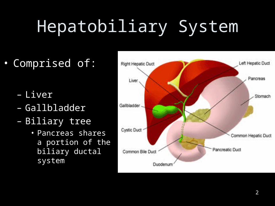
Hepatobiliary System
• Comprised of:
– Liver– Gallbladder– Biliary tree
• Pancreas shares a portion of the biliary ductal system
2
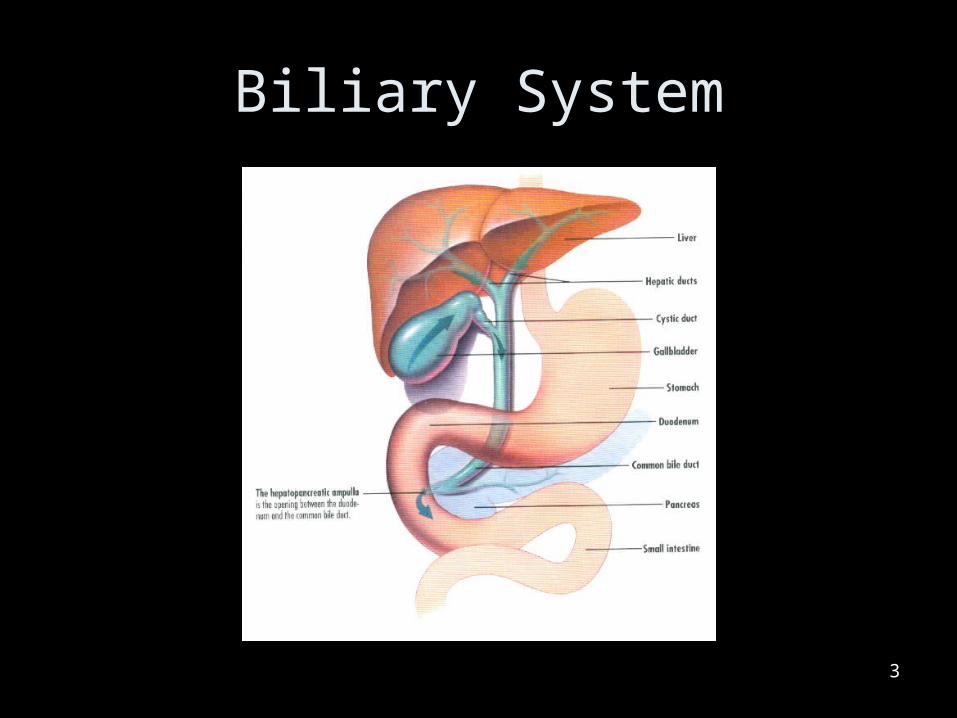
Biliary System
3
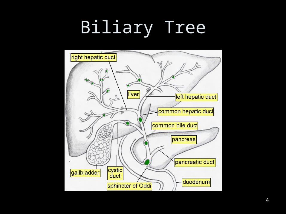
Biliary Tree
4
Hepatobiliary
5

Inflammatory Diseases
6
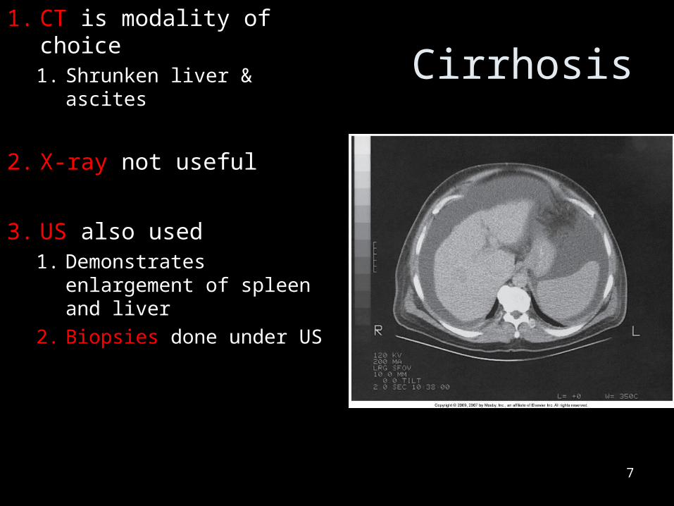
Cirrhosis1. CT is modality of choice
1. Shrunken liver & ascites
2. X-ray not useful
3. US also used1. Demonstrates
enlargement of spleen and liver
2. Biopsies done under US
7
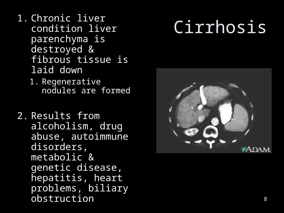
Cirrhosis1. Chronic liver
condition liver parenchyma is destroyed & fibrous tissue is laid down1. Regenerative
nodules are formed
2. Results from alcoholism, drug abuse, autoimmune disorders, metabolic & genetic disease, hepatitis, heart problems, biliary obstruction 8
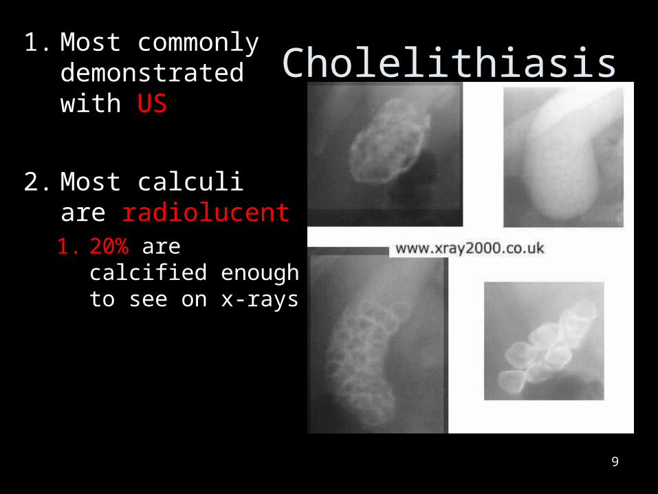
Cholelithiasis1. Most commonly
demonstrated with US
2. Most calculi are radiolucent1. 20% are calcified
enough to see on x-rays
9
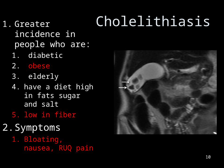
Cholelithiasis1. Greater incidence in people who are:
1. diabetic
2. obese
3. elderly
4. have a diet high in fats sugar and salt
5. low in fiber
2. Symptoms1. Bloating, nausea,
RUQ pain
10
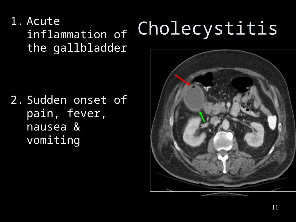
Cholecystitis1. Acute inflammation of the gallbladder
2. Sudden onset of pain, fever, nausea & vomiting
11
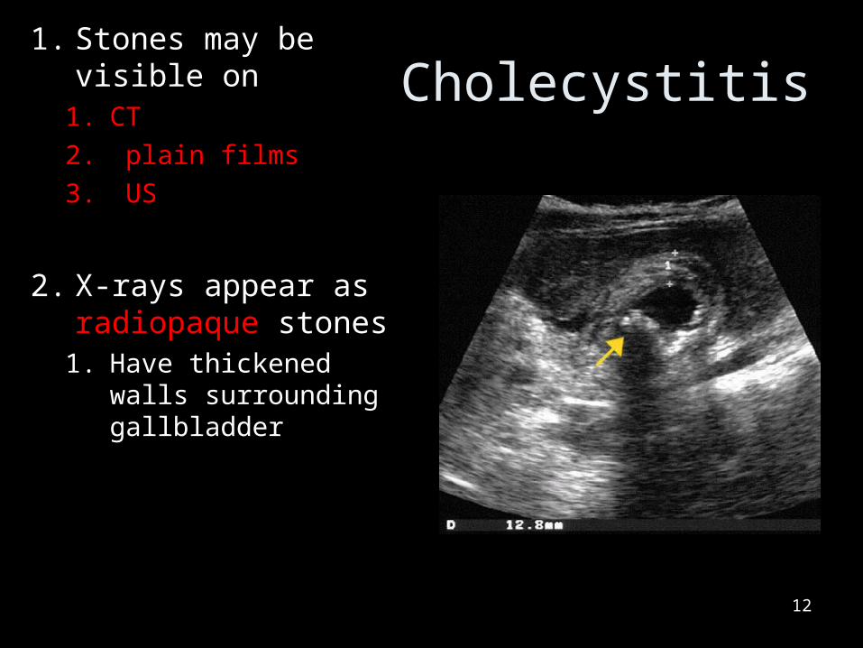
Cholecystitis1. Stones may be
visible on 1. CT
2. plain films
3. US
2. X-rays appear as radiopaque stones
1. Have thickened walls surrounding gallbladder
12
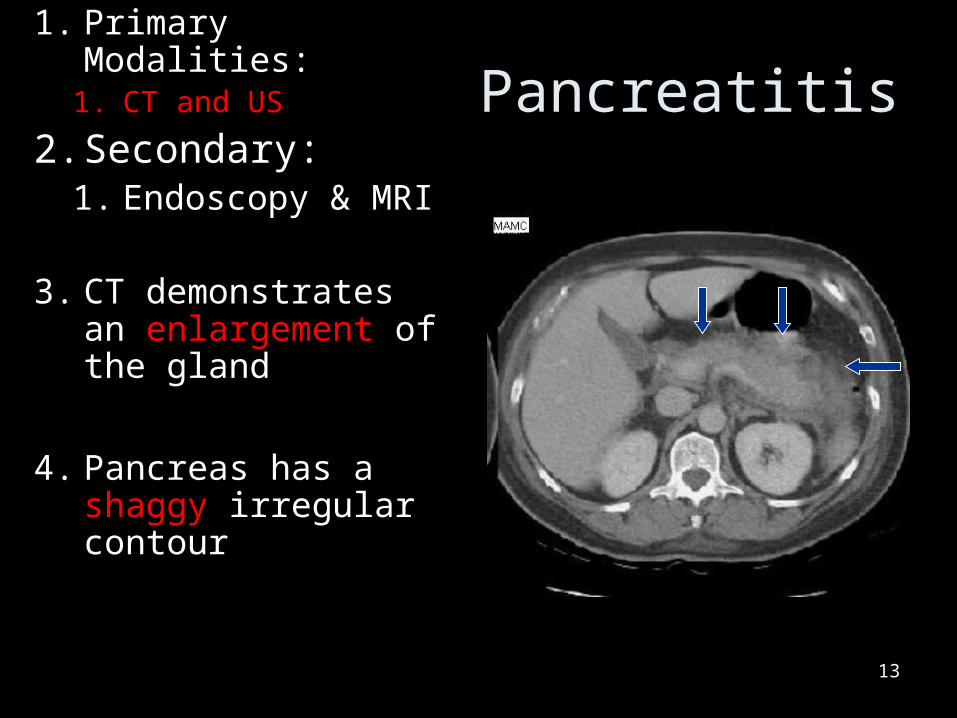
Pancreatitis1. Primary Modalities:
1. CT and US
2. Secondary:1. Endoscopy & MRI
3. CT demonstrates an enlargement of the gland
4. Pancreas has a shaggy irregular contour
13
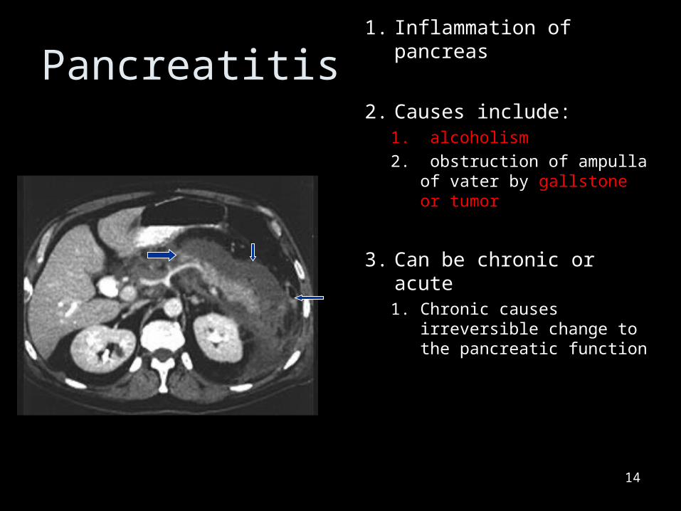
Pancreatitis1. Inflammation of
pancreas
2. Causes include:1. alcoholism
2. obstruction of ampulla of vater by gallstone or tumor
3. Can be chronic or acute1. Chronic causes
irreversible change to the pancreatic function
14

Neoplastic Diseases
15
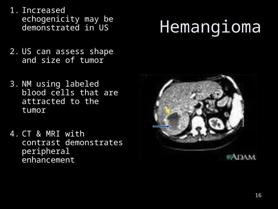
Hemangioma1. Increased echogenicity
may be demonstrated in US
2. US can assess shape and size of tumor
3. NM using labeled blood cells that are attracted to the tumor
4. CT & MRI with contrast demonstrates peripheral enhancement
16
Hemangioma
17
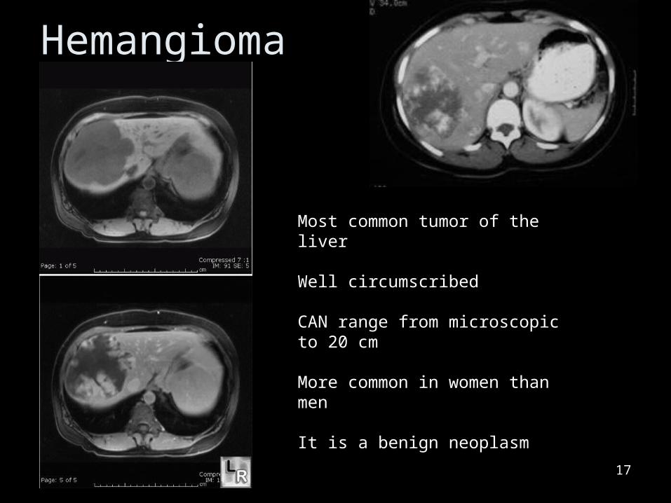
Most common tumor of the liver
Well circumscribed
CAN range from microscopic to 20 cm
More common in women than men
It is a benign neoplasm
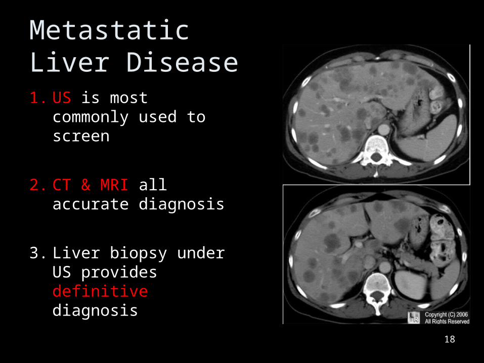
Metastatic Liver Disease1. US is most
commonly used to screen
2. CT & MRI all accurate diagnosis
3. Liver biopsy under US provides definitive diagnosis
18
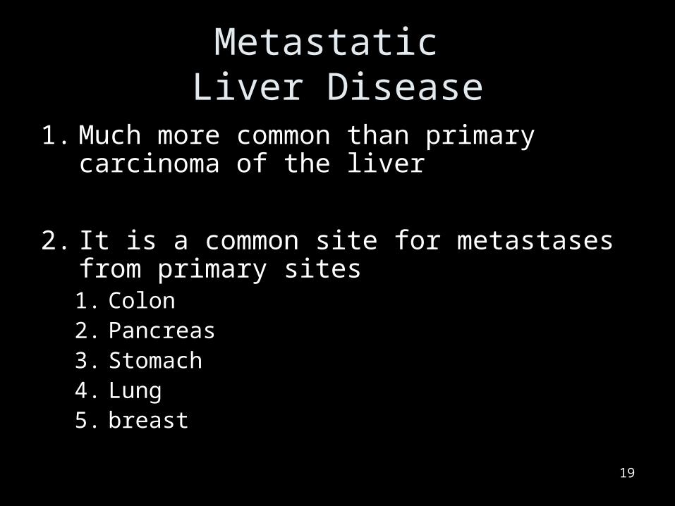
Metastatic Liver Disease
1. Much more common than primary carcinoma of the liver
2. It is a common site for metastases from primary sites1. Colon2. Pancreas3. Stomach4. Lung5. breast
19
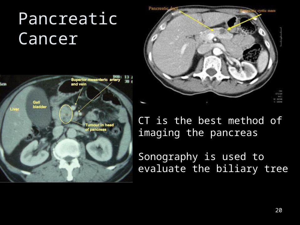
Pancreatic Cancer
20
CT is the best method of imaging the pancreas
Sonography is used to evaluate the biliary tree
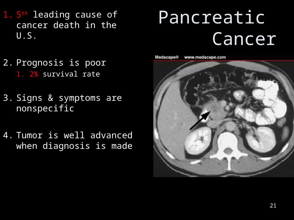
Pancreatic Cancer
1. 5th leading cause of cancer death in the U.S.
2. Prognosis is poor1. 2% survival rate
3. Signs & symptoms are nonspecific
4. Tumor is well advanced when diagnosis is made
21
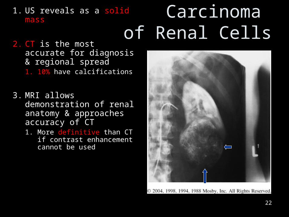
Carcinoma of Renal Cells
1. US reveals as a solid mass
2. CT is the most accurate for diagnosis & regional spread1. 10% have calcifications
3. MRI allows demonstration of renal anatomy & approaches accuracy of CT1. More definitive than CT if
contrast enhancement cannot be used
22

MISC pathologies ofHepatobiliary System
23
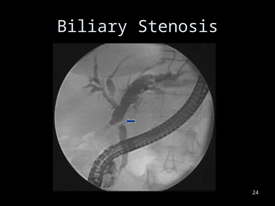
Biliary Stenosis
24

Genitourinary System
25
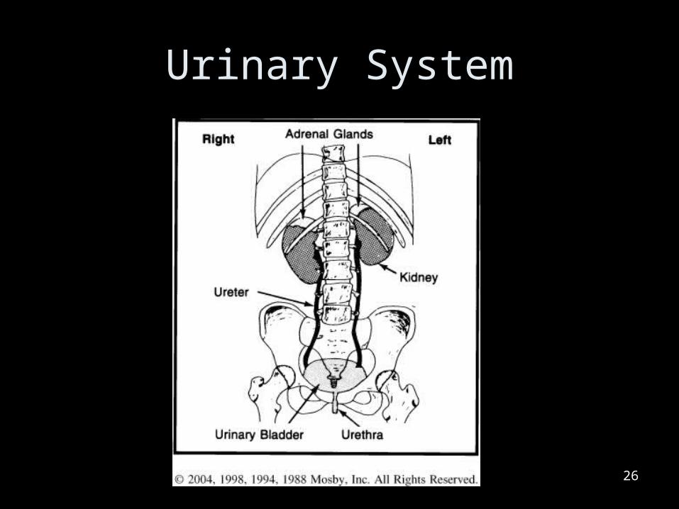
Urinary System
26
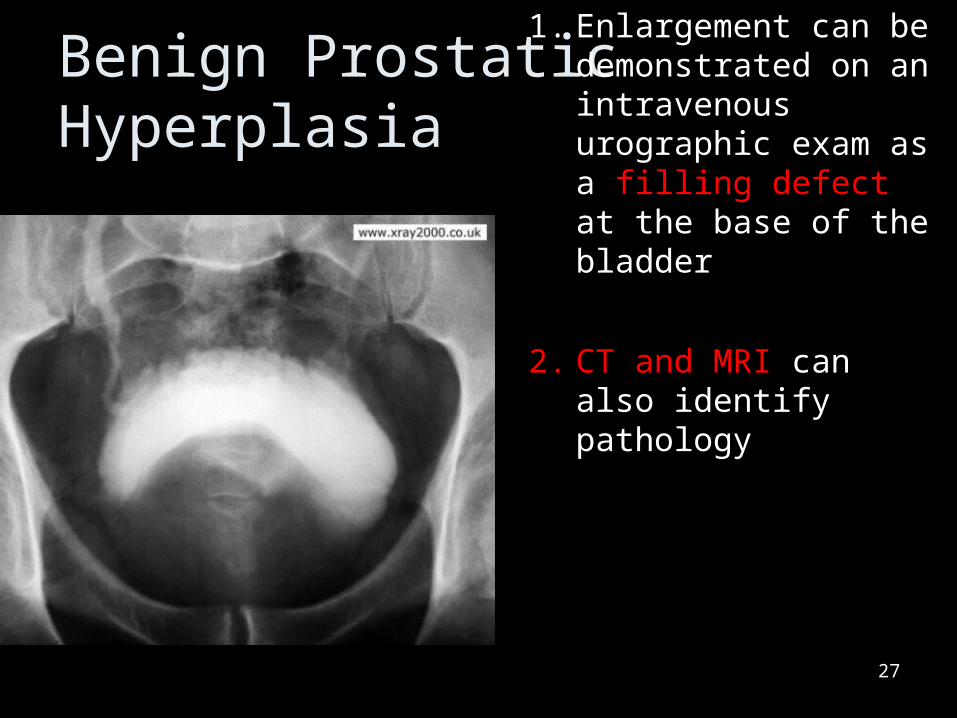
Benign Prostatic Hyperplasia
1. Enlargement can be demonstrated on an intravenous urographic exam as a filling defect at the base of the bladder
2. CT and MRI can also identify pathology
27
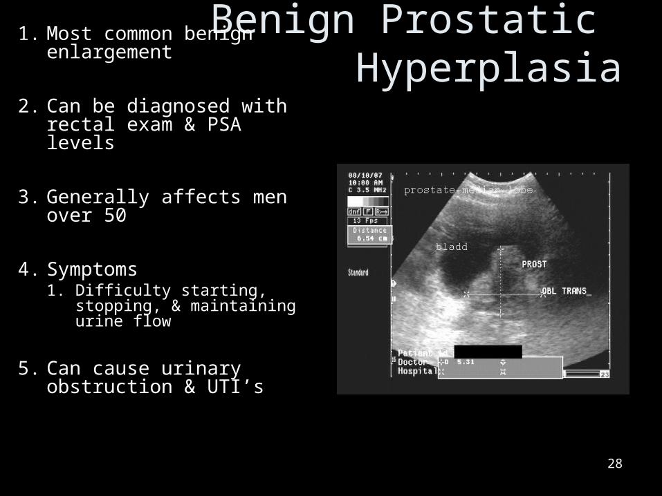
Benign Prostatic Hyperplasia
1. Most common benign enlargement
2. Can be diagnosed with rectal exam & PSA levels
3. Generally affects men over 50
4. Symptoms1. Difficulty starting,
stopping, & maintaining urine flow
5. Can cause urinary obstruction & UTI’s
28

Congenital Anomaly
29
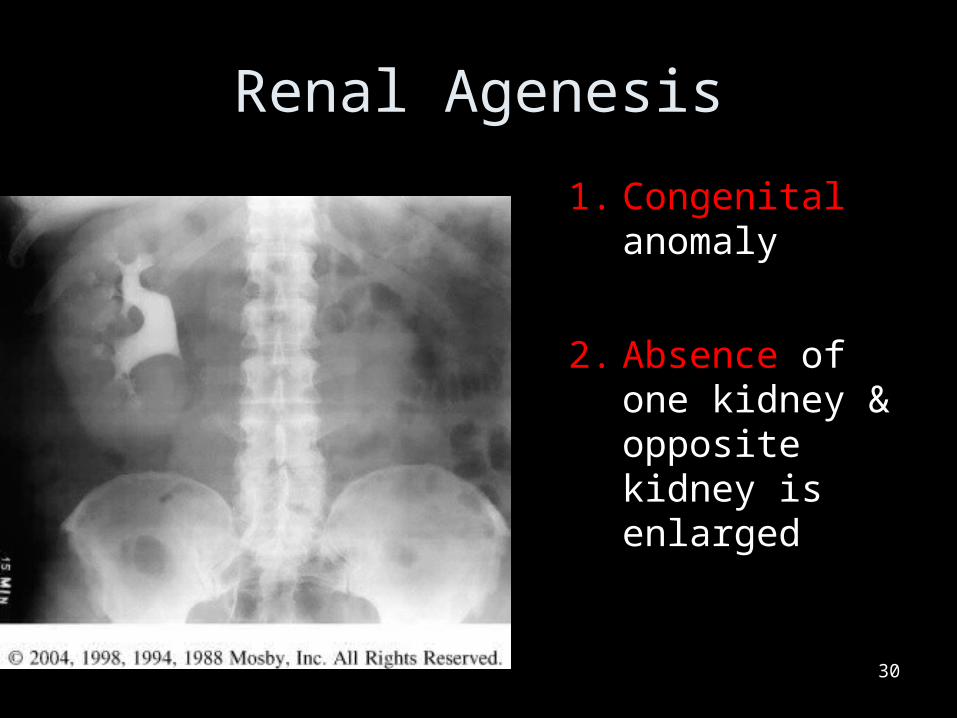
Renal Agenesis
1. Congenital anomaly
2. Absence of one kidney & opposite kidney is enlarged
30
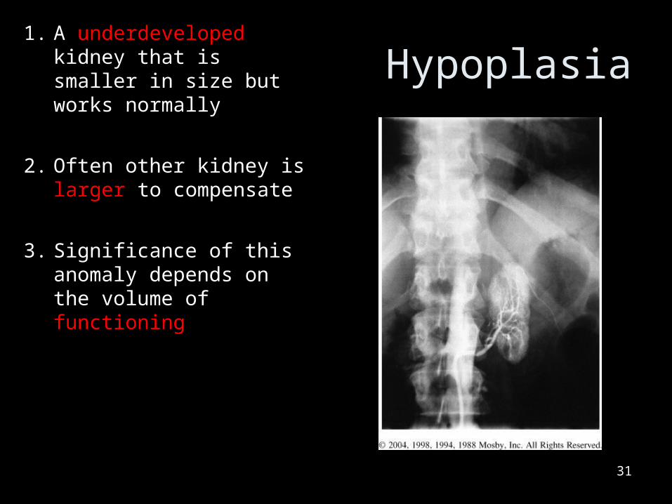
Hypoplasia1. A underdeveloped
kidney that is smaller in size but works normally
2. Often other kidney is larger to compensate
3. Significance of this anomaly depends on the volume of functioning
31
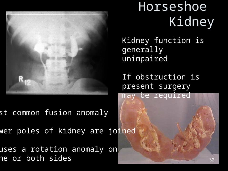
Horseshoe Kidney
32
Most common fusion anomaly
Lower poles of kidney are joined
Causes a rotation anomaly on one or both sides
Kidney function is generally unimpaired
If obstruction is present surgery may be required
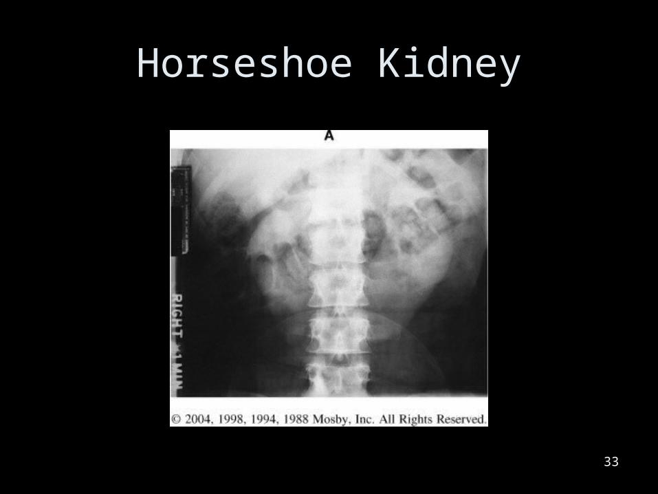
Horseshoe Kidney
33
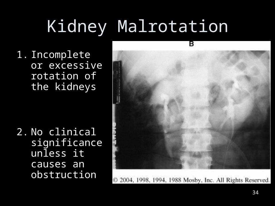
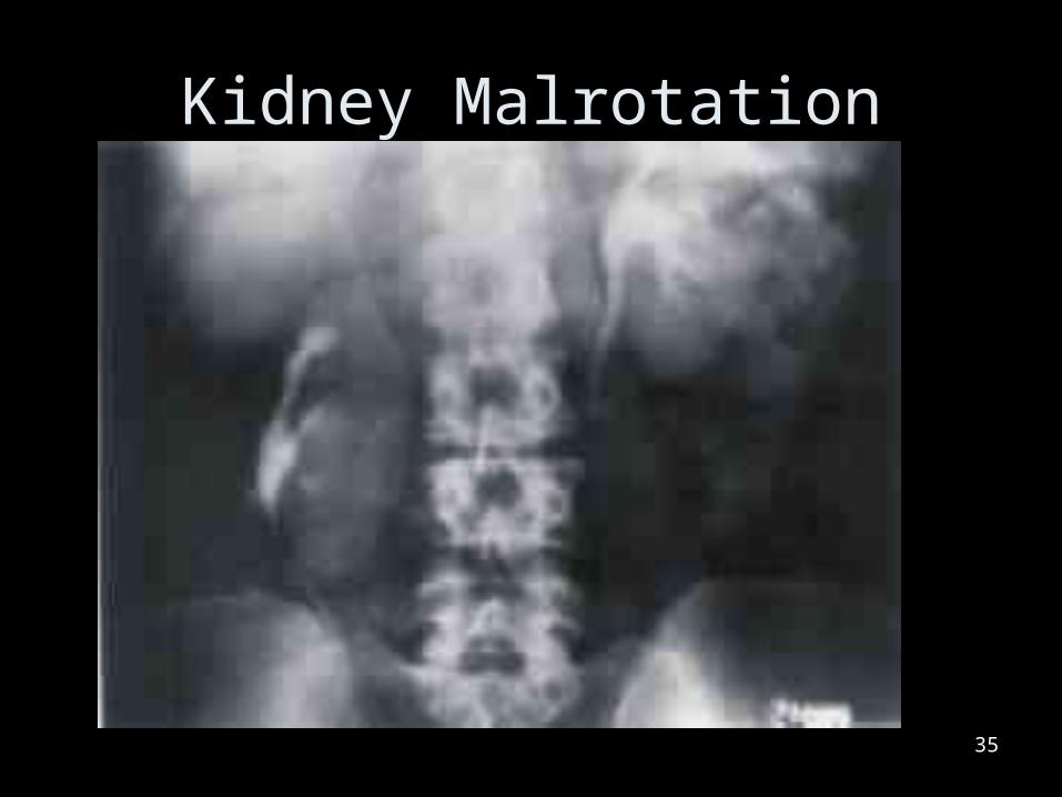
Kidney Malrotation
1. Incomplete or excessive rotation of the kidneys
2. No clinical significance unless it causes an obstruction
34
Kidney Malrotation
35
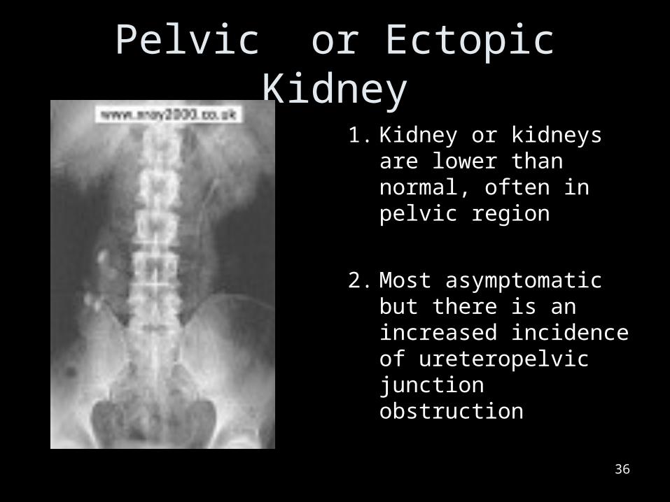
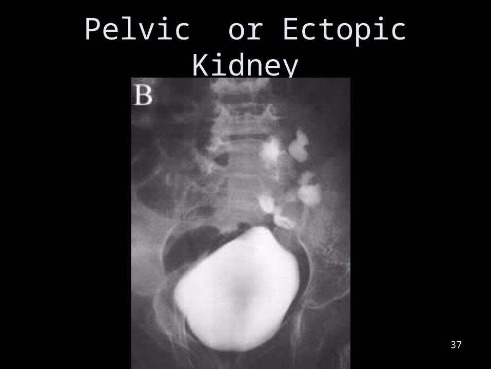
Pelvic or Ectopic Kidney
1. Kidney or kidneys are lower than normal, often in pelvic region
2. Most asymptomatic but there is an increased incidence of ureteropelvic junction obstruction
36
Pelvic or Ectopic Kidney
37
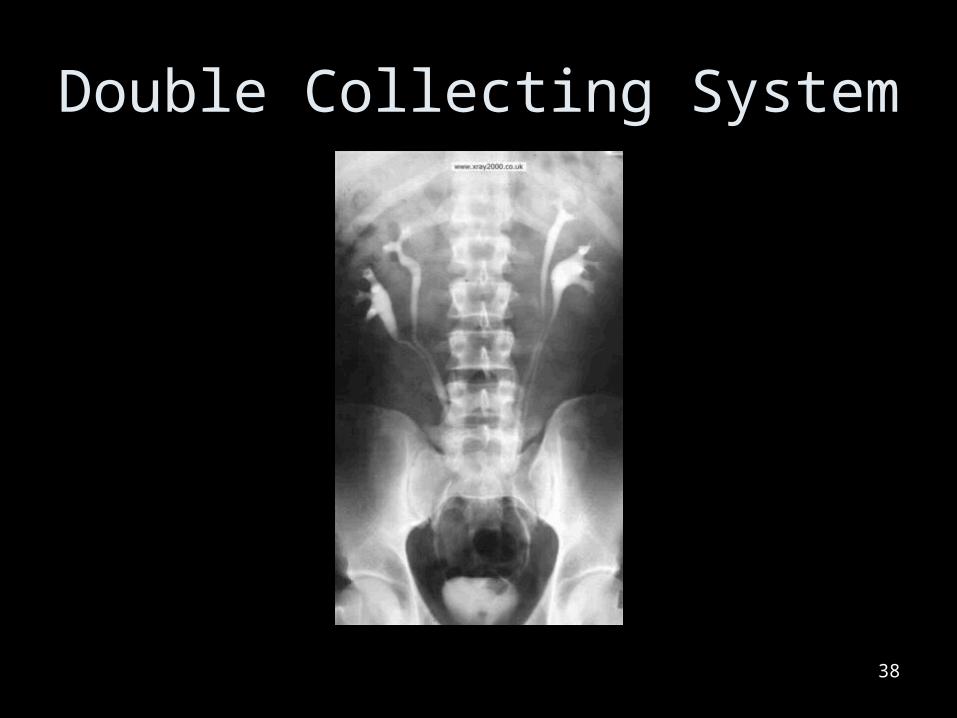
Double Collecting System
38
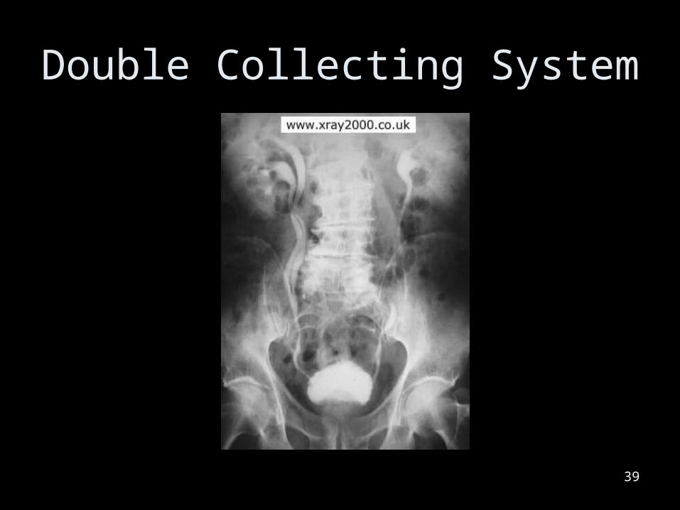
Double Collecting System
39
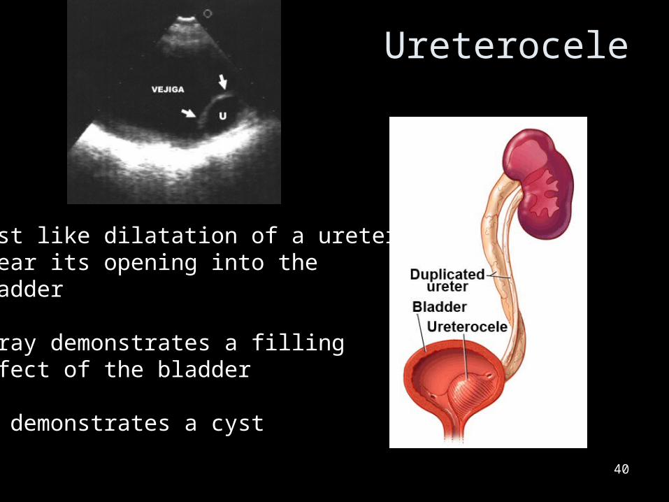
Ureterocele
40
Cyst like dilatation of a ureter near its opening into the bladder
X-ray demonstrates a filling defect of the bladder
US demonstrates a cyst
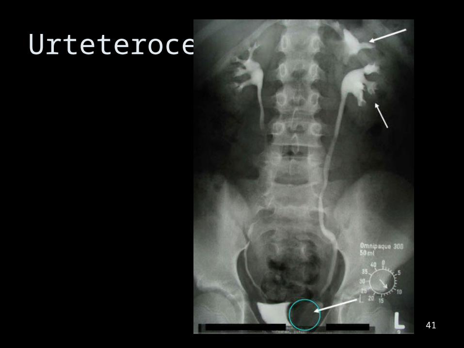
Urteterocele
41

Bladder Diverticula
• Con occur congenitally or caused by chronic bladder obstruction and infection
42
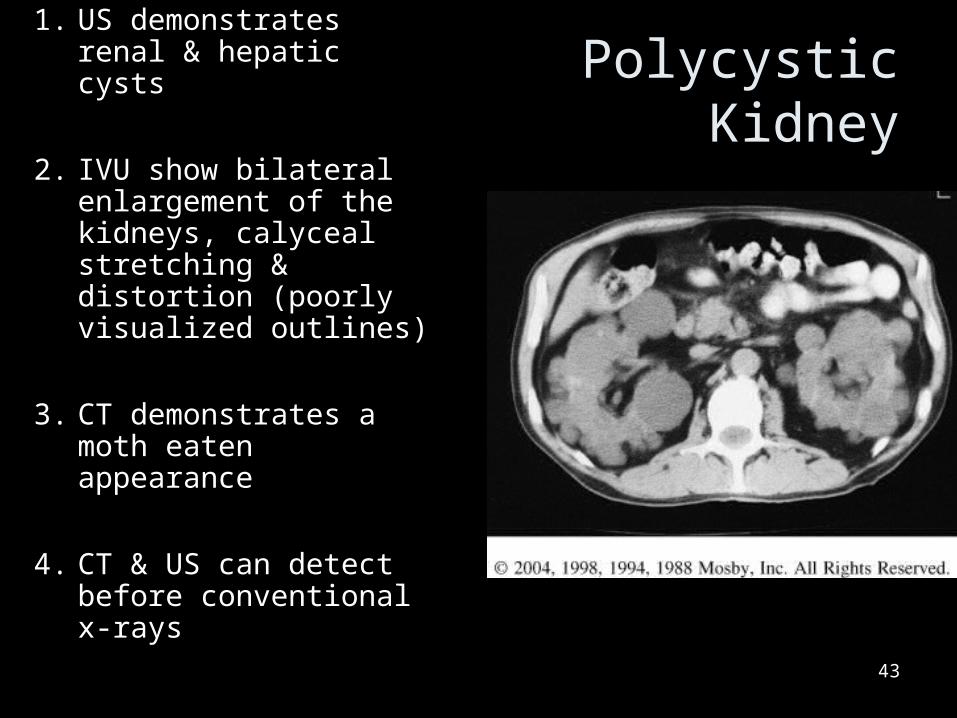
Polycystic Kidney
1. US demonstrates renal & hepatic cysts
2. IVU show bilateral enlargement of the kidneys, calyceal stretching & distortion (poorly visualized outlines)
3. CT demonstrates a moth eaten appearance
4. CT & US can detect before conventional x-rays
43

Polycystic Kidney1. Congenital disease2. Cysts enlarge as pt
ages
3. Enlargement destroys normal tissues
4. It is the cause of 10% of end-stage renal disease
44

Inflammatory Diseases
45

Pyelonephritis1. Can be
demonstrated on a CT and US
2. IVU will often look normal in a acute attack
3. Interstitial edema causes less visualization of collecting structures
46
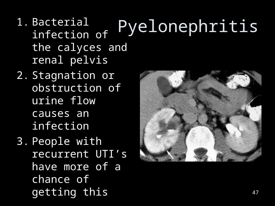
Pyelonephritis1. Bacterial infection of the calyces and renal pelvis
2. Stagnation or obstruction of urine flow causes an infection
3. People with recurrent UTI’s have more of a chance of getting this
47
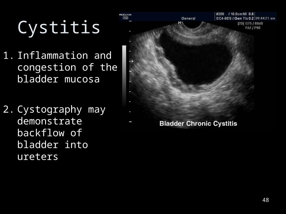
Cystitis
1. Inflammation and congestion of the bladder mucosa
2. Cystography may demonstrate backflow of bladder into ureters
48

Urinary System Calcifications
49
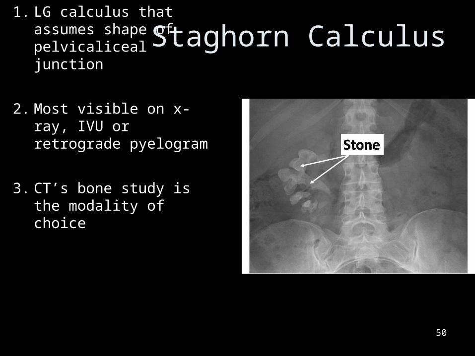
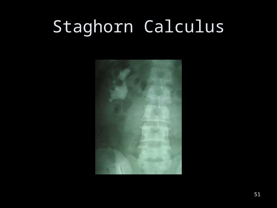
Staghorn Calculus1. LG calculus that
assumes shape of pelvicaliceal junction
2. Most visible on x-ray, IVU or retrograde pyelogram
3. CT’s bone study is the modality of choice
50
Staghorn Calculus
51
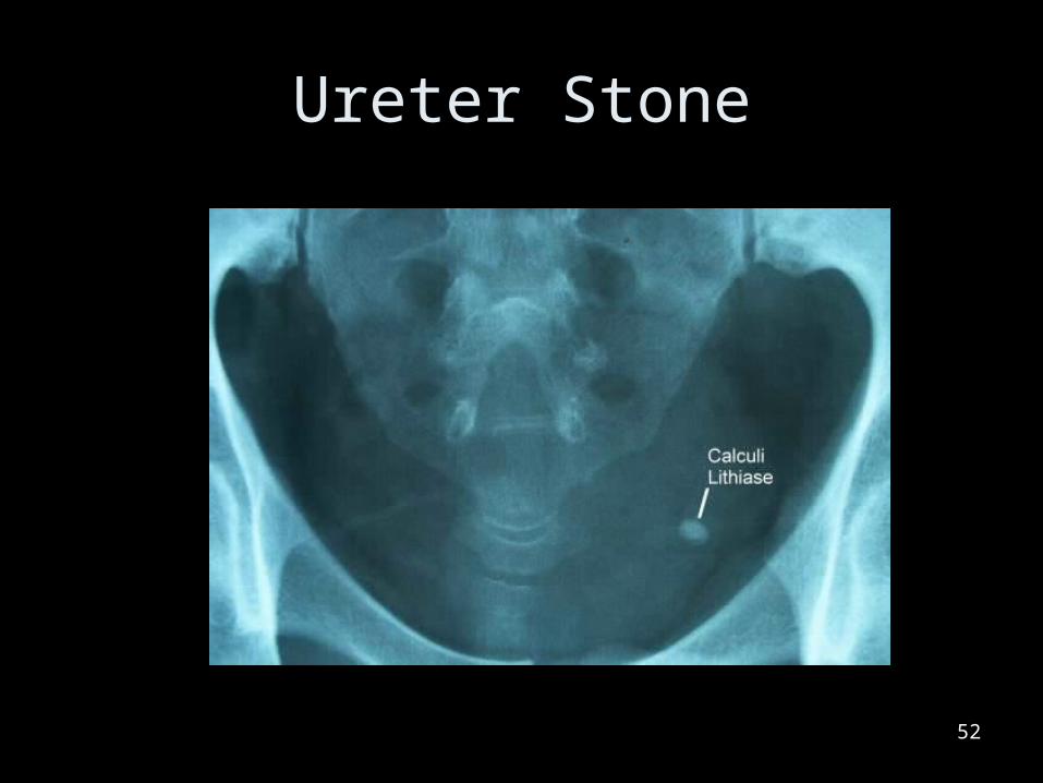
Ureter Stone
52
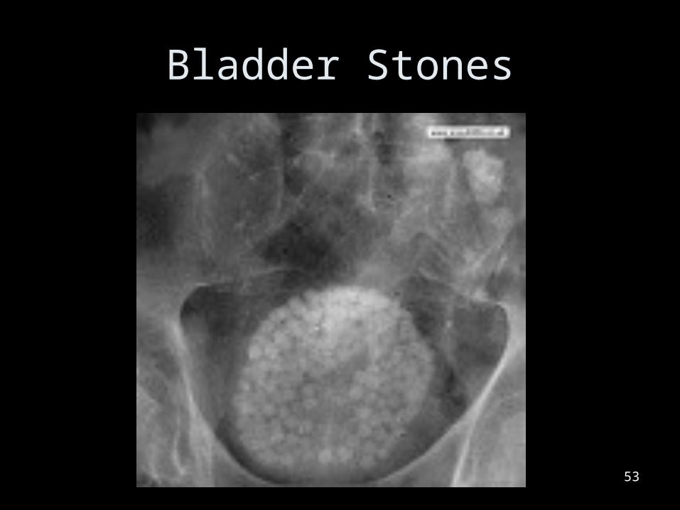
Bladder Stones
53
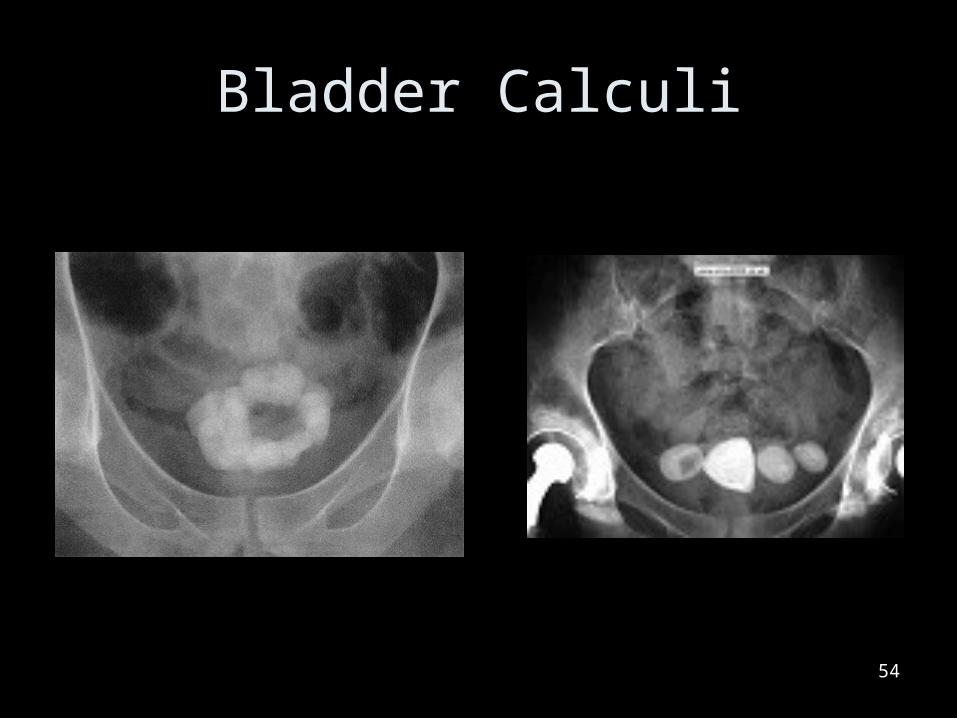
Bladder Calculi
54
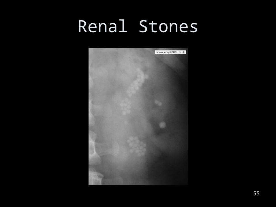
Renal Stones
55
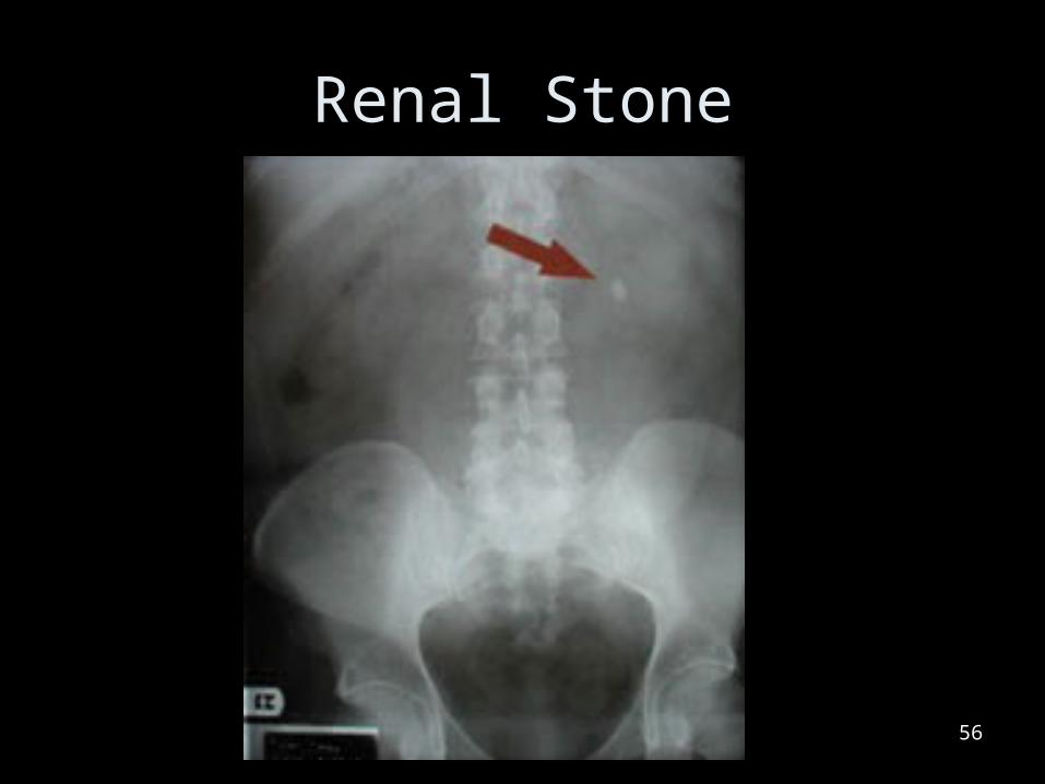
Renal Stone
56

Degenerative Diseases
57
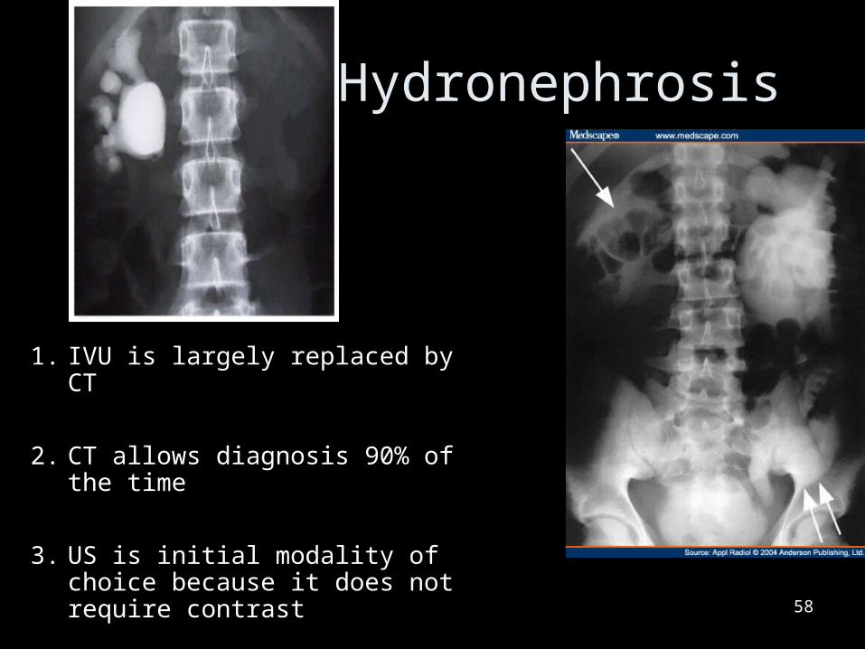
Hydronephrosis
1. IVU is largely replaced by CT
2. CT allows diagnosis 90% of the time
3. US is initial modality of choice because it does not require contrast 58

Neoplastic Diseases
59
Wilms Tumor
•Malignant renal tumor
•1 in every 13,500 births
60
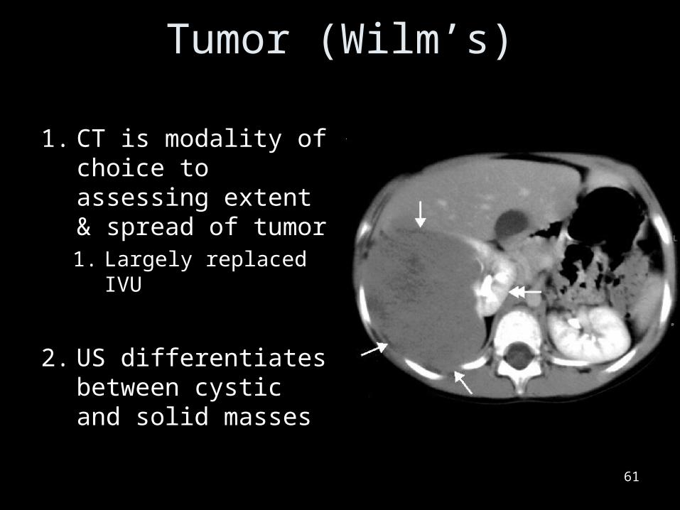
Tumor (Wilm’s)
1. CT is modality of choice to assessing extent & spread of tumor1. Largely replaced IVU
2. US differentiates between cystic and solid masses
61
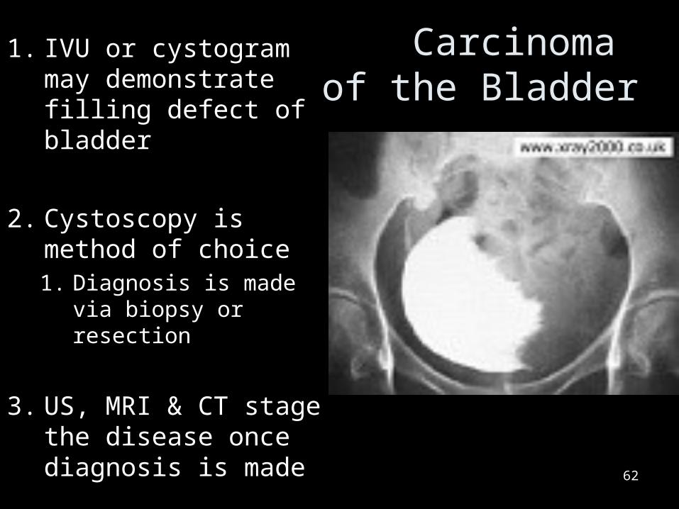
Carcinoma of the Bladder
1. IVU or cystogram may demonstrate filling defect of bladder
2. Cystoscopy is method of choice1. Diagnosis is made via
biopsy or resection
3. US, MRI & CT stage the disease once diagnosis is made
62

Carcinoma of the Bladder
63
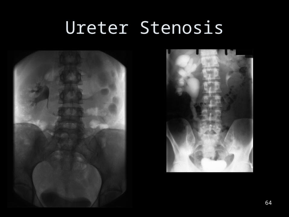
Ureter Stenosis
64
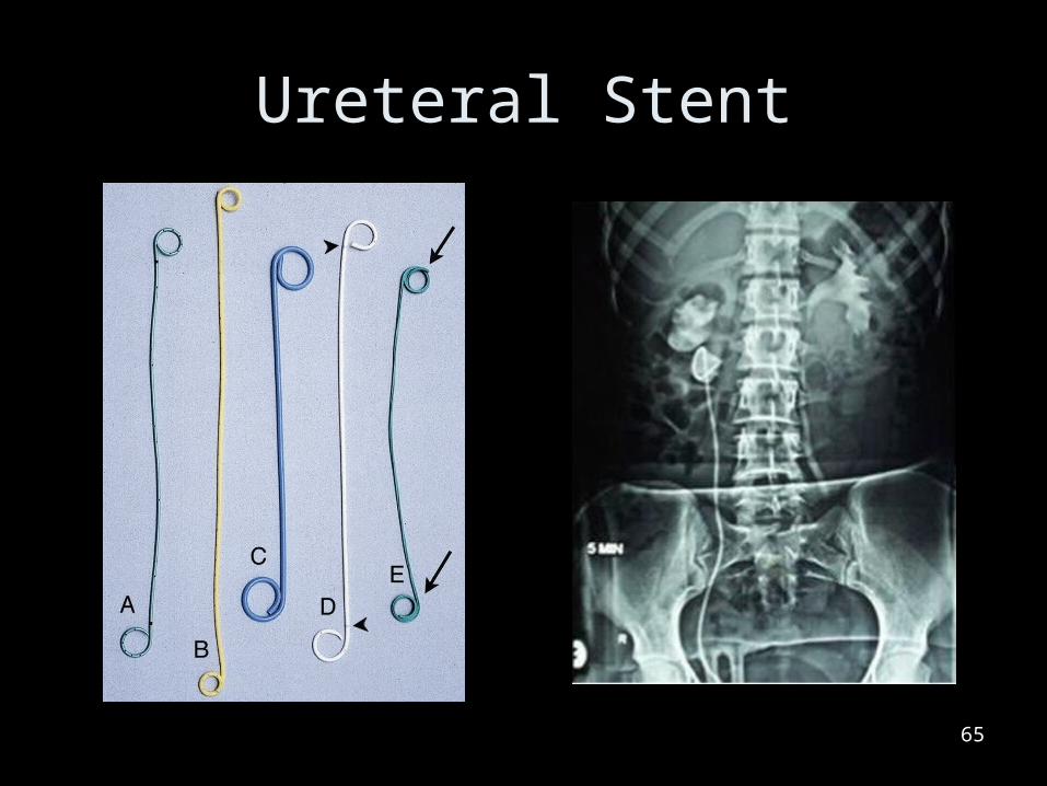
Ureteral Stent
65
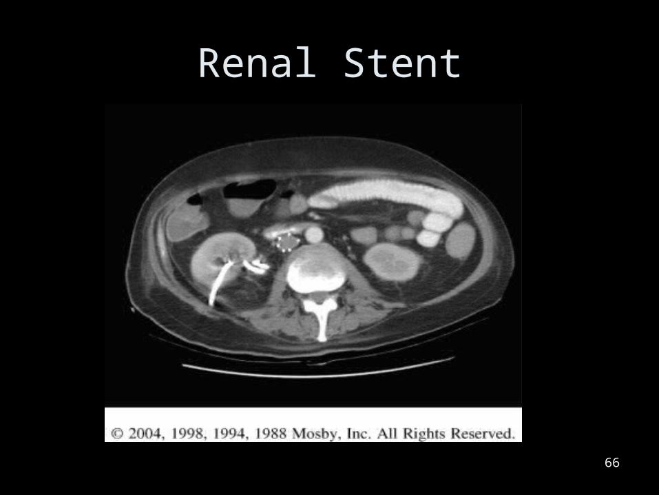
Renal Stent
66
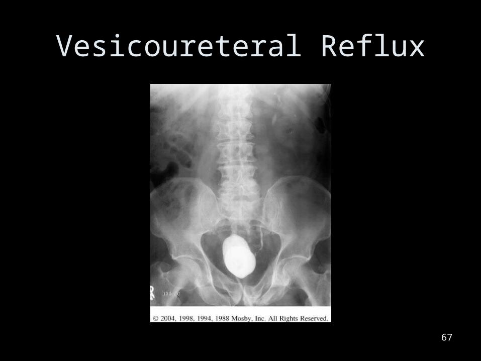
Vesicoureteral Reflux
67

Vesicoureteral Reflux
68


