ARTICLE IN PRESS
JID: HBPD [m5G; November 6, 2018;13:49 ]
Hepatobiliary & Pancreatic Diseases International xxx (xxxx) xxx
Contents lists available at ScienceDirect
Hepatobiliary & Pancreatic Diseases International
journal homepage: www.elsevier.com/locate/hbpd
Original Article/Liver
Potential application of ultrasound-guided thermal ablation in rare
liver tumors
Li-Li Wu
a , Jia-Xin Chen
a , Kai Li a , Zhong-Zhen Su
b , Ying-Lin Long
a , Li-Ping Luo
a , Er-Jiao Xu
a , ∗, Rong-Qin Zheng
a
a Department of Medical Ultrasonics, Third Affiliated Hospital of Sun Yat-sen University, Guangdong Key Laboratory of Liver Disease Research, Guangzhou
510630, China b Department of Medical Ultrasonics, Fifth Affiliated Hospital of Sun Yat-sen University, Zhuhai 5190 0 0, China
a r t i c l e i n f o
Article history:
Received 4 May 2018
Accepted 15 October 2018
Available online xxx
Keywords:
Contrast-enhanced ultrasound
Thermal ablation
Rare liver tumor
Fusion imaging
a b s t r a c t
Background: With the advances of imaging techniques, the detection rate of rare liver tumor is increased.
However, the therapeutic strategies of the rare liver tumors remain limited.
Methods: We analyzed twelve pathologically confirmed rare liver tumors in 8 patients. All of the pa-
tients underwent ultrasound (US) guided biopsy and subsequent thermal ablation. The tumors were ab-
lated according to the preoperative plans and monitored by real-time US. CT/MRI fused with contrast
enhanced US (CEUS) or three-dimensional (3D) US-CEUS images were used to guide and assess the abla-
tion zone more accurately during thermal ablation. The rate of technical efficacy was assessed based on
the contrast-enhance CT/MRI (CECT/MRI) results one month after ablation. Local tumor progression (LTP),
recurrence and complications were followed up and recorded.
Results: Among these twelve nodules, nine were subject to US-guided thermal ablation, whereas
the other three inconspicuous nodules were subject to CEUS-guided thermal ablation. Intra-procedure
CT/MRI-CEUS or 3D US-CEUS fusion imaging assessments demonstrated that the ablation zone sufficiently
covered the original tumor, and no immediate supplementary ablation was required. Additionally, no ma-
jor complications were observed during the follow-up period. The postoperative CECT/MRI confirmed that
the technique success rate was 100%. Within the surveillance period of 13 months, no LTP or recurrence
was noted.
Conclusions: US-guided thermal ablation was feasible and safe for rare liver tumors. The use of fusion
imaging technique might make US-guided thermal ablation as effective as surgical resection, and this
technique might serve as a potential therapeutic modality for rare liver tumors in the future.
© 2018 First Affiliated Hospital, Zhejiang University School of Medicine in China. Published by Elsevier
B.V. All rights reserved.
I
w
i
c
c
n
m
w
d
s
t
t
m
fi
n
t
w
m
c
t
t
a
a
h
1
ntroduction
Liver cancer is the second leading cause of death from cancer
orldwide, and the diagnosis and treatment of this cancer are crit-
cal [1–3] . The International Classification of Diseases by the WHO
ites various types of liver tumors. The most common types in-
lude hepatocellular carcinoma (HCC), intrahepatic cholangiocarci-
oma (ICC), liver metastasis from colorectal cancer and hepatic he-
angioma [4] . In addition, there are a number of rare liver tumors
ith a low incidence. These types of liver tumors are histologically
iverse and clinically asymptomatic, and their diagnoses rely con-
iderably on histological examination. However, some of these liver
umors are malignant or borderline, and removal of these liver
∗ Corresponding author.
E-mail address: [email protected] (E.-J. Xu).
n
t
ttps://doi.org/10.1016/j.hbpd.2018.10.002
499-3872/© 2018 First Affiliated Hospital, Zhejiang University School of Medicine in Chin
Please cite this article as: L.-L. Wu, J.-X. Chen and K. Li et al., Potentia
tumors, Hepatobiliary & Pancreatic Diseases International, https://doi.o
umors is necessary. Due to the low incidence of rare liver tu-
ors and the lack of symptoms or biochemical indices, it is dif-
cult to differentiate these tumors in preoperative imaging exami-
ation. Hence, the golden standard for the diagnosis is histology.
Prior studies have demonstrated that ultrasound (US) guided
hermal ablation is as effective as resection for early-stage HCC
ith respect to the long-term survival rate [5 , 6] . Additionally, ther-
al ablation is superior given its increased safety, reduced compli-
ations and more rapid recovery. The 2014 Japanese guidelines for
he treatment of liver cancer recommended radiofrequency abla-
ion as a first-line treatment for early HCC [7] . Nevertheless, the
pplication of thermal ablation in rare liver tumors has not been
ssessed to date.
In the present study, we retrospectively analyzed the effective-
ess of US-guided thermal ablation on the treatment of rare liver
umors.
a. Published by Elsevier B.V. All rights reserved.
l application of ultrasound-guided thermal ablation in rare liver
rg/10.1016/j.hbpd.2018.10.002
2 L.-L. Wu, J.-X. Chen and K. Li et al. / Hepatobiliary & Pancreatic Diseases International xxx (xxxx) xxx
ARTICLE IN PRESS
JID: HBPD [m5G; November 6, 2018;13:49 ]
Table 1
Demographic and examination data of the rare liver tumor cases.
Case no. Age (yr) Gender Lesion no. Position Diameter (mm) Pathology Ablation technique
1 29 F 1 S5 25 Inflammatory pseudotumor-like follicular dendritic cell RFA
2 47 F 2 S8 14 Inflammatory myofibroblastic tumor RFA
3 46 F 3 S4 22 Inflammatory myofibroblastic tumor RFA
4 64 F 4 S5 21 Mucosa-associated lymphoid tissue lymphomas RFA
5 S2 9
6 S4 7
5 34 F 7 S4 8 Metastasis of spleen inflammatory myofibroblastic tumor RFA
8 S7/8 13
6 28 M 9 S6/7 14 Tissue cells and dendritic cell tumors RFA
7 49 F 10 S5/8 14 B-cell-derived lymphoma or dendritic cell tumor RFA
11 S2 6
8 47 M 12 S8 22 EBV-associated lymphoepithelioma-like cholangiocarcinoma MWA
F: female; M: male; RFA: radiofrequency ablation; MWA: microwave ablation.
Table 2
Conventional US characteristics and contrast-enhanced US patterns of liver nodule.
Case
No.
Lesion
No.
Echogenicity Margin CEUS pattern
Arterial
phase
Portal
phase
Delay
phase
1 1 Hypo-echoic Clear Hyper- Hypo- Hypo-
2 2 Hypo-echoic Clear Hyper- Hypo- Hypo-
3 3 Hypo-echoic Obscure Hyper- Iso- Iso-
4 4 Hypo-echoic Clear Hyper- Hypo- Hypo-
5 Hypo-echoic Clear Iso- Hypo- Hypo-
6 Iso-echoic Obscure Hyper- Hypo- Hypo-
5 7 Hypo-echoic Clear Hyper- Hypo- Hypo-
8 Hypo-echoic Clear Hyper- Hypo- Hypo-
6 9 Hypo-echoic Obscure Hyper- Hypo- Hypo-
7 10 Hypo-echoic Clear Hyper- Hypo- Hypo-
11 Hypo-echoic Clear Hyper- Hypo- Hypo-
8 12 Hypo-echoic Clear Hyper- Hypo- Hypo-
US: ultrasound; CEUS: contrast-enhanced ultrasound.
U
E
r
T
l
a
T
(
w
1
t
i
w
8
i
u
t
w
t
a
C
A
v
w
A
c
l
t
t
M
f
s
a
s
c
i
a
r
a
a
Methods
Patients
This study was performed in compliance with the Declaration
of Helsinki and approved by the Institutional Ethics Review Board
of the Third Affiliated Hospital of Sun Yat-sen University. Informed
consent of ablation was obtained from each patient. Informed con-
sent of this study was waived due to the retrospective nature of
the study. All procedures were performed by two proficient radi-
ologists (LK and XEJ) with more than 10 years of experience in US
and US-guided thermal ablation.
Recruited patients included those who were diagnosed with
rare liver tumors by pathological examination and subsequently re-
ceived US-guided thermal ablation in our hospital from January
2014 to December 2016. The inclusion criteria were as follows:
(1) histological confirmation of rare liver tumors; (2) a single tu-
mor with a diameter ≤ 5 cm or less than 3 tumor nodules with the
maximal diameter ≤ 3 cm; (3) no invasion to major vessels or bile
ducts, adjacent or distant tissues/organs; (4) Child-Pugh Class A or
B liver function; and (5) underwent US-guided liver thermal ab-
lation successfully. The exclusion criteria were as follows: (1) de-
ficiency of the pre-procedure or intra-procedure imaging data re-
sulting in unanalyzable; and (2) without contrast-enhance CT/MRI
(CECT/CEMRI) evaluation one month after ablation and regular
follow-up every three months.
All the included lesions were confirmed by histology that were
obtained by US-guided tumor biopsy shortly before liver thermal
ablation. U
Please cite this article as: L.-L. Wu, J.-X. Chen and K. Li et al., Potentia
tumors, Hepatobiliary & Pancreatic Diseases International, https://doi.o
ltrasound-guided thermal ablation
quipment
Two types of thermal ablation were performed in this study:
adiofrequency ablation (RFA) and microwave ablation (MWA).
he former employed a Radionics Cool-tip RFA System (Valley-
ab, Mansfield, MA, USA) with a maximal output power of 200 W
nd single-pole, internally cooled ablation needles with 3-cm tips.
he latter employed a KY-20 0 0 Water-cooled MWA Instrument
Kangyou Co., Nanjing, China) comprising a microwave generator
ith an emission frequency of 2450 ± 50 MHz, output power of
0–100 W, and a 15-G water-cycle internally cooled microwave an-
enna.
Two types of US apparatuses with CnTI contrast-specific imag-
ng, namely, MyLab Twice and MyLab ClassC (Esoate, Genoa, Italy),
ere employed. Convex probes CA431 and CA541 (frequency: 1–
MHz) were used for US scanning and guidance. The fusion imag-
ng system Virtual Navigator (VN; Esoate) was composed of a main
nit in the US machine, a corresponding probe sensor adhered to
he probe and a magnetic field generator. The dedicated software
as installed on the US machine. The magnetic field generator and
he probe sensor ensured the exact position between the sensor
nd the patient.
ontrast-enhanced US (CEUS)
SonoVue (Bracco, Milan, Italy) was used for CEUS examination.
bolus of SonoVue (1.5–2.0 mL) was injected via the antecubital
ein, followed by 5 mL saline solution. When necessary, SonoVue
as injected repeatedly.
blation strategy
All patients were intubated with general anesthesia. After per-
utaneous biopsy, US-guided thermal ablation was conducted fol-
owing the preoperative plan. For RFA, the RF generator was set
o the impedance mode with maximal output, and each RF elec-
rode was inserted into the lesion within approximately 12 min. For
WA, the microwave generator was set to 60 W and maintained
or up to 6 min in each microwave antenna insertion.
Ablation was guided by real-time US or CEUS. When neces-
ary, ancillary means, such as the application of artificial ascites
nd artificial pleural fluids, were administered to improve le-
ion visualization and prevent damage to the surrounding criti-
al organs and tissues. In our hospital, CT/MRI-US fusion imag-
ng, which combined CT or MRI images with real-time US im-
ges using the electromagnetic positioning system of VN, was
outinely employed for thermal ablation procedures. This method
ided in the detection of inconspicuous lesions, puncture guidance
nd immediate evaluation of therapeutic response. The CT/MRI-
S fusion imaging procedure included the outline of the index
l application of ultrasound-guided thermal ablation in rare liver
rg/10.1016/j.hbpd.2018.10.002
L.-L. Wu, J.-X. Chen and K. Li et al. / Hepatobiliary & Pancreatic Diseases International xxx (xxxx) xxx 3
ARTICLE IN PRESS
JID: HBPD [m5G; November 6, 2018;13:49 ]
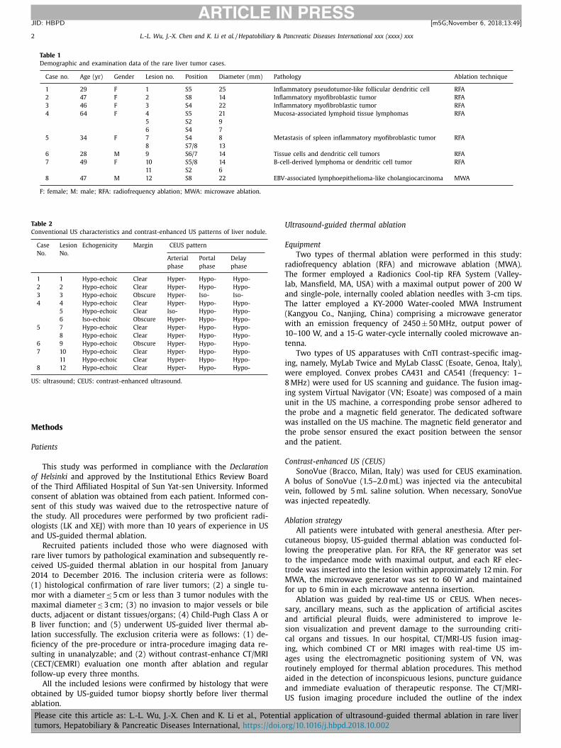
Fig. 1. A 47-year-old female patient with inflammatory myofibroblastic tumor. A –C : The contrast enhanced ultrasound (CEUS) images demonstrating the hyper-enhancement
in arterial phase and hypo-enhancement in portal phase; D –F : Preoperative MRI images demonstrating the hyper-enhancement in arterial phase and hypo-enhancement in
portal and venous phase; G : The 3D US-CEUS fusion imaging evaluation carried out immediately after the radiofrequency ablation showing the non-enhanced zone of CEUS
covered the blue ring of index tumor and the red ring of preset ablative margin, which indicated the tumor and its 5 mm ablative margin were completely ablated except
the adjacent large vessels; H : One month post-operatively, contrast enhanced MRI image showing the completely necrosis of the index tumor.
l
t
d
I
c
t
i
z
b
s
s
a
s
a
a
a
i
w
P
2
t
a
e
w
I
t
esion and its ablative margins on CT/MRI images, registration of
wo sets of images, alignment by fine-tuning and navigation. The
etails of the procedure were described in our previous report [8] .
f the index lesion was inconspicuous, CT/MRI-US fusion imaging
ould be used to locate the lesion and guide the subsequent punc-
ure. After the thermal ablation procedure, CT/MRI-CEUS fusion
maging was generally employed to assess whether the ablated
one covered the index lesion and its ablative margin. If possi-
le, 5 mm ablative margin was required during intraoperative as-
essment unless the lesion was adjacent to the major hepatic ves-
els or liver capsule. Moreover, 3D US-CEUS fusion imaging was
lso used for the immediate evaluation of therapeutic response in
ome other patients if the lesions were conspicuous on US im-
ges. Briefly, 3D US-US fusion imaging fused the real-time US im-
ges with the 3D US images that were acquired before ablation
aPlease cite this article as: L.-L. Wu, J.-X. Chen and K. Li et al., Potentia
tumors, Hepatobiliary & Pancreatic Diseases International, https://doi.o
nd during the procedure using the VN electromagnetic position-
ng system. Details of the 3D US-CEUS fusion imaging procedure
ere also described in our previous report [9] .
ostoperative evaluation and follow-up
All patients underwent conventional US examination within
4–72 h after thermal ablation to exclude early-stage complica-
ions. CECT/CEMRI was performed one month after ablation to
ssess technique efficacy. A tumor was regarded to have been
ffectively ablated when there was no longer any enhancement
ithin the ablation zone during the arterial phase on CECT/CEMRI.
f any residual tumor was present, the residual tumor was
hen ablated and re-evaluated. If the ablation was considered to
chieve technique efficacy, the patient was followed up every
l application of ultrasound-guided thermal ablation in rare liver
rg/10.1016/j.hbpd.2018.10.002
4 L.-L. Wu, J.-X. Chen and K. Li et al. / Hepatobiliary & Pancreatic Diseases International xxx (xxxx) xxx
ARTICLE IN PRESS
JID: HBPD [m5G; November 6, 2018;13:49 ]
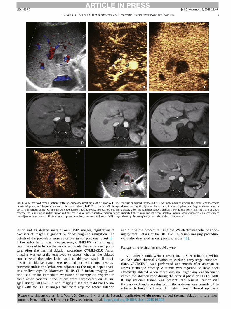
Fig. 2. A 49-year-old female patient with B-cell-derived lymphoma or dendritic cell tumor. A–C: The preoperative MRI images demonstrating the hyper-enhancement in arte-
rial phase and hypo-enhancement in the portal and venous phase of the lesion; D –F : The contrast enhanced ultrasound (CEUS) images demonstrating the hyper-enhancement
in arterial phase and hypo-enhancement in the portal phase; G : The 3D US-CEUS fusion imaging evaluation carried out immediately after the radiofrequency ablation show-
ing the non-enhanced zone of CEUS covered the index tumor and the yellow ring of preset ablative margin, which indicated the tumor and its 5-mm ablative margin were
completely ablated; H : One month post-operatively, contrast enhanced MRI image showing the completely necrosis of the index tumor.
R
D
s
a
n
h
s
e
s
i
three months after thermal ablation to evaluate recurrence and
complications.
Local tumor progression (LTP) was defined as the appearance of
new tumor foci one month after intervention at the edge of the ab-
lation zone, which was often characterized as hyper-enhancement
during arterial phase with hypo-enhancement in the portal venous
system or the delayed phase on CECT/CEMRI images.
Statistical analysis
All statistical analyses were performed using SPSS software
(Version 22.0, SPSS Inc., Chicago, IL, USA). Quantitative and qualita-
tive data were presented as the median (range) and number (per-
centage), respectively.
Please cite this article as: L.-L. Wu, J.-X. Chen and K. Li et al., Potentia
tumors, Hepatobiliary & Pancreatic Diseases International, https://doi.o
esults
emographic data of patients and lesions
In total, eight patients with twelve tumors were enrolled in this
tudy, including two male and six female patients with a median
ge of 47 (28–64) years. Among these eight patients, one had three
odules, and two patients had two nodules. Only one patient had a
istory of hepatitis B. The demographic and baseline data are pre-
ented in Table 1 . All of the nodules were confirmed by pathology
xamination.
In total, 91.7% (11/12) of the lesions presented as hypoechoic le-
ions on US images. In CEUS examinations, all of the lesions exhib-
ted “wash-out” features. In addition, 91.7% (11/12) of the lesions
l application of ultrasound-guided thermal ablation in rare liver
rg/10.1016/j.hbpd.2018.10.002
L.-L. Wu, J.-X. Chen and K. Li et al. / Hepatobiliary & Pancreatic Diseases International xxx (xxxx) xxx 5
ARTICLE IN PRESS
JID: HBPD [m5G; November 6, 2018;13:49 ]
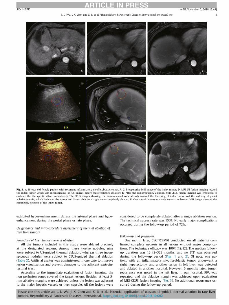
Fig. 3. A 46-year-old female patient with recurrent inflammatory myofibroblastic tumor. A –C : Preoperative MRI image of the index tumor; D : MRI-US fusion imaging located
the index tumor which was inconspicuous on US images before radiofrequency ablation; E : After the radiofrequency ablation, MRI-CEUS fusion imaging was employed to
evaluate the therapeutic effect immediately. The CEUS images showing the non-enhanced zone already covered the blue ring of index tumor and the red ring of preset
ablative margin, which indicated the tumor and 5-mm ablative margin were completely ablated; F : One month post-operatively, contrast enhanced MRI image showing the
completely necrosis of the index tumor.
e
e
U
r
P
a
w
s
(
l
t
n
m
t
c
T
o
F
fi
t
u
d
t
r
a
r
r
o
c
xhibited hyper-enhancement during the arterial phase and hypo-
nhancement during the portal phase or late phase.
S guidance and intra-procedure assessment of thermal ablation of
are liver tumors
rocedure of liver tumor thermal ablation
All the tumors included in this study were ablated precisely
t the designated regions. Among these twelve nodules, nine
ere subject to US-guided thermal ablation, whereas three incon-
picuous nodules were subject to CEUS-guided thermal ablation
Table 2 ). Artificial ascites was administered in one case to improve
esion visualization and prevent damages to the adjacent gastroin-
estinal tract.
According to the immediate evaluation of fusion imaging, the
on-perfusion zones covered the target lesions. Besides, at least 5-
m ablative margins were achieved unless the lesion was adjacent
o the major hepatic vessels or liver capsule. All the lesions were
Please cite this article as: L.-L. Wu, J.-X. Chen and K. Li et al., Potentia
tumors, Hepatobiliary & Pancreatic Diseases International, https://doi.o
onsidered to be completely ablated after a single ablation session.
he technical success rate was 100%. No early major complications
ccurred during the follow-up period of 72 h.
ollow-up and prognosis
One month later, CECT/CEMRI conducted on all patients con-
rmed complete necrosis in all lesions without major complica-
ions. The technique efficacy was 100% (12/12). The median follow-
p duration was 13 (2–32) months, and no LTP was observed
uring the follow-up period ( Figs. 1 and 2 ). Of note, one pa-
ient with an inflammatory myofibroblastic tumor underwent a
ight hepatectomy, and another lesion in left liver was detected
nd ablated in another hospital. However, 5 months later, tumor
ecurrence was noted in the left liver. In our hospital, RFA was
epeated, and the ablative margin was achieved upon evaluation
f MRI-CEUS fusion imaging ( Fig. 3 ). No additional recurrence oc-
urred during the follow-up period.
l application of ultrasound-guided thermal ablation in rare liver
rg/10.1016/j.hbpd.2018.10.002
6 L.-L. Wu, J.-X. Chen and K. Li et al. / Hepatobiliary & Pancreatic Diseases International xxx (xxxx) xxx
ARTICLE IN PRESS
JID: HBPD [m5G; November 6, 2018;13:49 ]
C
i
t
L
s
t
F
g
d
o
2
F
E
B
C
f
j
R
Discussion
CECT or CEMRI is recognized as major imaging modality for the
differentiation of liver tumors [10] . However, for rare liver tumors,
the enhancement pattern was atypical, and only a few studies have
been reported. CEUS is a novel imaging modality that is an ef-
fective method for the differentiation of most common liver tu-
mors according to enhancement patterns [11–14] . However, only
a few reports have provided enhancement patterns of rare liver
tumors [15] . In our study, all the lesions were “wash-out” in the
portal/delay phase, indicating the malignant nature of the tumor
according to the CEUS-LI-RADS [16] . Therefore, the acquisition of
pathological confirmation and removal of the liver tumor were se-
lected as the treatment strategy. In this study, all the enrolled pa-
tients refused liver resection, and US-guided biopsy and thermal
ablation were recommended for these patients after multidisci-
plinary consultation.
Previous studies reported that the most common treatment for
rare liver tumors is surgery [17] . Traditional surgical intervention
suggests that not only the target tumor but also the surrounding
liver parenchyma should be resected to reduce tumor volume and
the risk of recurrence. However, the clinical application of liver
resection was largely limited due to serious surgical trauma and
long recovery time. Local thermal ablation achieves the therapeutic
effect by killing tumor cells using high temperature. Compared
with the traditional surgical treatment, percutaneous thermal ab-
lation of liver tumors can effectively reduce trauma and surgery-
related complications [18,19] . However, due to the limited ab-
lative region, residual or LTP was more common. Our previous
reports demonstrated that the fusion imaging technique was a use-
ful tool to improve the local curative effect of HCC. With the help
of CT/MRI-US fusion imaging or 3D US-US fusion imaging, the ab-
lative margin could be simultaneously outlined and displayed on
real-time US images [8,20] . After ablation, immediate evaluation
using CT/MRI-CEUS fusion imaging or 3D US-CEUS fusion imag-
ing could be employed to assess whether the ablative region cov-
ers the entire lesion and its surrounding ablative margin. Thus,
we could enlarge the ablative region as far as possible in a man-
ner similar to surgical resection to reduce residual and LTP. In this
study, no residual or LTP was noted in all the enrolled patients.
However, one patient with a splenic inflammatory myofibroblastic
tumor and liver metastases experienced recurrence after RFA treat-
ment, which was performed at another hospital. She underwent
RFA in our hospital such that the ablation region covered the target
tumor and the ablative margin under the guidance of CT/MRI-CEUS
fusion imaging. No recurrence was noted during the subsequent
15-month follow-up. Therefore, local thermal ablation is also feasi-
ble for rare liver tumors, and the fusion imaging technique might
allow US-guided thermal ablation to become as effective as surgi-
cal resection. The risk of trauma and complication could be also
greatly reduced with thermal ablation when compared with resec-
tion.
Nevertheless, some limitations of this study should be noted.
First, the number of cases involved is small partly due to the low
incidence of rare liver tumors. This limitation may lead to a bi-
ased conclusion. Second, the follow-up duration is relatively short.
Hence, the recurrence and survival rates could differ after a longer
period of observation despite the fact that all the cases experi-
enced successful and complete ablation of their tumors with no
recurrence during the specified follow-up period.
In conclusion, US-guided thermal ablation is feasible and safe
for rare liver tumors. The use of a fusion imaging technique might
allow US-guided thermal ablation to become as effective as sur-
gical resection and serve as a potential therapeutic mode for rare
liver tumors in the future.
Please cite this article as: L.-L. Wu, J.-X. Chen and K. Li et al., Potentia
tumors, Hepatobiliary & Pancreatic Diseases International, https://doi.o
ontributors
WLL, CJX and XEJ conceived, designed and performed the exper-
ments, analyzed the data, wrote the paper, and reviewed drafts of
he paper. LK, SZZ and ZRQ reviewed drafts of the paper. LYL and
LP collected the data. All the authors have read and approved this
ubmission. WLL and CJX contributed equally to this work. XEJ is
he guarantor.
unding
This study was supported by grants from National Key R&D Pro-
ram of China ( 2017YFC01120 0 0 ), National Natural Science Foun-
ation of China ( 81430038 and 81401434 ), Science and Technol-
gy Planning Project of Guangdong Province ( 2015A020214009 ,
016A020215072 , and 2017A020215082 ); and Natural Science
oundation of Guangdong Province ( 2016A030313205 ).
thical approval
This study was approved by the Institutional Ethics Review
oard of the Third Affiliated Hospital of Sun Yat-sen University.
ompeting interest
No benefits in any form have been received or will be received
rom a commercial party related directly or indirectly to the sub-
ect of this article.
eferences
[1] Bruix J , Sherman M American Association for the Study of Liver Dis-
eases. Management of hepatocellular carcinoma: an update. Hepatology2011;53:1020–1022 .
[2] Chen W , Zheng R , Baade PD , Zhang S , Zeng H , Bray F , et al. Cancer statistics in
China, 2015. CA Cancer J Clin 2016;66:115–132 . [3] Parisi A , Desiderio J , Trastulli S , Castellani E , Pasquale R , Cirocchi R ,
et al. Liver resection versus radiofrequency ablation in the treatment of cir-rhotic patients with hepatocellular carcinoma. Hepatobiliary Pancreat Dis Int
2013;12:270–277 . [4] Lee DH , Lee JM . Primary malignant tumours in the non-cirrhotic liver. Eur J
Radiol 2017;95:349–361 .
[5] Feng K , Yan J , Li X , Xia F , Ma K , Wang S , et al. A randomized controlled trial ofradiofrequency ablation and surgical resection in the treatment of small hepa-
tocellular carcinoma. J Hepatol 2012;57:794–802 . [6] Lau WY , Lau SH . The current role of radiofrequency ablation in the treat-
ment of hepatocellular carcinoma. Hepatobiliary Pancreat Dis Int 2017;16:122–126 .
[7] Kudo M , Matsui O , Izumi N , Iijima H , Kadoya M , Imai Y , et al. JSH consen-
sus-based clinical practice guidelines for the management of hepatocellularcarcinoma: 2014 update by the liver cancer study group of Japan. Liver Cancer
2014;3:458–468 . [8] Li K , Su ZZ , Xu EJ , Ju JX , Meng XC , Zheng RQ . Improvement of ablative mar-
gins by the intraoperative use of CEUS-CT/MR image fusion in hepatocellularcarcinoma. BMC Cancer 2016;16:277 .
[9] Xu EJ , Lv SM , Li K , Long YL , Zeng QJ , Su ZZ , et al. Immediate evalua-
tion and guidance of liver cancer thermal ablation by three-dimensional ul-trasound/contrast-enhanced ultrasound fusion imaging. Int J Hyperthermia
2018;34:870–876 . [10] An C , Rakhmonova G , Choi JY , Kim MJ . Liver imaging reporting and data sys-
tem (LI-RADS) version 2014: understanding and application of the diagnosticalgorithm. Clin Mol Hepatol 2016;22:296–307 .
[11] Giorgio A , Montesarchio L , Gatti P , Amendola F , Matteucci P , Santoro B ,
et al. Contrast-enhanced ultrasound: a simple and effective tool in defining arapid diagnostic work-up for small nodules detected in cirrhotic patients dur-
ing surveillance. J Gastrointest Liver Dis 2016;25:205–211 . [12] Konopke R , Bunk A , Kersting S . The role of contrast-enhanced ultra-
sound for focal liver lesion detection: an overview. Ultrasound Med Biol2007;33:1515–1526 .
[13] Claudon M , Dietrich CF , Choi BI , Cosgrove DO , Kudo M , Nolsøe CP , et al. Guide-lines and good clinical practice recommendations for contrast enhanced ul-
trasound (CEUS) in the liver - update 2012: a WFUMB-EFSUMB initiative in
cooperation with representatives of AFSUMB, AIUM, ASUM, FLAUS and ICUS.Ultrasound Med Biol 2013;39:187–210 .
[14] Tominaga K , Kamimura K , Sakamaki A , Terai S . Intraductal papillary neoplasmof the bile duct: a rare liver tumor complicated by malignancy. Hepatology
2017;66:1695–1697 .
l application of ultrasound-guided thermal ablation in rare liver
rg/10.1016/j.hbpd.2018.10.002
L.-L. Wu, J.-X. Chen and K. Li et al. / Hepatobiliary & Pancreatic Diseases International xxx (xxxx) xxx 7
ARTICLE IN PRESS
JID: HBPD [m5G; November 6, 2018;13:49 ]
[
[15] Liu LN , Xu HX , Zheng SG , Sun LP , Guo LH , Zhang YF , et al. Ultrasound findingsof intraductal papillary neoplasm in bile duct and the added value of con-
trast-enhanced ultrasound. Ultraschall Med 2015;36:594–602 . [16] Piscaglia F , Wilson SR , Lyshchik A , Cosgrove D , Dietrich CF , Jang HJ , et al. Amer-
ican college of radiology contrast enhanced ultrasound liver imaging reportingand data system (CEUS LI-RADS) for the diagnosis of hepatocellular carcinoma:
a pictorial essay. Ultraschall Med 2017;38:320–324 . [17] Ismail H , Dembowska-Bagi ́nska B , Broniszczak D , Kalici ́nski P , Maruszewski P ,
Kluge P , et al. Treatment of undifferentiated embryonal sarcoma of the liver in
children–single center experience. J Pediatr Surg 2013;48:2202–2206 .
Please cite this article as: L.-L. Wu, J.-X. Chen and K. Li et al., Potentia
tumors, Hepatobiliary & Pancreatic Diseases International, https://doi.o
[18] Lei JY , Wang WT , Yan LN , Wen TF , Li B . Radiofrequency ablation versus surgicalresection for small unifocal hepatocellular carcinomas. Medicine (Baltimore)
2014;93:e271 . [19] Cho YK , Kim JK , Kim WT , Chung JW . Hepatic resection versus radiofrequency
ablation for very early stage hepatocellular carcinoma: a Markov model analy-sis. Hepatology 2010;51:1284–1290 .
20] Li K , Su Z , Xu E , Huang Q , Zeng Q , Zheng R . Evaluation of the ablation mar-gin of hepatocellular carcinoma using CEUS-CT/MR image fusion in a phantom
model and in patients. BMC Cancer 2017;17:61 .
l application of ultrasound-guided thermal ablation in rare liver
rg/10.1016/j.hbpd.2018.10.002