JOURNAL OF VIROLOGY, Aug. 1996, p. 5255–5265 Vol. 70, No. 8 0022-538X/96/$04.0010 Copyright q 1996, American Society for Microbiology Herpes Simplex Virus Type 1 Protein IE63 Affects the Nuclear Export of Virus Intron-Containing Transcripts A. PHELAN, J. DUNLOP, AND J. B. CLEMENTS* Institute of Virology, University of Glasgow, Glasgow, Scotland G11 5JR Received 11 March 1996/Accepted 29 April 1996 Using in situ hybridization labelling methods, we have determined that the herpes simplex virus type 1 immediate-early protein IE63 (ICP27) affects the cellular localization of virus transcripts. Intronless tran- scripts from the IE63, UL38, and UL44 genes are rapidly exported to and accumulate in the cytoplasm throughout infection, in either the presence or absence of IE63 expression. The intron-containing transcripts from the IE110 and UL15 genes, while initially cytoplasmic, are increasingly retained in the nucleus in distinct clumps as infection proceeds, and the clumps colocalize with the redistributed small nuclear ribonucleoprotein particles. Infections with the IE63 mutant virus 27-lacZ demonstrated that in the absence of IE63 expression, nuclear retention of intron-containing transcripts was lost. The nuclear retention of UL15 transcripts, which demonstrated both nuclear and cytoplasmic label, was not as pronounced as that of the IE110 transcripts, and we propose that this is due to the late expression of UL15. Infections with the mutant virus 110C1, in which both introns of IE110 have been precisely removed (R. D. Everett, J. Gen. Virol. 72:651–659, 1991), demon- strated IE110 transcripts in both the nucleus and the cytoplasm; thus, exon definition sequences which regulate viral RNA transport are present in the IE110 transcript. By in situ hybridization a stable population of polyadenylated RNAs was found to accumulate in the nucleus in spots, most of which were separate from the small nuclear ribonucleoprotein particle clumps. The IE63 protein has an involvement, either direct or indirect, in the regulation of nucleocytoplasmic transport of viral transcripts, a function which contrasts with the recently proposed role of herpes simplex virus type 1 Us11 in promoting the nuclear export of partially spliced or unspliced transcripts (J.-J. Diaz, M. Duc Dodon, N. Schaerer-Uthurraly, D. Simonin, K. Kindbeiter, L. Gazzolo, and J.-J. Madjar, Nature [London] 379:273–277, 1996), the significance of which is discussed. Primary transcripts of genes transcribed by RNA polymerase II generally are processed by 59 capping, splicing, and 39 pro- cessing before transportation to the cytoplasm (reviewed in references 35, 36, and 71). Direct labelling methods have dem- onstrated that splicing occurs at sites of transcription (80); however, polyadenylated unspliced intermediates can be de- tected both in vivo and in vitro (53). Splicing of 59-proximal introns can precede polyadenylation of large transcripts (39), and Liu and Mertz (40) have demonstrated that poly(A) site selection, but not necessarily cleavage, precedes excision of the 39-terminal intron in vivo; the temporal order in which these RNA processing functions occur is variable. The 59 cap and poly(A) tail are believed to act as RNA transport signals (13), whereas the presence of introns inhibits transport to the cyto- plasm as splicing factors are recruited to intron-containing transcripts which are then retained in the nucleus until they are spliced and/or actively transported to the cytoplasm (2). A number of factors govern the nucleocytoplasmic transport of mRNA, although this process is still poorly understood. Nuclear export occurs in two stages: (i) transport from the gene to the nuclear periphery and (ii) translocation across the nuclear membrane (13). The first step involves ribonucleopro- teins (RNPs) (45), which bind RNA cotranscriptionally (51, 57). The mechanism by which a transcript passes through the nuclear pore is thought to involve RNP interactions; binding of heterogeneous nuclear RNP L correlates with efficient mRNA processing and transport, and heterogeneous nuclear RNP L facilitates binding of other heterogeneous nuclear RNPs to the RNA (41). Regulation of nucleocytoplasmic transport has been identi- fied in a number of viral systems. The human immunodefi- ciency virus (HIV) type 1 protein Rev together with a cellular factor, Rab, induces the cytoplasmic appearance of unspliced and partially spliced virus transcripts via a direct and sequence- specific interaction with the Rev response element, which is contained within all intron-containing HIV transcripts (11, 18); a similar mechanism has been identified for the human T-cell leukemia virus (HTLV) type 1 Rex protein (3). In contrast to Rev and Rex, the influenza virus NS1 protein has an inhibitory effect on the transport of spliced polyadenylated RNAs and inhibits splicing (19, 59, 60). The adenovirus E1B protein en- hances the relocation of viral mRNAs to particular subnuclear compartments and facilitates their export to the cytoplasm (38, 55). The herpes simplex virus type 1 (HSV-1) Us11 late protein has been implicated as having effects similar to those of Rev and Rex, as it can functionally substitute for their functions in vitro (12). We show here that the HSV-1 IE63 protein acts to inhibit nuclear export of intron-containing transcripts; whether Us11 acts to promote export of unspliced transcripts in HSV- infected cells, in which case IE63 and Us11 would be in ap- parent competition, remains to be seen. HSV-1 is a nuclear-replicating DNA virus (64) the genes of which are expressed in a temporal cascade (9, 30). Immediate- early (IE) regulatory genes are expressed first; these stimulate early gene expression, which provides many of the functions required for viral DNA synthesis; and finally the late virus genes, which encode mainly structural proteins, are expressed (64, 77). Of the more than 70 HSV-1 genes, only 4 of those expressed during lytic infection contain introns (21). Thus, the virus has only a very limited requirement for the cell’s splicing * Corresponding author. Mailing address: Institute of Virology, Uni- versity of Glasgow, Church St., Glasgow, Scotland G11 5JR. Phone: 141 330 4017. Fax: 141 337 2236. 5255
Transcript
JOURNAL OF VIROLOGY, Aug. 1996, p. 5255–5265 Vol. 70, No. 80022-538X/96/$04.0010Copyright q 1996, American Society for Microbiology
Herpes Simplex Virus Type 1 Protein IE63 Affects the NuclearExport of Virus Intron-Containing Transcripts
A. PHELAN, J. DUNLOP, AND J. B. CLEMENTS*
Institute of Virology, University of Glasgow, Glasgow, Scotland G11 5JR
Received 11 March 1996/Accepted 29 April 1996
Using in situ hybridization labelling methods, we have determined that the herpes simplex virus type 1immediate-early protein IE63 (ICP27) affects the cellular localization of virus transcripts. Intronless tran-scripts from the IE63, UL38, and UL44 genes are rapidly exported to and accumulate in the cytoplasmthroughout infection, in either the presence or absence of IE63 expression. The intron-containing transcriptsfrom the IE110 and UL15 genes, while initially cytoplasmic, are increasingly retained in the nucleus in distinctclumps as infection proceeds, and the clumps colocalize with the redistributed small nuclear ribonucleoproteinparticles. Infections with the IE63 mutant virus 27-lacZ demonstrated that in the absence of IE63 expression,nuclear retention of intron-containing transcripts was lost. The nuclear retention of UL15 transcripts, whichdemonstrated both nuclear and cytoplasmic label, was not as pronounced as that of the IE110 transcripts, andwe propose that this is due to the late expression of UL15. Infections with the mutant virus 110C1, in whichboth introns of IE110 have been precisely removed (R. D. Everett, J. Gen. Virol. 72:651–659, 1991), demon-strated IE110 transcripts in both the nucleus and the cytoplasm; thus, exon definition sequences which regulateviral RNA transport are present in the IE110 transcript. By in situ hybridization a stable population ofpolyadenylated RNAs was found to accumulate in the nucleus in spots, most of which were separate from thesmall nuclear ribonucleoprotein particle clumps. The IE63 protein has an involvement, either direct orindirect, in the regulation of nucleocytoplasmic transport of viral transcripts, a function which contrasts withthe recently proposed role of herpes simplex virus type 1 Us11 in promoting the nuclear export of partiallyspliced or unspliced transcripts (J.-J. Diaz, M. Duc Dodon, N. Schaerer-Uthurraly, D. Simonin, K. Kindbeiter,L. Gazzolo, and J.-J. Madjar, Nature [London] 379:273–277, 1996), the significance of which is discussed.
Primary transcripts of genes transcribed by RNA polymeraseII generally are processed by 59 capping, splicing, and 39 pro-cessing before transportation to the cytoplasm (reviewed inreferences 35, 36, and 71). Direct labelling methods have dem-onstrated that splicing occurs at sites of transcription (80);however, polyadenylated unspliced intermediates can be de-tected both in vivo and in vitro (53). Splicing of 59-proximalintrons can precede polyadenylation of large transcripts (39),and Liu and Mertz (40) have demonstrated that poly(A) siteselection, but not necessarily cleavage, precedes excision of the39-terminal intron in vivo; the temporal order in which theseRNA processing functions occur is variable. The 59 cap andpoly(A) tail are believed to act as RNA transport signals (13),whereas the presence of introns inhibits transport to the cyto-plasm as splicing factors are recruited to intron-containingtranscripts which are then retained in the nucleus until they arespliced and/or actively transported to the cytoplasm (2).A number of factors govern the nucleocytoplasmic transport
of mRNA, although this process is still poorly understood.Nuclear export occurs in two stages: (i) transport from thegene to the nuclear periphery and (ii) translocation across thenuclear membrane (13). The first step involves ribonucleopro-teins (RNPs) (45), which bind RNA cotranscriptionally (51,57). The mechanism by which a transcript passes through thenuclear pore is thought to involve RNP interactions; binding ofheterogeneous nuclear RNP L correlates with efficient mRNAprocessing and transport, and heterogeneous nuclear RNP L
facilitates binding of other heterogeneous nuclear RNPs to theRNA (41).Regulation of nucleocytoplasmic transport has been identi-
fied in a number of viral systems. The human immunodefi-ciency virus (HIV) type 1 protein Rev together with a cellularfactor, Rab, induces the cytoplasmic appearance of unsplicedand partially spliced virus transcripts via a direct and sequence-specific interaction with the Rev response element, which iscontained within all intron-containing HIV transcripts (11, 18);a similar mechanism has been identified for the human T-cellleukemia virus (HTLV) type 1 Rex protein (3). In contrast toRev and Rex, the influenza virus NS1 protein has an inhibitoryeffect on the transport of spliced polyadenylated RNAs andinhibits splicing (19, 59, 60). The adenovirus E1B protein en-hances the relocation of viral mRNAs to particular subnuclearcompartments and facilitates their export to the cytoplasm (38,55). The herpes simplex virus type 1 (HSV-1) Us11 late proteinhas been implicated as having effects similar to those of Revand Rex, as it can functionally substitute for their functions invitro (12). We show here that the HSV-1 IE63 protein acts toinhibit nuclear export of intron-containing transcripts; whetherUs11 acts to promote export of unspliced transcripts in HSV-infected cells, in which case IE63 and Us11 would be in ap-parent competition, remains to be seen.HSV-1 is a nuclear-replicating DNA virus (64) the genes of
which are expressed in a temporal cascade (9, 30). Immediate-early (IE) regulatory genes are expressed first; these stimulateearly gene expression, which provides many of the functionsrequired for viral DNA synthesis; and finally the late virusgenes, which encode mainly structural proteins, are expressed(64, 77). Of the more than 70 HSV-1 genes, only 4 of thoseexpressed during lytic infection contain introns (21). Thus, thevirus has only a very limited requirement for the cell’s splicing
* Corresponding author. Mailing address: Institute of Virology, Uni-versity of Glasgow, Church St., Glasgow, Scotland G11 5JR. Phone:141 330 4017. Fax: 141 337 2236.
5255
machinery; however, lytically expressed viral transcripts arecapped and polyadenylated. Of the four spliced transcripts,three (IE110, IE68, and IE12) are expressed from IE timespostinfection, and the fourth (UL15) is expressed from latetimes (10). Another late gene, UL44, has been associated withsplicing, as it is apparently located within an intron and itstranscripts are produced only in the absence of splicing (21).HSV-1 infection represses host protein synthesis (17, 29),
and this is initially accomplished by the virion host shutofffunction, the action of an incoming viral tegument protein (37,61, 72), which acts by induction of an RNase activity whichdestabilizes viral and cellular mRNAs and disrupts polyribo-somes (34, 37, 75). A second stage of host shutoff, whichrequires virus gene expression, further reduces the levels ofhost protein synthesis (16), and the IE63 protein was requiredfor efficient reduction of host protein synthesis (65), implicat-ing this protein as a strong candidate for the second hostshutoff function.An inhibition of splicing has been identified following HSV
infection. Schroder et al. (69) found that splicing of b-tubulinRNA was reduced, and Hardwicke and Sandri-Goldin (24)demonstrated a decrease in the levels of three spliced cellularRNAs which was not due to effects on transcription or RNAstability. More recently, Hardy and Sandri-Goldin (26), usingan IE63 mutant virus, 27-lacZ, demonstrated that IE63 wasrequired for inhibition of splicing in vitro, and Hibbard andSandri-Goldin (28) found a reduction in cellular mRNA levelsand nuclear accumulation of pre-mRNAs which required IE63expression. Inhibition of splicing resulted in the nuclear exportof some unspliced transcripts (26), suggesting that regulationof RNA export was also affected.The IE63 (ICP27) protein is a nuclear phosphoprotein and
is one of only two IE functions essential for lytic virus replica-tion (62). IE63 plays a key role in the switch from early to lategene expression at the posttranscriptional level (46, 48, 49, 62,65, 68), has both positive and negative effects on gene expres-sion (25, 50, 70, 73), and functionally interacts with two otherIE proteins, IE175 and IE110 (70, 81). IE63 affects the phos-phorylation states of a number of proteins (one of which isthought to be the U1 small nuclear RNP [snRNP]) (66), bindsRNA directly (32), and acts to stabilize the 39 ends of normallylabile mRNAs (6). Further, IE63 has been shown to be bothnecessary and sufficient to cause a redistribution of the cellularsplicing snRNPs from a diffuse speckled pattern within thenucleus to a highly punctate distribution (56); this redistribu-tion effect was originally observed by Martin et al. (44). Fur-thermore, we showed that IE63 colocalizes with the snRNPs(56), and more recently, Sandri-Goldin and Hibbard (66) haveshown this interaction by coimmunoprecipitation studies. Thedistribution of the polyadenylation factors remains largely un-affected during HSV infection (48).Disruption of snRNP organization can be observed under a
number of conditions; for example, inhibition of RNA poly-merase II transcription by a-amanitin (7), inhibition of splicingby using antisense RNA molecules or antibodies (54), and heatshock treatments (4) cause snRNPs to cycle into interchroma-tin granule storage sites. Infections with adenovirus (5, 33, 58)and influenza virus (20) also cause a redistribution of thesnRNPs. Adenovirus RNAs are extensively spliced, and thesplicing factors are recruited into virus transcription and pro-cessing sites; in contrast, influenza virus has little requirementfor splicing and inhibits cellular splicing, indicating that theredistributed snRNPs in influenza virus-infected cells are in-active aggregates (20).IE63 can inhibit splicing and can interact and colocalize with
the snRNPs, which suggests that this protein inhibits splicing
by removing snRNPs from sites of transcription and polyade-nylation. Although splicing is largely inhibited by HSV-1 in-fection, there is evidence for a residual amount of ongoingsplicing, as the few intron-containing viral transcripts and somecellular genes are processed. Presumably this residual splicingcould be carried out by the remaining snRNPs diffusely dis-tributed throughout the nucleoplasm.Previous studies which have examined the effects of HSV-1
on splicing have been based largely on in vitro assays andtransient-expression systems. In this study we have directlyexamined the intracellular localization of spliced and unsplicedviral RNAs by using in situ hybridization experiments over atime course of HSV-1 infection. The IE and late intron-con-taining transcripts studied (IE110 and UL15) were increasinglyretained in the nucleus in distinct clumps as infection pro-ceeded, whereas intronless IE and late transcripts (IE63,UL38, and UL44) were detected predominantly in the cyto-plasm. Double-labelling experiments have demonstrated thatthese intron-containing transcripts colocalize with the redis-tributed snRNP clumps. IE63 is required for the nuclear re-tention of intron-containing transcripts, since in its absence, alltranscripts were predominantly cytoplasmic. Cells infectedwith an IE110 mutant virus in which both introns have beenprecisely removed (14) demonstrated a localization of IE110transcripts in both the nucleus and cytoplasm, showing morenuclear label than would be expected for an intronless tran-script but more cytoplasmic label than seen with intron-con-taining transcripts. This suggests the presence of exon defini-tion sequences in the IE110 transcript by which splicing signalsare recognized (53, 80). Interestingly, the UL15 late transcriptdemonstrated a similar intermediate phenotype with both nu-clear and cytoplasmic label, and we believe that this is a re-flection of the time postinfection of its expression. We haveutilized poly(dT) in situ hybridization to locate polyadenylatedRNAs and found poly(A)1 RNA in the cytoplasm and indistinct nuclear spots at later times postinfection. However,only a small proportion of these distinct spots colocalized withthe redistributed snRNPs, suggesting the presence of either astable population of poly(A)1 RNA which is retained in thenucleus but whose function is unknown (8, 31) or transientstorage forms of incompletely or abnormally processed pre-mRNA (76).
MATERIALS AND METHODS
Cell culture and viruses. HeLa cells were grown as monolayers in Dulbecco’smodified minimal essential medium supplemented with 5% newborn calf serumand 5% fetal calf serum. Vero 2-2 cells (73) were maintained in Glasgow minimalessential medium supplemented with 5% newborn calf serum and 5% fetal calfserum. BHK cells were grown in Glasgow minimal essential medium supple-mented with 10% newborn calf serum. All cells were grown at 378C in anatmosphere of 5% CO2.Stocks of wild-type HSV-1 (strain 171) and the HSV-1 mutant 110C1, a gift
from R. Everett, in which both introns of IE110 have been precisely removed(14), were grown at 378C on BHK monolayers. Virus 27-lacZ, a gift from R.Sandri-Goldin, in which the IE63 gene is inactivated by insertion of a lacZcassette, was grown on the complementing Vero 2-2 cell line (73).HSV-1 infection of cell monolayers. All cells were grown as monolayers on
sterile glass coverslips previously treated with 10% poly-L-lysine for 10 min aftersterilization. Subconfluent cell monolayers were infected with either wild-type ormutant HSV-1 at a multiplicity of infection of 10 PFU per cell. After 1 h at 378C,the infected medium was removed and replaced with fresh prewarmed mediumuntil the time of harvesting. Coverslips were harvested at 2-h intervals until 16 hpostinfection. Mock-infected (uninfected) cells were treated in an identical man-ner with the omission of virus.RNA in situ hybridization. RNA in situ hybridization was performed by two
methods.(i) Riboprobe in situ. Cells fixed for 10 min at room temperature (RT) in 3.7%
paraformaldehyde in CSK buffer {100 mM NaCl, 300 mM sucrose, 10 mMPIPES [piperazine-N,N9-bis(2-ethanesulfonic acid)], 3 mMMgCl2, 1 mM EGTA[ethylene glycol-bis(b-aminoethyl ether)-N,N,N9,N9-tetraacetic acid], pH 8} werelabelled as described in the Amersham RNA Colour Kit protocol with the
5256 PHELAN ET AL. J. VIROL.
omission of the proteinase K step. Fluorescein-UTP-labelled riboprobes between200 and 400 bp in length were transcribed in vitro to produce both sense (Sp6RNA polymerase) and antisense (T7 RNA polymerase) riboprobes. Sites ofhybridization were detected with anti-fluorescein alkaline phosphatase-conju-gated antibodies and alkaline phosphatase color development (Amersham kit).Digoxigenin-UTP-labelled sense and antisense riboprobes were synthesized fromthe same set of templates. Regions of hybridized probe were detected by usingan anti-digoxigenin fluorescein-conjugated antibody at a dilution of 1:10 (Boehr-inger Mannheim).A number of negative control experiments were performed. Treatment with
RNase A (200 mg/ml) and RNase H (100 U/ml) at 378C for 20 min prior tohybridization removed all signal. RNase H treatment after hybridization re-moved any DNA-RNA hybrids and reduced the background label but did notaffect the genuine signal; RNase A treatment after hybridization removed allunhybridized probe. Treatment with DNase (100 U/ml) for 1 h prior to hybrid-ization removed the background label but had no effects on the signal obtained.(ii) Biotinylated RNA oligonucleotides. Cells grown as monolayers on cover-
slips were permeabilized with 0.5% Triton in CSK buffer for 30 s on ice prior tofixation for 10 min at RT with 3.7% paraformaldehyde in CSK buffer. Biotiny-lated 29-O-methyl RNA oligonucleotides were hybridized for 1 h at RT at a finalprobe concentration of 1 pmol/ml (7) in a humidified chamber as follows. Fixedcells were rinsed in 63 SSPE (13 SSPE is 0.18 M NaCl, 10 mM NaH2PO4, and1 mM EDTA [pH 7.7]) and incubated for 5 to 15 min with tRNA (0.5 mg/ml in63 SSPE–53 Denhardt’s solution), the probe was added in an equal volume of63 SSPE–53 Denhardt’s solution, and hybridization was for 1 h at RT. Cellswere washed in 63 SSPE, and the probe was detected with Avidin-fluoresceinisothiocyanate (FITC) diluted 1:500 in Avidin wash buffer (20 mM HEPES[N-2-hydroxyethylpiperazine-N9-2-ethanesulfonic acid] [pH 7.9], 150 mM KCl,0.05% Tween). Coverslips were either mounted with Mowiol or double labelledby using a monoclonal anti-B0 U2 snRNP antibody and a rhodamine-conjugatedsecondary antibody as described in “Immunofluorescence and antibodies” below.In situ riboprobes. Fluorescein-UTP- or digoxigenin-UTP-labelled sense and
antisense riboprobes were synthesized as described in the Amersham RNAColour Kit. Essentially, riboprobes of between 200 and 400 bp in length weretranscribed in vitro by using Sp6 or T7 RNA polymerase with linearized plasmidsencoding the various gene-specific probe regions as the template. Probes againstthe following HSV-1 gene transcripts were generated: IE63 transcriptional trans-activator (genomic map positions [47] 115035 to 115435), 400-bp probe; IE110transcriptional transactivator, exon 3 (120480 to 121033), 210-bp probe; UL15DNA packaging protein, exon 1 (29419 to 29823), 404-bp probe; UL44 glyco-protein C (97444 to 97844), 400-bp probe; and UL38 capsid protein (86016 to85616), 400-bp probe.Biotinylated 29-O-methyl RNA oligonucleotides kindly provided by Cruachem,
Ltd., Glasgow, Scotland, were generated against U1 snRNP (biotin-CC UGCCAG GUA AGU AU) (16-mer) (7), IE110 exon 3 (biotin-UGU UAC UGCUGC CGU GU) (17-mer), and IE110 intron 1 (biotin-UAU GUG UUG GGGGUC UGU A) (19-mer). These probes were used in conjunction with immuno-fluorescence assays.DNA in situ hybridization. A biotinylated poly(dT) DNA oligonucleotide
(18-mer) was used to locate polyadenylated RNAs in the nucleus, using themethod described for the RNA oligonucleotides.Immunofluorescence and antibodies. Indirect-immunofluorescence experi-
ments were performed as previously described (56). All antibodies were dilutedin phosphate-buffered saline before use. The U2 splicing snRNPs were labelledwith the B0 monoclonal antibody 4G3 (23) at a dilution of 1:5.Microscopy. Labelled cells were examined with a Nikon light and fluorescence
microscope. Photographs of alkaline phosphatase-labelled cells were taken with160T Fuji films. Fluorescently labelled cells were photographed with ASA 400Ektachrome films.
RESULTS
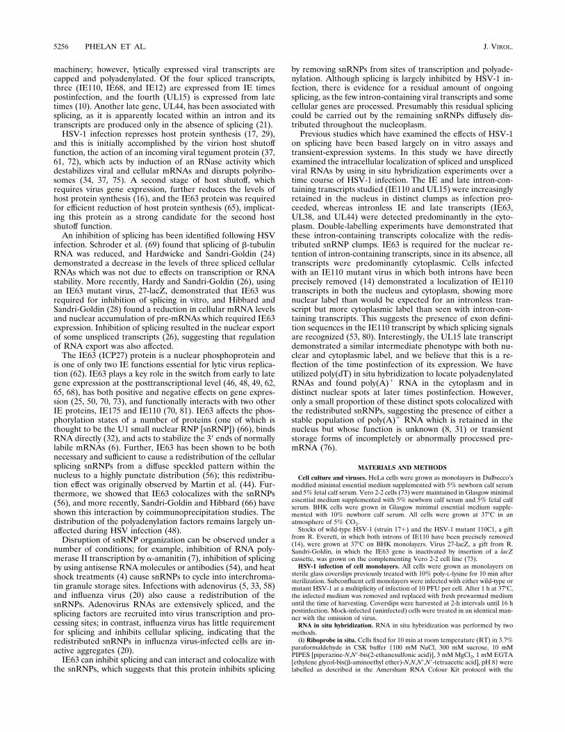
Intron-containing transcripts are retained in the nuclei ofinfected cells. HeLa cells infected with wild-type HSV-1 for 0,2, 4, 8, and 16 h were labelled with antisense fluorescein-labelled riboprobes directed against the coding portion ofIE63, an intronless transcript (Fig. 1a to d), and exon 3 ofIE110, a transcript which contains two introns (Fig. 1e to h),and the hybridized probes were detected by alkaline phos-phatase color development. The viral probes hybridized spe-cifically to virus transcripts and did not label the uninfectedcells (Fig. 1a and e). At 2 h postinfection, cells labelled with theanti-IE63 riboprobe demonstrated predominantly cytoplasmiclabel with some faint nuclear stain (Fig. 1b). As infectionproceeded, labelling became more intense, with the patternremaining constant and with IE63 transcripts being predomi-nantly cytoplasmic at the 8-h (Fig. 1c) and 16-h (Fig. 1d) timepoints. The IE110 exonic riboprobe demonstrated a strikingly
different labelling pattern. At 2 h postinfection, the label waslargely cytoplasmic with some quantity of nuclear stain (Fig.1f). By 4 to 6 h a large proportion of the label was found in thenucleus in distinct densely staining clumps, and cytoplasmiclabel was noticeably reduced (Fig. 1g). This became morepronounced, and by 8 h (not shown) and 16 h (Fig. 1h) thenucleus was packed with accumulated IE110 transcripts (some-what obscuring the discrete clump formation), and little cyto-plasmic label was seen. RNase A (Fig. 1k) and RNase H (Fig.1l) treatments of cells labelled with an anti-IE63 riboprobe 8 hpostinfection demonstrated that the hybridization was specific:RNase A removed any unhybridized probe and reduced thebackground label slightly; RNase H digested any RNA-DNAhybrids and removed some background label, but the bulk ofthe signal was unaffected. Sense riboprobes against IE110 (Fig.1i) and IE63 (Fig. 1j) did not hybridize.To investigate whether these two distinctive labelling pat-
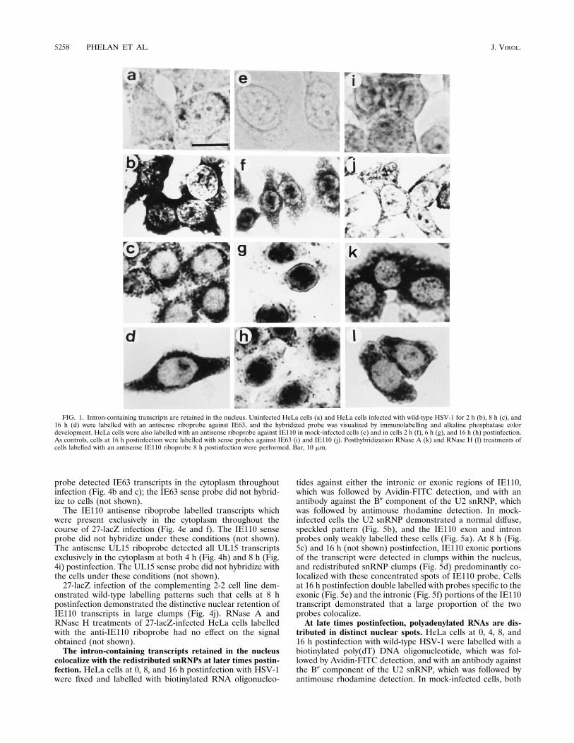
terns were exhibited by other spliced and unspliced viralRNAs, the locations of three additional viral transcripts wereexamined throughout infection. HeLa cells were labelled withantisense riboprobes against the late genes UL38 (Fig. 2a to c),UL44 (Fig. 2d to f), and UL15 (Fig. 2g to j). The intronlessUL38 and UL44 transcripts were detected almost exclusively inthe cytoplasm throughout infection (Fig. 2a to c and d to f,respectively), with very little nuclear label observed. Occa-sional nonspecific label was found associated with nucleoli(Fig. 2f); this could be removed by posthybridization RNase Atreatment (not shown). An antisense riboprobe against theUL15 transcript, which contains a single intron, labelled in-fected nuclei with an intermediate nuclear and cytoplasmicphenotype (Fig. 2g to j). Throughout infection (0 to 16 h),UL15 transcripts could be detected in the nucleus in largeclumps, but there was also a significant amount of cytoplasmiclabel, a phenotype intermediate between those of IE63 andIE110 transcripts. Sense riboprobes against UL38, UL44, andUL15 (not shown) exhibited no hybridization under these con-ditions. RNase A, RNase H, and DNase I treatments of thecells hybridized with the antisense UL15 probe after labellingat 8 h postinfection did not alter the labelling patterns (notshown), except that nonspecific nucleolar labelling was re-moved.IE110 transcripts which lack introns are still retained in the
nucleus. HeLa cells infected with the virus 110C1, in whichboth introns of IE110 have been precisely removed, were la-belled with an antisense IE110 riboprobe from exon 3 over atime course of infection, and sites of hybridization were de-tected by alkaline phosphatase color development.The mock-infected cells were not labelled with this probe
(Fig. 3a). Cells labelled 2, 4, 8, and 16 h postinfection with110C1 (Fig. 3b to e) demonstrated an intermediate phenotype,with considerably more label retained in the nucleus than ob-served for a transcript without introns (compare Fig. 3c withFig. 1c) and also with more cytoplasmic label than seen withthe IE110 intron-containing transcripts (compare Fig. 3c withFig. 1g). The IE110 sense riboprobe did not label the cells (Fig.3f), and RNase A and RNase H treatments posthybridizationhad no effect on the signal obtained (Fig. 3g and h).IE63 protein is required for the nuclear retention of intron-
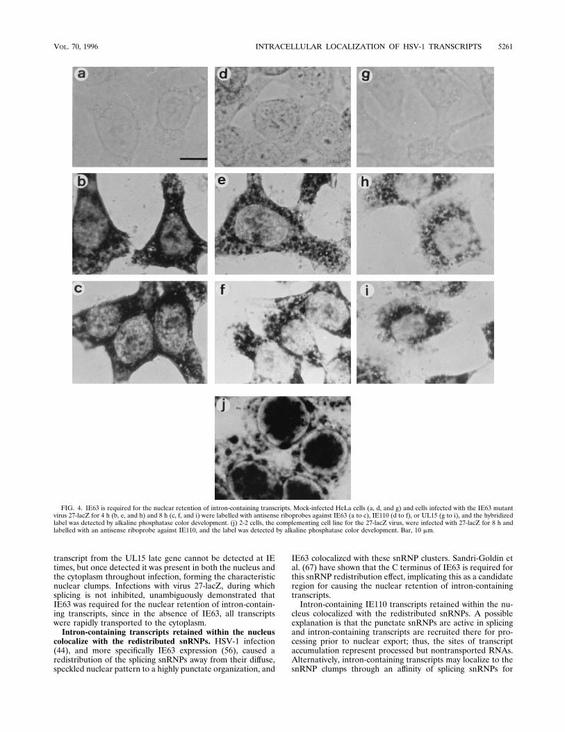
containing viral transcripts. HeLa cells infected with the IE63mutant virus 27-lacZ for 0, 4, 8, and 16 h (not shown) werelabelled with an antisense fluorescein-labelled riboprobe di-rected against the coding portion of IE63 (Fig. 4a to c), IE110exon 3 (Fig. 4d to f), and UL15 exon 2 (Fig. 4g to i), and thehybridized label was detected by alkaline phosphatase colordevelopment. Mock-infected cells were not labelled with any ofthe probes used (Fig. 4a, d, and g). The antisense IE63 ribo-
VOL. 70, 1996 INTRACELLULAR LOCALIZATION OF HSV-1 TRANSCRIPTS 5257
probe detected IE63 transcripts in the cytoplasm throughoutinfection (Fig. 4b and c); the IE63 sense probe did not hybrid-ize to cells (not shown).The IE110 antisense riboprobe labelled transcripts which
were present exclusively in the cytoplasm throughout thecourse of 27-lacZ infection (Fig. 4e and f). The IE110 senseprobe did not hybridize under these conditions (not shown).The antisense UL15 riboprobe detected all UL15 transcriptsexclusively in the cytoplasm at both 4 h (Fig. 4h) and 8 h (Fig.4i) postinfection. The UL15 sense probe did not hybridize withthe cells under these conditions (not shown).27-lacZ infection of the complementing 2-2 cell line dem-
onstrated wild-type labelling patterns such that cells at 8 hpostinfection demonstrated the distinctive nuclear retention ofIE110 transcripts in large clumps (Fig. 4j). RNase A andRNase H treatments of 27-lacZ-infected HeLa cells labelledwith the anti-IE110 riboprobe had no effect on the signalobtained (not shown).The intron-containing transcripts retained in the nucleus
colocalize with the redistributed snRNPs at later times postin-fection. HeLa cells at 0, 8, and 16 h postinfection with HSV-1were fixed and labelled with biotinylated RNA oligonucleo-
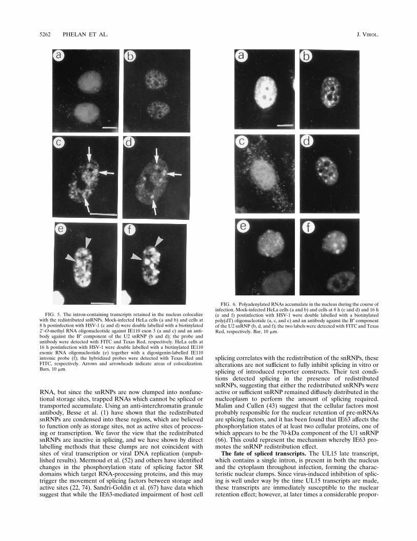
tides against either the intronic or exonic regions of IE110,which was followed by Avidin-FITC detection, and with anantibody against the B0 component of the U2 snRNP, whichwas followed by antimouse rhodamine detection. In mock-infected cells the U2 snRNP demonstrated a normal diffuse,speckled pattern (Fig. 5b), and the IE110 exon and intronprobes only weakly labelled these cells (Fig. 5a). At 8 h (Fig.5c) and 16 h (not shown) postinfection, IE110 exonic portionsof the transcript were detected in clumps within the nucleus,and redistributed snRNP clumps (Fig. 5d) predominantly co-localized with these concentrated spots of IE110 probe. Cellsat 16 h postinfection double labelled with probes specific to theexonic (Fig. 5e) and the intronic (Fig. 5f) portions of the IE110transcript demonstrated that a large proportion of the twoprobes colocalize.At late times postinfection, polyadenylated RNAs are dis-
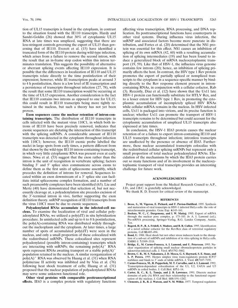
tributed in distinct nuclear spots. HeLa cells at 0, 4, 8, and16 h postinfection with wild-type HSV-1 were labelled with abiotinylated poly(dT) DNA oligonucleotide, which was fol-lowed by Avidin-FITC detection, and with an antibody againstthe B0 component of the U2 snRNP, which was followed byantimouse rhodamine detection. In mock-infected cells, both
FIG. 1. Intron-containing transcripts are retained in the nucleus. Uninfected HeLa cells (a) and HeLa cells infected with wild-type HSV-1 for 2 h (b), 8 h (c), and16 h (d) were labelled with an antisense riboprobe against IE63, and the hybridized probe was visualized by immunolabelling and alkaline phosphatase colordevelopment. HeLa cells were also labelled with an antisense riboprobe against IE110 in mock-infected cells (e) and in cells 2 h (f), 6 h (g), and 16 h (h) postinfection.As controls, cells at 16 h postinfection were labelled with sense probes against IE63 (i) and IE110 (j). Posthybridization RNase A (k) and RNase H (l) treatments ofcells labelled with an antisense IE110 riboprobe 8 h postinfection were performed. Bar, 10 mm.
5258 PHELAN ET AL. J. VIROL.
FIG. 2. Intronless transcripts are rapidly exported to the cytoplasm. Mock-infected HeLa cells (a and d) and HeLa cells infected with HSV-1 (b, c, e, and f) werelabelled with antisense riboprobes against UL38 (a to c) and UL44 (d to f) at 8 h (b and e) and 16 h (c and f) post-infection. Mock-infected cells (g) and infected cells(h to j) were labelled with an antisense riboprobe against UL15 at 4 h (h), 8 h (i), and 16 h (j) postinfection. The hybridized probe was detected by alkaline phosphatasecolor development. Bars, 10 mm.
VOL. 70, 1996 INTRACELLULAR LOCALIZATION OF HSV-1 TRANSCRIPTS 5259
demonstrated a diffuse, speckled labelling pattern (Fig. 6a andb) with a ringing of the dT label around the nucleoli (Fig. 6a).At 4 h the snRNPs had become clumped in appearance, butthe poly(dT) label remained unaltered (not shown) except fora noticeable increase in the amount of cytoplasmic polyadeny-lated RNA. By 8 h postinfection, the snRNPs were quite punc-tate in distribution (Fig. 6d) and the poly(A)1 RNA was dis-tributed throughout the nucleus and cytoplasm (Fig. 6c). At16 h postinfection, the snRNPs were highly punctate in distri-bution (Fig. 6f) and the poly(A)-containing RNA was presentin a large number of brightly labelled nuclear foci in most cellsand diffusely throughout the cytoplasm, but the intensity of thecytoplasm relative to the nucleus means that the cytoplasmiclabel is not clearly visible at the exposure shown (Fig. 6e). Onlya small proportion of the poly(dT) foci colocalize with theredistributed snRNP spots. DNase treatment prior to hybrid-ization did not remove the signal, demonstrating that the probewas RNA specific (not shown).
DISCUSSION
Cellular locations of spliced and unspliced viral transcripts.The data of Hardy and Sandri-Goldin (26) and Hardwicke andSandri-Goldin (24) showed that HSV infection causes an in-
hibition of splicing both in vitro and in vivo such that pre-mRNAs accumulate in the nucleus and, further, that IE63protein is required for this effect. Here, we have directly ex-amined the accumulated intracellular localizations of viraltranscripts which contain or lack introns and which are ex-pressed at different times postinfection, and we have deter-mined the involvement of IE63 in their localization. Intronlesstranscripts such as those from the IE63, UL38, and UL44genes were almost exclusively cytoplasmic in location, and verylittle RNA was found in the nucleus, indicative of rapid nucle-ocytoplasmic transport. The IE110 transcript which containstwo introns was detected predominantly in the cytoplasm at IEtimes; however, as infection proceeded from 4 to 6 h onwards,very little cytoplasmic label was detected and the nucleus be-came filled with accumulated transcripts present in largeclumps. Double-label experiments demonstrated that intronicand exonic IE110 probes colocalized in the nucleus at latertimes, but whether this represents unspliced transcripts orspliced RNAs with excised introns remaining close by cannotbe distinguished. Xing et al. (79) have shown that excisedneurotensin introns were free to diffuse, whereas unprocessedRNAs remained associated with the nuclear substructure; thus,colocalization of the intronic and exonic IE110 probes mostlikely represents unspliced pre-mRNAs. The intron-containing
FIG. 3. Exon sequences can define the nuclear retention of IE110 transcripts. Mock-infected HeLa cells (a) and cells infected with the IE110 mutant virus 110C1for 2 h (b), 4 h (c), 8 h (d), and 16 h (e) were labelled with an antisense riboprobe against the IE110 transcript, and the hybridized label was detected by alkalinephosphatase color development. Cells at 16 h postinfection with 110C1 were also labelled with a sense IE110 probe (f). Posthybridization RNase A (g) and RNase H(h) treatments were performed on cells 16 h postinfection, labelled with the antisense IE110 riboprobe. Bar, 10 mm.
5260 PHELAN ET AL. J. VIROL.
transcript from the UL15 late gene cannot be detected at IEtimes, but once detected it was present in both the nucleus andthe cytoplasm throughout infection, forming the characteristicnuclear clumps. Infections with virus 27-lacZ, during whichsplicing is not inhibited, unambiguously demonstrated thatIE63 was required for the nuclear retention of intron-contain-ing transcripts, since in the absence of IE63, all transcriptswere rapidly transported to the cytoplasm.Intron-containing transcripts retained within the nucleus
colocalize with the redistributed snRNPs. HSV-1 infection(44), and more specifically IE63 expression (56), caused aredistribution of the splicing snRNPs away from their diffuse,speckled nuclear pattern to a highly punctate organization, and
IE63 colocalized with these snRNP clusters. Sandri-Goldin etal. (67) have shown that the C terminus of IE63 is required forthis snRNP redistribution effect, implicating this as a candidateregion for causing the nuclear retention of intron-containingtranscripts.Intron-containing IE110 transcripts retained within the nu-
cleus colocalized with the redistributed snRNPs. A possibleexplanation is that the punctate snRNPs are active in splicingand intron-containing transcripts are recruited there for pro-cessing prior to nuclear export; thus, the sites of transcriptaccumulation represent processed but nontransported RNAs.Alternatively, intron-containing transcripts may localize to thesnRNP clumps through an affinity of splicing snRNPs for
FIG. 4. IE63 is required for the nuclear retention of intron-containing transcripts. Mock-infected HeLa cells (a, d, and g) and cells infected with the IE63 mutantvirus 27-lacZ for 4 h (b, e, and h) and 8 h (c, f, and i) were labelled with antisense riboprobes against IE63 (a to c), IE110 (d to f), or UL15 (g to i), and the hybridizedlabel was detected by alkaline phosphatase color development. (j) 2-2 cells, the complementing cell line for the 27-lacZ virus, were infected with 27-lacZ for 8 h andlabelled with an antisense riboprobe against IE110, and the label was detected by alkaline phosphatase color development. Bar, 10 mm.
VOL. 70, 1996 INTRACELLULAR LOCALIZATION OF HSV-1 TRANSCRIPTS 5261
RNA, but since the snRNPs are now clumped into nonfunc-tional storage sites, trapped RNAs which cannot be spliced ortransported accumulate. Using an anti-interchromatin granuleantibody, Besse et al. (1) have shown that the redistributedsnRNPs are condensed into these regions, which are believedto function only as storage sites, not as active sites of process-ing or transcription. We favor the view that the redistributedsnRNPs are inactive in splicing, and we have shown by directlabelling methods that these clumps are not coincident withsites of viral transcription or viral DNA replication (unpub-lished results). Mermoud et al. (52) and others have identifiedchanges in the phosphorylation state of splicing factor SRdomains which target RNA-processing proteins, and this maytrigger the movement of splicing factors between storage andactive sites (22, 74). Sandri-Goldin et al. (67) have data whichsuggest that while the IE63-mediated impairment of host cell
splicing correlates with the redistribution of the snRNPs, thesealterations are not sufficient to fully inhibit splicing in vitro orsplicing of introduced reporter constructs. Their test condi-tions detected splicing in the presence of redistributedsnRNPs, suggesting that either the redistributed snRNPs wereactive or sufficient snRNP remained diffusely distributed in thenucleoplasm to perform the amount of splicing required.Malim and Cullen (43) suggest that the cellular factors mostprobably responsible for the nuclear retention of pre-mRNAsare splicing factors, and it has been found that IE63 affects thephosphorylation states of at least two cellular proteins, one ofwhich appears to be the 70-kDa component of the U1 snRNP(66). This could represent the mechanism whereby IE63 pro-motes the snRNP redistribution effect.The fate of spliced transcripts. The UL15 late transcript,
which contains a single intron, is present in both the nucleusand the cytoplasm throughout infection, forming the charac-teristic nuclear clumps. Since virus-induced inhibition of splic-ing is well under way by the time UL15 transcripts are made,these transcripts are immediately susceptible to the nuclearretention effect; however, at later times a considerable propor-
FIG. 5. The intron-containing transcripts retained in the nucleus colocalizewith the redistributed snRNPs. Mock-infected HeLa cells (a and b) and cells at8 h postinfection with HSV-1 (c and d) were double labelled with a biotinylated29-O-methyl RNA oligonucleotide against IE110 exon 3 (a and c) and an anti-body against the B0 component of the U2 snRNP (b and d); the probe andantibody were detected with FITC and Texas Red, respectively. HeLa cells at16 h postinfection with HSV-1 were double labelled with a biotinylated IE110exonic RNA oligonucleotide (e) together with a digoxigenin-labelled IE110intronic probe (f); the hybridized probes were detected with Texas Red andFITC, respectively. Arrows and arrowheads indicate areas of colocalization.Bars, 10 mm.
FIG. 6. Polyadenylated RNAs accumulate in the nucleus during the course ofinfection. Mock-infected HeLa cells (a and b) and cells at 8 h (c and d) and 16 h(e and f) postinfection with HSV-1 were double labelled with a biotinylatedpoly(dT) oligonucleotide (a, c, and e) and an antibody against the B0 componentof the U2 snRNP (b, d, and f); the two labels were detected with FITC and TexasRed, respectively. Bar, 10 mm.
5262 PHELAN ET AL. J. VIROL.
tion of UL15 transcripts is found in the cytoplasm, in contrastto the situation found with the IE110 transcripts. Hardy andSandri-Goldin (26) showed that 30% of cytoplasmic UL15RNA at late times was unspliced, suggesting that there areless-stringent controls governing the export of UL15 than gov-erning that of IE110. Everett et al. (15) have identified atruncated form of the IE110 protein during wild-type infection,which arises from a failure to remove the second intron, withthe result that an in-frame stop codon within this intron ter-minates translation. This suggests the possibility of alternativeor aberrant splicing events affecting IE110 expression. It ispossible that the differences in localization of IE110 and UL15transcripts relate directly to the time postinfection of theirexpression; however, while IE transcription peaks at around 3to 4 h postinfection, there is a low level of IE transcription anda persistence of transcripts throughout infection (27, 78), withthe result that some IE110 transcription would be occurring atthe time of UL15 expression. Another consideration is the factthat IE110 contains two introns as opposed to one for UL15;this could result in IE110 transcripts being more tightly re-tained in the nucleus, but such a theory has not yet beenproven.Exon sequences cause the nuclear retention of intron-con-
taining transcripts. The distribution of IE110 transcripts incells infected with the mutant virus 110C1, in which both in-trons of IE110 have been precisely removed, indicates thatexonic sequences are dictating the interaction of this transcriptwith the splicing snRNPs. A considerable amount of IE110transcripts was detected in the cytoplasm throughout infectionwith 110C1; however, the IE110 exonic probe labelled thenuclei in large spots from early times, a pattern different fromthat shown by the wild-type IE110 intron-containing transcript,in which very little cytoplasmic RNA was present even at earlytimes. Niwa et al. (53) suggest that the exon rather than theintron is the unit of recognition in vertebrate splicing; factorsbinding 39 and 59 splice sites communicate across exons todefine them as the first units of spliceosome assembly, whichprecedes the definition of introns for removal. Sequences lo-cated within an exon downstream of a 39 splice site can facil-itate initial spliceosome complex formation, and a number ofsuch preassembly complexes have been identified (63). Liu andMertz (40) have demonstrated that selection of, but not nec-essarily cleavage at, a polyadenylation site precedes excision ofthe 39-terminal intron in vivo, further supporting the exondefinition theory. snRNP recognition of IE110 transcripts fromthe virus 110C1 must be due to exonic sequences.Polyadenylated RNAs accumulate in the infected-cell nu-
cleus. To examine the localization of viral and cellular poly-adenylated RNAs, we utilized a poly(dT) in situ hybridizationprocedure. In uninfected cells and up to 6 to 8 h postinfection,the poly(A)-containing RNA was distributed widely through-out the nucleoplasm and the cytoplasm. At later times, a largenumber of spots of accumulated poly(dT) were seen in thenucleus, and only a small proportion of these colocalized withthe redistributed snRNPs. These colocalized sites representpolyadenylated (possibly intron-containing) transcripts whichare interacting with snRNPs; the remaining poly(A)1 RNAspots represent RNAs awaiting transport or a stable poly(A)1
population retained in the nucleus. A similar reorganization ofpoly(A)1 RNA was observed by Huang et al. (31) when RNApolymerase II activity was inhibited and by Visa et al. (76)following heat shock treatment of cells; Huang et al. (31)proposed that the nuclear population of polyadenylated RNAsmay serve some unknown functional role.Other viral proteins with comparable posttranscriptional
effects. IE63 is a complex protein with regulatory functions
affecting virus transcription, RNA processing, and DNA rep-lication. Its posttranscriptional functions have counterparts inother viral systems. During influenza virus infection, thesnRNP and associated factors become more punctate in dis-tribution, and Fortes et al. (20) determined that the NS1 pro-tein was essential for this effect. NS1 causes an inhibition ofsplicing of its own mRNA (42, 60) with a resulting accumula-tion of splicing intermediates (19) and has been found to in-duce a generalized block of mRNA nucleocytoplasmic trans-port (19, 59). Like that of HSV-1, the influenza virus genomecontains few introns (20); hence, an inhibition of splicing pri-marily affects the host. In contrast, the HIV type 1 Rev proteinpromotes the export of partially spliced or nonspliced tran-scripts to the cytoplasm in a sequence-specific manner by bind-ing directly to the Rev response element present in intron-containing RNAs, in conjunction with a cellular cofactor, Rab(3). Recently, Diaz et al. (12) have shown that the Us11 lateHSV-1 protein can functionally substitute for HTLV Rex, andto a lesser extent for HIV type 1 Rev, in promoting the cyto-plasmic accumulation of incompletely spliced HIV RNAswhile cellular mRNA remains in the nucleus. In HSV-infectedcells, Us11 is packaged into virions, and its precise function isunclear; whether Us11 can promote the transport of HSV-1transcripts remains to be determined but could account for thecytoplasmic accumulation of unspliced UL15 transcripts alsoexpressed at late times.In conclusion, the HSV-1 IE63 protein causes the nuclear
retention of or a failure to export intron-containing IE110 andUL15 transcripts throughout infection; intronless transcriptsare unaffected by the presence or absence of IE63. Further-more, these nuclear accumulated transcripts colocalize withthe redistributed cellular splicing snRNPs but represent only asmall proportion of total nuclear polyadenylated RNAs. Elu-cidation of the mechanisms by which the IE63 protein carriesout so many functions and of its involvement in the nucleocy-toplasmic transport of viral transcripts provides an interestingchallenge for future work.
ACKNOWLEDGMENTS
Project grant support from the Medical Research Council to A.P.,J.D., and J.B.C. is gratefully acknowledged.We thank R. D. Everett for critical review of the manuscript.
REFERENCES
1. Besse, S., M. Vigeron, E. Pichard, and F. Puvion-Dutilleul. 1995. Synthesisand maturation of viral transcripts in HSV-1 infected HeLa cells: the role ofinterchromatin granules. Gene Exp. 4:143–161.
2. Boelens, W. C., C. Dargemony, and I. W. Mattaj. 1995. Export of mRNAthrough the nuclear pore complex, p. 173–183. In A. I. Lamond (ed.),Pre-mRNA processing. Springer-Verlag, R.G. Landes Company, Austin,Tex.
3. Bogerd, H. P., R. A. Fridell, S. Madore, and B. R. Cullen. 1995. Identificationof a novel cellular cofactor for the Rev/Rex class of retroviral regulatoryproteins. Cell 82:485–494.
4. Bond, U. 1988. Heat shock but not other stress inducers leads to the disrup-tion of a sub-set of snRNPs and inhibition of in vitro splicing in HeLa cells.EMBO J. 7:3509–3518.
5. Bridge, E., M. Carmo-Fonseca, A. I. Lamond, and U. Petersson. 1993. Nu-clear organization of splicing small nuclear ribonucleoprotein particles inadenovirus-infected cells. J. Virol. 67:5792–5802.
6. Brown, C. R., M. S. Nakamura, J. D. Mosca, G. S. Hayward, S. E. Staus, andL. P. Perera. 1995. Herpes simplex virus trans-regulatory protein ICP27stabilizes and binds to 39 ends of labile mRNA. J. Virol. 69:7187–7195.
7. Carmo-Fonseca, M., R. Pepperkok, M. T. Carvalho, and A. I. Lamond. 1992.Transcription-dependent colocalisation of the U1, U2, U4/U6 and U5snRNPs in coiled bodies. J. Cell Biol. 117:1–14.
8. Carter, K. C., K. L. Taneja, and J. B. Lawrence. 1991. Discrete nucleardomains of poly (A) RNA and their relationship to the functional organi-sation of the nucleus. J. Cell Biol. 115:1191–1202.
9. Clements, J. B., R. J. Watson, and N. M. Wilkie. 1977. Temporal regulation
VOL. 70, 1996 INTRACELLULAR LOCALIZATION OF HSV-1 TRANSCRIPTS 5263
of HSV-1 transcription: localisation of transcripts on the viral genome. Cell12:275–285.
10. Costa, R. H., K. G. Draper, T. J. Kelly, and E. K. Wagner. 1985. An unusualspliced herpes simplex virus type 1 transcript with sequence homology toEpstein-Barr virus. J. Virol. 54:317–328.
11. Cullen, B. R. 1996. New tricks from an old foe. Nature (London) 379:208–209.
12. Diaz, J.-J., M. Duc Dodon, N. Schaerer-Uthurraly, D. Simonin, K. Kindbe-iter, K. Gazzolo, and J.-J. Madjar. 1996. Post-transcriptional transactivationof human retroviral envelope glycoprotein expression by HSV Us11 protein.Nature (London) 379:273–277.
13. Elliot, D. J., F. Stutz, A. Lescure, and M. Rosbash. 1994. mRNA nuclearexport. Curr. Opin. Genet. Dev. 4:305–309.
14. Everett, R. D. 1991. Construction and characterisation of HSV viruses with-out introns of immediate-early gene 1. J. Gen. Virol. 72:651–659.
15. Everett, R. D., A. Cross, and A. Orr. 1993. A truncated form of herpessimplex virus type 1 immediate-early protein Vmw110 is expressed in a celltype dependent manner. Virology 197:751–756.
16. Fenwick, M. L., and J. Clark. 1982. Early and delayed shuttoff of host proteinsynthesis in cells infected with HSV. J. Gen. Virol. 61:121–125.
17. Fenwick, M. L., and M. J. Walker. 1978. Suppression of the synthesis ofcellular macromolecules of HSV. J. Gen. Virol. 41:37–51.
18. Fischer, U., J. Huber, W. C. Boelens, I. W. Mattaj, and R. Luhrmann. 1995.The HIV-1 rev activation domain is a nuclear export signal that accesses anexport pathway used by specific cellular RNAs. Cell 82:475–483.
19. Fortes, P., A. Beloso, and J. Ortin. 1994. Influenza virus NS1 protein inhibitspre-mRNA splicing and blocks nucleocytoplasmic transport. EMBO J. 13:704–712.
20. Fortes, P., A. I. Lamond, and J. Ortin. 1995. Influenza virus NS1 proteinalters the subnuclear localisation of cellular splicing components. J. Gen.Virol. 76:1001–1007.
21. Frink, R. J., R. Eisenberg, G. Cohen, and E. K. Wagner. 1983. Detailedanalysis of the portion of the herpes simplex virus type 1 genome encodingglycoprotein C. J. Virol. 45:634–647.
22. Gui, J.-F., W. S. Lane, and X.-D. Fu. 1994. A serine kinase regulates intra-cellular localisation of splicing factors in the cell cycle. Nature (London)369:678–682.
23. Habets, W. J., M. H. Hoet, B. A. W. de Jong, A. van der Kemp, and W. J. vanVenrooij. 1989. Mapping of B cell epitopes on small nuclear ribonucleopro-teins that react with human autoantibodies as well as with experimentally-induced mouse monoclonal antibodies. J. Immunol. 143:2560–2566.
24. Hardwicke, M. A., and R. M. Sandri-Goldin. 1994. The herpes simplex virusregulatory protein ICP27 contributes to the decrease in cellular mRNAlevels during infection. J. Virol. 68:4797–4810.
25. Hardwicke, M. A., P. J. Vaughan, R. E. Sekulovich, R. O’Conner, and R. M.Sandri-Goldin. 1989. The regions important for the activator and repressorfunctions of herpes simplex virus type 1 a protein ICP27 map to the C-terminal half of the molecule. J. Virol. 63:4590–4602.
26. Hardy, W. R., and R. M. Sandri-Goldin. 1994. Herpes simplex virus inhibitshost cell splicing, and regulatory protein ICP27 is required for this effect.J. Virol. 68:7790–7799.
27. Harris-Hamilton, E., and S. L. Bachenheimer. 1985. Accumulation of herpessimplex virus type 1 RNAs of different kinetic classes in the cytoplasm ofinfected cells. J. Virol. 53:144–151.
28. Hibbard, M. K., and R. M. Sandri-Goldin. 1995. Arginine-rich regions suc-ceeding the nuclear localization region of the herpes simplex virus type 1regulatory protein ICP27 are required for efficient nuclear localization andlate gene expression. J. Virol. 69:4656–4667.
29. Hill, T. M., R. K. Sinden, and J. R. Sadler. 1983. Herpes simplex virus types1 and 2 induce shutoff of host protein synthesis in Friend erythroleukemiacells. J. Virol. 45:241–250.
30. Honess, R. W., and B. Roizman. 1974. Regulation of herpesvirus macromo-lecular synthesis: cascade regulation of the synthesis of three groups of viralproteins. J. Virol. 14:8–19.
31. Huang, S., T. J. Deerinck, M. H. Ellisman, and D. L. Spector. 1994. In vivoanalysis of the stability and transport of nuclear poly (A)1RNA. J. Cell Biol.126:877–899.
32. Ingram, A., A. Phelan, J. Dunlop, and J. B. Clements. Immediate earlyprotein IE63 of HSV-1 binds RNA directly. J. Gen. Virol., in press.
33. Jimenez-Garcia, L. F., and D. L. Spector. 1993. In vivo evidence that tran-scription and splicing are co-ordinated by a recruiting mechanism. Cell73:47–59.
34. Jones, F. E., C. A. Smibert, and J. R. Smiley. 1995. Mutational analysis of theherpes simplex virus virion host shutoff protein: evidence that vhs functionsin the absence of other viral proteins. J. Virol. 69:4863–4871.
35. Keller, W. 1995. 39-end cleavage and polyadenylation, p. 113–128. In A. I.Lamond (ed.), Pre-mRNA processing. Springer-Verlag, R.G. Landes Com-pany, Austin, Tex.
36. Kramer, W. 1995. The biochemistry of pre-mRNA splicing, p. 35–55. In A. I.Lamond (ed.), Pre-mRNA processing. Springer-Verlag, R. G. Landes Com-pany, Austin, Tex.
37. Kwong, A. D., J. A. Kruper, and N. Frenkel. 1988. Herpes simplex virus
virion host shutoff function. J. Virol. 62:912–921.38. Leppard, K. N., and T. Shenk. 1989. The adenovirus E1B 55kDa protein
influences mRNA transport via an intranuclear effect on RNA metabolism.EMBO J. 8:2329–2336.
39. Levitt, N., D. Briggs, A. Gil, and N. J. Proudfoot. 1989. Definition of anefficient synthetic poly (A) site. Genes Dev. 3:1019–1025.
40. Liu, X., and J. E. Mertz. 1993. Polyadenylation site selection cannot occur invivo after excision of the 39 terminal intron. Nucleic Acids Res. 21:5256–5263.
41. Liu, X., and J. E. Mertz. 1995. HnRNP L binds a cis-acting RNA sequenceelement that enables intron-independent gene expression. Genes Dev.9:1766–1780.
42. Lu, Y., X.-Y. Qian, and R. M. Krug. 1994. The influenza virus NS1 protein:a novel inhibitor of pre-mRNA splicing. Genes Dev. 8:1817–1828.
43. Malim, M. H., and B. R. Cullen. 1993. Rev and the fate of pre-mRNA in thenucleus: implications for the regulation of RNA processing in eukaryotes.Mol. Cell. Biol. 13:6180–6189.
44. Martin, T. E., S. C. Barghusen, G. P. Leser, and P. G. Spear. 1987. Redis-tribution of the nuclear ribonucleoprotein antigens during herpes simplexvirus infection. J. Cell Biol. 105:2069–2082.
45. Matunis, M. J., J. Xing, and G. Dreyfuss. 1994. The hnRNP F protein:unique primary structure, nucleic acid binding properties and subcellularlocalisation. Nucleic Acids Res. 22:1059–1067.
46. McCarthy, A. M., L. McMahon, and P. A. Schaffer. 1989. Herpes simplexvirus 1 ICP27 mutants exhibit altered patterns of transcription and are DNAdeficient. J. Virol. 63:18–27.
47. McGeoch, D. J., M. A. Dalrymple, A. J. Davison, M. C. Frame, D. McNab,L. J. Perry, J. E. Scott, and P. Taylor. 1988. The complete DNA sequence ofthe long unique region in the genome of HSV-1. J. Gen. Virol. 69:1531–1574.
48. McGregor, F., A. Phelan, J. Dunlop, and J. B. Clements. 1996. Regulation ofherpes simplex virus poly(A) site usage: the early-late switch and the actionof immediate-early protein IE63. J. Virol. 70:1931–1940.
49. McLauchlan, J., A. Phelan, C. Loney, R. M. Sandri-Goldin, and J. B. Clem-ents. 1992. Herpes simplex virus IE63 acts at the posttranscriptional level tostimulate viral mRNA 39 processing. J. Virol. 66:6939–6945.
50. McMahon, L., and P. A. Schaffer. 1990. The repressing and enhancingfunctions of the herpes simplex virus regulatory protein ICP27 map toC-terminal regions. J. Virol. 64:3471–3485.
51. Mehlin, H., B. Danehalt, and U. Skoglund. 1992. Translocation of a specificpre-messenger ribonucleoprotein particle through the nuclear pore studiedwith electron microscope tomography. Cell 69:605–613.
52. Mermoud, J. E., P. T. W. Cohen, and A. I. Lamond. 1994. Regulation ofmammalian spliceosome assembly by a protein phosphorylation mechanism.EMBO J. 13:5679–5688.
53. Niwa, M., C. C. MacDonald, and S. M. Berget. 1992. Are vertebrate exonsscanned during splice-site selection? Nature (London) 360:277–280.
54. O’Keefe, R. T., A. Mayeda, C. L. Sadowski, A. R. Krainer, and D. L. Spector.1994. Disruption of pre-mRNA splicing in vivo results in reorganisation ofsplicing factors. J. Cell Biol. 124:246–260.
55. Ornelles, D. A., and T. Shenk. 1991. Localization of the adenovirus earlyregion 1B 55-kilodalton protein during lytic infection: association with nu-clear viral inclusions requires the early region 4 34-kilodalton protein. J. Vi-rol. 65:424–429.
56. Phelan, A., M. Carmo-Fonseca, J. McLauchlan, A. I. Lamond, and J. B.Clements. 1993. A herpes simplex virus type-1 immediate early gene product,IE63, regulates small nuclear ribonucleoprotein distribution. Proc. Natl.Acad. Sci. USA 90:9056–9060.
57. Pinol-Roma, S., and G. Dreyfuss. 1992. Shuttling of pre-mRNA bindingproteins between the nucleus and cytoplasm. Nature (London) 355:730–732.
58. Puvion-Dutilleul, F., J.-P. Bachellerie, N. Visa, and E. Puvion. 1994. Rear-rangements of intranuclear structures involved in RNA processing in re-sponse to adenovirus infection. J. Cell Sci. 107:1457–1468.
59. Qiu, Y., and R. M. Krug. 1994. The influenza virus NS1 protein is a poly(A)-binding protein that inhibits nuclear export of mRNAs containing poly(A).J. Virol. 68:2425–2432.
60. Qiu, Y., M. Nemeroff, and R. M. Krug. 1995. The influenza virus NS1 proteinbinds to a specific region in human U6 snRNA and inhibits U6-U2 andU6-U4 interactions during splicing. RNA 1:304–316.
61. Read, G. S., and N. Frenkel. 1983. Herpes simplex virus mutants defective inthe virion-associated shut off of host polypeptide synthesis and exhibitingabnormal synthesis of a (immediate early) polypeptides. J. Virol. 46:498–512.
62. Rice, S. A., V. Lam, and D. M. Knipe. 1993. The acidic amino-terminal regionof herpes simplex virus type 1 a protein ICP27 is required for an essentiallytic function. J. Virol. 67:1778–1787.
63. Robberson, B. L., G. J. Cote, and S. M. Berget. 1990. Exon definition mayfacilitate splice site selection in RNAs with multiple exons. Mol. Cell. Biol.10:84–94.
64. Roizman, B., and A. E. Sears. 1993. Herpes simplex viruses and their repli-cation, p. 11–68. In R. J. Whitley and C. Lopez (ed.), The human herpesvi-ruses. Raven Press, New York.
65. Sacks, W. R., C. C. Green, D. P. Ascaan, and P. A. Scaffer. 1985. Herpes
5264 PHELAN ET AL. J. VIROL.
simplex virus type 1 ICP27 is an essential regulatory protein. J. Virol. 55:796–805.
66. Sandri-Goldin, R. M., and M. K. Hibbard. 1996. The herpes simplex virustype 1 regulatory protein ICP27 coimmunoprecipitates with anti-Sm anti-serum, and the C terminus appears to be required for this interaction.J. Virol. 70:108–118.
67. Sandri-Goldin, R. M., M. K. Hibbard, and M. A. Hardwicke. 1995. TheC-terminal repressor region of herpes simplex virus type 1 ICP27 is requiredfor the redistribution of small nuclear ribonucleoproteins and splicing factorSC35; however, these alterations are not sufficient to inhibit host cell splic-ing. J. Virol. 69:6063–6076.
68. Sandri-Goldin, R. M., and G. E. Mendoza. 1992. A herpesvirus regulatoryprotein appears to act post-transcriptionally by affecting mRNA processing.Genes Dev. 6:848–863.
69. Schroder, H. C., D. Falke, K. Weise, M. Bachman, M. Carmo-Fonseca, T.Zaubitzer, and W. E. G. Muller. 1989. Change of processing and nucleo-plasmic transport of mRNA in HSV-1 infected cells. Virus Res. 13:61–78.
70. Sekulovich, R. E., K. Leary, and R. M. Sandri-Goldin. 1988. The herpessimplex virus type 1 a protein ICP27 can act as a trans-repressor or atrans-activator in combination with ICP4 and ICP0. J. Virol. 62:4510–4522.
71. Sharp, P. A. 1994. Split genes and RNA splicing. Cell 77:805–815.72. Smibert, C. A., D. C. Johnson, and J. R. Smiley. 1992. Identification and
characterisation of the virion-induced host shutoff product of herpes simplexvirus gene UL41. J. Gen. Virol. 73:467–470.
73. Smith, I. L., M. A. Hardwicke, and R. M. Sandri-Goldin. 1992. Evidence thatthe HSV immediate early protein ICP27 acts post transcriptionally duringinfection to regulate gene expression. Virology 186:74–86.
74. Spector, D. L. 1993. Nuclear organisation of pre-mRNA processing. Curr.Opin. Cell Biol. 5:442–448.
75. Sydiskis, R. J., and B. Roizman. 1967. The disaggregation of host polysomesin productive and abortive infection with HSV. Virology 32:678–686.
76. Visa, N., F. Puvion-Dutilleul, F. Harper, J.-P. Bachellerie, and E. Puvion.1993. Intranuclear distribution of poly A RNA determined by electron mi-croscopy in situ hybridisation. Exp. Cell Res. 208:19–34.
77. Wagner, E. K. 1985. Individual HSV transcripts, p. 45–104. In B. Roizman(ed.), The herpesviruses, vol. 3. Plenum Publishing Corp., New York.
78. Weinheimer, S. P., and S. L. McKnight. 1987. Transcriptional and post-transcriptional controls establish the cascade of herpes simplex virus proteinsynthesis. J. Mol. Biol. 195:819–833.
79. Xing, Y., C. V. Johnson, P. R. Dobner, and J. Bentley-Lawrence. 1993.Higher level organisation of individual gene transcription and RNA splicing.Science 259:1326–1329.
80. Zhang, G., K. L. Taneja, R. H. Singer, and M. R. Green. 1994. Localisationof pre-mRNA splicing in mammalian cells. Nature (London) 372:809–812.
81. Zhu, Z., W. Cai, and P. A. Schaffer. 1994. Cooperativity among herpessimplex virus type 1 immediate-early regulatory proteins: ICP4 and ICP27affect the intranuclear localization of ICP0. J. Virol. 68:3027–3040.
VOL. 70, 1996 INTRACELLULAR LOCALIZATION OF HSV-1 TRANSCRIPTS 5265


![Immunology of Herpes Simplex Virus Infection: …...[CANCER RESEARCH 36, 836-844, February 1976] Immunology of Herpes Simplex Virus Infection: Relevance to Herpes Simplex Virus Vaccines](https://static.documents.pub/doc/80x56/5e3c207dedbcb80872726a41/immunology-of-herpes-simplex-virus-infection-cancer-research-36-836-844.jpg)


