204

Herpes Zoster
Monographs in VirologyVol. 26
Series Editor
H.W. Doerr Frankfurt a.M.
From the Herpes Management Forum of the Paul-Ehrlich-Gesellschaft

Herpes ZosterRecent Aspects of Diagnosis and Control
Basel · Freiburg · Paris · London · New York ·
Bangalore · Bangkok · Singapore · Tokyo · Sydney
Volume Editors
Gerd Gross Rostock
Hans-Wilhelm Doerr Frankfurt a.M.
52 figures, 24 in color, and 18 tables, 2006

Prof. Gerd Gross Prof. H.W. DoerrDepartment of Dermatology and Institute of Virology, Director
Venerology, Director University of Frankfurt
University of Rostock Paul-Erhlich-Strasse 40
Augustenstrasse 80–84 DE–60596 Frankfurt am Main (Germany)
DE–18055 Rostock (Germany)
Bibliographic Indices. This publication is listed in bibliographic services, including Current Contents® and
Index Medicus.
Disclaimer. The statements, options and data contained in this publication are solely those of the individ-
ual authors and contributors and not of the publisher and the editor(s). The appearance of advertisements in the
book is not a warranty, endorsement, or approval of the products or services advertised or of their effectiveness,
quality or safety. The publisher and the editor(s) disclaim responsibility for any injury to persons or property
resulting from any ideas, methods, instructions or products referred to in the content or advertisements.
Drug Dosage. The authors and the publisher have exerted every effort to ensure that drug selection and
dosage set forth in this text are in accord with current recommendations and practice at the time of publication.
However, in view of ongoing research, changes in government regulations, and the constant flow of information
relating to drug therapy and drug reactions, the reader is urged to check the package insert for each drug for
any change in indications and dosage and for added warnings and precautions. This is particularly important when
the recommended agent is a new and/or infrequently employed drug.
All rights reserved. No part of this publication may be translated into other languages, reproduced or
utilized in any form or by any means electronic or mechanical, including photocopying, recording, microcopying,
or by any information storage and retrieval system, without permission in writing from the publisher.
© Copyright 2006 by S. Karger AG, P.O. Box, CH–4009 Basel (Switzerland)
www.karger.com
Printed in Switzerland on acid-free paper by Reinhardt Druck, Basel
ISBN-10: 3–8055–7982–9
ISBN-13: 978–3–8055–7982–7
Library of Congress Cataloging-in-Publication Data
Herpes zoster : recent aspects of diagnosis and control / volume
editors, Gerd Gross, Hans-Wilhelm Doerr.
p. ; cm. – (Monographs in virology ; v. 26)
Includes bibliographical references and index.
ISBN-13: 978-3-8055-7982-7 (hard cover : alk. paper)
ISBN-10: 3-8055-7982-9 (hard cover : alk. paper)
1. Shingles (Disease) 2. Ophthalmic zoster. 3. Varicella-zoster
virus. I. Gross, G. (Gerd) II. Doerr, Hans Wilhelm. III. Series.
[DNLM: 1. Herpes Zoster. 2. Herpes Zoster–complications.
3. Herpes Zoster–therapy. W1 MO569P v.26 2006 / WC 575 H563
2006]
RC147.H6H47 2006
616.9'112–dc22
2006025294
V
Contents
VII Preface
Virology and Laboratory Investigations
1 Molecular Biology of Varicella–Zoster VirusRahaus, M.; Desloges, N.; Wolff, M.H. (Witten)
9 Latency and Reactivation of VZVLungu, O.; Gershon, A.A. (New York, N.Y.)
13 Laboratory Confirmation of Herpes ZosterLudwig, B.; Buxbaum, S.; Doerr, H.W. (Frankfurt am Main)
Spectrum of Clinical Manifestations and Outcome
20 Clinical Picture and Complications of Herpes Zoster:The View of the DermatologistGross, G. (Rostock)
37 Ophthalmic Manifestations of Herpes Zoster InfectionGuembel, H. (Ulm)
47 (Herpes) Zoster OticusPau, H.W. (Rostock)
58 Neuroanatomy of Pain and Neuropathology of Herpes Zoster andPostherpetic NeuralgiaWree, A.; Schmitt, O. (Rostock); Usunoff, K.G. (Rostock/Sofia)
69 Postherpetic Neuralgia and Other Neurologic ComplicationsBaron, R. (Kiel)
81 Varicella–Zoster Virus Infections during PregnancySauerbrei, A.; Wutzler, P. (Jena)
93 Herpes Zoster in the Immunocompromised HostSchöfer, H. (Frankfurt)
107 Chickenpox and Zoster in Marrow Transplant RecipientsGenvresse, I.; Maschmeyer, G. (Potsdam)
Therapy
117 General Aspects of TherapyGross, G. (Rostock)
123 Antiviral Therapy of Shingles in DermatologyLilie, H.M.; Wassilew, S.W. (Krefeld)
131 Highly Potent and Selective Inhibition of Varicella–Zoster Virus Replication by Bicyclic Furo[2,3-d] pyrimidine Nucleoside Analogues (BCNAs)De Clercq, E. (Leuven)
143 Therapy of Zoster Pain, Postherpetic Neuralgia and Other Neurological ComplicationsBaron, R. (Kiel)
Epidemiology and Control of Herpes Zoster
154 Epidemiology of Herpes Zoster: What has Changed?Allwinn, R.; Buxbaum, S.; Doerr, H.W. (Frankfurt am Main)
164 Live Attenuated Varicella VaccineGershon, A.A. (New York, N.Y.)
170 Appendix
189 Subject Index
Contents VI
Preface
Herpes zoster is a serious neurocutaneous disease which has been under-
estimated in terms of its burden, particularly in the elderly population. The
overall incidence of herpes zoster in Europe is about 3 per 1,000 people and
more than 10 per 1,000 people per year aged more than 80 years. Due to the
growing life expectancy of the Central European population, the incidence of
herpes zoster and its burden is very likely to increase further in the near future.
The onset of herpes zoster is almost always associated with waning
varicella–zoster virus (VZV) specific cellular immunity. Herpes zoster is charac-
terised by a more or less painful vesicular rash usually restricted to a defined area
of the skin (dermatoma), which is innervated by the branches of a single sensory or
a cranial nerve. The most common debilitating complication of herpes zoster is
postherpetic neuralgia, which is especially seen in patients beyond 50 years of age.
The large majority of zoster cases are seen in elderly people. Nevertheless, a
significant and increasing number is also diagnosed in young adults, in children
and even in infants. As a rule of thumb, the risk to fall ill with zoster can be esti-
mated as one-fourth of the age life span in years. In very early records, an associ-
ation was recognized between herpes zoster and varicella, which is an ubiquitous
and easily transmissible disease. After varicella has been clinically distinguished
from smallpox, Steiner [1] postulated an identical infectious agent causing both
herpes zoster and varicella. Kundratitz [2] described an identical histology of vesi-
cles in varicella and zoster. Smears of affected cells show intranuclear inclusions
in contrast to smears of smallpox vesicles presenting cytoplasmatic inclusions.
Ruska, who had invented electron microscopy, first described the ultrastructurally
indistinguishable morphology of herpes zoster virus and varicella virus [3]. In the
same year (1943), herpes zoster was supposed by Garland [4] to be due to the
VII
reactivated varicella virus infection. It took 10 more years, until Weller and
Stoddard [5] succeeded in developing suitable cell cultures for the isolation of the
herpes zoster agent. They and others detected indistinguishable cytopathological
effects caused by this and by varicella virus. This finally leads to the description of
VZV as identical virus causing both varicella and herpes zoster. For a long time
herpes simplex and herpes zoster were considered to be different manifestations
of basically the same recurrent infection. The term herpes describes microefflo-
rescences on the cornea both of herpes simplex and herpes zoster patients (herpes
reflects the Greek word herpein � to creek). The development of methods to
propagate VZV in cell cultures helped to establish diagnostic serology and to
determine the VZV-specific humoral immune status. These investigations
revealed that preexisting VZV-serum antibodies protect against varicella, but not
against herpes zoster. On the contrary, people without antibodies do not develop
herpes zoster. With other words: Herpes zoster virus could only be isolated from
people with a history of varicella confirmed by the detection of serum antibodies.
Serologic assays also revealed the difference between VZV and HSV. Using mol-
ecular biological techniques (DNA—DNA hybridization) VZV was grouped into
the same subfamily of alpha-herpesvirinae as HSV. Restriction endonuclease
analysis of genomic DNA extracted from VZV strains isolated from patients, who
first had varicella and later on herpes zoster, definitely proved the identity of the
virus and its reactivation in the same patient [6].
Occurrence and course of herpes zoster and herpes simplex are strikingly dif-
ferent: While in predisposed individuals herpes simplex is frequently recurrent
throughout life, herpes zoster is usually a unique disease of the elderly and of
immunocompromised individuals of any age. Much more than herpes simplex, the
eruption of herpes zoster is rather strictly correlated to a waning antiviral cell-
mediated immunity. Thus, the diagnosis of zoster in a patient younger than 50
years demands to check for an immunocompromising disease such as leukaemia,
Morbus Hodgkin, HIV-infection, AIDS etc. Herpes zoster in infants is a rather rare
finding. Commonly it results from a prenatally or perinatally acquired VZV infec-
tion, when the cell-mediated immune system of the newborn is still immature.
Despite the advent of antiviral therapy, herpes zoster remains a challenge
for both physicians and scientists. In particular in older people, the rate of
severe herpes zoster complications is increasing, e.g. meningitis, less fre-
quently encephalitis and optic nerve damage. Zoster may be associated with
chronic pain, so called postherpetic neuralgia, which is especially harmful in
the head region innervated by the trigeminus nerve. The ganglion Gasseri is a
predilection site of VZV latency and similarly also of HSV latency. Nucleic
acids of both herpes viruses have been detected simultaneously at this site.
However, in contrast to HSV, VZV may be present also at all spinal ganglia after
primary VZV-infection (chickenpox).
Preface VIII

To fight the complications, it is mandatory to establish rapid clinical and, if
necessary, laboratory diagnosis and to begin antiviral therapy in time. Correct
diagnosis and indication of therapy challenge dermatologists, neurologists,
ophthalmologists and otologists. This lead to controversial discussions in the
past. Thus, medical and scientific societies in many countries have established
specific guidelines [7, 8].
Actually scientific interest focuses on VZV persistence. Similar to other
herpesviruses two different forms of persistence seem to exist: (a) Proviral
latency, which means genomic persistence without virus production and (b) low
level VZV production. In this context immune escape has to be elucidated. It is
obvious, that investigations, which study how VZV genome transcription is
switched on and off, have great pharmaceutical relevance. This is particularly
true with regard to the development of vaccines and new antiviral therapies.
Based on the preparation of clinical and laboratory medical guidelines for
the management of zoster patients, the editors of this book intended to bring
together leading specialists of clinical and scientific disciplines in order to com-
pile the various insights and experiences concerning herpes zoster and VZV. It
seems to be very useful at this time to present the state of the art and to describe
the direction of further research activities, which will be focused on very early
prevention of chronic zoster pain by a combined antiviral and analgetic therapy
and on prevention of herpes zoster by use of a VZV-specific zoster vaccine.
H.W. Doerr, Frankfurt am Main
G. Gross, Rostock
References
1 Steiner G: Zur Inokulation der Varicellen. Wien Med Wochenschr 1875;25:306.
2 Kundratitz K: Über die Ätiologie der Varicellen und ihre Beziehung zum Zoster. Wien Klin
Wochenschr 1925;38:502–503.
3 Ruska H: Über das Virus der Varicellen und des Zoster. Wien Klin Wochenschr 1943;22:703–704.
4 Garland J: Varicella following exposure to herpes zoster. N Engl J Med 1943;228:336–337.
5 Weller TH, Stoddard MB: Intranuclear inclusion bodies in cultures of human tissue inoculated with
varicella vesicle fluid. J Immunol 1952;68:311–319.
6 Straus SE, Reinhold W, Smith HA, Ruyechan WT, Henderson DK, Blaese RM, Hay J: Endonuclease
analysis of viral DNA from varicella and subsequent zoster infection in the same patient. N Engl J
Med 1984;311:1362–1364.
7 Gross G, Schöfer H, Wassilew S, Friese K, Timm A, Guthoff R, Pau HW, Malin JP, Wutzler P,
Doerr HW: Herpes zoster guideline of the German Dermatology Society (DDG). J Clin Virol
2003;26:277–289.
8 Volpi A, Gross G, Hercogova J, Johnson RW: Current management of herpes zoster: The European
view. Am J Clin Dermatol 2005;6:317–325.
Preface IX


Gross G , Doerr HW (eds): Herpes Zoster.
Monogr Virol. Basel, Karger, 2006, vol 26, pp 1–8
Molecular Biology of Varicella–Zoster Virus
Markus Rahaus, Nathalie Desloges, Manfred H. Wolff
Institute of Microbiology and Virology, Private University of Witten-Herdecke,
Witten, Germany
Morphology of the Virion and Genome Organisation
Varicella–zoster virus (VZV), also known as human herpesvirus 3
(HHV3) belongs to the herpesvirus family (Herpesviridae). This classification
is based on the morphological characteristics of the virus and its physical and
chemical properties. The Herpesvirus Study Group of the International
Committee on the Taxonomy of Viruses (ICTV) divided the members of this
family into three subfamilies: Alphaherpesvirinae, Betaherpesvirinea and
Gammaherpesvirinea. Based on its host spectrum, the length of the replicative
cycle, the cytopathic effect in vitro and the particularities in the establishment
of latency, VZV together with herpes simplex virus type 1 (HSV1; HHV1) and
type 2 (HSV2; HHV2) were grouped into the subfamily of Alphaherpesvirinae.
Moreover, by its genome organisation, VZV was classified into the genus vari-
cellovirus, whereas HSV was classified into the genus simplexvirus, for
overview see [1].
Though symptoms of an infection with one of these herpesviruses differ
strongly from each other, the morphology of the particles and the biological prop-
erties are very similar. VZV is characterised by a strongly limited spectrum of
infectable host cells, which are, in fact, exclusive cells of human or simian origin.
An important characteristic of herpesviruses is the architecture of the virion.
Its size varies between 120 and 300 nm and is described to have a polygonal or
round shape with a clearly visible central dot [2, 3]. Until now, it is not exactly
known, how many polypeptides are involved in the assembly of the virion, but an
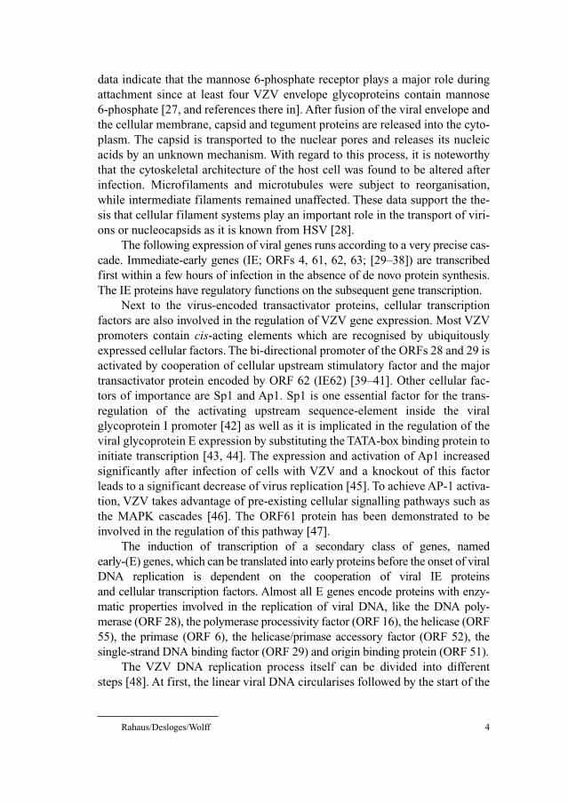
average of 30–35 is reported. The virion is structured by four distinct compo-
nents: envelope, tegument, capsid and core with the genome (fig. 1a).
Virology and Laboratory Investigations
Rahaus/Desloges/Wolff 2
The outer covering envelope has a typical trilaminar appearance [4]. It con-
sists of different membranous elements captured during the transport of the new
particles through the nuclear membrane network, Golgi apparatus, rough endo-
plasmic reticulum, cytoplasmic vesicles and cell surface elements [4–7]. The
envelope is interspersed by spikes made up of viral glycoproteins. The VZV
genome encodes glycoproteins gB, gC, gE, gH, gI, gK and gL as well as the
putative glycoproteins gM and gN [8–10]. The enveloped particles have a final
diameter of 180–200 nm and a pleomorphic to spherical shape.
The next inside layer, located directly underneath the envelope, is the tegu-
ment [11]. It does not have any distinct properties, but its thickness seems to be
variable: virions located in cytoplasmic vacuoles obviously have a thicker tegu-
ment than those located in the perinuclear space [12]. The proteins encoded by
the open reading frames (ORF) 4, 10, 47, 62 and 63 are found inside the tegu-
ment [13, 14]. The tegument surrounds the nucleocapsid.
The nucleocapsid has an icosahedric shape of 100–110 nm in diameter. It is
composed of exactly 162 capsomers. Due to the morphology of this capsid struc-
ture, it is not possible to distinguish between members of the Herpesviridae. All
capsomers occur in a 5:3:2 axial symmetry in which pentameric proteins form the
vertices of an 80–120 nm icosahedron. The facets are comprised by hexameric
Envelopecontains IEproteins4, 10, 47,62, 63
build up byproducts ofORFs 20, 23,33, 33.5,40, 41
linear dsDNA of125 kbp,wrapped on aprotein axis
containsglycoproteins:gB, gC, gI, gE,gL, gK, gH
Tegument
Capsid
Core withlinear DNA
TRL
IRL IRS
TRSUL
US
ori ori
P
IS
a c
b
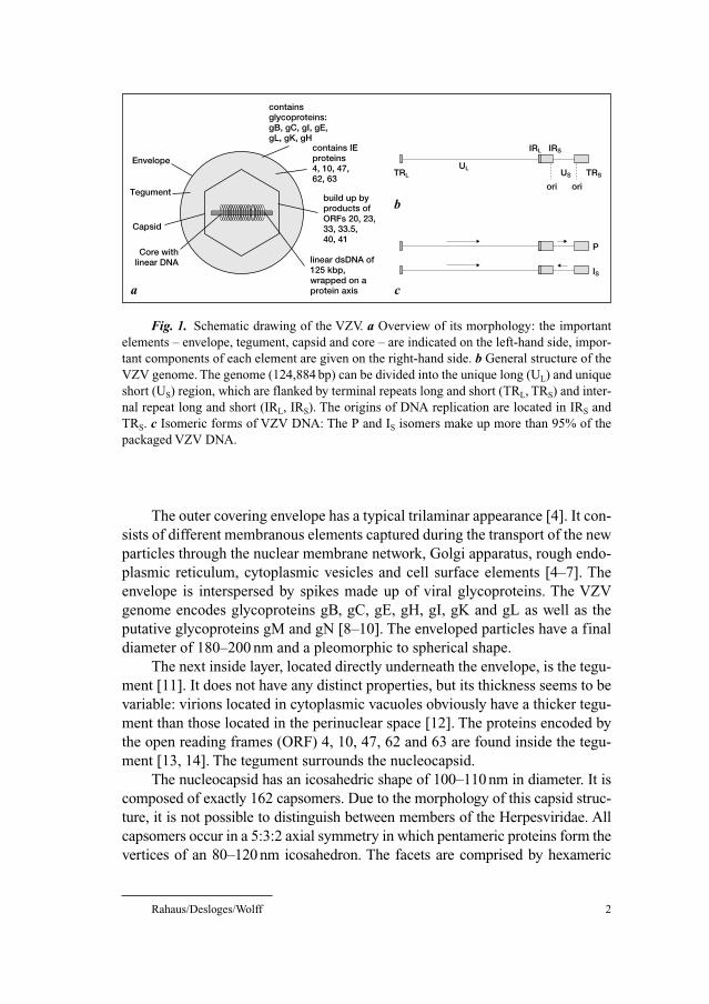
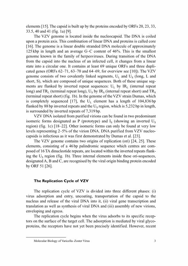
Fig. 1. Schematic drawing of the VZV. a Overview of its morphology: the important
elements – envelope, tegument, capsid and core – are indicated on the left-hand side, impor-
tant components of each element are given on the right-hand side. b General structure of the
VZV genome. The genome (124,884 bp) can be divided into the unique long (UL) and unique
short (US) region, which are flanked by terminal repeats long and short (TRL, TRS) and inter-
nal repeat long and short (IRL, IRS). The origins of DNA replication are located in IRS and
TRS. c Isomeric forms of VZV DNA: The P and IS isomers make up more than 95% of the
packaged VZV DNA.
Molecular Biology of Varicella–Zoster Virus 3
elements [15]. The capsid is built up by the proteins encoded by ORFs 20, 23, 33,
33.5, 40 and 41 (fig. 1a) [9].
The VZV genome is located inside the nucleocapsid. The DNA is coiled
upon a protein axis. This combination of linear DNA and proteins is called core
[16]. The genome is a linear double stranded DNA molecule of approximately
125 kbp in length and an average G–C content of 46%. This is the smallest
genome known in the family of herpesviruses. During transition of the DNA
from the capsid into the nucleus of an infected cell, it changes from a linear
state into a circular one. It contains at least 69 unique ORFs and three dupli-
cated genes (ORFs 62–71, 63–70 and 64–69, for overview see [10]). The VZV
genome consists of two covalently linked segments, UL and US (long, L and
short, S), which are composed of unique sequences. Both of these unique seg-
ments are flanked by inverted repeat sequences: UL by IRL (internal repeat
long) and TRL (terminal repeat long), US by IRS (internal repeat short) and TRS
(terminal repeat short) (fig. 1b). In the genome of the VZV strain Dumas, which
is completely sequenced [17], the UL element has a length of 104,836 bp
flanked by 88 bp inverted repeats and the US region, which is 5,232 bp in length,
is surrounded by inverted repeats of 7,319 bp.
VZV DNA isolated from purified virions can be found in two predominant
isomeric forms designated as P (prototype) and IS (showing an inverted US
region) (fig. 1c) [18–22]. Other isomeric forms can only be found at very low
levels representing 2–5% of the virion DNA. DNA purified from VZV nucleo-
capsids is infectious as it was first demonstrated by Dumas et al. [23].
The VZV genome contains two origins of replication (ori) [24, 25]. These
elements, consisting of a 46 bp palindromic sequence which centres are com-
posed of 16 TA dinucleotide repeats, are located within the inverted repeats flank-
ing the US region (fig. 1b). Three internal elements inside these ori-sequences,
designated A, B and C, are recognised by the viral origin binding protein encoded
by ORF 51 [26].
The Replication Cycle of VZV
The replication cycle of VZV is divided into three different phases: (i)
virus adsorption and entry, uncoating, transportation of the capsid to the
nucleus and release of the viral DNA into it, (ii) viral gene transcription and
translation as well as synthesis of viral DNA and (iii) assembly of new virions,
enveloping and egress.
The replication cycle begins when the virus adsorbs to its specific recep-
tors on the surface of the target cell. The adsorption is mediated by viral glyco-
proteins, the receptors have not yet been precisely identified. However, recent
Rahaus/Desloges/Wolff 4
data indicate that the mannose 6-phosphate receptor plays a major role during
attachment since at least four VZV envelope glycoproteins contain mannose
6-phosphate [27, and references there in]. After fusion of the viral envelope and
the cellular membrane, capsid and tegument proteins are released into the cyto-
plasm. The capsid is transported to the nuclear pores and releases its nucleic
acids by an unknown mechanism. With regard to this process, it is noteworthy
that the cytoskeletal architecture of the host cell was found to be altered after
infection. Microfilaments and microtubules were subject to reorganisation,
while intermediate filaments remained unaffected. These data support the the-
sis that cellular filament systems play an important role in the transport of viri-
ons or nucleocapsids as it is known from HSV [28].
The following expression of viral genes runs according to a very precise cas-
cade. Immediate-early genes (IE; ORFs 4, 61, 62, 63; [29–38]) are transcribed
first within a few hours of infection in the absence of de novo protein synthesis.
The IE proteins have regulatory functions on the subsequent gene transcription.
Next to the virus-encoded transactivator proteins, cellular transcription
factors are also involved in the regulation of VZV gene expression. Most VZV
promoters contain cis-acting elements which are recognised by ubiquitously
expressed cellular factors. The bi-directional promoter of the ORFs 28 and 29 is
activated by cooperation of cellular upstream stimulatory factor and the major
transactivator protein encoded by ORF 62 (IE62) [39–41]. Other cellular fac-
tors of importance are Sp1 and Ap1. Sp1 is one essential factor for the trans-
regulation of the activating upstream sequence-element inside the viral
glycoprotein I promoter [42] as well as it is implicated in the regulation of the
viral glycoprotein E expression by substituting the TATA-box binding protein to
initiate transcription [43, 44]. The expression and activation of Ap1 increased
significantly after infection of cells with VZV and a knockout of this factor
leads to a significant decrease of virus replication [45]. To achieve AP-1 activa-
tion, VZV takes advantage of pre-existing cellular signalling pathways such as
the MAPK cascades [46]. The ORF61 protein has been demonstrated to be
involved in the regulation of this pathway [47].
The induction of transcription of a secondary class of genes, named
early-(E) genes, which can be translated into early proteins before the onset of viral
DNA replication is dependent on the cooperation of viral IE proteins
and cellular transcription factors. Almost all E genes encode proteins with enzy-
matic properties involved in the replication of viral DNA, like the DNA poly-
merase (ORF 28), the polymerase processivity factor (ORF 16), the helicase (ORF
55), the primase (ORF 6), the helicase/primase accessory factor (ORF 52), the
single-strand DNA binding factor (ORF 29) and origin binding protein (ORF 51).
The VZV DNA replication process itself can be divided into different
steps [48]. At first, the linear viral DNA circularises followed by the start of the
Molecular Biology of Varicella–Zoster Virus 5
replication process, which involves the rolling circle-mechanism leading to the
formation of head-to-tail concatemers [24]. Isomerisation may occur by homol-
ogous recombination between the inverted repeats. Finally, the concatemers are
cleaved to generate linear DNA which is packaged into virions.
After DNA replication has begun, late (L) genes are transcribed. Proteins
belonging into the group of L products are the glycoproteins as well as those
proteins that build up the virus particles.
Due to the aim to achieve a strict and efficient expression of all classes of
genes and to repress an up-come of host defence mechanisms, VZV mediates a
process known as host shut-off which results in the degradation of cellular
mRNA. In contrast to HSV-1, the VZV mediated shut-off is not an immediate-
early process but a delayed one. The ORF17 protein, which is the homologue to
the HSV virion host shut-off (vhs) factor UL41, is not the main actor to gain the
shut-off [49, 50]. Due to its transrepressing properties, recent reports indicate a
role of IE63 in putting on the shut-off effect [51, 52].
The degradation of mRNA includes also transcripts of VZV IE genes what
is thought to be a part of the switching process from the IE to the E and L gene
transcription during the replication cascade [53]. Viral E and L transcripts are
also degraded as a consequence of the shut-off [53, 54]. However, evidence is
increasing that a broad range of cellular genes are not influenced by the shut-off.
In addition to the host shut-off as a mechanism against host defence, VZV
is also capable to prevent the induction of interferon-stimulated anti-viral sys-
tems such as PKR and RNase L [55, 56].
After the expression of all three classes of genes has occurred, the newly
replicated genomes are wrapped on the protein core, packed inside the
newly synthesised capsids and transported outside the host cell. It is still not def-
initely clear how and in which form the nucleocapsids are transported out of the
nucleus and towards the egress. Different hypotheses are still proposed. A widely
accepted model is that the capsids get a temporary envelope gained from inner
nuclear membranes while entering the perinuclear space. These newly formed
particles reach the lumen of the rough endoplasmatic reticulum (rER). The enve-
lope fuses with the rER membrane. The processes resulting in this temporary
enveloping at the inner nuclear membrane and the fusion with the rER mem-
brane are not understood yet. Further hypotheses give unknown functions of
some glycoproteins in these events. Following this dis-envelopment, naked par-
ticles bud into large cytoplasmic vesicles. The viral glycoproteins are released
from the trans-Golgi network in additional vesicles that fuse with the cytoplas-
mic vesicles prior to virion formation. The assembly of fully enveloped virions
with functional glycoproteins occurs in these vesicles while they are forwarded
to the cell surface. The viral particles are released by exocytosis [8, 57–59].
According to another scenario, cytosolic capsids are wrapped by cisternae of the
Rahaus/Desloges/Wolff 6
trans-Golgi network which already contain the glycoproteins. The tegument is
thought to bind to those glycoproteins [60].
References
1 Rahaus M, Schunemann S, Wolff MH: Morphological and biological characteristics of varicella-
zoster virus in Wolff MH, Schünemann S, Schmidt A (eds): Varicella-zoster Virus: Molecular
Biology, Pathogenesis, and Clinical Aspects. Contributions to Microbiology. Basel, Karger, 1999,
vol 3, pp 1–9.
2 Ruska H: Ziele und Erfolge der Übermikroskopie in der medizinischen Forschung. Scientia
1943;37:16.
3 Ruska H: Über das Virus der Varizellen und des Zoster. Wien Klin Wochenschr 1943;22:703–704.
4 Epstein MA: Observations on the mode of release of herpes virus from infected HeLa cells. J Cell
Biol 1962;12:589–597.
5 Achong BG, Meurisse EV: Observations on the fine structure and replication of varicella virus in
cultivated human amnion cells. J Gen Virol 1968;3:305–308.
6 Cok ML, Stevens JG: Replication of varicella-zoste virus in cell culture: an ultrastructural study.
J Ultrastruct Res 1970;32:334–350.
7 Stackpole CW: Herpes-type virus of the frog renal adenocarcinoma. I. Virus development in
tumor transplants maintained at low temperature. J Virol 1969;4:75–93.
8 Grose C: Glycoproteins encoded by varicella-zoster virus: biosynthesis, phosphorylation, and
intracellular trafficking. Annu Rev Microbiol 1990;44:59–80.
9 Kinchington PR, Cohen JI: Viral proteins; in Arvin AM, Gershon AA (eds): Varicella-zoster Virus,
Virology and Clinical Managment. Cambridge, Cambridge University Press, 2003, pp 74–104.
10 Ruyechan WT, Hay J: DNA replication; in Arvin AM, Gershon AA (eds): Varicella-zoster Virus,
Virology and Clinical Management. Cambridge, Cambridge University Press, 2003, pp 51–73.
11 Roizman B, Furlong D: The replication of herpes viruses; in Fraenkel-Conrat H, Wagner RR (eds):
Comprehensive Virology. New York, Plenum, 1974, vol 3, pp 229–403.
12 Falke D, Siegert R, Vogell W: Elektronen-mikroskopische Befunde zur Frage der
Doppelmembranbildung des Herpes-simplex Virus. Arch Gesamte Virusforsch 1959;9:484–496.
13 Besser J, Ikoma M, Fabel K, Sommer MH, Zerboni L, Grose C, Arvin AM: Differential require-
ment for cell fusion and virion formation in the pathogenesis of varicella-zoster virus infection in
skin and T cells. J Virol 2004;78:13293–13305.
14 Kinchington PR, Hougland JK, Arvin AM, Ruyechan WT, Hay J: The varicella-zoster
virus immediate-early protein IE62 is a major component of virus particles. J Virol 1992;66:
359–366.
15 Almeida JD, Howatson AF, Williams MG: Morphology of varicella (chicken pox) virus. Virology
1962;16:353–355.
16 Puvion-Dutilleul F, Pichard E, Laithier M, Leduc EH: Effect of dehydrating agents on DNA
organization in herpes viruses. J Histochem Cytochem 1987;35:635–645.
17 Davison AJ, Scott JE: The complete DNA sequence of varicella-zoster virus. J Gen Virol
1986;67(pt 9):1759–1816.
18 Davison AJ, Scott JE: Molecular cloning of the varicella-zoster virus genome and derivation of six
restriction endonuclease maps. J Gen Virol 1983;64(pt 8):1811–1814.
19 Dumas AM, Geelen JL, Weststrate MW, Wertheim P, van der Noordaa J: XbaI, PstI, and BglII
restriction enzyme maps of the two orientations of the varicella-zoster virus genome. J Virol
1981;39:390–400.
20 Ecker JR, Hyman RW: Varicella zoster virus DNA exists as two isomers. Proc Natl Acad Sci USA
1982;79:156–160.
21 Straus SE, Aulakh HS, Ruyechan WT, Hay J, Casey TA, Vande Woude GF, Owens J, Smith HA:
Structure of varicella-zoster virus DNA. J Virol 1981;40:516–525.
Molecular Biology of Varicella–Zoster Virus 7
22 Straus SE, Owens J, Ruyechan WT, Takiff HE, Casey TA, Vande Woude GF, Hay J: Molecular
cloning and physical mapping of varicella-zoster virus DNA. Proc Natl Acad Sci USA 1982;79:
993–997.
23 Dumas AM, Geelen JL, Maris W, Van der Noordaa J: Infectivity and molecular weight of
varicella-zoster virus DNA. J Gen Virol 1980:47.
24 Stow ND, Davison AJ: Identification of a varicella-zoster virus origin of DNA replication
and its activation by herpes simplex virus type 1 gene products. J Gen Virol 1986;67(pt 8):
1613–1623.
25 Stow ND, Weir HM, Stow EC: Analysis of the binding sites for the varicella-zoster virus gene 51
product within the viral origin of DNA replication. Virology 1990;177:570–577.
26 Koff A, Tegtmeyer P: Characterization of major recognition sequences for a herpes simplex virus
type 1 origin-binding protein. J Virol 1988;62:4096–4103.
27 Chen JJ, Zhu Z, Gershon AA, Gershon MD: Mannose 6-phosphate receptor dependence of vari-
cella zoster virus infection in vitro and in the epidermis during varicella and zoster. Cell 2004;119:
915–926.
28 Kuhn M, Desloges N, Rahaus M, Wolff MH: Varicella-zoster virus infection influences expression
and organization of actin and alpha-tubulin but does not affect lamin A and vimentin.
Intervirology 2005;48:312–320.
29 Debrus S, Sadzot-Delvaux C, Nikkels AF, Piette J, Rentier B: Varicella-zoster virus gene 63 encodes
an immediate-early protein that is abundantly expressed during latency. J Virol 1995;69:3240–3245.
30 Defechereux P, Debrus S, Baudoux L, Rentier B, Piette J: Varicella-zoster virus open reading
frame 4 encodes an immediate-early protein with posttranscriptional regulatory properties. J Virol
1997;71:7073–7079.
31 Disney GH, McKee TA, Preston CM, Everett RD: The product of varicella-zoster virus gene 62
autoregulates its own promoter. J Gen Virol 1990;71:2999–3003.
32 Inchauspe G, Nagpal S, Ostrove JM: Mapping of two varicella-zoster virus-encoded genes that
activate the expression of viral early and late genes. Virology 1989;173:700–709.
33 Kinchington PR, Bookey D, Turse SE: The transcriptional regulatory proteins encoded by
varicella-zoster virus open reading frames (ORFs) 4 and 63, but not ORF 61, are associated with
purified virus particles. J Virol 1995;69:4274–4282.
34 Kinchington PR, Vergnes JP, Turse SE: Transcriptional mapping of varicella-zoster virus regula-
tory proteins. Neurology 1995;45:S33–35.
35 Moriuchi M, Moriuchi H, Straus SE, Cohen JI: Varicella-zoster virus (VZV) virion-associated
transactivator open reading frame 62 protein enhances the infectivity of VZV DNA. Virology
1994;200:297–300.
36 Ng TI, Keenan L, Kinchington PR, Grose C: Phosphorylation of varicella-zoster virus open read-
ing frame (ORF) 62 regulatory product by viral ORF 47-associated protein kinase. J Virol
1994;68:1350–1359.
37 Stevenson D, Colman KL, Davison AJ: Characterization of the varicella-zoster virus gene 61 pro-
tein. J Gen Virol 1992;73:521–530.
38 Stevenson D, Xue M, Hay J, Ruyechan WT: Phosphorylation and nuclear localization of the
varicella-zoster virus gene 63 protein. J Virol 1996;70:658–662.
39 Meier JL, Luo X, Sawadogo M, Straus SE: The cellular transcription factor USF cooperates with
varicella-zoster virus immediate-early protein 62 to symmetrically activate a bidirectional viral
promoter. Mol Cell Biol 1994;14:6896–6906.
40 Rahaus M, Desloges N, Yang M, Ruyechan WT, Wolff MH: Transcription factor USF, expressed dur-
ing the entire phase of varicella-zoster virus infection, interacts physically with the major viral trans-
activator IE62 and plays a significant role in virus replication. J Gen Virol 2003;84: 2957–2967.
41 Yang M, Hay J, Ruyechan WT: The DNA element controlling expression of the varicella-zoster
virus open reading frame 28 and 29 genes consists of two divergent unidirectional promoters
which have a common USF site. J Virol 2004;78:10939–10952.
42 Ito H, Sommer MH, Zerboni L, He H, Boucaud D, Hay J, Ruyechan W, Arvin AM: Promoter
sequences of varicella-zoster virus glycoprotein I targeted by cellular transactivating factors Sp1
and USF determine virulence in skin and T cells in SCIDhu mice in vivo. J Virol 2003;77:
489–498.
Rahaus/Desloges/Wolff 8
43 Rahaus M, Wolff MH: Influence of different cellular transcription factors on the regulation of vari-
cella-zoster virus glycoproteins E (gE) and I (gI) UTR’s activity. Virus Res 1999;62:77–88.
44 Rahaus M, Wolff MH: Transcription factor Sp1 is involved in the regulation of varicella-zoster
virus glycoprotein E. Virus Res 2000;69:69–81.
45 Rahaus M, Wolff MH: Reciprocal effects of varicella-zoster virus (VZV) and AP1: activation of
jun, fos and ATF-2 after VZV infection and their importance for the regulation of viral genes.
Virus Res 2003;92:9–21.
46 Rahaus M, Desloges N, Wolff MH: Replication of varicella-zoster virus is influenced by the levels
of JNK/SAPK and p38/MAPK activation. J Gen Virol 2004;85:3529–3540.
47 Rahaus M, Desloges N, Wolff MH: ORF61 protein of Varicella-zoster virus influences
JNK/SAPK and p38/MAPK phosphorylation. J Med Virol 2005;76:424–433.
48 Cohen JI, Straus SE: Varicella-zoster virus and its replication; in Fields BN, Knipe DM, Howley
PM (eds): Field’s Virology. Philadelphia, Lippincott-Raven, 1996, vol 2, pp 2525–2545.
49 Sato H, Callanan LD, Pesnicak L, Krogmann T, Cohen JI: Varicella-zoster virus (VZV) ORF17
protein induces RNA cleavage and is critical for replication of VZV at 37 degrees C but not 33
degrees C. J Virol 2002;76:11012–11023.
50 Waterboer T, Rahaus M, Wolff MH: Varicella-zoster virus (VZV) mediates a delayed host shutoff
independent of open reading frame (ORF) 17 expression. Virus Genes 2002;24:49–56.
51 Desloges N, Rahaus M, Wolff MH: The varicella-zoster virus-mediated delayed host shutoff: open
reading frame 17 has no major function, whereas immediate-early 63 protein represses heterolo-
gous gene expression. Microbes Infect 2005;7:1519–1529.
52 Di Valentin E, Bontems S, Habran L, Jolois O, Markine-Goriaynoff N, Vanderplasschen A,
Sadzot-Delvaux C, Piette J: Varicella-zoster virus IE63 protein represses the basal transcription
machinery by disorganizing the pre-initiation complex. Biol Chem 2005;386:255–267.
53 Hamouda T, McPhee R, Hsia SC, Read GS, Holland TC, King SR: Inhibition of human immuno-
deficiency virus replication by the herpes simplex virus virion host shut-off protein. J Virol
1997;71:5521–5527.
54 Oroskar AA, Read GS: Control of mRNA stability by the virion host shutoff function of herpes
simplex virus. J Virol 1989;63:1897–1906.
55 Desloges N, Rahaus M, Wolff MH: Varicella-zoster virus does not significantly induce the cell
defence mechanism mediated by the 2–5A/RNase L pathway during its replication cycle. Med
Microbiol Immunol (Berl) 2004;194:25–31.
56 Desloges N, Rahaus M, Wolff MH: Role of the protein kinase PKR in the inhibition of
varicella-zoster virus replication by beta interferon and gamma interferon. J Gen Virol 2005;86: 1–6.
57 Gershon MD, Gershon AA: Role of glycoproteins in varicella-zoster virus infection; in Wolff MH,
Schünemann S, Schmidt A (eds): Varicella-zoster Virus, Molecular Biology, Pathogenesis, and
Clinical Aspects. Contributions to Microbiology. Basel, Karger, 1999, vol 3, pp 43–60.
58 Jones F, Grose C: Role of cytoplasmic vacuoles in varicella-zoster virus glycoprotein trafficking
and virion envelopment. J Virol 1988;62:2701–2711.
59 Sadzot-Delvaux C, Baudoux L, Defechereux P, Piette J, Rentier B: Overview of the replication
cycle of varicella-zoster virus; in Wolff MH, Schünemann S, Schmidt A (eds): Varicella-zoster
Virus: Molecular Biology, Pathogenesis and Clinical Aspects. Contributions to Microbiology.
Basel, Karger, 1999, vol 3, pp 21–42.
60 Gershon AA, Sherman DL, Zhu Z, Gabel CA, Ambron RT, Gershon MD: Intracellular transport of
newly synthesized varicella-zoster virus: final envelopment in the trans-Golgi network. J Virol
1994;68:6372–6390.
Manfred H. Wolff
Institute of Microbiology and Virology
Private University of Witten-Herdecke
Stockumer Strasse 10
DE–58448 Witten (Germany)
Tel. �49 2302 926107, Fax �49 2302 926220, E-Mail [email protected]
Gross G, Doerr HW (eds): Herpes Zoster.
Monogr Virol. Basel, Karger, 2006, vol 26, pp 9–12
Latency and Reactivation of VZV
Octavian Lungua, Anne A. Gershonb
Departments of aMicrobiology and bPediatrics, Columbia University, College of
Physicians and Surgeons, New York, N.Y., USA
Primary, Latent and Reactivated VZV Infection
Like other herpes viruses, VZV is produced following a temporally coordi-
nated expression of its genes. Upon entering a naïve cell viral DNA and virion-
associated proteins are transported to the cell nucleus and immediate early (IE)
genes are transcribed. After the corresponding mRNAs are translated in the cell
cytoplasm, the specific IE proteins return to the nucleus to initiate the expres-
sion of early (E) genes which are involved in viral replication. Then, IE and E
gene products prompt the expression of late (L) genes whose products are
required for packaging of the newly synthesized viral DNA and assembly of
mature virions. Virus then exits the cell to infect adjacent uninfected cells.
Complete or partial interruption of this regulated cycle of virus gene expression
results in latency. The re-establishment of the co-ordinated sequence of gene
expression results in reactivation.
Wild-type VZV infects varicella-susceptible individuals through the respira-
tory route. After an initial replication in the respiratory tract lymphoid tissue, a
primary viremia occurs, then the virus reaches the central lymphoid system where
it continues to replicate. Due to a subsequent secondary viremia of larger magni-
tude than the first, the skin is productively infected and the vesicular rash and
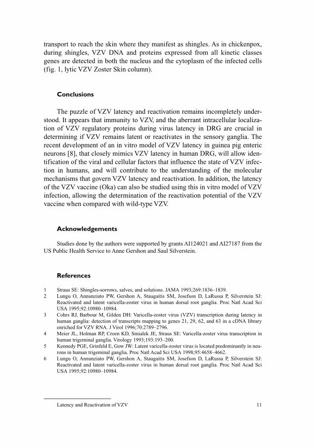
other clinical signs of chickenpox are apparent. During chickenpox VZV DNA
and proteins expressed by IE, E and L genes are detected in both the nucleus and
the cytoplasm of the infected cells (fig. 1, lytic VZV Varicella Skin column).
During primary infection through retrograde neuronal transport from the
skin and possibly hematogeneuos spread, or both routes, the virus reaches dor-
sal root ganglia (DRG) and establishes latent infection in the sensory neurons
and the supporting non-neuronal ganglionic cells [3, 4] (fig. 1, latent VZV
DNA DRG panel). Note that viral DNA is present in the nucleus of a neuron

Lungu/Gershon 10
and several satellite cells. As opposed to the closely related herpes simplex
viruses, which are silent during their latency in DRG, VZV expresses at least 5
IE and E genes while in the DRG. Transcripts encoded by IE 4, 62, 63 and E 21,
29 VZV genes [5, 6] and the corresponding regulatory proteins translated by
these transcripts [2, 7] can be identified in latently infected DRG. Late VZV
genes are not expressed during latency and L proteins are not detected in
latently infected DRG [2] (fig. 1, Latent VZV Late proteins DRG panel). IE and
E proteins localize to the cytoplasm of neurons during latency [2] (fig. 1, latent
VZV IE and E proteins DRG panel). The exclusion of IE and E proteins from
the cell nucleus, where they perform their regulatory functions during produc-
tive infection, is a hallmark of VZV latency and is thought to be a crucial factor
for the maintenance of virus latency.
For reasons yet to be defined, VZV can exit its latent state to cause a pro-
ductive infection in the infected ganglia. During reactivation VZV IE and E
proteins reach the nuclei of cells [2] (fig. 1, reactivated VZV IE and E proteins
DRG panel), re-establishing the co-ordinated cycle of VZV genes expression
that results in the production of L proteins [2] (fig. 1, reactivated VZV Late proteins
DRG panel) and lytic infection. The mature virions identified in the cytoplasm
of neurons as VZV DNA [2] (fig. 1, reactivated VZV DNA DRG panel – light
dark staining in the cytoplasm of a large cell) then use anterograde neuronal
Lytic VZVvaricella
LatentVZV
Skin SkinDRG
DNA
IE and Eproteins
Lateproteins
DRG
Lytic VZV zoster
Reactivated VZV
Fig. 1. VZV DNA and proteins identified by in situ hybridization and immunohisto-
chemistry during VZV primary infection or VZV latency and reactivation. The presence of
virus DNA or proteins (IE63p, L68/gE – as examples) is indicated by a dark colouration of
cells nuclei, cytoplasm or both.
Latency and Reactivation of VZV 11
transport to reach the skin where they manifest as shingles. As in chickenpox,
during shingles, VZV DNA and proteins expressed from all kinetic classes
genes are detected in both the nucleus and the cytoplasm of the infected cells
(fig. 1, lytic VZV Zoster Skin column).
Conclusions
The puzzle of VZV latency and reactivation remains incompletely under-
stood. It appears that immunity to VZV, and the aberrant intracellular localiza-
tion of VZV regulatory proteins during virus latency in DRG are crucial in
determining if VZV remains latent or reactivates in the sensory ganglia. The
recent development of an in vitro model of VZV latency in guinea pig enteric
neurons [8], that closely mimics VZV latency in human DRG, will allow iden-
tification of the viral and cellular factors that influence the state of VZV infec-
tion in humans, and will contribute to the understanding of the molecular
mechanisms that govern VZV latency and reactivation. In addition, the latency
of the VZV vaccine (Oka) can also be studied using this in vitro model of VZV
infection, allowing the determination of the reactivation potential of the VZV
vaccine when compared with wild-type VZV.
Acknowledgements
Studies done by the authors were supported by grants AI124021 and AI27187 from the
US Public Health Service to Anne Gershon and Saul Silverstein.
References
1 Straus SE: Shingles-sorrows, salves, and solutions. JAMA 1993;269:1836–1839.
2 Lungu O, Annunziato PW, Gershon A, Staugaitis SM, Josefson D, LaRussa P, Silverstein SJ:
Reactivated and latent varicella-zoster virus in human dorsal root ganglia. Proc Natl Acad Sci
USA 1995;92:10980–10984.
3 Cohrs RJ, Barbour M, Gilden DH: Varicella-zoster virus (VZV) transcription during latency in
human ganglia: detection of transcripts mapping to genes 21, 29, 62, and 63 in a cDNA library
enriched for VZV RNA. J Virol 1996;70:2789–2796.
4 Meier JL, Holman RP, Croen KD, Smialek JE, Straus SE: Varicella-zoster virus transcription in
human trigeminal ganglia. Virology 1993;193:193–200.
5 Kennedy PGE, Grinfeld E, Gow JW: Latent varicella-zoster virus is located predominantly in neu-
rons in human trigeminal ganglia. Proc Natl Acad Sci USA 1998;95:4658–4662.
6 Lungu O, Annunziato PW, Gershon A, Staugaitis SM, Josefson D, LaRussa P, Silverstein SJ:
Reactivated and latent varicella-zoster virus in human dorsal root ganglia. Proc Natl Acad Sci
USA 1995;92:10980–10984.

Lungu/Gershon 12
7 Mahalingam R, Wellish M, Cohrs R, Debrus S, Piette J, Rentier B, Gilden DH: Expression of pro-
tein encoded by varicella-zoster virus open reading frame 63 in latently infected human ganglionic
neurons. Proc Natl Acad Sci USA 1996;93:2122–2124.
8 Chen JJ, Gershon AA, Li ZS, Lungu O, Silverstein S, Gershon MD: Latent and lytic infection of iso-
lated guinea pig enteric ganglia by varicella zoster virus. J Med Virol 2003;70(suppl 1):S71–S78.
Octavian Lungu
Department of Pediatrics, Columbia University
College of Physicians and Surgeons
701 West 168th Street
New York, NY 10032 (USA)
Tel. �1 212 305 4137, Fax �1 212 305 5106, E-Mail [email protected]
Gross G, Doerr HW (eds): Herpes Zoster.
Monogr Virol. Basel, Karger, 2006, vol 26, pp 13–19
Laboratory Confirmation ofHerpes Zoster
B. Ludwig, S. Buxbaum, H.W. Doerr
Institute of Virology, University of Frankfurt, Frankfurt am Main, Germany
The diagnosis of herpes zoster is usually a clinical diagnosis and therefore
laboratory confirmation is not necessary. In cases where herpes zoster is in
question a rapid laboratory diagnosis may be helpful, especially in immuno-
compromised patients, when antiviral therapy must be initiated as soon as pos-
sible. Similarly, laboratory diagnosis may be useful for the determination of the
antibody status and the diagnostic clarification of ophthalmic and central nerve
system complications of a VZV infection. Serology is a helpful tool to recog-
nise VZV seronegative individuals having a risk to acquire an infection (e.g.
non-immune women during the first third of the pregnancy). Generally, we can
distinguish between the direct prove of the pathogen itself and the immunologi-
cal reaction of the infected person (humoral or cellular immunity). Today viro-
logical methods for the direct detection of infectious virus, viral DNA or viral
proteins are electron microscopy, cell culture, immunofluorescence assays and
polymerase chain reaction (PCR) (figs. 1–3). Serological diagnosis of VZV
infection/reactivation covers a number of different methods. Detection of cellu-
lar immunity is difficult to carry out and has therefore no role in routine diag-
nosis so far.
Direct Examination of Clinical Material
MicroscopyElectron microscopy (EM) can be used, if a rapid diagnosis is needed or
differentiation of VZV and smallpox is required. Typical herpesvirus particles
can be detected in the fluid of early vesicles or in infected cells scraped from
the ground of a lesion. Since the viruses of the herpes group have all the
same morphology, EM does not differentiate between VZV and HSV unless
Ludwig/Buxbaum/Doerr 14
combined with further immunological techniques [1–3] which are normally not
used in routine diagnosis (fig. 1).
Direct Immunofluorescence AssayDirect immunofluorescent antibody staining with monoclonal antibodies
allows a rapid and specific diagnosis of VZV suspected lesions. Even cells from
Fig. 1. VZV vesicles: electron microscopy.
101
100
10�1
10�2
0 2 4 6 8 10 12 14 16 18 20 22
Cycle
24 26 28 30 32 34 36 4038 42 44
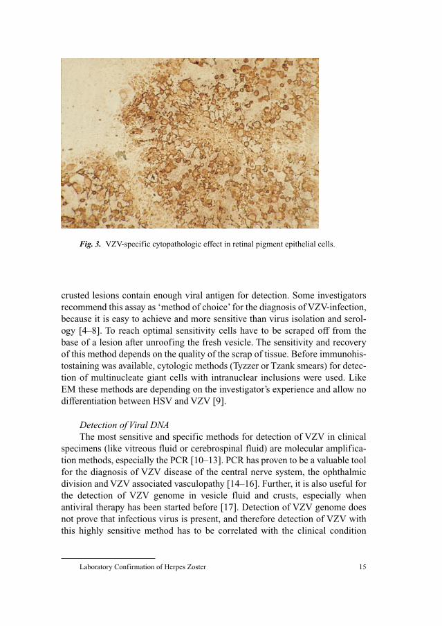
Fig. 2. PCR investigation by real-time technique.
Laboratory Confirmation of Herpes Zoster 15
crusted lesions contain enough viral antigen for detection. Some investigators
recommend this assay as ‘method of choice’ for the diagnosis of VZV-infection,
because it is easy to achieve and more sensitive than virus isolation and serol-
ogy [4–8]. To reach optimal sensitivity cells have to be scraped off from the
base of a lesion after unroofing the fresh vesicle. The sensitivity and recovery
of this method depends on the quality of the scrap of tissue. Before immunohis-
tostaining was available, cytologic methods (Tyzzer or Tzank smears) for detec-
tion of multinucleate giant cells with intranuclear inclusions were used. Like
EM these methods are depending on the investigator’s experience and allow no
differentiation between HSV and VZV [9].
Detection of Viral DNAThe most sensitive and specific methods for detection of VZV in clinical
specimens (like vitreous fluid or cerebrospinal fluid) are molecular amplifica-
tion methods, especially the PCR [10–13]. PCR has proven to be a valuable tool
for the diagnosis of VZV disease of the central nerve system, the ophthalmic
division and VZV associated vasculopathy [14–16]. Further, it is also useful for
the detection of VZV genome in vesicle fluid and crusts, especially when
antiviral therapy has been started before [17]. Detection of VZV genome does
not prove that infectious virus is present, and therefore detection of VZV with
this highly sensitive method has to be correlated with the clinical condition
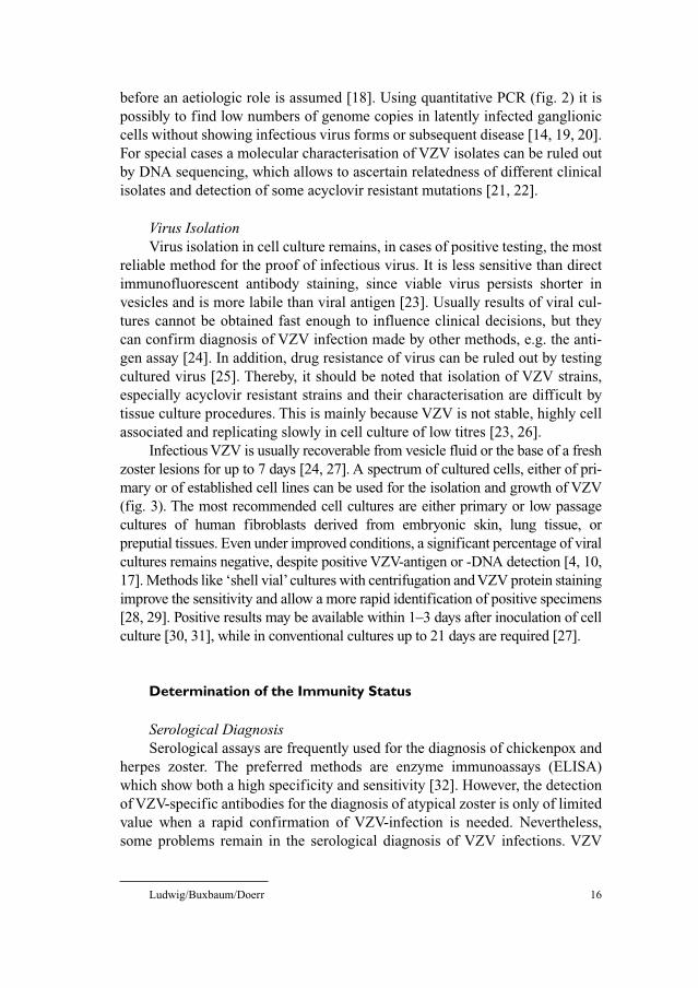
Fig. 3. VZV-specific cytopathologic effect in retinal pigment epithelial cells.
Ludwig/Buxbaum/Doerr 16
before an aetiologic role is assumed [18]. Using quantitative PCR (fig. 2) it is
possibly to find low numbers of genome copies in latently infected ganglionic
cells without showing infectious virus forms or subsequent disease [14, 19, 20].
For special cases a molecular characterisation of VZV isolates can be ruled out
by DNA sequencing, which allows to ascertain relatedness of different clinical
isolates and detection of some acyclovir resistant mutations [21, 22].
Virus IsolationVirus isolation in cell culture remains, in cases of positive testing, the most
reliable method for the proof of infectious virus. It is less sensitive than direct
immunofluorescent antibody staining, since viable virus persists shorter in
vesicles and is more labile than viral antigen [23]. Usually results of viral cul-
tures cannot be obtained fast enough to influence clinical decisions, but they
can confirm diagnosis of VZV infection made by other methods, e.g. the anti-
gen assay [24]. In addition, drug resistance of virus can be ruled out by testing
cultured virus [25]. Thereby, it should be noted that isolation of VZV strains,
especially acyclovir resistant strains and their characterisation are difficult by
tissue culture procedures. This is mainly because VZV is not stable, highly cell
associated and replicating slowly in cell culture of low titres [23, 26].
Infectious VZV is usually recoverable from vesicle fluid or the base of a fresh
zoster lesions for up to 7 days [24, 27]. A spectrum of cultured cells, either of pri-
mary or of established cell lines can be used for the isolation and growth of VZV
(fig. 3). The most recommended cell cultures are either primary or low passage
cultures of human fibroblasts derived from embryonic skin, lung tissue, or
preputial tissues. Even under improved conditions, a significant percentage of viral
cultures remains negative, despite positive VZV-antigen or -DNA detection [4, 10,
17]. Methods like ‘shell vial’cultures with centrifugation and VZV protein staining
improve the sensitivity and allow a more rapid identification of positive specimens
[28, 29]. Positive results may be available within 1–3 days after inoculation of cell
culture [30, 31], while in conventional cultures up to 21 days are required [27].
Determination of the Immunity Status
Serological DiagnosisSerological assays are frequently used for the diagnosis of chickenpox and
herpes zoster. The preferred methods are enzyme immunoassays (ELISA)
which show both a high specificity and sensitivity [32]. However, the detection
of VZV-specific antibodies for the diagnosis of atypical zoster is only of limited
value when a rapid confirmation of VZV-infection is needed. Nevertheless,
some problems remain in the serological diagnosis of VZV infections. VZV
Laboratory Confirmation of Herpes Zoster 17
reactivation induces often a significant rise of IgG and IgA [33, 34] antibodies,
which are found in 50–60% [10, 35] of the patients. However IgM antibodies
are also sometimes detectable. Thus differentiation of primary and recurrent
infection can be difficult [36]. Furthermore, sharing of antigens between VZV
and HSV can result in high anti HSV and anti VZV levels and it will be difficult
to differentiate between both diseases without further information or additional
tests [23, 37]. The main value of serologic assays is the determination of the
immune status of individuals, whose history of chickenpox is unknown [24]. In
patients with pain syndromes or facial paresis due to zoster sine herpete a rise
of VZV-IgG values in consecutive serum samples may be helpful to identify the
aetiology [38]. Other serological test systems like the neutralisation assay and
the fluorescent-antibody membrane antigen assay are too complex to carry out
in routine diagnostic testing [23].
Determination of Cell-Mediated ImmunityThe main role of cell-mediated immunity (CMI) is to prevent reactivation
and to limit an established infection. Detection of CMI can be carried out by
measuring the proliferative response [39] or by detection of cytokine produc-
tion [40–42]. Examples for such methods are the determination of intracellular
cytokines by flow cytometry [43] or of secreted extracellular cytokines by
ELISA [44] or ELISPOT [45]. Generally all procedures for measurement of
CMI are technically complex, time consuming, labour intensive and therefore
used only in special cases on small scales. Nevertheless, in the future the
demand for clinical studies concerning VZV-specific CMI on a large scale will
rise considerably and so the development of well reproducible and easily practi-
cable diagnosis methods will be necessary.
References
1 Folkers E, Vreeswijk J, Oranje AP, Duivenvoorden JN: Rapid diagnosis in varicella and herpes
zoster: re-evaluation of direct smear (Tzanck test) and electron microscopy including colloidal gold
immuno-electron microscopy in comparison with virus isolation. Br J Dermatol 1989;121:287–296.
2 Folkers E, Vreeswijk J, Oranje AP, Wagenaar F, Duivenvoorden JN: Improved detection of HSV by
electron microscopy in clinical specimens using ultracentrifugation and colloidal gold immuno-
electron microscopy: comparison with viral culture and cytodiagnosis. J Virol Methods 1991;34:
273–289.
3 Vreeswijk J, Folkers E, Wagenaar F, Kapsenberg JG: The use of colloidal gold immunoelectron
microscopy to diagnose varicella-zoster virus (VZV) infections by rapid discrimination between
VZV, HSV-1 and HSV-2. J Virol Methods 1988;22:255–271.
4 Dahl H, Marcoccia J, Linde A: Antigen detection: the method of choice in comparison with virus
isolation and serology for laboratory diagnosis of herpes zoster in human immunodeficiency
virus-infected patients. J Clin Microbiol 1997;35:347–349.
5 Drew WL, Mintz L: Rapid diagnosis of varicella-zoster virus infection by direct immunofluores-
cence. Am J Clin Pathol 1980;73:699–701.
Ludwig/Buxbaum/Doerr 18
6 Gleaves CA, Lee CF, Bustamante CI, Meyers JD: Use of murine monoclonal antibodies for labo-
ratory diagnosis of varicella-zoster virus infection. J Clin Microbiol 1988;26:1623–1625.
7 Perez JL, Garcia A, Niubo J, Salva J, Podzamczer D, Martin R: Comparison of techniques and
evaluation of three commercial monoclonal antibodies for laboratory diagnosis of varicella-zoster
virus in mucocutaneous specimens. J Clin Microbiol 1994;32:1610–1613.
8 Coffin SE, Hodinka RL: Utility of direct immunofluorescence and virus culture for detection of
varicella-zoster virus in skin lesions. J Clin Microbiol 1995;33:2792–2795.
9 Waner JL, Weller TH, Schmidt NJ, Emmons RW (eds): Diagnostic Procedures for Viral,
Rickettsial And Chlamydial Infections, ed 6. Varicella-Zoster Virus. Washington, DC, American
Public Health Association, 1998, vol 13, pp 379–406.
10 Sauerbrei A, Eichhorn U, Schacke M, Wutzler P: Laboratory diagnosis of herpes zoster. J Clin
Virol 1999;14:31–36.
11 Weidmann M, Meyer-Konig U, Hufert FT: Rapid detection of herpes simplex virus and varicella-
zoster virus infections by real-time PCR. J Clin Microbiol 2003;41:1565–1568.
12 van Doornum GJ, Guldemeester J, Osterhaus AD, Niesters HG: Diagnosing herpesvirus infections
by real-time amplification and rapid culture. J Clin Microbiol 2003;41:576–580.
13 Nahass GT, Goldstein BA, Zhu WY, Serfling U, Penneys NS, Leonardi CL: Comparison of Tzanck
smear, viral culture, and DNA diagnostic methods in detection of herpes simplex and varicella-
zoster infection. JAMA 1992;268:2541–2544.
14 Kleinschmidt-DeMasters BK, Gilden DH: Varicella-Zoster virus infections of the nervous system:
clinical and pathologic correlates. Arch Pathol Lab Med 2001;125:770–780.
15 Madhavan HN, Priya K: The diagnostic significance of enzyme linked immuno-sorbent assay for
herpes simplex, varicella zoster and cytomegalovirus retinitis. Indian J Ophthalmol 2003;51:
71–75.
16 Gargiulo F, De Francesco MA, Nascimbeni G, et al: Polymerase chain reaction as a rapid diagnos-
tic tool for therapy of acute retinal necrosis syndrome. J Med Virol 2003;69:397–400.
17 Beards G, Graham C, Pillay D: Investigation of vesicular rashes for HSV and VZV by PCR. J Med
Virol 1998;54:155–157.
18 Schunemann S, Mainka C, Wolff MH: Subclinical reactivation of varicella-zoster virus in
immunocompromised and immunocompetent individuals. Intervirology 1998;41:98–102.
19 Liedtke W, Opalka B, Zimmermann CW, Lignitz E: Age distribution of latent herpes simplex virus
1 and varicella-zoster virus genome in human nervous tissue. J Neurol Sci 1993;116:6–11.
20 Mahalingam R, Wellish M, Lederer D, Forghani B, Cohrs R, Gilden D: Quantitation of latent
varicella-zoster virus DNA in human trigeminal ganglia by polymerase chain reaction. J Virol
1993;67:2381–2384.
21 Morfin F, Thouvenot D, Turenne-Tessier M, Lina B, Aymard M, Ooka T: Phenotypic and genetic
characterization of thymidine kinase from clinical strains of varicella-zoster virus resistant to acy-
clovir. Antimicrob Agents Chemother 1999;43:2412–2416.
22 Boivin G, Edelman CK, Pedneault L, Talarico CL, Biron KK, Balfour HH Jr: Phenotypic and
genotypic characterization of acyclovir-resistant varicella-zoster viruses isolated from persons
with AIDS. J Infect Dis 1994;170:68–75.
23 Gershon AA, Forghani B, Lennette EH, Lennette DA, Lennette ET (eds): Diagnostic Procedures
for Viral, Rickettsial and Chlamydial Infections, ed 7. Varicella-Zoster Virus. Washington, DC,
American Public Health Association, 1995, Vol 42, pp 601–613.
24 Arvin AM, Knipe DM, Howley PM (eds): Fields Virology, ed 4. Varicella-Zoster Virus.
Philadelphia, Lippincott Willians and Wilkins, 2001, Vol 79, pp 2731–2767.
25 Pillay D, Emery VC, Mutimer D, et al: Guidelines for laboratory monitoring of treatment of per-
sistent virus infections. J Clin Virol 2002;25:73–92.
26 Sahli R, Andrei G, Estrade C, Snoeck R, Meylan PR: A rapid phenotypic assay for detection of
acyclovir-resistant varicella-zoster virus with mutations in the thymidine kinase open reading
frame. Antimicrob Agents Chemother 2000;44:873–878.
27 Breuer J, Harper DR, Kangro HO, Zuckerman A, Banatvala JE, Pattison JR (eds): Principles and
Practice Of Clinical Virology, ed 4. Varicella Zoster. Baffins Lane, Chichester, West Sussex PO19
1UD, England, John Wiley and Sons, 2000, vol 2B, pp 47–78.
Laboratory Confirmation of Herpes Zoster 19
28 Schirm J, Meulenberg JJ, Pastoor GW, Voorst Vader PC, Schroder FP: Rapid detection of varicella-
zoster virus in clinical specimens using monoclonal antibodies on shell vials and smears. J Med
Virol 1989;28:1–6.
29 West PG, Aldrich B, Hartwig R, Haller GJ: Increased detection rate for varicella-zoster virus with
combination of two techniques. J Clin Microbiol 1988;26:2680–2681.
30 Brinker JP, Doern GV: Enhancement of varicella-zoster virus detection in A-549 shell vials by use
of freeze-thawed specimens, extended incubation, and ‘a centrifuged, not incubated’ direct detec-
tion method. Diagn Microbiol Infect Dis 1998;31:555–558.
31 Brinker JP, Doern GV: Comparison of MRC-5 and A-549 cells in conventional culture tubes and shell
vial assays for the detection of varicella-zoster virus. Diagn Microbiol Infect Dis 1993;17:75–77.
32 Weinberg A, Hayward AR, Masters HB, Ogu IA, Levin MJ: Comparison of two methods for
detecting varicella-zoster virus antibody with varicella-zoster virus cell-mediated immunity.
J Clin Microbiol 1996;34:445–446.
33 Wültzer P, Porstmann T (eds): Virusdiagnostik. Varicella-Zoster-Virus. Berlin, Wien, Blackwell
Wissenschafts-Verlag, 1996, pp 291–302.
34 Doerr HW, Rentschler M, Scheifler G: Serologic detection of active infections with human herpes
viruses (CMV, EBV, HSV, VZV): diagnostic potential of IgA class and IgG subclass-specific anti-
bodies. Infection 1987;15:93–98.
35 Wittek AE, Arvin AM, Koropchak CM: Serum immunoglobulin A antibody to varicella-zoster
virus in subjects with primary varicella and herpes zoster infections and in immune subjects.
J Clin Microbiol 1983;18:1146–1149.
36 Arvin AM, Koropchak CM: Immunoglobulins M and G to varicella-zoster virus measured by
solid-phase radioimmunoassay: antibody responses to varicella and herpes zoster infections.
J Clin Microbiol 1980;12:367–374.
37 Schmidt NJ: Further evidence for common antigens in herpes simplex and varicella-zoster
viruses. J Med Virol 1982;9:27–36.
38 Gilden DH, Dueland AN, Devlin ME, Mahalingam R, Cohrs R: Varicella-zoster virus reactivation
without rash. J Infect Dis 1992;166(suppl 1):S30–S34.
39 Hayward AR, Zerbe GO, Levin MJ: Clinical application of responder cell frequency estimates
with four years of follow up. J Immunol Methods 1994;170:27–36.
40 Zhang Y, Cosyns M, Levin MJ, Hayward AR: Cytokine production in varicella zoster virus-
stimulated limiting dilution lymphocyte cultures. Clin Exp Immunol 1994;98:128–133.
41 Zhang Y, White CJ, Levin M, Hayward A: Cytokine production in varicella-zoster virus-stimulated
lymphocyte cultures. Neurology 1995;45(suppl 8):S38–S40.
42 Warnatz K, Draeger R, Schlesier M, Peter HH: Successful IL-2 therapy for relapsing herpes zoster
infection in a patient with idiopathic CD4� T lymphocytopenia. Immunobiology 2000;202:204–211.
43 Asanuma H, Sharp M, Maecker HT, Maino VC, Arvin AM: Frequencies of memory T cells spe-
cific for varicella-zoster virus, herpes simplex virus, and cytomegalovirus by intracellular detec-
tion of cytokine expression. J Infect Dis 2000;181:859–866.
44 Svahn A, Linde A, Thorstensson R, Karlen K, Andersson L, Gaines H: Development and evalua-
tion of a flow-cytometric assay of specific cell-mediated immune response in activated whole
blood for the detection of cell-mediated immunity against varicella-zoster virus. J Immunol
Methods 2003;277:17–25.
45 Smith JG, Liu X, Kaufhold RM, Clair J, Caulfield MJ: Development and validation of a gamma
interferon ELISPOT assay for quantitation of cellular immune responses to varicella-zoster virus.
Clin Diagn Lab Immunol 2001;8:871–879.
Professor H.W. Doerr, MD
Institute of Virology, University of Frankfurt
Paul-Ehrlich-Strasse 40
DE–60596 Frankfurt am Main (Germany)
Tel. �49 69 63015219, Fax �49 69 63016477, E-Mail [email protected]
Gross G, Doerr HW (eds): Herpes Zoster.
Monogr Virol. Basel, Karger, 2006, vol 26, pp 20–36
Clinical Picture and Complicationsof Herpes Zoster: The View of theDermatologist
G. Gross
Department of Dermatology and Venereology, University of Rostock,
Rostock, Germany
Herpes zoster (shingles, zoster) is a common neurocutaneous disease
resulting from reactivation of latent varicella–zoster virus (VZV) infection
acquired during primary VZV-infection (varicella, chickenpox). Herpes zoster
presents as a painful characteristically unilateral cutaneous rash in the sensory
innervation region of a spinal nerve or a cranial nerve. Unlike varicella herpes
zoster is a sporadic disease with an estimated lifetime incidence of 10–20% [1].
Whereas, varicella is generally a disease of childhood, herpes zoster becomes
more common with increasing age. Factors that decrease immune function,
such as human immunodeficiency virus infection, chemotherapy, malignancies
and chronic corticosteroid use may also increase the risk of developing herpes
zoster. The main risk factor for the development of herpes zoster, however is
increasing age, leading to a decline of VZV-specific cell-mediated immunity.
Incidence of zoster rises steadily until adulthood and remains constant with
about 2–3 cases per 1,000 per year until the end of the fourth decade of life. In
persons older than 50 years of age the incidence strongly increases to approxi-
mately 5 cases per 1,000 persons per year. Individuals in the sixth to seventh
decade have an incidence rate of 6–7 cases per 1,000 and individuals beyond
the age of 80 have an incidence of more than 10 cases per 1,000 per year.
According to Hope-Simpson [2] more than half of all people who reach
85 years of age will develop herpes zoster at any point of their life.
Persons older than 50 years of age affected by herpes zoster may suffer a
significant decrease of quality of life. These persons and immunocompromised
individuals of any age are at increased risk for severe complications involving
the skin, the eye, internal organs and the peripheral and central nervous system.
Spectrum of Clinical Manifestations and Outcome
Clinical Picture and Complications of Herpes Zoster 21
About 20% of patients with shingles develop prolonged pain and postherpetic
neuralgia (PHN). The most established risk factor for PHN is again age. This
complication occurs nearly 50 times more often in patients older than 50 years
of age. Other possible risk factors for the development of PHN are ophthalmic
zoster, zoster oticus and a history of prodromal pain before appearance of the
rash. Growing life expectancy and the increasing number of elderly in Europe
has resulted in a higher population risk for herpes zoster and chronic zoster
pain. HIV-infected individuals and adults suffering from cancer have a much
higher herpes zoster incidence than immunocompetent persons of the same age
[3, 4]. The occurrence of herpes zoster in HIV-infected patients, however does
not appear to increase the risk of acquired immunodeficiency syndrome
(AIDS) and is less dependent on the CD4-count than AIDS-related opportunis-
tic infections [5]. Furthermore, there is no evidence that herpes zoster heralds
the onset of an underlying malignancy [6].
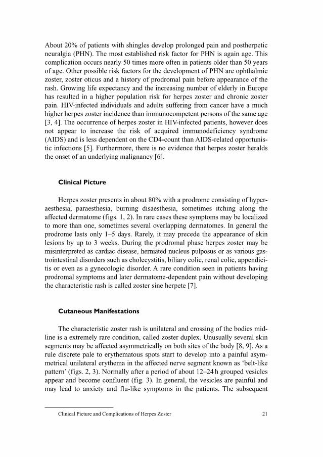
Clinical Picture
Herpes zoster presents in about 80% with a prodrome consisting of hyper-
aesthesia, paraesthesia, burning disaesthesia, sometimes itching along the
affected dermatome (figs. 1, 2). In rare cases these symptoms may be localized
to more than one, sometimes several overlapping dermatomes. In general the
prodrome lasts only 1–5 days. Rarely, it may precede the appearance of skin
lesions by up to 3 weeks. During the prodromal phase herpes zoster may be
misinterpreted as cardiac disease, herniated nucleus pulposus or as various gas-
trointestinal disorders such as cholecystitis, biliary colic, renal colic, appendici-
tis or even as a gynecologic disorder. A rare condition seen in patients having
prodromal symptoms and later dermatome-dependent pain without developing
the characteristic rash is called zoster sine herpete [7].
Cutaneous Manifestations
The characteristic zoster rash is unilateral and crossing of the bodies mid-
line is a extremely rare condition, called zoster duplex. Unusually several skin
segments may be affected asymmetrically on both sites of the body [8, 9]. As a
rule discrete pale to erythematous spots start to develop into a painful asym-
metrical unilateral erythema in the affected nerve segment known as ‘belt-like
pattern’ (figs. 2, 3). Normally after a period of about 12–24 h grouped vesicles
appear and become confluent (fig. 3). In general, the vesicles are painful and
may lead to anxiety and flu-like symptoms in the patients. The subsequent
Gross 22
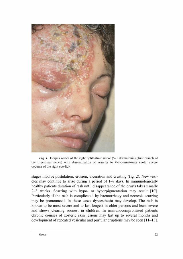
stages involve pustulation, erosion, ulceration and crusting (fig. 2). New vesi-
cles may continue to arise during a period of 1–7 days. In immunologically
healthy patients duration of rash until disappearance of the crusts takes usually
2–3 weeks. Scarring with hypo- or hyperpigmentation may result [10].
Particularly if the rash is complicated by haemorrhagy and necrosis scarring
may be pronounced. In these cases dysaesthesia may develop. The rash is
known to be most severe and to last longest in older persons and least severe
and shows clearing soonest in children. In immunocompromised patients
chronic courses of zosteric skin lesions may last up to several months and
development of repeated vesicular and pustular eruptions may be seen [11–13].
Fig. 1. Herpes zoster of the right ophthalmic nerve (V-1 dermatome) (first branch of
the trigeminal nerve) with dissemination of vesicles to V-2-dermatomes (note: severe
oedema of the right eye-lid).
Clinical Picture and Complications of Herpes Zoster 23
Localization of Herpes Zoster
Although any dermatome may be involved, dermatomes of the trunk from
Th3 to L1 are most frequently affected (figs. 3, 4). The second most frequently
involved nerve dermatome is the ophthalmic division of the trigeminal nerve.
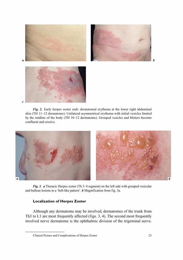
a
c
b
Fig. 2. Early herpes zoster rash: dermatomal erythema at the lower right abdominal
skin (TH 11–12 dermatome). Unilateral asymmetrical erythema with initial vesicles limited
by the midline of the body (TH 10–12 dermatome). Grouped vesicles and blisters become
confluent and erosive.
ba
Fig. 3. a Thoracic Herpes zoster (Th 3–4 segment) on the left side with grouped vesicular
and bullous lesions in a ‘belt-like pattern’. b Magnification from fig. 3a.
Gross 24
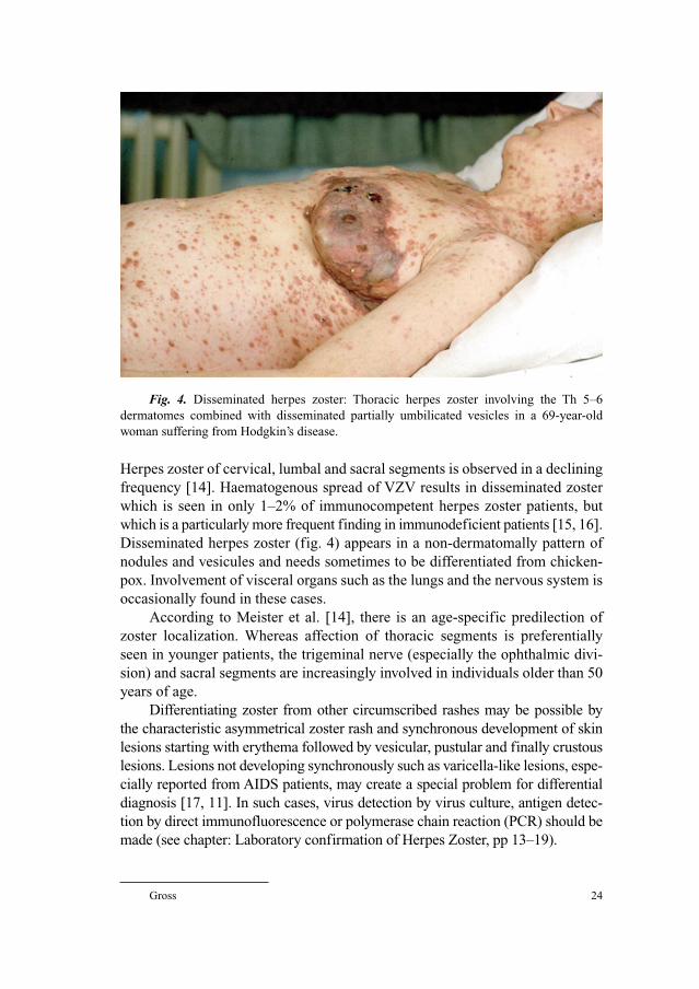
Herpes zoster of cervical, lumbal and sacral segments is observed in a declining
frequency [14]. Haematogenous spread of VZV results in disseminated zoster
which is seen in only 1–2% of immunocompetent herpes zoster patients, but
which is a particularly more frequent finding in immunodeficient patients [15, 16].
Disseminated herpes zoster (fig. 4) appears in a non-dermatomally pattern of
nodules and vesicules and needs sometimes to be differentiated from chicken-
pox. Involvement of visceral organs such as the lungs and the nervous system is
occasionally found in these cases.
According to Meister et al. [14], there is an age-specific predilection of
zoster localization. Whereas affection of thoracic segments is preferentially
seen in younger patients, the trigeminal nerve (especially the ophthalmic divi-
sion) and sacral segments are increasingly involved in individuals older than 50
years of age.
Differentiating zoster from other circumscribed rashes may be possible by
the characteristic asymmetrical zoster rash and synchronous development of skin
lesions starting with erythema followed by vesicular, pustular and finally crustous
lesions. Lesions not developing synchronously such as varicella-like lesions, espe-
cially reported from AIDS patients, may create a special problem for differential
diagnosis [17, 11]. In such cases, virus detection by virus culture, antigen detec-
tion by direct immunofluorescence or polymerase chain reaction (PCR) should be
made (see chapter: Laboratory confirmation of Herpes Zoster, pp 13–19).
Fig. 4. Disseminated herpes zoster: Thoracic herpes zoster involving the Th 5–6
dermatomes combined with disseminated partially umbilicated vesicles in a 69-year-old
woman suffering from Hodgkin’s disease.

Clinical Picture and Complications of Herpes Zoster 25
Symptoms
Characteristically the clinical appearance of herpes zoster is accompanied
by dermatomal pain, which may be continuous or intermittent and presenting
with varying intensity. By definition, pain occurring before and after the der-
matomal rash is called zoster-associated-pain. Postzoster neuralgia or PHN is
defined as pain, which appears or continues after cutaneous symptoms have
disappeared (see chapter: Postherpetic Neuralgia and Other Neurologic
Complications, pp 69–80). PHN is the most frequent and important complica-
tion of herpes zoster affecting the nervous system [18].
Herpes Zoster Ophthalmicus
Herpes zoster ophthalmicus involves the ophthalmic branch (V-1 der-
matome), which is the first division of the trigeminal nerve. According to sev-
eral studies about 7–18% of reported herpes zoster cases affect the ophthalmic
division of the trigeminal nerve [19–21]. While cases of zoster ophthalmicus
occur approximately in 10% of zoster patients under the age of 10 years, almost
30% of 80-year-old and older patients suffer from this condition. There is no
doubt that ophthalmic zoster is seen particularly more frequently in patients
older than 50 years of age [14, 22, 23]. The rash of ophthalmic zoster may
extend from the level of the eye to the vertex of the skull. Characteristically it
does not cross the midline of the forehead. About 50% of patients with herpes
zoster ophthalmicus will develop ocular complications if they do not receive
antiviral therapy [12]. Involvement of the nasociliary branch of the ophthalmic
nerve which is evidenced by a zosteric rash on the tip and side of the nose
(Hutchinson’s sign) is seen in about one-third of patients and is usually accom-
panied by ocular symptoms (fig. 5).
Thus, when ophthalmic zoster affects the side and the tip of the nose, care-
ful attention must be given to the condition of the eye and immediate ophthal-
mologic consultation is necessary in order to prevent complications of the eye
and central nerve system (see chapter: Ophthalmic Manifestations of Herpes
Zoster Infection, pp 37–46) [24].
Herpes Zoster Oticus
Herpes zoster affecting the second and third division of the trigeminal nerve
and other cranial nerves is likely uncommon. When it occurs, however it may pro-
duce symptoms and lesions in the mouth, ear, pharynx or larynx. The Ramsay

Gross 26
Hunt syndrome consists of facial palsy combined with ipsilateral herpes zoster of
the external auditory meatus or tympanic membrane. Severe facial pain, facial
palsy, decreased hearing or even deafness, tinnitus, vertigo and loss of taste in the
anterior part of the tongue may coexist resulting from the involvement of the
geniculate ganglion (compare chapter: (Herpes) Zoster Oticus, pp 47–57).
Herpes Zoster in Children
Herpes zoster can develop in immunocompetent children as young as a
few weeks of age and should be considered in the differential diagnosis of
vesicular eruptions in infants. Zoster in children most frequently involves the
thoracic dermatomes (65%). In contrast to adult zoster patients, cranial sites are
rarely affected (13 vs. 5%, respectively). Sacral and disseminated zoster are
very rare findings (�5%) [25–27].
Herpes zoster certainly occurs in pediatric populations, although by far
less frequent compared with adults [28]. Affected children are almost exclu-
sively those, who had an intrauterine exposure to VZV, VZV exposure until the
fourth year of age or who are immunosuppressed [25] (fig. 6). Pediatric malig-
nancies most frequently associated with zoster are leukaemia and Hodgkin’s
lymphoma. As a rule zoster does not precede the clinical manifestations of
these malignancies but is associated with chemotherapy or relapse [29]. If
immunocompetent children are affected, the course of the disease is rather mild
and pain and PHN are extremely uncommon findings. It is quite likely that the
vesicular lesions of herpes zoster in this age group are misdiagnosed as
impetigo or other cutaneous disorders [30].
a b
Fig. 5. a Herpes zoster ophthalmicus with involvement of the left tip and side of the
nose (nasociliary branch of V-1 (Hutchinson’s sign)) in a 84-year-old woman. b Severe ulcer-
ation despite optimal intravenous acyclovir therapy.

Clinical Picture and Complications of Herpes Zoster 27
With the increasingly widespread use of varicella-vaccine the frequency of
herpes zoster in children will probably continue to decrease. Studies of herpes
zoster in normal children [26] have not shown underlying problems and have
resulted in the conclusion that laboratory studies in healthy children with her-
pes zoster are not helpful. The majority of children presenting with herpes
zoster is not infected with HIV. Undocumented HIV infection in a young child
should however be considered when herpes zoster develops with a short interval
after an eruption of chickenpox. This is particularly true if there are no data
about the HIV serologic status of the mother [12].
Herpes Zoster in Immunodeficient Patients
Individuals with pathologic or iatrogenic immunodeficiency may present
atypical zoster. In some cases, the rash may appear mitigated with only few symp-
toms. More often the rash shows severe inflammation in part with haemorrhages
and necrosis of the skin. Involvement of more than one dermatome may be seen
and the rash may persist for a longer period than in immunocompetent persons.
Disseminated zoster with varicella-like lesions (fig. 4) combined with involve-
ment of inner organs and the nervous system is more common in immunodefi-
cient persons [16, 31] than in immunocompetent individuals. In persons suffering
from lymphocytic leukaemia or Hodgkins disease zoster rash with atypical vari-
celliform or papular and even verrucous lesions was reported [11, 17]. Since her-
pes zoster is regarded as an early marker for HIV-infection [32], it is generally
a b
Fig. 6. a Herpes zoster affecting dermatomes S1–S2 in a 10-months-old girl.
b Grouped vesicles on the left S2 dermatome.

Gross 28
accepted to serologically test zoster patients under the age of 50 for HIV infection
(see chapter: Herpes Zoster in the Immunocompromised Host, pp 93–106).
Herpes Zoster during Pregnancy
Maternal zoster is hardly considered a risk for the unborn. This is in con-
trast to maternal varicella (see chapter: Varicella-Zoster Virus Infections during
Pregnancy, pp 81–92). Zoster during pregnancy should only be treated with
intravenous acyclovir in exceptional cases. In general local and symptomatic
treatment is sufficient.
Pregnant zoster patients in a gynaecologic practice, however should be
treated in such a way that non-pregnant women without immune protection are
not put at risk of varicella [13].
Clinical Diagnosis
The clinical picture of herpes zoster is almost always distinctive enough for
diagnosis and laboratory diagnosis is not required. Characteristically, the asym-
metrical rash with grouped vesicles is located on inflamed and erythematous
dermatomal skin. This and limitation of the rash by the midline as well as pro-
dromal and zoster-associated segmental pain are mostly sufficient to establish
the diagnosis. Diagnostic difficulties may arise during the prodromal period and
the early phase of zoster and also when atypical skin lesions appear or an atypi-
cal site of the body is affected. In such situations laboratory confirmation is
required. Equally laboratorial diagnosis is important for VZV infections of
pregnant women, newborn infants and particularly if a VZV-infection of the cen-
tral nervous system is suspected.
The golden standard of laboratorial diagnosis is viral culture (see chapter:
Laboratory Confirmation of Herpes Zoster, pp 13–19). The PCR is a useful
method to detect VZV-DNA in fluids and in tissues. In case of neurological
manifestations PCR is the test of choice to detect VZV in cerebro-spinal fluid
[18]. Detection of VZV in blood by the PCR can be predictive of PHN or even
diagnostic in some cases of zoster sine herpete. In everyday practice, direct
immunofluorescence assays using labelled VZV-specific monoclonal antibod-
ies are a diagnostic approach suitable in terms of low costs and the option to
distinguish VZV-infection from HSV-infection. The Tzanck test can be of help
to examine the cytopathic effect of VZV in the epidermis with multinucleated
giant cells and intranuclear inclusions (fig. 7). The test is done by taking a swab
from the base of a blister, transferring the cells to a microscopic glass, and
observing the cells under the light-microscope after giemsa staining. This
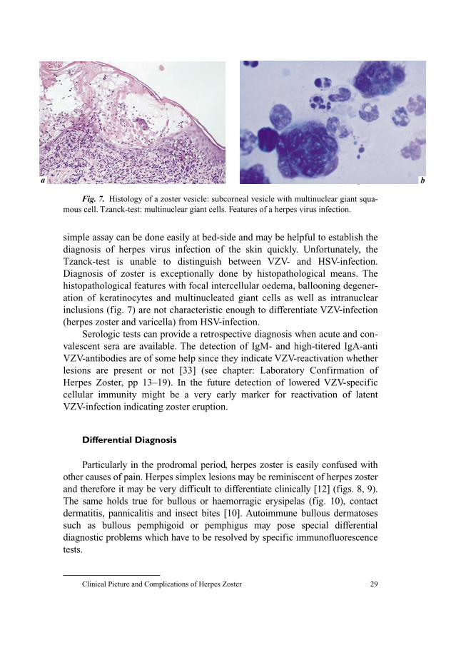
Clinical Picture and Complications of Herpes Zoster 29
simple assay can be done easily at bed-side and may be helpful to establish the
diagnosis of herpes virus infection of the skin quickly. Unfortunately, the
Tzanck-test is unable to distinguish between VZV- and HSV-infection.
Diagnosis of zoster is exceptionally done by histopathological means. The
histopathological features with focal intercellular oedema, ballooning degener-
ation of keratinocytes and multinucleated giant cells as well as intranuclear
inclusions (fig. 7) are not characteristic enough to differentiate VZV-infection
(herpes zoster and varicella) from HSV-infection.
Serologic tests can provide a retrospective diagnosis when acute and con-
valescent sera are available. The detection of IgM- and high-titered IgA-anti
VZV-antibodies are of some help since they indicate VZV-reactivation whether
lesions are present or not [33] (see chapter: Laboratory Confirmation of
Herpes Zoster, pp 13–19). In the future detection of lowered VZV-specific
cellular immunity might be a very early marker for reactivation of latent
VZV-infection indicating zoster eruption.
Differential Diagnosis
Particularly in the prodromal period, herpes zoster is easily confused with
other causes of pain. Herpes simplex lesions may be reminiscent of herpes zoster
and therefore it may be very difficult to differentiate clinically [12] (figs. 8, 9).
The same holds true for bullous or haemorragic erysipelas (fig. 10), contact
dermatitis, pannicalitis and insect bites [10]. Autoimmune bullous dermatoses
such as bullous pemphigoid or pemphigus may pose special differential
diagnostic problems which have to be resolved by specific immunofluorescence
tests.
ba b
Fig. 7. Histology of a zoster vesicle: subcorneal vesicle with multinuclear giant squa-
mous cell. Tzanck-test: multinuclear giant cells. Features of a herpes virus infection.
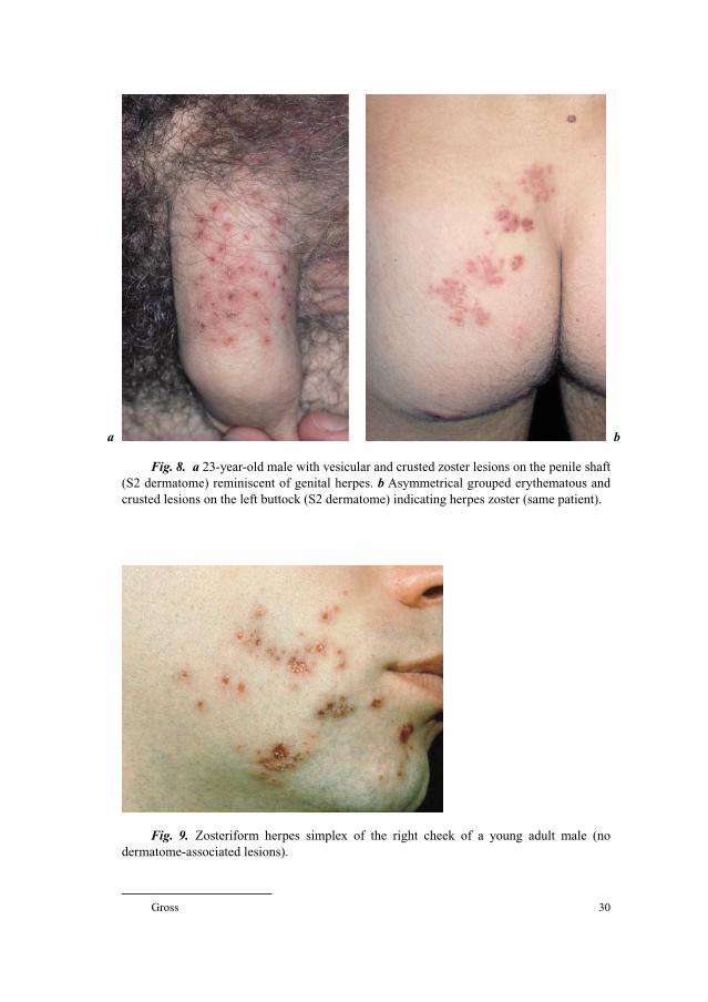
Gross 30
a b
Fig. 8. a 23-year-old male with vesicular and crusted zoster lesions on the penile shaft
(S2 dermatome) reminiscent of genital herpes. b Asymmetrical grouped erythematous and
crusted lesions on the left buttock (S2 dermatome) indicating herpes zoster (same patient).
Fig. 9. Zosteriform herpes simplex of the right cheek of a young adult male (no
dermatome-associated lesions).
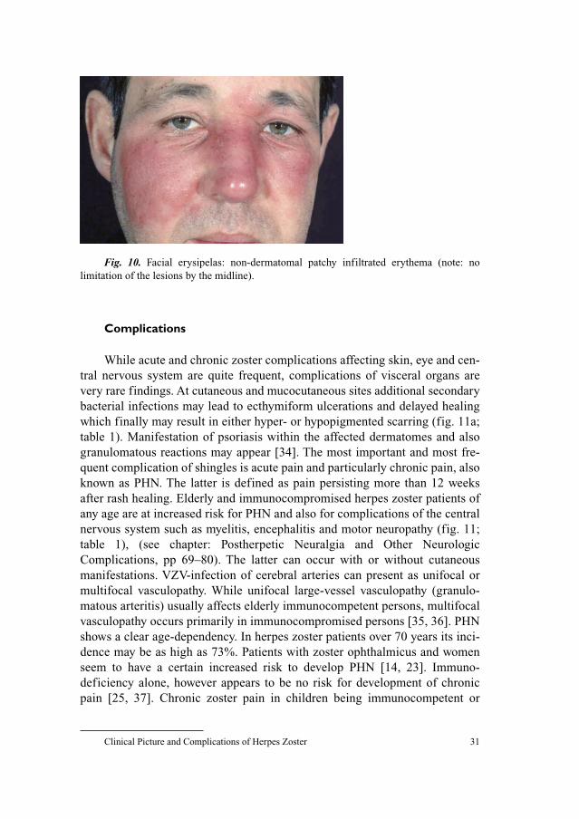
Clinical Picture and Complications of Herpes Zoster 31
Complications
While acute and chronic zoster complications affecting skin, eye and cen-
tral nervous system are quite frequent, complications of visceral organs are
very rare findings. At cutaneous and mucocutaneous sites additional secondary
bacterial infections may lead to ecthymiform ulcerations and delayed healing
which finally may result in either hyper- or hypopigmented scarring (fig. 11a;
table 1). Manifestation of psoriasis within the affected dermatomes and also
granulomatous reactions may appear [34]. The most important and most fre-
quent complication of shingles is acute pain and particularly chronic pain, also
known as PHN. The latter is defined as pain persisting more than 12 weeks
after rash healing. Elderly and immunocompromised herpes zoster patients of
any age are at increased risk for PHN and also for complications of the central
nervous system such as myelitis, encephalitis and motor neuropathy (fig. 11;
table 1), (see chapter: Postherpetic Neuralgia and Other Neurologic
Complications, pp 69–80). The latter can occur with or without cutaneous
manifestations. VZV-infection of cerebral arteries can present as unifocal or
multifocal vasculopathy. While unifocal large-vessel vasculopathy (granulo-
matous arteritis) usually affects elderly immunocompetent persons, multifocal
vasculopathy occurs primarily in immunocompromised persons [35, 36]. PHN
shows a clear age-dependency. In herpes zoster patients over 70 years its inci-
dence may be as high as 73%. Patients with zoster ophthalmicus and women
seem to have a certain increased risk to develop PHN [14, 23]. Immuno-
deficiency alone, however appears to be no risk for development of chronic
pain [25, 37]. Chronic zoster pain in children being immunocompetent or
Fig. 10. Facial erysipelas: non-dermatomal patchy infiltrated erythema (note: no
limitation of the lesions by the midline).
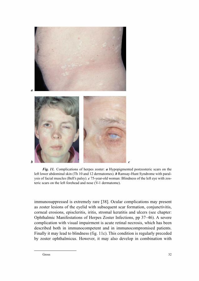
Gross 32
immunosuppressed is extremely rare [38]. Ocular complications may present
as zoster lesions of the eyelid with subsequent scar formation, conjunctivitis,
corneal erosions, episcleritis, iritis, stromal keratitis and ulcers (see chapter:
Ophthalmic Manifestations of Herpes Zoster Infections, pp 37–46). A severe
complication with visual impairment is acute retinal necrosis, which has been
described both in immunocompetent and in immunocompromised patients.
Finally it may lead to blindness (fig. 11c). This condition is regularly preceded
by zoster ophthalmicus. However, it may also develop in combination with
a
b c
Fig. 11. Complications of herpes zoster: a Hypopigmented postzosteric scars on the
left lower abdominal skin (Th 10 and 12 dermatomes). b Ramsay-Hunt Syndrome with paral-
ysis of facial muscles (Bell’s palsy). c 75-year-old woman: Blindness of the left eye with zos-
teric scars on the left forehead and nose (V-1 dermatome).
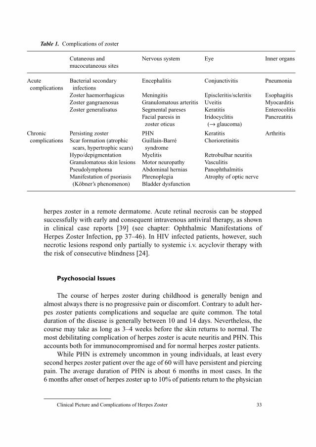
Clinical Picture and Complications of Herpes Zoster 33
herpes zoster in a remote dermatome. Acute retinal necrosis can be stopped
successfully with early and consequent intravenous antiviral therapy, as shown
in clinical case reports [39] (see chapter: Ophthalmic Manifestations of
Herpes Zoster Infection, pp 37–46). In HIV infected patients, however, such
necrotic lesions respond only partially to systemic i.v. acyclovir therapy with
the risk of consecutive blindness [24].
Psychosocial Issues
The course of herpes zoster during childhood is generally benign and
almost always there is no progressive pain or discomfort. Contrary to adult her-
pes zoster patients complications and sequelae are quite common. The total
duration of the disease is generally between 10 and 14 days. Nevertheless, the
course may take as long as 3–4 weeks before the skin returns to normal. The
most debilitating complication of herpes zoster is acute neuritis and PHN. This
accounts both for immunocompromised and for normal herpes zoster patients.
While PHN is extremely uncommon in young individuals, at least every
second herpes zoster patient over the age of 60 will have persistent and piercing
pain. The average duration of PHN is about 6 months in most cases. In the
6 months after onset of herpes zoster up to 10% of patients return to the physician
Table 1. Complications of zoster
Cutaneous and Nervous system Eye Inner organs
mucocutaneous sites
Acute Bacterial secondary Encephalitis Conjunctivitis Pneumonia
complications infections
Zoster haemorrhagicus Meningitis Episcleritis/scleritis Esophagitis
Zoster gangraenosus Granulomatous arteritis Uveitis Myocarditis
Zoster generalisatus Segmental pareses Keratitis Enterocolitis
Facial paresis in Iridocyclitis Pancreatitis
zoster oticus (→ glaucoma)
Chronic Persisting zoster PHN Keratitis Arthritis
complications Scar formation (atrophic Guillain-Barré Chorioretinitis
scars, hypertrophic scars) syndrome
Hypo/depigmentation Myelitis Retrobulbar neuritis
Granulomatous skin lesions Motor neuropathy Vasculitis
Pseudolymphoma Abdominal hernias Panophthalmitis
Manifestation of psoriasis Phrenoplegia Atrophy of optic nerve
(Köbner’s phenomenon) Bladder dysfunction
Gross 34
with recurrent complaints [18]. This complication has led to a variety of treat-
ment regimens such as long-term administration of analgesics and very early
administration of systemic antiviral therapy together with analgesics in acute
herpes zoster (see chapter: Postherpetic Neuralgia and Other Neurologic
Complications, pp 69–80). It has become clear that herpes zoster may have a
tremendous impact on the everyday’s life of affected individuals older than 50
years of age. The patients may suffer from fluctuating levels of pain for many
months or even years. As a consequence treatment of PHN can lead to a range
of drug therapies. Nevertheless, the chronicity and intensity of chronic pain can
lead to depression and even suicide, particularly among patients over 70 years
of age. In older patients who experience the disease on the head and face, which
are approximately 15% of all zoster patients, a slight touch, contact with cloth-
ing or even a breeze can trigger an unbearable pain (allodynia) [18]. During or
after shingles focal itch also may develop. So far this complaint has attracted
only little medical attention. According to Oaklander et al. [40] postherpetic
itch can lead to self-injury from scratching skin that has lost protective sensa-
tion. Postherpetic itch seems to occur particularly in patients with herpes zoster
involving the sensory innervation region of the trigeminal nerve or other cranial
nerves. Physicans should be aware of the importance to treat shingles early and
aggressively with systemic antivirals, analgesics and tricyclic antidepressants to
minimize zoster-related complications. Urgent indications for systemic antivi-
ral therapy are herpes zoster in patients beyond the age of 50 years and patients
at any age with herpes zoster in the head and neck area. Another urgent indica-
tion is severe herpes zoster on the trunk and on the extremities, herpes zoster in
immunosuppressed patients and zoster in patients with severe atopic dermatitis
and severe eczema [13, 41]. Once PHN has established, an interdisciplinary
approach to the care of these patients should comprise behavioural and physical
therapy, as well as supportive psychotherapy [40].
References
1 Balfour HH: Varicella zoster virus infections in immunocompromised hosts. A review of the nat-
ural history and management. Am J Med 1988;85:68–73.
2 Hope-Simpson RE: The nature of herpes zoster: a long term study and new hypothesis. Proc R Soc
Med 1965;58:9–20.
3 Friedman-Kien AE, Lafleur FL, Gendler E: Herpes zoster: a possible early clinical sign for devel-
opment of acquired immunodeficiency syndrome in high-risk individuals. J Am Acad Dermatol
1986;14:1023–1028.
4 Kost RG, Straus SE: Postherpetic neuralgia – pathogenesis, treatment, and prevention. N Engl J
Med 1996;335:32–42.
5 Aliegro MB, Dorruccci M, Pezzotti P, Rezza G, Sinicco A, Barbanera M: Herpes zoster and pro-
gression to AIDS in a cohort of individuals who seroconverted to human immunodeficiency virus.
Italian HIV Seroconversion Study. Clin Infect Dis 1996;23:990–995.
Clinical Picture and Complications of Herpes Zoster 35
6 Smith JB, Fenske NA: Herpes zoster and internal malignancy. South Med J 1995;88:1089–1092.
7 Rudra T: Zoster sine herpete. Br J Clin Pract 1990;44:284.
8 Vu AQ, Radonich M, Heald PW: Herpes zoster in seven desperate dermatomes (zoster multiplex):
report of a case and review of the literature. J Am Acad Dermatol 1999;40:868–869.
9 Bloss G, Ebisch MA, Kunz M, Gross G: Bilateral asymmetric herpes zoster in adolescence.
Hautarzt 2001;52:335–338.
10 Gross G: Herpes zoster. Dtsch Med Wochenschr 1997;122:132–139.
11 Hoppenjans WB, Bibler MR, Orne RL: Prolonged cutaneous herpes zoster in acquired immuno-
deficiency syndrome. Arch Dermatol 1990;126:1048–1050.
12 Gnann JW Jr, Whitley RJ: Clinical practice. Herpes zoster. N Engl J Med 2002;347:340–346.
13 Gross G, Schöfer H, Wassilew S, Friese K, Timm A, Guthoff R, Pau HW, Malin JP, Doerr HW:
Herpes zoster guideline of the German Dermatology Society (DDG). J Clin Virol 2003;26:
277–289.
14 Meister W, Neiss A, Gross G, Doerr H, Holbel W, Malin J, von Essen J, Reimann B, Witke C,
Wutzler P: Demography, symptomatology, and course of disease in ambulatory zoster patients. A
physician-based survey in Germany. Intervirology 1998;41:272–277.
15 Meier JL, Strauss SE: Comparative biology of latent varicella-zoster virus and herpes simplex
virus infections. J Infect Dis 1992;166(suppl 1):S13–S23.
16 Cohen RR, Beltraini VP, Grossmann ME: Disseminated herpes zoster in patients with human
immunodeficiency virus infection. Am J Med 1998;84:1076–1080.
17 Gallagher JG, Merigan TC: Prolonged herpes-zoster infection associated with immunosuppres-
sive therapy. Am Intern Med 1979;91:842–846.
18 Gilden DH, Kleinschmidt-DeMasters BK, La Guardia JJ, Mahalingam R, Cohrs RJ: Neurological
complications of the reactivation of varicella-zoster virus. N Engl J Med 2000;342:635–645.
19 Burgoon CF, Burgoon JS, Baldrige GD: The natural history of herpes zoster. JAMA 1957;164:
265–269.
20 De Moragas JM, Kierland R: The outcome of patients with herpes zoster. Arch Dermatol
1957;75:193–196.
21 Ragozzino MW, Melton LJ III, Kurland LT, Chu CP, Perry HO: Population-based study of herpes
zoster and its sequelae. Medicine (Baltimore) 1982;61:310–316.
22 Hardening SP, Lipton JR, Wells JCD: Natural history of herpes zoster ophthalmicus: predictors of
postherpetic neuralgia and ocular involvement. Br J Ophthalmol 1987;71:353–358.
23 Meister W, Neiss A, Gross G, Doerr HW, Holbel W, Malin JP, von Essen J, Reimann BY, Witke C,
Wutzler P: A prognostic score for postherpetic neuralgia in ambulatory patients. Infection
1998;26:359–363.
24 Liesegang TJ: Diagnosis and therapy of herpes zoster ophthalmicus. Ophthalmology 1991;98:
1216–1229.
25 Guess HA, Broughton DD, Melton LJ III, Kurland LT, Chu CP, Perry HO: Epidemiology of herpes
zoster in children and adolescents: a population-based study. Pediatrics 1985;76:512–517.
26 Smith CG, Glaser DA: Herpes zoster in childhood: case report and review of the literature. Pediatr
Dermatol 1996;13:226–229.
27 Terada K, Kawano S, Yoshihiro K: Characteristics of herpes zoster in otherwise normal children.
Pediatr Infect Dis J 1993;12:960–961.
28 Donahue JG, Choo PW, Manson JE: The incidence of herpes zoster. Arch Intern Med
1995;155:1605–1609.
29 Bacon GE, Olivier WJ, Shapiro BA: Factors contributing to severity of herpes zoster in children.
J Pediatr 1965;67:768–771.
30 Kurlan JG, Conelly BL, Lucky AW: Herpes zoster in the first year of life following postnatal
exposure to Varicella-zoster virus. Arch Dermatol 2004;140:1268–1272.
31 Rusthoven JJ, Ahlgren P, Elhakim T, Pinfold P, Reid J, Stewart L, Feld R: Varicella zoster infection
in adult cancer patients: a population study. Arch Intern Med 1988;148:1561–1566.
32 Melbye M, Grossman RJ, Goebert JJ: Risk of AIDS after herpes zoster. Lancet 1978;1:728–730.
33 Doerr HW, Rentschler M, Scheifler G: Serologic detection of active infections with human her-
pesviruses (CMV, EBV, HSV, VZV): diagnostic potential of IgA class and IgG-subclass-specific
antibodies. Infection 1987;15:93–98.
Gross 36
34 Requena L, Kutzner H, Pescalonilla P, Otiz S, Schaller J, Rohwedder A: Cutaneous reactions at
sites of herpes zoster scars: an expanded spectrum. Br J Dermatol 1998;138:161–168.
35 Gilden DH: Varicella zoster virus vasculopathy and disseminated encephalomyelitis. J Neurol Sci
2002;195:99–101.
36 Gilden DH, Lipton HL, Wolf JS, Akenbrandt W, Smith JE, Mahalingam R, Forghani B: Two patients
with unusual forms of varicella-zoster virus vasculopathy. N Engl J Med 2002;347:1500–1503.
37 Hope-Simpson RE: Postherpetic neuralgia. J R Coll Gen Pract 1975;25:571–575.
38 Malin JP: Zoster and the nervous system. Dtsch Med Wochenschr 1996;121:635–638.
39 Nau R, Lansch M, Stiefel M, Polak T, Reiber H: Varicella zoster virus-associated focal vasculitis
without herpes zoster: recovery after treatment with acyclovir. Neurology 1998;51:914–915.
40 Oaklander AL, Bowsher D, Galer B, Haanpaa M, Jensen P: Herpes zoster itch: preliminary epi-
demiologic data. J Pain 2003;4:338–343.
41 Volpi A, Gross G, Hercogova J, Johnson RW: Current management of herpes zoster: the European
view. Am J Clin Dermatol 2005;6:317–325.
Gerd Gross, MD
Professor of Dermatology and Venereology, Director
Department of Dermatology and Venereology, University of Rostock
Augustenstrasse 80–84
DE–18055 Rostock (Germany)
Tel. �49 381 4949700, Fax �49 381 4949702, E-Mail [email protected]
Gross G , Doerr HW (eds): Herpes Zoster.
Monogr Virol. Basel, Karger, 2006, vol 26, pp 37–46
Ophthalmic Manifestations ofHerpes Zoster Infection
Hermann Guembel
Bundeswehrkrankenhaus Ulm, Academic Teaching Hospital of the
University Clinics of Ulm, Ulm, Germany
The varicella–zoster virus (VZV) frequently reactivates within the oph-
thalmic part of the trigeminal nerve [1, 2]. Herpes zoster ophthalmicus (HZO)
is manifested by localized pain over the distribution of the first division of the
trigeminus cutaneous vesicular eruption around the forehead. There are also
frequently lymph node enlargement in the defined area, headache, sometimes
neck stiffness and a red eye.
Approximately 10–25% [3] of all cases of herpes zoster affect the oph-
thalmic division of the trigeminal nerve. The condition is then referred to as HZO.
Direct ocular involvement is specifically correlated with age, gender, or
severity of disease. Serious sequelae include chronic inflammation, vision loss
and disabling pain. HZO starts normally with an influenza-like illness with
malaise and low-grade fever that lasts up to 5 days before the rash on the forehead
appears. Subsequently, erythematous macules appear along the involved der-
matome, rapidly progressing over several days to papules and vesicles containing
clear serous fluid and pustules. These lesions rupture and get crusted. Very rarely,
the eye can become involved when the maxillary nerve is involved. Affection of
the nasociliary nerve (Hutchinson’s sign) is quite common and supplies the side
and the tip of the nose. The Hutchinson’s sign occurs in about one-third of patients
with HZO and correlates significantly with ocular complications [4].
However, there is no correlation between ocular complications and age,
sex or severity of the skin rash. Severe ocular complications may therefore
develop in patients with only a slight rash. Approximately 50% of people with
VZV infection involving the ophthalmic division of the fifth cranial nerve have
ocular involvement, often in association with typical skin lesions.
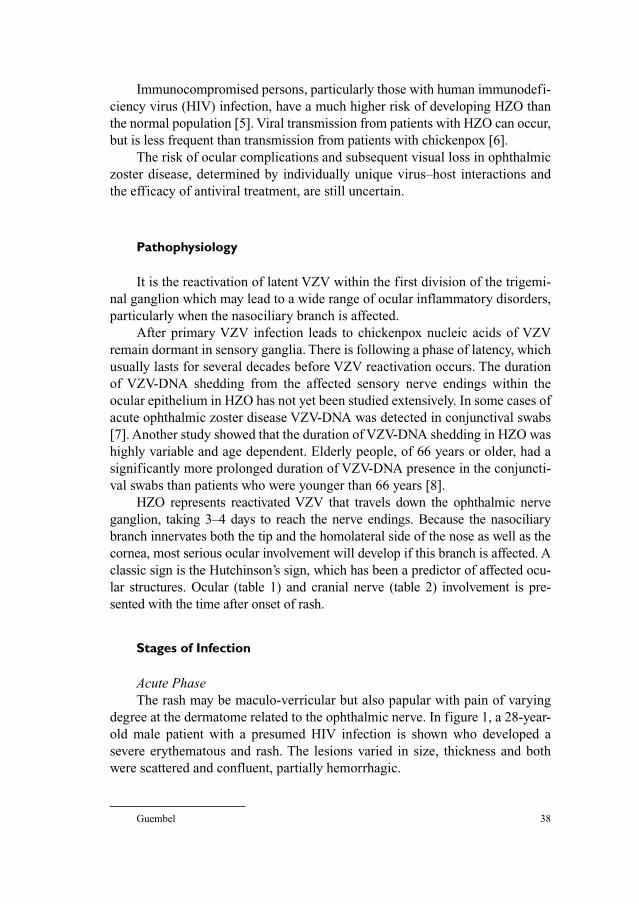
Guembel 38
Immunocompromised persons, particularly those with human immunodefi-
ciency virus (HIV) infection, have a much higher risk of developing HZO than
the normal population [5]. Viral transmission from patients with HZO can occur,
but is less frequent than transmission from patients with chickenpox [6].
The risk of ocular complications and subsequent visual loss in ophthalmic
zoster disease, determined by individually unique virus–host interactions and
the efficacy of antiviral treatment, are still uncertain.
Pathophysiology
It is the reactivation of latent VZV within the first division of the trigemi-
nal ganglion which may lead to a wide range of ocular inflammatory disorders,
particularly when the nasociliary branch is affected.
After primary VZV infection leads to chickenpox nucleic acids of VZV
remain dormant in sensory ganglia. There is following a phase of latency, which
usually lasts for several decades before VZV reactivation occurs. The duration
of VZV-DNA shedding from the affected sensory nerve endings within the
ocular epithelium in HZO has not yet been studied extensively. In some cases of
acute ophthalmic zoster disease VZV-DNA was detected in conjunctival swabs
[7]. Another study showed that the duration of VZV-DNA shedding in HZO was
highly variable and age dependent. Elderly people, of 66 years or older, had a
significantly more prolonged duration of VZV-DNA presence in the conjuncti-
val swabs than patients who were younger than 66 years [8].
HZO represents reactivated VZV that travels down the ophthalmic nerve
ganglion, taking 3–4 days to reach the nerve endings. Because the nasociliary
branch innervates both the tip and the homolateral side of the nose as well as the
cornea, most serious ocular involvement will develop if this branch is affected. A
classic sign is the Hutchinson’s sign, which has been a predictor of affected ocu-
lar structures. Ocular (table 1) and cranial nerve (table 2) involvement is pre-
sented with the time after onset of rash.
Stages of Infection
Acute PhaseThe rash may be maculo-verricular but also papular with pain of varying
degree at the dermatome related to the ophthalmic nerve. In figure 1, a 28-year-
old male patient with a presumed HIV infection is shown who developed a
severe erythematous and rash. The lesions varied in size, thickness and both
were scattered and confluent, partially hemorrhagic.

Ophthalmic Manifestations of Herpes Zoster Infection 39
Table 1. Herpes zoster ophthalmicus – disorders of the anterior eye segment
Involved structures Signs Starting clinical symptoms
(day 0 � onset of, up to weeks)
Eyelid and conjunctivaBlepharoconjunctivitis cutaneus rash respecting midline day 0 and painful dermatom
conjunctival edema/inflammation days 2–3
vesicular crusts and lesions 6 days
Secondary bacterial yellow colored crusts 1–2 weeks
infection
Episclera/scleraEpiscleritis/scleritis diffuse or localized redness, 5–8 days
edema and pain
CorneaPunctate epithelial keratitis edema of corneal surface 1–2 days
Dendritic keratitis worm-like epithelial lesions 4–6 days
Anterior stromal keratitis infiltrating stromal structures 1–2 weeks
Deep stromal keratitis lipid infiltrates and deep stromal 1 month to years
inflammation and neovascularization
Neurotrophic keratopathy punctate keratitis and erosions months to years
persistent epithelial defects
Anterior chamberUveitis inflammation, cells and synechias 2 weeks to years
Table 2. Herpes zoster ophthalmicus – disorders of the posterior eye segment
Involved structures Signs Starting clinical symptoms
(day 0 � onset of, up to weeks)
RetinaAcute retinal necrosis syndrome/ retinal whitening, intraretinal no time schedule possible
progressive outer retinal necrosis hemorrhages occlusive
syndrome vasculitis, vitreous floaters
Cranial nervesNeuritis nervi optici edema of the optic nerve no time schedule possible
Oculomotor palsies motional abnormalities of no time schedule possible
the eye muscles
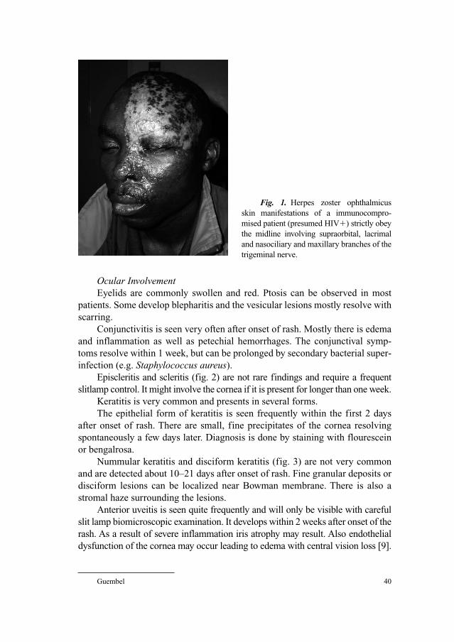
Guembel 40
Ocular InvolvementEyelids are commonly swollen and red. Ptosis can be observed in most
patients. Some develop blepharitis and the vesicular lesions mostly resolve with
scarring.
Conjunctivitis is seen very often after onset of rash. Mostly there is edema
and inflammation as well as petechial hemorrhages. The conjunctival symp-
toms resolve within 1 week, but can be prolonged by secondary bacterial super-
infection (e.g. Staphylococcus aureus).
Episcleritis and scleritis (fig. 2) are not rare findings and require a frequent
slitlamp control. It might involve the cornea if it is present for longer than one week.
Keratitis is very common and presents in several forms.
The epithelial form of keratitis is seen frequently within the first 2 days
after onset of rash. There are small, fine precipitates of the cornea resolving
spontaneously a few days later. Diagnosis is done by staining with flourescein
or bengalrosa.
Nummular keratitis and disciform keratitis (fig. 3) are not very common
and are detected about 10–21 days after onset of rash. Fine granular deposits or
disciform lesions can be localized near Bowman membrane. There is also a
stromal haze surrounding the lesions.
Anterior uveitis is seen quite frequently and will only be visible with careful
slit lamp biomicroscopic examination. It develops within 2 weeks after onset of the
rash. As a result of severe inflammation iris atrophy may result. Also endothelial
dysfunction of the cornea may occur leading to edema with central vision loss [9].
Fig. 1. Herpes zoster ophthalmicus
skin manifestations of a immunocompro-
mised patient (presumed HIV�) strictly obey
the midline involving supraorbital, lacrimal
and nasociliary and maxillary branches of the
trigeminal nerve.
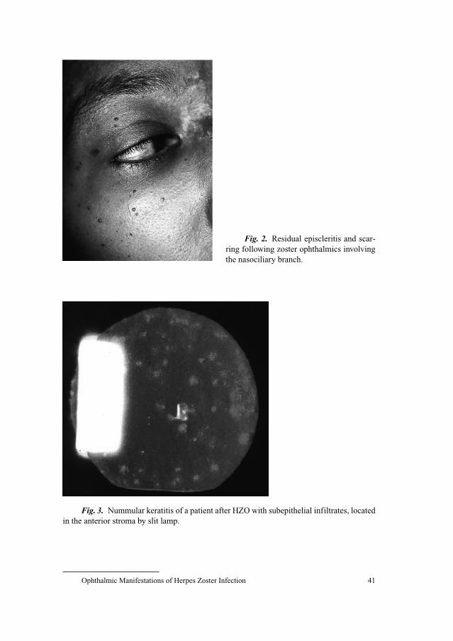
Ophthalmic Manifestations of Herpes Zoster Infection 41
Fig. 2. Residual episcleritis and scar-
ring following zoster ophthalmics involving
the nasociliary branch.
Fig. 3. Nummular keratitis of a patient after HZO with subepithelial infiltrates, located
in the anterior stroma by slit lamp.

Guembel 42
Chronic Phase
From a clinical point of view there are more cases presenting signs of
severe chronical involvement of the sclera and cornea than with acute clinical
findings.
Stromal or deep stromal keratitis is a later stage of stromal keratitis char-
acterized by infiltrates of differing degree. It develops 3–4 months after the first
onset of inflammation. Usually it is localized in the center of the cornea. A
chronic relapse of keratitis is not unusual and as a result of a uncontrolled
chronic disease deep corneal neovascularizations and lipid infiltrates may be
detectable in time.
Neurotrophic keratopathy is a clinical endpoint of keratitis. Destruction of
the cornea may lead to decreased corneal sensitivity, often followed by injuries.
Another pathway to vision loss results in corneal thinning with bullous ker-
atopathy and corneal perforation.
Acute retinal necrosis syndrome (ARN) and progressive outer retinal
necrosis syndrome (PORN) are fortunately rare findings in young patients and
in immunocompromised patients (fig. 4). First signs of ARN and PORN are
blurred vision in one or both eyes (30% bilateral involvement) and pain.
Clinically the fundus of ARN shows whitening and peripheral patches with
occlusive vasculitis and vitreous inflammation. In contrast in PORN vitreous
cells are absent, since immunocompromised patients are unable to produce an
inflammatory response. In such cases visual prognosis is bad despite antiviral
therapy and more than 70% develop retinal detachment.
Differential Diagnosis
Because of the typical clinical findings (rash, pain etc.) in the acute
phase of HZO there is no really clear differential diagnosis. Erysipelas may
however initiate herpes zoster at an early stage. A chronic HSV infection can
imitate similar corneal destruction and thinning as seen in HZO with ocular
affection.
Diagnostic Tools in Herpes Zoster Ophthalmicus
There are several aspects to detect VZV in the eye during a chronic course
of HZO. It is easy to diagnose acute HZO with its typical rash, involving the
nasociliary branch of the ophthalmic nerve. There are quite more difficulties to
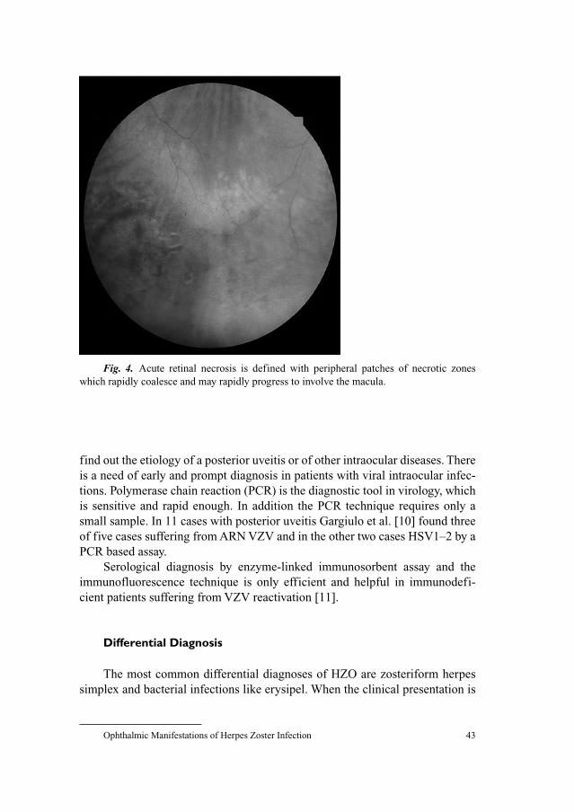
Ophthalmic Manifestations of Herpes Zoster Infection 43
find out the etiology of a posterior uveitis or of other intraocular diseases. There
is a need of early and prompt diagnosis in patients with viral intraocular infec-
tions. Polymerase chain reaction (PCR) is the diagnostic tool in virology, which
is sensitive and rapid enough. In addition the PCR technique requires only a
small sample. In 11 cases with posterior uveitis Gargiulo et al. [10] found three
of five cases suffering from ARN VZV and in the other two cases HSV1–2 by a
PCR based assay.
Serological diagnosis by enzyme-linked immunosorbent assay and the
immunofluorescence technique is only efficient and helpful in immunodefi-
cient patients suffering from VZV reactivation [11].
Differential Diagnosis
The most common differential diagnoses of HZO are zosteriform herpes
simplex and bacterial infections like erysipel. When the clinical presentation is
Fig. 4. Acute retinal necrosis is defined with peripheral patches of necrotic zones
which rapidly coalesce and may rapidly progress to involve the macula.
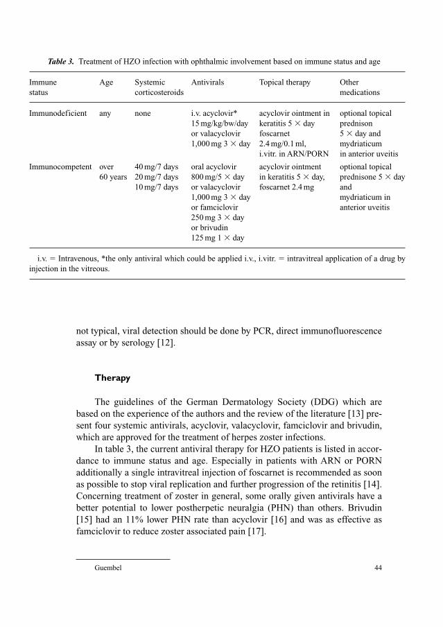
Guembel 44
not typical, viral detection should be done by PCR, direct immunofluorescence
assay or by serology [12].
Therapy
The guidelines of the German Dermatology Society (DDG) which are
based on the experience of the authors and the review of the literature [13] pre-
sent four systemic antivirals, acyclovir, valacyclovir, famciclovir and brivudin,
which are approved for the treatment of herpes zoster infections.
In table 3, the current antiviral therapy for HZO patients is listed in accor-
dance to immune status and age. Especially in patients with ARN or PORN
additionally a single intravitreal injection of foscarnet is recommended as soon
as possible to stop viral replication and further progression of the retinitis [14].
Concerning treatment of zoster in general, some orally given antivirals have a
better potential to lower postherpetic neuralgia (PHN) than others. Brivudin
[15] had an 11% lower PHN rate than acyclovir [16] and was as effective as
famciclovir to reduce zoster associated pain [17].
Table 3. Treatment of HZO infection with ophthalmic involvement based on immune status and age
Immune Age Systemic Antivirals Topical therapy Other
status corticosteroids medications
Immunodeficient any none i.v. acyclovir* acyclovir ointment in optional topical
15 mg/kg/bw/day keratitis 5 � day prednison
or valacyclovir foscarnet 5 � day and
1,000 mg 3 � day 2.4 mg/0.1 ml, mydriaticum
i.vitr. in ARN/PORN in anterior uveitis
Immunocompetent over 40 mg/7 days oral acyclovir acyclovir ointment optional topical
60 years 20 mg/7 days 800 mg/5 � day in keratitis 5 � day, prednisone 5 � day
10 mg/7 days or valacyclovir foscarnet 2.4 mg and
1,000 mg 3 � day mydriaticum in
or famciclovir anterior uveitis
250 mg 3 � day
or brivudin
125 mg 1 � day
i.v. � Intravenous, *the only antiviral which could be applied i.v., i.vitr. � intravitreal application of a drug by
injection in the vitreous.

Ophthalmic Manifestations of Herpes Zoster Infection 45
It is well known that VZV may become resistant to nucleoside analogs
such as the above mentioned antivirals acyclovir, valacyclovir, famciclovir and
brivudin. The resistance is mainly due to a mutation of the thymidin kinase.
Drug of choice in patients with resistant strains of VZV is intravenous foscar-
net, 3 times 40 mg or twice 50 mg per kg body weight per day [18]. In cases
where foscarnet would also be ineffective, treatment with cidofovir is indicated
as an alternative [19]. Both antivirals have some severe side effects (nephrotox-
icity, ocular hypotony etc.) and should be admitted very carefully [18, 19].
References
1 Hope-Simpson RE: The nature of herpes zoster: a long term study and new hypothesis. Proc R Soc
Med 1965;58:9–20.
2 Wilson JB: Thirty one years of herpes zoster in a rural practice. Br Med J 1986;22:1349–1351.
3 Naumann G, Gass JD, Font RL: Histopathology of herpes zoster ophthalmicus. Am J Ophthalmol
1968;65:533–541.
4 Liesegang TJ: Herpes zoster virus infection. Curr Opin Ophthalmol 2004;15:531–536.
5 Sandor EV, Millman A, Croxson TS, Mildvan D: Herpes zoster ophthalmicus in patients at
risk for the acquired immune deficiency syndrome (AIDS). Am J Ophthalmol 1986;101:
153–157.
6 Baratz KH, Goins K, Cobo M: Varicella-zoster viral infections; in Kaufman HE (ed): The Cornea.
New York, Livingstone, 1988.
7 Yamamoto S, Pavan-Langston D, Kinoshita S, Nishida K, Shimomura Y, Tano Y: Detecting herpes
virus DNA in uveitis using polymerase chain reaction. Br J Ophthalmol 1996;80:465–468.
8 Zaal MJW, Völker-Dieben HJ, Wienesen M, DÀmaro J, Kijlstra A: Longitudinal analysis of
varicella-zoster virus DNA on the ocular surface associated with herpes zoster ophthalmicus. Am
J Ophthalmol 2001;131:25–29.
9 Zaal MJW, Völker-Dieben HJ, DÁmaro J: Visual prognosis in immunocompetent patients with
herpes zoster ophthalmicus. Acta Ophthalmol Scand 2003;81:216–220.
10 Gargiulo F, Francesco de MA, Nascimbeni G, Turano R, Perandin F, Gandolfo E, Manca N:
Polymerase chain reaction as a rapid diagnostic tool for therapy of acute retinal necrosis syn-
drome. J Med Virol 2003;69:397–400.
11 Wutzler P, Doerr HW: Herpes zoster: a herpes non-simplex disease. Dtsch Med Wochenschr
1995;120:1133–1138.
12 Gross G, Doerr HW: Herpes zoster guidelines of German Dermatological Society. J Clin Virol
2003;27:308–309.
13 Gross G, Schöfer H, Wassilew S, Friese K, Timm A, Guthoff R, Pau HW, Malin JP, Wutzler P,
Doerr HW: Herpes zoster guideline of the German Dermatology Society (DDG). J Clin Virol
2003;26:277–289.
14 Gümbel H, Ohrloff C: Opportunistic infections of the eye in immunocompromised patients.
Ophthalmologica 1997;211:53–61.
15 Vij O, Bornfeld N, Roggendorf M, Fiedler M, Schilling H: Brivudin as an alternative systemic
therapy to acyclovir and ganciclovir in acute retinal necrosis syndrome due to varicella zoster
virus. Klin Monatsbl Augenheilkd 2003;220:710–715.
16 Wassilew SW, Wutzler P, Brivudin Herpes Zoster Study Group: Oral brivudin in comparision with
acyclovir for herpes zoster: a survey study on postherpetic neuralgia. Antiviral Res 2003;59:
57–60.
17 Wassilew SW, Wutzler P, Brivudin Herpes Zoster Study Group: Oral brivudin in comparison with
acyclovir for improved therapy of herpes zoster in immunocompetent patients: results of a ran-
domized double-blind, multicentered study. Antiviral Res 2003;59:49–56.

Guembel 46
18 Breton G, Fillet AM, Katlama C, Bricaire F, Caumes E: Acyclovir-resistant herpes zoster in human
immunodeficiency virus-infected patients: results of foscarnet therapy. Clin Infect Dis 1998;27:
1525–1527.
19 Safrin S, Cherrington J, Jaffe HS: Clinical uses of cidofovir. Rev Med Virol 1997;7:145–156.
Hermann Guembel, MD
Bundeswehrkrankenhaus Ulm
Academic Teaching Hospital of the University Clinics of Ulm
Oberer Eselsberg 40
DE–89081 Ulm (Germany)
Tel. �49 731 17101401, Fax �49 731 17101407, E-Mail [email protected]

Gross G, Doerr HW (eds): Herpes Zoster.
Monogr Virol. Basel, Karger, 2006, vol 26, pp 47–57
(Herpes) Zoster Oticus
H.W. Pau
Department of Otorhinolaryngology, Head and Neck Surgery, Medical Faculty,
University of Rostock, Rostock, Germany
In literature, the term ‘(herpes) zoster oticus’ is mostly attributed to the
neurologist James Ramsay Hunt, who described the disease in 1907 [1]. To our
knowledge, however, he was proceeded by the German otologist Otto Körner
(one of the author’s predecessors in Rostock), who published about ‘zoster
oticus’ in 1904 [2].
The disease is the clinical expression of an infection with – or better reac-
tivation of – the varicella–zoster virus (VZV) in the head – especially the ear
region. The incidence is roughly estimated as 1% of all zoster localizations.
Patients with (herpes) zoster oticus typically present with earache, vesicular
eruptions in the auricle or its surroundings (sometimes also in the buccal or
oropharyngeal mucosa) and different alterations of the VII, VIII (sometimes
also V, IX, and X) cranial nerves [3] (fig. 1).
VZV (first contact: chickenpox, varicella) may persist in various, mostly
neuronal structures – in case of zoster oticus most often in the geniculate gan-
glion. From these sites, dormant viruses may be reactivated by certain stimuli,
causing the clinical outbreak of the disease [4, 5]. However, occasional cases
could be attributed to reinfection of immunized hosts by re-exposition to VZV
(contact with varicella or zoster patients) [6].
Etiology and Pathogenesis
In 1907, Ramsay Hunt suggested that the disease resulted from a genicu-
late ganglionitis after reactivation of surviving viruses (VZV) – a theory that
recently proved true for many cases examined [2, 7]. In histological temporal
bone sections, VZV DNA could be identified in the region of the geniculate
ganglion using the polymerase chain reaction.
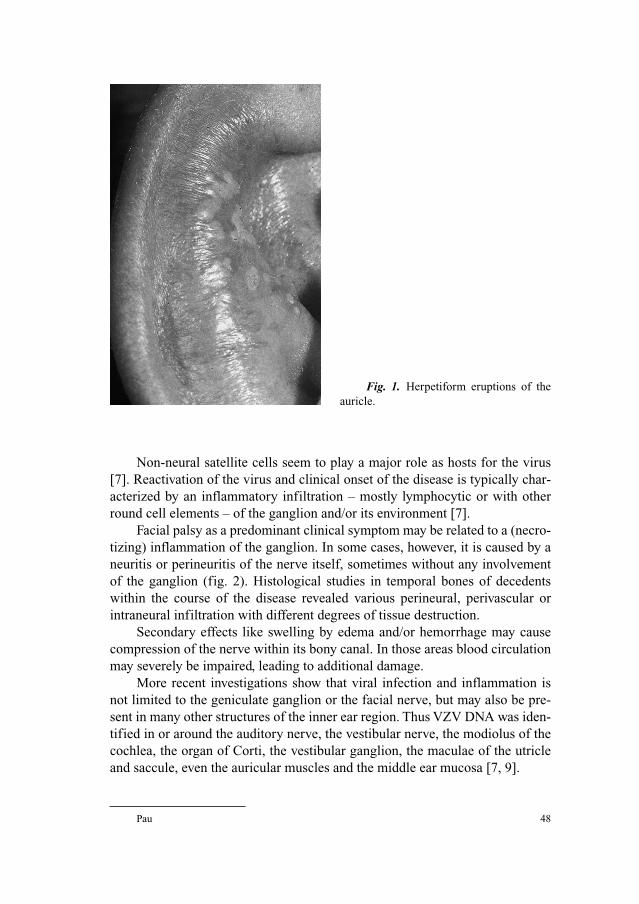
Pau 48
Non-neural satellite cells seem to play a major role as hosts for the virus
[7]. Reactivation of the virus and clinical onset of the disease is typically char-
acterized by an inflammatory infiltration – mostly lymphocytic or with other
round cell elements – of the ganglion and/or its environment [7].
Facial palsy as a predominant clinical symptom may be related to a (necro-
tizing) inflammation of the ganglion. In some cases, however, it is caused by a
neuritis or perineuritis of the nerve itself, sometimes without any involvement
of the ganglion (fig. 2). Histological studies in temporal bones of decedents
within the course of the disease revealed various perineural, perivascular or
intraneural infiltration with different degrees of tissue destruction.
Secondary effects like swelling by edema and/or hemorrhage may cause
compression of the nerve within its bony canal. In those areas blood circulation
may severely be impaired, leading to additional damage.
More recent investigations show that viral infection and inflammation is
not limited to the geniculate ganglion or the facial nerve, but may also be pre-
sent in many other structures of the inner ear region. Thus VZV DNA was iden-
tified in or around the auditory nerve, the vestibular nerve, the modiolus of the
cochlea, the organ of Corti, the vestibular ganglion, the maculae of the utricle
and saccule, even the auricular muscles and the middle ear mucosa [7, 9].
Fig. 1. Herpetiform eruptions of the
auricle.
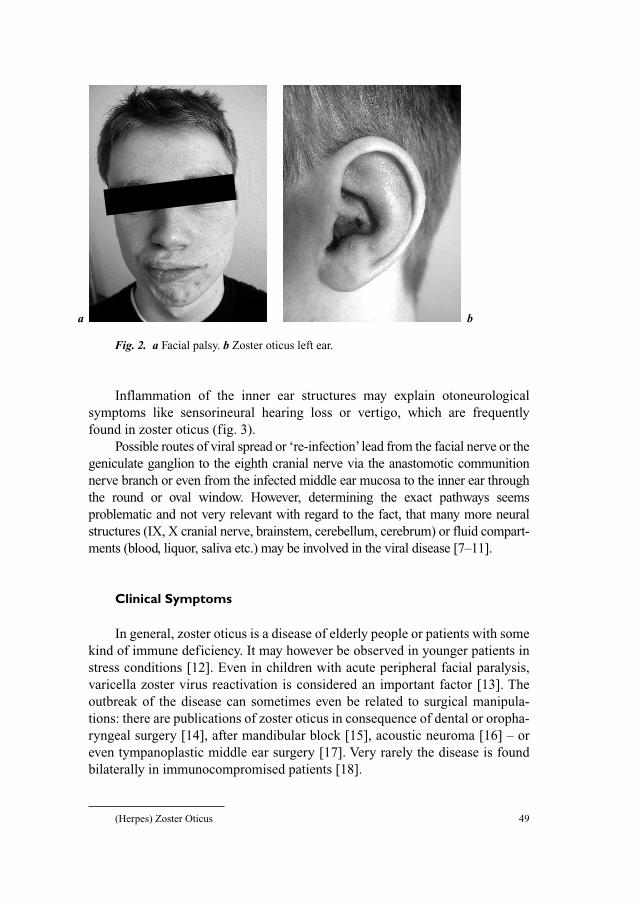
(Herpes) Zoster Oticus 49
Inflammation of the inner ear structures may explain otoneurological
symptoms like sensorineural hearing loss or vertigo, which are frequently
found in zoster oticus (fig. 3).
Possible routes of viral spread or ‘re-infection’ lead from the facial nerve or the
geniculate ganglion to the eighth cranial nerve via the anastomotic communition
nerve branch or even from the infected middle ear mucosa to the inner ear through
the round or oval window. However, determining the exact pathways seems
problematic and not very relevant with regard to the fact, that many more neural
structures (IX, X cranial nerve, brainstem, cerebellum, cerebrum) or fluid compart-
ments (blood, liquor, saliva etc.) may be involved in the viral disease [7–11].
Clinical Symptoms
In general, zoster oticus is a disease of elderly people or patients with some
kind of immune deficiency. It may however be observed in younger patients in
stress conditions [12]. Even in children with acute peripheral facial paralysis,
varicella zoster virus reactivation is considered an important factor [13]. The
outbreak of the disease can sometimes even be related to surgical manipula-
tions: there are publications of zoster oticus in consequence of dental or oropha-
ryngeal surgery [14], after mandibular block [15], acoustic neuroma [16] – or
even tympanoplastic middle ear surgery [17]. Very rarely the disease is found
bilaterally in immunocompromised patients [18].
a b
Fig. 2. a Facial palsy. b Zoster oticus left ear.
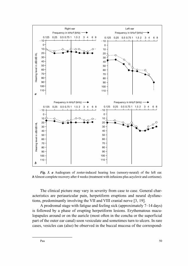
Pau 50
The clinical picture may vary in severity from case to case. General char-
acteristics are periauricular pain, herpetiform eruptions and neural dysfunc-
tions, predominantly involving the VII and VIII cranial nerve [3, 19].
A prodromal stage with fatigue and feeling sick (approximately 7–14 days)
is followed by a phase of erupting herpetiform lesions. Erythematous macu-
lopapules around or on the auricle (most often in the concha or the superficial
part of the outer ear canal) soon vesiculate and sometimes turn to ulcers. In rare
cases, vesicles can (also) be observed in the buccal mucosa of the correspond-
Right ear Left ear
0.125�10
0
10
20
30
40
50
Hea
ring
leve
l in
dB
/dB
HL
60
70
80
90
100
110
0.25 0.5 0.75 1 1.5 2 3 4 6 8 0.125�10
0
10
20
30
40
50
60
70
80
90
100
110
0.25 0.5 0.75 1 1.5 2 3 4 6 8
0.125�10
0
10
20
30
40
50
Hea
ring
leve
l in
dB
/dB
HL
60
70
80
90
100
110
0.25 0.5 0.75 1 1.5 2 3 4 6 8 0.125�10
0
10
20
30
40
50
60
70
80
90
100
110
0.25 0.5 0.75 1 1.5 2 3 4 6 8
Frequency in kHz/f (kHz) Frequency in kHz/f (kHz)
Frequency in kHz/f (kHz)Frequency in kHz/f (kHz)
a
b
Fig. 3. a Audiogram of zoster-induced hearing loss (sensory-neural) of the left ear.
bAlmost complete recovery after 4 weeks (treatment with infusions plus acyclovir and cortisone).
(Herpes) Zoster Oticus 51
ing side or in the oropharynx. Body temperature may slightly be raised, usually
there is pain – mostly moderate to severe – in the ear and its environment. After
approximately 1 week, the lesions turn dry and crust. Regional lymphadenopa-
thy is rather common. One of the leading symptoms is local pain (deep earache
till pain around the auricle), which may persist even after the crust fell off (‘pos-
therpetic neuralgia’).
Cutaneous zoster eruptions are in no way obligatory: 8–15% of the patients
present without any rash or of vesicles (‘zoster sine herpete’ [20]).
Special attention must be turned to neural disorders: about 70% of the
zoster oticus patients show some kind of involvement of the facial nerve, the
incidence of inner ear affections (deafness, vertigo) seems to range within equal
dimensions.
Signs of facial palsy mostly emerge later than the rash, typically proceed-
ing rapidly from mild facial weakness to a complete unilateral peripheral
paralysis.
Some patients complain of dysgeusia due to loss of chorda tympani-related
functions. So unilaterally taste on the antero-lateral portion of the tongue may
be diminished.
Without adequate treatment, the prognosis of facial palsy is very poor.
Untreated, the chance of a restitutio ad integrum is as little as 10% in complete
and about 70% incomplete paralysis [21]. With the nowadays generally
accepted treatment the outcome is much more favorable.
Zoster-related hearing disorders may occur at different intervals in the
course of the disease, but mostly follow the cutaneous symptoms. Often, there
is an abrupt onset, resembling sudden hearing loss. The common finding is a sen-
sorineural deafness, mostly in the high frequency range, sometimes accompanied
by tinnitus. Profound inner ear hearing loss or even complete deafness is a very
rare event. In some cases, so-called hyperacousis is found, meaning that the
pain threshold for noise or tones is decreased, though (mostly) the hearing
threshold is raised.
Vertigo may come along with the hearing disorders or appear at intervals,
often with sensations of spinning or twirling motion.
Zoster oticus infection may cause neurologic complications such as cere-
brospinal fluid changes, involvement of other cranial nerves (V, IX, X), aseptic
meningitis, myelitis, encephalitis, acute ascending polyradiculitis, thrombotic
cerebral vasculopathy etc. There are publications describing extensive involvement
of more cranial nerves such as XI and XII [22], very rarely I, II, III and IV, cervical
dermatomes C2–4 or even bilateral facial paralysis [18, 23]. In those cases, the
spread of infection through meningeal inflammation should be considered.
Post-herpetic neuralgia can persist for long time after the disease, meaning
a special therapeutic challenge.
Pau 52
Diagnosis
In cases with characteristic pathology (pain, herpetiform eruptions of the
auricle, facial palsy, may be unilateral hearing loss, vertigo) the diagnosis may
be fairly easy. However, there are many atypical courses, especially those with-
out any cutaneous symptoms (‘zoster sine herpete’), in which only serologic
examinations can verify the diagnosis ‘zoster’.
Serological assays confirming the diagnosis include verification of signi-
ficant increase of Anti-VZV-antibodies of the IgG-, IgM-, respectively, IgA-
fraction (Enzym-linked immunosorbent assay or immune fluorescence).
In some cases (and for scientific purpose), detection of viral DNA (VZV
DNA) via polymerase chain reaction in cerebrospinal fluid, saliva tissue etc.
can be helpful [24, 25].
In case of facial nerve involvement, the severity code is classified by the
criteria of House and Brackmann [26]. The course of facial nerve palsy can be
followed electrophysiologically (electromyography, electroneurography).
Hearing loss in the context of zoster oticus infection is documented and
controlled by audiometry (pure tone audiometry, supra-threshold tests, imped-
ance audiometry). In certain cases, objective audiometry via evoked potentials
(e.g. BERA) can be helpful for identifying the site of the lesion (cochlear/retro-
cochlear) [27, 28]. In early stages registration of otoacoustic emissions can
make sense [29].
In cases of zoster-linked dizziness or vertigo [30, 31], detection and regis-
tration of nystagmus is important. For clinical purpose, observation of eye
movement, e.g. with special glasses (‘Frenzel’s goggles’) in a darkened room
may be sufficient. For more detailed examinations nystagmography (electro-
nystagmography, or video-oculography) is inevitable (fig. 4). Caloric testing
with water [32] for detecting unilateral vestibular disorders may be painful
because of zoster papules, even the use of heated or cooled air may not be toler-
ated in single cases.
Additional hints concerning the site of the zoster-linked lesion may be
given by magnetic resonance tomography of the temporal bone (fig. 5).
Typically, gadolinium enhancement in the course of the facial nerve, the area of
the geniculate ganglion, the labyrinth or the meatus acousticus internus may
indicate inflammation within the temporal bone and help estimating the main
focus of the disease [33–35]. Some authors advocate transcranial magnetic stim-
ulation of the nerve, by which a more precise localization of the lesion within the
Fallopian channel is possible. Moreover, additional electric stimulation via the
same route may help to establish correct diagnosis and prognosis [36].
If there are further neurological signs or any apprehension of meningitis,
lumbal puncture should be performed for examining CNS fluid.

(Herpes) Zoster Oticus 53
Calibration
Spontane nystagmus
10˚
�10˚
Fig. 4. Electronystamography presenting signs of spontaneous nystagmus in conse-
quence of zoster oticus. The ‘sawteeth’ of the curve indicate spontaneous rhythmic eye move-
ments, indicating unilateral peripheral vestibular dysfunction.
Fig. 5. MRI of the temporal bone showing zoster-related gadolinum enhancement of
the intratemporal portion of the facial nerve.
Pau 54
Therapy
As soon as the diagnosis zoster oticus has been made, systemic antiviral
chemotherapy should be started urgently. Substances like acyclovir, valacy-
clovir, famciclovir and brivudin are available. In most studies, high doses of
acyclovir were administered intravenously (e.g. 5–10 mg, 3 times a day for
7–10 days), with good results [37, 38]. So far, in Germany oral application of
antiviral agents, even brivudin as a very potent drug, has not been widely
accepted for zoster oticus treatment, though in other localization of zoster it
proved to be effective.
Once there are any signs of involvement of the VIIth or VIIIth cranial
nerve (facial weakness, hearing loss, tinnitus, dizziness/vertigo), a simu-
ltaneous application of cortisone is indicated (e.g. 60 mg prednisolone daily
for the first 10 days, subsequently 10 mg for another 10 days or starting with
200 mg prednisolone i.v., progressively decreasing till 10 mg p.o. within 2
weeks).
It seems crucial, not to delay the onset of the combined therapy (at least
stay within the 72 h range), because the prognosis deteriorates markedly [39,
40].
With this therapy, prognosis of facial palsy has improved so much, that
indications for surgical procedures for the nerve became very rare [41]. Some
decades ago, nerve decompression for stopping ‘self-strangulation’ of the ede-
matous nerve by removing the bony walls of its canal was a rather common
operation [42]. Surgery ranged from simple mastoidectomy (descending part of
the nerve) to transtemporal approaches, from which the entire course of the
nerve from the internal auditory canal till the foramen stylomastoideum could
be exposed [42, 43].
If the VIIIth cranial nerve is involved (hearing loss, tinnitus, dizziness/
vertigo), a simultaneous infusion therapy (similar to that in sudden hearing
loss) can be added. In Germany, the combination of plasma volume expanders
(like hydroxyethyl starch) and vasoactive substances (e.g. pentoxiphylline) is
widely used (e.g. 500 ml HAES plus pentoxiphylline per day for approximately
1 week).
In cases of vestibular imbalance with dizziness and vertigo sometimes
causing vomitus, additional treatment with antivertiginous drugs: e.g. anti-
cholinergics, antihistamines, Ca-channel blockers may be indicated.
Heavy pain is treated with analgesics (e.g. tramadol) in appropriate doses,
best combined with a neuroactive agent (e.g. amitriptyline) [44].
Local treatment of the skin includes antisepsis and drying in the
early stages (e.g. lotio alba, vioform zinc mixture), later cautious removal of
crusts.
(Herpes) Zoster Oticus 55
Prognosis
Prior to the era of early and simultaneous antiviral and antiphlogistic ther-
apy, zoster-related affections of the VIIth and VIIIth cranial nerves had a poor
prognosis. Nowadays, in case of early onset of medical treatment (as possible
within the first 72 h), in most cases an almost or complete restitution can be
expected. The main difficulty for the otologist is the correct interpretation of
clinical symptoms, which may be misleading at least in the early phase of the
disease. As indicative cutaneous symptoms may be missing (‘zoster sine her-
pete’), the combination of local pain (earache) and hearing loss (sensorineural),
vertigo or any kind of facial weakness should always be suspicious for zoster
oticus.
References
1 Hunt JR: On herpetic inflammations of the geniculate ganglion. A new syndrome and its compli-
cations. J Nerv Ment Dis 1907;34:73–96.
2 Körner O: Über den Herpes zoster oticus. Munch Med Wochenschr 1904;1:6–10.
3 Walther LE, Prosowsky K, Walther A, Gudziol H: Herpes zoster oticus: symptom constellation
and serological diagnosis. Laryngorhinootologie 2004;83:355–362.
4 Straus SE: Overview: the biology of varicella-zoster virus infection. Ann Neurol 1994;35(suppl):
4–8.
5 Gilden DH, Mahalingam R, Dueland AN, Cohrs R: Herpes zoster: pathogenesis and latency. Prog
Med Virol 1992;39:19–75.
6 Gershon AA (ed): First international conference on the varicella-zoster virus. J Infect Dis
1992;166(suppl 1):1–68.
7 Wackym PA: Molecular temporal bone pathology: II. Ramsay Hunt syndrome (herpes zoster oticus).
Laryngoscope 1997;107:1165–1175.
8 Meier JL, Straus SE: Comparative biology of latent varicella-zoster virus and herpes simplex virus
infections. J Infect Dis 1992;166:13–23.
9 Murakami S, Nakashiro Y, Mizobuchi M, Hato N, Honda N, Gyo K: Varicella-zoster virus distrib-
ution in Ramsay Hunt syndrome revealed by polymerase chain reaction. Acta Otolaryngol
1998;118:145–149.
10 Gold E: Serologic and virus isolation studies of patients with varicella or herpes zoster infection.
N Engl J Med 1966;274:181–185.
11 Bessho K, Ogino S: Detection of varicella-zoster virus DNA by polymerase chain reaction in
patients with Hunt syndrome and clinical progression. J Otolaryngol Jpn 1994;97:1019–1027.
12 Meister W, Neiss A, Gross D, Doerr HW, Höbel W, Malin JP, von Essen J, Reimann BY, Witke C,
Wutzler P: Demography, symptomatology and course of disease in ambulatory zoster patients.
Intervirology 1998;41:272–277.
13 Furuta Y, Ohtani F, Aizawa H, Kukuda S, Kawabata H, Bergstrom T: Varicella-zoster virus reacti-
vation is an important cause of acute peripheral facial paralysis in children. Pediatr Inf Dis J
2005;24:97–101.
14 Furuta Y, Ohtani F, Fukuta S, Inuymama Y, Nagashima K: Reactivation of varicella-zoster virus in
delayed facial palsy after dental treatment abd oro-facial surgery. J Laryngol Otol 2000;62:42–45.
15 Maina S, Preece M: Herpes zoster oticus following mandibular block. J Laryngol Otol 2000;114:
212–213.
16 Magliulo G, D’Amico R, Celebrini A, Cuiuli G: Postoperative Ramsay-Hunt syndrome after
acoustic neuroma resection. Viral reactivation. An Otorrinolaringol Ibero Am 2005;32:253–259.

Pau 56
17 Gyo K, Honda N: Delayed facial palsy after middle-ear surgery due to reactivation of varicella-
zoster virus. J Laryngol Otol 1999;113:914–915.
18 Syal R, Tyagi I, Goyal A: Bilateral Ramsay Hunt syndrome in a diabetic patient. BMC Ear Nose
Throat Disord 2004;4:3.
19 Robillard RB, Hilsinger RL, Adour KK: Ramsay Hunt facial paralysis: clinical analyses of 185
patients. Otolaryngol Head Neck Surg 1986;95:292–297.
20 Rudra T: Zoster sine herpete. Br J Clin Pract 1990;44:284.
21 Devriese PP, Moesker WH: The natural history of facial paralysis in herpes zoster. Clin
Otolaryngol 1988;13:289–298.
22 De S, Pfleiderer AG: An extreme and unusual variant of Ramsay Hunt syndrome. J Laryngol Otol
1999;113:670–671.
23 Hamado T, Takano A, Miyao S, Teramoto J, Wanibe S: Bilateral facial palsy following trigeminal
zoster with zoster oticus. Intern Med 2000;39:80–81.
24 Furuta Y, Ohtani F, Sawa H, Fukuda S, Inuyama Y: Quantification of varicella-zoster virus DNA
in patients with Ramsay Hunt syndrome and zoster sine herpete. J Clin Microbiol 2001;39:
2856–2859.
25 Hiroshige K, Ikeda M, Hondo R: Detection of varicella virus DNA in tear fluid and saliva of
patients with Ramsay Hunt syndrome. Otol Neurotol 2002;23:602–607.
26 House JW, Brackmann DE: Facial nerve grading system. Otolaryngol Head Neck Surg 1985;93:
146–147.
27 Abramovich S, Prasher DK: Electrocochleography and brain-stem potentials in Ramsay Hunt syn-
drome. Arch Otolaryngol Head Neck Surg 1986;112:925–928.
28 Wayman DM, Pham HN, Byl FM, Adour KK: Audiological manifestations of Ramsay Hunt syn-
drome. J Laryngol Otol 1990;104:8.
29 Kaberos A, Balatsouras DG, Korres SG, Kandiloros D, Economou C: Audiological assessment in
Ramsay Hunt syndrome. Ann Otol Rhinol Laryngol 2002;111:68–76.
30 Proctor L, Perlmann H, Lindsay J, Matz G: Acute vestibular paralysis in herpes zoster oticus. Ann
Otol Rhinol Laryngol 1979;88:303–310.
31 Kuhweide R, van de Steene V, Vlaminck S, Casselman JW: Ramsay Hunt syndrome: pathophysi-
ology of cochleovestibular symptoms. J Laryngol Otol 2002;16:844–848.
32 Lu YC, Young YH: Vertigo from herpes zoster oticus: superior or inferior vestibular origin?
Laryngoscope 2003;113:307–311.
33 Casselmann JW, Kuhweide R, Ampe W, Meeus L, Steyaert C: Pathology of the membraneous
labyrinth: a comparison of T1- and T2-weighted and gadolinium-enhanced spin-echo and 3DFT-
CISS imaging. Am J Neuroradiol 1993;14:59–69.
34 Korzec K, Sobol SM, Kubal W, Mester SJ, Winzberg G, May M: Gadolinium-enhanced magnetic
resonance imaging of the facial nerve in zoster oticus and Bell’s plasy: clinical implications. Am J
Otol 1991;12:163–168.
35 Brandle P, Saretti-Schefer S, Bohm A, Wichmann W, Fisch U: Correlation of MRI, clinical and
electroneuronographic findings in acute facial palsy. Am J Otol 1996;17:154–161.
36 Nowak DA, Linder S, Topka H: Diagnostic relevance of transcranial magnetic and electric stimu-
lation of the facial nerve in the management of facial palsy. Clin Neurophysiol 2005;116:
2051–2057.
37 Dickens JRE, Smith JT, Graham SS: Herpes zoster oticus: treatment with intravenous acyclovir.
Laryngoscope 1988;98:776–779.
38 Kinishi M, Amatsu M, Mohri M, Saito M, Hasegawa T, Hasegawa S: Acyclovir improves recovery
rate of facial nerve palsy in Ramsay Hunt syndrome. Auris Nasus Larynx 2001;28:223–226.
39 Uri N, Greenberg E, Meyer W, Kitzes-Cohen R: Herpes zoster oticus: treatment with acyclovir.
Ann Otol Rhinol Laryngol 1992;101:161–162.
40 Murakami S, Hato N, Horiuchi J, Honda N, Gyo K, Yanagihara N: Treatment of Ramsay Hunt
syndrome with acyclovir-prednisone: significance of early diagnosis and treatment. Ann Neurol
1997;41:353–357.
41 Ko JY, Sheen TS, Hsu MM: Herpes zoster oticus treated with acyclovir and prednisolone: clinical
manifestations and analysis of prognostic factors. Clin Otolaryngol 2000;25:139–142.
(Herpes) Zoster Oticus 57
42 Fisch U, Esslen E: Total intratemporal exposition on the facial nerve. Arch Otolaryng 1972;95:
335–341.
43 Fisch U: Surgery for bell’s palsy and herpes zoster oticus. Proceedings of the Symposium of
Neurological Surgery of the Ear. Sarasota, 1979.
44 Malin JP: Zoster und Nervensystem. Dtsch Med Wochenschr 1996;121:635–638.
Prof. Dr. H. W. Pau
Klinik und Poliklinik für Hals-Nasen-Ohrenheilkunde
Kopf- und Hals-Chirurgie, ‘Otto Körner’ der Universität Rostock
Doberaner Strasse 137–139
DE–18057 Rostock (Germany)
Tel. �49 381 494 8300, Fax �49 381 494 8302, E-Mail [email protected]
Gross G, Doerr HW (eds): Herpes Zoster.
Monogr Virol. Basel, Karger, 2006, vol 26, pp 58–68
Neuroanatomy of Pain andNeuropathology of Herpes Zosterand Postherpetic Neuralgia
Andreas Wreea, Oliver Schmitta, Kamen G. Usunoff a–c
aInstitute of Anatomy, Medical Faculty, University of Rostock, Rostock, Germany;bDepartment of Anatomy and Histology, Preclinical University Center, Medical University
of Sofia, cInstitute of Physiology, Bulgarian Academy of Sciences, Sofia, Bulgaria
Pain is an important biological signal for danger and necessary for the
integrity of the organism. There are two different kinds of pain. One kind is
typical for threat of tissue damage (pinprick sensation). It is rapidly conducted to
consciousness and well localized. The other kind of pain occurs when tissue
damage has already taken place. It is slowly conducted and poorly localized. But
sustained or chronic pain can result in secondary symptoms. A typical example
is postherpetic neuralgia (PHN), characterized by chronic and severe intractable
pain [1–3].
Functional Neuroanatomy of the Pain System
Primary Afferent NeuronThe primary afferent (PA) neuron is a pseudounipolar cell, localized in
spinal (dorsal root) ganglia (SG), and in sensory ganglia of the Vth, VIIth, IXth
and Xth cranial nerves. Their perikarya are round to elliptical and emit a single
process that bifurcates in a peripheral and central process. The central one
enters the central nervous system (CNS), and the peripheral one runs to its zone
of sensory innervation. There are two basic types of pseudounipolar perikarya:
large, light A-cells and small, dark B-cells. The largest A-cells are the propri-
oreceptor neurons, and the small B-cells are the typical nociceptor neurons [4,
5]. Their transmitter is glutamate. In addition, the B-cells contain various neu-
ropeptides, especially substance P (SP) [6, 7]. The peripheral processes of the

Neuroanatomy of Pain and Neuropathology of HZ and PHN 59
B-cells are thin fibers of two types, both terminating as free nerve endings [8]:
A�-fibers are thinly myelinated (diameter of 1–3 �m, conduction velocity of
5–30 m/s), and C-fibers are unmyelinated (diameter of 1 �m, conduction veloc-
ity of only 0.5–2 m/s). Stimulation of A�-nociceptors leads to pricking, that of
C-nociceptors to burning or dull pain [9]. The free nerve endings are found
throughout the body, mainly in the adventitia of small blood vessels, outer and
inner epithelia, connective tissue capsules and the periosteum. They are most
densely in the cornea, tympanic membrane, dental pulp, skin and mucosa of the
head, skin of the fingers, parietal pleura and peritoneum. Except for avascular
structures, the nociceptors are adjacent to capillaries and mast cells. This triad
is a functional nociceptive response unit [7]. The firing of nociceptors at the site
of tissue injury causes release of vesicles containing peptides that act in sensi-
tizing the nociceptor and increase its firing rate [7]. Cellular damage and
inflammation increase concentrations of other chemical mediators (histamine,
bradykinin, prostaglandins) also, augmenting the transmission of nociceptive
impulses along sensory afferent fibers [7]. In addition, pronociceptive roles
have been proposed for ‘exotic’ species, including protons, ATP, cytokines, neu-
rotrophins and NO [9]. There are nociceptors that normally are inactive and
rather unresponsive. Inflammation leads to sensitization of these fibers, becom-
ing sensitive to peripheral stimulation [10].
Termination of PA Fibers in the Spinal Cord and Spinal Trigeminal NucleusAs central processes of SG neurons approach the dorsal root entry zone, the
nociceptive axons enter lateral portions of the dorsal horn (DH). They terminate
primarily in laminae I and II (substantia gelatinosa) [11], A�-fibers in laminae I
and V, C-fibers in lamina II. The mechanoreceptive A�-axons reach laminae
III–VI [4, 12]. The large lamina I neurons are the source of about one-half of the
spinothalamic tract (STT). Lamina II contains small cells with rich dendritic
trees. In lamina II neurons the inhibitory transmitters �-aminobutyric acid and
glycine coexist. Lamina II functions as a controlling system modulating synaptic
transmission from PA neurons to secondary sensory systems. After complex local
processing in the DH [4], nociceptive signals are conveyed to higher centers
through projection neurons whose axons form several ascending fiber systems.
The central processes of trigeminal ganglion neurons enter the brainstem via the
trigeminal root. Some fibers bifurcate to give a rostral branch to the principal
trigeminal nucleus and a caudal one that joins the spinal trigeminal tract reaching
the ipsilateral spinal trigeminal nucleus (STN) [5]. The PA fibers terminate soma-
totopically: the ophthalmic fibers are located ventrically, the maxillary in the
middle and the mandibular fibers are found dorsally. The PA axons emit collater-
als to all three subnuclei of the STN (oralis, interpolaris and caudalis), and
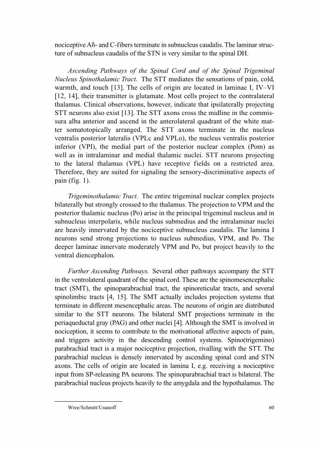
Wree/Schmitt/Usunoff 60
nociceptive A�- and C-fibers terminate in subnucleus caudalis. The laminar struc-
ture of subnucleus caudalis of the STN is very similar to the spinal DH.
Ascending Pathways of the Spinal Cord and of the Spinal TrigeminalNucleus Spinothalamic Tract. The STT mediates the sensations of pain, cold,
warmth, and touch [13]. The cells of origin are located in laminae I, IV–VI
[12, 14], their transmitter is glutamate. Most cells project to the contralateral
thalamus. Clinical observations, however, indicate that ipsilaterally projecting
STT neurons also exist [13]. The STT axons cross the midline in the commis-
sura alba anterior and ascend in the anterolateral quadrant of the white mat-
ter somatotopically arranged. The STT axons terminate in the nucleus
ventralis posterior lateralis (VPLc and VPLo), the nucleus ventralis posterior
inferior (VPI), the medial part of the posterior nuclear complex (Pom) as
well as in intralaminar and medial thalamic nuclei. STT neurons projecting
to the lateral thalamus (VPL) have receptive fields on a restricted area.
Therefore, they are suited for signaling the sensory-discriminative aspects of
pain (fig. 1).
Trigeminothalamic Tract. The entire trigeminal nuclear complex projects
bilaterally but strongly crossed to the thalamus. The projection to VPM and the
posterior thalamic nucleus (Po) arise in the principal trigeminal nucleus and in
subnucleus interpolaris, while nucleus submedius and the intralaminar nuclei
are heavily innervated by the nociceptive subnucleus caudalis. The lamina I
neurons send strong projections to nucleus submedius, VPM, and Po. The
deeper laminae innervate moderately VPM and Po, but project heavily to the
ventral diencephalon.
Further Ascending Pathways. Several other pathways accompany the STT
in the ventrolateral quadrant of the spinal cord. These are the spinomesencephalic
tract (SMT), the spinoparabrachial tract, the spinoreticular tracts, and several
spinolimbic tracts [4, 15]. The SMT actually includes projection systems that
terminate in different mesencephalic areas. The neurons of origin are distributed
similar to the STT neurons. The bilateral SMT projections terminate in the
periaqueductal gray (PAG) and other nuclei [4]. Although the SMT is involved in
nociception, it seems to contribute to the motivational affective aspects of pain,
and triggers activity in the descending control systems. Spino(trigemino)
parabrachial tract is a major nociceptive projection, rivalling with the STT. The
parabrachial nucleus is densely innervated by ascending spinal cord and STN
axons. The cells of origin are located in lamina I, e.g. receiving a nociceptive
input from SP-releasing PA neurons. The spinoparabrachial tract is bilateral. The
parabrachial nucleus projects heavily to the amygdala and the hypothalamus. The
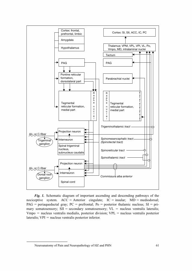
Neuroanatomy of Pain and Neuropathology of HZ and PHN 61
Cortex: frontal,prefrontal, limbic
Amygdala
Hypothalamus
Cortex: SI, SII, ACC, IC, PC
Thalamus: VPM, VPL, VPI, VL, Po,Vmpo, MD, intralaminar nuclei
Tectum
PAGPAG
Pontine reticularformation,dorsolateral part
Parabrachial nuclei
Tegmentalreticular formation,medial part
Tegmentalreticular formation,medial part
Projection neuron
Trigeminothalamic tract
Trigeminalganglion
Interneuron
A�- or C-fiber
Interneuron
Spinal trigeminalnucleus,subnucleus caudalis
Spinomesencephalic tract(Spinotectal tract)
Spinoreticular tract
Projection neuron
Spinothalamic tract
Spinal cord
Commissura alba anterior
Raphe nuclei
Raphe nuclei
Dorsal rootganglion
A�- or C-fiber
Fig. 1. Schematic diagram of important ascending and descending pathways of the
nociceptive system. ACC � Anterior cingulate; IC � insular; MD � mediodorsal;
PAG � periaqueductal gray; PC � prefrontal; Po � posterior thalamic nucleus; SI � pri-
mary somatosensory; SII � secondary somatosensory; VL � nucleus ventralis lateralis;
Vmpo � nucleus ventralis medialis, posterior division; VPL � nucleus ventralis posterior
lateralis; VPI � nucleus ventralis posterior inferior.
Wree/Schmitt/Usunoff 62
spino–parabrachio–amygdalar/hypothalamic nocispecific multineuronal chain is
probably concerned with the intensity of pain. The spino–reticulo–thalamic path-
ways may play an important role in distributing pain signals to the forebrain. In
addition, there are direct spino(trigemino)hypothalamic and spino(trigemino)
limbic tracts. The spinohypothalamic tract is an unexpectedly massive projection,
terminating in most of the hypothalamic divisions. Thus, the spinohypothalamic
tract provides nociceptive input to many areas that are involved in the multifaceted
responses to noxious stimuli.
Cerebral Cortices Involved in Pain PerceptionThere is a multiregional organization of cortical pain processing [16] and
areas involved are the primary (SI) and the secondary (SII) somatosensory, the
insular (IC), the anterior cingulate (ACC) and the prefrontal (PC) cortices.
These areas differ functionally as seen in electrophysiological and functional
imaging studies: the sensory-discriminative aspect of pain (localization, inten-
sity, duration, quality) is presented in SI (and SII), receiving input from lateral
thalamic nuclei, the motivational-affective aspect (subjective suffering,
unpleasantness, aversive emotions) and the cognitive-evaluative aspects of
pain are presented in IC, ACC and PC, receiving input from medial thalamic
nuclei.
SI is Located in the Postcentral Gyrus. Two classes of neurons are activated
in SI: neurons with a wide dynamic range react already to not painful stimuli,
showing highest activity to painful stimuli, having large receptive fields and
coding pain intensity; specific nociceptive neurons react to painful stimuli only,
having small receptive fields, being somatotopically located and enabling
localization, intensity and temporal attributes of painful stimuli. The SI neurons
get their afferences from the lateral thalamic nuclei (VPL, VPM, VPI) and also
heavily project back to them. Lesions of the respective thalamic nuclei, the
thalamo-cortical connections or of SI result in a decrease in temperature and
pain perception. But there is no complete analgesia, pain is still interpreted as
uncomfortable [17].
SII is Located just Lateral and Slightly Anterior to the Lateral end of theCentral Fissure. SII neurons seemingly are not involved in discrimination of
location and/or intensity of painful stimuli, but have an important role in
recognition, learning and memory of painful events [18]. There is significant
pain-related activation of SII [19]. The SII neurons get their bilateral afferences
from the lateral thalamic nuclei partly different from those projecting to SI,
namely from VPI and the dorsal part of Po. Thus, SII may play a role in relaying
Neuroanatomy of Pain and Neuropathology of HZ and PHN 63
nociceptive information to IC and temporal lobe limbic structures providing
fast access to pain related learning and memory.
Functional Imaging Studies Show an Increased Blood Flow of the ICduring Painful Stimuli, Either Contralaterally or Bilaterally [19]. Patients with
lesions of the IC had an elevated pain tolerance and loss of or inadequate
emotional reactions to painful stimuli although recognizing pain (asymbolia for
pain) [20]. The IC gets thalamic afferents from the VMpo, mediodorsal (MD)
and intralaminar nuclei and from SII, and projects to the amygdala, speaking in
favor of the importance of IC in the motivational-affective aspect of pain and in
autonomic reactions to noxious stimuli.
The ACC is Involved in Cognition and Emotion. The subarea involved in
the motivational-affective aspect of pain is located in the rostral part of
Brodmann’s area 24 and the adjoining area 32. Patients with ACC lesions lost
the emotional reactions to painful stimuli although pain could be correctly
localized. In ACC pain receptive neurons were found with large, often bilateral
receptive fields. The ACC gets thalamic afferents from VMpo, MD and
intralaminar nuclei, from IC and PC, and projects to the amygdala, MD, PAG,
motor nuclei of the brainstem and IC. The ACC is involved in motivational-
affective aspects of pain and in conditioned fear reaction, having a pivotal role
in interrelating attentional functions with that of establishing emotional valence
and response properties [21].
There are Still Doubts with Respect to the Function of the PC in PainPerception. The PC functions as ‘supervisory attention system’ are correlated
with the cognitive-evaluative aspect of pain. The PC gets thalamic afferents from
VMpo, MD and intralaminar nuclei, and projects to MD and ACC. Functional
imaging studies show activation of parts PC (probably Brodmann’s areas 9 and 10)
during painful stimuli, mostly in the right hemisphere. Patients with unilateral
lesions of the PC show changes in both the sensory-discriminative and the
motivational-affective aspects of pain.
Descending Modulatory PathwaysThe communication of Reynolds [22] being able to perform abdominal
surgery in rats by stimulating the midbrain PAG, initiated investigations on the
‘descending analgesia systems’. The PAG neurons project to the serotoninergic
raphe nuclei and to the noradrenergic nuclei in the dorsolateral pons.
Both neuronal groups project heavily to the spinal cord and to the STN. The
projections are predominantly crossed, innervating laminae I, II and V. The
Wree/Schmitt/Usunoff 64
neurochemistry of the transmitters and receptors in the multineuronal
antinociceptive pathway arising in the PAG is complex. Along serotonin and
noradrenaline, also endogenous opiates and glutamate, �-amino butyric acid
and glycine are involved.
Herpes Zoster and Postherpetic Neuralgia
NeuropathologyCNS complications occur in less than 1% of chickenpox cases [23]. The
children have mild meningitic symptoms. The most common abnormality is the
cerebellar ataxia; very rarely transverse myelitis. After varicella resolves, VZV
becomes latent in sensory ganglia, persisting throughout lifetime [23]. VZV is
localized predominantly in the pseudounipolar PA neurons and/or in the per-
ineuronal satellite cells. During latency, VZV is not infectious and does not
transcribe most of its genetic material, thereby escaping from detection and
clearance by the host immune system. The likelihood of viral reactivation
to herpes zoster (HZ) increases with each advancing decade of age.
Immunocomprised patients are at an especially high risk [23]. With reactiva-
tion, the virus spreads transaxonally to the skin, causing a rash with a der-
matomal distribution, and severe radicular pain. Thoracic HZ is the most
common one, affecting one to two, rarely more dermatomes, followed by the
ophthalmic division of the trigeminal nerve [23]. The involvement of the facial
nerve results in HZ oticus: geniculate neuralgia described by Ramsay Hunt
[24]. Similar combination of painful dermatomal rash with myotomal motor
weakness might occur also in spinal nerve HZ. Frequently, a prodrome of der-
matomal pain starts before the appearance of the rash. Dermatomal pain with-
out a rash (‘zoster sine herpete’) occurs rarely. HZ is mostly monophasic in
immunocompetent patients. In contrast, in immunocompromized patients
(especially in AIDS) HZ is recurrent, protracted, and often accompanied with
severe neurological complications [25].
The basic neuropathological substrate for HZ is ganglionic hemorrhage,
necrosis and inflammation [23]. The histopathologic features include mononu-
clear and lymphocytic infiltration, neuronal degeneration, neuronal phagocyto-
sis by satellite cells, empty neuronal cell beds and fibrous scarring of the
ganglia. Vasculitis in the adjacent nerve results in damage of axons and destruc-
tion of myelin sheaths [26]. Rarely, VZV spreads in the CNS [23] causing
myelitis. In HZ ophthalmicus, the virus might spread via trigeminal afferent
fibers to the large blood vessels at the base of the brain, with resultant vessel
thrombosis, vessel wall inflammation, and large, ipsilateral brain infarctions.
Mostly, HZ resolves without sequelae. However, many elderly patients have
Neuroanatomy of Pain and Neuropathology of HZ and PHN 65
prolonged, debilitating pain, known as PHN [25]. Besides PHN, also other
peripheral nerve injuries (cf. traumatic injury, diabetes, malignancy) might
result in neuropathic pain (NP). NP conditions are characterized by: sponta-
neous, continuous pain, usually of a burning character, paroxysmal (lancinat-
ing); evoked pain to mechanical or thermal stimuli, such as allodynia and
hyperalgesia. Allodynia can be produced in two ways: by the action of low
threshold myelinated A�-fibers on an altered CNS, and by a reduction in the
threshold of nociceptive fibers in the periphery. The fact that pain is often
located in hypoesthetic or anesthetic areas appears paradoxical and implies that
NP not only depends on the messages from nociceptors, but also on other
mechanisms as well, in contrast to ‘nociceptive pain’ [27].
The pathology of PHN is just beginning to be understood [23]. Smith [28]
found cystic distortion of thoracic SG removed 2.5 months after the onset of
HZ, and persistent chronic inflammatory cells. In a patient with removed SG 2
years after the onset of PHN he found ‘ghost cells’, hypothesizing that the
altered structure of surviving cells might contribute to the intractable pain.
Furthermore, DH atrophy and cell, axon and myelin loss were encountered
in PHN patients. Interestingly, a greater loss of small cutaneous nerve endings
in skin biopsies obtained from patients with HZ who developed PHN was
demonstrated [29].
Axons of uninjured PA neurons terminating in the DH can collaterally
sprout following chronic NP [30]. Peripheral nerve injury results in a
rearrangement of the highly ordered laminar termination of PA fibers within
appropriate regions of the DH. Peripheral axotomy causes long-lasting sprout-
ing of A-fibers into lamina II, an area they do not normally terminate. This A-
fiber sprouting appears to be a result of two phenomena: firstly, the presence
of vacant synaptic sites within the superficial laminae following the degenera-
tion of C-axons, secondly the induction of a regenerative capacity in the
injured neurons. The result of A-fiber sprouting is that lamina II begins to
receive information about non-noxious stimuli, an information being misinter-
preted by the CNS as noxious (mechanical allodynia). Thus, peripheral and
central mechanisms contribute to PHN pain. Some PHN patients show abnor-
mal sensitization of unmyelinated nociceptors (irritable nociceptors), having
minimal sensory loss. Others have pain associated with small fiber deafferen-
tiation: pain and temperature sensations are profoundly impaired but mechani-
cal stimuli can produce severe pain (allodynia). In those, allodynia may be due
to the formation of new connections between non-nociceptive thick (A�) PA
fibers and central pain transmission neurons. The third class complains from
severe spontaneous pain without hyperalgesia or allodynia, having lost both
large and small diameter fibers, and pain is due to increased spontaneous
activity in deafferented central neurons and/or reorganization of central
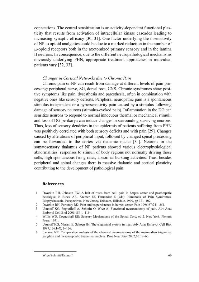
Wree/Schmitt/Usunoff 66
connections. The central sensitization is an activity-dependent functional plas-
ticity that results from activation of intracellular kinase cascades leading to
increasing synaptic efficacy [30, 31]. One factor underlying the insensitivity
of NP to opioid analgetics could be due to a marked reduction in the number of
�-opioid receptors both in the axotomized primary sensory and in the lamina
II neurons. In consequence, due to the different neuropathological mechanisms
obviously underlying PHN, appropriate treatment approaches in individual
patients vary [32, 33].
Changes in Cortical Networks due to Chronic PainChronic pain or NP can result from damage at different levels of pain pro-
cessing: peripheral nerve, SG, dorsal root, CNS. Chronic syndromes show posi-
tive symptoms like pain, dysesthesia and paresthesia, often in combination with
negative ones like sensory deficits. Peripheral neuropathic pain is a spontaneous
stimulus-independent or a hypersensitivity pain caused by a stimulus following
damage of sensory neurons (stimulus-evoked pain). Inflammation in the DG can
sensitize neurons to respond to normal innocuous thermal or mechanical stimuli,
and loss of DG perikarya can induce changes in surrounding surviving neurons.
Thus, loss of sensory dendrites in the epidermis of patients suffering from PHN
was positively correlated with both sensory deficits and with pain [29]. Changes
caused by alterations of peripheral input, followed by changed spinal processing
can be forwarded to the cortex via thalamic nuclei [34]. Neurons in the
somatosensory thalamus of NP patients showed various electrophysiological
abnormalities: responses to stimuli of body regions not normally driving those
cells, high spontaneous firing rates, abnormal bursting activities. Thus, besides
peripheral and spinal changes there is massive thalamic and cortical plasticity
contributing to the development of pathological pain.
References
1 Dworkin RH, Johnson RW: A belt of roses from hell: pain in herpes zoster and postherpetic
neuralgia; in Block AR, Kremer EF, Fernandez E (eds): Handbook of Pain Syndromes:
Biopsychosocial Perspectives. New Jersey, Erlbaum, Hillsdale, 1999, pp 371–402.
2 Dworkin RH, Portenoy RK: Pain and its persistence in herpes zoster. Pain 1996;67:241–251.
3 Usunoff KG, Popratiloff A, Schmitt O, Wree A: Functional neuroanatomy of pain. Adv Anat
Embryol Cell Biol 2006;184:1–119.
4 Willis WD, Coggeshall RE: Sensory Mechanisms of the Spinal Cord, ed 2. New York, Plenum
Press, 1991.
5 Usunoff KG, Marani E, Schoen JH: The trigeminal system in man. Adv Anat Embryol Cell Biol
1997;136:I–X, 1–126.
6 Lazarov NE: Comparative analysis of the chemical neuroanatomy of the mammalian trigeminal
ganglion and mesencephalic trigeminal nucleus. Prog Neurobiol 2002;66:19–60.

Neuroanatomy of Pain and Neuropathology of HZ and PHN 67
7 McHugh JM, McHugh WB: Pain: neuroanatomy, chemical mediators, and clinical implications.
AACN Clin Issues 2000;11:168–178.
8 Heppelmann B, Messlinger K, Neiss WF, Schmidt RF: Fine sensory innervation of the knee joint
capsule by group III and IV nerve fibers in the cat. J Comp Neurol 1995;351:415–428.
9 Millan MJ: The induction of pain: an integrative review. Prog Neurobiol 1999;57:1–164.
10 Schaible HG, Schmidt RF: Time course of mechanosensitivity changes in articular afferents dur-
ing a developing experimental arthritis. J Neurophysiol 1988;60:2180–2195.
11 Rexed B: Some aspects of the cytoarchitectonics and synaptology of the spinal cord; in Eccles JC,
Schade JP (eds): Organization of the Spinal Cord. Prog Brain Res, vol 2. Amsterdam, Elsevier,
1964, pp 58–92.
12 Craig AD: Pain, temperature, and the sense of the body; in Franzen O, Johansson R, Terenius L
(eds): Somesthesis and the Neurobiology of the Somatosensory Cortex. Basel, Birkhäuser, 1996,
pp 27–39.
13 Nathan PW, Smith M, Deacon P: The crossing of the spinothalamic tract. Brain 2001;124:
793–803.
14 Andrew D, Craig AD: Spinothalamic lamina I neurons selectively sensitive to histamine: a central
neural pathway for itch. Nat Neurosci 2001;4:72–77.
15 Willis WD, Westlund KN: Neuroanatomy of the pain system and of the pathways that modulate
pain. J Clin Neurophysiol 1997;14:2–31.
16 Peyron R, Laurent B, Garcia-Larrea L: Functional imaging of brain responses to pain: a review
and meta-analysis. Neurophysiol Clin 2000;30:263–288.
17 Ploner M, Freund HJ, Schnitzler A: Pain affect without pain sensation in a patient with postcentral
lesion. Pain 1999;81:211–214.
18 Schnitzler A, Ploner M: Neurophysiology and functional neuroanatomy of pain perception. J Clin
Neurophysiol 2000;17:592–603.
19 Treede RD, Apkarian AV, Bromm B, Greenspan JD, Lenz FA: Cortical representation of
pain: functional characterization of nociceptive areas near the lateral sulcus. Pain 2000;87:
113–119.
20 Greenspan JD, Lee RR, Lenz FA: Pain sensitivity alterations as a function of lesion location in the
parasylvian cortex. Pain 1999;81:273–282.
21 Price DD: Psychological and neural mechanisms of the affective dimension of pain. Science
2000;288:1769–1772.
22 Reynolds DV: Surgery in the rat during electrical analgesia induced by focal brain stimulation.
Science 1969;164:444–445.
23 Kleinschmidt-DeMasters BK, Gilden DH: Varicella-zoster virus infections of the nervous system.
Clinical and pathologic correlates. Arch Pathol Lab Med 2001;125:770–780.
24 Hunt JR: On herpetic inflammations of the geniculate ganglion: a new syndrome and its compli-
cations. J Nerv Ment Dis 1907;34:73–96.
25 Gilden DH, Kleinschmidt-DeMasters BK, LaGuardia JJ, Mahalingham R, Cohrs RJ: Neurologic
complications of the reactivation of varicella-zoster virus. N Engl J Med 2000;342:635–645.
26 Fabian WA, Wood B, Crowley P, Kakulas BA: Herpes zoster brachial plexus neuritis. Clin
Neuropathol 1997;16:61–64.
27 Attal N, Bouhassira D: Mechanisms of pain in peripheral neuropathy. Acta Neurol Scand
1999;173(suppl):12–24.
28 Smith FP: Pathological studies of spinal nerve ganglia in relation to intractable intercostal pain.
Surg Neurol 1978;10:50–53.
29 Oaklander AL, Romans K, Horasek S, Stocks H, Hauer P, Meyer RA: Unilateral postherpetic neu-
ralgia is associated with bilateral neuron damage. Ann Neurol 1998;44:789–795.
30 Ji RR, Woolf CJ: Neuronal plasticity and signal transduction in nociceptive neurons: implications
for the initiation and maintenance of pathological pain. Neurobiol Dis 2001;8:1–10.
31 Hunt SP, Mantyh PW: The molecular dynamics of pain control. Nat Rev Neurosci 2001;2:83–91.
32 Baron R: Post-herpetic neuralgia case study: optimizing pain control. Eur J Neurol 2004;11(suppl 1):
3–11.

Wree/Schmitt/Usunoff 68
33 Wasner G, Kleinert A, Binder A, Schattschneider J, Baron R: Postherpetic neuralgia: topical lido-
caine is effective in nociceptor-deprived skin. J Neurol 2005;252:677–686.
34 Hsieh JC, Belfrage M, Stone-Elander S, Hansson P, Ingvar M: Central representation of chronic
ongoing neuropathic pain studied by positron emission topography. Pain 1995;63:225–236.
Andreas Wree, MD
Institute of Anatomy, University of Rostock
PO Box 100888
DE–18055 Rostock (Germany)
Tel. �49 381 494 8401, Fax �49 381 494 8402, E-Mail [email protected]
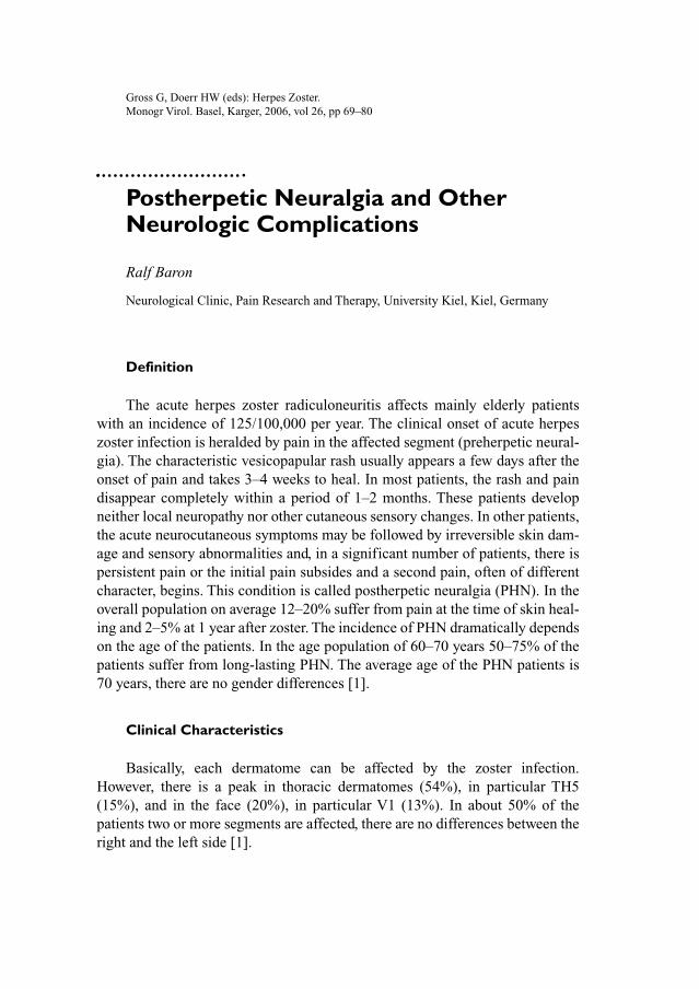
Gross G, Doerr HW (eds): Herpes Zoster.
Monogr Virol. Basel, Karger, 2006, vol 26, pp 69–80
Postherpetic Neuralgia and OtherNeurologic Complications
Ralf Baron
Neurological Clinic, Pain Research and Therapy, University Kiel, Kiel, Germany
Definition
The acute herpes zoster radiculoneuritis affects mainly elderly patients
with an incidence of 125/100,000 per year. The clinical onset of acute herpes
zoster infection is heralded by pain in the affected segment (preherpetic neural-
gia). The characteristic vesicopapular rash usually appears a few days after the
onset of pain and takes 3–4 weeks to heal. In most patients, the rash and pain
disappear completely within a period of 1–2 months. These patients develop
neither local neuropathy nor other cutaneous sensory changes. In other patients,
the acute neurocutaneous symptoms may be followed by irreversible skin dam-
age and sensory abnormalities and, in a significant number of patients, there is
persistent pain or the initial pain subsides and a second pain, often of different
character, begins. This condition is called postherpetic neuralgia (PHN). In the
overall population on average 12–20% suffer from pain at the time of skin heal-
ing and 2–5% at 1 year after zoster. The incidence of PHN dramatically depends
on the age of the patients. In the age population of 60–70 years 50–75% of the
patients suffer from long-lasting PHN. The average age of the PHN patients is
70 years, there are no gender differences [1].
Clinical Characteristics
Basically, each dermatome can be affected by the zoster infection.
However, there is a peak in thoracic dermatomes (54%), in particular TH5
(15%), and in the face (20%), in particular V1 (13%). In about 50% of the
patients two or more segments are affected, there are no differences between the
right and the left side [1].
Baron 70
Dermatological and Neurological Complications of Acute Herpes ZosterAcute zoster in the first trigeminal distribution (ophthalmic zoster) very
often leads to severe ocular complications (fig. 1). The cornea is very densely
innervated by trigeminal afferent fibers. Therefore, in 25–70% the cutaneous
zoster is associated with keratitis, iritis or chorioiritis which may lead to blindness.
If the facial nerve and the geniculate ganglion is affected by the acute
zoster infection (otic zoster), the rash will appear in the outer ear tube and a
peripheral facial palsy occurs that often has a poor long-term outcome.
If the acute zoster infection affects the ventral root or the ventral horn of
the spinal cord, in addition to sensory symptoms a severe muscle weakness may
develop. Does the zoster infection involve the brachial or lumbosacral plexus,
in 1–5% of the cases proximal pareses of the skeletal muscles will occur (fig. 2).
If the virus is transported centrally to the nervous system polyradiculitis,
myelitis and encephalitis may occur [2].
In immuno-compromised patients generalization of the rash to the entire
body (zoster generalisatus) is a common complications of acute zoster.
Pain Chronification – Postherpetic NeuralgiaAlthough histopathological studies have demonstrated peripheral and
central degenerative changes in many PHN patients, i.e. ganglion cell loss,
fibrosis and atrophy of the dorsal horn, dorsal root ganglion, dorsal root and
peripheral nerve [3–7], the most incapacitating symptom of PHN is the nearly
intractable pain and not just simple sensory loss. Most patients with PHN are
able to distinguish three distinct components of their discomfort: (1) a con-
stant, spontaneous usually deep burning pain, (2) a brief recurrent shooting
tic-like pain and (3) a sharp radiating pain of burning character evoked by very
light touching of the skin, which is called dynamic mechanical allodynia
(evoked-pain). The patient may undergo extraordinary efforts to protect the
diseased area from innocuous mechanical stimuli. Yet, firm compression of the
skin mostly does not exacerbate the pain and may even provide relief. Clinical
investigations show that negative (i.e. sensory deficits) and positive (i.e.
mechanical allodynia) sensory signs may coexist within the affected der-
matome. Extreme allodynia to light touch is often restricted to reproducable
areas surrounding scarred skin or lying at the border of affected and unaffected
dermatomes. In some patients the allodynic area expands far into formerly
unaffected adjacent dermatomes already indicating that some central nervous
system mechanisms must be involved in the generation of allodynia. Besides
mechanically evoked pain types up to 30% of patients suffer from heat evoked
pain (heat hyperalgesia), whereas pain induced by cold stimuli is a very rare
phenomenon (�10%).
PHN and Other Complications 71
Pathophysiological Mechanisms of Pain Generation
Considerable advances in the understanding of chronic pain mechanisms
have been achieved in the last years mainly using animal models with a variety
of different nerve lesions and the more recent models of VZV infection [8, 9].
In the latter, the striking PHN symptoms of hyperalgesia and allodynia can be
Fig. 1. Severe acute herpes zoster
within the right first trigeminal distribution.
The patient suffered from severe pain and
visual loss. Involvement of the cornea which
is densely innervated by trigeminal afferent
fibers.
Fig. 2. Magnetic resonance image of a patient with acute herpes zoster. Spinal cord in
the cervical affected region. Note the contrast enhancement in the dorsal root, dorsal horn of
the spinal cord and lesser also in the ventral root. The patient was severely affected with pain
and motor weakness at the left arm (T1 image with contrast medium). Photo courtesy of
Prof. Dr. T. Tölle, Munich.
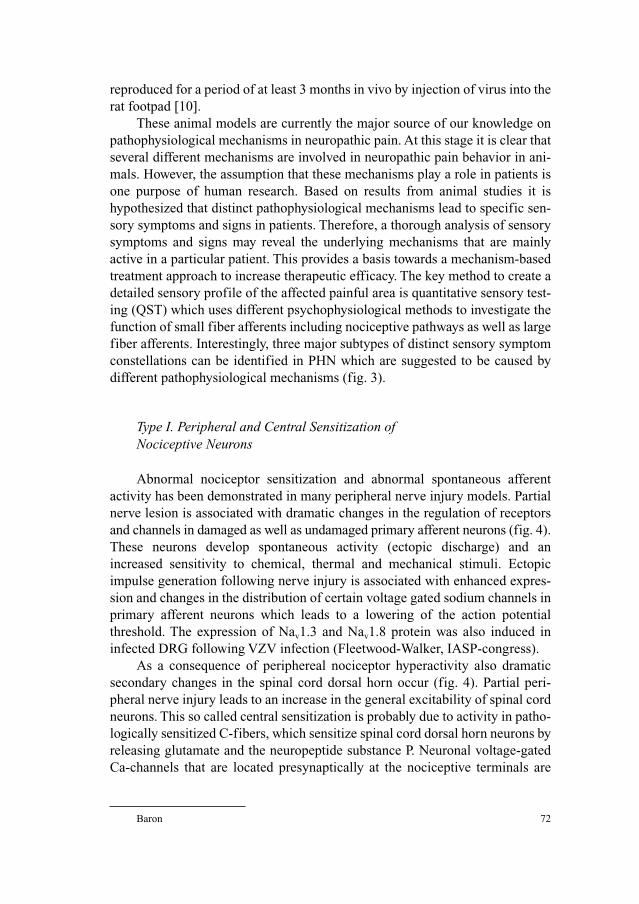
Baron 72
reproduced for a period of at least 3 months in vivo by injection of virus into the
rat footpad [10].
These animal models are currently the major source of our knowledge on
pathophysiological mechanisms in neuropathic pain. At this stage it is clear that
several different mechanisms are involved in neuropathic pain behavior in ani-
mals. However, the assumption that these mechanisms play a role in patients is
one purpose of human research. Based on results from animal studies it is
hypothesized that distinct pathophysiological mechanisms lead to specific sen-
sory symptoms and signs in patients. Therefore, a thorough analysis of sensory
symptoms and signs may reveal the underlying mechanisms that are mainly
active in a particular patient. This provides a basis towards a mechanism-based
treatment approach to increase therapeutic efficacy. The key method to create a
detailed sensory profile of the affected painful area is quantitative sensory test-
ing (QST) which uses different psychophysiological methods to investigate the
function of small fiber afferents including nociceptive pathways as well as large
fiber afferents. Interestingly, three major subtypes of distinct sensory symptom
constellations can be identified in PHN which are suggested to be caused by
different pathophysiological mechanisms (fig. 3).
Type I. Peripheral and Central Sensitization of Nociceptive Neurons
Abnormal nociceptor sensitization and abnormal spontaneous afferent
activity has been demonstrated in many peripheral nerve injury models. Partial
nerve lesion is associated with dramatic changes in the regulation of receptors
and channels in damaged as well as undamaged primary afferent neurons (fig. 4).
These neurons develop spontaneous activity (ectopic discharge) and an
increased sensitivity to chemical, thermal and mechanical stimuli. Ectopic
impulse generation following nerve injury is associated with enhanced expres-
sion and changes in the distribution of certain voltage gated sodium channels in
primary afferent neurons which leads to a lowering of the action potential
threshold. The expression of Nav1.3 and Nav1.8 protein was also induced in
infected DRG following VZV infection (Fleetwood-Walker, IASP-congress).
As a consequence of periphereal nociceptor hyperactivity also dramatic
secondary changes in the spinal cord dorsal horn occur (fig. 4). Partial peri-
pheral nerve injury leads to an increase in the general excitability of spinal cord
neurons. This so called central sensitization is probably due to activity in patho-
logically sensitized C-fibers, which sensitize spinal cord dorsal horn neurons by
releasing glutamate and the neuropeptide substance P. Neuronal voltage-gated
Ca-channels that are located presynaptically at the nociceptive terminals are
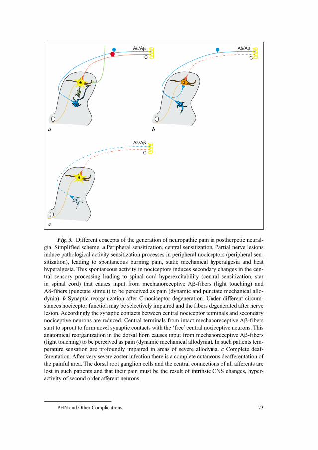
PHN and Other Complications 73
C
A�/A� A�/A�
C
A�/A�
C
a
c
b
Fig. 3. Different concepts of the generation of neuropathic pain in postherpetic neural-
gia. Simplified scheme. a Peripheral sensitization, central sensitization. Partial nerve lesions
induce pathological activity sensitization processes in peripheral nociceptors (peripheral sen-
sitization), leading to spontaneous burning pain, static mechanical hyperalgesia and heat
hyperalgesia. This spontaneous activity in nociceptors induces secondary changes in the cen-
tral sensory processing leading to spinal cord hyperexcitability (central sensitization, star
in spinal cord) that causes input from mechanoreceptive A�-fibers (light touching) and
A�-fibers (punctate stimuli) to be perceived as pain (dynamic and punctate mechanical allo-
dynia). b Synaptic reorganization after C-nociceptor degeneration. Under different circum-
stances nociceptor function may be selectively impaired and the fibers degenerated after nerve
lesion. Accordingly the synaptic contacts between central nociceptor terminals and secondary
nociceptive neurons are reduced. Central terminals from intact mechanoreceptive A�-fibers
start to sprout to form novel synaptic contacts with the ‘free’ central nociceptive neurons. This
anatomical reorganization in the dorsal horn causes input from mechanoreceptive A�-fibers
(light touching) to be perceived as pain (dynamic mechanical allodynia). In such patients tem-
perature sensation are profoundly impaired in areas of severe allodynia. c Complete deaf-
ferentation. After very severe zoster infection there is a complete cutaneous deafferentation of
the painful area. The dorsal root ganglion cells and the central connections of all afferents are
lost in such patients and that their pain must be the result of intrinsic CNS changes, hyper-
activity of second order afferent neurons.
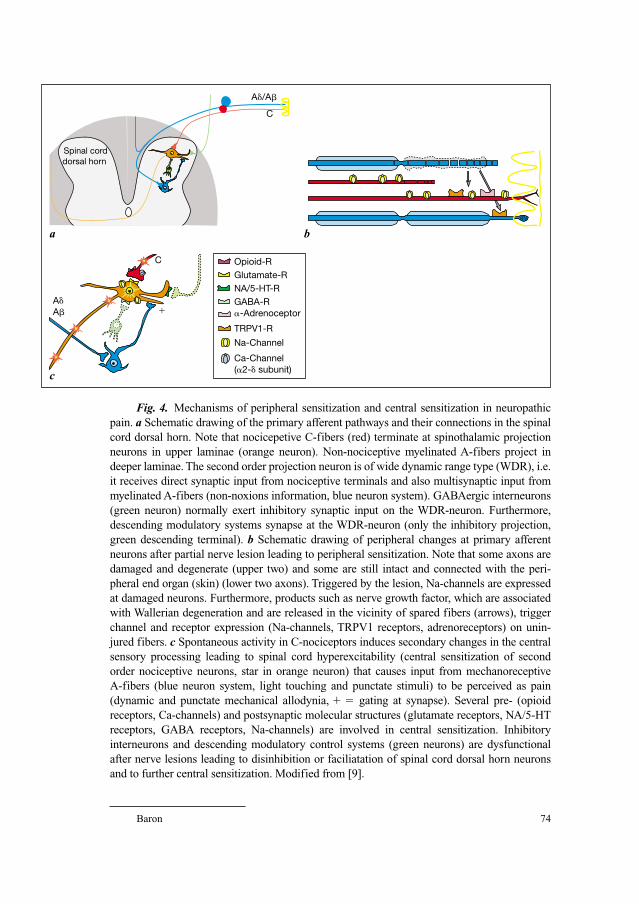
Baron 74
C
�
C
A�/A�
Spinal cord dorsal horn
A�A�
Opioid-RGlutamate-RNA/5-HT-R
Na-Channel
Ca-Channel (�2-� subunit)
�-AdrenoceptorGABA-R
TRPV1-R
a
c
b
Fig. 4. Mechanisms of peripheral sensitization and central sensitization in neuropathic
pain. a Schematic drawing of the primary afferent pathways and their connections in the spinal
cord dorsal horn. Note that nocicepetive C-fibers (red) terminate at spinothalamic projection
neurons in upper laminae (orange neuron). Non-nociceptive myelinated A-fibers project in
deeper laminae. The second order projection neuron is of wide dynamic range type (WDR), i.e.
it receives direct synaptic input from nociceptive terminals and also multisynaptic input from
myelinated A-fibers (non-noxions information, blue neuron system). GABAergic interneurons
(green neuron) normally exert inhibitory synaptic input on the WDR-neuron. Furthermore,
descending modulatory systems synapse at the WDR-neuron (only the inhibitory projection,
green descending terminal). b Schematic drawing of peripheral changes at primary afferent
neurons after partial nerve lesion leading to peripheral sensitization. Note that some axons are
damaged and degenerate (upper two) and some are still intact and connected with the peri-
pheral end organ (skin) (lower two axons). Triggered by the lesion, Na-channels are expressed
at damaged neurons. Furthermore, products such as nerve growth factor, which are associated
with Wallerian degeneration and are released in the vicinity of spared fibers (arrows), trigger
channel and receptor expression (Na-channels, TRPV1 receptors, adrenoreceptors) on unin-
jured fibers. c Spontaneous activity in C-nociceptors induces secondary changes in the central
sensory processing leading to spinal cord hyperexcitability (central sensitization of second
order nociceptive neurons, star in orange neuron) that causes input from mechanoreceptive
A-fibers (blue neuron system, light touching and punctate stimuli) to be perceived as pain
(dynamic and punctate mechanical allodynia, � � gating at synapse). Several pre- (opioid
receptors, Ca-channels) and postsynaptic molecular structures (glutamate receptors, NA/5-HT
receptors, GABA receptors, Na-channels) are involved in central sensitization. Inhibitory
interneurons and descending modulatory control systems (green neurons) are dysfunctional
after nerve lesions leading to disinhibition or faciliatation of spinal cord dorsal horn neurons
and to further central sensitization. Modified from [9].
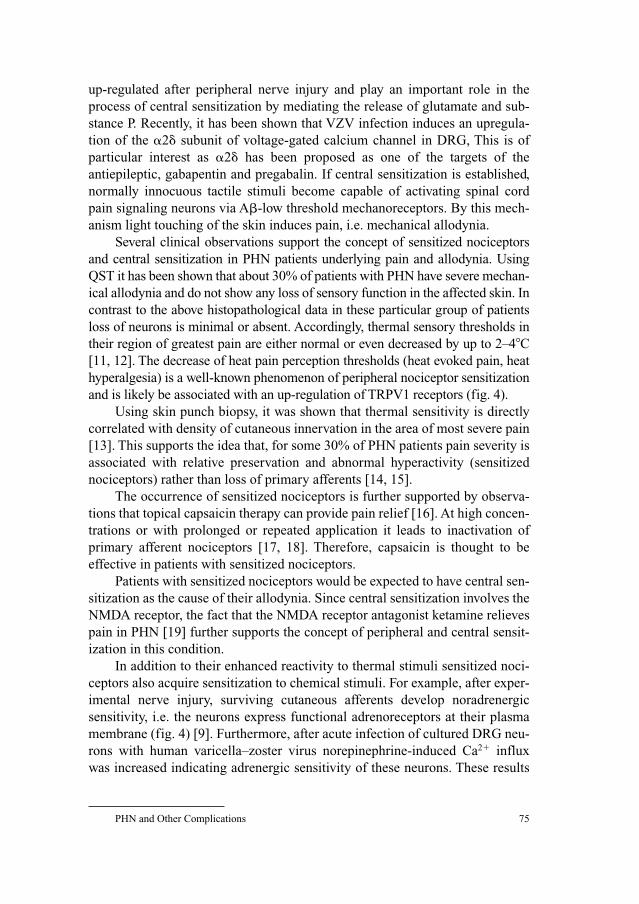
PHN and Other Complications 75
up-regulated after peripheral nerve injury and play an important role in the
process of central sensitization by mediating the release of glutamate and sub-
stance P. Recently, it has been shown that VZV infection induces an upregula-
tion of the �2� subunit of voltage-gated calcium channel in DRG, This is of
particular interest as �2� has been proposed as one of the targets of the
antiepileptic, gabapentin and pregabalin. If central sensitization is established,
normally innocuous tactile stimuli become capable of activating spinal cord
pain signaling neurons via A�-low threshold mechanoreceptors. By this mech-
anism light touching of the skin induces pain, i.e. mechanical allodynia.
Several clinical observations support the concept of sensitized nociceptors
and central sensitization in PHN patients underlying pain and allodynia. Using
QST it has been shown that about 30% of patients with PHN have severe mechan-
ical allodynia and do not show any loss of sensory function in the affected skin. In
contrast to the above histopathological data in these particular group of patients
loss of neurons is minimal or absent. Accordingly, thermal sensory thresholds in
their region of greatest pain are either normal or even decreased by up to 2–4�C[11, 12]. The decrease of heat pain perception thresholds (heat evoked pain, heat
hyperalgesia) is a well-known phenomenon of peripheral nociceptor sensitization
and is likely be associated with an up-regulation of TRPV1 receptors (fig. 4).
Using skin punch biopsy, it was shown that thermal sensitivity is directly
correlated with density of cutaneous innervation in the area of most severe pain
[13]. This supports the idea that, for some 30% of PHN patients pain severity is
associated with relative preservation and abnormal hyperactivity (sensitized
nociceptors) rather than loss of primary afferents [14, 15].
The occurrence of sensitized nociceptors is further supported by observa-
tions that topical capsaicin therapy can provide pain relief [16]. At high concen-
trations or with prolonged or repeated application it leads to inactivation of
primary afferent nociceptors [17, 18]. Therefore, capsaicin is thought to be
effective in patients with sensitized nociceptors.
Patients with sensitized nociceptors would be expected to have central sen-
sitization as the cause of their allodynia. Since central sensitization involves the
NMDA receptor, the fact that the NMDA receptor antagonist ketamine relieves
pain in PHN [19] further supports the concept of peripheral and central sensit-
ization in this condition.
In addition to their enhanced reactivity to thermal stimuli sensitized noci-
ceptors also acquire sensitization to chemical stimuli. For example, after exper-
imental nerve injury, surviving cutaneous afferents develop noradrenergic
sensitivity, i.e. the neurons express functional adrenoreceptors at their plasma
membrane (fig. 4) [9]. Furthermore, after acute infection of cultured DRG neu-
rons with human varicella–zoster virus norepinephrine-induced Ca2� influx
was increased indicating adrenergic sensitivity of these neurons. These results
Baron 76
may explain that not only mechanical lesion but also viral infection may lead to
phenotypical changes of afferent neurons [20].
Clinical studies support the idea that zoster infection may trigger cate-
cholamine sensitivity in nociceptors. In postherpetic neuralgia intracutaneous
injection of adrenaline and phenylephrine increase spontaneous pain and allo-
dynia on the affected side [21]. There are also reports that sympathetic blocks,
i.v. phentolamine and topical clonidine [22] transiently relieve pain in the occa-
sional PHN patient.
Type II. Predominant Degeneration of Nociceptive Neurons
In contrast to the patient population with sensitized nociceptors up to 60%
of PHN patients show considerable signs of neuronal degeneration and loss of
function within the affected tissues. Interestingly, many of these patients still
suffer from severe dynamic mechanical allodynia although the function of
nociceptors is diminished or absent in the same skin area.
Accordingly, Head and Campbell [3] reported extensive damage in dorsal root
ganglia supplying the affected dermatomes in cases with herpes zoster, however,
they made no attempt to correlate the pathology with the distribution of the pain.
Subsequent pathological studies have also demonstrated loss of nerve fibers or
cell bodies in the peripheral nerve, the dorsal root ganglion and also the spinal
cord dorsal horn [4–7]. This is not surprising since there is viral nucleic acid in
the satellite cells of dorsal root ganglia and viral reactivation produces significant
local inflammation with infection and damage of primary afferents (fig. 2).
Using immunohistochemical methods to visualize nerve terminals in the
skin it has been shown that cutaneous innervation is reduced in some PHN
patients. Punch skin biopsies and the anti-PGP 9.5 antibody, a pan-axonal
marker, in PHN patients and zoster patients without pain were used in order to
quantify sensory neurites in the affected skin and compared the numbers with
the homologous contralateral site [13, 23, 24]. Furthermore, a skin site distant
from the shingles involvement was also analyzed. In PHN a severe dendritic
loss could be demonstrated at the affected side (20% as compared with the con-
trols). Neurite loss was more prominent in the epidermis than in the dermis.
Furthermore, the PHN group also had lost half of the neurites in the contra-
lateral epidermis whereas distant areas where unaffected.
Functional studies support the concept of degeneration of cutaneous
C-nociceptors. By using these C-fiber axon reflex reactions it is possible to
objectively assess cutaneous C-fiber function in the human skin. In some patients
the histamine evoked axon reflex vasodilatation and flare size was impaired
or abolished in skin regions with intense dynamic allodynia [25, 26]. Using
PHN and Other Complications 77
quantitative thermal sensory testing to assess C- and A�-fibers function, some
patients with acute herpes zoster [27] and some chronic PHN patients have
extremely high thermal thresholds in areas with marked dynamic allodynia [21,
27, 28]. Thus, there is a subset of PHN patients with pain and loss of cutaneous
C-nociceptor function in a region that is coextensive with allodynic skin.
The question remains which mechanisms will induce dynamic mechanical
allodynia in the subgroup of patients characterized by loss of nociceptive neurons.
One interesting type of central anatomical reorganization might produce mech-
anical allodynia. Neurons in lamina II (substantia gelatinosa) of the spinal cord
dorsal horn normally receive direct input from small diameter (A�-, C-)fibers and
respond best to noxious stimulation. Peripheral C-fiber loss results in a substantial
degeneration of C-fiber terminals in lamina II. As a consequence of this loss of
synaptic contacts normally made by C-fiber afferents onto pain signaling neurons
in lamina II, the central terminals of A�-mechanoreceptive afferents which nor-
mally terminate in deeper laminae (III and IV) grow into lamina II and directly
contact the deafferented cells [29]. This sprouting of A�-terminals depends crit-
ically on the extent of C-fiber degeneration. Such changes might be the neural cor-
relate of the dynamic mechanical allodynia observed in some patients who have
loss of C-fiber function rather than C-fiber sensitization.
This charming idea was supported by histological experiments showing
specific A�-fiber labelling with cholera toxin B in superficial layers after nerve
injury. However, recent publications question the role of A�-sprouting in induc-
ing allodynia. It was shown that nerve injury induces neuroplastic changes in
C-fiber nociceptors so that besides A�-fibers also C-fibers are labeled with
cholera toxin B. In addition, large A�-fibers showed only very limited sprout-
ing into inner lamina II [30, 31].
Therefore, alternative explanations for pain and allodynia in the setting of
impaired cutaneous C-fiber function must be considered: (1) The nociceptive
C-fibers may degenerate exclusively in the peripheral branch leaving the dorsal
root ganglion soma and the central axon branch intact. The cell bodies of these
injured neurons could then generate ectopic impulse activity that would main-
tain the pathologic central sensitization in the presence of analgesic skin. (2) In
theory, since sensory testing have only studied the function of cutaneousC-fibers, ongoing activity that drives central sensitization might originate in
intact nociceptors of deep somatic tissues (e.g. muscle, ligaments, etc.).
Type III. Complete Skin Deafferentation
Clinically a third smaller group of PHN patients can be distinguished
which accounts for less than 10%. These patients have severe spontaneous pain,
Baron 78
profound sensory loss but no evoked sensations (hyperalgesia or allodynia). In
association with pain there is a complete cutaneous deafferentation of the
painful area (anesthesia dolorosa). It must be assumed that the dorsal root gan-
glion cells and the central connections of all afferents are lost in such patients
and that their pain must be the result of intrinsic CNS changes. In animal stud-
ies, following complete primary afferent loss of a spinal segment, many dorsal
horn cells begin to fire spontaneously at high frequencies [32].
Mechanism-Based Treatment Approach in Postherpetic Neuralgia
Based on the described different types of PHN patients the first prospec-
tive mechanism-based treatment approach was performed using topical lido-
caine [33]. All included 18 patients suffered from spontaneous burning pain and
mechanical allodynia but differ concerning their cutaneous nociceptor function
investigated by QST. Six patients had evidence for sensitized nociceptors in the
affected dermatome. The other twelve patients demonstrated severe partial
nerve injury associated with functional deafferentation including nociceptive
C-fiber afferents of the affected skin area. Topical lidocaine (patch, 5%) was
chosen for treatment, because it has been proven to be effective in PHN [34].
Additionally, it is generally assumed that its pain relieving effect is caused by
acting on sensitized cutaneous nociceptors that have expressed voltage-gated
sodium channels within the superficial layers of the skin [35]. Therefore, it was
hypothesized that patients with sensitized nociceptors respond well to lido-
caine. Surprisingly, topical lidocaine was more effective in patients with pre-
dominant degeneration of nociceptive neurons without inducing significant
relief in patients with sensitized nociceptors. Though, the heterogeneous distri-
bution of the patients in the two groups (6 vs. 12) weakens the statistical analy-
ses, the striking finding that patients with nociceptor-deprived skin responded
significantly to dermal lidocaine therapy cannot be ignored.
Three Distinct Sensory Profiles in PHN-Patients – Clinically Relevant?
As attractive, the PHN subtype-classification based on the nociceptor
function and evoked pain types might be it should be emphasized that not all
patients fit exactly into one category or the other. In a large group of PHN
patients many heterogenous patterns of sensory dysfunction were detected [12].
Accordingly, detailed sensory testing in one PHN patient clearly showed areas

PHN and Other Complications 79
of relative preservation in close vicinity to impaired thermal sensation, both
within the affected dermatome [15]. Furthermore, the sensory patterns showed
a variation over the time course of PHN.
On the other hand, several pain underlying mechanisms are probably
not identified so far. For example, an explanation for the lidocaine effect in
nociceptor-deprived skin might be that other intact afferents that have survived
the virus infection are targeted by lidocaine after expression of sodium channels
during the acute inflammatory disease process.
However, despite these limitations continuing investigations of mechanism-
based treatment approaches seem to be a promising way to establish an optimal
therapy with drugs that address the specific mechanisms in each patient in the
future.
References
1 Portenoy RK, Duma C, Foley KM: Acute herpetic and postherpetic neuralgia: clinical review and
current management. Ann Neurol 1986;20:651–664.
2 Haanpaa M, Hakkinen V, Nurmikko T: Motor involvement in acute herpes zoster. Muscle Nerve
1997;20:1433–1438.
3 Head H, Campbell AW: The pathology of herpes zoster and its bearing on sensory location. Brain
1900;23:353–523.
4 Noordenbos W: Pain. Amsterdam, Elsevier, 1959.
5 Zacks SI, Langfitt RW, Elliott FA: Herpetic neuritis: a light and electron microscopic study.
Neurology 1964;14:744–750.
6 Watson CP, Morshead C, Van der Kooy D, Deck J, Evans RJ: Post-herpetic neuralgia: post-mortem
analysis of a case. Pain 1988;34:129–138.
7 Watson CP, Deck JH, Morshead C, Van der Kooy D, Evans RJ: Post-herpetic neuralgia: further
post-mortem studies of cases with and without pain. Pain 1991;44:105–117.
8 Baron R: Peripheral neuropathic pain: from mechanisms to symptoms. Clin J Pain 2000;16
(suppl):S12–S20.
9 Baron R: Mechanisms of disease: neuropathic pain – a clinical perspective. Nat Clin Pract Neurol
2006;2:95–106.
10 Fleetwood-Walker SM, Quinn JP, Wallace C, Blackburn-Munro G, Kelly BG, Fiskerstrand CE,
Nash AA, Dalziel RG: Behavioural changes in the rat following infection with varicella-zoster
virus. J Gen Virol 1999;80(pt 9):2433–2436.
11 Rowbotham MC, Fields HL: The relationship of pain, allodynia and thermal sensation in post-
herpetic neuralgia. Brain 1996;119(pt 2):347–354.
12 Pappagallo M, Oaklander AL, Quatrano-Piacentini AL, Clark MR, Raja SN: Heterogenous pat-
terns of sensory dysfunction in postherpetic neuralgia suggest multiple pathophysiologic mech-
anisms. Anesthesiology 2000;92:691–698.
13 Rowbotham MC, Yosipovitch G, Connolly MK, Finlay D, Forde G, Fields HL: Cutaneous innerva-
tion density in the allodynic form of postherpetic neuralgia. Neurobiol Dis 1996;3:205–214.
14 Fields HL, Rowbotham M, Baron R: Postherpetic neuralgia: irritable nociceptors and deafferenta-
tion. Neurobiol Dis 1998;5:209–227.
15 Petersen KL, Fields HL, Brennum J, Sandroni P, Rowbotham MC: Capsaicin evoked pain and allo-
dynia in post-herpetic neuralgia. Pain 2000;88:125–133.
16 Bernstein JE, Korman NJ, Bickers DR, Dahl MV, Millikan LE: Topical capsaicin treatment of
chronic postherpetic neuralgia. J Am Acad Dermatol 1989;21(pt 1):265–270.

Baron 80
17 Lynn B: Capsaicin: actions on nociceptive C-fibres and therapeutic potential. Pain 1990;41:
61–69.
18 Rains C, Bryson HM: Topical capsaicin. A review of its pharmacological properties and therapeu-
tic potential in post-herpetic neuralgia, diabetic neuropathy and osteoarthritis. Drugs Aging
1995;7:317–328.
19 Eide PK, Jorum E, Stubhaug A, Bremnes J, Breivik H: Relief of post-herpetic neuralgia with the
N-methyl-D-aspartic acid receptor antagonist ketamine: a double-blind, cross-over comparison
with morphine and placebo. Pain 1994;58:347–354.
20 Kress M, Fickenscher H: Infection by human varicella-zoster virus confers norepinephrine sensi-
tivity to sensory neurons from rat dorsal root ganglia. FASEB J 2001;15:1037–1043.
21 Choi B, Rowbotham MC: Effect of adrenergic receptor activation on post-herpetic neuralgia pain
and sensory disturbances. Pain 1997;69:55–63.
22 Abadir AR, Kraynack BJ, Mayda J 2nd, Gintautas J: Postherpetic neuralgia: response to topical
clonidine. Proc West Pharmacol Soc 1996;39:47–48.
23 Oaklander AL: The density of remaining nerve endings in human skin with and without post-
herpetic neuralgia after shingles. Pain 2001;92:139–145.
24 Oaklander AL: Unilateral postherpetic neuralgia is associated with bilateral sensory neuron dam-
age. Neurology 1998;44:789–795.
25 Baron R, Saguer M: Mechanical allodynia in postherpetic neuralgia: evidence for central mech-
anisms depending on nociceptive C-fiber degeneration. Neurology 1995;45(suppl 8):S63–S65.
26 Baron R, Saguer M: Postherpetic neuralgia. Are C-nociceptors involved in signalling and mainte-
nance of tactile allodynia? Brain 1993;116(pt 6):1477–1496.
27 Nurmikko T, Bowsher D: Somatosensory findings in postherpetic neuralgia. J Neurol Neurosurg
Psychiatry 1990;53:135–141.
28 Nurmikko T, Wells C, Bowsher D. Sensory dysfunction in postherpetic neuralgia; in Boivie J,
Hansson P, Lindblom U (eds): Touch, Temperature, and Pain in Health and Disease: Mechanisms
and Assessments. Seattle, IASP Press, 1994, pp 133–141.
29 Woolf CJ, Shortland P, Coggeshall RE: Peripheral nerve injury triggers central sprouting of myeli-
nated afferents. Nature 1992;355:75–78.
30 Santha P, Jancso G: Transganglionic transport of choleragenoid by capsaicin-sensitive C-fibre
afferents to the substantia gelatinosa of the spinal dorsal horn after peripheral nerve section.
Neuroscience 2003;116:621–627.
31 Bao L, Wang HF, Cai HJ, Tong YG, Jin SX, Lu YJ, Grant G, Hokfelt T, Zhang X: Peripheral axo-
tomy induces only very limited sprouting of coarse myelinated afferents into inner lamina II of rat
spinal cord. Eur J Neurosci 2002;16:175–185.
32 Lombard MC, Larabi Y: Electrophysiological study of cervical dorsal horn cells in partially deaf-
ferented rats; in Bonica JJ (ed): Advances in Pain Research and Therapy. New York, Raven Press,
1983, pp 147–154.
33 Wasner G, Kleinert A, Binder A, Schattschneider J, Baron R: Postherpetic neuralgia: topical lido-
caine is effective in nociceptor-deprived skin. J Neurol 2005;252:677–686.
34 Meier T, Wasner G, Faust M, Kuntzer T, Ochsner F, Hueppe M, Bogousslavsky J, Baron R:
Efficacy of lidocaine patch 5% in the treatment of focal peripheral neuropathic pain syndromes: a
randomized, double-blind, placebo-controlled study. Pain 2003;106:151–158.
35 Argoff CE: New analgesics for neuropathic pain: the lidocaine patch. Clin J Pain 2000;16(suppl):
S62–S66.
Prof. Dr. Med. Ralf Baron
Klinik für Neurologie, UK-SH, Campus Kiel
Sektion Neurologische Schmerzforschung und Therapie
Schittenhelmstr. 10
DE–24105 Kiel (Germany)
Tel. �49 431 597 8505, Fax �49 431 597 8530, E-Mail [email protected]
Gross G, Doerr HW (eds): Herpes Zoster.
Monogr Virol. Basel, Karger, 2006, vol 26, pp 81–92
Varicella–Zoster Virus Infectionsduring Pregnancy
Andreas Sauerbrei, Peter Wutzler
Institute of Virology and Antiviral Therapy, Friedrich-Schiller University,
Jena, Germany
Frequency and Consequences of Varicella–Zoster Virus Infections during Pregnancy
In most industrial countries, chickenpox is a rare disease during pregnancy
as more than 90% of women of child-bearing age possess virus-specific IgG class
antibodies. Only 3–4% of women in Germany were found to be susceptible to pri-
mary varicella–zoster virus (VZV) infection [1]. In early reports, the average inci-
dence of varicella in pregnant women was calculated as 0.7 per 1,000 pregnancies
[2, 3], but the current rates appear to be 2–3 per 1,000 pregnancies [4].
Varicella during pregnancy may occasionally lead to serious maternal and
fetal diseases (table 1). Pregnant women who contract varicella are at risk of
severe pneumonia associated with life-threatening ventilatory compromise and
death. The disease seems to occur more often in the third trimester [5] and must
be regarded as a medical emergency. In the general population, varicella pneu-
monia has a mortality of 10–20% but in pregnancy it may be as high as 45%.
More recent studies, however, suggest that the mortality has decreased to
10–11% for both non-pregnant and pregnant patients most likely due to the
effects of antiviral therapy and better respiratory management [6].
At any stage during pregnancy, chickenpox may cause intrauterine infec-
tion. Maternal varicella resulting in viremia may transmit the virus to the fetus
by either transplacental spread, or by ascending infection from lesions in the
birth canal. Furthermore, direct contact or respiratory droplet can lead to infec-
tion after birth. The consequences for the infant depend on the time of maternal
disease. They range from asymptomatic infection to fetal loss especially in case
of severe maternal disease. Primary VZV infection during first two trimesters
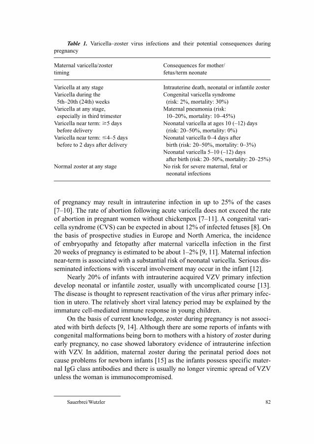
Sauerbrei/Wutzler 82
of pregnancy may result in intrauterine infection in up to 25% of the cases
[7–10]. The rate of abortion following acute varicella does not exceed the rate
of abortion in pregnant women without chickenpox [7–11]. A congenital vari-
cella syndrome (CVS) can be expected in about 12% of infected fetuses [8]. On
the basis of prospective studies in Europe and North America, the incidence
of embryopathy and fetopathy after maternal varicella infection in the first
20 weeks of pregnancy is estimated to be about 1–2% [9, 11]. Maternal infection
near-term is associated with a substantial risk of neonatal varicella. Serious dis-
seminated infections with visceral involvement may occur in the infant [12].
Nearly 20% of infants with intrauterine acquired VZV primary infection
develop neonatal or infantile zoster, usually with uncomplicated course [13].
The disease is thought to represent reactivation of the virus after primary infec-
tion in utero. The relatively short viral latency period may be explained by the
immature cell-mediated immune response in young children.
On the basis of current knowledge, zoster during pregnancy is not associ-
ated with birth defects [9, 14]. Although there are some reports of infants with
congenital malformations being born to mothers with a history of zoster during
early pregnancy, no case showed laboratory evidence of intrauterine infection
with VZV. In addition, maternal zoster during the perinatal period does not
cause problems for newborn infants [15] as the infants possess specific mater-
nal IgG class antibodies and there is usually no longer viremic spread of VZV
unless the woman is immunocompromised.
Table 1. Varicella–zoster virus infections and their potential consequences during
pregnancy
Maternal varicella/zoster Consequences for mother/
timing fetus/term neonate
Varicella at any stage Intrauterine death, neonatal or infantile zoster
Varicella during the Congenital varicella syndrome
5th–20th (24th) weeks (risk: 2%, mortality: 30%)
Varicella at any stage, Maternal pneumonia (risk:
especially in third trimester 10–20%, mortality: 10–45%)
Varicella near term: �5 days Neonatal varicella at ages 10 (–12) days
before delivery (risk: 20–50%, mortality: 0%)
Varicella near term: �4–5 days Neonatal varicella 0–4 days after
before to 2 days after delivery birth (risk: 20–50%, mortality: 0–3%)
Neonatal varicella 5–10 (–12) days
after birth (risk: 20–50%, mortality: 20–25%)
Normal zoster at any stage No risk for severe maternal, fetal or
neonatal infections
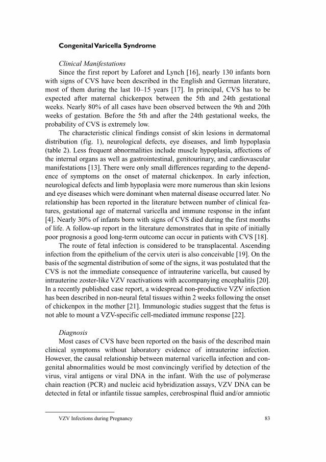
VZV Infections during Pregnancy 83
Congenital Varicella Syndrome
Clinical ManifestationsSince the first report by Laforet and Lynch [16], nearly 130 infants born
with signs of CVS have been described in the English and German literature,
most of them during the last 10–15 years [17]. In principal, CVS has to be
expected after maternal chickenpox between the 5th and 24th gestational
weeks. Nearly 80% of all cases have been observed between the 9th and 20th
weeks of gestation. Before the 5th and after the 24th gestational weeks, the
probability of CVS is extremely low.
The characteristic clinical findings consist of skin lesions in dermatomal
distribution (fig. 1), neurological defects, eye diseases, and limb hypoplasia
(table 2). Less frequent abnormalities include muscle hypoplasia, affections of
the internal organs as well as gastrointestinal, genitourinary, and cardiovascular
manifestations [13]. There were only small differences regarding to the depend-
ence of symptoms on the onset of maternal chickenpox. In early infection,
neurological defects and limb hypoplasia were more numerous than skin lesions
and eye diseases which were dominant when maternal disease occurred later. No
relationship has been reported in the literature between number of clinical fea-
tures, gestational age of maternal varicella and immune response in the infant
[4]. Nearly 30% of infants born with signs of CVS died during the first months
of life. A follow-up report in the literature demonstrates that in spite of initially
poor prognosis a good long-term outcome can occur in patients with CVS [18].
The route of fetal infection is considered to be transplacental. Ascending
infection from the epithelium of the cervix uteri is also conceivable [19]. On the
basis of the segmental distribution of some of the signs, it was postulated that the
CVS is not the immediate consequence of intrauterine varicella, but caused by
intrauterine zoster-like VZV reactivations with accompanying encephalitis [20].
In a recently published case report, a widespread non-productive VZV infection
has been described in non-neural fetal tissues within 2 weeks following the onset
of chickenpox in the mother [21]. Immunologic studies suggest that the fetus is
not able to mount a VZV-specific cell-mediated immune response [22].
DiagnosisMost cases of CVS have been reported on the basis of the described main
clinical symptoms without laboratory evidence of intrauterine infection.
However, the causal relationship between maternal varicella infection and con-
genital abnormalities would be most convincingly verified by detection of the
virus, viral antigens or viral DNA in the infant. With the use of polymerase
chain reaction (PCR) and nucleic acid hybridization assays, VZV DNA can be
detected in fetal or infantile tissue samples, cerebrospinal fluid and/or amniotic
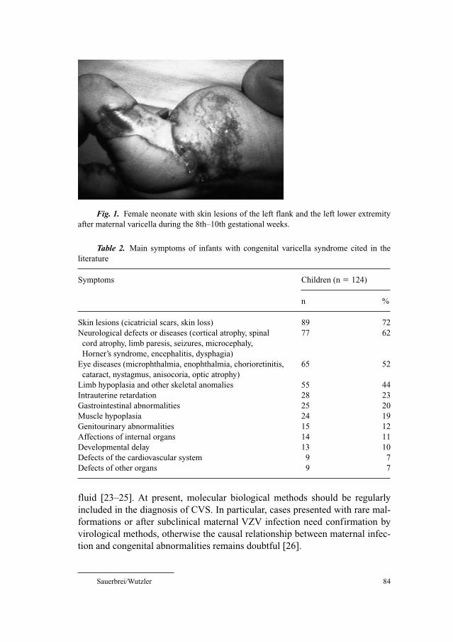
Sauerbrei/Wutzler 84
fluid [23–25]. At present, molecular biological methods should be regularly
included in the diagnosis of CVS. In particular, cases presented with rare mal-
formations or after subclinical maternal VZV infection need confirmation by
virological methods, otherwise the causal relationship between maternal infec-
tion and congenital abnormalities remains doubtful [26].
Fig. 1. Female neonate with skin lesions of the left flank and the left lower extremity
after maternal varicella during the 8th–10th gestational weeks.
Table 2. Main symptoms of infants with congenital varicella syndrome cited in the
literature
Symptoms Children (n � 124)
n %
Skin lesions (cicatricial scars, skin loss) 89 72
Neurological defects or diseases (cortical atrophy, spinal 77 62
cord atrophy, limb paresis, seizures, microcephaly,
Horner’s syndrome, encephalitis, dysphagia)
Eye diseases (microphthalmia, enophthalmia, chorioretinitis, 65 52
cataract, nystagmus, anisocoria, optic atrophy)
Limb hypoplasia and other skeletal anomalies 55 44
Intrauterine retardation 28 23
Gastrointestinal abnormalities 25 20
Muscle hypoplasia 24 19
Genitourinary abnormalities 15 12
Affections of internal organs 14 11
Developmental delay 13 10
Defects of the cardiovascular system 9 7
Defects of other organs 9 7
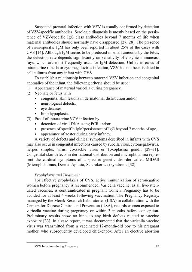
VZV Infections during Pregnancy 85
Suspected prenatal infection with VZV is usually confirmed by detection
of VZV-specific antibodies. Serologic diagnosis is mostly based on the persis-
tence of VZV-specific IgG class antibodies beyond 7 months of life when
maternal antibodies should normally have disappeared [27, 28]. The presence
of virus-specific IgM has only been reported in about 25% of the cases with
CVS [14]. Although IgM seems to be produced in small amounts by the fetus,
the detection rate depends significantly on sensitivity of enzyme immunoas-
says, which are most frequently used for IgM detection. Unlike in cases of
intrauterine rubella or cytomegalovirus infection, VZV has not been isolated in
cell cultures from any infant with CVS.
To establish a relationship between maternal VZV infection and congenital
anomalies of the infant, the following criteria should be used:
(1) Appearance of maternal varicella during pregnancy,
(2) Neonate or fetus with
• congenital skin lesions in dermatomal distribution and/or
• neurological defects,
• eye diseases,
• limb hypoplasia.
(3) Proof of intrauterine VZV infection by
• detection of viral DNA using PCR and/or
• presence of specific IgM/persistence of IgG beyond 7 months of age,
• appearance of zoster during early infancy.
A variety of defects and clinical symptoms described in infants with CVS
may also occur in congenital infections caused by rubella virus, cytomegalovirus,
herpes simplex virus, coxsackie virus or Toxoplasma gondii [29–31].
Congenital skin defects in dermatomal distribution and microphthalmia repre-
sent the cardinal symptoms of a specific genetic disorder called MIDAS
(Microphthalmus, Dermal Aplasia, Sclerokornea) syndrome [32].
Prophylaxis and TreatmentFor effective prophylaxis of CVS, active immunization of seronegative
women before pregnancy is recommended. Varicella vaccine, as all live-atten-
uated vaccines, is contraindicated in pregnant women. Pregnancy has to be
avoided for at least 4 weeks following vaccination. The Pregnancy Registry,
managed by the Merck Research Laboratories (USA) in collaboration with the
Centers for Disease Control and Prevention (USA), records women exposed to
varicella vaccine during pregnancy or within 3 months before conception.
Preliminary results show no hints to any birth defects related to vaccine
exposure [33]. In a case report, it was documented that the varicella vaccine
virus was transmitted from a vaccinated 12-month-old boy to his pregnant
mother, who subsequently developed chickenpox. After an elective abortion

Sauerbrei/Wutzler 86
between the 7th and 8th weeks of gestation, no virus was detected in the fetal
tissue [34].
Vaccinated persons can develop mild varicella that occurs 42 days after
vaccination and represents wild virus infection. These cases have been referred
as to breakthrough. The rates vary between 1 and 4% per year independent of
time since immunization [35]. Most breakthrough diseases are very mild, the
infectivity is relatively low and there is a low or no risk for complications [36].
Therefore, the risk for CVS from breakthrough varicella can be regarded as
considerably lower than that for CVS in unvaccinated women with varicella.
However, since data about the risk for CVS after breakthrough varicella are not
available to date, measures should be considered as in unvaccinated women
who develop varicella.
Non-immune pregnant women should be advised to avoid exposure to chick-
enpox and zoster. VZV-specific IgG antibodies should be measured without delay
in pregnant women exposed to VZV and with a negative or indeterminate history
of varicella. A woman should be regarded as susceptible, if no antibodies can be
detected and there is an indeterminate or unknown status of immunity. In this
case, the application of varicella–zoster immune globulin (VZIG) within 72 (–96)
h has been recommended [37, 38]. The prescribed dose administered intramuscu-
larly is 125 U/10 kg of body weight, up to a maximum of 625 U [39] or 0.5 ml/kg
of body weight [40]. As alternative, 1 ml/kg of body weight can be administered
intravenously [40]. Although passive immunization may theoretically reduce the
risk of fetal infection, there is no evidence that this prevents fetal viremia or CVS.
Thus, the primary reason for VZIG is to prevent severe chickenpox and compli-
cations in the mother. If there is a definitive past history of chickenpox, it is rea-
sonable to assume that the woman is immune to varicella.
Pregnant women, who were adequately vaccinated with 2 doses should be
regarded as immune to varicella because 99% of persons become seropositive
after the second dose of vaccination [41]. Thus, following exposure, routine sero-
logic testing and administration of VZIG are not considered necessary.
Furthermore, currently used enzyme immunoassays may be too insensitive to
detect vaccine-induced VZV-specific IgG class antibodies [42]. On the other hand,
sensitive fluorescent antibody to membrane antigen assay or tests for the determi-
nation of the cell-mediated immune response are too laborious and/or time con-
suming for daily routine. Nevertheless, if a vaccinated pregnant woman was tested
VZV IgG-negative, she should be managed as an vaccinated seronegative preg-
nant woman without varicella vaccination. However, in most cases, seronegative
vaccines should have acquired VZV-specific cell-mediated immunity.
Mothers with varicella during the first or second trimester should be care-
fully monitored since an intrauterine infection may lead to CVS. Fetal ultra-
sound and magnetic resonance imaging at 16–22 weeks gestational age or 5 weeks
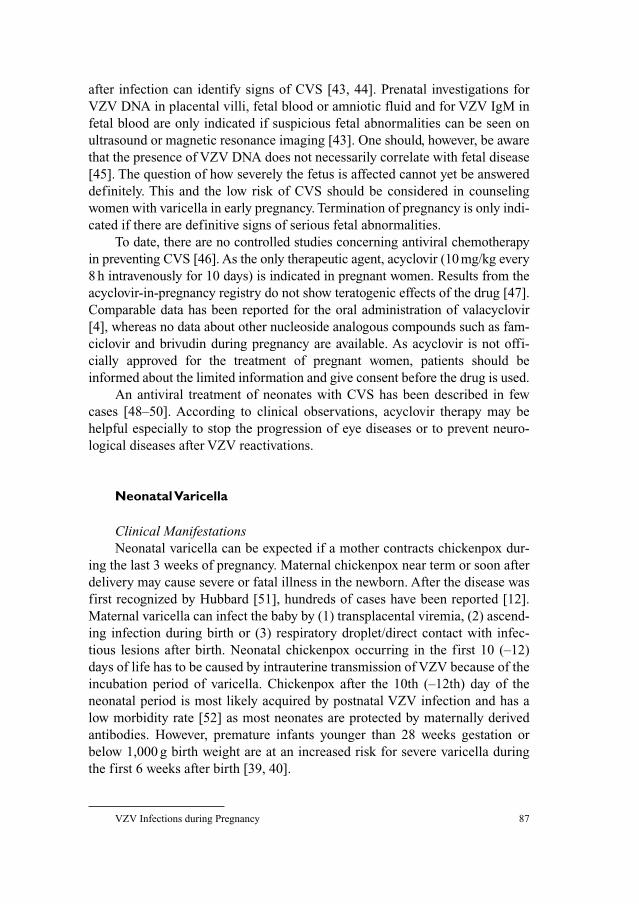
VZV Infections during Pregnancy 87
after infection can identify signs of CVS [43, 44]. Prenatal investigations for
VZV DNA in placental villi, fetal blood or amniotic fluid and for VZV IgM in
fetal blood are only indicated if suspicious fetal abnormalities can be seen on
ultrasound or magnetic resonance imaging [43]. One should, however, be aware
that the presence of VZV DNA does not necessarily correlate with fetal disease
[45]. The question of how severely the fetus is affected cannot yet be answered
definitely. This and the low risk of CVS should be considered in counseling
women with varicella in early pregnancy. Termination of pregnancy is only indi-
cated if there are definitive signs of serious fetal abnormalities.
To date, there are no controlled studies concerning antiviral chemotherapy
in preventing CVS [46]. As the only therapeutic agent, acyclovir (10 mg/kg every
8 h intravenously for 10 days) is indicated in pregnant women. Results from the
acyclovir-in-pregnancy registry do not show teratogenic effects of the drug [47].
Comparable data has been reported for the oral administration of valacyclovir
[4], whereas no data about other nucleoside analogous compounds such as fam-
ciclovir and brivudin during pregnancy are available. As acyclovir is not offi-
cially approved for the treatment of pregnant women, patients should be
informed about the limited information and give consent before the drug is used.
An antiviral treatment of neonates with CVS has been described in few
cases [48–50]. According to clinical observations, acyclovir therapy may be
helpful especially to stop the progression of eye diseases or to prevent neuro-
logical diseases after VZV reactivations.
Neonatal Varicella
Clinical ManifestationsNeonatal varicella can be expected if a mother contracts chickenpox dur-
ing the last 3 weeks of pregnancy. Maternal chickenpox near term or soon after
delivery may cause severe or fatal illness in the newborn. After the disease was
first recognized by Hubbard [51], hundreds of cases have been reported [12].
Maternal varicella can infect the baby by (1) transplacental viremia, (2) ascend-
ing infection during birth or (3) respiratory droplet/direct contact with infec-
tious lesions after birth. Neonatal chickenpox occurring in the first 10 (–12)
days of life has to be caused by intrauterine transmission of VZV because of the
incubation period of varicella. Chickenpox after the 10th (–12th) day of the
neonatal period is most likely acquired by postnatal VZV infection and has a
low morbidity rate [52] as most neonates are protected by maternally derived
antibodies. However, premature infants younger than 28 weeks gestation or
below 1,000 g birth weight are at an increased risk for severe varicella during
the first 6 weeks after birth [39, 40].
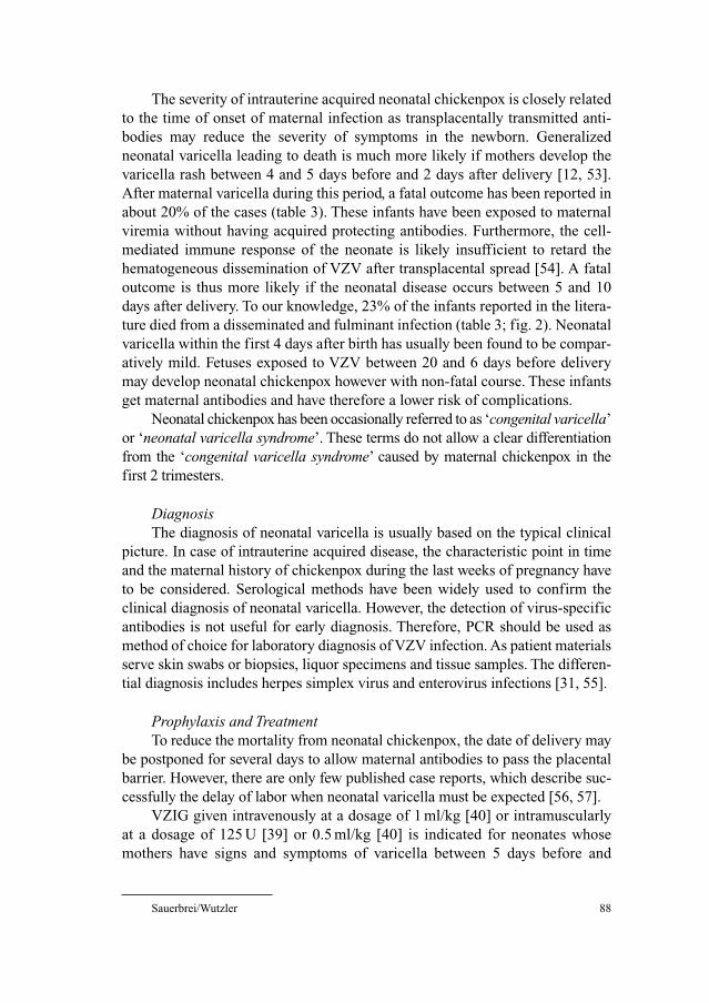
Sauerbrei/Wutzler 88
The severity of intrauterine acquired neonatal chickenpox is closely related
to the time of onset of maternal infection as transplacentally transmitted anti-
bodies may reduce the severity of symptoms in the newborn. Generalized
neonatal varicella leading to death is much more likely if mothers develop the
varicella rash between 4 and 5 days before and 2 days after delivery [12, 53].
After maternal varicella during this period, a fatal outcome has been reported in
about 20% of the cases (table 3). These infants have been exposed to maternal
viremia without having acquired protecting antibodies. Furthermore, the cell-
mediated immune response of the neonate is likely insufficient to retard the
hematogeneous dissemination of VZV after transplacental spread [54]. A fatal
outcome is thus more likely if the neonatal disease occurs between 5 and 10
days after delivery. To our knowledge, 23% of the infants reported in the litera-
ture died from a disseminated and fulminant infection (table 3; fig. 2). Neonatal
varicella within the first 4 days after birth has usually been found to be compar-
atively mild. Fetuses exposed to VZV between 20 and 6 days before delivery
may develop neonatal chickenpox however with non-fatal course. These infants
get maternal antibodies and have therefore a lower risk of complications.
Neonatal chickenpox has been occasionally referred to as ‘congenital varicella’
or ‘neonatal varicella syndrome’. These terms do not allow a clear differentiation
from the ‘congenital varicella syndrome’ caused by maternal chickenpox in the
first 2 trimesters.
DiagnosisThe diagnosis of neonatal varicella is usually based on the typical clinical
picture. In case of intrauterine acquired disease, the characteristic point in time
and the maternal history of chickenpox during the last weeks of pregnancy have
to be considered. Serological methods have been widely used to confirm the
clinical diagnosis of neonatal varicella. However, the detection of virus-specific
antibodies is not useful for early diagnosis. Therefore, PCR should be used as
method of choice for laboratory diagnosis of VZV infection. As patient materials
serve skin swabs or biopsies, liquor specimens and tissue samples. The differen-
tial diagnosis includes herpes simplex virus and enterovirus infections [31, 55].
Prophylaxis and TreatmentTo reduce the mortality from neonatal chickenpox, the date of delivery may
be postponed for several days to allow maternal antibodies to pass the placental
barrier. However, there are only few published case reports, which describe suc-
cessfully the delay of labor when neonatal varicella must be expected [56, 57].
VZIG given intravenously at a dosage of 1 ml/kg [40] or intramuscularly
at a dosage of 125 U [39] or 0.5 ml/kg [40] is indicated for neonates whose
mothers have signs and symptoms of varicella between 5 days before and

VZV Infections during Pregnancy 89
2 days after delivery. The Department of Health Joint Committee on Vacci-
nation and Immunisation (UK) recommends passive immunization for a
period between 7 days before and 2 days after delivery [58] and the Committee
on Infectious Diseases of the American Academy of Pediatrics (USA) recom-
mends VZIG for infants whose mothers develop chickenpox between 5 days
before and 3 days after delivery [8]. However, VZIG is probably not necessary
for neonates whose mothers have signs of varicella �5 days before or �2 days
after delivery, because those infants are not at risk of severe varicella.
Table 3. Prognosis of neonatal varicella without anti-
viral treatment in 136 term infants cited in the literature
Day of rash onset Cases
non-fatal fatal
Mother�5* (n � 57) 57 (100%) 0
4*–2** (n � 79) 65 (82%) 14 (18%)
Neonate1
0–4*** (n � 35) 34 (97%) 1 (3%)
5–10*** (n � 47) 36 (77%) 11 (23%)
*Days before delivery; **days after delivery;
***days after birth.1Data of 54 neonates have not been described.
Fig. 2. Necropsy of neonate died from neonatal varicella: focal necroses on the surface
of the liver.
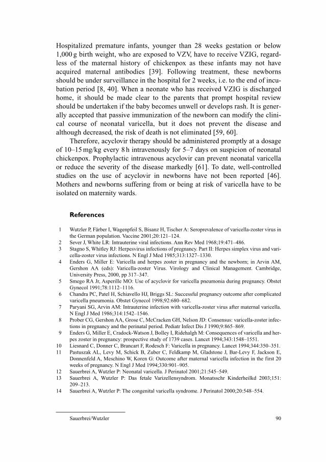
Sauerbrei/Wutzler 90
Hospitalized premature infants, younger than 28 weeks gestation or below
1,000 g birth weight, who are exposed to VZV, have to receive VZIG, regard-
less of the maternal history of chickenpox as these infants may not have
acquired maternal antibodies [39]. Following treatment, these newborns
should be under surveillance in the hospital for 2 weeks, i.e. to the end of incu-
bation period [8, 40]. When a neonate who has received VZIG is discharged
home, it should be made clear to the parents that prompt hospital review
should be undertaken if the baby becomes unwell or develops rash. It is gener-
ally accepted that passive immunization of the newborn can modify the clini-
cal course of neonatal varicella, but it does not prevent the disease and
although decreased, the risk of death is not eliminated [59, 60].
Therefore, acyclovir therapy should be administered promptly at a dosage
of 10–15 mg/kg every 8 h intravenously for 5–7 days on suspicion of neonatal
chickenpox. Prophylactic intravenous acyclovir can prevent neonatal varicella
or reduce the severity of the disease markedly [61]. To date, well-controlled
studies on the use of acyclovir in newborns have not been reported [46].
Mothers and newborns suffering from or being at risk of varicella have to be
isolated on maternity wards.
References
1 Wutzler P, Färber I, Wagenpfeil S, Bisanz H, Tischer A: Seroprevalence of varicella-zoster virus in
the German population. Vaccine 2001;20:121–124.
2 Sever J, White LR: Intrauterine viral infections. Ann Rev Med 1968;19:471–486.
3 Stagno S, Whitley RJ: Herpesvirus infections of pregnancy. Part II: Herpes simplex virus and vari-
cella-zoster virus infections. N Engl J Med 1985;313:1327–1330.
4 Enders G, Miller E: Varicella and herpes zoster in pregnancy and the newborn; in Arvin AM,
Gershon AA (eds): Varicella-zoster Virus. Virology and Clinical Management. Cambridge,
University Press, 2000, pp 317–347.
5 Smego RA Jr, Asperille MO: Use of acyclovir for varicella pneumonia during pregnancy. Obstet
Gynecol 1991;78:1112–1116.
6 Chandra PC, Patel H, Schiavello HJ, Briggs SL: Successful pregnancy outcome after complicated
varicella pneumonia. Obstet Gynecol 1998;92:680–682.
7 Paryani SG, Arvin AM: Intrauterine infection with varicella-zoster virus after maternal varicella.
N Engl J Med 1986;314:1542–1546.
8 Prober CG, Gershon AA, Grose C, McCracken GH, Nelson JD: Consensus: varicella-zoster infec-
tions in pregnancy and the perinatal period. Pediatr Infect Dis J 1990;9:865–869.
9 Enders G, Miller E, Cradock-Watson J, Bolley I, Ridehalgh M: Consequences of varicella and her-
pes zoster in pregnancy: prospective study of 1739 cases. Lancet 1994;343:1548–1551.
10 Liesnard C, Donner C, Brancart F, Rodesch F: Varicella in pregnancy. Lancet 1994;344:350–351.
11 Pastuszak AL, Levy M, Schick B, Zuber C, Feldkamp M, Gladstone J, Bar-Levy F, Jackson E,
Donnenfeld A, Meschino W, Koren G: Outcome after maternal varicella infection in the first 20
weeks of pregnancy. N Engl J Med 1994;330:901–905.
12 Sauerbrei A, Wutzler P: Neonatal varicella. J Perinatol 2001;21:545–549.
13 Sauerbrei A, Wutzler P: Das fetale Varizellensyndrom. Monatsschr Kinderheilkd 2003;151:
209–213.
14 Sauerbrei A, Wutzler P: The congenital varicella syndrome. J Perinatol 2000;20:548–554.

VZV Infections during Pregnancy 91
15 Miller E, Cradock-Watson JE, Ridehalgh MK: Outcome in newborn babies given anti-varicella-
zoster immunoglobulin after perinatal maternal infection with varicella-zoster virus. Lancet
1989;2:371–373.
16 Laforet EG, Lynch CJ Jr: Multiple congenital dafects following maternal varicella. N Engl J Med
1947;236:534–537.
17 Sauerbrei A, Wutzler P: Varicella-zoster virus infections during pregnancy: epidemiology, clinical
symptoms, diagnosis, prevention and therapy. Curr Pediatr Rev 2005;1:205–216.
18 Schulze A, Dietzsch HJ: The natural history of varicella embryopathy: a 25-year follow-up.
J Pediatr 2000;137:871–874.
19 Birch CJ, Druce JD, Catton MC, MacGregor L, Read T: Detection of varicella-zoster virus in
genital specimens using a multiplex polymerase chain reaction. Sex Transm Infect 2003;79:298–300.
20 Higa K, Dan K, Manabe H: Varicella-zoster virus infections during pregnancy: hypothesis
concerning the mechanisms of congenital malformation. Obstet Gynecol 1987;69:214–221.
21 Nikkels A, Delbecque K, Pierard GE, Wienkötter P, Schalasta G, Enders M: Distribution of
varicella-zoster virus DNA and gene products in tissues of a first-trimester varicella-infected
fetus. J Infect Dis 2005;191:540–545.
22 Grose C: Congenital varicella-zoster virus infection and the failure to establish virus-specific cell-
mediated immunity. Mol Biol Med 1989;6:453–462.
23 Scharf A, Scherr O, Enders G, Helftenbein E: Virus detection in the fetal tissue of a
premature delivery with congenital varicella syndrome: a case report. J Perinatol Med 1990;18:
317–322.
24 Michie CA, Acolet D, Charlton R, Stevens JP, Happerfield LC, Bobrow LG, Kangro H, Gau G,
Modi N: Varicella-zoster contracted in the second trimester of pregnancy. Pediatr Infect Dis J
1992;11:1050–1053.
25 Sauerbrei A, Müller D, Eichhorn U, Wutzler P: Detection of varicella-zoster virus in congenital
varicella syndrome: a case report. Obstet Gynecol 1996;88:687–689.
26 Al-Katawee YA, Al-Hassoun YA, Taha MN, Al-Moslem K: Congenital varicella-zoster virus infec-
tion. A rare case of severe brain and ocular malformations without limb or cutaneous involvement
in a newborn after maternal subclinical infection. Saudi Med J 2005;26:869–871.
27 Gershon AA, Raker R, Steinberg S, Topf-Olstein B, Drusin LM: Antibody to varicella-zoster virus
in parturient women and their offspring during the first year of life. Pediatrics 1976;58:692–696.
28 Akisu M, Yalaz M, Aksu G, Arslanoglu S, Genel F, Kutukculer N, Kultursay N: Maternally
acquired varicella-zoster virus antibodies disappear at 6 months of age in prematurely born chil-
dren. Panminerva Med 2003;45:155–156.
29 Koskimies O, Lapinleimu K, Saxén L: Infections and other maternal factors as risk indicators for
congenital malformations: a case-control study with paired serum samples. Pediatrics 1978;61:
832–837.
30 Johansson AB, Rassart A, Blum D, Van Beers D, Liesnard C: Lower-limb hypoplasia due to
intrauterine infection with herpes simplex virus type 2: possible confusion with intrauterine vari-
cella-zoster syndrome. Clin Infect Dis 2004;38:e57–e62.
31 Sauerbrei A, Glück B, Jung K, Bittrich H, Wutzler P: Congenital skin lesions caused by intrauter-
ine coxsackievirus B3 infection: a case report. Infection 2000;28:326–328.
32 Spranger S, Stute H, Blankenagel A, Jauch A, Hager D, Tariverdian G: MIDAS-Syndrom – Eine
X-chromosomale Erkrankung. Differenzialdiagnose zum kongenitalen Varizellensyndrom.
Monatsschr Kinderheilkd 1998;146:761–765.
33 Shields KE, Galil K, Seward J, Sharrar RG, Cordero JF, Slater E: Varicella vaccine exposure dur-
ing pregnancy: data from the first 5 years of the pregnancy registry. Obstet Gynecol 2001;98:
14–19.
34 Salzman MB, Sharrar RG, Steinberg S, LaRussa P: Transmission of varicella-vaccine virus from a
healthy 12-month-old child to his pregnant mother. J Pediatr 1997;131:151–154.
35 Watson B: Varicella: a vaccine preventable disease? J Infect 2002;44:220–225.
36 Vázquez M, Shapiro ED: Varicella vaccine and infection with varicella-zoster virus. N Engl J Med
2005;352:439–440.
37 Royal College of Obstetricians and Gynecologists: Guideline No. 13. Chickenpox in Pregnancy.
2001.
Sauerbrei/Wutzler 92
38 Robert Koch-Institut: Empfehlungen der Ständigen Impfkommission (STIKO) am Robert Koch
Institut/Stand: Juli 2004. Epidemiol Bull 2004;30:235–250.
39 Advisory Committee on Immunization Practices (ACIP): Prevention of varicella. MMWR
1996;45:1–36.
40 Deutsche Gesellschaft für Pädiatrische Infektiologie: Handbuch 2003: Infektionen im Kindesalter.
München, Futuramed, 2003, pp 732–739.
41 Kuter BJ, Ngai A, Patterson CM, Staehle BO, Cho I, Matthews H, Provost PJ, White CJ: Safety,
tolerability, and immunogenicity of two regimens of Oka/Merck varicella vaccine (Varivax®) in
healthy adolescents and adults. Vaccine 1995;13:967–972.
42 Sauerbrei A, Wutzler P: Labordiagnostik der Varizellen. Kinderärztl Prax 2004, Sonderheft
Impfen, pp 18–21.
43 Pretorius DH, Hayward I, Jones KL, Stamm E: Sonographic evaluation of pregnancies with mater-
nal varicella infection. J Ultrasound Med 1992;11:459–463.
44 Verstraelen H, Vanzieleghem B, Deroort P, Vanhaesebrouck P, Temmerman M: Prenatal ultrasound
and magnetic resonance imaging in fetal varicella syndrome: correlation with pathology findings.
Prenat Diagn 2003;23:705–709.
45 Mouly F, Mirlesse V, Meritet JF, Rozenberg F, Poissonier MH, Lebon P, Daffos F: Prenatal diagno-
sis of fetal varicella-zoster virus infection with polymerase chain reaction of amniotic fluid in 107
cases. Am J Obstet Gynecol 1997;177:894–898.
46 Tan MP, Koren G. Chickenpox in pregnancy: revisited. Reprod Toxicol 2006;21:410–420, DOI:
10.1016/j.reprotox.2005.04.011.
47 Andrews EB: Acyclovir does not increase risk in pregnancy. Am Pharm 1994;NS34:6.
48 Wutzler P, Sauerbrei A, Scholz H, Müller D, Wiedersberg H: Varicella-Zoster-Virusinfektionen in
der Schwangerschaft. Paediatr Prax 1990–1991;41:213–224.
49 Sauerbrei A, Pawlak J, Luger C, Wutzler P: Intracerebral VZV reactivation in congenital varicella
syndrome? A case report. Dev Med Child Neurol 2003;45:837–840.
50 Schulze-Oechtering F, Roth B, Enders G, Grosser R: Kongenitales Varizellensyndrom – besteht
eine Infektionsgefahr für die Umgebung? Dtsch Med Wochenschr 2004;208:25–28.
51 Hubbard TW: Varicella occurring in an infant twenty four hours after birth. Br Med J 1878;1:822.
52 Heidl M: Varicella-Zoster-Virus-Infektion in der Schwangerschaft, beim Neugeborenen und jun-
gen Säugling. Z Klin Med 1985;40:245–250.
53 Sterner G, Forsgren M, Enocksson E, Grandien M, Granström G: Varicella-zoster infections in
late pregnancy. Scand J Infect Dis 1990;71:30–35.
54 Baba K, Yabuuchi H, Takahashi M, Ogra PL: Immunologic and epidemiologic aspects of varicella
infection acqired during infancy and early childhood. J Pediatr 1982;100:881–885.
55 Gershon AA: Varicella-zoster virus; in Feigin RD, Cherry JD (eds): Textbook of Pediatric
Infectious Diseases, ed 4. Philadelphia, W. B. Saunders Company, 1998, pp 1769–1777.
56 Paulman PM, McLellan R: Varicella during pregnancy: the timing of effective treatment. J Am
Board Fam Pract 1990;3:121–123.
57 Zieger W, Friese K, Weigel M, Becker KP, Melchert F: Varizellen-Infektion am Geburtstermin.
Z Geburtshilfe Perinatol 1994;198:134–137.
58 Miller E: Varicella-zoster virus infection in pregnancy. Arch Dis Child 1994;70:F157–F158.
59 Holland P, Isaacs D, Moxon ER: Fatal neonatal varicella infection. Lancet 1986;2:1156.
60 King SM, Gorensek M, Ford-Jones EL, Read SE: Fatal varicella-zoster infection in a newborn
treated with varicella-zoster immunoglobulin. Pediatr Infect Dis 1986;5:588–589.
61 Carter PE, Duffty P, Lloyd DJ: Neonatal varicella infection Lancet 1986;2:1459–1460.
Dr. Andreas Sauerbrei
Institute of Virology and Antiviral Therapy
Friedrich-Schiller University Jena
Hans-Knöll-Strasse 2
DE–07745 Jena (Germany)
Tel. �49 3641 657300, Fax �49 3641 657301, E-Mail [email protected]

Gross G, Doerr HW (eds): Herpes Zoster.
Monogr Virol. Basel, Karger, 2006, vol 26, pp 93–106
Herpes Zoster in theImmunocompromised Host
Helmut Schöfer
Department of Dermatology, University Hospital, J.W. Goethe University,
Frankfurt, Germany
After primary chickenpox infection and varicella–zoster virus (VZV)
viraemia, which usually occurs during childhood (with or without a varicella
exanthem), VZV persists as a residual latent infection in the dorsal root ganglia.
At the age of 13–14 years, endemic VZV infection in Germany exceeds 90%.
The latent infection is controlled by cellular immune functions (specific CD4�-
T-lymphocytes) and neutralising antibodies [1]. Although the pathomechanisms
are not yet understood in detail, exogenic reinfection and/or immunological
dysbalance, due to the decrease of T-cell related cellular immune functions and
the loss of neutralising antibodies, may lead to a dermatomal reactivation in
patients carrying the latent VZV infection. Clinically, this results in the mani-
festation of herpes zoster. Triggers for reactivation may be:
In patients �50 years:
• Age related (physiological) deficiency of cellular immune functions: Loss
of CD4� T-lymphocytes by numbers (CD4�-count ↓) and loss of T-cell
activity.
• Therapy with immunosuppressives [2].
• Chemotherapy.
• Severe consumptive diseases.
In patients �50 years:
• Incomplete immunologic response to prenatal chickenpox infection.
• Incomplete immunologic response to chickenpox infection in the first year
of life.
• Atopic diathesis.
• HIV-infection (CD4-count ↓).
• Therapy with immunosuppressives.
• Severe consumptive diseases.

Schöfer 94
• Distress (emotional, physical).
• Severe insolation (UV-irradiation).
• Pregnancy (?).
• In HIV� patients: Immune reconstitution, following highly active anti-
retroviral therapy (HAART).
Representing a disease of the elderly, the occurrence of herpes zoster in
patients younger than 50 years of age is suspicious of any kind of cellular
immunodeficiency. In times prior to the HIV-epidemic, malignant systemic
diseases, like leukaemia or Hodgkin’s disease were most likely to be the cause of
shingles in young patients (�50 years of age). Since the early eighties of the
20th century the HIV-infection became more and more relevant and nowadays it
is the most important disease to be excluded in young zoster patients [3].
Herpes Zoster in HIV-Infected Patients
EpidemiologyOnly 3 years after the first descriptions of AIDS as a new disease Mathur-
Wagh and Mildvan [4] reported, that herpes zoster is very common in the
pre-AIDS stage of the HIV-disease, which was named ‘lymphadenopathy
syndrome’ at that time. 24% of these patients had a herpes zoster manifestation
prior to full-blown AIDS, which was diagnosed exclusively by clinical and
immunological parameters until HIV was discovered (1983) and first antibody tests
had been established (1984). In 1986, Friedman-Kien et al. [5] pointed out, that
herpes zoster is an early clinical sign of the development of AIDS. He calculated a
7-fold risk for HIV-infected patients to suffer from herpes zoster compared to non-
HIV-infected adults. Later Buchbinder et al. [6] showed that the age-adjusted over-
all relative risk of herpes zoster among HIV-infected homosexuals was 16.9
compared to non-HIV-infected homosexual men and that herpes zoster might occur
during all stages of HIV disease. The herpes zoster incidence ranges from 2.9 to 5.1
episodes per 1,000 person years [6–9]. In 1986, the Centres for Disease Control
(CDC, Atlanta) included shingles involving more than one dermatome in the symp-
tomatic phase of the HIV disease and AIDS classification (B symptoms).
Today it is well-established that herpes zoster in patients less than 50 years
of age, is the most striking marker disease of HIV-infection. Marker diseases
are defined as illnesses that are attracting the physician’s attention to the possi-
bility of an underlying acquired immunodeficiency, i.e. HIV-infection by their
mere appearance or special clinical features [10–12]. Among 188 patients of the
Department of Dermatology, Frankfurt/M University Hospital, in which skin
disease had led to the diagnosis of HIV-infection, herpes zoster was the pre-
senting marker disease in 25 cases (unpublished data, Schöfer).
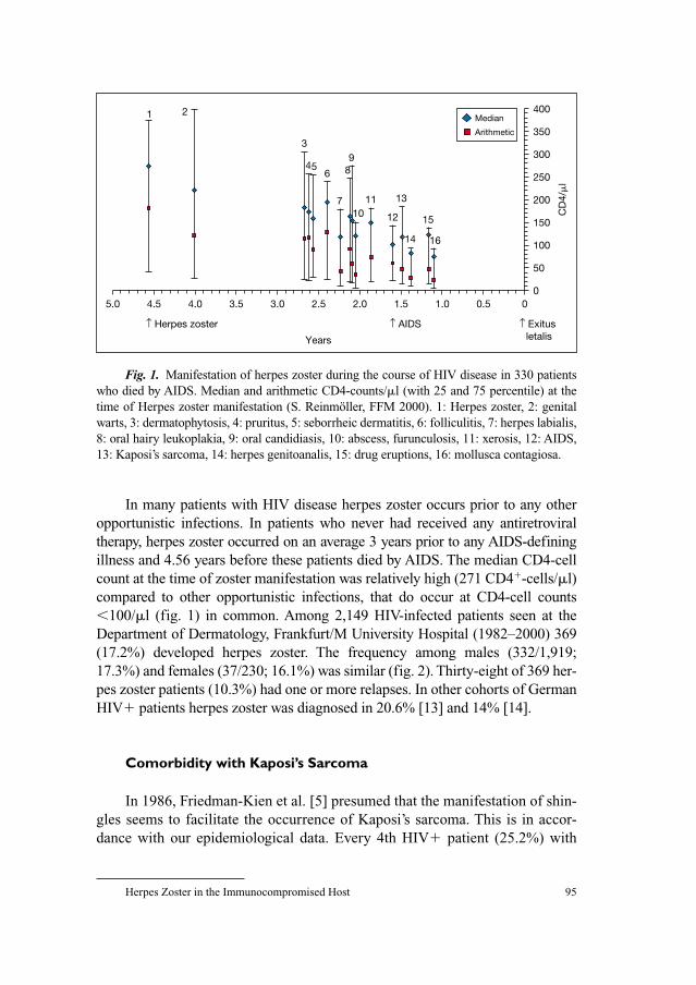
Herpes Zoster in the Immunocompromised Host 95
In many patients with HIV disease herpes zoster occurs prior to any other
opportunistic infections. In patients who never had received any antiretroviral
therapy, herpes zoster occurred on an average 3 years prior to any AIDS-defining
illness and 4.56 years before these patients died by AIDS. The median CD4-cell
count at the time of zoster manifestation was relatively high (271 CD4�-cells/�l)
compared to other opportunistic infections, that do occur at CD4-cell counts
�100/�l (fig. 1) in common. Among 2,149 HIV-infected patients seen at the
Department of Dermatology, Frankfurt/M University Hospital (1982–2000) 369
(17.2%) developed herpes zoster. The frequency among males (332/1,919;
17.3%) and females (37/230; 16.1%) was similar (fig. 2). Thirty-eight of 369 her-
pes zoster patients (10.3%) had one or more relapses. In other cohorts of German
HIV� patients herpes zoster was diagnosed in 20.6% [13] and 14% [14].
Comorbidity with Kaposi’s Sarcoma
In 1986, Friedman-Kien et al. [5] presumed that the manifestation of shin-
gles seems to facilitate the occurrence of Kaposi’s sarcoma. This is in accor-
dance with our epidemiological data. Every 4th HIV� patient (25.2%) with
5.0 4.5 4.0
1 2
3
456 8
7
9
1011 13
12
14
15
16
3.5 3.0 2.5
Years
2.0 1.5 1.0 0.5 00
50
100
150
200
CD
4/�
l250
300
350
400
↑ Herpes zoster ↑ AIDS ↑ Exitus letalis
Median
Arithmetic
Fig. 1. Manifestation of herpes zoster during the course of HIV disease in 330 patients
who died by AIDS. Median and arithmetic CD4-counts/�l (with 25 and 75 percentile) at the
time of Herpes zoster manifestation (S. Reinmöller, FFM 2000). 1: Herpes zoster, 2: genital
warts, 3: dermatophytosis, 4: pruritus, 5: seborrheic dermatitis, 6: folliculitis, 7: herpes labialis,
8: oral hairy leukoplakia, 9: oral candidiasis, 10: abscess, furunculosis, 11: xerosis, 12: AIDS,
13: Kaposi’s sarcoma, 14: herpes genitoanalis, 15: drug eruptions, 16: mollusca contagiosa.
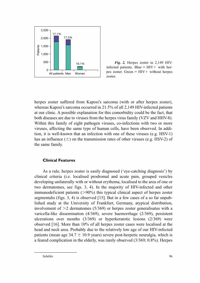
Schöfer 96
0
500
1,000
1,500
2,000
2,500
All patients Men Women
17.3%
16.1%
17.1%P
atie
nts
Fig. 2. Herpes zoster in 2,149 HIV-
infected patients. Blue � HIV� with her-
pes zoster. Green � HIV� without herpes
zoster.
herpes zoster suffered from Kaposi’s sarcoma (with or after herpes zoster),
whereas Kaposi’s sarcoma occurred in 21.5% of all 2,149 HIV-infected patients
at our clinic. A possible explanation for this comorbidity could be the fact, that
both diseases are due to viruses from the herpes virus family (VZV and HHV-8).
Within this family of eight pathogen viruses, co-infections with two or more
viruses, affecting the same type of human cells, have been observed. In addi-
tion, it is well-known that an infection with one of these viruses (e.g. HSV-1)
has an influence (�) on the transmission rates of other viruses (e.g. HSV-2) of
the same family.
Clinical Features
As a rule, herpes zoster is easily diagnosed (‘eye-catching diagnosis’) by
clinical criteria (i.e. localised prodromal and acute pain, grouped vesicles
developing unilaterally with or without erythema, localised to the area of one or
two dermatomes, see figs. 3, 4). In the majority of HIV-infected and other
immunodeficient patients (�90%) this typical clinical aspect of herpes zoster
segmentalis (figs. 3, 4) is observed [15]. But in a few cases of a so far unpub-
lished study at the University of Frankfurt, Germany, atypical distribution,
involvement of �2 dermatomes (5/369) or herpes zoster generalisatus with a
varicella-like dissemination (4/369), severe haemorrhage (2/369), persistent
ulcerations over months (3/369) or hyperkeratotic lesions (2/369) were
observed [16]. More than 10% of all herpes zoster cases were localised at the
head and neck area. Probably due to the relatively low age of our HIV-infected
patients (mean age 34.7 � 10.9 years) severe post-herpetic neuralgia, which is
a feared complication in the elderly, was rarely observed (3/369; 0.8%). Herpes
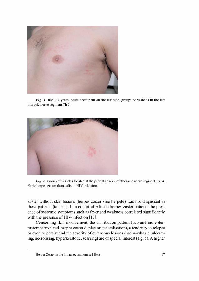
Herpes Zoster in the Immunocompromised Host 97
zoster without skin lesions (herpes zoster sine herpete) was not diagnosed in
these patients (table 1). In a cohort of African herpes zoster patients the pres-
ence of systemic symptoms such as fever and weakness correlated significantly
with the presence of HIV-infection [17].
Concerning skin involvement, the distribution pattern (two and more der-
matomes involved, herpes zoster duplex or generalisation), a tendency to relapse
or even to persist and the severity of cutaneous lesions (haemorrhagic, ulcerat-
ing, necrotising, hyperkeratotic, scarring) are of special interest (fig. 5). A higher
Fig. 3. RM, 34 years, acute chest pain on the left side, groups of vesicles in the left
thoracic nerve segment Th 3.
Fig. 4. Group of vesicles located at the patients back (left thoracic nerve segment Th 3).
Early herpes zoster thoracalis in HIV-infection.
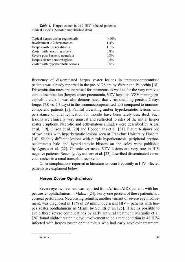
Schöfer 98
frequency of disseminated herpes zoster lesions in immunocompromised
patients was already reported in the pre-AIDS era by Weber and Pelecchia [18].
Dissemination rates are increased for cutaneous as well as for the very rare vis-
ceral dissemination (herpes zoster pneumonia, VZV hepatitis, VZV meningoen-
cephalitis etc.). It was also demonstrated, that virus shedding persists 2 days
longer (7.0 vs. 5.3 days) in the immunocompromised host compared to immuno-
competend patients [5]. Painful ulcerating and/or hyperkeratotic lesions with
persistance of viral replication for months have been rarely described. Such
lesions are clinically very unusual and restricted to sites of the initial herpes
zoster eruptions. Necrotic and ecthymatous shingles were described by Alessi
et al. [19], Gilson et al. [20] and Hoppenjans et al. [21]. Figure 6 shows one
of two cases with hyperkeratotic lesions seen at Frankfurt University Hospital
[16]. Slightly different lesions with purple hyperkeratosis, peripheral erythro-
oedematous halo and hyperkeratotic blisters on the soles were published
by Agusto et al. [22]. Chronic verrucous VZV lesions are very rare in HIV
negative patients. Recently, Jeyaratnam et al. [23] described disseminated verru-
cous rashes in a renal transplant recipient.
Other complications reported in literature to occur frequently in HIV-infected
patients are explained below.
Herpes Zoster Ophthalmicus
Severe eye involvement was reported from African AIDS patients with her-
pes zoster ophthalmicus in Malawi [24]. Forty-one percent of these patients had
corneal perforation. Necrotising retinitis, another variant of severe eye involve-
ment, was diagnosed in 17% of 29 immunodeficient HIV� patients with her-
pes zoster ophthalmicus in Miami by Sellitti et al. [25]. It seems possible to
avoid these severe complications by early antiviral treatment: Margolis et al.
[26] found sight-threatening eye involvement to be a rare condition in 48 HIV-
infected with herpes zoster ophthalmicus who had early acyclovir treatment.
Table 1. Herpes zoster in 369 HIV-infected patients:
clinical aspects (Schöfer, unpublished data)
Typical herpes zoster segmentalis �90%
Involvement �2 dermatomes 1.4%
Herpes zoster generalisatus 1.1%
Zoster with persisting ulcers 0.8%
Severe post-herpetic neuralgia 0.8%
Herpes zoster hemorrhagicus 0.5%
Zoster with hyperkeratotic lesions 0.5%
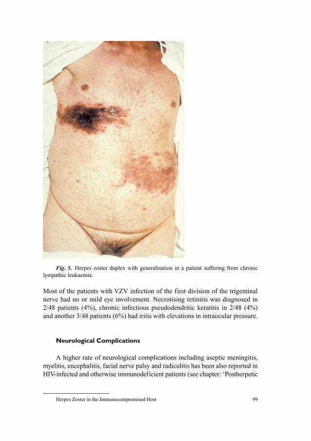
Herpes Zoster in the Immunocompromised Host 99
Most of the patients with VZV infection of the first division of the trigeminal
nerve had no or mild eye involvement. Necrotising retinitis was diagnosed in
2/48 patients (4%), chronic infectious pseudodendritic keratitis in 2/48 (4%)
and another 3/48 patients (6%) had iritis with elevations in intraocular pressure.
Neurological Complications
A higher rate of neurological complications including aseptic meningitis,
myelitis, encephalitis, facial nerve palsy and radiculitis has been also reported in
HIV-infected and otherwise immunodeficient patients (see chapter: ‘Postherpetic
Fig. 5. Herpes zoster duplex with generalisation in a patient suffering from chronic
lympathic leukaemia.

Schöfer 100
Neuralgia and Other Neurologic Complications’ by R. Baron, pp 69–80), but
some authors [27, 28] demonstrated, that immunodeficiency does not increase
the risk of post-herpetic neuralgia. The incidence of this complication seems to
be related above all to the patient’s age.
Herpes Zoster, HIV and the Immune Reconstitution Syndrome
Severe immunodeficiency (CD4�-T-lymphocytes �200/�l), as observed
in untreated AIDS patients, seems to hinder the clinical manifestation of herpes
Fig. 6. Hyperkeratotic lesions of chronic herpes zoster infection (5 months).
Herpes Zoster in the Immunocompromised Host 101
zoster. However, in cases where HAART had been introduced successfully
(CD4�-T-lymphocytes ↑, HIV viral load ↓) and had led to a relevant immune
reconstitution, herpes zoster was reported to occur more frequently [29–33].
This was also observed in patients, who switched from a rather ineffective anti-
retroviral monotherapy to HAART [29]. In these patients herpes zoster
occurred on an average 16.6 weeks after HAART had been started, but half of
the patients had herpes zoster already within the first 4 weeks. Domingo et al.
[30] found that shingles in connection with the immune reconstitution syn-
drome occurred especially in those patients who had an substantial increase of
CD8�-T-lymphocytes after HAART had been started. Only a few cases of
zoster in the course of immune reconstitution were reported to be recurrences.
In contrast to the data published in literature, we identified only one of 94
HIV� herpes zoster patients, who had a herpes zoster manifestation in direct
association (within 3 months after the start of HAART) with immune reconsti-
tution (unpublished data: Department of Dermatology, University Hospital
Frankfurt/M, Germany 1995–2003).
Herpes Zoster and the Progression of HIV Disease Towards AIDS
Several longitudinal investigations that tried to answer the question,
whether herpes zoster in HIV-infected patients increases the risk of AIDS man-
ifestation (independent from CD4-cell counts) did not show any significant
influence on the outcome of HIV-infection and survival [6, 34–37].
Herpes Zoster in Organ Transplant Recipients
It is now more than 35 years ago, that the first kidney was transplanted.
Immunosuppressive regimens have changed several times since those pioneer
days, but still there are substantial problems to prevent abortion of the trans-
planted organ. Iatrogenic immunosuppression with corticosteroids, cyclosporin
A, azathioprin or mycophenolat mofetil favours viral infections. Severe
cytomegalovirus and recurrent herpes simplex virus infections are frequent and
alarming complications. Primary VZV infections harbour the danger of severe
chickenpox with life threatening organ manifestations like VZV pneumonia,
encephalitis and disseminated intravascular coagulopathy. Herpes zoster is
diagnosed in 8–30% of all transplant recipients within the first 6 months of
immunosuppression. Highest rates (50% within 12 months) occur in bone
marrow recipients who did not have an acyclovir prophylaxis. Postherpetic
Schöfer 102
neuralgia and a prolonged duration of skin disease is reported in organ trans-
plant recipients [38]. Gourishankar et al. [39] analysed 869 recipients of solid
organs for the incidence and clinical features of herpes zoster. An overall inci-
dence of 8.6% was found. Herpes zoster was almost twice as frequent in heart
and lung recipients (15.1–16.8%) compared to liver and kidney recipients
(5.7–7.4%) and started 9 months (median time) after transplantation. The
female gender and immunosuppressive therapy with mycophenolate mofetil
were found to be independent risk factors in these patients [40]. Cutaneous
scarring (18.7%) and post-herpetic neuralgia (42.7%) were frequent complica-
tions [39], whereas verrucous lesions are rare in transplant recipients [23].
In cases where a transplantation can be planned in advance, VZV vaccina-
tion is substantial in VZV negative patients and might be helpful as a booster
vaccination in the elderly patient. If immunosuppression has started, vaccina-
tion with attenuated virus is no longer allowed and specific hyperimmunoglob-
ulins have to be considered instead. In addition, a prolonged CMV prophylaxis
seems to be also effective to reduce the risk of herpes zoster [39].
Herpes Zoster in Patients with Malignancies and Chemotherapy
Especially patients with malignant lymphomas (Hodgkin’s disease as well
as non-Hodgkin lymphoma) or leukaemia are endangered to suffer from herpes
zoster due to the disease related immunodeficiency or due to chemotherapy
[41–43] or bone marrow transplantation. Clinical course and manifestations do
not differ much from shingles in HIV-infected patients, but cutaneous dissemi-
nation is observed in 9–16% of all cases associated with malignancies and in
6.7–25.9% in patients with Hodgkin’s disease [44]. Some cases of visceral
involvement masked as abdominal pain or acute abdomen have been reported in
patients after bone marrow transplantation [45–47]. The overall relative risk for
shingles in tumour patients (age adapted) was calculated 5.0. Guess et al. [48]
reviewed 173 cases of herpes zoster in childhood and found a 122-fold risk of
herpes zoster for children with acute lymphocytic leukaemia. Shingles in these
children caused very few complications and not a single case of postherpetic
neuralgia. After bone marrow and stem cell transplantation acyclovir or valaci-
clovir prophylaxis is recommended [49, 50].
It is not reasonable to screen for occult tumours in all herpes zoster seg-
mentalis patients. The rate of occult tumours found in otherwise asymptomatic
herpes zoster patients is less than 5% of the overall relative risk 1.1 [51]. In
patients with disseminated zoster malignancies are found in a higher rate and
tumour screening is always recommended [52].
Herpes Zoster in the Immunocompromised Host 103
Therapy
Only acyclovir and famciclovir (in patients older than 25 years) are regis-
tered pharmaceuticals for the treatment of herpes zoster in the immunocom-
promised host [53, 54]. Recommended doses are depending on the grade of
immunodeficiency (with the absolute CD4-cell count as a surrogate marker)
and the severity of the disease:
In patients with CD4�-T-lymphocytes �200/�l and segmental zoster:
Acyclovir 5–7.5 mg/kg BW every 8 h i.v., Famciclovir 3 � 250 mg/day
perorally.
In patients with CD4�-T-lymphocytes �200/�l and/or clinical complica-
tions (haemorrhage, dissemination, neurological symptoms etc.): 10 mg/kg BW
every 8 h i.v.
In case of acyclovir resistance: Foscarnet (60 mg/kg BW BID or 40 mg/kg
BW TID i.v.) may be tried as an alternative. Foscarnet efficacy is independent
from viral thymidinkinase.
Oral antivirals like valacyclovir or brivudin have been effective in single
cases, but are not registered for the immunodeficient host. Recently, Breton
et al. [55] reported the failure of valacyclovir in a HIV-infected patient with
only moderate immunodeficiency.
Conclusion
As a rule, herpes zoster in the immunocompromised host is a frequent
finding, but in general (�90% of all cases) as long as it is treated early and
effectively there is no difference clinically from herpes zoster in the elderly
(herpes zoster segmentalis). Although rare, severe courses and a wide spectrum
of clinical variants and complications as well as postherpetic neuralgia are seen
occasionally and cause diagnostic and therapeutic problems. Relapsing and per-
sisting shingles with ulcerating or hyperkeratotic lesions are exclusively seen in
the immunodeficient host, where virus replication cannot be controlled by
immunological surveillance and acyclovir resistant strains might develop under
antiviral treatment. Therapy is linked to the severity of cellular immune defi-
ciency and to clinical complications. A new finding is the manifestation of her-
pes zoster as a part of the immune reconstitution syndrome in HIV-infected
patients under HAART. This complication of HAART usually occurs during the
first 3 months of effective treatment and is accompanied by a marked increase
of CD8�-T-lymphocytes. In addition, herpes zoster is an important clinical
marker disease for acquired immunodeficiency. It occurs frequently as the first
clinical symptom of HIV-infection, but can also be diagnosed in all other stages
Schöfer 104
of HIV disease. Therefore, herpes zoster in young adults (�50 years of age) is
an important indication for anti-HIV antibody testing.
References
1 Weksler ME: Immune senescence. Ann Neurol 1994;35:35–37.
2 Spencer ES, Anderson HK: Clinical evident, non-terminal infections with herpes viruses and the
wart virus in immunosuppressed renal allograft recipients. Br Med J 1970;1:251–254.
3 Schöfer H, Oswald A, Allwinn R: Zoster bei 223 Patienten unter 50 Jahren – eine retrospektive
Vergleichsanalyse von Patienten mit und ohne HIV-Infektion. J Dtsch Dermatol Ges 2003;
1(suppl 1):S85.
4 Mathur-Wagh U, Mildvan D: Prodromal syndromes in AIDS. Ann N Y Acad Sci 1984;437:
184–191.
5 Friedman-Kien AE, Lafleur FL, Gendler E, Hennessey NP, Montagna R, Halbert S, Rubinstein P,
Krasinski K, Zang E, Poiesz B: Herpes zoster: a possible early clinical sign for development of
acquired immunodeficiency syndrome in high-risk individuals. J Am Acad Dermatol 1986;14:
1023–1028.
6 Buchbinder SP, Katz MH, Hessol NA, Liu JY, O’Malley PM, Underwood R, Holmberg SD:
Herpes zoster and human immunodeficiency virus infection. J Infect Dis 1992;166:1153–1156.
7 Glesby MJ, Moore RD, Chaisson RE: Clinical spectrum of herpes zoster in adults infected with
human immunodeficiency virus. Clin Infect Dis 1995;21:370–375.
8 Insinga RP, Itzler RF, Pellissier JM, Saddier P, Nikas AA: The incidence of herpes zoster in a
United States administrative database. J Gen Intern Med 2005;20:748–753.
9 Veenstra J, Krol A, van Praag RM, Frissen PH, Schellekens PT, Lange JM, Coutinho RA, van der
Meer JT: Herpes zoster, immunological deterioration and disease progression in HIV-1 infection.
AIDS 1995;9:1153–1158.
10 Meigel W, Kern P, Dietrich M: Dermatologic diseases in patients with AIDS. Hautarzt
1986;37:250–254.
11 Rigopoulos D, Paparizos V, Katsambas A: Cutaneous markers of HIV infection. Clin Dermatol
2004;22:487–498.
12 Schöfer H: Frühsymptome der HIV-Erkrankung an Haut- und Schleimhäuten. AIDS Forsch
1991;6:633–648.
13 Garbe C, Husak R, Orfanos CE: HIV-associated dermatoses and their prevalence in 456 HIV-
infected patients. Relation to immune status and its importance as a diagnostic marker. Hautarzt
1994;45:623–629.
14 Hartmann M, Petzoldt D: Viruserkrankungen der Haut bei HIV-Infizierten. Allergologie
1994;17:316–320.
15 Disler RS, Dover JS: Chronic localized herpes zoster in the acquired immunodeficiency syn-
drome. Arch Dermatol 1990;126:1105.
16 Schöfer H, Baur SI, Gregel C, Wolter M, Milbradt R: Hyperkoratotic varicella zoster virus infec-
tion in an HIV-infected patient. Successful treatment of persistent lesions with cryosurgery. Br J
Dermatol 1998;138:714–715.
17 Onunu AN, Uhunwangho A: Clinical spectrum of herpes zoster in HIV-infected versus non-HIV
infected patients in Benin City, Nigeria. West Afr J Med 2004;23:300–304.
18 Weber DM, Pelecchia JA: Varicella pneumonia: study of prevalence in adult men. JAMA
1965;192:572–577.
19 Alessi E, Cusini M, Zerboni R, Cavicchini S, Uberti-Foppa C, Galli M, Moroni M: Unusual vari-
cella zoster virus infection in patients with the acquired immunodeficiency syndrome. Arch
Dermatol 1988;124:1011–1013.
20 Gilson IH, Barnett JH, Conant MA, Laskin OL, Williams J, Jones PG: Disseminated ecthymatous
herpes varicella-zoster virus infection in patients with acquired immunodeficiency syndrome. J
Am Acad Dermatol 1989;20:637–642.
Herpes Zoster in the Immunocompromised Host 105
21 Hoppenjans WB, Bibler MR, Orme RL, Solinger AM: Prolonged cutaneous herpes zoster in
acquired immunodeficiency syndrome. Arch Dermatol 1990;126:1048–1050.
22 Agusto V, Franca I, Mansinho K, Araujo C, Borges F, Champalimaud JL, Poiares-Baptista A,
Martins C, Ricardo JL: Verrucous herpes zoster in AIDS patients. Acta Med Port 1997;10:497–501.
23 Jeyaratnam D, Robson AM, Hextall JM, Wong W, MacMahon E: Concurrent verrucous and vari-
celliform rashes following renal transplantation. Am J Transplant 2005;5:1777–1780.
24 Lewallen S: Herpes zoster ophthalmicus in Malawi. Ophthalmology 1994;101:1801–1804.
25 Sellitti TP, Huang AJW, Schiffman J, Davis JL: Association of herpes zoster ophthalmicus with
acquired immunodeficiency syndrome. Am J Ophthalmol 1993;116:297–301.
26 Margolis TP, Milner MS, Shama A, Hodge W, Seiff S: Herpes zoster ophthalmicus in patients with
human immunodeficiency virus infection. Am J Ophthalmol 1998;125:285–291.
27 Choo PW, Galil K, Donahue JG, Walker AM, Spiegelman D, Platt R: Risk factors for postherpetic
neuralgia. Arch Intern Med 1997;157:1217–1224.
28 Kost RG, Straus SE: Postherpetic neuralgia – pathogenesis, treatment and prevention. N Engl J
Med 1996;335:23–42.
29 Aldeen T, Hay P, Davidson F, Lau R: Herpers zoster infection in HIV-seropositive patients associ-
ated with highly active antiretroviral therapy. AIDS 1998;12:1719–1720.
30 Domingo P, Torres OH, Ris J, Vazquez G: Herpes zoster as an immune reconstitution disease after
initiation of combination antiretroviral therapy in patients with human immunodeficiency virus
type-1 infection. Am J Med 2001;110:605–609.
31 Dunic I, Djurkovic-Djakovic O, Vesic S, Zerjav S, Jevtovic D: Herpes zoster as an immune
restoration disease in AIDS patients during therapy including protease inhibitors. Int J STD AIDS
2005;16:475–478.
32 Jevtovic DJ, Salemovic D, Ranin J, Pesic I, Zerjav S, Djurkovic-Djakovic O: The prevalence and
risk of immune restoration disease in HIV-infected patients treated with highly active antiretrovi-
ral therapy. HIV Med 2005;6:140–143.
33 Martinez E, Gatell ME, Moran Y, Aznar E, Buira E, Guelar A, Mallolas J, Soriano E: High inci-
dence of herpes zoster early after starting antiretroviral therapy with a protease inhibitor. Fifth
Conference on Retroviruses and Opportunistic Infections. Chicago, February 1998, Abstract 501.
34 Alliegro MB, Dorrucci M, Pezzotti P, Rezza G, Sinicco A, Barbanera M, Castelli F, Tarantini G,
Petrucci A: Herpes zoster and progression to AIDS in a cohort of individuals who seroconverted to
human immunodeficiency virus. Italian HIV Seroconversion Study. Clin Infect Dis 1996;23:
990–995.
35 McNulty A, Li Y, Radtke U, Kaldor J, Rohrsheim R, Cooper DA, Donovan B: Herpes zoster and
the stage and prognosis of HIV-1 infection. Genitourin Med 1997;73:467–470.
36 Moss AR, Bacchetti P, Osmond D, Krampf W, Chaisson RE, Stites D, Wilber J, Allain JP,
Carlson J: Seropositivity for HIV and the development of AIDS or AIDS related condition: three
year follow-up of the San Francisco General Hospital cohort. BMJ 1988;296:745–750.
37 Rogues AM, Dupon M, Ladner J, Ragnaud JM, Pellegrin JL, Dabis F: Herpes zoster and human
immunodeficiency virus infection: a cohort study of 101 coinfected patients. Groupe
d’Epidemiologie clinique du SIDA en Aquitaine. J Infect Dis 1993;45.
38 Galagher JG, Merigan TC: Prolonged herpes zoster infection associated with immunosuppressive
therapy. Am Intern Med 1979;91:842–846.
39 Gourishankar S, McDermid JC, Jhangri GS, Preiksaitis JK: Herpes zoster infection following
solid organ transplantation: incidence, risk factors and outcomes in the current immunosuppres-
sive era. Am J Transplant 2004;4:108–115.
40 Lauzurica R, Bayes B, Frias C, Fontsere N, Hernandez A, Matas L, Jimenez A, Bonet J, Romero R:
Disseminated varicella infection in adult renal allograft recipients: role of mycophenolate mofetil.
Transplant Proc 2003;35:1758–1759.
41 Metzner B, Gruneisl R, Gebauer W, Reschke D, Ost E, Muller TH, Reichert D, Rosien B, Del Valle F,
Zirpel I, Kohse KP, Schunter F, Illiger HJ: Late infectious complications after high-dose therapy
and autologous blood stem cell transplantation. Med Klin 2002;97:650–658.
42 Rusthoven JJ, Ahlgren P, Elhakim T, Pinfold P, Reid J, Stewart L, Feld R: Varicella zoster infection
in adult cancer patients: a population study. Arch Intern Med 1988;148:1561–1566.

Schöfer 106
43 Zambelli A, Montagna D, Da Prada GA, Maccario R, Zibera C, Moretta A, Ponchio L, Lozza L,
Baiardi P, Maserati R, Marone P, Della Cuna GR: Evaluation of infectious complications and
immune recovery following high-dose chemotherapy (HDC) and autologous peripheral blood
progenitor cell transplantation (PBPC-T) in 148 breast cancer patients. Anticancer Res 2002;22:
3701–3708.
44 Dolin R, Reichman RC, Mazur MH, Whitley RJ. Herpes zoster-varicella infections in the
immunosuppressed patients. Ann Intern Med 1978;89:375–388.
45 De Jong MD, Weel JF, van Oers MH, Boom R, Wertheim-van Dillen PM: Molecular diagnosis of
visceral herpes zoster. Lancet 2001;357:2101–2102.
46 Itoh M, Kawaguchi S, Yago K, Shimada H, Muro H: Acute abdominal pain preceding cutaneous
manifestations of varicella zoster infection after allogeneic bone marrow transplantation. Tohoku
J Exp Med 2001;195:61–63.
47 O’Loughlin CJ, Karnam US, Barkin JS: Visceral varicella zoster after bone marrow transplanta-
tion: an obscure cause of an ‘acute abdomen’. Dig Dis Sci 2002;47:1962–1964.
48 Guess HA, Broughton DD, Melton LJ 3rd, Kurland LT: Epidemiology of herpes zoster in children
and adolescents: a population-based study. Pediatrics 1985;76:512–517.
49 Kanda Y, Mineishi S, Saito T, Saito A, Yamada S, Ohnishi M, Chizuka A, Niiya H, Suenaga K,
Nakai K, Takeuchi T, Makimoto A, Tanosaki R, Kami M, Tanaka Y, Fujita S, Watanabe T,
Kobayashi Y, Tobinai K, Takaue Y: Long-term low-dose acyclovir against varicella-zoster virus
reactivation after allogeneic hematopoietic stem cell transplantation. Bone Marrow Transplant
2001;2:689–692.
50 Sullivan KM, Dykewicz CA, Longworth DL, Boeckh M, Baden LR, Rubin RH, Sepkowitz KA:
Preventing opportunistic infections after hematopoietic stem cell transplantation: the Centers for
Disease Control and Prevention, Infectious Diseases Society of America, and American Society
for Blood and Marrow Transplantation Practice Guidelines and beyond. Hematology (Am Soc
Hematol Educ Program) 2001;2001:392–421.
51 Ragozzino MW, Melton LJ 3rd, Kurland LT, Chu CP, Perry HO: Population-based study of herpes
zoster and its sequelae. Medicine 1982;61:310–316.
52 Mazur MH, Dolin R. Herpes zoster at the NIH: a 20 year experience. Am J Med 1978;65:
738–744.
53 Gross G, Schöfer H, Wassilew S, Friese K, Timm A, Guthoff R, Pau HW, Malin JP, Wutzler P,
Doerr HW: Herpes zoster guideline of the German Dermatology Society (DDG). J Clin Virol
2003;26:277–289.
54 Tyring S, Belanger R, Bezwoda W, Ljungman P, Boon R, Saltzman RL: A randomized, double-
blind trial of famciclovir versus acyclovir for the treatment of localized dermatomal herpes zoster
in immunocompromised patients. Cancer Invest 2001;19:13–22.
55 Breton G, Bouldouyre MA, Gervais A, Duval X, Longuet P, Leport C, Vilde JL: Failure of valacy-
clovir for herpes zoster in a moderately immunocompromised HIV-infected patient. AIDS Patient
Care STDS 2004;18:255–257.
Helmut Schöfer, MD
Department of Dermatology, University Hospital, J.W. Goethe University
Theodor Stern Kai 7
DE–60590 Frankfurt (Germany)
E-Mail [email protected]

Gross G, Doerr HW (eds): Herpes Zoster.
Monogr Virol. Basel, Karger, 2006, vol 26, pp 107–116
Chickenpox and Zoster in MarrowTransplant Recipients
I. Genvressea, G. Maschmeyerb
aDepartment of Haematology and Oncology, Charité University Hospital, Campus
Virchow-Klinikum, Berlin; bDepartment of Haematology and Oncology, Klinikum
Ernst von Bergmann, Potsdam, Germany
Incidence, Onset and Risk Factors of Varicella–Zoster VirusInfection in Marrow Transplant Recipients
Varicella–zoster virus (VZV) is one of the most common infectious agents
encountered in patients undergoing bone marrow transplantation (BMT), mostly
through reactivation of latent virus. Both allogeneic and autologous BMT
patients have an increased incidence of VZV infections. In two large retrospec-
tive series, VZV infection among allogeneic BMT patients occurred with an
incidence of 21 and 41%, respectively [1, 2]. In autologous BMT recipients, due
to the less degree of the immunological impairment experienced by these
patients, the incidence of VZV infection is generally lower than among allo-
geneic recipients, averaging 15–25% [3], although some authors reported higher
incidence rates [4]. Most VZV infections occur within the first year after trans-
plantation. Typically, VZV reactivation develops after a median of 5–7 months
post-BMT. In the study by Koc et al. [2], 68% of VZV infections emerged within
1 year after transplantation, however, only 12% were observed within the first 3
months. In the study by Steer et al. [1], who used a prolonged antiviral prophy-
laxis with low-dose acyclovir (ACV) and/or ganciclovir (GCV) given for at least
6 months after allogeneic transplantation, a delay in the onset of VZV reactiva-
tion to a median onset of 16 months post-BMT was reported (table 1).
Various putative risk factors have been studied to identify patient groups at
increased risk of VZV reactivation. Of these, age �10 years [4, 5], VZV seropos-
itivity [4], pre-transplant total body or total lymphatic irradiation [4], presence
of acute or extensive chronic GVHD [1, 2, 5] and the use of post-transplant
antithymocyte globulin [5] have been associated with a higher incidence of
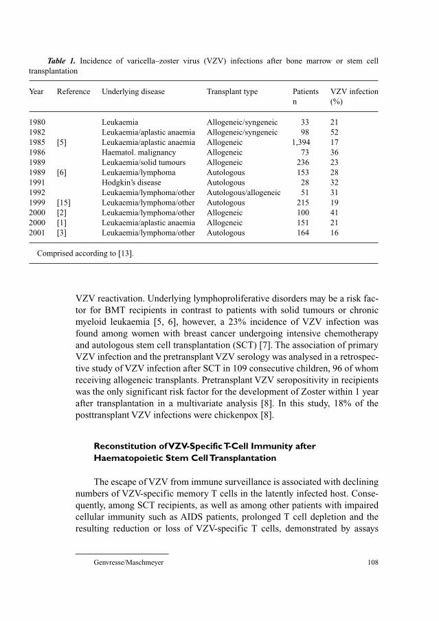
Genvresse/Maschmeyer 108
VZV reactivation. Underlying lymphoproliferative disorders may be a risk fac-
tor for BMT recipients in contrast to patients with solid tumours or chronic
myeloid leukaemia [5, 6], however, a 23% incidence of VZV infection was
found among women with breast cancer undergoing intensive chemotherapy
and autologous stem cell transplantation (SCT) [7]. The association of primary
VZV infection and the pretransplant VZV serology was analysed in a retrospec-
tive study of VZV infection after SCT in 109 consecutive children, 96 of whom
receiving allogeneic transplants. Pretransplant VZV seropositivity in recipients
was the only significant risk factor for the development of Zoster within 1 year
after transplantation in a multivariate analysis [8]. In this study, 18% of the
posttransplant VZV infections were chickenpox [8].
Reconstitution of VZV-Specific T-Cell Immunity afterHaematopoietic Stem Cell Transplantation
The escape of VZV from immune surveillance is associated with declining
numbers of VZV-specific memory T cells in the latently infected host. Conse-
quently, among SCT recipients, as well as among other patients with impaired
cellular immunity such as AIDS patients, prolonged T cell depletion and the
resulting reduction or loss of VZV-specific T cells, demonstrated by assays
Table 1. Incidence of varicella–zoster virus (VZV) infections after bone marrow or stem cell
transplantation
Year Reference Underlying disease Transplant type Patients VZV infection
n (%)
1980 Leukaemia Allogeneic/syngeneic 33 21
1982 Leukaemia/aplastic anaemia Allogeneic/syngeneic 98 52
1985 [5] Leukaemia/aplastic anaemia Allogeneic 1,394 17
1986 Haematol. malignancy Allogeneic 73 36
1989 Leukaemia/solid tumours Allogeneic 236 23
1989 [6] Leukaemia/lymphoma Autologous 153 28
1991 Hodgkin’s disease Autologous 28 32
1992 Leukaemia/lymphoma/other Autologous/allogeneic 51 31
1999 [15] Leukaemia/lymphoma/other Autologous 215 19
2000 [2] Leukaemia/lymphoma/other Allogeneic 100 41
2000 [1] Leukaemia/aplastic anaemia Allogeneic 151 21
2001 [3] Leukaemia/lymphoma/other Autologous 164 16
Comprised according to [13].
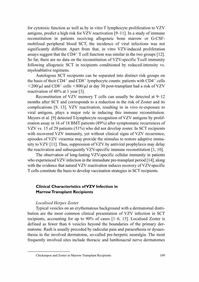
Chickenpox and Zoster in Marrow Transplant Recipients 109
for cytotoxic function as well as by in vitro T lymphocyte proliferation to VZV
antigens, predict a high risk for VZV reactivation [9–11]. In a study of immune
reconstitution in patients receiving allogeneic bone marrow or G-CSF-
mobilised peripheral blood SCT, the incidence of viral infections was not
significantly different. Apart from that, in vitro VZV-induced proliferation
assays suggest that the CD4� T cell function was similar in the two groups [12].
So far, there are no data on the reconstitution of VZV-specific T-cell immunity
following allogeneic SCT in recipients conditioned by reduced-intensity vs.
myeloablative regimens.
Autologous SCT recipients can be separated into distinct risk groups on
the basis of their CD4� and CD8� lymphocyte counts: patients with CD4� cells
�200/�l and CD8� cells �800/�l at day 30 post-transplant had a risk of VZV
reactivation of 48% at 1 year [3].
Reconstitution of VZV memory T cells can usually be detected at 9–12
months after SCT and corresponds to a reduction in the risk of Zoster and its
complications [9, 13]. VZV reactivation, resulting in in vivo re-exposure to
viral antigens, plays a major role in inducing this immune reconstitution.
Meyers et al. [9] detected T-lymphocyte recognition of VZV antigens by prolif-
eration assay in 16 of 18 BMT patients (89%) after symptomatic recurrences of
VZV vs. 15 of 29 patients (51%) who did not develop zoster. In SCT recipients
with recovered VZV immunity, yet without clinical signs of VZV recurrence,
episodes of VZV viraemia may provide the stimulus to restore adaptive immu-
nity to VZV [11]. Thus, suppression of VZV by antiviral prophylaxis may delay
the reactivation and subsequently VZV-specific immune reconstitution [1, 10].
The observation of long-lasting VZV-specific cellular immunity in patients
who experienced VZV infection in the immediate pre-transplant period [14], along
with the evidence that natural VZV reactivation induces recovery of VZV-specific
T cells constitute the basis to develop vaccination strategies in SCT recipients.
Clinical Characteristics of VZV Infection in Marrow Transplant Recipients
Localised Herpes ZosterTypical vesicles on an erythematous background with a dermatomal distri-
bution are the most common clinical presentation of VZV infection in SCT
recipients, accounting for up to 90% of cases [1–6, 15]. Localised Zoster is
defined as fewer than 6 vesicles beyond the boundaries of the primary der-
matome. Rash is usually preceded by radicular pain and paraesthesia or dysaes-
thesia in the involved dermatome, so-called pre-herpetic neuralgia. The most
frequently involved sites include thoracic and lumbosacral nerve dermatomes
Genvresse/Maschmeyer 110
followed by cranial and cervical dermatomes [2, 3, 8, 15]. Delayed or inade-
quate host response results in an average time for cessation of new lesion
formation of 8 days, compared with 3–5 days in the immunocompetent host.
The time to complete healing is also prolonged.
Local scarring and bacterial superinfection are also more common in SCT
recipients [2, 5, 6, 15] and significantly more frequent among patients with VZV
during the first 9 months after transplant than among patients with late infections
[5]. However, postherpetic neuralgia remains the most common complication,
observed in 32–41% of affected SCT recipients [2, 3, 14], who therefore appear
to be at higher risk compared to immuncompetent individuals [5].
Some SCT patients have a chronic cutaneous VZV reactivation, indicating
continued viral replication in ganglia and skin and a failure of the host response
to overcome the immune evasion mechanisms of the virus.
Cutaneous and Visceral DisseminationDisseminated Zoster consists of 6 skin lesions beyond the boundaries of
the primary dermatome and/or visceral involvement. Before the introduction of
VZV prophylaxis, 45% of SCT recipients experienced cutaneous and visceral
dissemination with an overall mortality of up to 10% [5]. More recent studies
using antiviral prophylaxis showed incidences of cutaneous dissemination of
3.8–25% among autologous SCT recipients [3, 15] and 17–38% after allo-
geneic BMT [1, 2], but a very low rate of visceral dissemination (0–7%).
Consequently, the mortality from VZV infection after BMT is now below 1%.
The most common sites of VZV organ infection are lungs and liver, result-
ing in pneumonia, hepatitis and intravascular coagulopathy, but myelitis and
meningoencephalitis have been reported as well [16]. The fatality of these dis-
seminated visceral forms approaches 30–50%. Fatal disseminated VZV infec-
tions are most often due to pneumonia [4].
There has been a small number of case reports of BMT patients where vis-
ceral disseminated disease was not preceded by any skin involvement [17, 18],
demonstrating that viraemia can occur without replication of the virus in the
skin, presumably by entry of virus into T cells that traffic through sensory gan-
glia. These rare presentations may remain undiagnosed.
Second Episodes of ZosterSome SCT recipients have recurrent zoster within days after discontinuation
of antiviral therapy, indicating a failure to re-establish latency in the short-term.
ChickenpoxChickenpox is defined as generalised onset of vesicular skin eruptions
without dermatomal localisation in a VZV seronegative patient without clinical
Chickenpox and Zoster in Marrow Transplant Recipients 111
history of previous chickenpox. Before the introduction of ACV prophylaxis,
chickenpox emerged in 25% of children during the first 6 months after BMT
[19]. After introduction of low-dose ACV and/or GCV prophylaxis, this rate has
dropped to 5.5% [8].
However, because the absence of previous VZV infection (history of
chickenpox and presence of VZV-specific antibodies) does not exclude the pos-
sibility of post-transplant VZV reactivation [8], chickenpox cannot be distin-
guished from disseminated cutaneous Zoster, so that both clinical entities are
often referred as ‘varicella-like’ cutaneous involvement accounting for 17% in
adult SCT recipients [11].
Management of VZV Infections in Marrow Transplant Recipients
DiagnosisThe diagnosis of VZV infection is established by clinical findings.
However, confirmation by rapid, sensitive laboratory methods such as immuno-
fluorescence, enzyme immunoassay or PCR is essential in many patients due to
unusual clinical manifestations of VZV infection in this population. Serologic
diagnosis of VZV reactivation is not reliable in SCT patients because the rise of
VZV IgG antibodies or detection of IgM antibodies is delayed or absent.
Detection of a PCR product after amplification of VZV DNA from peripheral
blood mononuclear cells or from plasma or serum to document VZV viraemia
in SCT patients appears compelling. In a retrospective survey, transient VZV
viraemia was documented with this method in 19% of the allogeneic BMT
recipients [2]. In cases of visceral disseminated VZV infection without skin
involvement, VZV DNA can be detected in the serum by PCR [18, 20].
Antiviral TreatmentThe recommended therapy for chickenpox or zoster in BMT patients is
intravenous (iv) ACV at a dosage of 10 mg/kg or 500 mg/m2 every 8 h for at
least 7 days. This treatment has been shown to prevent the dissemination of the
disease, even when it is initiated only 6 days after the onset of rash [21, 22]. For
localised dermatomal zoster, oral ACV, 800 mg every 4–5 h, was compared with
i.v. ACV in a small randomised study in allogeneic SCT patients, and the out-
come was comparable [23]. Oral famciclovir in a dosage of 500 mg every 8 h
was compared with oral ACV (800 mg q 4–5 h) in SCT, solid organ transplant
and oncological patients, and the results indicated similar efficacy [24]. No
controlled study has been performed with valacyclovir given for the treatment
Genvresse/Maschmeyer 112
of zoster in SCT recipients, but it is likely that valacyclovir would have compa-
rable antiviral effects in this patient group as well.
VZV resistance to ACV is rare but has been reported after BMT [25]. In
some cases, ACV-resistant VZV strains emerge during therapy, particularly under
prolonged, low-dose regimens. Because most resistant VZV strains show muta-
tions in thymidine kinase, ACV, valacyclovir, and famciclovir will be ineffective.
VZV-ImmunoglobulinVaricella–zoster immune globulin is the recommended prophylactic mea-
sure in seronegative SCT recipients after a close or household exposure to vari-
cella, if it can be given within 4 days of exposure [26]. Varicella–zoster immune
globulin is not used in patients with zoster, because VZV IgG titres are main-
tained despite the loss of cell-mediated immunity to the virus. There is no evi-
dence that antibody prophylaxis increases antibody titres or reduces the risk of
VZV reactivation after SCT.
Antiviral ProphylaxisBecause the median onset of VZV reactivation is 5–7 months after SCT, the
duration of antiviral prophylaxis must be long enough to prevent VZV disease. A
summary of studies of long-term ACV prophylaxis after BMT is presented in
table 2. Two randomised, controlled studies have been performed comparing 6
months of prophylactic ACV with placebo: in the first one, i.v. ACV (250 mg/m2
every 8 h, starting 5 days before transplantation) for 5 weeks and followed by oral
ACV (1,200 mg daily) prevented zoster in allogeneic BMT patients [27]. Similar
results were obtained in a study on higher doses of ACV (3,200 mg daily) [28].
Both studies, along with a trial in autologous BMT recipients [29], showed that
although reactivation was suppressed during the period of prophylaxis, no overall
reduction of the VZV reactivation rate could be achieved 12 months after trans-
plantation, due to delayed onset after ACV discontinuation. Possibly, prolonged
(�6 months) oral antiviral prophylaxis after transplantation could reduce the inci-
dence of VZV infection post-BMT, allowing patients to restore their cellular
immunity. This was studied by Kanda et al. [30], using low-dose ACV (400 mg
p.o. daily) continued until the end of immunosuppressive therapy after SCT.
Thereby, the first-year cumulative incidence of VZV reactivation could be
reduced to 29%. Low-dose prophylaxis might permit subclinical VZV reactiva-
tion, leading to an immune recovery against VZV. The major concern against pro-
longed low-dose ACV prophylaxis is the emergence of VZV strains resistant to
ACV. However, in studies on low-dose ACV prophylaxis, no breakthrough reacti-
vation of VZV could be observed and symptomatic VZV reactivation after dis-
continuing ACV was always successfully treated with a therapeutic dose of ACV
[1, 30]. Another concern is the cost of long-term prophylaxis.
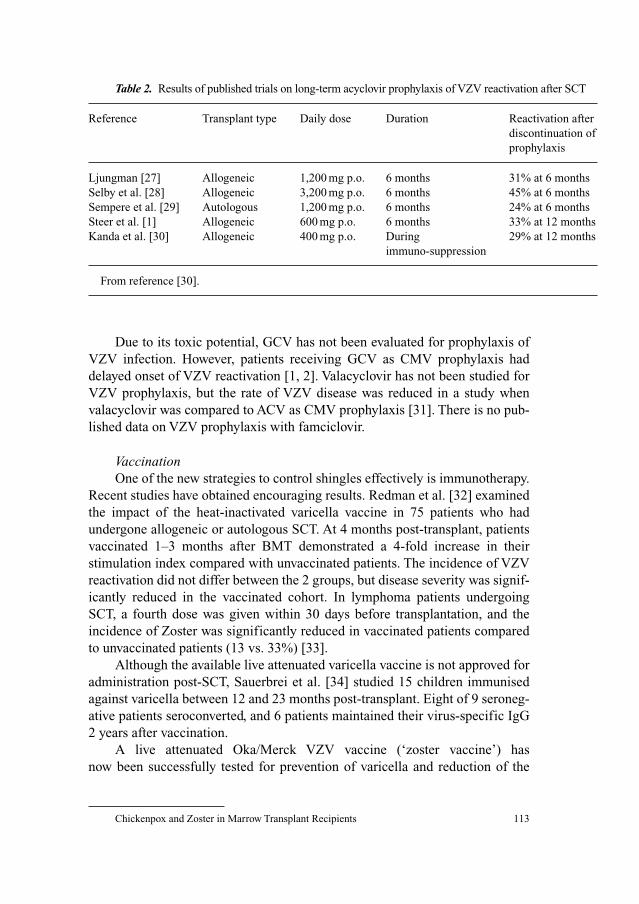
Chickenpox and Zoster in Marrow Transplant Recipients 113
Due to its toxic potential, GCV has not been evaluated for prophylaxis of
VZV infection. However, patients receiving GCV as CMV prophylaxis had
delayed onset of VZV reactivation [1, 2]. Valacyclovir has not been studied for
VZV prophylaxis, but the rate of VZV disease was reduced in a study when
valacyclovir was compared to ACV as CMV prophylaxis [31]. There is no pub-
lished data on VZV prophylaxis with famciclovir.
VaccinationOne of the new strategies to control shingles effectively is immunotherapy.
Recent studies have obtained encouraging results. Redman et al. [32] examined
the impact of the heat-inactivated varicella vaccine in 75 patients who had
undergone allogeneic or autologous SCT. At 4 months post-transplant, patients
vaccinated 1–3 months after BMT demonstrated a 4-fold increase in their
stimulation index compared with unvaccinated patients. The incidence of VZV
reactivation did not differ between the 2 groups, but disease severity was signif-
icantly reduced in the vaccinated cohort. In lymphoma patients undergoing
SCT, a fourth dose was given within 30 days before transplantation, and the
incidence of Zoster was significantly reduced in vaccinated patients compared
to unvaccinated patients (13 vs. 33%) [33].
Although the available live attenuated varicella vaccine is not approved for
administration post-SCT, Sauerbrei et al. [34] studied 15 children immunised
against varicella between 12 and 23 months post-transplant. Eight of 9 seroneg-
ative patients seroconverted, and 6 patients maintained their virus-specific IgG
2 years after vaccination.
A live attenuated Oka/Merck VZV vaccine (‘zoster vaccine’) has
now been successfully tested for prevention of varicella and reduction of the
Table 2. Results of published trials on long-term acyclovir prophylaxis of VZV reactivation after SCT
Reference Transplant type Daily dose Duration Reactivation after
discontinuation of
prophylaxis
Ljungman [27] Allogeneic 1,200 mg p.o. 6 months 31% at 6 months
Selby et al. [28] Allogeneic 3,200 mg p.o. 6 months 45% at 6 months
Sempere et al. [29] Autologous 1,200 mg p.o. 6 months 24% at 6 months
Steer et al. [1] Allogeneic 600 mg p.o. 6 months 33% at 12 months
Kanda et al. [30] Allogeneic 400 mg p.o. During 29% at 12 months
immuno-suppression
From reference [30].

Genvresse/Maschmeyer 114
burden-of-illness caused by herpes zoster in a large placebo-controlled trial in
healthy adults [35]. To what extent the inactivated vaccines will protect patients
after stem cell transplantation from zoster has to be addressed.
Recovery of VZV immunity in BMT patients might be further improved by
immunising donors and recipients before allogeneic transplantation or by initi-
ating the vaccine regimen at a shorter interval after transplantation.
References
1 Steer CB, Szer J, Sasadeusz J, Matthews JP, Beresford JA, Grigg A: Varicella-zoster infection after
allogeneic bone marrow transplantation: incidence, risk factors and prevention with low-dose aci-
clovir and ganciclovir. Bone Marrow Transplant 2000;25:657–664.
2 Koc Y, Miller KB, Schenkein DP, Griffith J, Akhtar M, DesJardin J, Snydman DR: Varicella zoster
virus infections following allogeneic bone marrow transplantation: frequency, risk factors, and
clinical outcome. Biol Blood Marrow Transplant 2000;6:44–49.
3 Offidani M, Corvatta L, Olivieri A, Mele A, Brunori M, Montanari M, Rupoli S, Scalari P, Leoni P:
A Predictive model of varicella-zoster virus infection after autologous peripheral blood progenitor
cell transplantation. Clin Infect Dis 2001;32:1414–1422.
4 Han CS, Miller W, Haake R, Weisdorf D: Varicella zoster infection after bone marrow trans-
plantation: incidence, risk factors and complications. Bone Marrow Transplant 1994;4:191–194.
5 Locksley RM, Flournoy N, Sullivan KM, Meyers JD: Infection with varicella-zoster virus after
marrow transplantation. J Infect Dis 1985;152:1172–1181.
6 Schuchter LM, Wingard JR, Piantadosi S, Burns WH, Santos GW, Saral R: Herpes zoster infection
after autologous bone marrow transplantation. Blood 1989;74:1424–1427.
7 Chandrasekar PH, Abraham OC, Klein J, Alangaden G, Chalasani G, Cassells L, Dansey R, Abella S,
Karanes C, Peters W, Baynes R: Low infectious morbidity after intensive chemotherapy and
autologous peripheral blood progenitor cell transplantation in the outpatient setting for women
with breast cancer. Clin Infect Dis 2001;32:546–551.
8 Leung TF, Chik KW, Li CK, Lai H, Shing MMK, Chan PKS, Lee V, Yuen PMP: Incidence, risk
factors and outcome of varicella-zoster virus infection in children after haematopoietic stem cell
transplantation. Bone Marrow Transplant 2000;25:167–172.
9 Meyers JD, Flournoy N, Thomas ED: Cell-mediated immunity to varicella-zoster virus after allo-
geneic marrow transplant. J Infect Dis 1980;141:479–487.
10 Ljungman P, Lonnqvist B, Gahrton G, Ringden O, Sundqvist VA, Wahren B: Clinical and subclin-
ical reactivations of varicella-zoster virus in immunocompromised patients. J Infect Dis 1986;153:
840–847.
11 Wilson A, Sharp M, Koropchak CM, Ting SF, Arvin AM: Subclinical varicella-zoster virus
viremia, herpes zoster, and T lymphocyte immunity to varicella-zoster viral antigens after bone
marrow transplantation. J Infect Dis 1992;165:119–126.
12 Storek J, Dawson MA, Storer B, Stevens-Ayers T, Maloney DG, Marr KA, Witherspoon RP,
Bensinger W, Flowers MED, Martin P, Storb R, Appelbaum FR, Boeckh M: Immune reconstitu-
tion after allogeneic marrow transplantation compared with blood stem cell transplantation. Blood
2001;97:3380–3389.
13 Arvin AM: Varicella-zoster virus: pathogenesis, immunity, and clinical management in
hematopoietic cell transplant recipients. Biol Blood Marrow Transplant 2000;6:219–230.
14 Kato S, Yabe H, Yabe M, Kimura M, Ito M, Tsuchida F, Tsuji K, Takahashi M: Studies on transfer
of varicella-zoster virus specific T-cell immunity from bone marrow donor to recipient. Blood
1990;75:806–809.
15 Bilgrami S, Chakraborty NG, Rodriguez-Pinero F, Khan AM, Feingold JM, Bona RD, Edwards RL,
Dorsky D, Clive J, Mukherji B, Tutschka PJ: Varicella zoster virus infection associated with high-
dose chemotherapy and autologous stem-cell rescue. Bone Marrow Transplant 1999;23: 469–474.

Chickenpox and Zoster in Marrow Transplant Recipients 115
16 Tauro S, Toh V, Osman H, Mahendra P: Varicella zoster meningoencephalitis following treatment
for dermatomal zoster in an alloBMT patient. Bone Marrow Transplant 2000;26:795–796.
17 Stemmer SM, Kinsman K, Tellschow S, Jones RB: Fatal noncutaneous visceral infection with
varicella-zoster virus in a patient with lymphoma after autologous bone marrow transplantation.
Clin Infect Dis 1993;16:497–499.
18 Yagi T, Karasuno T, Hasegawa T, Yasumi M, Kawamoto S, Murakami M, Uosima N, Nakamura H,
Hiraoka A, Masaoka T: Acute abdomen without cutaneous signs of varicella zoster virus infection
as a late complication of allogeneic bone marrow transplantation: importance of empiric therapy
with acyclovir. Bone Marrow Transplant 2000;25:1003–1005.
19 Wacker P, Hartmann O, Benhamou E, Salloum E, Lemerle J: Varicella-zoster virus infections after
autologous bone marrow transplantation in children. Bone Marrow Transplant 1989;4:191–194.
20 Rogers SY, Irving W, Harris A, Russel NH: Visceral varicella zoster infection after bone marrow
transplantation without skin involvement and the use of PCR for diagnosis. Bone Marrow
Transplant 1995;15:805–807.
21 Balfour HH Jr, Bean B, Laskin OL, Ambinder RF, Meyers JD, Wade JC, Zaia JA, Aeppli D, Kirk
LE, Segreti AC, Keeney RE: Acyclovir halts progression of herpes zoster in immunocompromised
patients. N Engl J Med 1983;308:1448–1453.
22 Shepp DH, Dandliker PS, Meyers JD: Treatment of varicella-zoster virus infection in severely
immunocompromised patients. A randomised comparison of acyclovir and vidarabine. N Engl J
Med 1986;314:208–212.
23 Ljungman P, Lönnqvist B, Ringdén O, Skinhöj P, Gahrton G: A randomised trial of oral versus
intravenous acyclovir for treatment of herpes zoster in bone marrow transplant recipients. Nordic
Bone Marrow Transplant Group. Bone Marrow Transplant 1989;4:613–615.
24 Tyring S, Belanger R, Bezwoda W, Ljungman P, Boon R, Saltzman RL: A randomised, double-
blind trial of famciclovir versus acyclovir for the treatment of localized dermatomal herpes zoster
in immunocompromised patients. Cancer Invest 2001;19:13–22.
25 Reusser P, Cordonnier C, Einsele H, Engelhard D, Link H, Locasciulli A, Ljungman P, for the
Infectious Diseases Working Party of the European Group for Blood and Marrow Transplantation
(EBMT): European survey of herpesvirus resistance to antiviral drugs in bone marrow transplant
recipients. Bone Marrow Transplant 1996;17:813–817.
26 Centers for Disease Control (CDC): Guidelines for preventing opportunistic infections among
hematopoietic stem cell transplant recipients. Recommendations of the CDC, the Infectious
Disease Society of America, and the American Society of Blood and Marrow Transplantation.
MMWR Morb Mortal Wkly Rep 2000;49:1–125.
27 Lundgren G, Wilczek H, Lönnqvist B, Lindholm A, Wahren B, Ringdén O: Acyclovir prophylaxis
in bone marrow transplant recipients. Scand J Infect Dis 1985;47(suppl):137–144.
28 Selby PJ, Powles RL, Easton D, Perren TJ, Stolle K, Jameson B, Fiddian AP, Tryhorn Y, Stern H:
The prophylactic role of intravenous and long-term oral acyclovir after allogeneic bone marrow
transplantation. Br J Cancer 1989;59:434–438.
29 Sempere A, Sanz GF, Senent L, de la Rubia J, Jarque I, López F, Arilla MJ, Guinot M, Martín G,
Martínez K, Marty ML, Sanz MA: Long-term acyclovir for prevention of varicella zoster virus
infection after autologous blood stem cell transplantation in patients with acute leukemia. Bone
Marrow Transplant 1992;10:495–498.
30 Kanda Y, Mineishi S, Saito T, Saito A, Yamada S, Ohnishi M, Chizuka A, Niiya H, Suenaga K,
Nakai K, Takeuchi T, Makimoto A, Tanosaki R, Kami M, Tanaka Y, Fujita S, Watanabe T,
Kobayashi Y, Tobinai K, Takaue Y: Long-term acyclovir against varicella-zoster virus reactivation
after allogeneic hematopoietic stem cell transplantation. Bone Marrow Transplant 2001;28:
689–692.
31 Ljungman P, Aschan J, Lewensohn-Fuch I, Carlens S, Larsson K, Lönnqvist B, Mattsson J,
Sparrelid E, Winiarski J, Ringdén O: Results of different strategies for reducing cytomegalovirus-
associated mortality in allogeneic stem cell transplant recipients. Transplantation 1998;66:
1330–1334.
32 Redman RL, Nader S, Zerboni L, Liu C, Wong RM, Brown BW, Arvin AM: Early reconstitution
of immunity and decreased severity of herpes zoster in bone marrow transplant recipients immu-
nized with inactivated varicella vaccine. J Infect Dis 1997;176:578–585.

Genvresse/Maschmeyer 116
33 Hata A, Asanuma H, Rinki M, Sharp M, Wong RM, Blume K, Arvin AM: Use of an inactivated
varicella vaccine in recipients of hematopoietic-cell transplants. N Engl J Med 2002;347:26–34.
34 Sauerbrei A, Prager J, Hengst U, Zintl F, Wutzler P: Varicella vaccination in children after bone
marrow transplantation. Bone Marrow Transplant 1997;20:381–383.
35 Oxman MN, Levin MJ, Johnson GR, Shamder KE et al. A vaccine to prevent herpes zoster and
postherpetic neuralgia in older adults. N Engl J Med 2005;352:2271–2284.
Georg Maschmeyer, MD
Professor of Internal Medicine, Director
Department of Haematology and Oncology, Klinikum Ernst von Bergmann
Charlottenstrasse 72
DE–14467 Potsdam (Germany)
Tel. �49 331 241 6001, Fax �49 331 241 6000, E-Mail [email protected]
Gross G, Doerr HW (eds): Herpes Zoster.
Monogr Virol. Basel, Karger, 2006, vol 26, pp 117–122
General Aspects of Therapy
G. Gross
Department of Dermatology and Venereology, University of Rostock,
Rostock, Germany
Patients suffering from herpes zoster should be encouraged to see a physi-
cian as early as possible for immediate medical care based on administration of
systemic antiviral therapy. In addition symptomatic local therapy and analgetic
therapy in order to achieve painlessness are equally important. Since years it
has become clear, that systemic antiviral therapy is indicated for most patients
suffering from herpes zoster. In general the aims of therapy for herpes zoster
comprise the following: decrease viral replication as early as possible, thus low-
ering the viral load, accelerate healing, limit or relieve severity and duration of
acute and chronic pain (postherpetic neuralgia, PHN). Further options are to
prevent or alleviate other acute and chronic herpes zoster complications and
reduce the risk of cutaneous extension and visceral dissemination of VZV,
which is particularly a problem in immunocompromised patients. Alternative
therapies such as hypnosis and others are definitely of little help in terms of
prevention of acute and chronic complications. Therefore, these therapies
should be omitted. Patients with herpes zoster involving cranial nerves, such as
ophthalmic zoster and zoster oticus, must be referred as early as possible for
specialist ophthalmic and otorhinolaryngologic management. There is available
evidence from clinical trials that early treatment with oral antiviral agents
achieves many of the mentioned aims [1–3]. The risk of ocular complications is
reduced from about 50% to 20–30% with oral antiviral therapy.
Antiviral Therapy
Systemic antiviral therapy is urgently indicated in herpes zoster patients
beyond the age of 50 years and in patients of any age, including children, with
herpes zoster affecting the head and neck area, especially zoster ophthalmicus
Therapy

Gross 118
and zoster oticus (table 1). Severe herpes zoster of the trunk and extremities as
well as herpes zoster in immunodeficient patients and patients with atopic der-
matitis or other eczema are also indications for antiviral therapy. Furthermore,
zoster ophthalmicus and zoster oticus as well as age over 50 years are major risk
factors for the development of PHN. Some authorities consider antiviral treat-
ment to be only optional in patients younger than 50 years of age with shingles
on the trunk or on the extremities. Nevertheless, antiviral therapy has potential
benefit and bears a limited risk. The same is true for immunocompetent chil-
dren. However, as a rule patients younger than 50 years should be checked sero-
logically for HIV-antibodies, because herpes zoster is an important marker for
HIV-infection and AIDS.
In 1998, the Zoster Study Group of the Dermatological Research
Cooperative Group (ADF) associated with the German Dermatology Society
(DDG) published a PHN prevention zoster score, which has shown to be an use-
ful aid for initiating systemic antiviral therapy [4, 5]. The zoster score is taking
into account the following risk factors: age beyond 50 years, female gender,
presence of more than 50 herpes zoster vesicles, haemorrhagic lesions and her-
pes zoster involvement of cranial and sacral dermatomes.
Four different antivirals (acyclovir, valacyclovir, famciclovir and brivudin)
(fig. 1) have become available for the treatment of herpes zoster over the years.
All of these are nucleoside analogues, which interfere with viral nucleic acid
chain synthesis, thereby inhibiting viral replication. In more recent years, oral
acyclovir has been replaced by its prodrug valacyclovir in the oral treatment of
herpes zoster. Acyclovir has a comparable lower oral bioavailability (20%) than
valacyclovir. Another nucleoside analogue is penciclovir, which has a similar
activity and mechanism compared to acyclovir. Its prodrug famciclovir is used
for oral treatment of VZV infections (herpes zoster and chickenpox). While
Table 1. Indications for systemic antiviral therapy of herpes zoster
Urgent indicationsHerpes zoster at any site of the body in patients beyond the age of 50
Herpes zoster in the head/neck area of patients at any age
Severe herpes zoster on the trunk and on the limb
Herpes zoster in immunodeficient patients
Herpes zoster in patients with severe atopic dermatitis and severe
eczema
Relative indicationsHerpes zoster on the trunk/on the extremities in patients younger
than 50 years
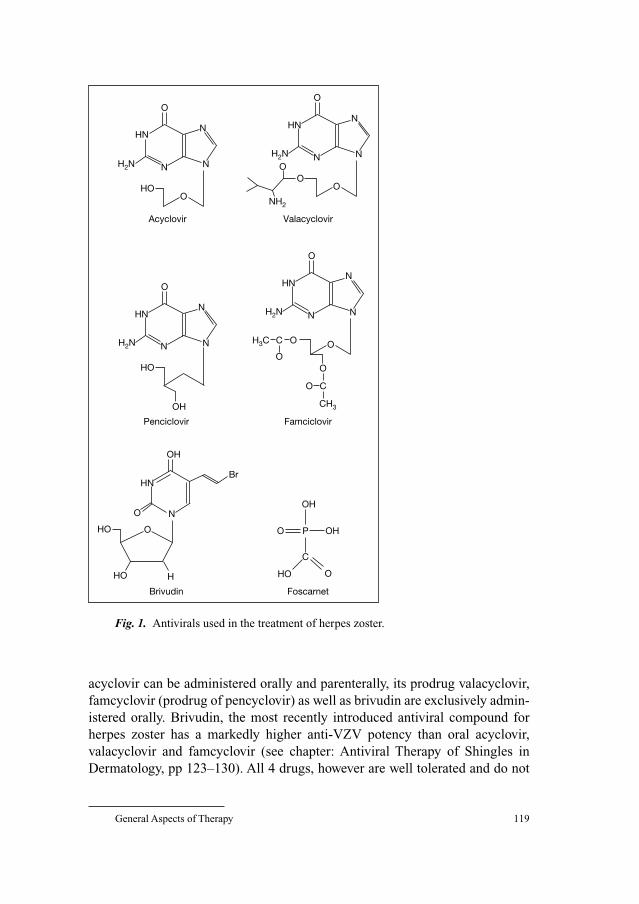
General Aspects of Therapy 119
acyclovir can be administered orally and parenterally, its prodrug valacyclovir,
famcyclovir (prodrug of pencyclovir) as well as brivudin are exclusively admin-
istered orally. Brivudin, the most recently introduced antiviral compound for
herpes zoster has a markedly higher anti-VZV potency than oral acyclovir,
valacyclovir and famcyclovir (see chapter: Antiviral Therapy of Shingles in
Dermatology, pp 123–130). All 4 drugs, however are well tolerated and do not
Valacyclovir
HN
H2N N
O
OO
NH2
ON
N
Acyclovir
HN
H2N N
O
O
HO
N
N
Penciclovir
HN
H2N N
O
HO
OH
N
N
Famciclovir
HN
H2N N
O
O
OCH3C
CH3
OO
CO
N
N
Brivudin
HN
HO
HO
O
O
N
Br
H
OH
Foscarnet
O
HO O
C
P
OH
OH
Fig. 1. Antivirals used in the treatment of herpes zoster.
Gross 120
differ widely in terms of efficacy and safety. However, because of improved
pharmaco-kinetic properties and simpler dosing regimes, recently oral valacy-
clovir, famcyclovir and brivudin have been used preferentially in comparison to
acyclovir. While acyclovir, valacyclovir and famcyclovir represent the common
antiviral treatment used for herpes zoster so far, brivudin has become actually a
frequently used antiviral compound for this indication particularly in European
countries [6].
In order to achieve successful results in patients with herpes zoster,
antiviral therapy should be started as early as possible (i.e. within 48 h to a
maximum of 72 h after onset of rash) and should be continued strictly for 7
days. A major limitation of antiviral therapy is the delay between onset of
symptoms and initiation of antiviral therapy. Although it is well accepted that
the likelihood of beneficial effects is higher with earlier intervention [7],
some patients will benefit from antiviral therapy even if it is started after
72 h. Patients with disseminated herpes zoster and evidence of immunodefi-
ciency should be always treated antivirally even later than 72 h after onset of
the rash.
When Intravenous Antiviral Therapy has to be Considered?
The standard antiviral treatment for herpes zoster in immunodeficient
patients is acyclovir intravenously in a dose of 8–10 mg per kg body weight,
3 times daily during 7–14 days [8]. While intravenous acyclovir was com-
pared with brivudin in a study of Wutzler et al. [9], no controlled studies com-
paring intravenous acyclovir with oral acyclovir, valacyclovir or famcyclovir
have been performed in immunodeficient persons suffering from herpes
zoster.
Other conditions where intravenous acyclovir therapy has to be considered
are particularly herpes zoster ophthalmicus and herpes zoster oticus at any age.
This applies also for children with herpes zoster affecting the VIth, the VIIth
and the VIIIth cranial nerve. In view of the unpredictable course, herpes zoster
in patients with HIV infection, herpes zoster in immunodeficient children and
also in atopic children (prevention of scar formation) should be treated with
acyclovir intravenously. Elderly and debilitated patients under multidrug ther-
apy are a further group of persons that should be considered to be treated with
acyclovir parenterally under stationary conditions. Herewith, harmful under-
treatment due to mistakes taking antiviral drugs continuously is intended to
prevent.
General Aspects of Therapy 121
Symptomatic Therapy
Adequate control of acute symptoms is important for optimal therapy. So
concurrent symptomatic treatment of cutaneous zoster lesions is always necces-
sary. Helpful measures for the local management include keeping lesions clean,
dry and protected with a sterile nonadherent dressing. However, topical anti-
viral agents have shown to be ineffective and thus definitely have no place in
the management of shingles [2, 3]. Topical lotions such as lotio alba, vioform
zinc mixture etc. or crust removal of older lesions using an antisepic cream may
help to control burning and itching which are common symptoms associated
with acute stage herpes zoster.
Painlessness is the most important goal of therapy in the acute phase of
herpes zoster. Consequent control of acute pain may prevent mechanisms
thought to be related to the development of PHN [10]. It is anticipated that the
risk of PHN might be reduced by a combined treatment consisting of a systemic
antiviral drug and an appropriately-dosed analgesic, a tricyclic antidepressant,
an antiepileptic or an adjunctive therapy such as gabapentin or pregabalin [2, 3].
So far, however it has not been demonstrated clearly, that systematic analgesia
in acute herpes zoster prevents the development of PHN. Acute pain manage-
ment in children with zoster can usually be accomplished with ibuprofen. For
acute severe pain codeine may be helpful [11].
The Role of Corticisteroids in the Management of Herpes Zoster
Oral corticosteroids are not recommended for the treatment of herpes
zoster [2, 3]. Some countries, however have approved them for combined
therapy with oral acyclovir therapy [12] in patients not being at risk for
corticosteroid-related complications.
Two large prospective studies [3, 13] showed that herpes zoster patients
treated with a combination therapy consisting of oral acyclovir and prednisone
had an improved quality of life and some improvement in acute herpes zoster
symptoms, particularly in alleviation of acute pain. However, combination of
oral acyclovir and corticosteroids had no effect on the incidence of postherpetic
neuralgia. The available study results have been achieved with a combination
therapy of oral acyclovir and oral prednisolone (60 mg per day, dose reduction
over about 10–14 days, to 5 mg per day). As a conclusion, use of oral corticos-
teroids with systemic antiviral therapy, if considered must be very carefully out-
weighed with the risk of adverse effects [12].

Gross 122
References
1 Bansal R, Tutrone WD, Weinberg JM: Viral skin infections in the elderly: diagnosis and manage-
ment. Drugs Aging 2002;19:503–514.
2 Gnann JW Jr, Whitley RJ: Clinical practice. Herpes zoster. N Engl J Med 2002;347:340–346.
3 Whitley RJ, Weiss H, Gnann W, Tyring S, Mertz GJ, Pappas PG, Schleupner CJ, Hayden F, Wolf J,
Soong SJ: Acyclovir with and without prednisone for the treatment of herpes zoster. A randomized
placebo-controlled trial. The National Institute of Allergy and Infectious Diseases. Collaborative
Antiviral Study Group. Ann Intern Med 1996;125:376–383.
4 Meister W, Neiss A, Gross G, Doerr H, Holbel W, Malin J, von Essen J, Reimann B, Witke C,
Wutzler P: Demography, symptomatology, and course of disease in ambulatory zoster patients.
A physician-based survey in Germany. Intervirology 1998;41:272–277.
5 Meister W, Neiss A, Gross G, Doerr HW, Holbel W, Malin JP, von Essen J, Reimann BY, Witke C,
Wutzler P: A prognostic score for postherpetic neuralgia in ambulatory patients. Infection
1998;26:359–363.
6 Volpi A, Gross G, Hercogova J, Johnson RW: Current management of herpes zoster: the European
view. Am J Clin Dermatol 2005;6:317–325.
7 Schmader K: Herpes zoster in adults. Clin Infect Dis 2001;32:1481–1486.
8 Gross G, Schofer H, Wassilew S, Friese K, Timm A, Guthoff R, Pau HW, Malin JP, Wutzler P,
Doerr HW: Herpes zoster guideline of the German Dermatology Society (DDG). J Clin Virol
2003;26: 277–289.
9 Wutzler P, De Clercq E, Wutke K, Farber I: Oral brivudin vs. intravenous acyclovir in the treatment
of herpes zoster in immunocompromised patients: a randomized double-blind trial. J Med Virol
1995;46:252–257.
10 Dworkin RH, Perkins FM, Nagasako EM: Prospects for the prevention of postherpetic neuralgia
in herpes zoster patients. Clin J Pain 2000;16(suppl):90–100.
11 Feder HM Jr, Hoss DM: Herpes zoster in otherwise healthy children. Pediatr Infect Dis J
2004;23:451–457; quiz 458–460. Review.
12 Santee JA: Corticosteroids for herpes zoster: what do they accomplish? Clin Dermatol 2002;3:
517–524.
13 Wood MJ, Johnson RW, Mc Kendrick MW, Taylor J, Mandal BK, Crooks J: A randomized trial of
acyclovir for 7 days or 21 days with and without prednisone for treatment of acute herpes zoster.
N Eng J Med 1994;330:896–900.
Gerd Gross, MD
Professor of Dermatology and Venereology, Director
Department of Dermatology and Venereology
University of Rostock
Augustenstrasse 80–84
DE–18055 Rostock (Germany)
Tel. �49 381 4949700, Fax �49 381 4949702, E-Mail [email protected]

Gross G, Doerr HW (eds): Herpes Zoster.
Monogr Virol. Basel, Karger, 2006, vol 26, pp 123–130
Antiviral Therapy of Shingles inDermatology
H. Martina Lilie, Sawko W. Wassilew
Dermatology Department, Klinikum Krefeld, Krefeld, Germany
The major goals of therapy in patients with herpes zoster are:
(1) shortening of virus replication,
(2) prevention of dissemination of skin lesions,
(3) reduction of acute zoster-associated pain (ZAP),
(4) prevention of chronic ZAP and postherpetic neuralgia (PHN),
(5) prevention of other complications, e.g. ophthalmic involvement.
Recent research has shown that antiviral therapy with acyclovir, valacy-
clovir, famciclovir, and brivudin, started as early as possible, can significantly
shorten viral replication, prevent lesion dissemination and reduce intensity and
duration of ZAP particularly in elderly patients, provided that treatment is
started early in the course of disease. This suggests that antiviral therapy (table 1)
should be offered to all patients as soon as herpes zoster is diagnosed, preferably
within 72 h after onset of rash. In patients of any age with ophthalmic herpes
zoster and in all immunocompromised patients antiviral therapy should be
started even later as long as viral replication can be considered in skin and
nerves, e.g. as long as new blisters appear in the skin.
Dermatologists are trained to diagnose early skin lesions as herpes zoster
and should be consulted in time.
In the immunocompetent patient antiviral therapy should normally be
given for 7 days, whereas in the immunocompromised host antiviral therapy
should be given intravenously and for at least 10 days.
Immunocompetent Patients
AcyclovirAcyclovir (9-{2-hydroxyethoxymethyl}guanine) is a nucleoside analog. After
penetrating into the infected cells acyclovir is converted to its monophosphate
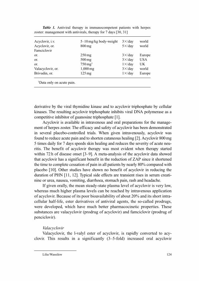
Lilie/Wassilew 124
derivative by the viral thymidine kinase and to acyclovir triphosphate by cellular
kinases. The resulting acyclovir triphosphate inhibits viral DNA polymerase as a
competitive inhibitor of guanosine triphosphate [1].
Acyclovir is available in intravenous and oral preparations for the manage-
ment of herpes zoster. The efficacy and safety of acyclovir has been demonstrated
in several placebo-controlled trials. When given intravenously, acyclovir was
found to reduce acute pain and to shorten cutaneous healing [2]. Acyclovir 800 mg
5 times daily for 7 days speeds skin healing and reduces the severity of acute neu-
ritis. The benefit of acyclovir therapy was most evident when therapy started
within 72 h of disease onset [3–9]. A meta-analysis of the acyclovir data showed
that acyclovir has a significant benefit in the reduction of ZAP since it shortened
the time to complete cessation of pain in all patients by nearly 80% compared with
placebo [10]. Other studies have shown no benefit of acyclovir in reducing the
duration of PHN [11, 12]. Typical side effects are transient rises in serum creati-
nine or urea, nausea, vomiting, diarrhoea, stomach pain, rash and headache.
If given orally, the mean steady-state plasma level of acyclovir is very low,
whereas much higher plasma levels can be reached by intravenous application
of acyclovir. Because of its poor bioavailability of about 20% and its short intra-
cellular half-life, ester derivatives of antiviral agents, the so-called prodrugs,
were developed, which have much better pharmacocinetic properties. These
substances are valacyclovir (prodrug of acyclovir) and famciclovir (prodrug of
penciclovir).
ValacyclovirValacyclovir, the l-valyl ester of acyclovir, is rapidly converted to acy-
clovir. This results in a significantly (3–5-fold) increased oral acyclovir
Table 1. Antiviral therapy in immunocompetent patients with herpes
zoster: management with antivirals, therapy for 7 days [30, 31]
Acyclovir, i.v. 5–10 mg/kg body-weight 3�/day world
Acyclovir, or. 800 mg 5�/day world
Famciclovir
or. 250 mg 3�/day Europe
or. 500 mg 3�/day USA
or. 750 mg1 1�/day UK
Valacyclovir, or. 1,000 mg 3�/day world
Brivudin, or. 125 mg 1�/day Europe
1Data only on acute pain.
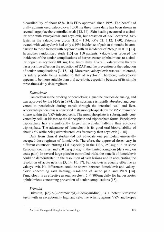
Antiviral Therapy of Shingles in Dermatology 125
bioavailability of about 65%. It is FDA approved since 1995. The benefit of
orally administered valacyclovir 1,000 mg three times daily has been shown in
several large placebo-controlled trials [13, 14]. Skin healing occurred at a simi-
lar time with valacyclovir and acyclovir, but cessation of ZAP occurred 34%
faster in the valacyclovir group (HR � 1.34, 95% CI: 1.12, 1.60). Patients
treated with valacyclovir had only a 19% incidence of pain at 6 months in com-
parison to those treated with acyclovir with an incidence of 26%, p � 0.02 [13].
In another randomized study [15] on 110 patients, valacyclovir reduced the
incidence of the ocular complications of herpes zoster ophthalmicus to a simi-
lar degree as acyclovir 800 mg five times daily. Overall, valacyclovir therapy
has a positive effect on the reduction of ZAP and the prevention or the reduction
of ocular complications [3, 15, 16]. Moreover, valacyclovir was well-tolerated,
its safety profile being similar to that of acyclovir. Therefore, valacyclovir
appears to be more suitable than oral acyclovir, especially because of its simple
three-times-daily dose regimen.
FamciclovirFamciclovir is the prodrug of penciclovir, a guanine nucleoside analog, and
was approved by the FDA in 1994. The substance is rapidly absorbed and con-
verted to penciclovir during transit through the intestinal wall and liver.
Afterwards penciclovir is converted to its monophosphate by the VZV thymidine
kinase within the VZV-infected cells. The monophosphate is subsequently con-
verted by cellular kinases to the diphosphate and triphosphate forms. Penciclovir
triphosphate has a significantly longer intracellular half-life than acyclovir
triphosphate. The advantage of famciclovir is its good oral bioavailability of
about 77% while being administered less frequently than acyclovir [3, 16].
Data from clinical studies did not advocate one particular, universally
accepted dose regimen of famciclovir. Therefore, the approved doses vary in
different countries: 500 mg t.i.d. especially in the USA, 250 mg t.i.d. in some
European countries, and 750 mg q.d. e.g. in the United Kingdom (data only on
acute pain). In several large placebo-controlled trials, the benefit of famciclovir
could be demonstrated in the resolution of skin lesions and in accelerating the
resolution of acute neuritis [3, 14, 16, 17]. Famciclovir is equally effective as
valacyclovir. No differences could be shown between famciclovir and valacy-
clovir concerning rash healing, resolution of acute pain and PHN [14].
Famciclovir is as effective as oral acyclovir 5 � 800 mg daily for herpes zoster
ophthalmicus concerning prevention of ocular complications [18].
BrivudinBrivudin, [(e)-5-(2-bromovinyl)-2�deoxyuridine], is a potent virostatic
agent with an exceptionally high and selective activity against VZV and herpes
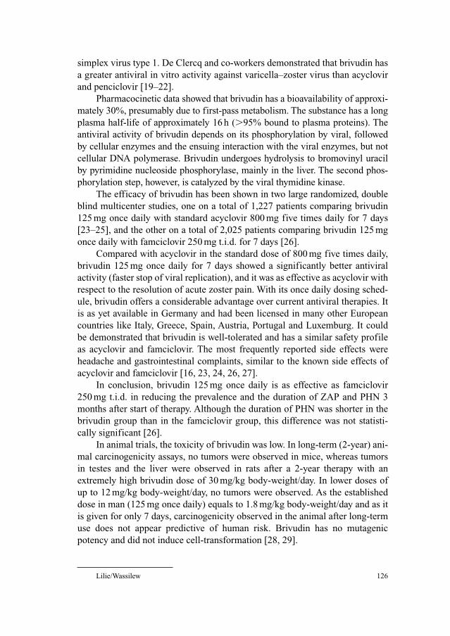
Lilie/Wassilew 126
simplex virus type 1. De Clercq and co-workers demonstrated that brivudin has
a greater antiviral in vitro activity against varicella–zoster virus than acyclovir
and penciclovir [19–22].
Pharmacocinetic data showed that brivudin has a bioavailability of approxi-
mately 30%, presumably due to first-pass metabolism. The substance has a long
plasma half-life of approximately 16 h (�95% bound to plasma proteins). The
antiviral activity of brivudin depends on its phosphorylation by viral, followed
by cellular enzymes and the ensuing interaction with the viral enzymes, but not
cellular DNA polymerase. Brivudin undergoes hydrolysis to bromovinyl uracil
by pyrimidine nucleoside phosphorylase, mainly in the liver. The second phos-
phorylation step, however, is catalyzed by the viral thymidine kinase.
The efficacy of brivudin has been shown in two large randomized, double
blind multicenter studies, one on a total of 1,227 patients comparing brivudin
125 mg once daily with standard acyclovir 800 mg five times daily for 7 days
[23–25], and the other on a total of 2,025 patients comparing brivudin 125 mg
once daily with famciclovir 250 mg t.i.d. for 7 days [26].
Compared with acyclovir in the standard dose of 800 mg five times daily,
brivudin 125 mg once daily for 7 days showed a significantly better antiviral
activity (faster stop of viral replication), and it was as effective as acyclovir with
respect to the resolution of acute zoster pain. With its once daily dosing sched-
ule, brivudin offers a considerable advantage over current antiviral therapies. It
is as yet available in Germany and had been licensed in many other European
countries like Italy, Greece, Spain, Austria, Portugal and Luxemburg. It could
be demonstrated that brivudin is well-tolerated and has a similar safety profile
as acyclovir and famciclovir. The most frequently reported side effects were
headache and gastrointestinal complaints, similar to the known side effects of
acyclovir and famciclovir [16, 23, 24, 26, 27].
In conclusion, brivudin 125 mg once daily is as effective as famciclovir
250 mg t.i.d. in reducing the prevalence and the duration of ZAP and PHN 3
months after start of therapy. Although the duration of PHN was shorter in the
brivudin group than in the famciclovir group, this difference was not statisti-
cally significant [26].
In animal trials, the toxicity of brivudin was low. In long-term (2-year) ani-
mal carcinogenicity assays, no tumors were observed in mice, whereas tumors
in testes and the liver were observed in rats after a 2-year therapy with an
extremely high brivudin dose of 30 mg/kg body-weight/day. In lower doses of
up to 12 mg/kg body-weight/day, no tumors were observed. As the established
dose in man (125 mg once daily) equals to 1.8 mg/kg body-weight/day and as it
is given for only 7 days, carcinogenicity observed in the animal after long-term
use does not appear predictive of human risk. Brivudin has no mutagenic
potency and did not induce cell-transformation [28, 29].
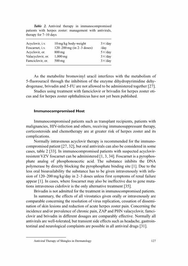
Antiviral Therapy of Shingles in Dermatology 127
As the metabolite bromovinyl uracil interferes with the metabolism of
5-fluorouracil through the inhibition of the enzyme dihydropyrimidine dehy-
drogenase, brivudin and 5-FU are not allowed to be administered together [27].
Studies using treatment with famciclovir or brivudin for herpes zoster oti-
cus and for herpes zoster ophthalmicus have not yet been published.
Immunocompromised Host
Immunocompromised patients such as transplant recipients, patients with
malignancies, HIV-infection and others, receiving immunosuppressant therapy,
corticosteroids and chemotherapy are at greater risk of herpes zoster and its
complications.
Normally intravenous acyclovir therapy is recommended for the immuno-
compromised patient [27, 32], but oral antivirals can also be considered in some
cases, table 2 [33]. In immunocompromised patients with suspected acyclovir-
resistent VZV foscarnet can be administered [1, 3, 34]. Foscarnet is a pyrophos-
phate analog of phosphonoacetic acid. The substance inhibits the DNA
polymerase by directly blocking the pyrophosphate binding site [1]. Due to the
less oral bioavailability the substance has to be given intravenously with infu-
sion of 120–200 mg/kg/day in 2–3 doses unless first symptoms of renal failure
appear [1]. In cases, where foscarnet may also be ineffective due to gene muta-
tions intravenous cidofovir is the only alternative treatment [35].
Brivudin is not admitted for the treatment in immunocompromised patients.
In summary, the effects of all virostatics given orally or intravenously are
comparable concerning the resolution of virus replication, cessation of dissemi-
nation of skin lesions and reduction of acute herpes zoster pain. Concerning the
incidence and/or prevalence of chronic pain, ZAP and PHN valacyclovir, famci-
clovir and brivudin in different dosages are comparably effective. Normally all
antivirals are well-tolerated, but transient side effects such as headache, gastroin-
testinal and neurological complaints are possible in all antiviral drugs [31].
Table 2. Antiviral therapy in immunocompromised
patients with herpes zoster: management with antivirals,
therapy for 7–10 days
Acyclovir, i.v. 10 mg/kg body-weight 3�/day
Foscarnet, i.v. 120–200 mg (in 2–3 doses) /day
Acyclovir, or. 800 mg 5�/day
Valacyclovir, or. 1,000 mg 3�/day
Famciclovir, or. 500 mg 3�/day
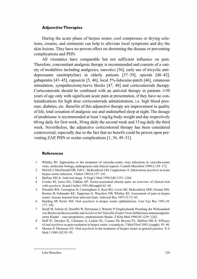
Lilie/Wassilew 128
Adjunctive Therapies
During the acute phase of herpes zoster, cool compresses or drying solu-
tions, creams, and ointments can help to alleviate local symptoms and dry the
skin lesions. They have no proven effect on shortening the disease or preventing
complications and PHN.
All virostatics have comparable but not sufficient influence on pain.
Therefore, concomitant analgesic therapy is recommended and consists of a vari-
ety of modalities including analgesics, narcotics [36], early use of tricyclic anti-
depressants (amitriptyline) in elderly patients [37–39], opioids [40–42],
gabapentin [43–45], capsaicin [5, 46], local 5%-lidocaine-patch [46], cutaneous
stimulation, sympathectomy/nerve blocks [47, 48] and corticosteroids therapy.
Corticosteroids should be combined with an antiviral therapy in patients �50
years of age only with significant acute pain at presentation, if they have no con-
traindications for high dose corticosteroids administration, i.e. high blood pres-
sure, diabetes, etc. Benefits of this adjunctive therapy are improvement in quality
of life, total cessation of analgesic use and undisturbed sleep at night. The dosage
of prednisone is recommended at least 1 mg/kg body-weight and day respectively
60 mg daily for first week, 30 mg daily the second week and 15 mg daily the third
week. Nevertheless, the adjunctive corticosteroid therapy has been considered
controversial, especially due to the fact that no benefit could be proven upon pre-
venting ZAP, PHN or ocular complications [1, 36, 49–51].
References
1 Whitley RJ: Approaches to the treatment of varicella-zoster virus infections in varicella-zoster
virus, molecular biology, pathogenesis and clinical aspects. Contrib Microbiol 1999;3:158–172.
2 McGill J, MacDonald DR, Fall C, McKendrick GD, Copplestone A: Intravenous acyclovir in acute
herpes zoster infection. J Infect 1983;6:157–161.
3 Balfour HH Jr: Antiviral drugs. N Engl J Med 1999;340:1255–1268.
4 Crooks RJ, Jones DA, Fiddian AP: Zoster-associated chronic pain: an overview of clinical trial
with acyclovir. Scand J Infect 1991;80(suppl):62–68.
5 Dworkin RH, Carrington D, Cunningham A, Kost RG, Levin MJ, McKendrick MW, Oxman MN,
Rentier B, Schmader KE, Tappeiner G, Wassilew SW, Whitley RJ: Assessment of pain in herpes
zoster: lessons learned from antiviral trials. Antiviral Res 1997;33:73–85.
6 Harding SP, Porter SM: Oral acyclovir in herpes zoster ophthalmicus. Curr Eye Res 1991;10:
177–182.
7 Heidl M, Scholz H, Doerffel W, Herrmann J, Wutzler P:Vergleichende Pruefung der Wirksamkeit
von Bromvinyldesoxyuridin und Aciclovir bei Varicella-Zoster-Virus-Infektionen immunsupprim-
ierter Kinder – eine prospektive, randomisierte Studie. Z Klin Med 1990;45:1259–1262.
8 Huff JC, Drucker JL, Clemmer A, Laskin OL, Connor JD, Bryson YL, Balfour HH Jr: Efficacy
of oral acyclovir on pain resolution in herpes zoster: a reanalysis. J Med Virol 1993;1(suppl): 93–96.
9 Morton P, Thomson AN: Oral acyclovir in the treatment of herpes zoster in general practice. N Z
Med J 1989;102:93–95.

Antiviral Therapy of Shingles in Dermatology 129
10 Wood MJ, Kay R, Dworkin RH, Soong SJ, Whitley RJ: Oral acyclovir therapy accelerates pain res-
olution in patients with herpes zoster: a meta-analysis of placebo-controlled trials. Clin Infect Dis
1996;22:341–347.
11 McKendrick MW, Care C, Burke C, Hickmott E, McKendrick GD: Oral acyclovir in herpes zoster.
J Antimicrob Chemother 1984;14:661–665.
12 van den Broek PJ, van der Meer JW, Mulder JD Versteeg J, Mattie H: Limited value to acyclovir in the
treatment of uncomplicated herpes zoster: a placebo-controlled study. Infection 1984;12: 338–341.
13 Beutner KR, Friedman DJ, Forszpaniak C, Andersen PL, Wood MJ: Valaciclovir compared with
acyclovir for improved therapy for herpes zoster in immunocompetent adults. Antimicrob Agents
Chemother 1995;39:1547–1553.
14 Tyring SK, Beutner KR, Tucker BA, Anderson WC, Crooks RJ: Antiviral therapy for herpes
zoster: randomized, controlled clinical trial of valaciclovir and famciclovir therapy in immuno-
competent patients 50 years and older. Arch Fam Med 2000;9:863–869.
15 Colin J, Prisant O, Cochener B, Lescale O, Rolland B, Hoang-Xuan T: Comparison of the efficacy
and safety of valaciclovir and acyclovir for the treatment of herpes zoster ophthalmicus.
Ophthalmology 2000;107:1507–1511.
16 Wutzler P, Doerr HW: Antivirale Therapie des Zoster. Dtsch Aerztbl 1998;95:79–81.
17 Dworkin RH, Boon RJ, Griffin DR, Phung D: Postherpetic neuralgia: impact of famciclovir, age,
rash severity, and acute pain in herpes zoster patients. J Infect Dis1998;178(suppl 1):76–80.
18 Tyring S, Engst R, Corriveau C, Robillard N, Trottier S, Van Slycken S, Crann RA, Locke, LA,
Saltzman R, Palestine AG: Famciclovir for ophthalmic zoster: a randomised aciclovir controlled
study. Br J Ophthalmol 2001;85:576–581.
19 Andrei G, Snoeck R, Reymen D, Liesnard C: Comparative activity of selected antiviral
compounds against clinical strains of varicella zoster virus. Eur J Clin Microbiol Infect Dis
1995;14: 318–329.
20 De Clercq E, Descamps J, De Somer P, Barr PJ, Jones AS, Walker RT: (E)-5-(2-Bromovinyl)-2�-deoxyuridine: a potent and selective anti-herpes agent Proc Natl Acad Sci USA 1979;76:
2947–2951.
21 De Clercq E, Degreef H, Wildiers J, De Jonge G, Drochmans A, Descamps J, De Somer P: Oral
(E)-5-(2-bromovinyl)-2�-deoxyuridine in severe Herpes zoster. Brit Med J 1980;281:1178.
22 De Clercq E, Descamps J, Verhelst G, Walker RT, Jones AS, Torrence PF, Shugar D: Comparative
efficacy of antiherpes drugs against different strains of Herpes simplex virus. J Infect Dis
1980;141:563–574.
23 Wassilew SW, Wutzler P: Oral brivudin in comparison with acyclovir for improved therapy of her-
pes zoster in immunocompetent patients: results of a randomized, double-blind, multicentered
study. Antiviral Res 2003;59:49–56.
24 Wassilew SW, Wutzler P: Oral brivudin in comparison with acyclovir for herpes zoster: a survey
study on postherpetic neuralgia. Antiviral Res 2003;59:57–60.
25 Wassilew SW: Virus- und Schmerzbekämpfung sind oberste Therapieziele. Pharm Ztg 2001;146:
43–49.
26 Wassilew SW, Stubinski BM, Koch I, Schumacher K, Städtler G: Brivudin compared to famciclovir
in the treatment of herpes zoster: effect on acute disease and chronic pain in immunocompetent
patients – a randomized multinational study. J Europ Acad Dermatol Venereol 2005;19: 47–55.
27 Wutzler P, De Clercq E, Wutke K, Faerber I: Oral brivudin vs. intravenous acyclovir in the treat-
ment of Herpes zoster in immunocompromised patients: a randomised double-blind trial. J Med
Virol 1995;46:252–257.
28 De Clercq E, Cassiman JJ: Mutagenic potential of anti-herpes agent. Life Science 1986;38:
281–289.
29 Wutzler P, Thust R: Genetic risks of antiviral nucleoside analogues – a survey. Antiviral Res
2001;49:55–74.
30 Gross G, Schöfer H, Wassilew SW, Friese K, Timm A, Guthoff R, Pau HW, Malin JP, Wutzler P,
Doerr HW: Herpes zoster guideline of the German Dermatology Society (DDG). J Clin Virol
2003;26:277–289.
31 Lilie HM, Wassilew SW: The role of antivirals in the management of neuropathic pain in the older
patient with herpes zoster. Drugs Aging 2003;20:561–570.

Lilie/Wassilew 130
32 Balfour HH Jr, Bean B, Laskin OL, Ambinder RF, Meyers JD, Wade JC, Zaia JA, Aeppli D, Kirk LE,
Segreti AC, Keeney RE: Acyclovir halts progression of herpes zoster in immunocompromised
patients. J Antimicrob Chemother 1983;12(suppl B):169–179.
33 Tyring S, Belanger R, Bezwoda W, Ljungman P, Boon R, Saltzman RL, Colaborative Famciclovir
Immunocompromised Study Group: A randomized, double-blind trial of famciclovir versus acy-
clovir for the treatment of localized dermatomal herpes zoster in immunocompromised patients.
Caner Invest 2001;19:13–22.
34 Breton G, Fillet AM, Katlama C, Bricaire F, Caumes E: Acyclovir-resistant herpes zoster in human
immunodeficiency virus-infected patients: results of foscarnet therapy. Clin Infect Dis 1998;27:
1525–1527.
35 Safrin S, Cherrington J, Jaffe HS: Clinical uses of cidovofir. Rev Med Virol 1997;7:145–156.
36 Pasqualucci A, Pasqualucci V, Galla F, De Angelis V, Marzocchi V, Colussi R, Paoletti F, Girardis M,
Lugano M, Del Sindaco F: Prevention of post-herpetic neuralgia: acyclovir and prednisolone ver-
sus epidural local anesthetic and methylprednisolone. Acta Anaesthesiol Scand 2000;44:910–918.
37 Bowsher D: The effects of pre-emptive treatment of postherpetic neuralgia with amitriptyline: a
randomized, double-blind, placebo-controlled trial. J Pain Symptom Manage 1997;13:327–331.
38 Graff-Radford SB, Shaw LR, Naliboff BN: Amitriptyline and fluphenazine in the treatment of
postherpetic neuralgia. Clin J Pain 2000;16:188–192.
39 Watson CPN, Vernich L, Chipman M, Reed K: Nortriptyline versus amitriptyline in postherpetic
neuralgia: a randomized trial. Neurology 1998;51:1166–1171.
40 Dühmke RM, Cornblath DD, Hollinshead JRF: Tramadol for neuropathic pain (Cochrane
Review); in The Cochrane Library, Issue 3, 2004.
41 Dworkin RH, Schmader KE: Treatment and prevention of postherpetic neuralgia. Clin Infect Dis
2003;36:877–882.
42 Rowbotham MC, Twilling L, Davies PS, Reisner L, Taylor K, Mohr D: Oral opioid therapy for
chronic periphal and central neuropathic pain. N Engl J Med 2003;348:1223–1232.
43 Curran MP, Wagstaff AJ: Gabapentin in postherpetic neuralgia. CNS Drugs 2003;17:975–982.
44 Rice ASC, Morton S, Postherpetic Neuralgia Study Group: Gabapentin in postherpetic neuralgia:
a randomised double blind, placebo controlled study. Pain 2001;94:215–224.
45 Rowbotham M, Harden N, Stacey B, Bernstein P, Magnus-Miller L, for the Gabapentin
Postherpetic Neuralgia Study Group: Gabapentin for the treatment of postherpetic neuralgia: a
randomized controlled trial. JAMA 1998;280:1837–1842.
46 Galer BS: Advances in the Treatment of Postherpetic Neuralgia: the topical lidocaine patch.
Todays Ther Trends 2000;18:1–20.
47 Winnie AP, Hartwell: Relationship between time of treatment of acute herpes zoster with sympathetic
blockade and prevention of post-herpetic neuralgia: clinical support for a new theory of the mecha-
nism by which sympathetic blockade provides therapeutic benefit. Reg Anesth 1993;18:277–282.
48 Wu CL, Marsh A, Dworkin RH: The role of sympathetic nerve blocks in herpes zoster and pos-
therpetic neuralgia. Pain 2000;87:121–129.
49 Gnann JW, Whitley RJ: Herpes zoster. N Eng J Med 2002;347:340–346.
50 Whitley RJ, Weiss H, Gnann JW Jr, Tyring S, Mertz GJ, Pappas PG, Schleupner CJ, Hayden F,
Wolf J, Soong SJ: Acyclovir with and without prednisone for the treatment of herpes zoster. A ran-
domized, placebo-controlled trial. The National Institute of Allergy and Infectious Diseases
Collaborative Antiviral Study Group. Ann Intern Med 1996;125:376–383.
51 Wood MJ, Johnson RW, McKendrick MW, Taylor J, Mandal BK, Crooks J: A randomized trial of
acyclovir for 7 days or 21 days with and without prednisolone for treatment of acute herpes zoster.
N Engl J Med 1994;330:896–900.
Dr. H. Martina Lilie
Dermatology Department, Klinikum Krefeld
Lutherplatz 40
DE–47805 Krefeld (Germany)
Tel. �49 2151 32 2880, Fax �49 2151 32 2005, E-Mail [email protected]

Gross G, Doerr HW (eds): Herpes Zoster.
Monogr Virol. Basel, Karger, 2006, vol 26, pp 131–142
Highly Potent and Selective Inhibitionof Varicella–Zoster Virus Replication byBicyclic Furo[2,3-d ]pyrimidineNucleoside Analogues (BCNAs)
Erik De Clercq
Rega Institute for Medical Research, Katholieke Universiteit Leuven,
Leuven, Belgium
Varicella-zoster virus (VZV) causes two distinct syndromes [1]: primary
infection presents as varicella (or chickenpox), a usually benign illness during
childhood, and subsequent reactivation of latent VZV in dorsal-root ganglia
results in a localized cutaneous eruption termed ‘herpes zoster’ (or ‘shingles’).
The annual incidence of herpes zoster is 0.2%, although in the elderly (persons
older than 75 years) and in immunocompromised patients it can exceed 1 and
2%, respectively [1]. Therapy for herpes zoster is warranted to accelerate heal-
ing, limit the severity and duration of acute and chronic pain, and reduce
complications (among which postherpetic neuralgia, defined as pain that
persists for more than 30 days after the onset of rash, is the most feared com-
plication in immunocompetent patients); in immunocompromised patients,
an additional therapeutic objective is to reduce the risk of dissemination of
VZV [1].
In the United States three drugs – acyclovir (ACV), valacyclovir and
famciclovir – are approved for the treatment of herpes zoster: ACV at a dosage
of 800 mg every 4 h (5 times daily) for 7–10 days, valacyclovir at a dosage of
1,000 mg every 8 h (3 times daily) for 7 days, and famciclovir at a dosage of
500 mg every 8 h (3 times daily) for 7 days: because of the simpler dosing regi-
mens, valacyclovir and famciclovir (which are therapeutically equipotent) are
preferred to ACV for the treatment of herpes zoster [1]. In addition to ACV,
valacyclovir and famciclovir, also brivudin (BVDU) has been licensed for the
therapy of herpes zoster in Germany and several other European Countries
(Austria, Belgium, Greece, Italy, Luxemburg, Portugal, Spain): BVDU has the
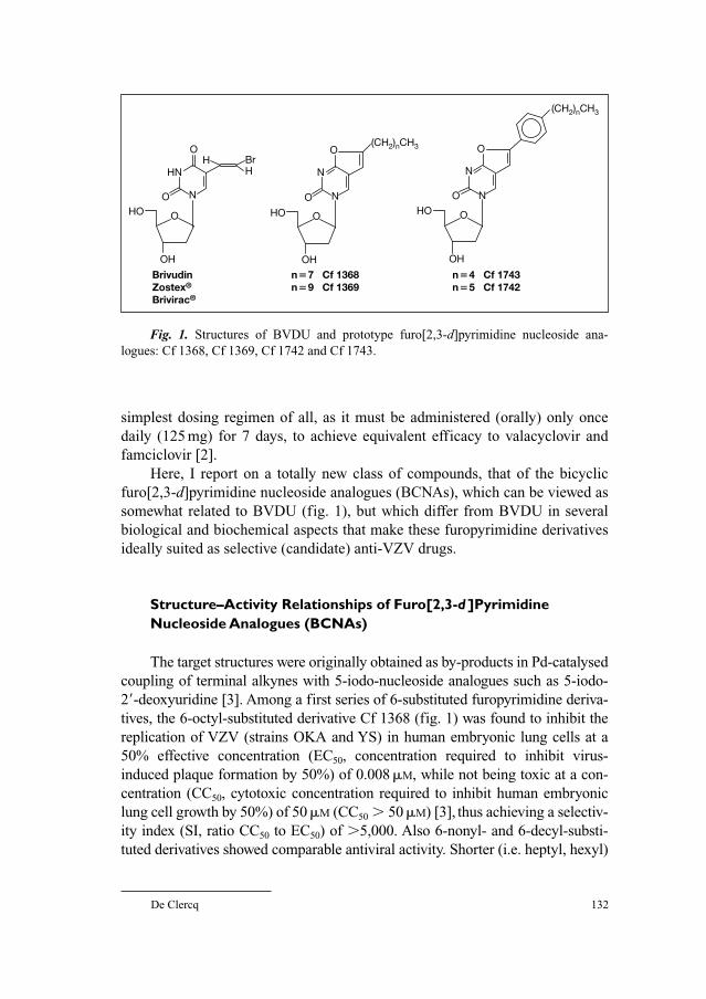
De Clercq 132
simplest dosing regimen of all, as it must be administered (orally) only once
daily (125 mg) for 7 days, to achieve equivalent efficacy to valacyclovir and
famciclovir [2].
Here, I report on a totally new class of compounds, that of the bicyclic
furo[2,3-d]pyrimidine nucleoside analogues (BCNAs), which can be viewed as
somewhat related to BVDU (fig. 1), but which differ from BVDU in several
biological and biochemical aspects that make these furopyrimidine derivatives
ideally suited as selective (candidate) anti-VZV drugs.
Structure–Activity Relationships of Furo[2,3-d ]PyrimidineNucleoside Analogues (BCNAs)
The target structures were originally obtained as by-products in Pd-catalysed
coupling of terminal alkynes with 5-iodo-nucleoside analogues such as 5-iodo-
2�-deoxyuridine [3]. Among a first series of 6-substituted furopyrimidine deriva-
tives, the 6-octyl-substituted derivative Cf 1368 (fig. 1) was found to inhibit the
replication of VZV (strains OKA and YS) in human embryonic lung cells at a
50% effective concentration (EC50, concentration required to inhibit virus-
induced plaque formation by 50%) of 0.008 �M, while not being toxic at a con-
centration (CC50, cytotoxic concentration required to inhibit human embryonic
lung cell growth by 50%) of 50 �M (CC50 � 50 �M) [3], thus achieving a selectiv-
ity index (SI, ratio CC50 to EC50) of �5,000. Also 6-nonyl- and 6-decyl-substi-
tuted derivatives showed comparable antiviral activity. Shorter (i.e. heptyl, hexyl)
HN
NO
H BrH
O
OH
HO
O
Brivudin Zostex®
Brivirac®
N
NO
O(CH2)nCH3
O
OH
HO
n�7 Cf 1368 n�9 Cf 1369
N
NO
O
(CH2)nCH3
O
OH
HO
n�4 Cf 1743 n�5 Cf 1742
Fig. 1. Structures of BVDU and prototype furo[2,3-d]pyrimidine nucleoside ana-
logues: Cf 1368, Cf 1369, Cf 1742 and Cf 1743.
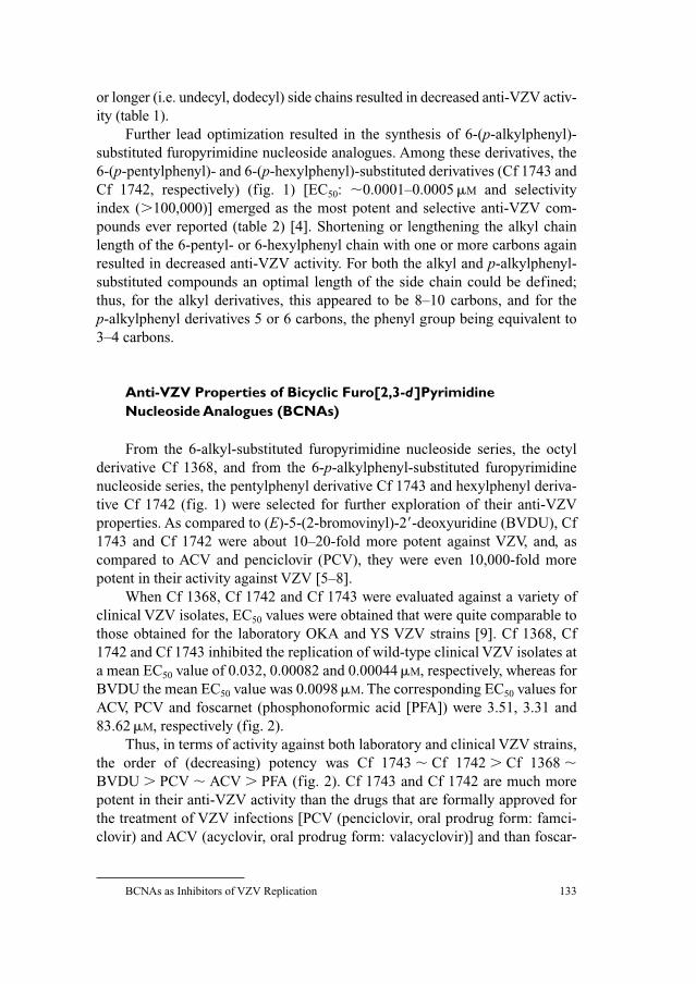
BCNAs as Inhibitors of VZV Replication 133
or longer (i.e. undecyl, dodecyl) side chains resulted in decreased anti-VZV activ-
ity (table 1).
Further lead optimization resulted in the synthesis of 6-(p-alkylphenyl)-
substituted furopyrimidine nucleoside analogues. Among these derivatives, the
6-(p-pentylphenyl)- and 6-(p-hexylphenyl)-substituted derivatives (Cf 1743 and
Cf 1742, respectively) (fig. 1) [EC50: �0.0001–0.0005 �M and selectivity
index (�100,000)] emerged as the most potent and selective anti-VZV com-
pounds ever reported (table 2) [4]. Shortening or lengthening the alkyl chain
length of the 6-pentyl- or 6-hexylphenyl chain with one or more carbons again
resulted in decreased anti-VZV activity. For both the alkyl and p-alkylphenyl-
substituted compounds an optimal length of the side chain could be defined;
thus, for the alkyl derivatives, this appeared to be 8–10 carbons, and for the
p-alkylphenyl derivatives 5 or 6 carbons, the phenyl group being equivalent to
3–4 carbons.
Anti-VZV Properties of Bicyclic Furo[2,3-d]Pyrimidine Nucleoside Analogues (BCNAs)
From the 6-alkyl-substituted furopyrimidine nucleoside series, the octyl
derivative Cf 1368, and from the 6-p-alkylphenyl-substituted furopyrimidine
nucleoside series, the pentylphenyl derivative Cf 1743 and hexylphenyl deriva-
tive Cf 1742 (fig. 1) were selected for further exploration of their anti-VZV
properties. As compared to (E)-5-(2-bromovinyl)-2�-deoxyuridine (BVDU), Cf
1743 and Cf 1742 were about 10–20-fold more potent against VZV, and, as
compared to ACV and penciclovir (PCV), they were even 10,000-fold more
potent in their activity against VZV [5–8].
When Cf 1368, Cf 1742 and Cf 1743 were evaluated against a variety of
clinical VZV isolates, EC50 values were obtained that were quite comparable to
those obtained for the laboratory OKA and YS VZV strains [9]. Cf 1368, Cf
1742 and Cf 1743 inhibited the replication of wild-type clinical VZV isolates at
a mean EC50 value of 0.032, 0.00082 and 0.00044 �M, respectively, whereas for
BVDU the mean EC50 value was 0.0098 �M. The corresponding EC50 values for
ACV, PCV and foscarnet (phosphonoformic acid [PFA]) were 3.51, 3.31 and
83.62 �M, respectively (fig. 2).
Thus, in terms of activity against both laboratory and clinical VZV strains,
the order of (decreasing) potency was Cf 1743 � Cf 1742 � Cf 1368 �BVDU � PCV � ACV � PFA (fig. 2). Cf 1743 and Cf 1742 are much more
potent in their anti-VZV activity than the drugs that are formally approved for
the treatment of VZV infections [PCV (penciclovir, oral prodrug form: famci-
clovir) and ACV (acyclovir, oral prodrug form: valacyclovir)] and than foscar-
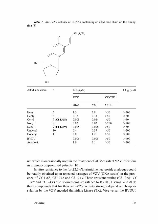
De Clercq 134
net which is occasionally used in the treatment of ACV-resistant VZV infections
in immunocompromised patients [10].
In vitro resistance to the furo[2,3-d]pyrimidine nucleoside analogues could
be readily obtained upon repeated passages of VZV (OKA strain) in the pres-
ence of Cf 1368, Cf 1742 and Cf 1743. These resistant strains (Cf 1368r, Cf
1742r and Cf 1743r) also showed cross-resistance to BVDU, BVaraU and ACV,
three compounds that for their anti-VZV activity strongly depend on phospho-
rylation by the VZV-encoded thymidine kinase (TK). Vice versa, the BVDUr,
Table 1. Anti-VZV activity of BCNAs containing an alkyl side chain on the furanyl
ring [3]
Alkyl side chain n EC50 (�M) CC50 (�M)
VZV VZV TK�
OKA YS YS-R
Hexyl 5 1.3 2.8 �50 �200
Heptyl 6 0.12 0.33 �50 �50
Octyl 7 (Cf 1368) 0.008 0.024 �50 �50
Nonyl 8 0.02 0.02 �200 �200
Decyl 9 (Cf 1369) 0.015 0.008 �50 �50
Undecyl 10 0.4 0.37 �50 �200
Dodecyl 11 0.8 1.2 �50 �200
BVDU 0.005 0.005 �50 �400
Acyclovir 1.9 2.1 �50 �200
N
NO
O
(CH2)nCH
3
O
OH
HO

BCNAs as Inhibitors of VZV Replication 135
BVaraUr and ACVr mutants also proved cross-resistant to the furopyrimidine
nucleoside analogues [9]. These data suggest that the furopyrimidine nucleo-
side analogues, akin to BVDU, BVaraU and ACV, select for drug-resistant VZV
mutants that are deficient in their TK-dependent phosphorylating capacity. On
the other hand, PCVr, PFAr and PMEAr strains [9], that were selected for resis-
tance to PCV, foscarnet or adefovir, respectively, retained marked susceptibility
Table 2. Anti-VZV activity of furo[2,3-d]pyrimidine
nucleoside analogues containing a p-alkyl-substituted phenyl
group as the side chain on the furanyl ring [4]
R EC50 (�M) CC50 (�M)
VZV
OKA YS
H 0.28 0.16 �200
Methyl 0.06 0.06 �200
Ethyl 0.09 0.07 123
Propyl 0.01 0.008 188
Butyl 0.0022 0.0005 �200
Pentyl (Cf 1743) 0.0003 0.0001 �200
Hexyl (Cf 1742) 0.0005 0.0001 18
Heptyl 0.0054 0.003 18
Octyl 0.04 0.027 �200
N
NO
O
R
O
OH
HO

De Clercq 136
to Cf 1742 and Cf 1743. This suggests that the PCV, PFA and PMEA resistance
phenotype must be attributed to mutations in an enzyme other than the viral TK,
most probably the viral DNA polymerase.
Mechanism of Antiviral Action of Bicyclic Furo[2,3-d]Pyrimidine Nucleoside Analogues (BCNAs)
All the compounds that are currently used in the treatment of VZV infec-
tions (i.e. ACV, PCV, BVDU) are also active against herpes simplex virus type 1
(HSV-1) and, furthermore, ACV and PCV are also active against herpes sim-
plex virus type 2 (HSV-2). The furopyrimidine nucleoside analogues Cf 1368
and Cf 1743, however, are selectively active against VZV (they are not active
against HSV-1 or HSV-2 or other herpes viruses such as cytomegalovirus). This
0.0001
0.001
0.01
0.1
1
10
100E
C50
(�M
)
Cf 1743 Cf 1742Cf 1368BVDU
ACV PCV PFA
Fig. 2. Activity of BCNAs and anti-VZV reference compounds [BVDU, ACV, PCV
and foscarnet (PFA)] against clinical VZV isolates (�) and VZV reference strains Oka (�)
and YS (�). Mean values for clinical isolates are indicated by horizontal bars. Data taken
from reference [9].

BCNAs as Inhibitors of VZV Replication 137
is because they are recognized as a substrate for phosphorylation by the VZV-
encoded TK [11]. In contrast, the HSV-1 TK, HSV-2 TK as well as the cytosolic
TK-1 and mitochondrial TK-2 do not recognize the furopyrimidine nucleoside
analogues as substrate. The VZV TK, due to its intrinsic dTMP kinase activity,
will successively phosphorylate the furopyrimidine nucleosides to the 5�-monophosphate and 5�-diphosphate form [11]. Whether the compounds are
then further phosphorylated to the 5�-triphosphate form could so far not be
clearly demonstrated.
Phosphorylation of the furopyrimidine nucleoside analogues by the VZV
TK is a prerequisite for their anti-VZV activity, but it clearly is not sufficient a
requirement. For a large series of furopyrimidine nucleosides that were exam-
ined for both their anti-VZV activity and their inhibitory effects on thymidine
phosphorylation by VZV TK (as a parameter of affinity of the compounds to
the viral enzyme), no correlation whatsoever was obtained. This means that,
while the viral TK is necessary, it is by itself not sufficient to explain the anti-
VZV activity of the furopyrimidine nucleoside analogues. How these com-
pounds accomplish their antiviral activity and with which molecular target they
eventually interact remain fascinating questions, subject of further investigation
in our laboratory.
Metabolic Interactions of Bicyclic Furo[2,3-d]PyrimidineNucleoside Analogues (BCNAs)
The pyrimidine nucleoside catabolic enzyme, thymidine phosphorylase
(TPase), is known to hydrolyse the N-glycosidic bond of various pyrimidine
nucleoside analogues, including BVDU [12, 13]. Since the aglycones (free
pyrimidine bases) are as such devoid of antiviral activity, the action of TPase
may annihilate the antiviral activity of the pyrimidine nucleoside analogues.
Interestingly, the furopyrimidine nucleoside analogues Cf 1368 and Cf 1743
were found to be entirely resistant to the phosphorolytic cleavage by both
human TPase and bacterial TPase [14].
The free nucleobase of BVDU, (E)-5-(2-bromovinyl)uracil (BVU), is an
efficient inhibitor of human dihydropyrimidine dehydrogenase (DPD) [15].
DPD is a key enzyme involved in the degradation of natural pyrimidines and
pyrimidine analogues such as 5-fluorouracil (5-FU). BVU, through its
inhibitory effect on DPD, may significantly potentiate the toxicity of 5-FU. The
relevance of this interaction has become dramatically clear in the past, when the
simultaneous administration of oral sorivudine (BVaraU) with 5-FU in cancer
patients was found to lead to a number of deaths as the result of an accrued
5-FU toxicity [16–18]. The cascade of reactions leading to these fatalities were
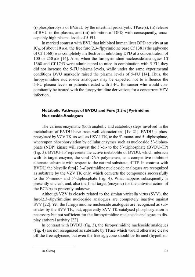
De Clercq 138
(i) phosphorolysis of BVaraU by the intestinal prokaryotic TPase(s), (ii) release
of BVU in the plasma, and (iii) inhibition of DPD, with consequently, unac-
ceptably high plasma levels of 5-FU.
In marked contrast with BVU that inhibited human liver DPD activity at an
IC50 of about 10 �M, the free furo[2,3-d]pyrimidine base Cf 1381 (the aglycone
of Cf 1368) was completely ineffective in inhibiting DPD at a concentration of
100 or 250 �M [14]. Also, when the furopyrimidine nucleoside analogues Cf
1368 and Cf 1743 were administered to mice in combination with 5-FU, they
did not increase the 5-FU plasma levels, while under the same experimental
conditions BVU markedly raised the plasma levels of 5-FU [14]. Thus, the
furopyrimidine nucleoside analogues may be expected not to influence the
5-FU plasma levels in patients treated with 5-FU for cancer who would con-
comitantly be treated with the furopyrimidine derivatives for a concurrent VZV
infection.
Metabolic Pathways of BVDU and Furo[2,3-d]PyrimidineNucleoside Analogues
The various enzymatic (both anabolic and catabolic) steps involved in the
metabolism of BVDU have been well characterized [19–21]. BVDU is phos-
phorylated by VZV TK, as well as HSV-1 TK, to the 5�-mono- and 5�-diphosphate,
whereupon phosphorylation by cellular enzymes such as nucleoside 5�-diphos-
phate (NDP) kinase will convert the 5�-di- to the 5�-triphosphate (BVDU-TP)
(fig. 3). BVDU-TP represents the active metabolite of BVDU, which interacts
with its target enzyme, the viral DNA polymerase, as a competitive inhibitor/
alternate substrate with respect to the natural substrate, dTTP. In contrast with
BVDU, the bicyclic furo[2,3-d]pyrimidine nucleoside analogues are recognized
as substrate by the VZV TK only, which converts the compounds successfully
to the 5�-mono- and 5�-diphosphate (fig. 4). What happens subsequently is
presently unclear, and, also the final target (enzyme) for the antiviral action of
the BCNAs is presently unknown.
Although VZV is closely related to the simian varicella virus (SVV), the
furo[2,3-d]pyrimidine nucleoside analogues are completely inactive against
SVV [22]. Yet, the furopyrimidine nucleoside analogues are recognized as sub-
strates by the SVV TK, but, apparently SVV TK-catalysed phosphorylation is
necessary but not sufficient for the furopyrimidine nucleoside analogues to dis-
play antiviral activity [22].
In contrast with BVDU (fig. 3), the furopyrimidine nucleoside analogues
(fig. 4) are not recognized as substrate by TPase which would otherwise cleave
off the free aglycone, but even the free aglycone should be formed (hypotheti-
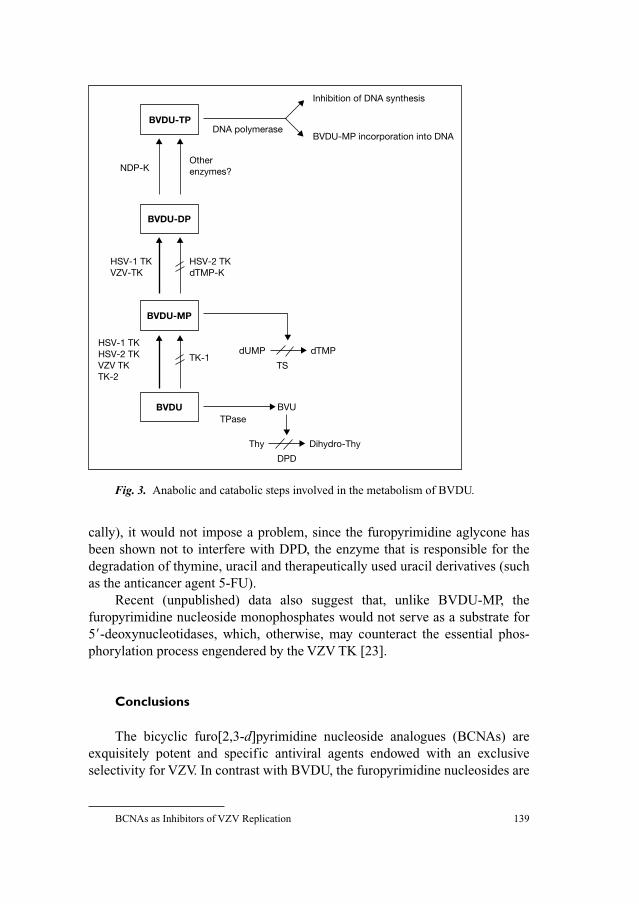
BCNAs as Inhibitors of VZV Replication 139
cally), it would not impose a problem, since the furopyrimidine aglycone has
been shown not to interfere with DPD, the enzyme that is responsible for the
degradation of thymine, uracil and therapeutically used uracil derivatives (such
as the anticancer agent 5-FU).
Recent (unpublished) data also suggest that, unlike BVDU-MP, the
furopyrimidine nucleoside monophosphates would not serve as a substrate for
5�-deoxynucleotidases, which, otherwise, may counteract the essential phos-
phorylation process engendered by the VZV TK [23].
Conclusions
The bicyclic furo[2,3-d]pyrimidine nucleoside analogues (BCNAs) are
exquisitely potent and specific antiviral agents endowed with an exclusive
selectivity for VZV. In contrast with BVDU, the furopyrimidine nucleosides are
Inhibition of DNA synthesis
BVDU-MP incorporation into DNADNA polymerase
Otherenzymes?NDP-K
HSV-1 TKVZV-TK
HSV-2 TKdTMP-K
HSV-1 TKHSV-2 TKVZV TKTK-2
TK-1dUMP dTMP
TS
BVDUTPase
Dihydro-Thy
DPD
BVDU-MP
BVDU-DP
BVDU-TP
BVU
Thy
Fig. 3. Anabolic and catabolic steps involved in the metabolism of BVDU.
De Clercq 140
recognized solely by the TK and (therewith associated thymidylate kinase activ-
ity) of VZV. They are not hydrolysed by human or bacterial TPases, and their
free bases do not inhibit human dihydropyrimidine dehydrogenase. They are
also characterized by a number of favourable properties: straightforward chem-
ical synthesis, good stability in biological fluids, virtual absence of cytotoxicity,
pronounced oral bioavailability, and no interference with the degradation of
5-FU. The furopyrimidine nucleoside analogues proved equally effective
against laboratory VZV strains as well as clinical VZV isolates. Cf 1742 and Cf
1743, the most potent among the furopyrimidine series, inhibited the replica-
tion of VZV in cell culture at subnanomolar concentrations. The precise mech-
anism of antiviral action of the furopyrimidine nucleoside analogues still
remains to be clarified. Their unprecedented potency, specificity and selectivity
for VZV highly justifies further development of these compounds as candidate
drugs for the treatment of VZV infections, i.e. varicella and herpes zoster.
Inhibition of DNA synthesis
BCNA-MP incorporation into DNADNA polymerase
?
NDP-K?
VZV-TK HSV-1 TKdTMP-K
VZV-TK HSV-1 TKHSV-2 TKTK-1TK-2
BCNATPase
Dihydro-ThyDPD
BCNA-MP
BCNA-DP
BCNA-TP
free base
Thy
Fig. 4. Anabolic and catabolic steps involved in the metabolism of bicyclic furo[2,3-d]
pyrimidine nucleoside analogues (BCNAs).
BCNAs as Inhibitors of VZV Replication 141
Acknowledgments
This review is based on the original investigations carried out at the Rega Institute
(G. Andrei, A. Camps, L. Naesens, R. Sienaert, R. Snoeck, L. van Berckelaer, R. Van
Berwaer, L. Vandenheurck, J. Balzarini and E. De Clercq), the University of Wales Cardiff
Medicinal Chemistry Group (headed by C. McGuigan), Oxford University (R. Esnouf,
D. Stammers), and the Academic Clinic Amsterdam (A. van Kuilenburg). I thank Christiane
Callebaut for her proficient editorial assistance.
References
1 Gnann JW Jr, Whitley RJ: Herpes zoster. N Engl J Med 2002;347:340–346.
2 Gross G, Schöfer H, Wassilew S, Friese K, Timm A, Guthoff R, Pau HW, Malin JP, Wutzler P,
Doerr HW: Herpes zoster guideline of the German Dermatology Society (DDG). J Clin Virol
2003;26:277–289.
3 McGuigan C, Yarnold CJ, Jones G, Velázquez S, Barucki H, Brancale A, Andrei G, Snoeck R, De
Clercq E, Balzarini J: Potent and selective inhibition of varicella-zoster virus (VZV) by nucleoside
analogues with an unusual bicyclic base. J Med Chem 1999;42:4479–4484.
4 McGuigan C, Barucki H, Blewett S, Carangio A, Erichsen JT, Andrei G, Snoeck R, De Clercq E,
Balzarini J: Highly potent and selective inhibition of varicella-zoster virus by bicyclic furopyrim-
idine nucleosides bearing an aryl side chain. J Med Chem 2000;43:4993–4997.
5 De Clercq E, Degreef H, Wildiers J, de Jonge G, Drochmans A, Descamps J, De Somer P: Oral
(E)-5-(2-bromovinyl)-2�-deoxyuridine in severe herpes zoster. Br Med J 1980;281:1178.
6 Shigeta S, Yokota T, Iwabuchi T, Baba M, Konno K, Ogata M, De Clercq E: Comparative efficacy
of antiherpes drugs against various strains of varicella-zoster virus. J Infect Dis 1983;147:576–584.
7 De Clercq E: The antiviral spectrum of (E)-5-(2-bromovinyl)-2�-deoxyuridine. J Antimicrob
Chemother 1984;14(suppl A):85–95.
8 Andrei G, Snoeck R, Reymen D, Liesnard D, Goubau P, Desmyter J, De Clercq E: Comparative
activity of selected antiviral compounds against clinical isolates of varicella-zoster virus. Eur J
Clin Microbiol Infect Dis 1995;14:318–328.
9 Andrei G, Sienaert R, Naesens L, McGuigan C, De Clercq E, Balzarini J, Snoeck R: Susceptibilities
of several clinical varicella-zoster virus (VZV) isolates and drug-resistant VZV strains to bicyclic
furano pyrimidine nucleosides. Antimicrob Agents Chemother 2005;49:1081–1086.
10 Safrin S, Berger TG, Gilson I, Wolfe PR, Wofsy CB, Mills J, Biron KK: Foscarnet therapy in five
patients with AIDS and acyclovir-resistant varicella-zoster virus infection. Ann Intern Med
1991;115:19–21.
11 Sienaert R, Naesens L, Brancale A, De Clercq E, McGuigan C, Balzarini J: Specific recognition
of the bicyclic pyrimidine nucleoside analogs, a new class of highly potent and selective inhibitors
of varicella-zoster virus (VZV), by the VZV-encoded thymidine kinase. Mol Pharmacol
2002;61:249–254.
12 Desgranges C, Razaka G, Rabaud M, Bricaud H, Balzarini J, De Clercq E: Phosphorolysis of (E)-5-
(2-bromovinyl)-2�-deoxyuridine (BVDU) and other 5-substituted 2�-deoxyuridines by purified
human thymidine phosphorylase and intact blood platelets. Biochem Pharmacol 1983;32:3583–3590.
13 Liermann B, Hermann G: (E)-5-(2-Bromovinyl)-2�-deoxyuridine: a good substrate for mam-
malian pyrimidine nucleoside phosphorylases. Biomed Biochim Acta 1983;42:K35–K38.
14 Balzarini J, Sienaert R, Liekens S, Van Kuilenburg A, Carangio A, Esnouf R, De Clercq E,
McGuigan C: Lack of susceptibility of bicyclic nucleoside analogs, highly potent inhibitors of
varicella-zoster virus, to the catabolic action of thymidine phosphorylase and dihydropyrimidine
dehydrogenase. Mol Pharmacol 2002;61:1140–1145.
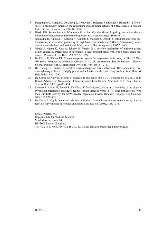
De Clercq 142
15 Desgranges C, Razaka G, De Clercq E, Herdewijn P, Balzarini J, Drouillet F, Bricaud H: Effect of
(E)-5-(2-bromovinyl)uracil on the catabolism and antitumor activity of 5-fluorouracil in rats and
leukemic mice. Cancer Res 1986;46:1094–1101.
16 Diasio RB: Sorivudine and 5-fluorouracil: a clinically significant drug-drug interaction due to
inhibition of dihydropyrimidine dehydrogenase. Br J Clin Pharmacol 1998;46:1–4.
17 Nakayama H, Kinouchi T, Kataoka K, Akimoto S, Matsuda Y, Ohnishi Y: Intestinal anaerobic bac-
teria hydrolyse sorivudine, producing the high blood concentration of 5-(E)-5-(2-bromovinyl)uracil
that increases the level and toxicity of 5-fluorouracil. Pharmacogenetics 1997;7:5–43.
18 Okuda H, Ogura K, Kato A, Takubo H, Watabe T: A possible mechanism of eighteen patient
deaths caused by interactions of sorivudine, a new antiviral drug, with oral 5-fluorouracil pro-
drugs. J Pharmacol Exp Ther 1998;287:791–799.
19 De Clercq E, Walker RT: Chemotherapeutic agents for herpesvirus infections; in Ellis GP, West
GB (eds): Progress in Medicinal Chemistry, vol 23. Amsterdam, The Netherlands, Elsevier
Science Publishers B.V. (Biomedical Division), 1986, pp 187–218.
20 De Clercq E: Towards a selective chemotherapy of virus infections. Development of bro-
movinyldeoxyuridine as a highly potent and selective anti-herpetic drug. Verh K Acad Geneesk
Belg 1986;48:261–290.
21 De Clercq E: Antiviral activity of nucleoside analogues: the BVDU connection; in Chu D (ed):
Recent Advances in Nucleosides: Chemistry and Chemotherapy. New York, NY, USA, Elsevier
Science B.V., 2002, pp 433–454.
22 Sienaert R, Andrei G, Snoeck R, De Clercq E, McGuigan C, Balzarini J: Inactivity of the bicyclic
pyrimidine nucleoside analogues against simian varicella virus (SVV) does not correlate with
their substrate activity for SVV-encoded thymidine kinase. Biochem Biophys Res Commun
2004;315:877–883.
23 De Clercq E: Highly potent and selective inhibition of varicella-zoster virus replication by bicyclic
furo[2,3-d]pyrimidine nucleoside analogues. Med Res Rev 2003;23:253–274.
Erik De Clercq, MD
Rega Institute for Medical Research
Minderbroedersstraat 10
BE–3000 Leuven (Belgium)
Tel. �32 16 337367, Fax �32 16 337340, E-Mail [email protected]

Gross G, Doerr HW (eds): Herpes Zoster.
Monogr Virol. Basel, Karger, 2006, vol 26, pp 143–153
Therapy of Zoster Pain, PostherpeticNeuralgia and Other NeurologicalComplications
Ralf Baron
Neurological Clinic, Pain Research and Therapy, University Kiel,
Kiel, Germany
Treatment of Acute Herpes Zoster
In most case acute herpes zoster is a self-limiting disease and will resolve
without complications with time. Analgesic drugs, i.e. NSAIDS and weak opi-
oids or in severe cases strong opioids, in combination with local antiinflammatory
ointments are used to establish adequate control of acute pain during healing of
the rash. Furthermore, antiviral therapy (e.g. acyclovir, famciclovir, valacy-
clovir and brivudin) should be initiated as soon as possible, especially in
older patients. If neurological complications, i.e. motor paresis, clinical signs of
myelitis or encephalitis or dermatological complications (involvement of the
cornea, zoster generalisatus) have developed as well as in immunocompro-
mised patients an intravenous therapy of virostatic drugs should be applied.
The pain in acute herpes zoster may have a sympathetically maintained
component, i.e. some percentage of the pain depends on the efferent sympa-
thetic innervation to the affected skin area. If the zoster is located in the face,
the upper or lower extremity diagnostic sympathetic blocks at the stellate gan-
glion or the lumbosacral chain can be performed particularly in severe cases.
In case of a sympathetically maintained pain component a series of several
sympathetic blocks may reduce acute pain. The important question whether
sympathetic blocks are capable of reducing the risk of chronic PHN is still
unresolved.

Baron 144
Prediction of Postherpetic Neuralgia
Despite the similarity of the acute symptoms only a subgroup of zoster
patients are at risk for the development of postherpetic neuralgia (PHN). Until
now there are no firm indicators the clinician can rely on in setting the progno-
sis for the patient with acute herpes zoster. However, some interesting associa-
tions have started to emerge. In general, patients of older age have a higher risk
to be left with PHN [1–3]. In acute herpes zoster there is a positive correlation
between elevation of T-lymphocyte CD4/CD8, indicative of the impairment of
cell mediated immunity, and duration of pain [4]. Several psychosocial
antecedents of the development of chronic pain could be established in herpes
zoster, i.e. disease conviction and depression [2]. Furthermore, neurophysiolog-
ical measures, i.e. elevation of thermal thresholds within the acutely affected
skin area, were associated with reports of pain at 3 months duration but the
small number of patients precluded assessment of association with chronic
established PHN [5]. By analogy with other chronic pain syndromes, i.e. phan-
tom limb pain [6] and chronic back pain [7], many investigators suggested that
the acute pain intensity, indicative of the severity of the herpes zoster infection
and nerve damage, may predict the development of PHN [2, 3]. In addition, it
was shown that patients with a preexisting large fiber polyneuropathy were at
higher risk to develop PHN [8].
Prevention of Postherpetic Neuralgia
Recent results from the shingles prevention study shows that vaccination
of adults is effective in prevention of PHN [9]. If vaccination of adults is
adopted, and shows similar long-term effects, it is possible that there could be a
dramatic reduction of PHN in decades to come. Combining this effect with the
long-term effect (40� years) of childhood vaccination against varicella, future
generations might be immune to the sometimes devastating results of PHN.
Early antiherpetic therapy, in particular with modern antiviral drugs [10]
significantly reduced the development of PHN [11].
The immediate treatment objective during the acute zoster phase is to
shorten as much as possible the duration of the pain phase. Although only few
controlled studies are available that assessed the effect of acute pain therapy on
development of PHN all modern concepts of pain generation suggest that every
acute pain input to the nervous system will lead to chronification.
According to the pathophysiological mechanisms several therapeutical
interventions to prevent PHN can be hypothesized: (1) an adequate analgesia
should be established in the acute phase with e.g. analgesics, anti-depressants
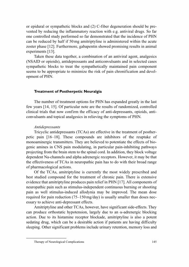
Therapy of Neurological Complications 145
or epidural or sympathetic blocks and (2) C-fiber degeneration should be pre-
vented by reducing the inflammatory reaction with e.g. antiviral drugs. So far
one controlled study performed so far demonstrated that the incidence of PHN
can be reduced by half if 50 mg amitriptyline is administered within the acute
zoster phase [12]. Furthermore, gabapentin showed promising results in animal
experiments [13].
Taken these data together, a combination of an antiviral agent, analgesics
(NSAID or opioids), antidepressants and anticonvulsants and in selected cases
sympathetic blocks to treat the sympathetically maintained pain component
seems to be appropriate to minimize the risk of pain chronification and devel-
opment of PHN.
Treatment of Postherpetic Neuralgia
The number of treatment options for PHN has expanded greatly in the last
few years [14, 15]. Of particular note are the results of randomized, controlled
clinical trials that now confirm the efficacy of anti-depressants, opioids, anti-
convulsants and topical analgesics in relieving the symptoms of PHN.
AntidepressantsTricyclic antidepressants (TCAs) are effective in the treatment of posther-
petic pain [16–18]. These compounds are inhibitors of the reuptake of
monoaminergic transmitters. They are believed to potentiate the effects of bio-
genic amines in CNS pain modulating, in particular pain-inhibiting pathways
projecting from the brain stem to the spinal cord. In addition, they block voltage
dependent Na-channels and alpha adrenergic receptors. However, it may be that
the effectiveness of TCAs in neuropathic pain has to do with their broad range
of pharmacological actions.
Of the TCAs, amitriptyline is currently the most widely prescribed and
best studied compound for the treatment of chronic pain. There is extensive
evidence that amitriptyline produces pain relief in PHN [17]. All components of
neuropathic pain such as stimulus-independent continuous burning or shooting
pain as well stimulus-induced allodynia may be improved. The mean dose
required for pain reduction (75–150 mg/day) is usually smaller than doses nec-
essary to achieve anti-depressant effects.
Amitriptyline and other TCAs, however, have significant side-effects. They
can produce orthostatic hypotension, largely due to an �-adrenergic blocking
action. Due to its histamine receptor blockade, amitriptyline is also a potent
sedating drug, which can be a desirable action if patients are having difficulty
sleeping. Other significant problems include urinary retention, memory loss and
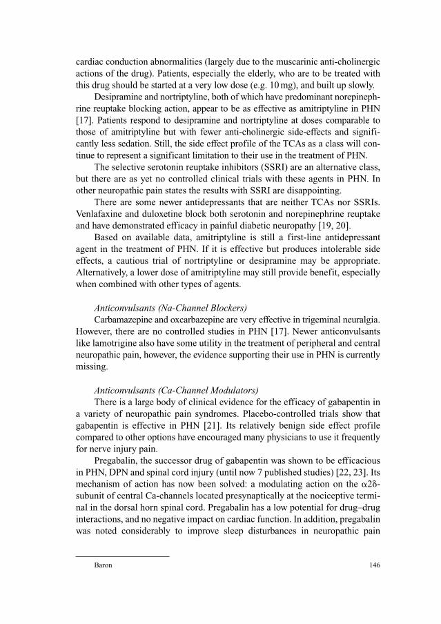
Baron 146
cardiac conduction abnormalities (largely due to the muscarinic anti-cholinergic
actions of the drug). Patients, especially the elderly, who are to be treated with
this drug should be started at a very low dose (e.g. 10 mg), and built up slowly.
Desipramine and nortriptyline, both of which have predominant norepineph-
rine reuptake blocking action, appear to be as effective as amitriptyline in PHN
[17]. Patients respond to desipramine and nortriptyline at doses comparable to
those of amitriptyline but with fewer anti-cholinergic side-effects and signifi-
cantly less sedation. Still, the side effect profile of the TCAs as a class will con-
tinue to represent a significant limitation to their use in the treatment of PHN.
The selective serotonin reuptake inhibitors (SSRI) are an alternative class,
but there are as yet no controlled clinical trials with these agents in PHN. In
other neuropathic pain states the results with SSRI are disappointing.
There are some newer antidepressants that are neither TCAs nor SSRIs.
Venlafaxine and duloxetine block both serotonin and norepinephrine reuptake
and have demonstrated efficacy in painful diabetic neuropathy [19, 20].
Based on available data, amitriptyline is still a first-line antidepressant
agent in the treatment of PHN. If it is effective but produces intolerable side
effects, a cautious trial of nortriptyline or desipramine may be appropriate.
Alternatively, a lower dose of amitriptyline may still provide benefit, especially
when combined with other types of agents.
Anticonvulsants (Na-Channel Blockers)Carbamazepine and oxcarbazepine are very effective in trigeminal neuralgia.
However, there are no controlled studies in PHN [17]. Newer anticonvulsants
like lamotrigine also have some utility in the treatment of peripheral and central
neuropathic pain, however, the evidence supporting their use in PHN is currently
missing.
Anticonvulsants (Ca-Channel Modulators)There is a large body of clinical evidence for the efficacy of gabapentin in
a variety of neuropathic pain syndromes. Placebo-controlled trials show that
gabapentin is effective in PHN [21]. Its relatively benign side effect profile
compared to other options have encouraged many physicians to use it frequently
for nerve injury pain.
Pregabalin, the successor drug of gabapentin was shown to be efficacious
in PHN, DPN and spinal cord injury (until now 7 published studies) [22, 23]. Its
mechanism of action has now been solved: a modulating action on the �2�-
subunit of central Ca-channels located presynaptically at the nociceptive termi-
nal in the dorsal horn spinal cord. Pregabalin has a low potential for drug–drug
interactions, and no negative impact on cardiac function. In addition, pregabalin
was noted considerably to improve sleep disturbances in neuropathic pain
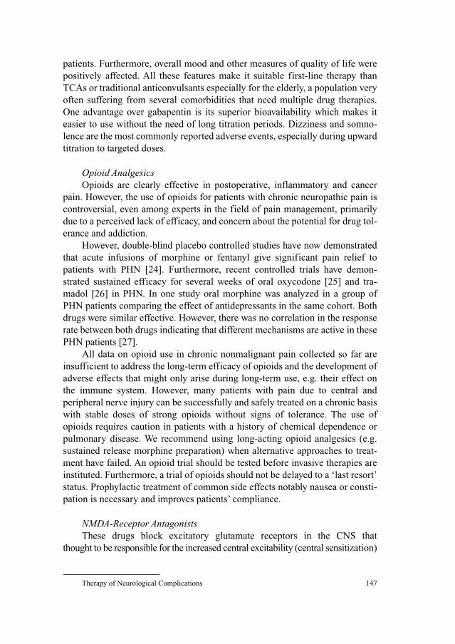
Therapy of Neurological Complications 147
patients. Furthermore, overall mood and other measures of quality of life were
positively affected. All these features make it suitable first-line therapy than
TCAs or traditional anticonvulsants especially for the elderly, a population very
often suffering from several comorbidities that need multiple drug therapies.
One advantage over gabapentin is its superior bioavailability which makes it
easier to use without the need of long titration periods. Dizziness and somno-
lence are the most commonly reported adverse events, especially during upward
titration to targeted doses.
Opioid AnalgesicsOpioids are clearly effective in postoperative, inflammatory and cancer
pain. However, the use of opioids for patients with chronic neuropathic pain is
controversial, even among experts in the field of pain management, primarily
due to a perceived lack of efficacy, and concern about the potential for drug tol-
erance and addiction.
However, double-blind placebo controlled studies have now demonstrated
that acute infusions of morphine or fentanyl give significant pain relief to
patients with PHN [24]. Furthermore, recent controlled trials have demon-
strated sustained efficacy for several weeks of oral oxycodone [25] and tra-
madol [26] in PHN. In one study oral morphine was analyzed in a group of
PHN patients comparing the effect of antidepressants in the same cohort. Both
drugs were similar effective. However, there was no correlation in the response
rate between both drugs indicating that different mechanisms are active in these
PHN patients [27].
All data on opioid use in chronic nonmalignant pain collected so far are
insufficient to address the long-term efficacy of opioids and the development of
adverse effects that might only arise during long-term use, e.g. their effect on
the immune system. However, many patients with pain due to central and
peripheral nerve injury can be successfully and safely treated on a chronic basis
with stable doses of strong opioids without signs of tolerance. The use of
opioids requires caution in patients with a history of chemical dependence or
pulmonary disease. We recommend using long-acting opioid analgesics (e.g.
sustained release morphine preparation) when alternative approaches to treat-
ment have failed. An opioid trial should be tested before invasive therapies are
instituted. Furthermore, a trial of opioids should not be delayed to a ‘last resort’
status. Prophylactic treatment of common side effects notably nausea or consti-
pation is necessary and improves patients’ compliance.
NMDA-Receptor AntagonistsThese drugs block excitatory glutamate receptors in the CNS that
thought to be responsible for the increased central excitability (central sensitization)
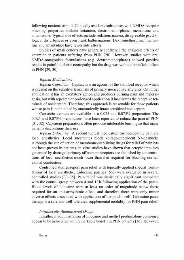
Baron 148
following noxious stimuli. Clinically available substances with NMDA receptor
blocking properties include ketamine, dextromethorphane, memantine and
amantadine. Typical side effects include sedation, nausea, disagreeable psycho-
logical disturbances or even frank hallucinations. Dextromethorphan, meman-
tine and amantadine have fewer side affects.
Studies of small cohorts have generally confirmed the analgesic effects of
ketamine in patients suffering from PHN [28]. However, studies with oral
NMDA-antagonists formulations (e.g. dextromethorphane) showed positive
results in painful diabetic neuropathy but the drug was without beneficial effect
in PHN [29, 30].
Topical MedicationsTopical Capsaicin: Capsaicin is an agonist of the vanilloid receptor which
is present on the sensitive terminals of primary nociceptive afferents. On initial
application it has an excitatory action and produces burning pain and hyperal-
gesia, but with repeated or prolonged application it inactivates the receptive ter-
minals of nociceptors. Therefore, this approach is reasonable for those patients
whose pain is maintained by anatomically intact sensitized nociceptors.
Capsaicin extracts are available in a 0.025 and 0.075% preparation. The
0.025 and 0.075% preparations have been reported to reduce the pain of PHN
[31, 32]. Capsaicin preparations often produce intolerable burning so that many
patients discontinue their use.
Topical Lidocaine: A second topical medication for neuropathic pain are
local anesthetics. Local anesthetics block voltage-dependent Na-channels.
Although the site of action of membrane-stabilizing drugs for relief of pain has
not been proven in patients, in vitro studies have shown that ectopic impulses
generated by damaged primary afferent nociceptors are abolished by concentra-
tions of local anesthetics much lower than that required for blocking normal
axonal conduction.
Controlled studies report pain relief with topically applied special formu-
lations of local anesthetic. Lidocaine patches (5%) were evaluated in several
controlled studies [33–35]. Pain relief was statistically significant compared
with the control group between 4 and 12 h following application of the patch.
Blood levels of lidocaine were at least an order of magnitude below those
required for an anti-arrhythmic effect, and therefore there were only minor
adverse effects associated with application of the patch itself. Lidocaine patch
therapy is a safe and well-tolerated supplemental modality for PHN pain relief.
Intrathecally Administered DrugsIntrathecal administration of lidocaine and methyl prednisolone combined
appear to be associated with remarkable benefit in PHN patients [36]. However,

Therapy of Neurological Complications 149
the therapy has potentially dangerous side effects and the trial has not yet been
replicated. Therefore, it is suggested to wait for further high-quality controlled
trials for this therapy before definite recommendations can be made [15].
Stimulation TechniquesTranscutaneous electrical nerve stimulation (TENS) may be effective in some
cases and has minimal side effects. It should be avoided to place the electrodes
within skin areas with allodynia since pain may be exaggerated. Alternatively
the electrodes may be fixed at adjacent dermatomes or even contralaterally.
Invasive stimulation techniques, epidural spinal cord stimulation and deep
brain stimulation (sensory thalamus, motor cortex), have been reported to be
effective in selected cases of PHN.
Treatment Guideline
In summary, adult vaccination seem to be effective for prevention of shin-
gles and PHN. In acute herpes zoster early antiviral therapy is recommended
and immediate pain treatment should be initiated. The following treatment
algorithm for PHN (fig. 1) is based on the results of available controlled trials
in PHN, several recent meta-analyses of therapy of neuropathic pain and
clinical experience (table 1) [14, 15, 17, 37]. The medical management of PHN
consists of four main classes of oral medication (serotonin/norepinephrine
reuptake blockers, Na-channel-anticonvulsants, Ca-channel-anticonvulsants,
opioids) and several categories of topical medications for patients with cuta-
neous allodynia and hyperalgesia (capsaicin and local anesthetics). However, it
should be noted that so far no controlled trials exists for carbamezepine, oxcar-
bazepine, lamotrigine, duloxetine and venlafaxine and most opioids in PHN
(table 1).
Since more than one mechanism of PHN is at work in most patients,
a combination of two or more analgesic agents to cover multiple types of
mechanisms will generally produce greater pain relief and fewer side effects.
Therefore, early combinations of two or three compounds out of different
classes may be more appropriate for some patients instead of a stepwise
proceeding with a successive monotherapy. This is indicated in the circles in
figure 1. Indeed, in a recent controlled four-period crossover trial gabapentin
and morphine combined achieved better analgesia at lower doses of each drug
than either as a single agent with constipation, sedation and dry mouth as the
most frequent adverse effects [38].
In particular cases intrathecal administration of lidocaine and methyl pred-
nisolone combined or invasive stimulation techniques like epidural spinal cord
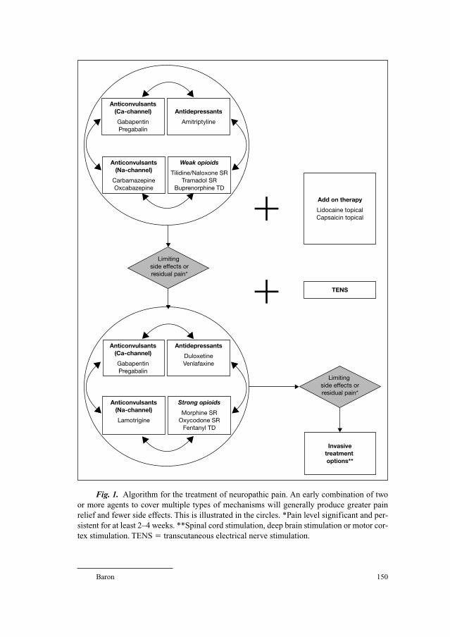
Baron 150
Anticonvulsants (Ca-channel)
Gabapentin Pregabalin
Antidepressants
Amitriptyline
TENS
Limiting side effects orresidual pain*
Invasivetreatment options**
Add on therapy
Lidocaine topical Capsaicin topical
Limiting side effects orresidual pain*
Anticonvulsants(Na-channel)
CarbamazepineOxcabazepine
Weak opioids
Tilidine/Naloxone SRTramadol SR
Buprenorphine TD
Anticonvulsants(Ca-channel)
GabapentinPregabalin
Antidepressants
DuloxetineVenlafaxine
Anticonvulsants(Na-channel)
Lamotrigine
Strong opioids
Morphine SROxycodone SR
Fentanyl TD
�
�
Fig. 1. Algorithm for the treatment of neuropathic pain. An early combination of two
or more agents to cover multiple types of mechanisms will generally produce greater pain
relief and fewer side effects. This is illustrated in the circles. *Pain level significant and per-
sistent for at least 2–4 weeks. **Spinal cord stimulation, deep brain stimulation or motor cor-
tex stimulation. TENS � transcutaneous electrical nerve stimulation.

Therapy of Neurological Complications 151
stimulation may be indicated. Transcutaneous electrical nerve stimulation may
be effective in some cases and has minimal side effects.
However, beyond these treatment approaches the importance of the
biopsychosocial model of chronic pain should be considered by additional
management of psychological and social aspects [15, 39].
The treatment of neuropathic pain is currently still unsatisfactory. The
hope is that in the future novel drugs will be developed that address specifically
the relevant combination of mechanisms in one particular patient leading to an
optimal individual polypragmatic therapy [40].
References
1 Watson CP, Watt VR, Chipman M, Birkett N, Evans RJ: The prognosis with postherpetic neural-
gia. Pain 1991;46:195–199.
2 Dworkin RH, Hartstein G, Rosner HL, Walther RR, Sweeney EW, Brand L: A high-risk method
for studying psychosocial antecedents of chronic pain: the prospective investigation of herpes
zoster. J Abnorm Psychol 1992;101:200–205.
3 Bruxelle J: Prospective epidemiologic study of painful and neurologic sequelae induced by herpes
zoster in patients treated early with oral acyclovir. Neurology 1995;45(suppl 8):S78–S79.
4 Higa K, Noda B, Manabe H, Sato S, Dan K: T-lymphocyte subsets in otherwise healthy patients
with herpes zoster and relationships to the duration of acute herpetic pain. Pain 1992;51:111–118.
Table 1. Pharmacological therapy of postherpetic neuralgia (doses for adults)
Compound Efficacious dose Dose interval Evidence
(maximal dose) [mg/day]
Anti-depressants
Amitriptyline 50–75 (150) 0–0–1 ⇑⇑Anti-convulsants
(Ca-channel)
Gabapentin 1,200–2,400 (3,600) 1–1–1 ⇑⇑Pregabalin 150 (600) 1–0–1 ⇑⇑Long-acting opioids
Tramadol SR Titration (600) 1–(1)–1 ⇑Morphine SR Titration no 1–(1)–1 ⇑Oxycodone Titration no 1–(1)–1 ⇑Topical therapy
Capsaicin cream – 4 � die ⇑Lidocain-patch – (3 patches/die) 1 � die ⇑⇑
Levels of evidence.
⇑ � at least 1 RCT; ⇑⇑ � Several RCT or metaanalyses.

Baron 152
5 Nurmikko T, Bowsher D: Somatosensory findings in postherpetic neuralgia. J Neurol Neurosurg
Psychiatry 1990;53:135–141.
6 Jensen TS, Krebs B, Nielsen J, Rasmussen P: Immediate and long-term phantom limb pain in
amputees: incidence, clinical characteristics and relationship to pre-amputation limb pain. Pain
1985;21:267–278.
7 Gatchel RJ, Polatin PB, Kinney RK: Predicting outcome of chronic back pain using clinical pre-
dictors of psychopathology: a prospective analysis. Health Psychol 1995;14:415–420.
8 Baron R, Haendler G, Schulte H: Afferent large fiber polyneuropathy predicts the development of
postherpetic neuralgia. Pain 1997;73:231–238.
9 Oxman MN, Levin MJ, Johnson GR, Schmader KE, Straus SE, Gelb LD, Arbeit RD, Simberkoff MS,
Gershon AA, Davis LE, Weinberg A, Boardman KD, Williams HM, Zhang JH, Peduzzi PN,
Beisel CE, Morrison VA, Guatelli JC, Brooks PA, Kauffman CA, Pachucki CT, Neuzil KM,
Betts RF, Wright PF, Griffin MR, Brunell P, Soto NE, Marques AR, Keay SK, Goodman RP,
Cotton DJ, Gnann JW, Jr., Loutit J, Holodniy M, Keitel WA, Crawford GE, Yeh SS, Lobo Z,
Toney JF, Greenberg RN, Keller PM, Harbecke R, Hayward AR, Irwin MR, Kyriakides TC, Chan CY,
Chan IS, Wang WW, Annunziato PW, Silber JL: A vaccine to prevent herpes zoster and postherpetic
neuralgia in older adults. N Engl J Med 2005;352:2271–2284.
10 Wassilew S: Brivudin compared with famciclovir in the treatment of herpes zoster: effects in acute
disease and chronic pain in immunocompetent patients. A randomized, double-blind, multi-
national study. J Eur Acad Dermatol Venereol 2005;19:47–55.
11 Wood MJ, Kay R, Dworkin RH, Soong SJ, Whitley RJ: Oral acyclovir therapy accelerates pain res-
olution in patients with herpes zoster: a meta-analysis of placebo-controlled trials. Clin Infect Dis
1996;22:341–347.
12 Bowsher D: The effects of pre-emptive treatment of postherpetic neuralgia with amitriptyline: a
randomized, double-blind, placebo-controlled trial. J Pain Symptom Manage 1997;13:327–331.
13 Kuraishi Y, Takasaki I, Nojima H, Shiraki K, Takahata H: Effects of the suppression of acute her-
petic pain by gabapentin and amitriptyline on the incidence of delayed postherpetic pain in mice.
Life Sci 2004;74:2619–2626.
14 Dworkin RH, Schmader KE: Treatment and prevention of postherpetic neuralgia. Clin Infect Dis
2003;36:877–882.
15 Hempenstall K, Nurmikko TJ, Johnson RW, A’Hern RP, Rice AS: Analgesic therapy in posther-
petic neuralgia: a quantitative systematic review. PLoS Med 2005;2:e164.
16 McQuay HJ, Tramer M, Nye BA, Carroll D, Wiffen PJ, Moore RA: A systematic review of anti-
depressants in neuropathic pain. Pain 1996;68:217–227.
17 Sindrup SH, Jensen TS: Efficacy of pharmacological treatments of neuropathic pain: an update
and effect related to mechanism of drug action. Pain 1999;83:389–400.
18 Collins SL, Moore RA, McQuay HJ, Wiffen P: Antidepressants and anticonvulsants for diabetic
neuropathy and postherpetic neuralgia: a quantitative systematic review. J Pain Symptom Manage
2000;20:449–458.
19 Goldstein DJ, Lu Y, Detke MJ, Lee TC, Iyengar S: Duloxetine vs. placebo in patients with painful
diabetic neuropathy. Pain 2005;116:109–118.
20 Rowbotham MC, Goli V, Kunz NR, Lei D: Venlafaxine extended release in the treatment of
painful diabetic neuropathy: a double-blind, placebo-controlled study. Pain 2004;110:697–706.
21 Rowbotham M, Harden N, Stacey B, Bernstein P, Magnus-Miller L: Gabapentin for the treatment
of postherpetic neuralgia: a randomized controlled trial. JAMA 1998;280:1837–1842.
22 Dworkin RH, Corbin AE, Young JP: Pregabalin for the treatment of postherpetic neuralgia: a ran-
domized, placebo-controlled trial. Neurology 2003;22:1274–1283.
23 Sabatowski R, Galvez R, Cherry DA, Jacquot F, Vincent E, Maisonobe P, Versavel M: Pregabalin
reduces pain and improves sleep and mood disturbances in patients with post-herpetic neuralgia:
results of a randomised, placebo-controlled clinical trial. Pain 2004;109:26–35.
24 Rowbotham MC, Reisner-Keller LA, Fields HL: Both intravenous lidocaine and morphine reduce
the pain of postherpetic neuralgia. Neurology 1991;41:1024–1028.
25 Watson CP, Babul N: Efficacy of oxycodone in neuropathic pain: a randomized trial in posther-
petic neuralgia [In Process Citation]. Neurology 1998;50:1837–1841.
Therapy of Neurological Complications 153
26 Göbel H, Stadler T: Treatment of post-herpes zoster pain with tramadol. Results of an open pilot
study versus clomipramine with or without levomepromazine. Drugs 1997;53(suppl 2):34–39.
27 Raja SN, Haythornthwaite JA, Pappagallo M, Clark MR, Travison TG, Sabeen S, Royall RM, Max
MB: Opioids versus antidepressants in postherpetic neuralgia: a randomized, placebo-controlled
trial. Neurology 2002;59:1015–1021.
28 Eide K, Stubhaug A, Oye I, Breivik H: Continuous subcutaneous administration of the N-methyl-
D-aspartic acid (NMDA) receptor antagonist ketamine in the treatment of post-herpetic neuralgia.
Pain 1995;61:221–228.
29 Nelson KA, Park KM, Robinovitz E, Tsigos C, Max MB: High-dose oral dextromethorphan versus
placebo in painful diabetic neuropathy and postherpetic neuralgia. Neurology 1997;48:1212–1218.
30 Sang CN, Booher S, Gilron I, Parada S, Max MB: Dextromethorphan and memantine in painful
diabetic neuropathy and postherpetic neuralgia: efficacy and dose-response trials. Anesthesiology
2002;96:1053–1061.
31 Watson CP, Tyler KL, Bickers DR, Millikan LE, Smith S, Coleman E: A randomized vehicle-
controlled trial of topical capsaicin in the treatment of postherpetic neuralgia. Clin Ther 1993;15:
510–526.
32 Bernstein JE, Korman NJ, Bickers DR, Dahl MV, Millikan LE: Topical capsaicin treatment of
chronic postherpetic neuralgia. J Am Acad Dermatol 1989;21(pt 1):265–270.
33 Galer BS, Rowbotham MC, Perander J, Friedman E: Topical lidocaine patch relieves postherpetic
neuralgia more effectively than a vehicle topical patch: results of an enriched enrollment study.
Pain 1999;80:533–538.
34 Meier T, Wasner G, Faust M, Kuntzer T, Ochsner F, Hueppe M, Bogousslavsky J, Baron R:
Efficacy of lidocaine patch 5% in the treatment of focal peripheral neuropathic pain syndromes: a
randomized, double-blind, placebo-controlled study. Pain 2003;106:151–158.
35 Rowbotham MC, Davies PS, Verkempinck C, Galer BS: Lidocaine patch: double-blind controlled
study of a new treatment method for post-herpetic neuralgia. Pain 1996;65:39–44.
36 Kotani N, Kushikata T, Hashimoto H, Kimura F, Muraoka M, Yodono M, Asai M, Matsuki A:
Intrathecal methylprednisolone for intractable postherpetic neuralgia. N Engl J Med 2000;343:
1514–1519.
37 Sindrup SH, Jensen TS: Pharmacologic treatment of pain in polyneuropathy. Neurology 2000;55:
915–920.
38 Gilron I, Bailey JM, Tu D, Holden RR, Weaver DF, Houlden RL: Morphine, gabapentin, or their
combination for neuropathic pain. N Engl J Med 2005;352:1324–1334.
39 Haythornthwaite JA, Benrud-Larson LM: Psychological aspects of neuropathic pain. Clin J Pain
2000;16(suppl):S101–S105.
40 Fields HL, Rowbotham M, Baron R: Postherpetic neuralgia: irritable nociceptors and deafferenta-
tion. Neurobiol Dis 1998;5:209–227.
Prof. Dr. Med. Ralf Baron
Klinik für Neurologie, UK-SH, Campus Kiel
Sektion Neurologische Schmerzforschung und Therapie
Schittenhelmstr. 10
DE–24105 Kiel (Germany)
Tel. �49 431 597 8505, Fax �49 431 597 8530, E-Mail [email protected]
Gross G, Doerr HW (eds): Herpes Zoster.
Monogr Virol. Basel, Karger, 2006, vol 26, pp 154–163
Epidemiology of Herpes Zoster:What has Changed?
Regina Allwinn, Sigune Buxbaum, Hans Wilhelm Doerr
Institute for Medical Virology, University Hospital of Frankfurt,
Frankfurt am Main, Germany
Herpes zoster (shingles) is an inflammatory neurodermatologic disease,
usually localized on a skin segment of the body which is innervated by a sen-
sory nerve. More than 100 years ago, an association between Varicella and her-
pes zoster has been suggested (see preface of the book). Shingles is a secondary
disease to passed Varicella virus (VZV) infection, which latently persists life-
long in the spinal ganglia of the host. The presumably proviral latency is
switched to productive infection by several trigger factors resulting in shingles
(‘girdle rose’). The most important trigger factor is a waning cell-mediated
immunity to VZV along a big time interval after primary infection during child-
hood. Thus, the majority of patients are elderly or those who suffer from
immunocompromising diseases. Nevertheless, many case reports remind that
herpes zoster occurs also in immunocompetent adolescents and even in chil-
dren. Numerous clinical observations have elucidated the epidemiology of her-
pes zoster. Figure 1 displays the viral circulation through the population.
Seroepidemiology
Similar to many other viral infections the spread of VZV can be estimated by
serum surveys, which means determination of IgG antibody prevalence in age
grouped population samples. IgG antibodies are formed after varicella or sub-
clinic VZV infection and persist lifelong in stable titers. Numerous prospective
studies or retrospective evaluations have shown that high prevalences are reached
already in childhood [1–3]. During the course of life, fluctuating antibody titer is
no marker for a subsequent development of herpes zoster. Figure 2 shows serum
surveys on different seroconversion titers of VZV-specific IgG antibodies and
Epidemiology and Control of Herpes Zoster
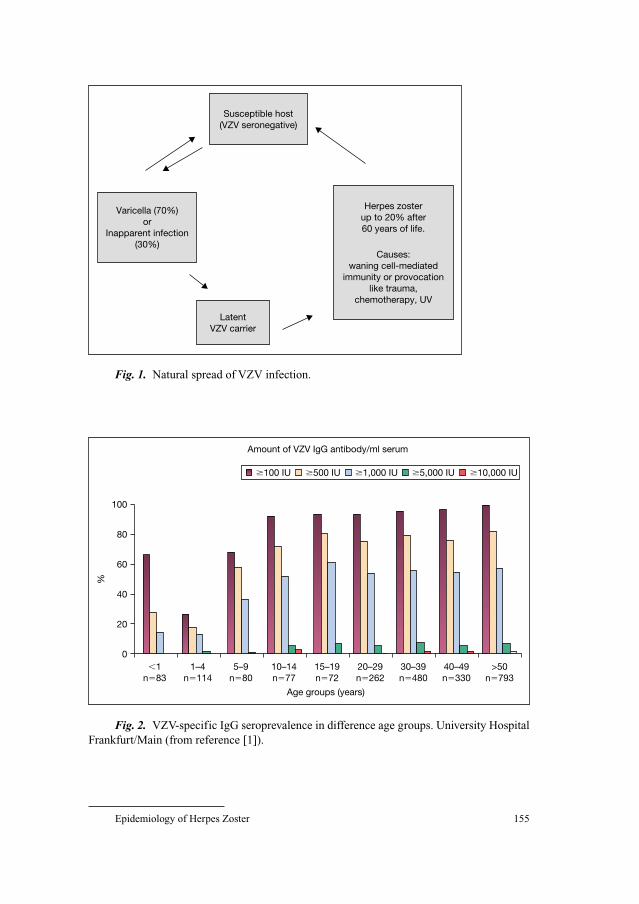
Epidemiology of Herpes Zoster 155
Latent VZV carrier
Susceptible host (VZV seronegative)
Varicella (70%) or
Inapparent infection (30%)
Herpes zoster up to 20% after 60 years of life.
Causes: waning cell-mediated
immunity or provocation like trauma,
chemotherapy, UV
Fig. 1. Natural spread of VZV infection.
0
20
40
60
80
100
�1 n�83
1–4 n�114
5–9 n�80
10–14 n�77
15–19 n�72
20–29 n�262
30–39 n�480
40–49 n�330
>50 n�793
%
Age groups (years)
Amount of VZV IgG antibody/ml serum
�100 IU �500 IU �10,000 IU�5,000 IU�1,000 IU
Fig. 2. VZV-specific IgG seroprevalence in difference age groups. University Hospital
Frankfurt/Main (from reference [1]).
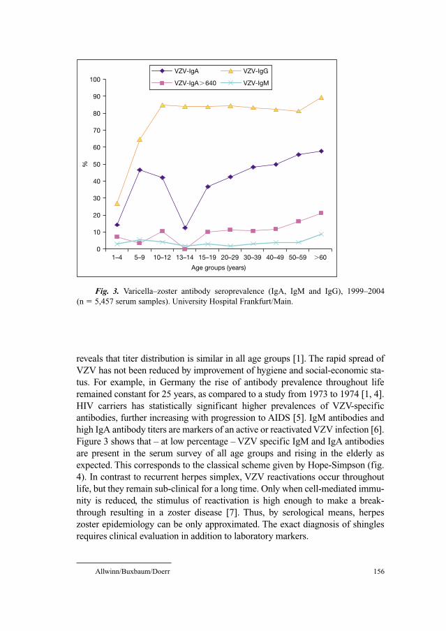
0
10
20
30
40
50
60
70
80
90
100
1–4 5–9 10–12 13–14 15–19 20–29 30–39 50–59 �60
VZV-IgA
VZV-IgA�640
VZV-IgG
VZV-IgM%
Age groups (years)
40–49
Fig. 3. Varicella–zoster antibody seroprevalence (IgA, IgM and IgG), 1999–2004
(n � 5,457 serum samples). University Hospital Frankfurt/Main.
Allwinn/Buxbaum/Doerr 156
reveals that titer distribution is similar in all age groups [1]. The rapid spread of
VZV has not been reduced by improvement of hygiene and social-economic sta-
tus. For example, in Germany the rise of antibody prevalence throughout life
remained constant for 25 years, as compared to a study from 1973 to 1974 [1, 4].
HIV carriers has statistically significant higher prevalences of VZV-specific
antibodies, further increasing with progression to AIDS [5]. IgM antibodies and
high IgA antibody titers are markers of an active or reactivated VZV infection [6].
Figure 3 shows that – at low percentage – VZV specific IgM and IgA antibodies
are present in the serum survey of all age groups and rising in the elderly as
expected. This corresponds to the classical scheme given by Hope-Simpson (fig.
4). In contrast to recurrent herpes simplex, VZV reactivations occur throughout
life, but they remain sub-clinical for a long time. Only when cell-mediated immu-
nity is reduced, the stimulus of reactivation is high enough to make a break-
through resulting in a zoster disease [7]. Thus, by serological means, herpes
zoster epidemiology can be only approximated. The exact diagnosis of shingles
requires clinical evaluation in addition to laboratory markers.

Epidemiology of Herpes Zoster 157
Incr
easi
ng le
vels
of h
ost
resi
stan
ce
Prim
ary
varic
ella
–zos
ter
viru
s in
fect
ion
Con
tact
with
a c
ase
of v
aric
ella
Var
icel
la (l
aten
t vi
rus
goes
to
sens
ory
gang
lia)
Containedreversions
Days
3 6 9 121518
Months Years
Containedreversions
Level of host resistance Her
pes
Zos
ter
(“su
cces
sful
” re
vers
ion)
Critical level of host resistance
Containedreversions
Fig. 4. Immune response after infection VZV (from reference [12]).
Clinical Epidemiology
Before the introduction of VZV vaccination, only a few population-basedstudies of the natural history and epidemiology of herpes zoster have been con-
ducted [8]. The two classic investigations have been performed in UK and USA
by Hope-Simpson [9, 10] and by Ragozzino et al. [11] who retrospectively
analyzed records of the time periods 1947–1962 and 1945–1959, respectively.
Beginning with the first year of life, the cumulative incidence of herpes zoster
rises slowly in an exponential way (fig. 5). After the 60th year of life, at least
20% of the population has once passed an episode of herpes zoster; only a few of
them have presented a second episode. Even in infants some cases are observed,
probably as a consequence of prenatal or early childhood VZV infection.
Now, 30 years later, the herpes zoster epidemiology has obviously
changed. Donahue et al. [12] investigated incident and recurrent herpes zoster
from 1990 to 1992 in a health maintenance organization and found significant
higher incidences of shingles. A ‘new’ epidemiology of shingles was also
ruled out in Central Europe by the ‘German Zoster Study Group’ [13] which
prospectively recruited 2,063 patients suffering from shingles in different der-
matomes between September 1994 and March 1995. Nearly 20% of patients
were younger than 30 years of age! Since 1990, it was observed, that children
are more afflicted by herpes zoster, going parallel with a broader use of corticoids
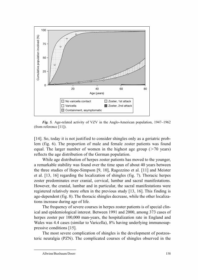
Allwinn/Buxbaum/Doerr 158
Cum
ulat
ive
pop
ulat
ion
invo
lved
(%)
100
75
50
25
020 40
Age (years)
60 80
No varicella contact Zoster, 1st attack
Zoster, 2nd attackVaricella
Containment, asymptomatic
Fig. 5. Age-related activity of VZV in the Anglo-American population, 1947–1962
(from reference [11]).
[14]. So, today it is not justified to consider shingles only as a geriatric prob-
lem (fig. 6). The proportion of male and female zoster patients was found
equal. The larger number of women in the highest age group (�70 years)
reflects the age distribution of the German population.
While age distribution of herpes zoster patients has moved to the younger,
a remarkable stability was found over the time span of about 40 years between
the three studies of Hope-Simpson [9, 10], Ragozzino et al. [11] and Meister
et al. [13, 16] regarding the localization of shingles (fig. 7). Thoracic herpes
zoster predominates over cranial, cervical, lumbar and sacral manifestations.
However, the cranial, lumbar and in particular, the sacral manifestations were
registered relatively more often in the previous study [13, 16]. This finding is
age-dependent (fig. 8). The thoracic shingles decrease, while the other localiza-
tions increase during age of life.
The frequency of severe courses in herpes zoster patients is of special clin-
ical and epidemiological interest. Between 1991 and 2000, among 373 cases of
herpes zoster per 100,000 man-years, the hospitalization rate in England and
Wales was 4.4 cases (similar to Varicella), 8% having underlying immunosup-
pressive conditions [15].
The most severe complication of shingles is the development of postzos-
teric neuralgia (PZN). The complicated courses of shingles observed in the

Epidemiology of Herpes Zoster 159
Pop
ulat
ion/
pat
ient
s (%
)Male patients (n�912)Female patients (n�1,146)
Male population(n�39.2�106)
Female population(n�41.8�106)
�10 10–19 20–29 30–39 40–49 50–59 60–69 �70
30
25
20
15
10
5
0
Age groups (years)
Fig. 6. Age and sex distribution among patients with herpes zoster compared with the
data from the German general population in 1993 (from reference [13]).
German study are compiled in table 1. To summarize, 320 among 2,063
patients (15.5%) required more intensive medical treatment.
Older and female patients had been more afflicted by PZN. Furthermore,
the number, as well as the hemorrhages of skin lesions with cranial or sacral
60
50
40
30%
20
10
0Thoracal Cranial Cervical
Localization
Lumbal Sacral Nodeclaration
Ragozzino et al. [11] (n�590)
Hope-Simpson [9, 10] [n�322]
Meister et al. (13, 16) [n�2,063]
Fig. 7. Localization of herpes zoster recorded in three different studies.
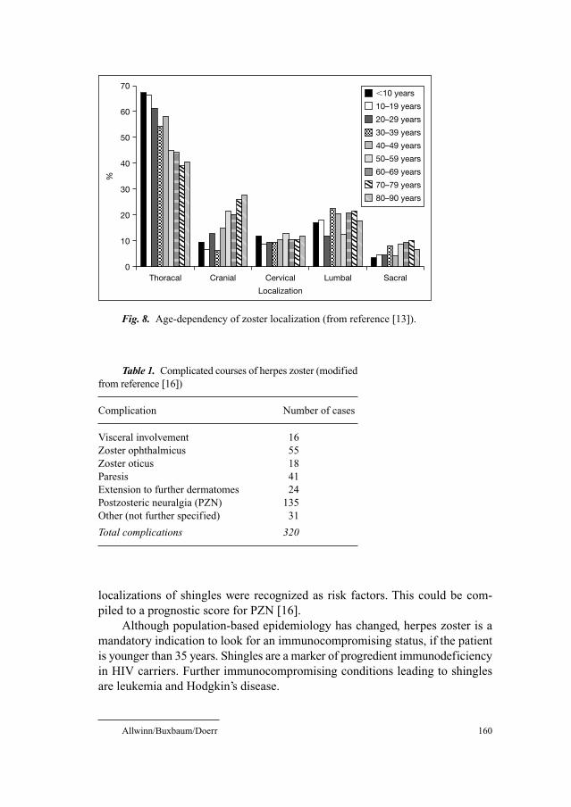
Table 1. Complicated courses of herpes zoster (modified
from reference [16])
Complication Number of cases
Visceral involvement 16
Zoster ophthalmicus 55
Zoster oticus 18
Paresis 41
Extension to further dermatomes 24
Postzosteric neuralgia (PZN) 135
Other (not further specified) 31
Total complications 320
Allwinn/Buxbaum/Doerr 160
localizations of shingles were recognized as risk factors. This could be com-
piled to a prognostic score for PZN [16].
Although population-based epidemiology has changed, herpes zoster is a
mandatory indication to look for an immunocompromising status, if the patient
is younger than 35 years. Shingles are a marker of progredient immunodeficiency
in HIV carriers. Further immunocompromising conditions leading to shingles
are leukemia and Hodgkin’s disease.
%
70
60
50
40
30
20
10
0Thoracal Cranial Cervical
Localization
Lumbal Sacral
�10 years
10–19 years
20–29 years
30–39 years
40–49 years
50–59 years
60–69 years
70–79 years
80–90 years
Fig. 8. Age-dependency of zoster localization (from reference [13]).
Epidemiology of Herpes Zoster 161
A similar high risk factor is therapeutic immunosuppression after organ
and in particular after bone marrow transplantation (see chapter: Herpes Zoster
in the Immunocompromised Host, pp 93–106). Thus, these people are recom-
mended to be vaccinated or screened on natural immunity before immunsup-
pression (see chapter: Live Attenuated Varicella Vaccine, pp 164–169).
Change of Epidemiology after General VZV Vaccination
After the successful vaccination of risk groups against VZV infection, gen-
eral immunization has been introduced into the USA in 1995. By the use of
consequent vaccination programs in early childhood, the occurrence of vari-
cella could be dramatically reduced (�90%) and the mortality decreased by two
third [17, 18]. In Germany, vaccination against VZV is generally recommended
since August 2004. In this way, the threshold of 90% population immunization
which is necessary for herd immunity can be reached easier. The vaccine is an
attenuated VZV strain (Oka strain) imitating the natural infection and thus
inducing immunity in a similar way. After immunization the vaccine virus
becomes latent. Later reactivation has been described in immunocompromised
and also healthy vaccinees. This usually appears as a mild course of herpes
zoster [19]. The persistence of vaccine-induced immunity does not last as long
as natural immunity. A growing number of ‘breakthrough Varicella’ among
immunized children was recognized [20–22].
Reactivation of vaccine virus or breakthrough of wild virus infection should
not limit the decrease of natural herpes zoster. However, if wild VZV circulation
is interrupted, the sub-clinical booster of immunity against natural herpes zoster
is reduced. Before varicella eradication, the huge pool of latent wild VZV repre-
sents a threat, namely that herpes zoster could become more frequent [7].
Paradoxically, a good VZV herd immunity of 90% could increase herpes zoster
incidences, which has been calculated with mathematical models [23].
Using the random-digit-dial Behavioral Risk Factor Surveillance System,
Yih et al. [24] monitored the incidence of varicella and herpes zoster in
Massachusetts between 1998 and 2003. They confirmed that due to general
vaccination the incidence of varicella has decreased as expected, while the
incidence of herpes zoster has significantly increased from 2.77/1,000 to
5.25/1,000. Thus, until VZV is eradicated, a booster vaccination of the elderly
might be recommended. A second vaccination with VZV decreases the herpes
zoster incidence, as recently shown in a large study on prevention of shingles
and postherpetic neuralgia [19]. At the moment, it cannot be decided whether
this is generally needed. More epidemiologic data are necessary for better
effective and reliable mathematical modeling [25].
Allwinn/Buxbaum/Doerr 162
References
1 Buxbaum S, Doerr HW, Allwinn R: Epidemiological analysis of immunity against vaccine-
preventable diseases: rubella, measles, mumps and chickenpox. Dtsch Med Wochenschr
2001;126:1289–1293.
2 Mossong J, Putz L, Schneider F: Seroprevalence and force of infection of varicella-zoster virus in
Luxembourg. Epidemiol Infect 2004;132:1121–1127.
3 Vyse, AJ, Gay NJ, Hesketh LM, Morgan-Capner P, Miller E: Seroprevalence of antibody to vari-
cella zoster virus in England and Wales in children and young adults. Epidemiol Infect 2004;132:
1129–1134.
4 Doerr HW, Lehmair H, Schmitz H, Kampa D, Luthardt TH: Simple mathematical deductions in
the seroepidemiology of viral infections. I. Herpesvirus group (Herpesvirus hominis; Varicella-
Zoster-Virus, Cytomegalovirus, Epstein-Barr-Virus). Zentrabl Bakteriol [Orig A] 1977;238:
149–164.
5 Enzensberger R, Braun W, July Ch, Helm EB, Doerr HW: Prevalence of antibodies to human her-
pesvirus and hepatitis B virus in patients at different stages of human immunodeficiency virus
(HIV). Infection 1991;19:140–145.
6 Doerr HW, Rentschler M, Scheifler G: Serologic detection of active infections with human herpes
viruses (CMV, EBV, HSV, VZV): diagnostic potential of IgA class and IgG subclass-specific anti-
bodies. Infection 1987;15:93–98.
7 Hambleton S, Gershon AA: Preventing varicella-zoster disease. Clin Microbiol Rev 2005;18:70–80.
8 Weller TH: Varicella and herpes zoster. Changing concepts of the natural history, control, and
importance of a not-so-benign virus (First of Two Parts). N Engl J Med 1983;309:1362–1368.
9 Hope-Simpson RE: Postherpetic neuralgia. J R Coll Gen Pract 1975;25:571–575.
10 Hope-Simpson: The nature of herpes zoster. A long-term study and a new hypothesis. Proc R Soc
Med 1965;58:9–20.
11 Ragozzino MW, Melton LJ 3rd, Kurland LT, Chu CP, Perry HO: Population-based study of herpes
zoster and its sequelae. Medicine (Baltimore) 1982;61:310–316.
12 Donahue JG, Choo PW, Manson JE, Platt R: The incidence of herpes zoster. Arch Intern Med
1995;155:1605–1609.
13 Meister W, Neiss A, Gross G, Doerr HW, Höbel W, Malin JP, von Essen J, Reimann BY, Witke C,
Wutzler P: Demography, symptomatology, and course of disease in ambulatory zoster patients. A
physican-based survey in Germany. Intervirology 1998;41:272–277.
14 Uebe B, Sauerbrei A, Burdach S, Horneff G: Herpes zoster by reactivated vaccine varicella zoster
virus in a healthy child. Eur J Pediatr 2002;161:442–444.
15 Brisson M, Edmunds WJ: Epidemiology of varicella-zoster virus in England and Wales. J Med
Virol 2003;70(suppl 1):9–14.
16 Meister W, Neiß A, Gross G, Doerr HW, Höbel W, Malin JP, von Essen J, Reimann BY, Witke C,
Wutzler P: A prognostic score for postherpetic neuralgia in ambulatory patients. Infection
1998;26:6.
17 Nguyen HQ, Jumaan AO, Seward JF: Decline in mortality due to varicella after implementation of
varicella vaccination in the Unites States. N Engl J Med 2005;352:450–458.
18 Vázques M, Shapiro ED: Varicella vaccine and infection with varicella-zoster virus. N Engl J Med
2005;352:439–440.
19 Oxmann MN, Levin MJ, Johnson GR, Schmader KE, Straus SE, Gelb LD, Arbeit RD, Simberkoff
MS, Gershon AA, Davis LE, Weinberg A, Boardman KD, Williams HM, Zhang JH, Peduzzi PN,
Beisel CE, Morrison VA, Guatelli JC, Brooks PA, Kauffman CA, Pachucki CT, Neuzil KM, Betts RF,
Wright PF, Griffin MR, Brunell P, Soto NE, Marques AR, Keay SK, Goodmann RP, Cotton DJ,
Gnann JW Jr, Loutit J, Holodniy M, Keitel WA, Crawford GE, Yeah SS, Lobo Z, Toney JF,
Greenberg RN, Keller PM, Harbecke R, Hayward AR, Irwin MR, Kyriakides TC, Chan CY, Chan
IS Wang WW, Annunziato PW, Silber JL, Shingles Prevention Group: A vaccine to prevent herpes
zoster and postherpetic neuralgia in older adults. N Engl J Med 2005;352:2271–2284.
20 Grose C: Varicella vaccination of children in the United States: assessment after the first decade
1995–2005. J Clin Virol 2005;33:89–95; discussion 96–98.
Epidemiology of Herpes Zoster 163
21 Haddad MB, Hill MB, Pavia AT, Green CE, Jumaan AO, De AK, Rolfs RT: Vaccine effectiveness dur-
ing a varicella outbreak among school children: Utah, 2002–2003. Pediatrics 2005;115:1488–1493.
22 Ludwig B, Kraus FB, Allwinn R, Keim S, Doerr HW, Buxbaum S: Loss of VZV-antibodies in a
health care worker – how to deal with the wane of immunity after vaccination? Infection, submitted.
23 Brisson M, Edmunds WJ, Gay NJ: Varicella vaccination: impact of vaccine efficacy on the epi-
demiology of VZV. J Med Virol 2003;70(suppl 1):31–37.
24 Yih WK, Brooks DR, Lett SM, Jumaan AO, Zhang Z, Clements KM, Seward JF: The incidence
of varicella and herpes zoster in Massachusetts as measured by the Behavioral Risk Factor
Surveillance System (BRFSS) during a period of increasing varicella vaccine coverage,
1998–2003. BMC Public Health 2005;5:68.
25 Wagenpfeil S, Neiss A, Wutzler P: Effects of varicella vaccination on herpes zoster incidence. Clin
Microbiol Infect 2004;10:954–960.
Professor H.W. Doerr, M.D.
Institute for Medical Virology, University Hospital of Frankfurt
Paul-Ehrlich-Strasse 40
DE–60596 Frankfurt/Main (Germany)
Tel. �49 69 63015219, Fax �49 69 63016244, E-Mail [email protected]
Gross G, Doerr HW (eds): Herpes Zoster.
Monogr Virol. Basel, Karger, 2006, vol 26, pp 164–169
Live Attenuated Varicella Vaccine
Anne A. Gershon
Department of Pediatrics, Columbia University College of Physicians and Surgeons,
New York, N.Y., USA
The Virus and Its Diseases
Varicella–zoster virus (VZV) is the etiologic agent of varicella and zoster.
Varicella is the primary infection, and zoster is due to reactivation of latent
VZV acquired during chickenpox. Each disease is characterized by a macu-
lopapular and vesicular skin eruption, which in varicella is generalized, and in
zoster is unilateral and usually localized. Varicella is often mild and uncompli-
cated in otherwise healthy children, but it may unpredictably be associated with
significant morbidity and even mortality. In the United States, in the pre-vac-
cine era, there were about 100 annual deaths from varicella and 11,000 hospi-
talizations [1]. Most deaths from varicella occurred in individuals who were
healthy before contracting varicella. The risk of developing zoster is increased
in the immunocompromised and the elderly, and zoster may be severe but is
rarely fatal.
Live Attenuated Varicella Vaccine: Background
A live attenuated varicella vaccine, the Oka strain, was developed in Japan in
the early 1970s [2]. This vaccine is now available worldwide. The most extensive
published experience with varicella vaccine comes from the United States, where
it was licensed in 1995 for routine use in healthy susceptible individuals over the
age of 1 year [1]. The vaccine is extremely safe [3, 4]. The main adverse effects in
healthy persons are both mild and transient: fever and a sore arm in less than 50%,
and a very mild rash in about 5%, usually occurring a month after immunization
[5]. VZV antibodies are demonstrable after about 2 weeks. In the United States,
Live Attenuated Varicella Vaccine 165
the epidemiology of varicella is changing, with a decreased incidence of chicken-
pox in both the vaccinated and the unvaccinated [6].
Transmission of the Oka strain from healthy vaccinees with rash to other sus-
ceptibles is extremely rare. It has been reported only on four occasions, although
over 30 million doses have been distributed in the United States alone. There is no
clinical evidence of reversion of the vaccine strain to virulence. Contact cases are
usually very mild or even subclinical [4, 7]. There are many mutations in the vac-
cine compared to the parental viral strains, although which are responsible for
attenuation is unknown [8–10]. Although a few cases of immunodeficiency have
been unmasked by complications following varicella vaccination, there have been
no reported fatalities from the Oka varicella strain [4].
In the United States, contraindications to varicella vaccine include preg-
nancy, allergy to vaccine components, and immunodeficiency. Children receiv-
ing steroids at a dose of 2 mg/kg/day of prednisone or greater (or of its
equivalent) should not be immunized. It is safe to immunize HIV-infected chil-
dren, as long as their levels of CD4 lymphocytes are relatively well-preserved
[11]. Healthy persons who have close contact with susceptible individuals who
are at high risk to develop severe varicella, such as pregnant women and chil-
dren with malignant disease, should be immunized to protect them because
transmission of vaccine is rare. The vaccine has been shown to be cost effective
in the United States and abroad [12–17].
Efficacy and Effectiveness of the Vaccine
Studies in vaccinated leukemic children showed not only that the vaccine
was safe but also that it was highly protective against varicella [18]. About 85%
of vaccinated leukemic children were completely protected against varicella
after household exposure, and those who developed breakthrough infection had
mild infections. Early studies in healthy children indicated a similar degree of
protection [19].
Two double-blind placebo controlled studies in healthy children were con-
ducted, indicating protection of about 90% [20–22]. Better protection resulted
from higher doses (roughly 3,000 plaque forming units {pfus} vs. 10,000 pfus
per dose). A post-licensure case-control effectiveness study in healthy children
indicated that the vaccine in the United States is about 85% effective in
preventing varicella [23]. The best evidence of protection from varicella vac-
cine, however, is the reported decline in disease since 1995, in the United
States [6].

Gershon 166
Does Immunity to Varicella Wane with Time after Immunization?
There are two types of vaccine failure, primary and secondary. Primary vac-
cine failure means there is no immune response to a vaccine. When a positive
immune response after vaccination is lost with time, secondary vaccine failure has
occurred. There is little evidence for secondary vaccine failure after immunization
of healthy children with the Oka vaccine. Persistence of VZV antibodies and cellu-
lar immunity for up to 20 years after vaccination has been reported [24–26]. Studies
involving over 400 vaccinated adults for up to 20 years revealed no decrease in
immunity to varicella with time. It is disquieting, however, that about 10% of chil-
dren may develop a modified form of varicella despite vaccination. In some studies,
the breakthrough rate of varicella ranged between 18 and 34% [27–30].
Outbreaks of varicella in young vaccinated children have recently been
reported in the United States [31–33]. There are a number of possible explanations
for these outbreaks. Improper storage of this labile vaccine may account for pri-
mary vaccine failure in some children. The ability to mount a protective immune
response may be impaired in children with asthma [34]. Children immunized at
less than 14 months old may have higher rates of breakthrough varicella than those
immunized when they were older [31, 32]. When varicella vaccine is administered
less than 1 month after another live vaccine, the incidence of breakthrough vari-
cella increases [35]. A recent outbreak of varicella in a day care center in New
Hampshire identified an interval of over 3 years since vaccination as the only sig-
nificant risk for developing breakthrough disease [33]. This small study is the only
one that suggests that waning immunity may be a factor in breakthrough disease.
However, the children involved were very young, and the age at vaccination could
also have been a factor. Continued investigations are necessary to determine
whether waning immunity is significant in developing breakthrough disease.
A second dose of varicella vaccine may prove useful to avoid possible pri-
mary and secondary vaccine failure [36]. Breakthrough varicella is almost
always mild. The few reports of varicella of normal or increased severity in vac-
cines probably represent primary vaccine failure [4]. Zoster, however, may be
more of a concern. Currently a second dose of varicella vaccine is being consid-
ered for all healthy children in the U.S. because of the realization that primary
immune failure may be more of a concern than was originally thought [37].
Zoster: Effects and Potential Effects on Its Incidence in the Vaccine Era
In immunocompromised vaccines, the incidence of zoster was lower than
after natural infection [25]. Therefore vaccination may also be protective
Live Attenuated Varicella Vaccine 167
against zoster in healthy children. Less than 50 cases of zoster have been
reported after distribution of over 30 million doses of vaccine between 1995
and 2002 in the United States [4].
Recently, it has been proposed that exposure to varicella is protective
against zoster, and there is concern that zoster may become more common as
the incidence of varicella decreases in a population. Studies in vaccinated
leukemic children with either household exposure and/or additional doses of
varicella vaccine correlated with a lower incidence of zoster than one dose of
vaccine [38]. A recent case-control study indicated that following natural vari-
cella, there is a lower incidence of zoster in individuals who have exposures to
children with VZV infections in comparison to those who do not [39]. It has
been projected that an epidemic of zoster with accompanying significant mor-
tality will occur in countries where varicella vaccination is routine [40]. These
observations, even though theoretical, have led to reluctance to use varicella
vaccine routinely in some countries. Since the possibility has been raised, it will
be important to study the situation further.
It is important, however, to put the idea of an increase of zoster into per-
spective. The reported incidence of zoster in healthy individuals aged 40–50 in
developed countries is 2–4 cases per 1,000 person-years of observation. It has
been projected that the rate of zoster may double in countries with routine vac-
cination [40]. This could lead to an incidence of 4–8 cases per 1,000 person-
years of observation in this age group. This incidence of zoster is still not very
high and is about that seen in vaccinated leukemic children. In similar unvacci-
nated children, the incidence is at least 3 times higher. In adults with AIDS it is
6 times higher. Thus the projected increase in the incidence of zoster based on
computer modeling might represent a significant increase, but it is likely to be
far from an epidemic. Moreover, the mortality of zoster is lower than that from
the primary infection (varicella). As yet, no actual increase in the incidence of
zoster has been observed in the United States.
References
1 Centers for Disease Control: Prevention of varicella: recommendations of the advisory committee
on immunization practices (ACIP). MMWR Morb Mortal Wkly Rep 1996;45:1–36.
2 Takahashi M, Otsuka T, Okuno Y, Asano Y, Yazaki T: Live vaccine used to prevent the spread of
varicella in children in hospital. Lancet 1974;2:1288–1290.
3 Baba K, Yabuuchi H, Takahashi M, Gershon AA, Ogra PL: Seroepidemiologic behavior of vari-
cella zoster virus infection in a semiclosed community after introduction of VZV vaccine.
J Pediatr 1984;105:712–716.
4 Sharrar RG, LaRussa P, Galea SA, Steinberg SP, Sweet AR, Keatley RM, Wells ME, Stephenson
WP, Gershon AA: The postmarketing safety profile of varicella vaccine. Vaccine 2000;19:916–923.
5 White CJ. Varicella-zoster virus vaccine. Clin Infect Dis 1997;24:753–763.
Gershon 168
6 Seward JF, Watson BM, Peterson CL, Mascola L, Pelosi JW, Zhang JX, Maupin TJ, Goldman GS,
Tabony LJ, Brodovicz KG, Jumaan AO, Wharton M: Varicella disease after introduction of vari-
cella vaccine in the United States, 1995–2000. JAMA 2000;287:606–611.
7 Tsolia M, Gershon AA, Steinberg SP, Gelb L: Live attenuated varicella vaccine: evidence that the
virus is attenuated and the importance of skin lesions in transmission of varicella-zoster virus.
J Pediatr 1990;116:184–189.
8 Gomi Y, Imagawa T, Takahashi M, Yamanishi K: Oka varicella vaccine is distinguishable from its
parental virus in DNA sequence of open reading frame 62 and its transactivation activity. J Med
Virol 2000;61:497–503.
9 Loparev VN, McCaustland K, Holloway BP, Krause PR, Takayama M, Schmid DS: Rapid geno-
typing of varicella-zoster virus vaccine and wild type strains with fluorophore-labeled hybridiza-
tion probes. J Clin Micro 2000;38:4315–4319.
10 Gomi Y, Imagawa T: Comparison of DNA sequence and transactivation activity of open reading
frame 62 of Oka varicella vaccine and its parental viruses. Arch Virol 2001;S17:49–56.
11 Gershon A, Takahashi M, et al: Live attenuated varicella vaccine; in Plotkin S, Orenstein W (eds):
Vaccines, ed 4. Philadelphia, WB Saunders, 2002.
12 Lieu T, Cochi S, Black SB, Halloran ME, Shinefield HR, Holmes SJ, Wharton M, Washington AE:
Cost-effectiveness of a routine varicella vaccination program for U.S. children. JAMA 1994;271:
375–381.
13 Beutels P, Clara R, Tormans G, Van Doorslaer E, Van Damme P: Costs and benefits of routine
varicella vaccination in German children. J Infect Dis 1996;174:S335–S341.
14 Burnham BR, Wells TS, Riddle JR: A cost-benefit analysis of a routine varicella vaccination pro-
gram for United States Air Force Academy cadets. Mil Med 1998;163:631–634.
15 Coudeville L, Paree F, Lebrun T, Sailly J: The value of varicella vaccination in healthy children:
cost-benefit analysis of the situation in France. Vaccine 1999;17:142–151.
16 Diez Domingo J, Ridao M, Latour J, Ballester A, Morant A: A cost benefit analysis of routine vari-
cella vaccination in Spain. Vaccine 1999;17:1306–1311.
17 Brisson M, Edmunds WJ: The cost-effectiveness of varicella vaccination in Canada. Vaccine
2002;20:1113–1125.
18 Gershon AA, Steinberg SP, Gelb L, Galasso G, Borkowsky W, LaRussa P, Farrara A: Live attenu-
ated varicella vaccine: efficacy for children with leukemia in remission. JAMA 1984;252:
355–362.
19 White CJ: Clinical trials of varicella vaccine in healthy children. Infect Dis Clin North Am
1996;10:595–608.
20 Weibel RB, Neff BJ, Kuter BJ, Guess HA, Rothenberger CA, Fitzgerald AJ, Connor KA, McLean
AA, Hilleman MR, Buynak EB: Live attenuated varicella virus vaccine: efficacy trial in healthy
children. N Engl J Med 1984;310:1409–1415.
21 Weibel R, Kuter BJ, Guess HA, Rothenberger CA, Fitzgerald AJ, Connor KA, McLean AA,
Hilleman MR, Buynak EB: Live Oka/Merck varicella vaccine in healthy children: further clinical
and laboratory assessment. JAMA 1985;245:2435–2439.
22 Varis T, Vesikari T: Efficacy of high titer live attenuated varicella vaccine in healthy young chil-
dren. J Infect Dis 1996;174:330–334.
23 Vazquez M, LaRussa PS, Vazquez M, LaRussa PS, Gershon AA, Steinberg SP, Freudigman K,
Shapiro ED: The effectiveness of the varicella vaccine in clinical practice. N Engl J Med
2001;344:955–960.
24 Asano Y, Suga S, Yoshikawa T, Kobayashi I, Yazaki T, Shibata M, Tsuzuki K, Ito S: Experience and
reason: twenty year follow up of protective immunity of the Oka live varicella vaccine. Pediatrics
1994;94:524–526.
25 Arvin A, Gershon AA: Live attenuated varicella vaccine. Annu Rev Microbiol 1996;50:59–100.
26 Ampofo K, Saiman L, LaRussa P, Steinberg S, Annunziato P, Gershon A: Persistence of immunity
to live attenuated varicella vaccine in healthy adults. Clin Infect Dis 2002;34:774–779.
27 Clements DA, Armstrong CB, Ursano AM, Moggio MM, Walter EB, Wilfert CM: Over five-year
follow-up of Oka/Merck varicella vaccine recipients in 465 infants and adolescents. J Pediatr
Infect Dis 1995;14:874–879.

Live Attenuated Varicella Vaccine 169
28 Johnson, Stancin CT, Fattlar D, Rome LP, Kumar ML: A long-term prospective study of varicella
vaccine in healthy children. Pediatrics 1997;100:761–766.
29 Takayama N, Minamitani M, Takayama M: High incidence of breakthrough varicella observed in
healthy Japanese children immunized with live varicella vaccine (Oka strain). Acta Paediatr Jpn
1997;39:663–668.
30 Clements D, Moreira SP, Coplan PM, Bland CL, Walter EB: Postlicensure study of varicella vac-
cine effectiveness in a day-care setting. J Pediatr Infect Dis 1999;18:1047–1050.
31 Dworkin MS, Jennings CE: An outbreak of varicella among children attending preschool and ele-
mentary school in Illinois. Clin Infect Dis 2002;35:102–104.
32 Galil K, Fair E, et al: Younger age at vaccination may increase risk of varicella vaccine failure.
J Infect Dis 2002;186:102–105.
33 Galil K, Lee B, et al: Outbreak of varicella at a day-care center despite vaccination. N Eng J Med
2002;347:1909–1915.
34 Shapiro E, LaRussa P: Vaccination for varicella – just do it. JAMA 1997;278:1529–1530.
35 Centers for Disease Control: Simultaneous administration of varicella vaccine and other recom-
mended childhood vaccines – United States, 1995–1999. MMWR Morb Mortal Wkly Rep
2001;50:1058–1061.
36 Gershon A: Varicella vaccine: are two doses better than one? N Engl J Med 2002;347:1962–1963.
37 Gershon AA: Control of varicella: why is a two-dose schedule necessary? Pediatr Infect Dis J
2006;25:475–476.
38 Gershon A, LaRussa P, et al: The protective effect of immunologic boosting against zoster: an analy-
sis in leukemic children who were vaccinated against chickenpox. J Infect Dis 1996;173:450–453.
39 Thomas S, Wheeler J, et al: Contacts with varicella or with children and protection against herpes
zoster in adults: a case-control study. Lancet 2002;360:678–682.
40 Brisson M, Gay N, et al: Exposure to varicella boosts immunity to herpes-zoster: implications for
mass vaccination against chickenpox. Vaccine 2002;20:2500–2507.
Anne A. Gershon, MD
Columbia University College of Physicians and Surgeons
650 W. 168th Street
New York, NY 10032 (USA)
Tel. �1 212 305 9445, Fax �1 212 342 5218, E-Mail [email protected]
Gross G, Doerr HW (eds): Herpes Zoster.
Monogr Virol. Basel, Karger, 2006, vol 26, pp 170–188
Appendix:Herpes Zoster Guideline1 of theGerman Dermatology Society (DDG)2
G. Grossa, H. Schöferb, S. Wassilewc, K. Friesed, A. Timme, R. Guthoff c,H.W. Pauf, J.P. Maling, P. Wutzlerh, H.W. Doerri
aUniversitätsklinik and Poliklinik für Dermatologie und Venerologie, Rostock;bHautklinik, Klinikum der Johann-Wolfgang-Goethe-Universität, Frankfurt a. M.;cStädtische Hautklinik, Krefeld, Krefeld; dUniversitäts-Frauenklinik und Poliklinik;eUniversitäts-Augenklinik, Universität Rostock, and fUniversitätsklinik für Hals-
Nasen-Ohren-Krankheiten, Universität Rostock, Rostock; gNeurologische Klinik,
Universität Bochum, Bochum; hInstitut für Antivirale Chemotherapie, Universität
Jena, Jena; iInstitut für Medizinische Virologie, Klinikum der Johann-Wolfgang-
Goethe-Universität, Frankfurt a. M., Germany.
1. Definition
Herpes zoster (zoster, shingles) is a neurocutaneous disease caused by the
varicella zoster virus (VZV) as a consequence of declining VZV-specific cell-
mediated immunity occurring physiologically with the aging process or by
immune suppression and conditions with immunodeficiency. The main risk fac-
tor for the development of zoster is increasing age. While the number of cases
1Established by an Expert Group on the occasion of a symposium on ‘Zoster and Zoster
pain’ held on 9th December 2000 in Rostock (Germany) consisting of H. Schöfer, Frankfurt
a. M.; S. Wassilew, Krefeld (Dermatology and Venereology); K. Friese (Gynaecology and
Obstetrics); H.W. Pau (Otorhinolaryngology); A. Timm, R. Guthoff (Ophthalmology); A. Wree
(Anatomy), Rostock; J.P. Malin (Neurology), Bochum; H.W. Doerr, Frankfurt a. M. (Virology);
P. Wutzler, Jena (Virology); under the leadership of G. Gross, Rostock (Dermatology and
Venereology).2Reprinted from Journal of Clinical Virology, V26(3): 277–289, Gross et al.: Herpes
zoster guideline of the German Dermatology Society (DDG) © 2003 Elsevier B.V., with
permission from Elsevier.
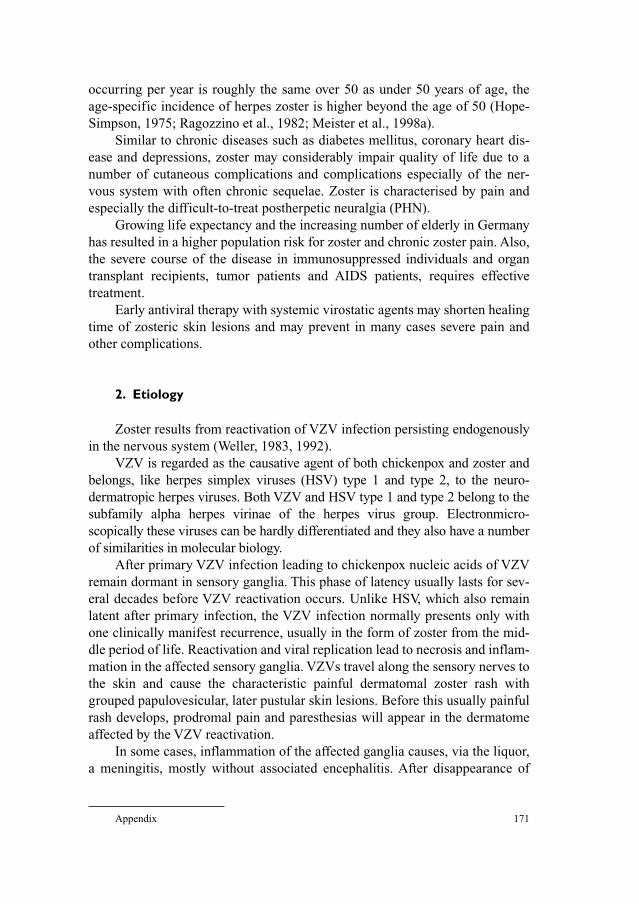
Appendix 171
occurring per year is roughly the same over 50 as under 50 years of age, the
age-specific incidence of herpes zoster is higher beyond the age of 50 (Hope-
Simpson, 1975; Ragozzino et al., 1982; Meister et al., 1998a).
Similar to chronic diseases such as diabetes mellitus, coronary heart dis-
ease and depressions, zoster may considerably impair quality of life due to a
number of cutaneous complications and complications especially of the ner-
vous system with often chronic sequelae. Zoster is characterised by pain and
especially the difficult-to-treat postherpetic neuralgia (PHN).
Growing life expectancy and the increasing number of elderly in Germany
has resulted in a higher population risk for zoster and chronic zoster pain. Also,
the severe course of the disease in immunosuppressed individuals and organ
transplant recipients, tumor patients and AIDS patients, requires effective
treatment.
Early antiviral therapy with systemic virostatic agents may shorten healing
time of zosteric skin lesions and may prevent in many cases severe pain and
other complications.
2. Etiology
Zoster results from reactivation of VZV infection persisting endogenously
in the nervous system (Weller, 1983, 1992).
VZV is regarded as the causative agent of both chickenpox and zoster and
belongs, like herpes simplex viruses (HSV) type 1 and type 2, to the neuro-
dermatropic herpes viruses. Both VZV and HSV type 1 and type 2 belong to the
subfamily alpha herpes virinae of the herpes virus group. Electronmicro-
scopically these viruses can be hardly differentiated and they also have a number
of similarities in molecular biology.
After primary VZV infection leading to chickenpox nucleic acids of VZV
remain dormant in sensory ganglia. This phase of latency usually lasts for sev-
eral decades before VZV reactivation occurs. Unlike HSV, which also remain
latent after primary infection, the VZV infection normally presents only with
one clinically manifest recurrence, usually in the form of zoster from the mid-
dle period of life. Reactivation and viral replication lead to necrosis and inflam-
mation in the affected sensory ganglia. VZVs travel along the sensory nerves to
the skin and cause the characteristic painful dermatomal zoster rash with
grouped papulovesicular, later pustular skin lesions. Before this usually painful
rash develops, prodromal pain and paresthesias will appear in the dermatome
affected by the VZV reactivation.
In some cases, inflammation of the affected ganglia causes, via the liquor,
a meningitis, mostly without associated encephalitis. After disappearance of
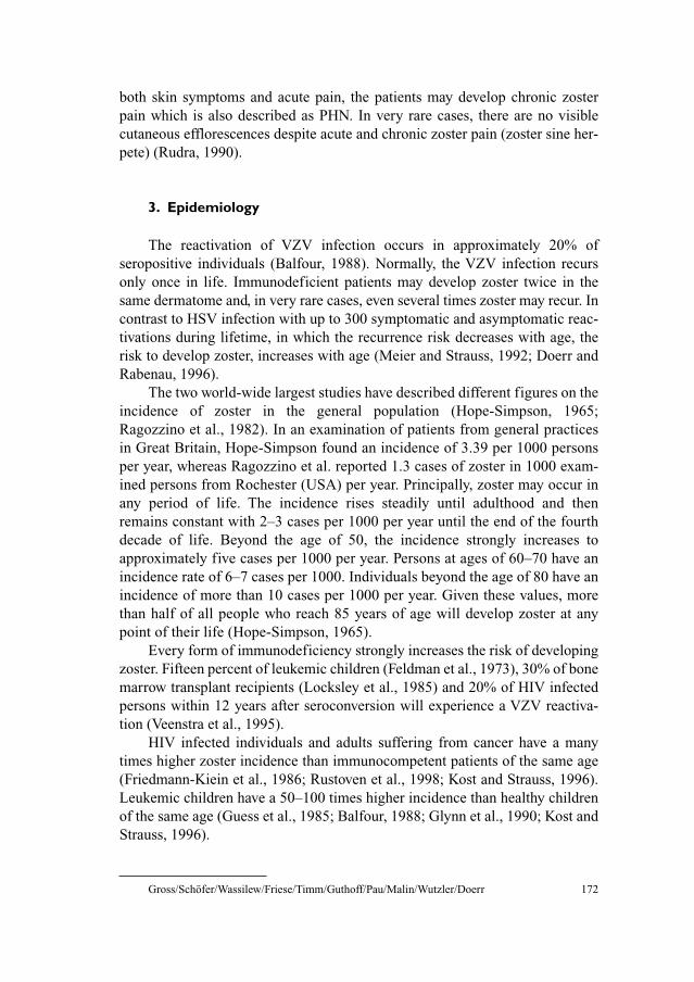
Gross/Schöfer/Wassilew/Friese/Timm/Guthoff/Pau/Malin/Wutzler/Doerr 172
both skin symptoms and acute pain, the patients may develop chronic zoster
pain which is also described as PHN. In very rare cases, there are no visible
cutaneous efflorescences despite acute and chronic zoster pain (zoster sine her-
pete) (Rudra, 1990).
3. Epidemiology
The reactivation of VZV infection occurs in approximately 20% of
seropositive individuals (Balfour, 1988). Normally, the VZV infection recurs
only once in life. Immunodeficient patients may develop zoster twice in the
same dermatome and, in very rare cases, even several times zoster may recur. In
contrast to HSV infection with up to 300 symptomatic and asymptomatic reac-
tivations during lifetime, in which the recurrence risk decreases with age, the
risk to develop zoster, increases with age (Meier and Strauss, 1992; Doerr and
Rabenau, 1996).
The two world-wide largest studies have described different figures on the
incidence of zoster in the general population (Hope-Simpson, 1965;
Ragozzino et al., 1982). In an examination of patients from general practices
in Great Britain, Hope-Simpson found an incidence of 3.39 per 1000 persons
per year, whereas Ragozzino et al. reported 1.3 cases of zoster in 1000 exam-
ined persons from Rochester (USA) per year. Principally, zoster may occur in
any period of life. The incidence rises steadily until adulthood and then
remains constant with 2–3 cases per 1000 per year until the end of the fourth
decade of life. Beyond the age of 50, the incidence strongly increases to
approximately five cases per 1000 per year. Persons at ages of 60–70 have an
incidence rate of 6–7 cases per 1000. Individuals beyond the age of 80 have an
incidence of more than 10 cases per 1000 per year. Given these values, more
than half of all people who reach 85 years of age will develop zoster at any
point of their life (Hope-Simpson, 1965).
Every form of immunodeficiency strongly increases the risk of developing
zoster. Fifteen percent of leukemic children (Feldman et al., 1973), 30% of bone
marrow transplant recipients (Locksley et al., 1985) and 20% of HIV infected
persons within 12 years after seroconversion will experience a VZV reactiva-
tion (Veenstra et al., 1995).
HIV infected individuals and adults suffering from cancer have a many
times higher zoster incidence than immunocompetent patients of the same age
(Friedmann-Kiein et al., 1986; Rustoven et al., 1998; Kost and Strauss, 1996).
Leukemic children have a 50–100 times higher incidence than healthy children
of the same age (Guess et al., 1985; Balfour, 1988; Glynn et al., 1990; Kost and
Strauss, 1996).
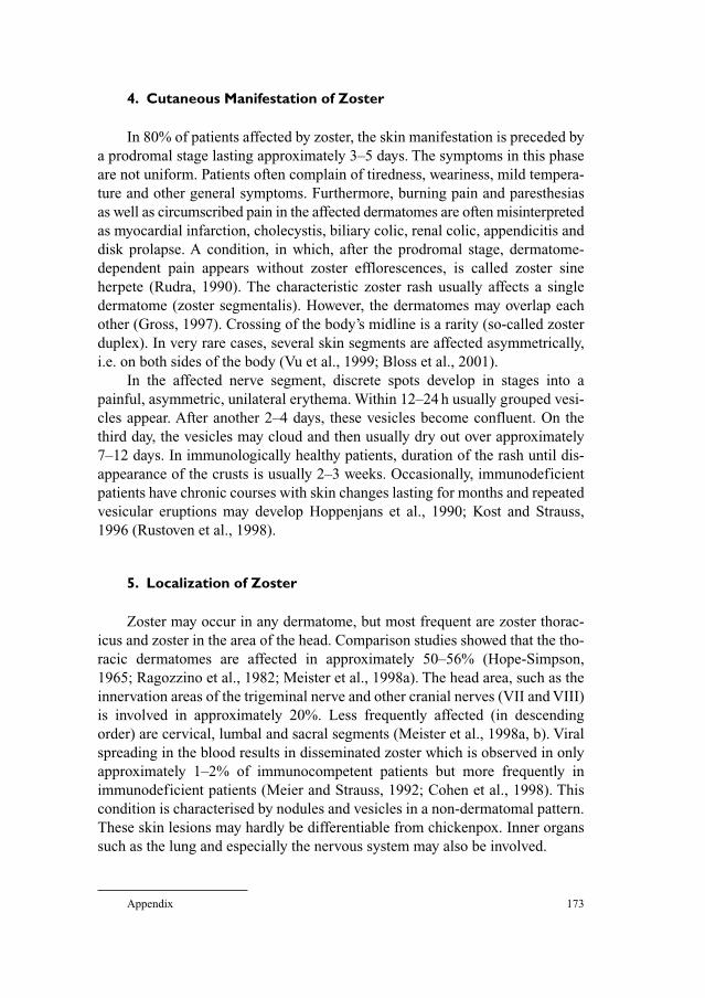
Appendix 173
4. Cutaneous Manifestation of Zoster
In 80% of patients affected by zoster, the skin manifestation is preceded by
a prodromal stage lasting approximately 3–5 days. The symptoms in this phase
are not uniform. Patients often complain of tiredness, weariness, mild tempera-
ture and other general symptoms. Furthermore, burning pain and paresthesias
as well as circumscribed pain in the affected dermatomes are often misinterpreted
as myocardial infarction, cholecystis, biliary colic, renal colic, appendicitis and
disk prolapse. A condition, in which, after the prodromal stage, dermatome-
dependent pain appears without zoster efflorescences, is called zoster sine
herpete (Rudra, 1990). The characteristic zoster rash usually affects a single
dermatome (zoster segmentalis). However, the dermatomes may overlap each
other (Gross, 1997). Crossing of the body’s midline is a rarity (so-called zoster
duplex). In very rare cases, several skin segments are affected asymmetrically,
i.e. on both sides of the body (Vu et al., 1999; Bloss et al., 2001).
In the affected nerve segment, discrete spots develop in stages into a
painful, asymmetric, unilateral erythema. Within 12–24 h usually grouped vesi-
cles appear. After another 2–4 days, these vesicles become confluent. On the
third day, the vesicles may cloud and then usually dry out over approximately
7–12 days. In immunologically healthy patients, duration of the rash until dis-
appearance of the crusts is usually 2–3 weeks. Occasionally, immunodeficient
patients have chronic courses with skin changes lasting for months and repeated
vesicular eruptions may develop Hoppenjans et al., 1990; Kost and Strauss,
1996 (Rustoven et al., 1998).
5. Localization of Zoster
Zoster may occur in any dermatome, but most frequent are zoster thorac-
icus and zoster in the area of the head. Comparison studies showed that the tho-
racic dermatomes are affected in approximately 50–56% (Hope-Simpson,
1965; Ragozzino et al., 1982; Meister et al., 1998a). The head area, such as the
innervation areas of the trigeminal nerve and other cranial nerves (VII and VIII)
is involved in approximately 20%. Less frequently affected (in descending
order) are cervical, lumbal and sacral segments (Meister et al., 1998a, b). Viral
spreading in the blood results in disseminated zoster which is observed in only
approximately 1–2% of immunocompetent patients but more frequently in
immunodeficient patients (Meier and Strauss, 1992; Cohen et al., 1998). This
condition is characterised by nodules and vesicles in a non-dermatomal pattern.
These skin lesions may hardly be differentiable from chickenpox. Inner organs
such as the lung and especially the nervous system may also be involved.

Gross/Schöfer/Wassilew/Friese/Timm/Guthoff/Pau/Malin/Wutzler/Doerr 174
Zoster is mainly differentiated from other circumscribed rashes by its
asymmetry, the synchronous development of skin lesions from erythemas to
vesicular, pustular and finally crustous lesions. Varicella-like forms have espe-
cially been reported in AIDS and tumor patients. The lesions often do not
develop synchronously in stages, thus creating a problem for differential diag-
nosis (Galagher and Merigan, 1979; Hoppenjans et al., 1990).
6. Symptoms
In most cases, the clinical appearance of zoster is preceded by prodromal
symptoms such as pain or paresthesias. Pain that occurs before, during or after
the dermatomal rash stage is called zoster-associated pain. Postzosteric or PHN
is defined as pain which appears or persists after the cutaneous symptoms
(Dworkin et al., 1997).
Different mechanisms in the pathogenesis of chronic zoster pain are being
discussed: inflammatory nerve lesions with destruction of peripheral nerve
structures or neurones in the sensory ganglia as well as modified signal
processes in the central nervous system which occur during and subsequent to
VZV reactivation (Kost and Strauss, 1996; Malin, 1996).
7. Complications and Sequelae of Zoster
Acute and chronic complications affecting skin, eye, and central nervous
system are rather frequent (Table 1), whereas complications in inner organs are
relatively rare.
In the acute stage, the skin is primarily affected by bacterial secondary
infections that can be accompanied by ecthymiform ulcerations. Other major
cutaneous complications are haemorrhagias (zoster haemorrhagicus), purulent
gangrene (zoster gangraenosus), persistence of lesions and dissemination
(zoster disseminatus). The latter occur especially in immunodeficient patients.
Chronic inconvenient sequelae on the skin are hypo- and depigmented-scars,
more seldom granulomatous reactions and manifestation of a psoriasis vulgaris
(Köbner’s phenomenon) (Gross, 1997).
Ophthalmologic complications comprise zoster lesions of the eyelid with
subsequent scar formation, conjunctivitis, episcleritis, scleritis, corneal involve-
ment with epithelial and stromal keratitis as well as endothelitis. Furthermore,
uveitis may occur with the risk of secondary glaucoma or even acute retinal
necrosis. The optic nerve can also be affected (Liesegang, 1991).
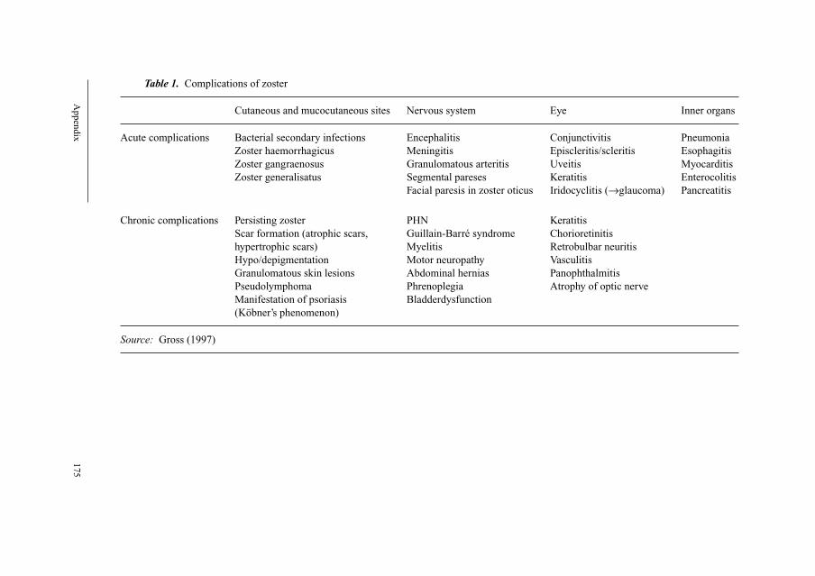
Appendix
175
Table 1. Complications of zoster
Cutaneous and mucocutaneous sites Nervous system Eye Inner organs
Acute complications Bacterial secondary infections Encephalitis Conjunctivitis Pneumonia
Zoster haemorrhagicus Meningitis Episcleritis/scleritis Esophagitis
Zoster gangraenosus Granulomatous arteritis Uveitis Myocarditis
Zoster generalisatus Segmental pareses Keratitis Enterocolitis
Facial paresis in zoster oticus Iridocyclitis (→glaucoma) Pancreatitis
Chronic complications Persisting zoster PHN Keratitis
Scar formation (atrophic scars, Guillain-Barré syndrome Chorioretinitis
hypertrophic scars) Myelitis Retrobulbar neuritis
Hypo/depigmentation Motor neuropathy Vasculitis
Granulomatous skin lesions Abdominal hernias Panophthalmitis
Pseudolymphoma Phrenoplegia Atrophy of optic nerve
Manifestation of psoriasis Bladderdysfunction
(Köbner’s phenomenon)
Source: Gross (1997)
Gross/Schöfer/Wassilew/Friese/Timm/Guthoff/Pau/Malin/Wutzler/Doerr 176
Chorioretinits and neuritis of the optic nerve are more frequently observed
in AIDS patients. Also, herpetic retinal necroses due to VZV infection with the
risk of bilateral blindness were reported in HIV positive patients.
Other neurologic complications comprise zoster meningitis, motor neu-
ropathies and paralysis, Guillain-Barré syndrome, granulomatous arteritis and
affection of cranial nerves (Malin, 1996). The most frequent and important
complication of zoster affecting the nervous system, is acute and chronic pain
also known as PHN. PHN is defined as pain that persists for longer than 4
weeks or that occurs 4 weeks after a pain-free interval. Approximately 10–20%
of zoster patients of all ages are affected by PHN. Chronic zoster pain in chil-
dren is extremely rare (Malin, 1996).
The manifestation rate of PHN shows a clear age-dependence. In zoster
patients over 55, 60 and 70 years it is 27, 47 and 73%, respectively. In women
and patients with zoster ophthalmicus, PHN seems to occur more frequently.
Immunodeficiency seems to be no risk for the development of chronic pain
(Hope-Simpson, 1975; Guess et al., 1985; Gross, 1997).
Best documented complications are postzosteric neuropathies and paraly-
sis such as phrenoplegias, abdominal hernias, bladder dysfunction and cystitis.
In some cases these symptoms may be misinterpreted (Malin, 1996). Another
neurologic complication is segmental paralysis which has a good outcome,
since in every second case complete regression occurs.
8. Zoster in Immunodeficient Patients
Zoster may present atypically in patients with pathologic or iatrogenic
immunodeficiency. The rash may occur mitigated with few symptoms.
However, in most cases, distinct inflammation, partly with hemorrhages and
occasionally with necroses, is found. In some cases, more than one dermatome
is involved and the rash may persist for a longer period than in immunocom-
petent patients.
Disseminated zoster with varicella-like skin involvement and involvement
of inner organs is observed more frequently in immunodeficient patients
(Cohen et al., 1998; Rustoven et al., 1998). Zoster is regarded as an early
marker for HIV infection (Melbye et al., 1978; Friedmann-Kiein et al., 1986;
Schöfer, 1991). Therefore, HIV infection should always be excluded serologi-
cally in zoster patients younger than 50.
With reduced cellular immunity, the zoster rash may be associated
with atypic varicelliform or even verrucous or ecthymiform lesions (Galagher
and Merigan, 1979; Hoppenjans et al., 1990; Schöfer, 1991; Schöfer et al.,
1998).
Appendix 177
9. Diagnosis
Zoster is usually diagnosed by inspection with an asymmetrical dermatomal
rash and grouped vesicles. Further evidence is found with the rash limited by the
midline, its dermatomal arrangement and, especially, prodromal and zoster-asso-
ciated segmental pain. Diagnostic problems may occur particularly in the pro-
dromal or early phase of zoster when only erythematous skin lesions exist.
9.1. Laboratory Diagnosis
The differentiation of the VZV infection from herpes simplex and bullous
dermatoses is an important indication for virological diagnosis. Also, VZV
infections of pregnant women and of newborn infants, atypical infections of
immunodeficient patients and suspected VZV infection of the central nervous
system must be confirmed by laboratory diagnosis.
Today, the VZV polymerase chain reaction (PCR) as well as the direct
detection of virus in cell cultures and the detection of specific antibodies to
VZV in special circumstances are recommended.
The enzyme-linked immunosorbent assay and the immunofluorescence
technique are especially suited for the detection of VZV-specific immunoglob-
ulins of classes IgG, IgM and IgA. VZV–IgG rises may occur spontaneously
and in recurrent HSV-infections due to cross reactivity of epitopes. However,
additional detection of IgM and high-titered IgA anti-VZV antibodies usually
indicates a reactivated VZV-infection regardless whether lesions are visible or
not. Generally the patient consults a physician 1–3 days after the occurrence of
symptoms. Thus clinical diagnosis is mostly supported by serologic findings
(Doerr et al., 1987; Wutzler and Doerr, 1995; Doerr and Rabenau, 1996).
Serological diagnosis is particularly efficient and helpful in immunodeficient
patients suffering from VZV reactivation (Wutzler and Doerr, 1995).
In very early zoster (erythematous stage), detection of VZV infection in
tissue by the VZV-specific PCR may occasionally be helpful (Lilie and
Wassilew, 1999). Additionally, VZV infection can be detected using the
immunofluorescence test. Another indirect but less sensitive detection proce-
dure is the so-called Tzanck test that examines the cytopathic effect of VZV in
the epidermis with characteristics multinucleate giant cells and intranuclear
inclusions (Barr et al., 1977; Solomon et al., 1986; Gross and Doerr, 1997).
Unfortunately, this cytopathic effect is not VZV-specific since it is also seen in
cutaneous HSV-infections.
Electron microscopy allows the morphological detection of herpes
viruses in vesicular fluids or smears. However, this again does not allow dif-
Gross/Schöfer/Wassilew/Friese/Timm/Guthoff/Pau/Malin/Wutzler/Doerr 178
ferentiation of the herpes viruses VZV, HSV-1 and HSV-2 (Barr et al., 1977;
Gross and Doerr, 1997). Furthermore electron microscopy cannot be used
routinely.
9.2. Differential Diagnosis
The major differential diagnoses of zoster are the zosteriform herpes sim-
plex and the different forms of erysipelas, such as hemorrhagic and bullous
erysipelos.
Further differential diagnoses are contact dermatitis, insect bites, bullous
dermatoses such as the bullous pemphigoid and pemphigus vulgaris. Phlegmones
and panniculitis pose less frequently differential diagnosis problems. In cases
where the clinical presentation is atypical, particularly if no symptoms exist, viral
detection should be done by PCR, or viral culture as well as by serology and also
by the Tzanck test (Gross and Doerr, 1997; Lilie and Wassilew, 1999).
If chronic pain persists after the zoster lesions have healed diagnosis can
be confirmed retrospectively by serologic tests. Viral detection is not possible
in this phase.
10. Therapy
The aim of zoster treatment is to relieve pain in the acute phase, to limit the
spread and duration of zosteric skin lesions and to prevent or alleviate PHN and
other acute and chronic complications (Table 1). It is of utmost importance that
ophthalmological complications should be referred for specialist ophthalmic
management as soon as possible. Complications related to zoster of other cra-
nial nerves such as zoster oticus always require consulting a specialist.
10.1. Symptomatic Therapy of Zoster
Depending on the stage of the rash, treatment will be done locally either
through drying and antisepsis, e.g. with wet dressings, with lotio alba, vioform
zinc mixture or later by crust removal. A satisfactory local therapy with proven
antiviral efficacy does not exist. Local zoster therapy with antiviral substances
has shown to be ineffective and is not recommended (Gross, 1997).
Painlessness should be achieved by appropriately-dosed analgesics (e.g.
tramadol), often in combination with a neuroactive agent, (e.g. amitriptylin)
(Malin, 1996). However, it is unknown as to whether systemic analgesia pre-
vents the development of PHN in the early stage of zoster.
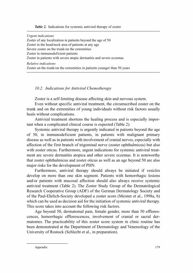
Appendix 179
10.2. Indications for Antiviral Chemotherapy
Zoster is a self-limiting disease affecting skin and nervous system.
Even without specific antiviral treatment, the circumscribed zoster on the
trunk and on the extremities of young individuals without risk factors usually
heals without complications.
Antiviral treatment shortens the healing process and is especially impor-
tant when a complicated clinical course is expected (Table 2).
Systemic antiviral therapy is urgently indicated in patients beyond the age
of 50, in immunodeficient patients, in patients with malignant primary
disease as well as in patients with involvement of cranial nerves, especially with
affection of the first branch of trigeminal nerve (zoster ophthalmicus) but also
with zoster oticus. Furthermore, urgent indications for systemic antiviral treat-
ment are severe dermatitis atopica and other severe eczemas. It is noteworthy
that zoster ophthalmicus and zoster oticus as well as an age beyond 50 are also
major risks for the development of PHN.
Furthermore, antiviral therapy should always be initiated if vesicles
develop on more than one skin segment. Patients with hemorrhagic lesions
and/or patients with mucosal affection should also always receive systemic
antiviral treatment (Table 2). The Zoster Study Group of the Dermatological
Research Cooperative Group (ADF) of the German Dermatology Society and
of the Paul-Ehrlich-Society developed a zoster score (Meister et al., 1998a, b)
which can be used as decision aid for the initiation of systemic antiviral therapy.
This score takes into account the following risk factors.
Age beyond 50, dermatomal pain, female gender, more than 50 efflores-
cences, hemorrhagic efflorescences, involvement of cranial or sacral der-
matomes. The practicability of this zoster score system in clinic routine has
been demonstrated at the Department of Dermatology and Venereology of the
University of Rostock (Schlecht et al., in preparation).
Table 2. Indications for systemic antiviral therapy of zoster
Urgent indicationsZoster of any localization in patients beyond the age of 50
Zoster in the head/neck area of patients at any age
Severe zoster on the trunk/on the extremities
Zoster in immunodeficient patients
Zoster in patients with severe atopic dermatitis and severe eczemas
Relative indicationsZoster on the trunk/on the extremities in patients younger than 50 years
Gross/Schöfer/Wassilew/Friese/Timm/Guthoff/Pau/Malin/Wutzler/Doerr 180
10.3. When to Start Systemic Antiviral Therapy
The success of each antiviral treatment is dependent on the starting point
of therapy. Systemic virostatic therapy must be initiated as soon as possible, i.e.
if possible within 48 to a maximum of 72 h after onset of skin symptoms. The
effect level of the antiviral drug must be achieved rapidly and maintained for an
optimum therapeutical success. In the following situations, systemic antiviral
therapy can also be started even at a later date (after 72 h).
Disseminated zoster with evidence of immuno-deficiency and affection of
inner organs, persisting zoster ophthalmicus and zoster oticus. Furthermore,
even after 72 h, antiviral treatment of zoster is urgently indicated in all patients
with known immunodeficiency. Some more recent data suggest that valacy-
clovir, the prodrug of acyclovir, continues to be beneficial in preventing PHN
even if given 72 h after onset of symptoms.
10.4. Antiviral Therapy of Zoster
A total of 4 different systemic antiviral substances for treatment of zoster
have become available in Germany: acyclovir, valacyclovir, famciclovir and
brivudin, which very recently has been approved (illustration), whereas in the
United States only three drugs – acyclovir, valacyclovir and famciclovir – are in
use (Gnann and Whitley, 2002).
All of these substances can be given orally (Gross and Laskowski, 2001).
Only acyclovir can be administered both enterally and parenterally. Up to now,
valacyclovir and famciclovir have been the world-wide most commonly used
antiviral drugs for the oral treatment of uncomplicated zoster. Better pharma-
cokinetics, better bioavailability and easier application make them superior to
oral acyclovir. A recent double-blind randomised study showed valacyclovir
and famciclovir to have similar effects on zoster-associated pain and on PHN
in immunocompetent patients (Tyring et al., 2000). Valacyclovir and famci-
clovir have to be administered orally 3 times daily for 7 days, whereas oral
acyclovir must be given in a dose of 800 mg, 5 times daily. Brivudin has the
great advantage of requiring only once daily dosing. In VZV infections,
brivudin has a markedly higher antiviral potency than acyclovir, valacyclovir
and famciclovir (De Clercq et al., 1979). Brivudin, when given once daily
(1 � 125 mg for 7 days), is therefore well effective and superior to oral acy-
clovir treatment (5 times daily dosing) (Table 3).
This is especially true for patients beyond the age of 50 who are at a
higher risk of developing PHN. Patients treated with brivudin had signifi-
cantly faster stop of viral replication compared to patients treated by oral
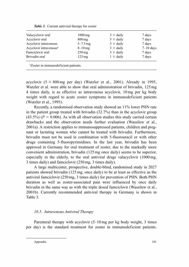
Appendix 181
acyclovir (5 � 800 mg per day) (Wutzler et al., 2001). Already in 1995,
Wutzler et al. were able to show that oral administration of brivudin, 125 mg
4 times daily, is as effective as intravenous acyclovir, 10 mg per kg body
weight with regard to acute zoster symptoms in immunodeficient patients
(Wutzler et al., 1995).
Recently, a randomised observation study showed an 11% lower PHN rate
in the patient group treated with brivudin (32.7%) than in the acyclovir group
(43.5%) (P � 0.006). As with all observation studies this study carried certain
drawbacks and the observation needs further evaluation (Wassilew et al.,
2001a). A restriction applies to immunosuppressed patients, children and preg-
nant or lactating women who cannot be treated with brivudin. Furthermore,
brivudin must not be used in combination with 5-fluorouracil or with other
drugs containing 5-fluoropyrimidines. In the last year, brivudin has been
approved in Germany for oral treatment of zoster, due to the markedly more
convenient administration, brivudin (125 mg once daily) seems to be superior,
especially in the elderly, to the oral antiviral drugs valacyclovir (1000 mg,
3 times daily) and famciclovir (250 mg, 3 times daily).
A large multicenter, prospective, double-blind, randomised study in 2027
patients showed brivudin (125 mg, once daily) to be at least as effective as the
antiviral famciclovir (250 mg, 3 times daily) for prevention of PHN. Both PHN
duration as well as zoster-associated pain were influenced by once daily
brivudin in the same way as with the triple dosed famciclovir (Wassilew et al.,
2001b). Currently recommended antiviral therapy in Germany is shown in
Table 3.
10.5. Intravenous Antiviral Therapy
Parenteral therapy with acyclovir (5–10 mg per kg body weight, 3 times
per day) is the standard treatment for zoster in immunodeficient patients.
Table 3. Current antiviral therapy for zoster
Valacyclovir oral 1000 mg 3 � daily 7 days
Acyclovir oral 800 mg 5 � daily 7 days
Acyclovir intravenous 5–7.5 mg 3 � daily 7 days
Acyclovir intravenousa 8–10 mg 3 � daily 7–10 days
Famciclovir oral 250 mg 3 � daily 7 days
Brivudin oral 125 mg 1 � daily 7 days
aZoster in immunodeficient patients.
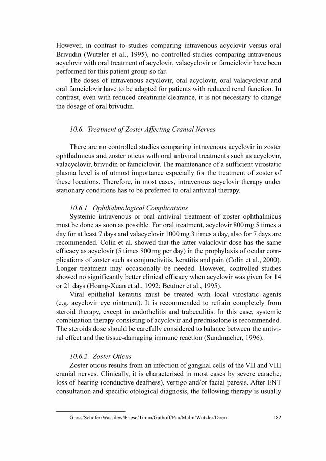
Gross/Schöfer/Wassilew/Friese/Timm/Guthoff/Pau/Malin/Wutzler/Doerr 182
However, in contrast to studies comparing intravenous acyclovir versus oral
Brivudin (Wutzler et al., 1995), no controlled studies comparing intravenous
acyclovir with oral treatment of acyclovir, valacyclovir or famciclovir have been
performed for this patient group so far.
The doses of intravenous acyclovir, oral acyclovir, oral valacyclovir and
oral famciclovir have to be adapted for patients with reduced renal function. In
contrast, even with reduced creatinine clearance, it is not necessary to change
the dosage of oral brivudin.
10.6. Treatment of Zoster Affecting Cranial Nerves
There are no controlled studies comparing intravenous acyclovir in zoster
ophthalmicus and zoster oticus with oral antiviral treatments such as acyclovir,
valacyclovir, brivudin or famciclovir. The maintenance of a sufficient virostatic
plasma level is of utmost importance especially for the treatment of zoster of
these locations. Therefore, in most cases, intravenous acyclovir therapy under
stationary conditions has to be preferred to oral antiviral therapy.
10.6.1. Ophthalmological ComplicationsSystemic intravenous or oral antiviral treatment of zoster ophthalmicus
must be done as soon as possible. For oral treatment, acyclovir 800 mg 5 times a
day for at least 7 days and valacyclovir 1000 mg 3 times a day, also for 7 days are
recommended. Colin et al. showed that the latter valaclovir dose has the same
efficacy as acyclovir (5 times 800 mg per day) in the prophylaxis of ocular com-
plications of zoster such as conjunctivitis, keratitis and pain (Colin et al., 2000).
Longer treatment may occasionally be needed. However, controlled studies
showed no significantly better clinical efficacy when acyclovir was given for 14
or 21 days (Hoang-Xuan et al., 1992; Beutner et al., 1995).
Viral epithelial keratitis must be treated with local virostatic agents
(e.g. acyclovir eye ointment). It is recommended to refrain completely from
steroid therapy, except in endothelitis and trabeculitis. In this case, systemic
combination therapy consisting of acyclovir and prednisolone is recommended.
The steroids dose should be carefully considered to balance between the antivi-
ral effect and the tissue-damaging immune reaction (Sundmacher, 1996).
10.6.2. Zoster OticusZoster oticus results from an infection of ganglial cells of the VII and VIII
cranial nerves. Clinically, it is characterised in most cases by severe earache,
loss of hearing (conductive deafness), vertigo and/or facial paresis. After ENT
consultation and specific otological diagnosis, the following therapy is usually
Appendix 183
indicated: high dose antiviral therapy (intravenously preferred) in combination
with glucocorticoids, infusion treatment with rheologics or physiological saline
solution with vasoactive substances. Analgesics and, in cases with strong ver-
tigo, anti-vertigo agents are additionally indicated.
10.7. Corticosteroid Therapy
The additive zoster therapy with high dose steroids shortens the duration of
acute zoster pain, but has no essential effect on chronic pain (PHN). This is the
result of 2 large prospective studies (Wood et al., 1994; Whitley et al., 1996).
The use of corticosteroids must be carefully considered, especially in view of
possible side effects.
It is not recommended to use corticosteroids without systemic antiviral
therapy. Study results are available only for a combination therapy of pred-
nisolone (40 and 60 mg per day, dose reduction over � 10–14 days to 5 mg per
day) and acyclovir. However, nothing speaks against possible combination of
prednisolone with valacyclovir, brivudin or famciclovir.
10.8. Therapy of Neuralgia: Step-by-Step Scheme
Patient-specific aspects and side effects are important considerations in
pain therapy. Early presentation to a pain therapist or a pain outpatient clinic
may be required.
1) Step 1: non-steroidal analgesics (e.g. paracetamol 1.5–5 g per day)
2) Step 2: additional low-potency opioid analgesics (e.g. tramadol
200–400 mg per day, codeine 120 mg per day), if necessary, combined
preparations.
3) Step 3: in addition to a ‘peripheral’ analgesic, administration of a high-
potency central opioid (e.g. buprenorphine 1.5–1.6 mg per day; oral mor-
phine 30–360 mg per day) is indicated. This refers to patients who fail to
respond the more measured treatment approaches.
In severe neuralgic pain, step 1 or 2 in combination with an anti-convulsant
(carbamazepine 400–1200 mg per day). Anti-convulsants (carbamazepine) can
reduce the lancinating pain, but are not effective for continuous pain. Another
option is gabapentine (900–2400 mg per day). Also, anti-depressants, amitryp-
tilin 10–75 mg) and neuroleptics (levomepromazin 20–150 mg per day) may be
efficient, especially in elderly patients (Malin, 1996; Wassilew, 2000).
Further treatment possibilities consist in local therapy with capsaicin, local
anesthetic blocking of sympathetic nerve, transcutaneous electric nerve
Gross/Schöfer/Wassilew/Friese/Timm/Guthoff/Pau/Malin/Wutzler/Doerr 184
stimulation, if necessary, neurosurgery (e.g. thermocoagulation of substantial
gelatinosa Rolandi) in exceptional cases.
10.9. Therapy of Other Neurologic Complications of Zoster
Zoster meningitis, zoster encephalitis and zoster myelitis should be treated
with parenteral acyclovir in a dose of 10 mg per kg body weight, 3 times daily.
Also in severe zoster ophthalmicus and zoster oticus, intravenous acyclovir
should be preferred to enteral therapy.
10.10. Therapy of Zoster in Children and Adolescents
Zoster in childhood is usually not a strict indication for systemic antiviral
therapy. An exception is zoster affecting cranial nerves. Also, children with
hereditary or acquired immunodeficiency as well as atopic children might ben-
efit from antiviral therapy. As long as there are no controlled treatment studies
in children with immunodeficiency and zoster available, one can only speculate
whether antivirals, given early, might prevent viral spread, reduce the severity of
inflammation and prevent secondary immunosuppression. It is likely, but not
yet proven, that severe tissue destruction, secondary bacterial infections and
scar formation, might be reduced by an early antiviral therapy. In analogy of the
sometimes severe, but individually not predictable course of zoster in patients
with HIV infection we suggest to treat immunodeficient and atopic children
with acyclovir intravenously.
10.11. Therapy of Zoster in Immunodeficient Patients
Only acyclovir has been approved so far for the treatment of zoster in
severely immunocompromized patients. Other oral virostatic agents such as
brivudin, valacyclovir and famciclovir showed positive results in case reports
especially in HIV-infected patients, but are not officially approved. In a given
case, the responsible physician may use these drugs after careful consideration
of the risks involved.
It has been shown that the therapeutical regimen of acyclovir is depen-
dent on the severity of immunodeficiency and clinical findings. If the CD4
cells are roughly within limits of normal (�400 cells per �l), segmental
zoster may be treated with a standard dose of acyclovir intravenous (5–7.5 mg
per kg body weight every 8 h). In severe immunodeficiency with extended
skin lesions, especially when neurologic symptoms are present, treatment

Appendix 185
with high dose intravenous acyclovir (10 mg per kg every 8 h) and continuous
monitoring of renal function is required. In severe immunodeficiency with
extended skin lesions, especially when neurologic symptoms are present,
treatment with high dose intravenous acyclovir (10 mg per kg every 8 h) is
required. As acyclovir has some nephrotoxic properties and cumulates in
patients with impaired renal function, serum creatinine clearance must be
tested with the first given dose of acyclovir. In case of reduced serum creati-
nine clearance the interval to the following acyclovir infusions must be
prolonged from 8 to 12 or even 24 h (according to the nomograms or tables
provided by the producers of acyclovir).
Acyclovir resistance has to be assumed if the clinical findings improve
only slowly or not at all. In such cases, another treatment option would be intra-
venous foscarnet which, however, is highly nephrotoxic (Breton et al., 1998).
Apart from the constant number of HIV patients in Germany (� 1500–
1700 new infections in 2000), especially the prophylaxis and therapy of other
immunodeficient patients such as organ transplant recipients, patients under
immunosuppressive agents and tumor patients strongly require therapeutics that
can be used if acyclovir resistance has developed. Also, zoster patients with
renal damage (contraindications for acyclovir and foscarnet) require new non-
nephrotoxic virostatic agents.
10.12. Therapy of Zoster in Pregnancy
Unlike varicella, maternal zoster is hardly considered a risk for the unborn.
Normally, there is no viremia that could pose a risk to the development of the
unborn. Antiviral therapy of the mother with intravenous acyclovir (3 times
5–10 mg per kg body weight for 7 days) is indicated only in exceptional cases.
If possible, zoster during pregnancy should be treated locally and symptomati-
cally. Topical acyclovir is definitely not recommended. The treatment of a preg-
nant zoster patient in a gynaecologic practice should be in such a way that
non-pregnant women without immune protection are not put at risk of varicella.
11. Resistance of Varicella Zoster Virus to Antivirals
VZV can become resistant to nucleoside analogues such as acyclovir, vala-
cyclovir, famciclovir and brivudin, probably due to mutations in the thymidin
kinase (TK) gene or mutations in the polymerase gene. Viral strains with muta-
tions in the TK gene are usually resistant to acyclovir, famciclovir and also to
ganciclovir. Such virus strains have been isolated from several HIV infected
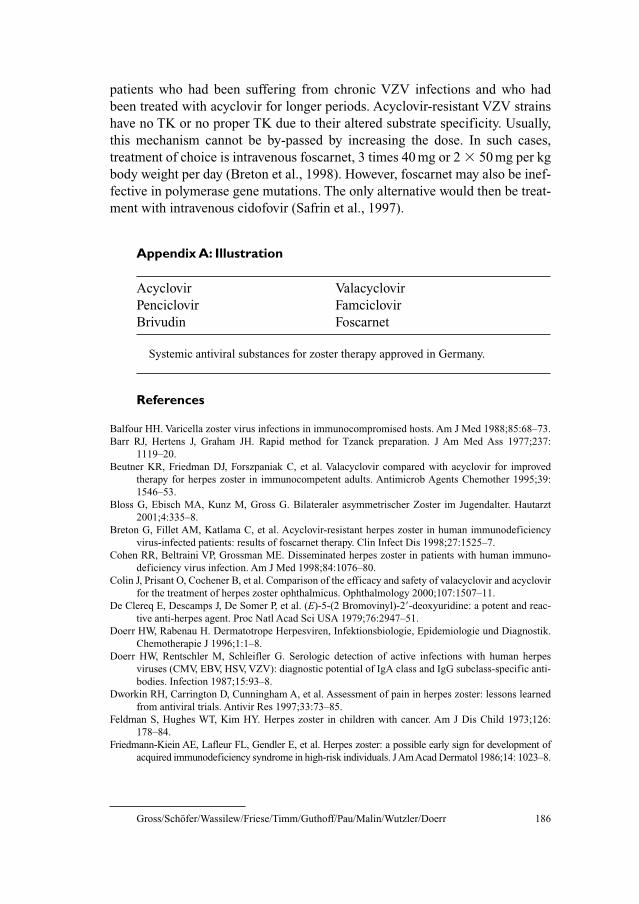
Gross/Schöfer/Wassilew/Friese/Timm/Guthoff/Pau/Malin/Wutzler/Doerr 186
patients who had been suffering from chronic VZV infections and who had
been treated with acyclovir for longer periods. Acyclovir-resistant VZV strains
have no TK or no proper TK due to their altered substrate specificity. Usually,
this mechanism cannot be by-passed by increasing the dose. In such cases,
treatment of choice is intravenous foscarnet, 3 times 40 mg or 2 � 50 mg per kg
body weight per day (Breton et al., 1998). However, foscarnet may also be inef-
fective in polymerase gene mutations. The only alternative would then be treat-
ment with intravenous cidofovir (Safrin et al., 1997).
Appendix A: Illustration
Acyclovir Valacyclovir
Penciclovir Famciclovir
Brivudin Foscarnet
Systemic antiviral substances for zoster therapy approved in Germany.
References
Balfour HH. Varicella zoster virus infections in immunocompromised hosts. Am J Med 1988;85:68–73.
Barr RJ, Hertens J, Graham JH. Rapid method for Tzanck preparation. J Am Med Ass 1977;237:
1119–20.
Beutner KR, Friedman DJ, Forszpaniak C, et al. Valacyclovir compared with acyclovir for improved
therapy for herpes zoster in immunocompetent adults. Antimicrob Agents Chemother 1995;39:
1546–53.
Bloss G, Ebisch MA, Kunz M, Gross G. Bilateraler asymmetrischer Zoster im Jugendalter. Hautarzt
2001;4:335–8.
Breton G, Fillet AM, Katlama C, et al. Acyclovir-resistant herpes zoster in human immunodeficiency
virus-infected patients: results of foscarnet therapy. Clin Infect Dis 1998;27:1525–7.
Cohen RR, Beltraini VP, Grossman ME. Disseminated herpes zoster in patients with human immuno-
deficiency virus infection. Am J Med 1998;84:1076–80.
Colin J, Prisant O, Cochener B, et al. Comparison of the efficacy and safety of valacyclovir and acyclovir
for the treatment of herpes zoster ophthalmicus. Ophthalmology 2000;107:1507–11.
De Clereq E, Descamps J, De Somer P, et al. (E)-5-(2 Bromovinyl)-2�-deoxyuridine: a potent and reac-
tive anti-herpes agent. Proc Natl Acad Sci USA 1979;76:2947–51.
Doerr HW, Rabenau H. Dermatotrope Herpesviren, Infektionsbiologie, Epidemiologie und Diagnostik.
Chemotherapie J 1996;1:1–8.
Doerr HW, Rentschler M, Schleifler G. Serologic detection of active infections with human herpes
viruses (CMV, EBV, HSV, VZV): diagnostic potential of IgA class and IgG subclass-specific anti-
bodies. Infection 1987;15:93–8.
Dworkin RH, Carrington D, Cunningham A, et al. Assessment of pain in herpes zoster: lessons learned
from antiviral trials. Antivir Res 1997;33:73–85.
Feldman S, Hughes WT, Kim HY. Herpes zoster in children with cancer. Am J Dis Child 1973;126:
178–84.
Friedmann-Kiein AE, Lafleur FL, Gendler E, et al. Herpes zoster: a possible early sign for development of
acquired immunodeficiency syndrome in high-risk individuals. J Am Acad Dermatol 1986;14: 1023–8.
Appendix 187
Galagher JG, Merigan TC. Prolonged herpes zoster infection associated with immunosuppressive ther-
apy. Am Intern Med 1979;91:842–6.
Glynn C, Crockford G, Garaghan D, et al. Epidemiology of shingles. J R Soc Med 1990;83:617–9.
Gnann JW, Whitley RJ. Herpes zoster. N Engl J Med 2002;347:340–6.
Gross G. Zoster. Dtsch med Wschr 1997;122:132–9.
Gross G, Doerr HW. Labordiagnose dermatotroper Virusinfektionen: Erregerisolierung. In:
Korting HC, Sterry W, editors. Diagnostische Verfahren in der Dermatologie. Berlin, Wien:
Blackwell Wissenschaftsverlag; 1997, p. 197–202.
Gross G, Laskowski J, Virustatika. In: Korting HC, Sterry W, editors. Therapeutische Verfahren in der
Dermatologie, Berlin, Wien: Blackwell Wissenschaftsverlag; 2001, p. 647–54.
Guess HA, Broughton DD, Melton LJ, Kurland LJ, III. Epidemiology of herpes zoster in children and
adolescents: a population-based study. Pediatrics 1985;76:512–7.
Hoang-Xuan T, Büchi R, Herbort CP, et al. Oral acyclovir for herpes zoster ophthalmicus. Ophthalmology
1992;99:1062–70.
Hope-Simpson RE. The nature of herpes zoster: a long term study and new hypothesis. Proc R Soc Med
1965;58:9–20.
Hope-Simpson RE. Postherpetic neuralgia. J R Coll Gen Practit 1975;25:571–5.
Hoppnjans WB, Bibler MR, Orne RL, et al. Prolonged cutaneous herpes zoster in acquired immunode-
ficiency syndrome. Arch Dermatol 1990;126:1048–50.
Kost RG, Strauss SE. Postherpetic neuralgia – pathogenesis, treatment and prevention. N Engl J Med
1996;335:23–42.
Liesegang TJ. Diagnosis and therapy of herpes zoster ophthalmicus. Ophthalmology 1991;98:1216–29.
Lilie HM, Wassilew SW. Shingles (Zoster). In: Wolff MH, Schünemann S, Schmidt A, editors, Varicella-
zoster-virus. Molecular biology pathogenesis and clinical aspects, vol. 3. Contrib Microbiol Basel
Karger, 1999;111–27.
Locksley RM, Flournoy N, Sullivan KM, et al. Infection with varicella-zoster virus after marrow trans-
plantation. J Infect Dis 1985;152:1172–81.
Malin JP. Zoster und Nervensystem. Dtsch med Wschr 1996;121:635–8.
Meier JL, Strauss SE. Comparative biology of latent varicella-zoster virus and herpes simplex virus
infections. J Inf Dis 1992;166(Suppl):S13–23.
Meister W, Neiss A, Gross G, et al. Demography, symptomatology and course of disease in ambulatory
zoster patients. Intervirology 1998a;41:272–7.
Meister W, Neiss A, Gross G, et al. A prognostic score for postherpetic neuralgia in ambulatory patients.
Infection 1998b;26(6):359–63.
Melbye M, Grossman RJ, Goebert JJ, et al. Risk of AIDS after herpes zoster. Lancet 1978;1:728–30.
Ragozzino MW, Melton LJ, Kurland TL, et al. Population based study of herpes zoster and its sequelae.
Medicine (Baltimore) 1982;61:310–6.
Rudra T. Zoster sine herpete. Brit J Clin Pract 1990;44:284.
Rustoven JJ, Ahlgren P, Elhakim T, et al. Varizella zoster infection in adult cancer patients: a population
study. Arch Intern Med 1998;148:1561–6.
Safrin S. Cherrington J, Jaffe HS. Clinical uses of cidofovir. Rev Med Virol 1997;7:145–56.
Solomon AR, Rasmussen JE, Weiss JS. A comparison of Tzanck smear and virus isolation in varizella
and herpes zoster. Arch Dermatol 1986;122:282–5.
Sundmacher R. Therapie der Viruserkrankungen des vorderen Augenabschnittes. In: Kampik A, editors.
Das äußere Auge: Hauptreferate der XXXI—Essener Fortbildung für Augenärzte, Bücherei des
Augenarztes; 1996, Vol. 137, p. 121–5.
Schlecht K, Laskowski J, Gross G. Zoster-Scoresystem zur Risikoabschätzung bezüglich der
Entwicklung einer post-zosterischen Neuralgie-Erste Anwendungen in der Praxis. In preparation.
Schöfer H. Frühsymptome der HIV-Erkrankung an Haut- und Schleimhäuten. AIDS-Forschung (AIFO)
1991;6:633–48.
Schöfer H, Baur S, Gregel C, et al. Hyperkeratotic varicella zoster virus infection in a HIV-infected patient.
Successful treatment of persistent lesions with cryosurgery. Br J Dermatol 1998;138:714–5.
Tyring SK, Beutner KR, Tucker BA, et al. Antiviral therapy for herpes zoster. Randomized, controlled
clinical trial of valacyclovir, and famciclovir therapy in immunocompetent patients of 50 years and
older. Arch Fam Med 2000;9:863–9.

Gross/Schöfer/Wassilew/Friese/Timm/Guthoff/Pau/Malin/Wutzler/Doerr 188
Veenstra J, Krool A, van Praag RM, et al. Herpes zoster, immunological deterioration and disease pro-
gression in HIV-1 infection. AIDS 1995;9:1153–8.
Vu AQ, Radonich MA, Heald PW. Herpes zoster in seven disparate dermatomes (zoster multiplex):
report of a case and review of the literature. J Am Acad Dermatol 1999;40:868–9.
Wassilew SW. Differentialtherapie der Herpesvirusinfektionen. In: Plettenberg A, Meigel WN, Moll I,
editors. Dermatologie an der Schwelle zum neuen Jahrtausend: Springer-Verlag; 2000, p. 315–8.
Wassilew SW, Schumacher K, Städtler G, et al. A randomised double-blind survey on the effect of brivudin
in the prevention of postherpetic pain in comparison with acyclovir. Fourth International Conference
on Varicella, Herpes Zoster, Postherpetic Neuralgia. La Jolla, USA; 2001, March, p. 3–5.
Wassilew SW, Schumacher K, Macher K, Städtler G, et al. Brivudin compared to famciclovir in the preven-
tion of postherpetic neuralgia: a randomised double-blind multi-center trial. Fourth International
Conference on Varicella, Herpes Zoster, Postherpetic Neuralgia. La Jolla, USA; 2001, March, p. 3–5.
Weller TH. Varicella and herpes zoster: changing concepts of the natural history, control and importance
of a not-so-benign virus. N Engl Med 1983;309:1362–8.
Weller TH. Varcella and herpes zoster. A perspective and overview. J Infect Dis 1992;166(Suppl 1):1–6.
Whitley RJ, Weiss H, Gnann IW, et al. Acyclovir with and without prednisone for the treatment of herpes
zoster. A randomised placebo-controlled trial. The National Institute of Allergy and Infectious
Diseases Collaborative Antiviral Study Group. Ann Intern Med 1996;125:376–83.
Wood MJ, Johnson RW, Mc Kendrick MW, et al. A randomised trial of acyclovir for 7 days or 21 days with
and without prednisolon for treatment of acute herpes zoster. N Engl J Med 1994;330:896–900.
Wutzler P, Doerr HW. Der Herpes zoster-ein Morbus herpes non simplex. Dtsch med Wschr 1995;120:
1133–8.
Wutzler P, de Clercq E, Wutke K, Färber I. Oral brivudin vs. intravenous acyclovir in the treatment of
herpes zoster in immunocompromised patients: a randomised double-blind trial. J Med Virol
1995;46:252–7.
Wutzler P. Stubinski BM, Koch I, et al. Brivudin compared to acyclovir in the treatment of acute herpes
zoster: a randomised, double-blind, multicenter trial. Fourth International Conference on Varicella,
Herpes Zoster, Postherpetic Neuralgia. La Jolla, USA; 2001, March, p. 3–5.
Gerd Gross, MD
Professor of Dermatology and Venereology, Director
Department of Dermatology and Venereology
University of Rostock
Augustenstrasse 80–84
DE–18055 Rostock (Germany)
Tel. �49 381 4949701, Fax �49 381 4949702, E-Mail [email protected]
189
Acute retinal necrosis syndrome, seeRetinal necrosis
Acyclovir, see also Antiviral therapy
bioavailability 118, 124
bone marrow transplant recipient herpes
zoster treatment and prophylaxis
111–113
congenital varicella syndrome
management 87
corticosteroid combination therapy 121
dosing 131, 180, 181
herpes zoster ophthalmicus management
44
herpes zoster oticus management 54
immunocompromised host, herpes zoster
management 103, 127
neonatal varicella management 90
resistance 185
shingles (herpes zoster) management
123, 124
structure 119
AIDS, see Human immunodeficiency virus
Amitriptyline
herpes zoster oticus management 54
postherpetic neuralgia management 128,
145
side effects 145, 146
Anterior cingulate cortex, pain system 62, 63
Anticonvulsants, postherpetic neuralgia
management 146, 147
Antiviral therapy, see also specific drugs
corticosteroid combination therapy 121,
128, 183
drug features 118–120
efficacy in zoster 180, 181
indications 117, 118, 179
initiation 120, 123, 143, 180
intravenous therapy indications 120, 181,
182
resistance 185
symptomatic therapy 121, 178
Ap1, viral replication role 4
Audiometry, herpes zoster oticus diagnosis
52
Bicyclic furo[2, 3-d]pyrimidine nucleoside
analogs (BCNAs)
antiviral action
mechanisms 136, 137
properties 133–136
metabolism 137–140
structure-activity relationships 132, 133
therapeutic prospects 140
Bone marrow transplant recipients,
varicella-zoster virus infection
clinical features
chickenpox 110, 111
cutaneous and visceral dissemination
110
localized herpes zoster 109, 110
second episodes 110
diagnosis 111
incidence 107
reconstitution of virus-specific T-cell
immunity after stem cell
transplantation 108, 109
risk factors 107, 108
treatment 111–114
Subject Index
Subject Index 190
Brivudin (BVDU), see also Antiviral
therapy
contraindications 181
dosing 180
drug–drug interactions 127
efficacy 119, 126
herpes zoster ophthalmicus management
44, 45
herpes zoster oticus management 54
immunocompromised host, herpes zoster
management 103
pharmacology 126
shingles (herpes zoster) management
125–127
structure 119, 132
toxicity 126
Capsaicin, postherpetic neuralgia
management 148
Capsid, structure 2, 3
Cell-mediated immunity assay, herpes
zoster confirmation 17
Chemotherapy
bone marrow transplant recipients, seeBone marrow transplant recipients
herpes zoster 102
Chickenpox, see Varicella
Children, herpes zoster 26, 27, 184
Cidofovir, herpes zoster ophthalmicus
management 45
Congenital varicella syndrome
clinical manifestations 83
diagnosis 83–85
prophylaxis and treatment 85–87
Corticosteroids
antiviral combination therapy 121, 128,
183
herpes zoster oticus management 54
Dermatome, herpes zoster localization
23, 24
Desipramine, postherpetic neuralgia
management 146
Dorsal root ganglia (DRG), infection
9, 10
Duloxetine, postherpetic neuralgia
management 146
Early genes, functions 4, 9, 10
Electron microscopy (EM), herpes zoster
confirmation 13, 14, 177, 178
Encephalitis, herpes zoster management
184
Envelope, particles 2
Enzyme-linked immunosorbent assay
(ELISA), herpes zoster confirmation
16, 17
Famciclovir, see also Antiviral therapy
active compound 118
dosing 125, 131, 180, 181
herpes zoster ophthalmicus management
44, 45
immunocompromised host, herpes zoster
management 103, 127
shingles (herpes zoster) management 125
structure 119
Foscarnet
herpes zoster ophthalmicus management
45
structure 119
‘Frenzel’s goggles’, herpes zoster oticus
diagnosis 52
Gabapentin, postherpetic neuralgia
management 146
Herpesvirus
classification 1
virion architecture 1–3
Herpes zoster
acute disease management 143
antiviral therapy, see Antiviral therapy;
specific drugs
children 26, 27, 184
clinical presentation 21, 25, 174
complications 31–33, 174–176
cutaneous manifestations 21–23, 70, 173
definition 170, 171
diagnosis, see Laboratory diagnosis,
herpes zoster
differential diagnosis 29, 178
epidemiology
clinical epidemiology 20, 157–161,
172
Subject Index 191
seroepidemiology 154, 156
vaccination impact 161, 167
etiology 171, 172
immunodeficient patients, see Human
immunodeficiency virus
localization 23, 24, 173, 174
ophthalmicus, see Ophthalmicus, herpes
zoster
oticus, see Oticus, herpes zoster
postherpetic neuralgia, see Postherpetic
neuralgia
psychosocial issues 33, 34
Human herpesvirus 3, see Varicella-zoster
virus
Human immunodeficiency virus (HIV)
infection, herpes zoster
AIDS progression effects 101
clinical features 96–98, 176
cutaneous manifestations 21, 27, 28, 97,
98
epidemiology 94, 95
highly active antiretroviral therapy and
immune reconstitution 100, 101, 103
Kaposi’s sarcoma comorbidity 95, 96
postherpetic neuralgia 99, 100
treatment 103, 184, 185
Hutchinson’s sign, herpes zoster
ophthalmicus 38
Immediate-early genes, functions 4, 9, 10
Immunocompromised hosts, seeChemotherapy; Human
immunodeficiency virus (HIV) infection,
herpes zoster; Organ transplantation
Immunofluorescence microscopy, herpes
zoster confirmation 14, 15
Insular cortex, pain system 63
Intrathecal therapy, postherpetic neuralgia
148
Kaposi’s sarcoma, herpes zoster
comorbidity 95, 96
Ketamine, postherpetic neuralgia
management 147
Laboratory diagnosis, herpes zoster
cell-mediated immunity assay 17
electron microscopy 13, 14, 177, 178
immunofluorescence microscopy 14, 15
isolation in cell culture 16, 28
overview 13
polymerase chain reaction 15, 16, 28,
177
serology 16, 17, 29
Tzanck test 28, 29
Late genes, functions 5, 9
Latency, mechanisms 10, 11
Lidocaine
adjunctive therapy in shingles (herpes
zoster) 128
postherpetic neuralgia management 78,
148
Magnetic resonance imaging (MRI), herpes
zoster oticus diagnosis 52, 53
Meningitis, herpes zoster management 184
Myelitis, herpes zoster management 184
Neonatal varicella
clinical manifestations 87, 88
diagnosis 88
prophylaxis and diagnosis 88–90
Nociceptive neurons
postherpetic neuralgia
degeneration 76, 77
sensitization 72, 75, 76
skin deafferentation 77, 78
primary afferent neurons 58, 59
Nortriptyline, postherpetic neuralgia
management 146
Nucleocapsid, structure 2, 3
Ophthalmicus, herpes zoster
complications 32
differential diagnosis 43, 44
epidemiology 37, 38
immunocompromised patients 38, 99,
100
laboratory diagnosis 43
overview 25
pathophysiology 38
postherpetic neuralgia association 43
stages of infection
acute phase 38, 39
Subject Index 192
chronic phase 42, 43
ocular involvement 40
treatment 44, 45, 182
Opioids, postherpetic neuralgia
management 147
Organ transplantation
bone marrow, see Bone marrow
transplant recipients, varicella-zoster
virus infection
herpes zoster in immunocompromised
hosts 101, 102
Oticus, herpes zoster
clinical presentation 47, 49–51
diagnosis 52
history of study 47
overview 25, 26
pathogenesis 47–49
prognosis 55
treatment 54, 182, 183
Pentoxiphylline, herpes zoster oticus
management 54
Periaqueductal gray (PAG), pain system 63,
64
Polymerase chain reaction (PCR)
bone marrow transplant recipient
infection diagnosis 111
herpes zoster confirmation 15, 16, 28,
177
herpes zoster ophthalmicus diagnosis 43
herpes zoster oticus diagnosis 52
neonatal varicella diagnosis 88
Posterior thalamic nucleus, pain system 50
Postherpetic itch, management 34
Postherpetic neuralgia (PHN)
animal models 71, 72
clinical manifestations 31, 33, 69, 70
epidemiology 21, 69
immunocompromised hosts 99, 100
neuropathology 64–66, 71, 72, 75–78
ophthalmicus association 43
pain chronification 70
pain system neuroanatomy 58–60, 62–64
prediction 144
prophylaxis 144, 145
psychosocial issues 33, 34
subtype classification 78, 79
treatment 34, 78, 145–151, 183, 184
Prefrontal cortex, pain system 63
Pregabalin, postherpetic neuralgia
management 146, 147
Pregnancy, varicella-zoster virus infection
congenital varicella syndrome, seeCongenital varicella syndrome
epidemiology 81
management 28, 185
neonatal varicella, see Neonatal varicella
sequelae 81, 82
Primary afferent neuron, pain system 58, 59
Progressive outer retinal necrosis syndrome,
see Retinal necrosis
Retinal necrosis
acute retinal necrosis syndrome 42, 43
herpes zoster ophthalmicus association 32
progressive outer retinal necrosis
syndrome 42, 43
treatment 33
Serology
congenital varicella syndrome diagnosis
85
herpes zoster confirmation 16, 17, 29
herpes zoster oticus diagnosis 52
Shingles, see Herpes zoster
Somatosensory cortex, pain system
SI 62
SII 62, 63
Spinal ganglia, pain system 59
Spinal trigeminal nucleus (STN), pain
system 59, 60, 63
Spinomesencephalic tract (SMT), pain
system 60
Spinothalamic tract (STT), pain system 59,
60
Substance P (SP), pain system 58
Tegument, structure 2
Tramadol, herpes zoster oticus management
54
Transcutaneous electrical nerve stimulation
(TENS), postherpetic neuralgia
management 149
Ophthalmicus (continued)

Subject Index 193
Tricyclic antidepressants (TCAs)
herpes zoster oticus management 54,
128, 145
side effects 145, 146
Trigeminothalamic tract, pain system 60
Tzanck test, herpes zoster diagnosis 28, 29
Vaccination, varicella-zoster virus
bone marrow transplant recipient herpes
zoster management 113, 114
booster 161
contraindications 165
efficacy 165
herpes zoster impact 161, 166, 167
immunity duration 166
safety 164, 165
varicella impact 161
Valacyclovir, see also Antiviral therapy
dosing 131, 180, 181
herpes zoster ophthalmicus management
44
immunocompromised host, herpes zoster
management 103, 124, 125, 127
structure 119
Varicella
mortality 164
neonatal varicella, see Neonatal varicella
vaccine impact 161
Varicella-zoster immunoglobulin (VZIG)
bone marrow transplant recipient, herpes
zoster management 112
congenital varicella syndrome
management 86
neonatal varicella management 88–90
Varicella-zoster virus (VZV)
envelope particles 2
genome features 3
herpes zoster, see Herpes zoster
host specificity 1
immunocompromised hosts, seeChemotherapy; Human
immunodeficiency virus (HIV)
infection, herpes zoster; Organ
transplantation
latency and reactivation 10, 11, 93, 154,
171, 172
pregnancy, see Pregnancy, varicella-
zoster virus infection
primary infection 9
replication cycle 3–6
virion architecture 1–3
Venlafaxine, postherpetic neuralgia
management 146
Viral replication, Sp1 role 4
Zoster, see Herpes zoster


