Heterobimetallic o-vanillin functionalized complexes: In vitro DNA binding validation, cleavage activity and molecular docking studies of Cu II eSn 2 IV analogs Sartaj Tabassum a, * , Shipra Yadav a , Iqbal Ahmad b a Department of Chemistry, Aligarh Muslim University, Aligarh 202002, India b Department of Agricultural Microbiology, Faculty of Agricultural Sciences, Aligarh Muslim University, Aligarh 202 002, India article info Article history: Received 12 September 2013 Received in revised form 2 November 2013 Accepted 19 November 2013 Keywords: Cu II eSn 2 IV complex In vitro DNA profiling 5 0 -AMP Oxidative cleavage Topo I inhibition Molecular docking abstract The heterobimetallic chemical entities 1e4 of o-vanillin functionalized Schiff base have been synthesized and characterized by elemental analysis and spectroscopic methods viz., UVevis, IR, ESIemass, NMR (in 2 and 4) and EPR (in 1 and 3). The Ni II eSn 2 IV analogs were synthesized only for structural elucidation by NMR spectroscopy. To evaluate the biological preference with the molecular target DNA, interaction of the Cu II eSn 2 IV entities 1 and 3 with CT DNA has been explored by employing various biophysical methods revealing the electrostatic mode of binding via oxygen of sugarephosphate backbone of DNA helix. The K b values of 1 and 3 were found to be 2.31 10 4 and 3.67 10 4 M 1 , respectively suggesting the greater binding propensity of 3. Furthermore, site of action was ascertained by the interaction studies of 1 and 3 with 5 0 -AMP employing UVevis titrations, 1 H and 31 P NMR studies which implicates the preferential selectivity of these complexes to N1 of adenosine moiety. Moreover, the antimicrobial activities of 1 and 3, revealed 3 as a good antimicrobial agent. The cleavage activity of 3 was evaluated by agarose gel electrophoresis assay with pBR322 DNA, revealing the involvement of singlet oxygen species via oxidative cleavage pathway. Additionally, 3 exhibited significant inhibitory effects on the catalytic ac- tivity of Topo I at a very low concentration, 15 mM, suggesting that 3 is an efficient catalytic inhibitor of human Topo I. The computer-aided molecular docking techniques were carried out to ascertain the mode of action toward the molecular target DNA and Topo I for 1 and 3. Ó 2013 Elsevier B.V. All rights reserved. 1. Introduction Cancer is leading cause of mortality globally, accounting for 7.6 million deaths around the world in 2008, and an estimated 13.1 million deaths by 2030 [1]. The serendipitous discovery of cisplatin, cis-diamminedichloroplatinum (II) e an archetypical inorganic anticancer drug [2], has triggered the design of new improved metal-based chemotherapeutic agents with fewer side effects. The role of cisplatin, its second generation analogs viz., carboplatin, oxaliplatin, etc. and multinuclear complexes like BBR3464 as chemotherapeutic anticancer drugs have been well established [3]. However, the clinical effectiveness of the existing anticancer drugs was not good enough owing to severe side effects [4] and acquisi- tion of resistance by tumor cells [5]. To address these limitations, optimization of the chemical entities to exert effective chemo- therapeutic potential was needed. The chemotherapeutic anticancer drugs exert their cytotoxic effect, and thereby therapeutic effect by interacting with DNA, topoisomerases or DNAetopoisomerase complexes. DNA Topo I is a ubiquitous cellular enzyme that catalyzes the topological changes of DNA during fundamental cellular processes such as replication, transcription, recombination and repair by triggering single- stranded breaks in DNA [6]. Topoisomerase targeting compounds are considered as an attractive target for design of cancer chemo- therapeutics, because they can cause permanent DNA damage that triggers a series of cellular events, inducing apoptosis leading to cell death [7]. In this respect, DNA Topo I inhibitors represent a class of anticancer agents forming the basis of many chemotherapy com- binations widely used in a broad spectrum of tumors. A series of drugs that specifically target DNA Topo I, such as camptothecin (CPT) and its derivatives, as well as other non-CPT Topo I inhibitors like indenoisoquinolines have been used clinically as antitumor agents in cancer chemotherapy [8]. Abbreviations: CT DNA, calf thymus DNA; EB, ethidium bromide; En, ethyl- enediamine; Topo I, topoisomerase I; UVevis, UVevisible. * Corresponding author. Tel.: þ91 9358255791. E-mail addresses: [email protected], [email protected](S. Tabassum). Contents lists available at ScienceDirect Journal of Organometallic Chemistry journal homepage: www.elsevier.com/locate/jorganchem 0022-328X/$ e see front matter Ó 2013 Elsevier B.V. All rights reserved. http://dx.doi.org/10.1016/j.jorganchem.2013.11.023 Journal of Organometallic Chemistry 752 (2014) 17e24
Transcript
lable at ScienceDirect
Journal of Organometallic Chemistry 752 (2014) 17e24
Heterobimetallic o-vanillin functionalized complexes: In vitro DNAbinding validation, cleavage activity and molecular docking studiesof CuIIeSn2
IV analogs
Sartaj Tabassum a,*, Shipra Yadav a, Iqbal Ahmad b
aDepartment of Chemistry, Aligarh Muslim University, Aligarh 202002, IndiabDepartment of Agricultural Microbiology, Faculty of Agricultural Sciences, Aligarh Muslim University, Aligarh 202 002, India
a r t i c l e i n f o
Article history:Received 12 September 2013Received in revised form2 November 2013Accepted 19 November 2013
Keywords:CuIIeSn2
IV complexIn vitro DNA profiling50-AMPOxidative cleavageTopo I inhibitionMolecular docking
0022-328X/$ e see front matter � 2013 Elsevier B.V.http://dx.doi.org/10.1016/j.jorganchem.2013.11.023
a b s t r a c t
The heterobimetallic chemical entities 1e4 of o-vanillin functionalized Schiff base have been synthesizedand characterized by elemental analysis and spectroscopic methods viz., UVevis, IR, ESIemass, NMR (in 2and 4) and EPR (in 1 and 3). The NiIIeSn2
IV analogs were synthesized only for structural elucidation byNMR spectroscopy. To evaluate the biological preference with the molecular target DNA, interaction ofthe CuIIeSn2
IV entities 1 and 3 with CT DNA has been explored by employing various biophysical methodsrevealing the electrostatic mode of binding via oxygen of sugarephosphate backbone of DNA helix. TheKb values of 1 and 3 were found to be 2.31 � 104 and 3.67 � 104 M�1, respectively suggesting the greaterbinding propensity of 3. Furthermore, site of action was ascertained by the interaction studies of 1 and 3with 50-AMP employing UVevis titrations, 1H and 31P NMR studies which implicates the preferentialselectivity of these complexes to N1 of adenosine moiety. Moreover, the antimicrobial activities of 1 and3, revealed 3 as a good antimicrobial agent. The cleavage activity of 3 was evaluated by agarose gelelectrophoresis assay with pBR322 DNA, revealing the involvement of singlet oxygen species viaoxidative cleavage pathway. Additionally, 3 exhibited significant inhibitory effects on the catalytic ac-tivity of Topo I at a very low concentration, 15 mM, suggesting that 3 is an efficient catalytic inhibitor ofhuman Topo I. The computer-aided molecular docking techniques were carried out to ascertain the modeof action toward the molecular target DNA and Topo I for 1 and 3.
� 2013 Elsevier B.V. All rights reserved.
1. Introduction
Cancer is leading cause of mortality globally, accounting for 7.6million deaths around the world in 2008, and an estimated 13.1million deaths by 2030 [1]. The serendipitous discovery of cisplatin,cis-diamminedichloroplatinum (II) e an archetypical inorganicanticancer drug [2], has triggered the design of new improvedmetal-based chemotherapeutic agents with fewer side effects. Therole of cisplatin, its second generation analogs viz., carboplatin,oxaliplatin, etc. and multinuclear complexes like BBR3464 aschemotherapeutic anticancer drugs have been well established [3].However, the clinical effectiveness of the existing anticancer drugswas not good enough owing to severe side effects [4] and acquisi-tion of resistance by tumor cells [5]. To address these limitations,
thidium bromide; En, ethyl-ible.
gmail.com (S. Tabassum).
All rights reserved.
optimization of the chemical entities to exert effective chemo-therapeutic potential was needed.
The chemotherapeutic anticancer drugs exert their cytotoxiceffect, and thereby therapeutic effect by interacting with DNA,topoisomerases or DNAetopoisomerase complexes. DNA Topo I is aubiquitous cellular enzyme that catalyzes the topological changesof DNA during fundamental cellular processes such as replication,transcription, recombination and repair by triggering single-stranded breaks in DNA [6]. Topoisomerase targeting compoundsare considered as an attractive target for design of cancer chemo-therapeutics, because they can cause permanent DNA damage thattriggers a series of cellular events, inducing apoptosis leading to celldeath [7]. In this respect, DNA Topo I inhibitors represent a class ofanticancer agents forming the basis of many chemotherapy com-binations widely used in a broad spectrum of tumors. A series ofdrugs that specifically target DNA Topo I, such as camptothecin(CPT) and its derivatives, as well as other non-CPT Topo I inhibitorslike indenoisoquinolines have been used clinically as antitumoragents in cancer chemotherapy [8].
S. Tabassum et al. / Journal of Organometallic Chemistry 752 (2014) 17e2418
The ligand design plays a decisive role in transporting andaddressing themolecule to the target, resisting untimely exchangeswith biomolecules [9]. Previous literature reports have demon-strated that Schiff bases derived from o-vanillin are biologicallymore significant due to their superior chelating ability, structuralflexibility [10] and diverse biological activities including antimi-crobial [11] and antitumor [12]. Further, the potency and selectivityof o-vanillin Schiff base derivative could provide an active phar-macophore scaffold for the design of chemotherapeutic drugs [12].
Organotin(IV) complexes have displayed remarkable in vitro andin vivo antiproliferative properties, owing to which they play acrucial role in cancer oncology [13]. Further, organotin(IV) com-plexes with Schiff base ligands have received considerable atten-tion owing to their fascinating chemical behavior, kinetically stable,relatively lipophilic nature and possessing attractive propertiessuch as lower toxicity than platinum drugs [14]. While coppere anessential bioelement involved in cellular respiration, antioxidantdefense, neurotransmission, connective tissue and DNA biosyn-thesis. CuII complexes possess diverse structures and have a highnucleobase affinity that enables them to cleave DNA effectively,which gives them potential value in the treatment of cancer [15].On other hand, organotin(IV) compounds have demonstrated highantitumor activity in vitro in a wide variety of human tumors and ithas been found that the organotin(IV) moiety bound preferentiallyto a phosphate group of the DNA backbone. Additionally, it has alsobeen established that organotin(IV) are involved in cancerchemotherapy because of their apoptotic inducing property [16].Further, the use of o-vanillin in association with 2-amino-2-methylpropane-1,3-diol ligand proved as good scaffold due to itspotential binding modes to metal center as well as participation inhydrogen bonding interactions, which are necessary for DNAbinding and Topo I inhibition.
Our interest focuses on the design of bioactive chemical entitieswhere two or more metal centers can be incorporated in a singlecomplex so as to achieve bifunctional architectures, preferablydivalent copper with organotin(IV), which produces a highlycationic species that exerts strong electrostatic attraction to theanionic phosphate backbone of DNA. Such metalated chemicalentities (with two or more active metal centers) act synergisticallyto cleave DNA with higher efficiency and also exhibit preferentialintrinsic DNA interactions inside the cell that make them inter-esting for continued investigation of their reactivity with DNA.
2. Experimental
2.1. Materials and measurements
Reagent grade chemicals were used without further purificationfor all syntheses and experiments. Ethylenediamine (Loba Chem.),2-hydroxy-3-methoxybenzaldehyde (o-vanillin), 2-amino-2-methylpropane-1,3-diol, dimethyltin(IV) dichloride, diphenylti-n(IV) dichloride, tris buffer {Tris(hydroxymethyl)aminomethane},NaN3, DMSO, SOD, methyl green, DAPI (SigmaeAldrich), copper(II)chloride dihydrate, nickel(II) chloride hexahydrate (E. Merck), 6Xloading dye (Fermentas Life Science), 50-AMP (Fluka), doxycycline,nystatin (Hi-Media) supercoiled plasmid pBR322 DNA (Genei) andTopo I (CalBioChem) were utilized as received. Disodium salt of CT(calf thymus) DNA purchased from Sigma Chem. Co. and was storedat 4 �C.
The 1H, 13C and 119Sn NMR spectra were obtained on a BrukerDRX-400 spectrometer operating at 400, 100 and 150 MHz,respectively. Infrared spectra were recorded on Interspec 2020 FTIRspectrometer in KBr pellets from 400 to 4000 cm�1. Electrospraymass spectra were recorded on Micromass Quattro II triple quad-rupol mass spectrometer. Microanalyses (C, H and N) were
performed on an Elementar Vario EL III. EPR spectra of coppercomplexes were recorded on Varian E 112 spectrometer at the X-band frequency (9.1 GHz). Electronic spectra were recorded on aUV-1700 PharmaSpec UVevis Spectrophotometer (Shimadzu).Fluorescence measurements were made on Shimadzu RFe5301PCSpectrofluorophotometer. Viscosity measurements were carriedout from observed flow time of CT DNA containing solution(t > 100 s) corrected for the flow time of buffer alone (t0), usingOstwald’s viscometer at 25� 0.01 �C. Flow timewasmeasuredwitha digital stopwatch. Molar conductance was measured at roomtemperature on Eutech con 510 electronic conductivity bridge.
2.2. DNA binding and cleavage experiments
DNA binding experiments which include absorption spectraltitrations, fluorescence titrations and viscosity measurementsconformed to the standard methods and practices previouslyadopted by our laboratory [17e20]. DNA cleavage experiment hasbeen performed by the standard protocol [21].
2.3. Topoisomerase I inhibition assay
One unit of the enzyme was defined as completely relax 1 mg ofnegatively supercoiled pBR322 DNA in 30 min at 310 K under thestandard assay conditions. The reaction mixture (30 mL) contained35 mM TriseHCl (pH 8.0), 72 mM KCl, 5 mM MgCl2, 5 mM DTT,2 mM spermidine, 0.1 mg/ml BSA, 0.25 mg pBR322 DNA, 2 Unit TopoI and complex 3. These reaction mixtures were incubated at 310 Kfor 30 min, and the reaction was terminated by addition of 4 mL of5� buffer solution consisting of 0.25% bromophenol blue, 4.5% SDSand 45% glycerol. The samples were electrophoresed through 1%agarose in TBE at 30 V for 8 h.
2.4. Molecular docking studies
The rigid molecular docking studies were performed by usingHEX 6.3 software [22], which is an interactive molecular graphicsprogram for calculating and displaying feasible docking modes ofpairs of protein, enzymes and DNA molecule. The structure of thecomplex was sketched by CHEMSKETCH (http://www.acdlabs.com)and converted to pdb format from mol format by OPENBABEL(http://www.vcclab.org/lab/babel/). The crystal structure of the B-DNA dodecamer d(CGCGAATTCGCG)2 (PDB ID: 1BNA) and humaneDNA Topo I complex (PDB ID: 1SC7) was downloaded from theprotein data bank (http://www.rcsb.org./pdb). Visualization of thedocked pose has been done by using CHIMERA (www.cgl.ucsf.edu/chimera) molecular graphics program.
IV analogs 1 and 3 were screenedfor in vitro antibacterial activity against two Gram-negative[Escherichia coli (ATCC 25922) and Pseudomonas aeruginosa (ATCC27853)] and two Gram-positive [Staphylococcus aureus (ATCC25923) and Bacillus subtilis (MTCC 121)] bacterial strains. The agarwell diffusion method was adopted for determining zones of in-hibition [23]. Briefly, all cultures were routinely maintained on NA(nutrient agar) and incubated at 37 �C overnight. The culture wascentrifuged at 1000 rpm and pellets were resuspended and dilutedin sterile normal saline solution to obtain viable count 105 cfu/ml.Volume of 0.1 ml of diluted bacterial culture suspensionwas spreaduniformly with the help of spreader on NA plates. Wells of 8 mmsize were cut and loaded with different concentrations of the testcomplexes. Antibiotic disc, doxycycline (30 mg/disc) and solvent
S. Tabassum et al. / Journal of Organometallic Chemistry 752 (2014) 17e24 19
were used as positive and negative control respectively. The plateswere then incubated for 24 h at 37 �C, and the resulting zones ofinhibition (in mm) were measured.
2.5.2. Antifungal activityAll cultures were routinely maintained on SDA and incubated at
28 �C. The inoculums of non-sporing fungi, Candida albicans wereperformed by growing the culture in SD broth at 37 �C for over-night. Volume of 0.1 ml of diluted fungal culture suspension wasspread with the help of spreader on SDA plates uniformly. Sterile8 mm discs were impregnated with the test compounds. Wells of8 mm size were cut and loaded with different concentrations of thetest samples. Antibiotic disc, nystatin (30 mg/disc) were used aspositive control. C. albicans plates were incubated at 37 �C for 18e48 h. Antifungal activity was determined by measuring the di-ameters of the inhibition zone.
2.6. Syntheses
2.6.1. Synthesis of ligand, LThe Schiff base ligand, L was synthesized from 2-amino-2-
methylpropane-1,3-diol (1.051 g, 10 mmol) and o-vanillin (1.52 g,10 mmol) by adopting the reported procedure [12].
2.6.2. Synthesis of [Cu(en)2]Cl2 and [Ni(en)2]Cl2The monometallic complexes of [Cu(en)2]Cl2 and [Ni(en)2]Cl2
were synthesized by adding ethylenediamine (1.34 ml, 20 mmol) ina methanolic solution of CuCl2$2H2O (1.70 g, 10 mmol)/NiCl2$6H2O(2.38 g, 10 mmol) in a 2:1 molar ratio as described in the earlierreported procedure [24].
2.6.3. Synthesis of heterobimetallic complexes 1e4Methanolic solution of dimethyltin dichloride (0.87 g,
4 mmol)/diphenyltin dichloride (1.37 g, 4 mmol) was addedto a stirring methanolic solution of [Cu(en)2]$Cl2 (0.50 g,2 mmol)/[Ni(en)2]$Cl2 (0.49 g, 2 mmol). The reaction mixture waskept on reflux on water bath for ca. 2 h, the reaction was moni-tored by TLC. To this methanolic solution of ligand, L (0.956 g,4 mmol) was added drop wise and the resulting solution wascontinued to reflux for 6 h. Depending upon the CuII/NiII complex,blue or green colored solids, respectively were precipitated outwhich were filtered, washed with diethyl ether and dried in vacuo.
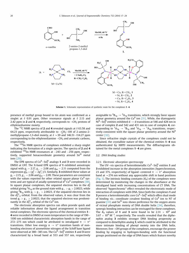
The synthetic route of new organotin(IV) analogs, 1e4 is rep-resented in Scheme 1. The proposed structures were formulated onthe basis of their elemental analysis, spectroscopic techniques (IR,NMR, ESIeMS, UVevis and EPR) and molar conductance values.The molar conductivity data of synthesized complexes 1e4 inDMSO (10�3 M) at 25 �C were too low to account for any disso-ciative ions in the complexes, consistent with their non-electro-lytic nature.
The IR spectrum of non-coordinated Schiff base ligand, L ex-hibits medium absorption band at 1637 cm�1 attributable to azo-methine y(C]N), which was perturbed to lower frequencies by 9e6 cm�1 upon complexation in 1e4 indicating donation of the lonepair of azomethine nitrogen to Sn atom. The characteristicstretching vibration of the phenolic oxygen n(CeO) was shifted tolower frequencies in the 1236e1228 cm�1 in 1e4, suggestinginvolvement of phenolic oxygen in coordination after deprotona-tion [25]. A medium intensity band at 3400e3300 cm�1 attribut-able to the eNH2 group of ethylenediamine disappearedcompletely in all complexes 1e4 and new bands appeared at 3192e3178 and 1583e1570 cm�1 corresponding to n(NeH) stretching andd(NeH) bending vibrations, respectively, supporting the coordina-tion of ethylenediamine to CuII/NiII and subsequently to SnIV metalions by the elimination of HCl [26]. Furthermore, the diagnosticassignments of 1e4 corresponding to y(SneC), y(SneO) and y(SneN) were found at 576e556, 539e530 and 446e432 cm�1, respec-tively [27]. Besides, all the complexes exhibited a sharp band in theregion 478e467 cm�1 attributed to y(Cu/NieN) vibrations sup-porting the coordination through nitrogen.
In the NMR spectra, the assignment of the proton resonanceswas made by their peak multiplicity, intensity pattern and com-parison of the integration values of the protons with the expectedcomposition. The 1H NMR spectra of 2 and 4 recorded in DMSO-d6demonstrated the absence ofeOH signal at dw12.6 ppm indicatingthe involvement of phenolic eOH group in coordination to SnIV
metal center through deprotonation [28]. The characteristic signalof eHC]N group at d 9.87 and 9.88 ppm in 2 and 4, respectively,revealed a significant chemical shift due to ligation of nitrogenatom to tin metal centers. In case of dimethyltin analog 2, the
Scheme 1. Schematic representation of synthetic route for the complexes 1e4.
S. Tabassum et al. / Journal of Organometallic Chemistry 752 (2014) 17e2420
presence of methyl group bound to tin atom was confirmed as asinglet at d 0.91 ppm. Other resonance signals at d 2.13 and2.42 ppm in 2 and 4, respectively, corresponds to eCH2 protons ofethylenediamine moiety.
The 13C NMR spectra of 2 and 4 revealed signals at d 63.58 and64.23 ppm, respectively attributable to eCH2eOH of 2-amino-2-methylpropane-1,3-diol moiety, at d w39 and 146.51e114.27 ppmcorresponding to the ethylenediamine eCH2 and aromatic carbons,respectively.
The 119Sn NMR spectra of complexes exhibited a sharp singletindicating the formation of a single species. The spectra of 2 and 4exhibited 119Sn NMR resonances at �243 and �256 ppm, respec-tively, supported hexacoordinate geometry around SnIV metalions [29].
The EPR spectra of CuIIeSn2IV analogs 1 and 3 were recorded in
DMSO at LNT. The X-band EPR spectra of 1 exhibited anisotropicsignal with gjj ¼ 2.17, gt ¼ 2.08 and gav ¼ 2.11 computed from theexpression gav2 ¼ (gjj2 þ 2gt2 )/3. Similarly, 3 exhibited these values atgjj ¼ 2.15, gt¼ 2.06 and gav¼ 2.09. These parameters are consistentwith the values reported for other related square planar CuII sys-tems and are typical of axially symmetrical d9 CuII complexes [30].In square planar complexes, the unpaired electron lies in the dz2
orbital giving 2A1g as the ground state with gt > gk > 2.0023, whilegiving 2B1g with gk > gt > 2.0023, if the unpaired electron lies inthe dx
2�y2 orbital. From the observed values, it was clear from the
trend gk > gt > 2.0023, that the unpaired electron was predomi-nantly in the dx
2�y2 orbital of the CuII ion.
The electronic absorption spectra can often provide quick andreliable information about the ligand arrangement in transitionmetal complexes. The electronic spectra of the chemical entities 1e4were recorded in DMSO at room temperature in the range of 190e1100 nm exhibited characteristic absorption bands in the range of272e275 nm which could be attributed to intraligand p / p*transitions. Other low energy n / p* transition bands of non-bonding electrons of azomethine nitrogen of the Schiff base ligandwere observed at 380e385 nm. The CuIIeSn2
IV entities 1 and 3werecharacterized by a broad band at 553 and 557 nm, respectively
assignable to 2B1g /2A1g transitions, which strongly favor square
planar geometry around the CuII ion [31]. While, the diamagneticNiIIeSn2
IV entities exhibited d/ d transitions at 546 and 428 nm incase of complex 2 and 542 and 431 nm in case of complex 4, cor-responding to 1A1g / 1B1g and 1A1g / 1A2g transitions, respec-tively consistent with the square planar geometry around the NiII
center [32].Since refractive single crystals of the complexes could not be
obtained, the crystalline nature of the chemical entities 1e4 wasauthenticated by XRPD measurements. The diffractograms ob-tained for the metal complexes 1e4 are given.
3.2. DNA binding studies
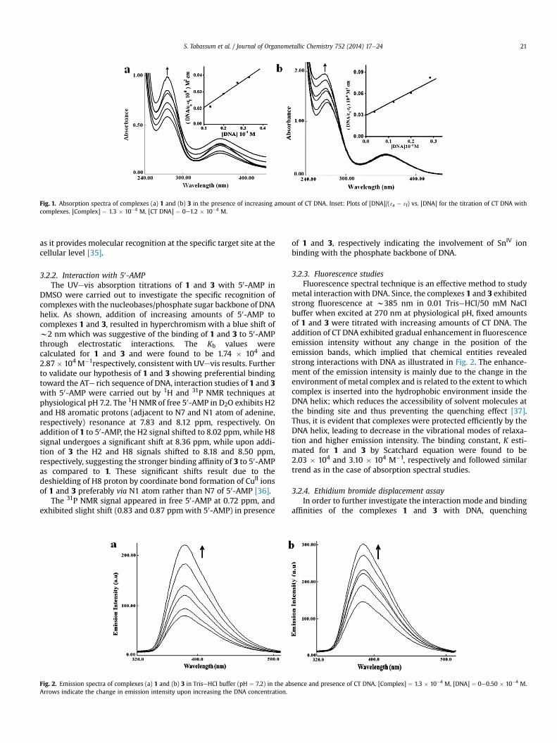
3.2.1. Electronic absorption spectroscopyThe UVevis spectra of heterobimetallic CuIIeSn2
IV entities 1 and3 exhibited increase in the absorption intensities (hyperchromism,23 and 37%, respectively) of ligandecentered p / p* absorptionband at w274 nm without any appreciable shift in band positions(Fig. 1). The intrinsic binding constants (Kb) of the complexes weredetermined by monitoring the changes in the absorbance at theintraligand band with increasing concentrations of CT DNA. Theobserved “hyperchromic” effect revealed the electrostatic mode ofinteraction of complexes with DNA. Since both the complexes 1 and3 possess a heterobimetallic core CuIIeSn2
IV which offers dual modeof binding viz.; coordinate covalent binding of CuII ion to N7 ofguanine [33] and SnIV ions shows preference for the oxygen atomsof vicinal phosphate moiety of DNA double helix [34], therefore,synergic bonding interaction can be envisaged for these complexes.The Kb values of 1 and 3 were found to be 2.31 � 104 and3.67 � 104 M�1, respectively. The results revealed that the diphe-nyltin analog 3 exhibits stronger DNA binding propensity ascompared to dimethyltin analog 1, which could be attributed to themore intimate binding of phenyl moiety into the DNA helix.Moreover, freeeOH groups of the complexes, encourage the groovebinding by engaging in hydrogen-bonding with the functionalgroups positioned on the edge of DNA bases which feature novelty
Fig. 1. Absorption spectra of complexes (a) 1 and (b) 3 in the presence of increasing amount of CT DNA. Inset: Plots of [DNA]/( 3a � 3f) vs. [DNA] for the titration of CT DNA withcomplexes. [Complex] ¼ 1.3 � 10�4 M, [CT DNA] ¼ 0e1.2 � 10�4 M.
S. Tabassum et al. / Journal of Organometallic Chemistry 752 (2014) 17e24 21
as it provides molecular recognition at the specific target site at thecellular level [35].
3.2.2. Interaction with 50-AMPThe UVevis absorption titrations of 1 and 3 with 50-AMP in
DMSO were carried out to investigate the specific recognition ofcomplexes with the nucleobases/phosphate sugar backbone of DNAhelix. As shown, addition of increasing amounts of 50-AMP tocomplexes 1 and 3, resulted in hyperchromism with a blue shift ofw2 nm which was suggestive of the binding of 1 and 3 to 50-AMPthrough electrostatic interactions. The Kb values werecalculated for 1 and 3 and were found to be 1.74 � 104 and2.87� 104 M�1respectively, consistent with UVevis results. Furtherto validate our hypothesis of 1 and 3 showing preferential bindingtoward the ATe rich sequence of DNA, interaction studies of 1 and 3with 50-AMP were carried out by 1H and 31P NMR techniques atphysiological pH 7.2. The 1H NMR of free 50-AMP in D2O exhibits H2and H8 aromatic protons (adjacent to N7 and N1 atom of adenine,respectively) resonance at 7.83 and 8.12 ppm, respectively. Onaddition of 1 to 50-AMP, the H2 signal shifted to 8.02 ppm, while H8signal undergoes a significant shift at 8.36 ppm, while upon addi-tion of 3 the H2 and H8 signals shifted to 8.18 and 8.50 ppm,respectively, suggesting the stronger binding affinity of 3 to 50-AMPas compared to 1. These significant shifts result due to thedeshielding of H8 proton by coordinate bond formation of CuII ionsof 1 and 3 preferably via N1 atom rather than N7 of 50-AMP [36].
The 31P NMR signal appeared in free 50-AMP at 0.72 ppm, andexhibited slight shift (0.83 and 0.87 ppmwith 50-AMP) in presence
Fig. 2. Emission spectra of complexes (a) 1 and (b) 3 in TriseHCl buffer (pH ¼ 7.2) in the abArrows indicate the change in emission intensity upon increasing the DNA concentration.
of 1 and 3, respectively indicating the involvement of SnIV ionbinding with the phosphate backbone of DNA.
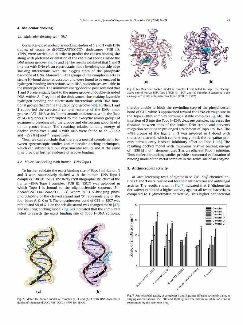
3.2.3. Fluorescence studiesFluorescence spectral technique is an effective method to study
metal interactionwith DNA. Since, the complexes 1 and 3 exhibitedstrong fluorescence at w385 nm in 0.01 TriseHCl/50 mM NaClbuffer when excited at 270 nm at physiological pH, fixed amountsof 1 and 3 were titrated with increasing amounts of CT DNA. Theaddition of CT DNA exhibited gradual enhancement in fluorescenceemission intensity without any change in the position of theemission bands, which implied that chemical entities revealedstrong interactions with DNA as illustrated in Fig. 2. The enhance-ment of the emission intensity is mainly due to the change in theenvironment of metal complex and is related to the extent to whichcomplex is inserted into the hydrophobic environment inside theDNA helix; which reduces the accessibility of solvent molecules atthe binding site and thus preventing the quenching effect [37].Thus, it is evident that complexes were protected efficiently by theDNA helix, leading to decrease in the vibrational modes of relaxa-tion and higher emission intensity. The binding constant, K esti-mated for 1 and 3 by Scatchard equation were found to be2.03 � 104 and 3.10 � 104 M�1, respectively and followed similartrend as in the case of absorption spectral studies.
3.2.4. Ethidium bromide displacement assayIn order to further investigate the interaction mode and binding
affinities of the complexes 1 and 3 with DNA, quenching
sence and presence of CT DNA. [Complex] ¼ 1.3 � 10�4 M, [DNA] ¼ 0e0.50 � 10�4 M.
Fig. 3. Agarose gel electrophoresis pattern for the cleavage of pBR322 supercoiled DNA(300 ng) by complex 3 in presence of (a) different activating agents at 310 K afterincubation for 45 min. Lane 1: DNA Control; Lane 2: 50 mM of complex þ DMSO(0.4 M) þ DNA; Lane 3: 50 mM of complex þ EtOH (0.4 M) þ DNA; Lane 4: 50 mM ofcomplex þ SOD (15 units) þ DNA Lane 5: 50 mM of complex þ sodium azide(0.4 M) þ DNA (b) DNA recognition agents at 310 K after incubation for 45 min. Lane 1,DNA control; Lane 2, 40 mM of complex 1 þ DNA þ methyl green (2.5 mL of a 0.01 mg/ml solution); Lane 3, 40 mM of complex 1 þ DNA þ DAPI (8 mM).
Fig. 4. Agarose gel electrophoresis pattern showing effect of different concentration ofcomplex 3 on the activity of DNA Topo I; Lane 1, DNA control; Lane 2, Topo I þ DNA;Lane 3, 7.5 mM of complex þ DNA þ Topo I; Lane 4: 10 mM of complex þ DNA þ Topo I;Lane 5: 12.5 mM of complex þ DNA þ Topo I; Lane 6: 15 mM of complex þ DNA þ Topo I.
S. Tabassum et al. / Journal of Organometallic Chemistry 752 (2014) 17e2422
experiments with EB were employed. The EB displacement tech-nique is based on the decrease of fluorescence resulting from thedisplacement of EB from a DNA sequence by a quencher, and thequenching is due to the reduction of the number of binding sites onthe DNA that is available to the EB. The increasing concentrations ofthe complexes 1 and 3 (0e1.2 � 10�4 M) to DNA pretreated with EB([DNA]/[EB] ¼ 1) (0.2 � 10�4 M) resulted in remarkable quenchingof the emission intensity, indicative of the competitive displace-ment of the bound EB from the CTeDNA by the complexes. Therelative binding propensity of the complexes to DNA is measuredfrom the extent of reduction in the emission intensity. As com-plexes 1 and 3 bind to DNA through electrostatic binding mode viathe phosphate backbone of DNA helix, the observed quenchingmaybe due to the photoelectron transfer mechanism [38]. The Ksvvalues calculated from SterneVolmer equation for complexes 1 and3were found to be 0.64 and 1.06, respectively. The greater decreasein the Ksv value for complex 3 compared to complex 1 was attrib-uted to the strong binding of the complex with DNA double helix.
3.2.5. Effect of ionic strengthTo evaluate the electrostatic contribution into the DNA-binding
event of the complexes 1 and 3, the effect of the ionic strength onthe emission spectrum of complexes was monitored. The fluores-cence intensity of DNA bound complexes was moderatelyquenched with increasing ionic strength through added NaCl (0e0.50 � 10�4 M). Due to the competitive interaction for phosphateanions, the addition of NaCl weakens the surface binding in-teractions as well as hydrogen bonding between the CT DNA andthe interacting molecules [39].
3.2.6. Viscosity measurementsThe plots of relative viscosities vs. [complex]/[DNA] are shown.
The relative specific viscosity of the DNA reduces steadily uponaddition of CuIIeSn2
IV analogs 1 and 3 support our contention thatthe complexes bind to DNA via non-covalent interactions [40]. Thedecreased relative viscosity of DNA may be explained by a bindingmode which produced bends or kinks in the DNA strand, therebydiminishing its effective length and concomitantly its viscosity.
3.3. DNA cleavage studies of pBR322 DNA
Since, 3 exhibited greater binding propensity to CT DNA, thecleavage activity of 3 was studied using supercoiled plasmidpBR322 DNA as a substrate in a medium of 5 mM TriseHCl/50 mMNaCl buffer (pH 7.2) under physiological conditions. A concentra-tion dependent DNA cleavage by 3 was performed at increasingcomplex concentrations (10e50 mM) which exhibiting significantcleavage at 50 mM without the formation of Form III, suggestingsingle strand DNA cleavage.
The mechanistic aspects of the DNA cleavage reaction of 3 werestudied in the presence of radical scavengers. The DNA cleavage inthe presence of hydroxyl radical scavengers (DMSO and EtOH),singlet oxygen quencher (NaN3) and SOD as superoxide radicalscavenger is shown in Fig. 3a. An enhancement in the DNA cleavagewas observed upon the addition of DMSO and EtOH, ruling out theinvolvement of hydroxyl radical in the cleavage process (Fig. 3a,Lanes 2 and 3). Since, no obvious inhibition was observed in thepresence of SOD, the possibility of involvement of superoxideradical in DNA cleavagewas ruled out (Fig. 3a, Lane 4). While, in thepresence of NaN3, DNA cleavage is significantly inhibited whichwasindicative of the involvement of the singlet oxygen or a singletoxygen-like entity in the cleavage process (Fig. 3a, Lane 5). Thisinhibitory activity of NaN3 can be ascribed to the affinity of theazide anion for transition metals [41].
From the results obtained abovewemaysuggest that 3 is capableof promoting DNA cleavage through an oxidative DNA damagepathway. A copper peroxide with DNA cleaving ability is formed byan active singlet oxygen species or a singlet oxygen-like entity. Theassumed involvement of coppereoxo species can be deduced fromthe reaction of copper(I) and endogenous dioxygen to give super-oxide anion that dismutates, giving rise to hydrogen peroxide thatcan react yielding copper(I) to obtain a coppereoxo species [42].
The DNA groove binding preference of 3 was studied using DNAmajor groove binder methyl green and DNA minor groove binderdistamycin. A significant inhibition in the chemical nuclease ac-tivity of 3was observed in the presence of distamycin, whilemethylgreen addition exhibited no apparent effect on the DNA cleavage(Fig. 3b). These results are of significance as majority of theoxidative cleavage reagents generally bind in the minor grooverather than major groove [43].
3.4. Topoisomerase I inhibition
Topo I inhibitors inhibit or reduce the rate of religation in theDNA cleavage complex and ultimately leads to cell death aftercollision of the cleavage complex, with the replication fork result-ing in double-strand breakage [44]. The gel electrophoresis patternwas obtained by incubating pBR322 DNA with Topo I in the pres-ence of different concentrations of 3. As shown in Fig. 4, supercoiledDNA was fully relaxed by the enzyme in the absence of 3 (Lane 2).However, upon increasing the concentrations of 3 (7.5e15 mM), thelevels of the relaxed form (OC) were inhibited (Lanes 3e6) [45]. At15 mM the DNA relaxation effect caused by Topo I was completelyinhibited by 3. These observations suggest that 3 inhibits Topo Icatalytic activity due to the relatively strong DNA binding affinity ofthe complex, preventing the enzyme from efficiently binding toDNA.
Fig. 6. (a) Molecular docked model of complex 1 was failed to target the cleavageactive site of human DNA Topo I (PDB ID: 1SC7) and (b) Complex 3 targeting in thecleavage active site of human DNA Topo I (PDB ID: 1SC7).
S. Tabassum et al. / Journal of Organometallic Chemistry 752 (2014) 17e24 23
4. Molecular docking
4.1. Molecular docking with DNA
Computer-aided molecular docking studies of 1 and 3with DNAduplex of sequence d(CGCGAATTCGCG)2 dodecamer (PDB ID:1BNA) were carried out in order to predict the chosen binding sitealong with preferred orientation of the chemical species inside theDNAminor groove (Fig. 5a and b). The results exhibited that 1 and 3interact with DNA via an electrostatic mode involving outside edgestacking interactions with the oxygen atom of the phosphatebackbone of DNA. Moreover, eOH groups of the complexes acts asstrong Hebond donor or acceptor and were found to be engaged inhydrogen-bonding interactions with DNA nucleobases available intheminor grooves. Theminimum energy docked pose revealed that1 and 3 preferentially bind to the minor groove of double-strandedDNA, within AeT regions of the dodecamer, thus making favorablehydrogen bonding and electrostatic interactions with DNA func-tional groups that define the stability of groove [46]. Further, 1 and3 supported the structural complementarity of the DNA minorgroove of ATeDNA, as its floor is smooth and convex, while the floorof GC-sequences is interrupted by the exocyclic amino groups ofguanines protruding into the groove and obstructing good fit of aminor groove-binder. The resulting relative binding energy ofdocked complexes 1 and 3 with DNA were found to be �252.2and �272.8 kJ mol�1 respectively.
Thus, we can conclude that there is a mutual complement be-tween spectroscopic studies and molecular docking techniques,which can substantiate our experimental results and at the sametime provides further evidence of groove binding.
4.2. Molecular docking with humaneDNA Topo I
To further validate the exact binding site of Topo I inhibition, 1and 3 were successively docked with the humaneDNA Topo Icomplex (PDB ID: 1SC7). The X-ray crystallographic structure of thehumaneDNA Topo I complex (PDB ID: 1SC7) was uploaded inwhich Topo I is bound to the oligonucleotide sequence 50eAAAAAGACTTsX-GAAAATTTTT-30, where ‘s’ is 50-bridging phos-phorothiolate of the cleaved strand and ‘X’ represents any of thefour bases A, G, C or T. The phosphoester bond of G12 in 1SC7 wasrebuilt and SH of G11 on the scissile strand was changed to OH [47].The resulting docking model (Fig. 6a) indicated that the complex 1failed to search the exact binding site of Topo IeDNA complex,
Fig. 5. Molecular docked model of complex (a) 1 and (b) 3 with DNA dodecamerduplex of sequence d(CGCGAATTCGCG)2 (PDB ID: 1BNA).
thereby unable to block the rewinding step of the phosphoesterbond of G12, while 3 approached toward the DNA cleavage site inthe Topo IeDNA complex forming a stable complex (Fig. 6b). Theinsertion of 3 into the Topo IeDNA cleavage complex increases thedistance between ends of the broken DNA strand and preventsrelegation resulting in prolonged attachment of Topo I to DNA. TheeOH groups of the ligand in 3 was involved in H-bond withthe scissile strand, which could strongly block the religation pro-cess, subsequently leads to inhibitory effect on Topo I [48]. Theresulting docked model with minimum relative binding energyof �330 kJ mol�1 demonstrates 3 as an efficient Topo I inhibitor.Thus, molecular docking studies provide a structural explanation ofbinding mode of the metal complex in the active site of an enzyme.
5. Antimicrobial activity
In vitro screening tests of synthesized CuIIeSn2IV chemical en-
tities 1 and 3were carried out for their antibacterial and antifungalactivity. The results shown in Fig. 7 indicated that 3 (diphenyltinderivative) exhibited a higher activity against all tested bacteria ascompared to 1 (dimethyltin derivative). This higher antibacterial
Fig. 7. Antimicrobial activity of complexes 1 and 3 against different bacterial strains, atvarying concentrations (250, 500 and 1000 mg/ml). The maximum inhibition zone isrepresented by the reference drug.
S. Tabassum et al. / Journal of Organometallic Chemistry 752 (2014) 17e2424
activity of 3 against most of bacteria strains could be attributed tothe high lipophilic character of diphenyltin moiety. The presence oftwo phenyl groups in 3 increases the solubility of the complex inlipids and hence it can cross through biological membranes withhigher efficiency [49]. However, both 1 and 3, exhibited lesser ac-tivity than the reference drug (doxycyclin).
The synthesized compounds were also screened for their anti-fungal activity against C. albicans. The results demonstrated thatboth the CuIIeSn2
IV DNA binding agents 1 and 3 exhibited goodantifungal activity.
6. Conclusions
This work describes the synthesis and characterization of het-erobimetallic CuII/NiIIeSn2
IV chemical entities 1e4, derived fromo-vanillin Schiff base. The comparative in vitro DNA binding profileof 1 and 3 was investigated by absorption, fluorescence and vis-cosity measurements and the results revealed electrostatic in-teractions along with the selective binding to the minor groove ofDNA. The intrinsic binding constant, Kb revealed higher bindingpropensity of diphenyltin analog, 3 as compared to dimethyltinanalog,1. The antimicrobial activity of 1 and 3was evaluated whichrevealed that 3 exhibited better antimicrobial activity than 1.Further, the pBR322 DNA cleaving ability of 3 was evaluated byagarose gel electrophoresis which revealed that the complex bindto double-stranded DNA possibly in the minor groove and cleavessupercoiled DNA through an oxidative cleavage mechanisminduced by a reactive oxygen species. Furthermore, 3 exhibitedsignificant inhibitory effects on Topo I activity. Additionally, mo-lecular docking studies of 1 and 3 were performed with moleculartarget DNA and the active site of topoisomerase enzyme in order tovalidate the experimental results. Therefore, from the above resultsit could be concluded the introduction of diphenyltin moiety couldsignificantly enhance the activity both in DNA binding and Topo Iinhibition, thereby complex 3 proving its worth as a robust andbetter choice for potent and safe chemotherapeutic drug design.
Acknowledgments
The authors are grateful to SAIF, Panjab University, Chandigarh;STIC, Cochin and IIT Bombay, India for providing ESIeMS, NMR,elemental analysis and EPR facility, respectively. They alsoacknowledge financial support to the Department of Chemistry,AMU through UGC assisted DRSeSAP and DST PURSE Programme.
References
[1] Cancer; Fact Sheet No. 297, World Health Organization, Geneva, Switzerland;http://www.who.int/mediacentre/factsheets/fs297/en/index.html.
[2] B. Rosenberg, L. Van camp, Cancer Res. 30 (1970) 1799e1802.[3] K.S. Lovejoy, S.J. Lippard, Dalton Trans. (2009) 10651e10659.[4] L.R. Kelland, Drugs 59 (2000) 1e8.[5] V. Srb, E. Kubzova, K. Kubikova, Neoplasma 33 (1986) 615e620.[6] J.J. Champoux, Annu. Rev. Biochem. 70 (2001) 369e413.
[7] Y. Pommier, Chem. Rev. 109 (2009) 2894e2902.[8] Y. Pommier, E. Leo, H.L. Zhang, C. Marchand, Chem. Biol. 17 (2010) 421e433.[9] A. Alama, B. Tasso, F. Novelli, F. Sparatore, Drug Discov. Today 14 (2009)
(2004) 985e987.[11] M.S. Nair, R.S. Joseyphus, Spectrochim. Acta Part A 70 (2008) 749e753.[12] S. Tabassum, S. Amir, F. Arjmand, C. Pettinari, F. Marchetti, N. Masciocchi,
G. Lupidi, R. Pettinari, Eur. J. Med. Chem. 60 (2013) 216e232.[13] M. Gielen, E.R.T. Tiekink, Metallotherapeutic Drugs and Metal Based Diag-
nostic Agents. The Use of Metals in Medicine, J. Wiley & Sons, 2005, pp.421e439.
[14] T.S. Basu Baul, S. Basu, D. de Vos, A. Linden, Invest. New Drugs 27 (2009)419e431.
[15] R. Buchtik, Z. Travnicek, J. Vanco, R. Herchel, Z. Dvorak, Dalton Trans. 40(2011) 9404e9412.
[16] S. Tabassum, S. Mathur, F. Arjmand, K. Mishra, K. Banerjee, Metallomics 4(2012) 205e217.
[17] J. Marmur, J. Mol. Biol. 3 (1961) 208e218.[18] M.E. Reicmann, S.A. Rice, C.A. Thomas, P. Doty, J. Am. Chem. Soc. 76 (1954)
3047e3053.[19] A. Wolfe, G.H. Shimer, T. Meehan, Biochemistry 26 (1987) 6392e6396.[20] J.R. Lakowiez, G. Webber, Biochemistry 12 (1973) 4161e4170.[21] F. Arjmand, S. Parveen, D.K. Mohapatra, Inorg. Chim. Acta 388 (2012) 1e10.[22] D.W. Ritche, V. Venkataraman, Bioinformatics 26 (2010) 2398e2405.[23] A. Rehman, M.I. Choudhary, W.J. Thomsen, Bioassay Techniques for Drug
Development, Harwood Academic Publishers, Amsterdam, The Netherlands,2001, p. 9.
[24] G. Wilkinson, R.D. Gillard, J.A. McCleverty, Comprehensive CoordinationChemistry, Peragamon Press, Oxford, 1987, pp. 516e550.
[25] K. Nakamoto, Infrared and Raman Spectra of Inorganic and CoordinationCompounds, third ed., John Wiley & Sons, New York, 1986.
[27] F. Arjmand, I. Yousuf, J. Organomet. Chem. 743 (2013) 55e62.[28] I. Kaya, A. Bilici, M. Gul, Polym. Adv. Technol. 19 (2008) 1154e1163.[29] R. Zhang, J. Sun, C. Ma, J. Organomet. Chem. 690 (2005) 4366e4372.[30] S. Srinivasan, P. Athappan, G. Rajagopal, Trans. Met. Chem. 26 (2001) 588e593.[31] H. Unver, Z. Hayvali, Spectrochim. Acta Part A 75 (2010) 782e788.[32] F. Arjmand, B. Mohani, S. Ahmad, Eur. J. Med. Chem. 40 (2005) 1103e1110.[33] S. Tabassum, S. Yadav, F. Arjmand, J. Organomet. Chem. 745e746 (2013)
226e234.[34] M. Chauhan, F. Arjmand, J. Organomet. Chem. 692 (2007) 5156e5164.[35] M. Baldini, M.-B. Ferrari, F. Bisceglie, G. Pelosi, S. Pinelli, P. Tarasconi, Inorg.
Chem. 42 (2003) 2049e2055.[36] R. Jastrzab, J. Inorg. Biochem. 103 (2009) 766e773.[37] S. Kashanian, M.B. Gholivand, F. Ahmadi, A. Taravati, A.H. Colagar, Spec-
trochim. Acta Part A 67 (2007) 472e478.[38] B. Selvakumar, V. Rajendiran, P.V. Maheswari, H.S. Evans, M. Palanaindavar,
J. Inorg. Biochem. 100 (2006) 316e330.[39] F.-Y. Wu, F.-Y. Xie, Y.-M. Wu, J.-I. Hong, J. Fluoresc. 18 (2008) 175e181.[40] U. Chaveerach, A. Meenongwa, Y. Trongpanich, C. Soikum, P. Chaveerach,
Polyhedron 29 (2010) 731e738.[41] F.B. ElAmrani, L. Perelló, J.A. Real, M.G. Alvarez, G. Alzuet, J. Borrás,
S.G. Granda, J.M. Bernardo, J. Inorg. Biochem. 100 (2006) 1208e1218.[42] D.-D. Li, J.-L. Tian, W. Gu, X. Liu, H.-H. Zeng, S.-P. Yan, J. Inorg. Biochem. 105
(2011) 894e901.[43] W.K. Pogozelski, T.D. Tullius, Chem. Rev. 98 (1998) 1089e1107.[44] Y. Pommier, P. Pourquier, Y. Urasaki, J. Wu, G.S. Laco, Drug Resist. Updat. 2
(1999) 307e318.[45] B. Montaner, W.C. Avila, M. Martinell, R. Ollinger, J. Aymami, E. Giralt,