35
Presented Presented by by K.Ravichandran K.Ravichandran 1 1 st st Msc.Medical Msc.Medical biochemistry biochemistry HETEROPOLYSACCHARIDES
PresentedPresented
byby
K.RavichandranK.Ravichandran
11stst Msc.Medical Msc.Medical biochemistrybiochemistry
HETEROPOLYSACCHARIDES

POLYSACCHARIDES – Most of the carbohydrates found in nature occur in the form of high molecular polymers calledpolysaccharides
Polysaccharides are of two types :
a) Homopolysaccharides
b) Heteropolysaccharides
HOMOPOLYSACCHARIDES – polymer of same monosaccharide
units
eg : Starch, Glycogen, Inulin, Cellulose, Pectin, Chitin.
HETEROPOLYSACCHARIDES – polymer of different
monosaccharide units eg : Mucopolysaccharides
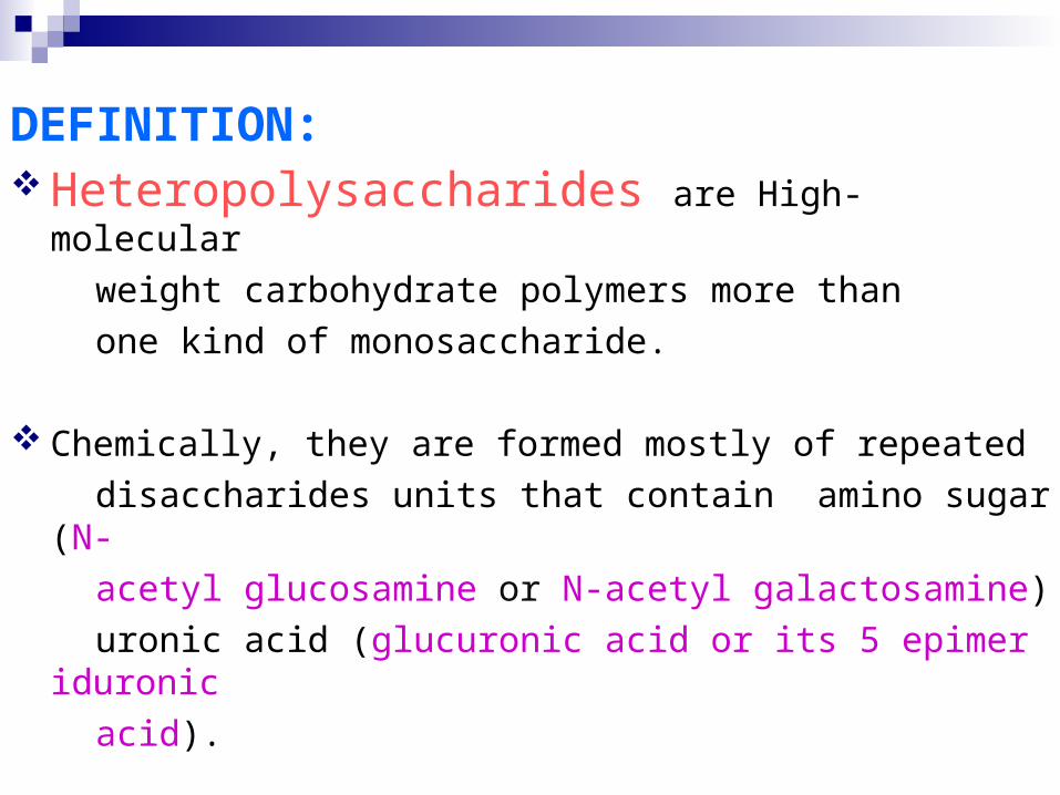
DEFINITION: Heteropolysaccharides are High-molecular
weight carbohydrate polymers more than
one kind of monosaccharide.
Chemically, they are formed mostly of repeated
disaccharides units that contain amino sugar (N-
acetyl glucosamine or N-acetyl galactosamine)
uronic acid (glucuronic acid or its 5 epimer iduronic
acid).
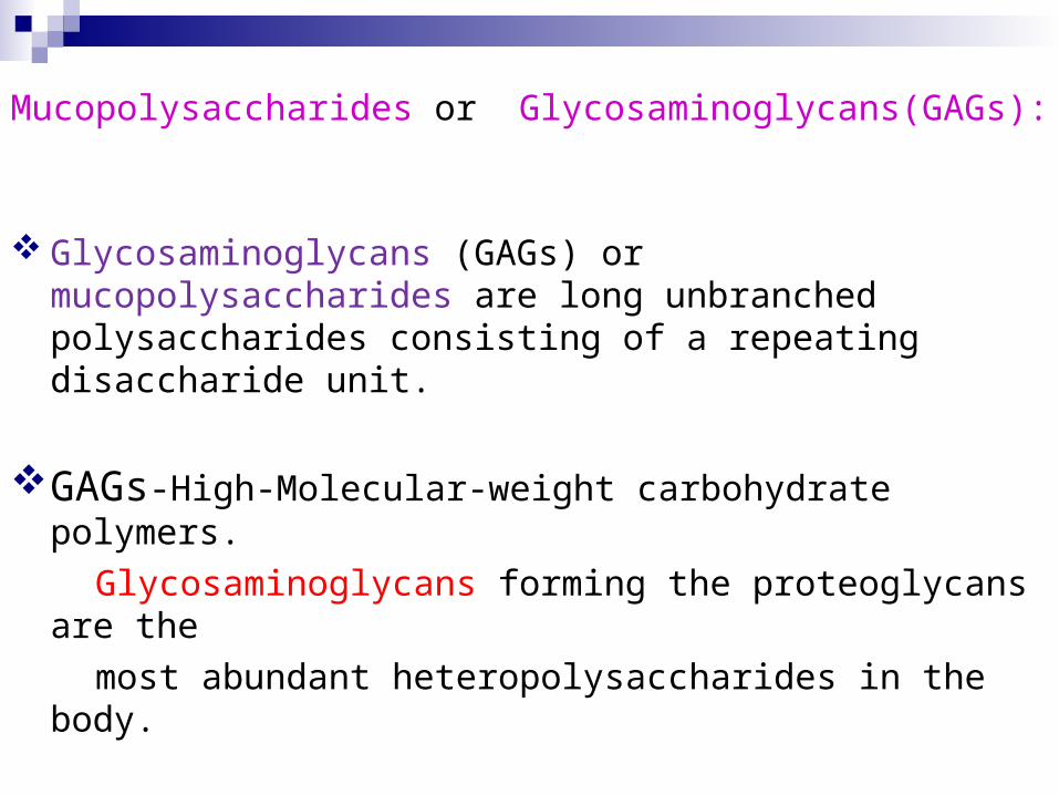
Mucopolysaccharides or Glycosaminoglycans(GAGs):
Glycosaminoglycans (GAGs) or mucopolysaccharides are long unbranched polysaccharides consisting of a repeating disaccharide unit.
GAGs-High-Molecular-weight carbohydrate polymers.
Glycosaminoglycans forming the proteoglycans are the
most abundant heteropolysaccharides in the body.
They are long unbranched molecules containing a repeating
disaccharides unit. One or both sugars contain Sulfate
Groups(the only exception is Hyaluronic acid).
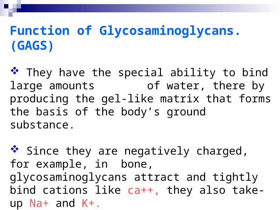
Function of Glycosaminoglycans. (GAGS)
They have the special ability to bind large amounts of water, there by producing the gel-like matrix that forms the basis of the body’s ground substance.
Since they are negatively charged, for example, in bone, glycosaminoglycans attract and tightly bind cations like ca++, they also take-up Na+ and K+.
GAGs stabilize and support cellular and fibrous components of tissue while helping maintain the water and salt balance of the body.
Its essential components of the extra cellular matrix, GAGs play an important role in mediating cell-cell interactions
Ground substance is a part of connective tissue, which is a gel like substance containing water, salt, proteins and polysaccharides. An example of specialized ground substance is the synovial fluid, which serves as a lubricant injoints, and tendon sheaths.
Types:
GAGs are either sulfate group free or sulfate group containing
GAGs are sulfate group free(Hyaluronic acid)
GAGs are sulfate group containing as Chondroitin sulfate
Heparin Keratan sulfate and Dermatan sulfate.
Hyaluronic acid: (also called Hyaluronan
or hyaluronate or HA)
A gel like aminoglycan that is found in the tissue space, the synovial fluid of joints, and the vitreous humor of the eyes and acts as a binding, lubricating, and protective agent.
Hyaluronic acid is unique among the GAGs because it does not contain any sulfate and is not found covalently attached to proteins.
It forms non-covalently linked complexes with proteoglycans in the ECM.
Hyaluronic acid polymers are very large (100-10,000 kD)and can displace a large volume of water.
Found in :Synovial fluid.
Vitreous humor of the eye.
Skin and loose connective tissue.
Cartilage
Epithelial
Neural tissues
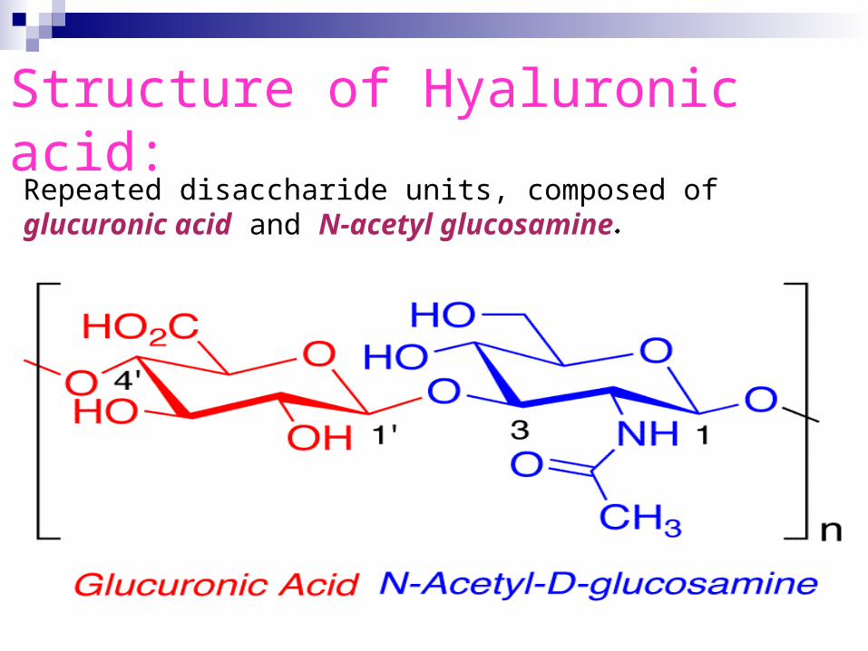
Structure of Hyaluronic acid:Repeated disaccharide units, composed of glucuronic acid and N-acetyl glucosamine.
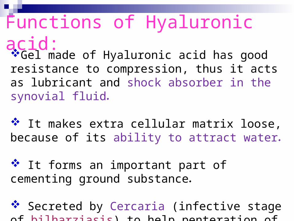
Functions of Hyaluronic acid:Gel made of Hyaluronic acid has good resistance to compression, thus it acts as lubricant and shock absorber in the synovial fluid.
It makes extra cellular matrix loose, because of its ability to attract water.
It forms an important part of cementing ground substance.
Secreted by Cercaria (infective stage of bilharziasis) to help penteration of skin.
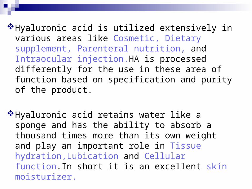
Hyaluronic acid is utilized extensively in various areas like Cosmetic, Dietary supplement, Parenteral nutrition, and Intraocular injection.HA is processed differently for the use in these area of function based on specification and purity of the product.
Hyaluronic acid retains water like a sponge and has the ability to absorb a thousand times more than its own weight and play an important role in Tissue hydration,Lubication and Cellular function.In short it is an excellent skin moisturizer.

Applications: Due to its high Biocompatibility and its common presence in the extracellular matrix of the tissues.
Hyaluronan is gaining popularity as a biomaterial Scaffold in tissue engineering research and producing a Hydrogel.
This added feature allows a researcher to form a desired shape as well as to deliver therapeutic molecules into a host.
Hyaluronan can be crosslinked by attaching Thiols Methacrylates and Thyramines.
Hyaluronan can also be crosslinked directly with Formaldehyde Divinylsulfone
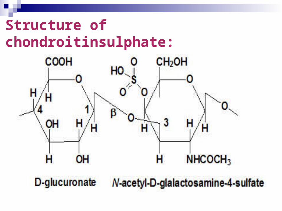
CHONDROITIN SULFATE Definition: Chondroitin sulfate is a sulfated glycosaminoglycan
(GAG) composed of a chain of alternating sugars (N- acetylgalactosamine
and glucuronic acid). It is usually found attached to proteins as part of a proteoglycan.
A chondroitin chain can have over 100 individual sugars, each of which can be sulfated in variable positions and quantities.
Chondroitin sulfate is a chemical that is normally found in cartilage around joints in the body. Chondroitin sulfate is manufactured from animal sources such as cow cartilage.
Structure of chondroitinsulphate:
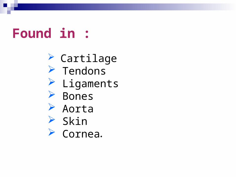
Found in :
Cartilage Tendons Ligaments Bones Aorta Skin Cornea.
Functions of chondroitin sulphates: It has a role in binding collagen of cartilage and
holding its fibers together. Compressibility of cartilage in weight bearing is due to chondroitin sulfate.
Chondroitin is in dietary supplements used as an alternative medicine to treat osteoarthritis and also approved and regulated as a symptomatic slow-acting drug for this disease (SYSADOA) in Europe and some other countries.
It is commonly sold together with glucosamine. Chondroitin and glucosamine are also used in veterinary medicine.
It has a weak anticoagulant property.
Heparin
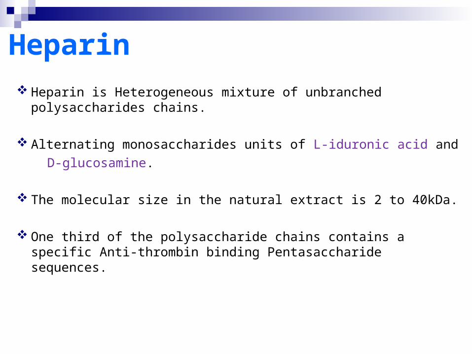
Heparin is Heterogeneous mixture of unbranched polysaccharides chains.
Alternating monosaccharides units of L-iduronic acid and
D-glucosamine.
The molecular size in the natural extract is 2 to 40kDa.
One third of the polysaccharide chains contains a specific Anti-thrombin binding Pentasaccharide sequences.
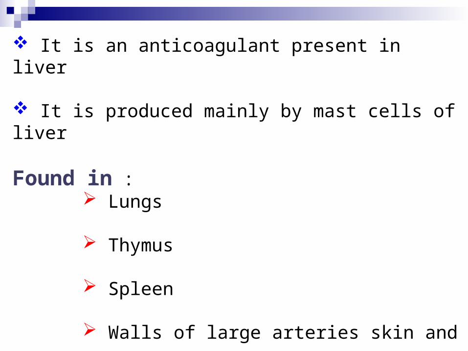
It is an anticoagulant present in liver
It is produced mainly by mast cells of liver
Found in : Lungs
Thymus
Spleen
Walls of large arteries skin and
Small quantities in blood
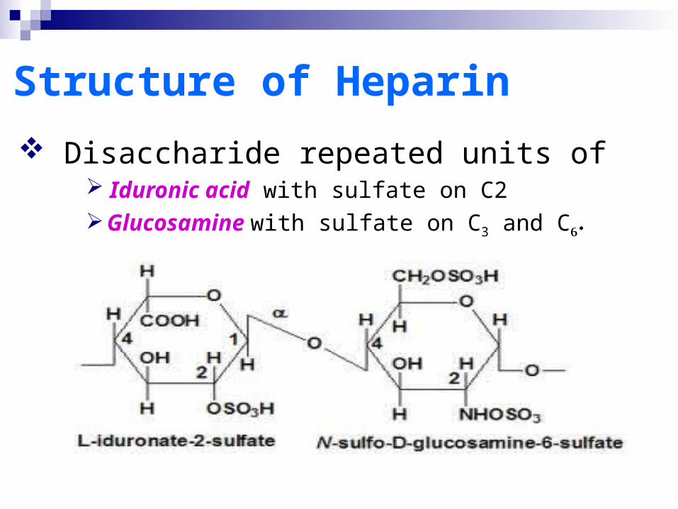
Structure of Heparin
Disaccharide repeated units of Iduronic acid with sulfate on C2 Glucosamine with sulfate on C3 and C6.

Heparin-Clinical Use Prophylactics of DVT and PE
-Preventation of formation of thrombin
-Low dose reginmens
-High risk groups (acute myocardial infraction, Surgery)
Treatment of DVT and PE -Preventation of further thrombin generation
-High dose reginmens
Coronary heart disease -Unstable angina
-Acute myocardial infraction
-After thrombolytic therapy with rt-PA

Functions of Heparin:An anticoagulant present in liver and produced
mainly by mast cells of liver.
Stimulates the release of lipoprotein lipase enzyme that hydrolyses the absorbed fats.
It is an extra cellular compounds, entering in the structure of receptors on the cell surface.It participate in the cell adhesion and cell-cell interaction.

Dermatan sulphate
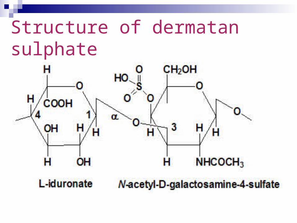
Structure: Disaccharide units composed of L-Iduronic acid and N-acetyl galactosamine with sulfate on C6.
It is widely distributed in animal tissue, resembling chondroitin sulfate and Heparan sulfate.
It has anti thrombotic properties similar to heparin.
Structure of dermatan sulphate
Functions of Dermatan Sulphate:
It is a predominant glycan present in skin.
Dermatan sulfate may have roles
Coagulation, Cardiovascular disease, Carcinogenesis, Infection, Wound repair, and Fibrosis.
Keratan sulfate (KS),also called Keratosulfat
Keratan Sulphate is either of two glycosaminoglycans (I and II),consisting of repeating disaccharides units of N-acetylglucosamine and galactose,but differing slightly in carbohydrate content and localization.
It occurs in cartilage, the cornea, and in the nucleus pulposus and is also an accumulation productin Morquio's syndrome.
KS types are to be composed of three regions A linkage region, at one end of which the KS chain is linked to the core protein. A repeat region, composed of the -3Galβ1-4GlcNAcβ1- repeating disaccharide unit and A chain capping region, occurring at the opposite end of
the KS chain to the protein linkage region. The designations KSI and KSII were originally assigned on the basis of the tissue type from which the keratan sulfate was isolated. KSI was isolated from corneal tissue and KSII from skeletal tissue. The major differences occur in the way each KS type is joined to its coreprotein.
The designations KSI and KSII are now based upon these protein linkage differences. KSI is N-linked to specific asparagine amino acids via acetylglucosamine and KSII is O-linked to specific Serine or Threonine amino acids vi N-acetyl galactosamine
The tissue based classification of KS no longer exists as KS types have been shown to be non tissue specific
A third type of KS (KSIII) has also been isolated from brain tissue that is O-linked to specific serine or threonine amino acids via mannose.
Found in :
Loose connective tissue KS (type-1)
Cornea
Cartilage and
Bone.
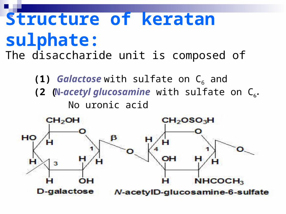
Structure of keratan sulphate:The disaccharide unit is composed of
(1) Galactose with sulfate on C6 and (2 (N-acetyl glucosamine with sulfate on C6. No uronic acid
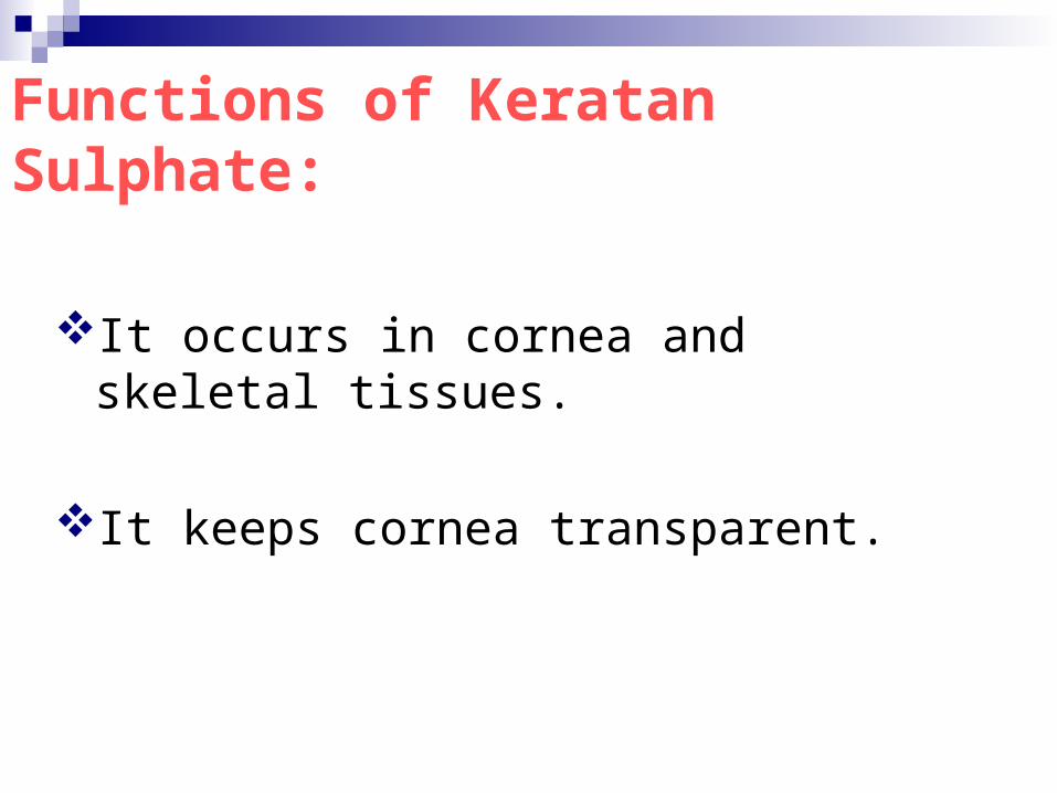
Functions of Keratan Sulphate:
It occurs in cornea and skeletal tissues.
It keeps cornea transparent.
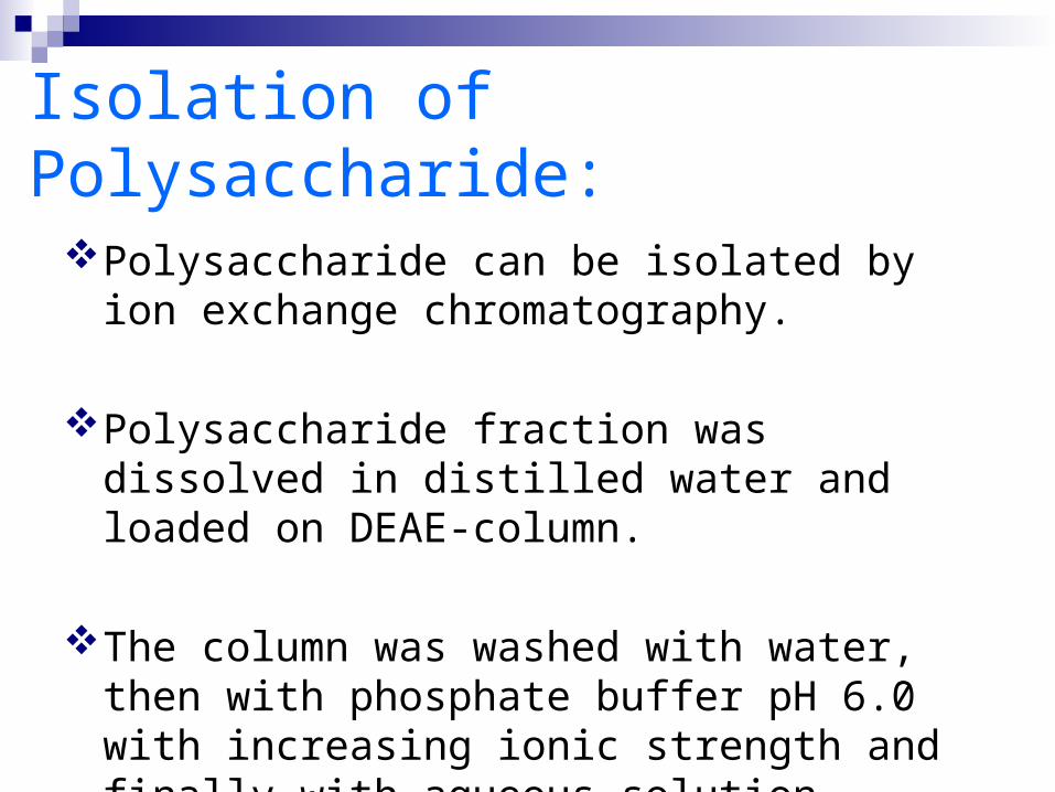
Isolation of Polysaccharide:
Polysaccharide can be isolated by ion exchange chromatography.
Polysaccharide fraction was dissolved in distilled water and loaded on DEAE-column.
The column was washed with water, then with phosphate buffer pH 6.0 with increasing ionic strength and finally with aqueous solution.
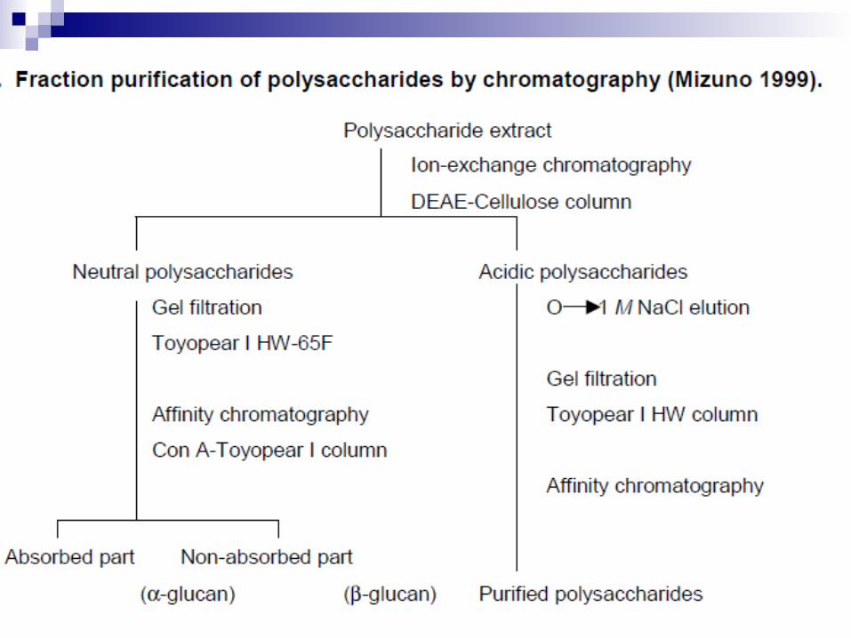
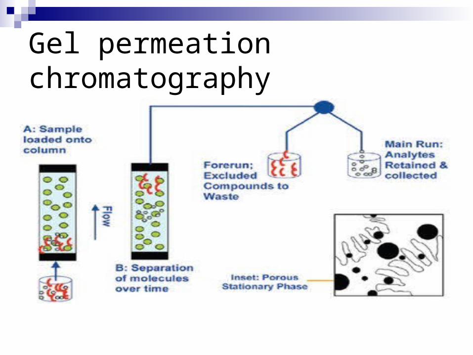
Gel permeation chromatography
