Hfq, a Novel Pleiotropic Regulator of Virulence-Associated Genes inFrancisella tularensis�
Karin L. Meibom,1,2 Anna-Lena Forslund,3,4 Kerstin Kuoppa,3 Khaled Alkhuder,1,2
Iharilalao Dubail,1,2 Marion Dupuis,1,2 Åke Forsberg,3,4 and Alain Charbit1,2*Universite Paris Descartes, Faculte de Medecine Necker-Enfants Malades, Paris F-75015, France1; INSERM, U570, Unit of
Pathogenesis of Systemic Infections, Paris F-75015, France2; FOI Swedish Defence Research Agency, Division ofCBRN Defence and Security, SE-901 82 Umeå, Sweden3; and Laboratory for Molecular Infection Medicine Sweden and
Umeå Centre for Microbial Research, Department of Molecular Biology Umeå University,SE-901 87 Umeå, Sweden4
Received 9 December 2008/Accepted 6 February 2009
Francisella tularensis is a highly infectious pathogen that infects animals and humans, causing tularemia. Theability to replicate within macrophages is central for virulence and relies on expression of genes located in theFrancisella pathogenicity island (FPI), as well as expression of other genes. Regulation of FPI-encodedvirulence gene expression in F. tularensis involves at least four regulatory proteins and is not fully understood.Here we studied the RNA-binding protein Hfq in F. tularensis and particularly the role that it plays as a globalregulator of gene expression in stress tolerance and pathogenesis. We demonstrate that Hfq promotes resis-tance to several cellular stresses (including osmotic and membrane stresses). Furthermore, we show that Hfqis important for the ability of the F. tularensis vaccine strain LVS to induce disease and persist in organs ofinfected mice. We also demonstrate that Hfq is important for stress tolerance and full virulence in a virulentclinical isolate of F. tularensis, FSC200. Finally, microarray analyses revealed that Hfq regulates expression ofnumerous genes, including genes located in the FPI. Strikingly, Hfq negatively regulates only one of twodivergently expressed putative operons in the FPI, in contrast to the other known regulators, which regulatethe entire FPI. Hfq thus appears to be a new pleiotropic regulator of virulence in F. tularensis, acting mostlyas a repressor, in contrast to the other regulators identified so far. Moreover, the results obtained suggest anovel regulatory mechanism for a subset of FPI genes.
Regulation of gene expression by noncoding or small RNAs(sRNAs) is prevalent in both prokaryotes and eukaryotes, andrecent studies have identified numerous sRNAs in differentorganisms. Most sRNAs encoded by bacterial chromosomesact by base pairing with mRNA targets that are encoded intrans, and these sRNAs commonly require Hfq in order tofunction (47). Hfq is a bacterial RNA-binding protein that wasinitially recognized as a host factor for replication of the Q�RNA phage in Escherichia coli (17). The Hfq protein is veryabundant, and it belongs to the eukaryotic and archaeal fam-ilies of Sm and Sm-like proteins, respectively, that form homo-hexameric structures (for reviews, see references 5 and 53).The importance of Hfq became clear when an E. coli hfq nullmutant was created. This mutant had pleiotropic phenotypes,such as a decreased growth rate, increased sensitivity to cellu-lar stresses, and increased cell length (51). Hfq is a posttran-scriptional regulator that binds sRNAs and mRNA and facil-itates RNA-RNA interaction (1, 18, 23, 32, 35, 55). For themost part, sRNA-mRNA interactions result in mRNA degra-dation and/or inhibition of translation, but they can also in-crease translation (for reviews, see references 1, 20, and 48).As might be expected from the pleiotropic phenotypes of anhfq mutant, deletion of hfq has been correlated with consider-
able changes in gene expression observed at the mRNA orprotein level (14, 15, 21, 42, 46).
In the last few years, the role of Hfq in the pathogenesis ofseveral bacterial species has been examined (11, 14, 30, 37, 39,41, 42, 45). A Salmonella enterica serovar Typhimurium hfqmutant was found to be severely attenuated for intracellularreplication in vitro and for virulence in mice (42). Similarly,decreased virulence was observed for hfq mutants of Brucellaabortus, Listeria monocytogenes, Pseudomonas aeruginosa,Vibrio cholerae, Shigella flexneri, and Legionella pneumophila(11, 14, 30, 39, 41, 45). In many cases, the hfq mutation alsorendered the bacteria more sensitive to cellular stress. In strik-ing contrast, deletion of hfq in three different Staphylococcusaureus strain backgrounds did not result in any detectablephenotype (4).
Bacteria have developed numerous regulatory mechanismsso that they are capable of a rapid molecular response withcoordinated gene expression in order to survive in a variety ofhostile environments. A macrophage is probably one of themost stressful environments that facultative intracellular bac-terial pathogens encounter. The aim of the present work was tounderstand how Hfq contributes to the stress response andvirulence of Francisella tularensis, a gram-negative facultativeintracellular pathogen that causes tularemia in humans and alarge number of animal species. Three F. tularensis subspeciesare capable of inducing disease in humans: F. tularensis subsp.tularensis (type A), F. tularensis subsp. holarctica (type B), andF. tularensis subsp. mediasiatica. The fourth subspecies of F.tularensis (“F. tularensis subsp. novicida”) causes disease in
* Corresponding author. Mailing address: Faculte de MedecineNecker, 156, rue de Vaugirard, 75730 Paris Cedex 15, France. Phone: 331-40 61 53 76. Fax: 33 1-40 61 55 92. E-mail: [email protected].
mice, but it is rarely pathogenic in humans. In humans, an F.tularensis infection can be acquired by ingestion of contami-nated water or food, through arthropod bites, from contami-nated animal carcasses, or by inhalation of bacteria. Due to itshigh infectivity and the severity of the disease, F. tularensis hasbeen classified by the Centers for Disease Control and Preven-tion as a potential biological weapon (for reviews, see refer-ences 29 and 43).
The pathogenicity of F. tularensis is believed to be depen-dent largely on its ability to replicate inside host cells, partic-ularly macrophages. Several factors important for virulencehave been identified, but the molecular mechanisms underly-ing disease are unclear. Several of the genes located in theFrancisella pathogenicity island (FPI) (38), such as iglC, iglD,iglA, pdpA, pdpB, and pdpD, have been demonstrated to berequired for virulence in at least one subspecies (12, 19, 24, 27,38, 40, 49, 54).
There are only three known regulators of virulence in F.tularensis, MglA, SspA, and PmrA, all of which control expres-sion of FPI genes (3, 7, 10, 25, 34). Each of these proteins alsoregulates expression of genes outside the FPI; however,whereas MglA and SspA regulate the same set of genes outsidethe FPI, PmrA controls a separate group of genes (7, 10, 34).Recently, a new protein regulator of FPI gene expression wasidentified (6). This protein, FevR, controls the genes in theMglA-SspA regulon and seems to act in parallel with MglA(6).
The genome of F. tularensis does not encode any completeregulatory two-component systems (34) and encodes only oneputative alternative sigma factor, �32 (31), suggesting that thisorganism has an exceptionally limited repertoire of pleiotropicregulatory mechanisms. Expression of the alternative sigmafactors �S and/or �E is controlled by the regulatory protein Hfqin several gram-negative bacteria, such as E. coli, S. Typhi-murium, P. aeruginosa, and V. cholerae, and the sigma factorsplay an integral role in the Hfq regulatory network in thesespecies (8, 14, 21, 36, 45). As the F. tularensis genome does notencode either �S or �E, the Hfq regulatory network could beexpected to be different from the regulatory networks in otherbacterial species.
This prompted us to address the role of Hfq in stress resis-tance and virulence in F. tularensis subsp. holarctica. For this,we created an hfq deletion mutant of the live vaccine strain,strain LVS, and demonstrated that it has pleiotropic pheno-types, such as slower growth and increased sensitivity to cellu-lar stress, and is severely attenuated for virulence in mice. Asimilar mutant of the virulent clinical isolate F. tularensis subsp.holarctica FSC200 was also investigated. Using this mutant, wecould verify that Hfq is also important for growth, stress tol-erance, and full virulence in a human-pathogenic Francisellastrain. Finally, we show that Hfq is a global regulator in F.tularensis LVS and that it controls expression of a subset ofgenes located in the FPI. Thus, Hfq is a novel pleiotropicregulator of virulence in F. tularensis.
MATERIALS AND METHODS
Bacterial strains and growth conditions. Bacterial strains and plasmids used inthis study are listed in Table 1. F. tularensis LVS and a �hfq derivative wereroutinely grown in liquid Schaedler medium containing vitamin K3 (SchaedlerK3 medium) (Biomerieux), in Chamberlain medium (9), on premade chocolate
agar plates (Biomerieux), or on chocolate agar plates prepared from Difco GCmedium supplemented with hemoglobin and IsoVitaleX enrichment (BD Diag-nostics). F. tularensis FSC200 and an �hfq derivative (FSC768) were routinelygrown in Chamberlain medium or on McLeod agar plates. E. coli strains weregrown in Luria-Bertani medium or on Luria-Bertani agar plates. When required,the medium was supplemented with kanamycin (10 to 20 �g/ml for F. tularensisand 50 �g/ml for E. coli), chloramphenicol (1.75 �g/ml for LVS and 10 �g/ml forE. coli), or polymyxin B (100 �g/ml).
Construction of hfq mutant strains. The counterselectable plasmid pPV-hfqused to generate a nonpolar hfq deletion mutant of strain LVS was constructedby overlap PCR. Primers Hfq-AF and Hfq2 amplify the 912-bp region upstreamof position 13, and primers Hfq3 and Hfq-BR amplify the 1,021-bp regiondownstream of position 210 (relative to the hfq translation start site). PrimersHfq3 and Hfq2 have 23 nucleotides of overlapping sequence, resulting in fusionof hfq codon 4 to hfq codon 71 after crossover PCR. PCR with primers Hfq-AFand Hfq2 and with primers Hfq3 and Hfq-BR were performed with Mango Taqpolymerase (Bioline, Massachusetts), and the products were purified using aQIAquick PCR purification kit (Qiagen, California). Two microliters of eachpurified product was included in a PCR mixture without primers and subjectedto 20 cycles of PCR (94°C for 40 s, 48°C for 40 s, and 72°C for 90 s) to anneal andextend the crossover product. Next, 2 �l of the extended crossover product wasused as a template for PCR using primers Hfq-AF and Hfq-BR. The gel-purifiedfragment was digested with NheI and SalI and cloned into XbaI-SalI-digestedpPV (19). The plasmid was introduced into F. tularensis LVS by conjugation fromE. coli strain S17-1. S17-1/pPV-hfq and LVS were grown to exponential phase,mixed (ratio, approximately 1:20) in 0.9% NaCl, and spotted onto Schaedlermedium agar plates (Bio-Rad) supplemented with MEM vitamins (Sigma-Al-drich). The plates were incubated at 25°C for 16 h, and bacterial material wasrecovered in Schaedler K3 medium (Biomerieux) supplemented with 100 �g/mlpolymyxin B (Sigma). After 3 h of incubation at 37°C (to eliminate the majorityof the E. coli cells), bacteria were spread on chocolate agar plates containing 100�g/ml polymyxin B and 1.75 �g/ml chloramphenicol. Colonies appeared after 5to 6 days of incubation at 37°C and were subsequently passaged once on plateswith selection, followed by one passage in liquid medium without selection (toallow recombination to occur). Next, bacteria were passaged once on agar platescontaining 5% sucrose. Isolated colonies were checked for loss of the wild-typehfq gene by analyzing the size of the fragment obtained after PCR using primers5up-hfqA and 3down-hfqB (which annealed to the regions outside the fragmentcloned in pPV-hfq). Furthermore, colonies were verified to be F. tularensiscolonies using primers TUL4-435 and TUL4-863 (44). One colony harboring anhfq gene that was smaller, as determined by PCR analysis, was used for furtherstudy. Genomic DNA was isolated and used as a template in a PCR with the5up-hfqA/3down-hfqB primer pair. The PCR product was directly sequencedusing primers Hfq-AF, Hfq-BR, and miaA-F870 to verify the presence of an hfqgene with an in-frame deletion of codons 5 to 70. As a control, genomic DNAfrom the LVS wild-type strain was used in the same PCR, and the product wassequenced. Sequencing confirmed the deletion of Hfq amino acids 5 to 70 andrevealed that a single nucleotide substitution resulted in an amino acid substi-tution (Gly3Cys) in the remaining peptide in the mutant clone.
The hfq mutant of the FSC200 strain was constructed essentially as previouslydescribed (19). Briefly, a deletion construct was amplified by PCR using primerpairs Hfq200-A/Hfq200-B and Hfq200-C/Hfq200-D with strain FSC200 genomicDNA as the template. The resulting overlap PCR fragment, the Hfq200-A/Hfq200-D fragment, was cloned into the pCR4TOPO cloning vector (Invitrogen)and subsequently cloned as a 2.4-kb PmeI-NotI fragment into SmaI-NotI-di-gested suicide mutagenesis vector pDMK2 (K. Kadzhaev, unpublished).pDMK2-hfq, carrying the deletion fragment, was introduced into recipient strainFSC200 by conjugal mating with E. coli S17-1�pir as the donor strain. Single-crossover allelic exchange of the suicide plasmid was selected by plating bacteriaon McLeod agar plates containing kanamycin and polymyxin B. To complete theallelic exchange, a second recombination event was selected for utilizing sacB-dependent sucrose sensitivity as described previously (33). The resulting mutantstrain (FSC768) was verified by PCR using primers Hfq200-E_For and Hfq200-F_Revand by loss of kanamycin resistance.
Determination of the transcription start site of hfq. The 5� end of the hfqmRNA was determined by 5� rapid amplification of cDNA ends (5�-RACE)using a 5�/3� RACE kit from Roche Applied Science. One microgram of RNAisolated from LVS grown in Schaedler K3 medium at exponential phase was usedas the template for cDNA synthesis using the gene-specific primer hfq_R194.The cDNA was purified using a QIAquick PCR purification kit (Qiagen, Valen-cia, CA), a poly(A) tail was added at the 3� end, and the cDNA was PCRamplified using the gene-specific primer hfq_R133 and the poly(A) tail-specificprimer oligo dT-anchor. An aliquot of the PCR product was used as the template
VOL. 77, 2009 F. TULARENSIS Hfq PROTEIN 1867
Dow
nloa
ded
from
http
s://j
ourn
als.
asm
.org
/jour
nal/i
ai o
n 07
Dec
embe
r 20
21 b
y 92
.49.
155.
40.
TABLE 1. Plasmids, strains, and primers used in this study
Plasmid, strain, or primer Genotype, description, or sequence Reference, source,or comment
PlasmidspFNLTP6 gro-gfp Shuttle plasmid with LVS groE operon promoter upstream of gfp, Kmr Apr 28p6-gro Shuttle plasmid derivative with groE promoter, Kmr Apr This studyp6-gro-hfq Shuttle plasmid derivative with groE promoter upstream of hfq, Kmr Apr This studypPV oriT sacB, Apr Cmr, Francisella suicide plasmid 19pPV-hfq pPV carrying hfq deletion fragment for mutagenesis of LVS This studypDMK2 mobRP4 oriR6K sacB, Kmr, Francisella suicide vector K. Kadzhaev, unpublishedpDMK-hfq pDMK2 carrying hfq deletion fragment for mutagenesis of FCS200 This studypCR4TOPO PCR cloning vector, Ampr Kmr InvitrogenpCR2.1TOPO PCR cloning vector, Ampr Kmr Invitrogen
StrainsF. tularensis LVS F. tularensis subsp. holarctica live vaccine strain, Pmr A. SjostedtF. tularensis �hfq mutant LVS strain with deletion of codons 5 to 71 of hfq This studyF. tularensis FSC200 F. tularensis subsp. holarctica, clinical isolate Human ulcer, SwedenF. tularensis FSC768 FSC200 with deletion of codons 9 to 113 of hfq This studyE. coli DH5� F 80lacZ�M15 endA1 recA1 hsdR17 supE44 thi-1 gyrA96 relA1 (lacZYA-
argF)U169Invitrogen
E. coli TOP10 (F) mcrA �(mrr-hsdRMS-mcrBC) 80lacZ�M15 �lacX74 recA1 deoR araD139(�ara-leu)7697 galU galK rpsL (Smr) endA1 nupG
a The SalI site is underlined.b The NheI site is underlined.c The sequence overlapping Hfq2 is underlined.d The NdeI site is underlined.e The BamHI site is underlined.f The sequence that overlaps part of Hfq200-B_rev is underlined.g See reference 7.
1868
Dow
nloa
ded
from
http
s://j
ourn
als.
asm
.org
/jour
nal/i
ai o
n 07
Dec
embe
r 20
21 b
y 92
.49.
155.
40.
in a second PCR with primers hfq_R133 and PCR-anchor, and the fragmentobtained was directly sequenced. Additionally, the PCR product was ligated intopCR2.1-TOPO and several clone sequences. This confirmed that in five clonesthe transcription start site was at position 65 relative to the ATG start codon,as determined by direct sequencing. Three clones had 5� ends at positions 29,�22, and �110, indicating that these clones were degraded transcripts.
Hfq antiserum and immunoblotting. The hfq gene of LVS was cloned into thepET-28a expression vector (Novagen) to create a protein with a histidine tag atthe N terminus. This protein was expressed in E. coli strain BL21(DE3), and a gelpiece containing the overexpressed protein was used for production of polyclonalantisera in rabbits (Centre de Production Animale, Olivet, France). Antiserumwas purified by incubation with an F. tularensis �hfq bacterial lysate before it wasused for immunoblotting. Aliquots of F. tularensis LVS (and the �hfq strain as acontrol) were harvested at different cell densities, and equal amounts of celllysate (corresponding to 2 � 108 bacteria per well) were deposited on a 12%sodium dodecyl sulfate (SDS)-polyacrylamide gel electrophoresis gel. After elec-trophoresis, proteins were transferred to a nitrocellulose membrane using asemidry Transblot apparatus (Bio-Rad) and incubated with the Hfq antiserum.After washing, horseradish peroxidase-linked secondary antibody was added, andbinding was detected using an ECL Plus kit (Amersham).
Construction of complementing plasmid p6gro-hfq. Plasmid pFNLTP6-gro-gfp(28) was digested with BamHI, which excised the gfp gene, and was subsequentlyself-ligated, yielding plasmid p6-gro. The hfq gene was PCR amplified usingprimers Hfq_FNdeI and Hfq_RBamHI and the Expand high-fidelity PCR system(Roche Applied Science). The product was digested with NdeI and BamHI andligated into NdeI-BamHI-digested p6-gro, producing p6-gro-hfq. This constructwas verified by sequencing and introduced into F. tularensis LVS by electropo-ration (28).
Stress resistance. F. tularensis LVS and its �hfq derivative were grown over-night in Schaedler K3 medium. For experiments with liquid media, the cultureswere diluted 10-fold in fresh medium containing either 1% or 2% NaCl andincubated at 37°C with agitation. Growth was assessed by measuring the opticaldensity at 600 nm (OD600). For heat resistance tests, overnight cultures werediluted in fivefold steps, and 3 �l of each dilution was spotted onto chocolateagar plates and incubated at 37°C or 40°C. For experiments with the comple-mented mutant strain, wild-type and mutant bacteria containing p6gro or p6gro-hfq were grown overnight in Schaedler K3 medium supplemented with kanamy-cin. The resulting cultures were diluted in 10-fold steps, and 3 �l of each dilutionwas spotted onto chocolate agar containing kanamycin and either 2% NaCl,0.2% SDS, or water (as a control). F. tularensis FSC200 and FSC768 were grownon McLeod agar plates and suspended in Chamberlain medium. Each suspen-sion was used to inoculate Chamberlain medium containing 1 or 2% NaCl andto prepare fivefold serial dilutions and was incubated at 37°C without agitation.NaCl resistance and heat resistance were assessed as described above. All theexperiments described above were performed at least twice, and results of asingle representative experiment are presented.
Multiplication in macrophages. J774 cells were propagated in RPMI mediumor Dulbecco’s modified Eagle’s medium containing 10% fetal calf serum. Cellswere seeded at a concentration of �2 � 105 cells per well in 12-well (forexperiments with LVS strains) or 24-well (for experiments with FSC200 strains)tissue plates (Falcon), and monolayers were used 24 h after seeding. Bonemarrow-derived macrophages (BMM) from BALB/c mice were obtained andcultured as described previously (13). RAW macrophages were propagated inRPMI medium. J774 macrophage monolayers were incubated for 90 min (LVS)or 120 min (FSC200), RAW macrophages were incubated for 90 min, and BMMwere incubated for 60 min at 37°C with the bacterial suspensions (multiplicitiesof infection, approximately 100) to allow the bacteria to enter. After washing(time zero of the kinetic analysis), the cells were incubated in fresh culturemedium containing gentamicin (10 �g/ml) to kill extracellular bacteria. At sev-eral time points, cells were washed three times in RPMI medium or phosphate-buffered saline (PBS) and processed for counting of surviving intracellular bac-teria. For this, bacteria were recovered by lysis of macrophages with distilledwater (LVS) or 0.1% sodium deoxycholate in PBS (FSC200). The titer of viablebacteria released from the cells was determined by spreading preparations onagar plates. For each strain and time in an experiment, the assay was performedin triplicate. Each experiment was independently repeated at least two times, andthe data presented below are the averages of two experiments (six wells).
Infection of mice and virulence assays. Bacterial stocks of LVS and the LVS�hfq mutant (in 16 to 20% glycerol) stored at 80°C were thawed and diluted toobtain the appropriate concentration (CFU/ml) in 0.15 M NaCl. Six- to 8-week-old female BALB/c mice (Janvier, Le Genest St. Isle, France) were inoculatedintraperitoneally (i.p.) with 200 �l of a bacterial suspension. Groups of five micewere inoculated with various doses of bacteria, and the mortality was followed
for 9 days. The actual number of viable bacteria was determined by platingdilutions of a bacterial suspension on chocolate plates. Animal experiments wereperformed according to the INSERM guidelines for laboratory animal hus-bandry.
For experiments examining the kinetics of infection, BALB/c mice were in-fected with �3 � 104 CFU of LVS or hfq mutant bacteria. At 2, 3, 4, and 7 daysafter infection, groups of five mice were sacrificed, and the numbers of bacteriain the liver and spleen were determined by plating homogenized samples onchocolate agar plates. Four mice infected with LVS died before day 7.
Infection with F. tularensis strain FSC200 and the FSC200 �hfq mutant wasperformed approximately as described above, except that the mice were infectedi.p. or subcutaneously with 100 �l of the appropriate bacterial suspension. Forthe competitive index (CI) infection studies, mice in groups of five animals wereinfected subcutaneously with approximately 100 bacteria using a 1:1 mixture ofthe wild-type and mutant strains (FSC200 and FSC768). Five days after infectionmice were sacrificed, and spleens were isolated and homogenized in PBS. Thehomogenates were serial diluted and spread on McLeod agar plates, and colo-nies were analyzed by using PCR to distinguish the mutant from the wild type.The output value after infection was determined by dividing the number ofmutant CFU by the number of wild-type CFU. This ratio was divided by theinitial ratio of the inoculum (verified by viable counting) to obtain a final CI.C57Black/6 female mice (Scanbur BK AB, Sollentuna, Sweden) were housedunder conventional conditions, given food and water ad libitum, and allowed toacclimatize for at least 7 days before infection. The study was approved by theLocal Ethics Committee on Laboratory Animals in Umeå, Sweden.
Transcriptional profiling using DNA microarrays. RNA was isolated fromLVS and the LVS �hfq mutant grown to exponential phase in Schaedler K3medium (OD600, �0.25) or Chamberlain medium (OD600, �0.35) and fromcolonies grown on chocolate agar using Trizol reagents (Invitrogen), followed bypurification of the aqueous phase on RNeasy columns (Qiagen). RNA sampleswere treated with DNase I (Ambion), and 3 �g of each sample was used in areverse transcription (RT) reaction. Cy3-labeled cDNA (LVS) and Cy5-labeledcDNA (LVS �hfq mutant) were prepared using the protocol for aminoallyllabeling of microbial RNA (The Institute for Genomic Research [TIGR]; http://pfgrc.jcvi.org/index.php/microarray/protocols.html). The labeled samples werecombined and hybridized as described at the TIGR website (http://pfgrc.jcvi.org/index.php/microarray/protocols.html) with an F. tularensis microarray providedby The Pathogen Funtional Genomic Resource Center (Rockville, MD). Thismicroarray contains 2,331 oligonucleotides (70 nucleotides) representing openreading frames from strains LVS and Schu S4, as well as plasmids pFNL10 andpOM1, each spotted four times. The experiments were each performed two orthree times, and duplicate labeling and hybridization experiments were per-formed for each set of RNAs. The microarray slides were scanned using aGenepix 4000B microarray scanner (Molecular Devices), and the images wereanalyzed using the Spotfinder and MIDAS software (TIGR). Statistically signif-icant changes in gene expression under each condition were identified by per-forming a one-class analysis using the significance analysis of microarray (SAM)program (52) with a 0% false discovery rate. Genes with an average change of atleast twofold (calculated using all microarray slides and the four values for eachslide) in at least two experiments are shown in Table 2.
Quantitative RT-PCR (qRT-PCR). One microgram of RNA was reverse tran-scribed using random hexamers and Superscript II reverse transcriptase (Invitro-gen) according to the protocol provided by the manufacturer. Real-time PCRwas performed with gene-specific primers using an ABI PRISM 7700 and SYBRgreen PCR master mixture (Applied Biosystems, Foster City, CA). To calculatethe amount of transcript, a standard curve was created for each gene-specificprimer set using a series of diluted genomic DNA from LVS. To compare thetranscript levels of LVS and the hfq mutant, the amounts of transcript werenormalized using the DNA helicase gene (FTL_1656), as the expression of thisgene has been shown to change little during growth (7). The differences betweenLVS and the hfq mutant were calculated using triplicate samples, and the resultswere expressed as averages standard deviations. Student’s t test was used todetermine if the difference between the normalized transcript level of LVS andthe normalized transcript level of the hfq mutant is significant.
Microarray data accession numbers. The raw and processed microarray datahave been submitted to the ArrayExpress database under accession numbersE-MEXP-1884, E-MEXP-1891, and E-MEXP-1892.
RESULTS
Promoter analysis and construction of hfq deletion mutantof F. tularensis LVS. The F. tularensis LVS Hfq protein (en-
VOL. 77, 2009 F. TULARENSIS Hfq PROTEIN 1869
Dow
nloa
ded
from
http
s://j
ourn
als.
asm
.org
/jour
nal/i
ai o
n 07
Dec
embe
r 20
21 b
y 92
.49.
155.
40.
TABLE 2. Hfq-regulated genesa
Locus Gene product Gene KEGG classification
Change (fold)b
Definedmedium
Agarplates Broth
Genes significantly downregulatedin the hfq mutant (n � 16)
FTL_0194 Cytochrome o ubiquinol oxidase subunit IV cyoD Metabolism 0.21 0.42FTL_0195 Protoheme IX farnesyltransferase cyoE Metabolism 0.28 0.32FTL_0221 Amino acid permease 0.50 0.41FTL_0317 Hypothetical protein 0.41 0.41 0.36FTL_0392 Type IV pilus fiber building block protein 0.42 0.47 0.33FTL_0421 Conserved hypothetical lipoprotein lpnA 0.46 0.31 0.47FTL_0568 Transposase isftu2 0.84 0.48 0.47FTL_0570 Hypothetical protein 0.34 0.42 0.39FTL_0898 Host factor I for bacteriophage Q� replication hfq 0.12 0.04 0.08FTL_0899 Protease, GTP-binding subunit hflX 0.37 0.47 0.75FTL_1016 Short-chain dehydrogenase 0.31 0.50 0.48FTL_1394 Galactose-proton symporter, major facilitator
FTL_0670 Exodeoxyribonuclease V, alpha subunit recD Genetic information 4.16 4.04 1.77FTL_0737 Hypothetical membrane protein processing 5.81 4.63 1.79FTL_0739 Glucose-inhibited division protein A gidA Cellular processes 3.18 2.03FTL_0788 Glutamine amidotransferase class II family
protein4.09 3.28 1.94
FTL_0797 Type IV pili associated protein Genetic informationprocessing
2.99 2.35 1.32
FTL_0798 Type IV pilus glycosylation protein Genetic informationprocessing
4.06 3.38 1.81
FTL_0827 Type IV pilus polytopic inner membraneprotein
Genetic informationprocessing
2.32 4.31 2.32
FTL_0882 Apolipoprotein N-acyltransferase 6.09 7.01FTL_0883 Metal ion transporter protein Environmental 3.33 3.15 1.74FTL_0884 Hypothetical protein information processing 1.49 3.09 2.55FTL_0885 PhoH-like protein phoH 2.31 4.40 1.77FTL_0893 ATP-dependent protease, ATP-binding
subunitclpX 2.22 3.42 1.81
FTL_0894 DNA-binding, ATP-dependent protease La lon 2.16 2.76 1.92FTL_0921 Endonuclease III Genetic information 2.78 2.11 1.75FTL_0993 HesA/MoeB/ThiF family protein processing 3.61 3.38 2.75
Continued on following page
1870 MEIBOM ET AL. INFECT. IMMUN.
Dow
nloa
ded
from
http
s://j
ourn
als.
asm
.org
/jour
nal/i
ai o
n 07
Dec
embe
r 20
21 b
y 92
.49.
155.
40.
coded by FTL_0898) was identified by in silico analysis of theLVS genome. It consists of 109 amino acids and is 100%identical to Hfq of another F. tularensis subsp. holarctica strain(FSC200) and 98% identical to the protein from F. tularensissubsp. tularensis strain Schu S4. Furthermore, it is 53% iden-tical (78% similar) to the protein from E. coli, and all residuesproposed to contribute to binding of RNA are conserved (Fig.1A) (5). The hfq gene is located downstream of the miaA gene[encoding tRNA delta(2)-isopentenylpyrophosphate trans-ferase] and upstream of hflX (encoding GTP-binding proteinHflX) (Fig. 1B), a gene organization that is conserved in manybacterial species. RT-PCR experiments demonstrated thatthere is cotranscription of hfq and hflX, but not of miaA and
hfq (Fig. 1B and C). 5� RACE experiments showed that thestart site of the hfq transcript is located at position 65 relativeto the ATG codon in the 110-bp miaA-hfq intergenic region(Fig. 1E). Putative 10 and 35 boxes are similar to the motifsrecognized by the �70-containing RNA polymerase and areseparated by 17 nucleotides, indicating that hfq is not depen-dent on �32.
To study the role of Hfq in F. tularensis, we constructed anhfq mutant of F. tularensis LVS in which there was an in-framedeletion of amino acids 5 to 70. Using RT-PCR, we verifiedthat the deletion in hfq had no polar effect on transcription ofthe downstream gene hflX (Fig. 1D). In some bacteria, expres-sion of Hfq has been reported to depend on the growth phase
TABLE 2—Continued
Locus Gene product Gene KEGG classification
Change (fold)b
Definedmedium
Agarplates Broth
FTL_0994 Hypothetical protein 5.66 4.91 2.55FTL_1004 D-Alanyl-D-alanine carboxypeptidase vanY 2.44 2.08 1.79FTL_1005 Hypothetical protein 10.6 4.06 1.86FTL_1043 Hypothetical protein 7.12 5.79 3.75FTL_1044 Hypothetical protein 6.70 3.46 4.03FTL_1045 Conserved hypothetical lipoprotein 3.27 3.01 2.27FTL_1046 D-Alanyl-D-alanine carboxypeptidase
(penicillin-binding protein) family proteindacB 11.6 3.70 2.78
FTL_1049 DNA primase dnaG Genetic informationprocessing
4.16 4.04
FTL_1050 RNA polymerase sigma-70 factor rpoD Genetic information 2.47 3.04 1.75FTL_1064 Hypothetical protein processing 4.10 4.47 2.10FTL_1065 ABC transporter, ATP-binding protein yhbG 2.64 7.30 3.39FTL_1066 Fumarylacetoacetate hydrolase family protein 2.37 2.59FTL_1067 Hypothetical protein 2.79 5.48 2.32FTL_1068 tRNA pseudouridine synthase A truA Genetic information 1.46 3.27 3.56FTL_1074 GDSL-like lipase/acylhydrolase family protein processing 1.72 2.54 2.51FTL_1106 Alanyl-tRNA synthetase alaS Metabolism and genetic 2.41 2.80 2.06FTL_1108 Cytosol aminopeptidase family protein information processing 2.49 2.65 1.65FTL_1174 Cystathionine beta-synthase (cysteine
synthase)cbs Metabolism 6.30 3.86 3.20
FTL_1177 tRNA modification GTPase trmE 2.71 2.37FTL_1190 Chaperone protein GrpE (heat shock protein
family 70 cofactor)grpE 2.65 2.03 3.27
FTL_1228 Hypothetical protein 3.15 1.43 2.41FTL_1229 ABC transporter, ATP-binding protein 4.57 1.35 3.29FTL_1230 Cysteine desulfurase activator complex subunit
SufB2.49 2.63 1.44
FTL_1261 Anthranilate synthase component II trpG Metabolism 6.60 3.05 3.42FTL_1262 Chorismate binding family protein 10.2 4.00 2.90FTL_1266 Lipase/esterase 5.90 2.91FTL_1267 Hypothetical protein 3.19 1.88 2.81FTL_1271 Adenosylmethionine-8-amino-7-oxononanoate
FTL_1616 Phosphoenolpyruvate carboxykinase pckA Metabolism 2.33 3.78FTL_1639 Hypothetical protein 2.59 2.99 1.59FTL_1806 Major facilitator superfamily (MFS) transport
protein3.37 5.45 1.22
FTL_1826 NADH dehydrogenase I, E subunit Metabolism 2.13 2.18 1.72
a Genes whose expression is significantly changed (at least twofold, as determined using the SAM program) in LVS and the hfq mutant under at least two conditionsare included.
b The values are average changes between the hfq mutant and LVS in all microarrays. Bold type indicates a gene whose expression is significantly changed (asdetermined using the SAM program), but the change is less than twofold.
VOL. 77, 2009 F. TULARENSIS Hfq PROTEIN 1871
Dow
nloa
ded
from
http
s://j
ourn
als.
asm
.org
/jour
nal/i
ai o
n 07
Dec
embe
r 20
21 b
y 92
.49.
155.
40.
or growth rate. In P. aeruginosa the level of Hfq is higher instationary phase (46), whereas in E. coli the level of this pro-tein has been reported both to decrease in stationary phase (2,22) and to increase in stationary phase (50). We assessed, usingimmunoblotting, the level of the Hfq protein in F. tularensisLVS in different growth phases. For this, we constructed arecombinant Hfq protein with an N-terminal histidine tag thatwas used for production of polyclonal anti-Hfq antibodies inrabbits (see Materials and Methods for details). As shown inFig. 1F, we did not observe any major differences between theprotein levels at the beginning of exponential phase (OD600,0.2), in mid-exponential phase (OD600, 0.4), and in early sta-tionary phase (OD600, 0.8).
Growth characteristics of the hfq mutant. On chocolate agarplates, the colonies formed by the hfq mutant are smaller thanthe colonies formed by the wild-type strain (Fig. 2A). Whengrown in broth, the mutant exhibited a decreased growth rateand grew to a slightly lower density (Fig. 2B). Growth pheno-types on plates and/or in broth have also been observed for hfqmutants of E. coli, S. Typhimurium, V. cholerae, B. abortus, P.aeruginosa, and L. pneumophila (14, 30, 39, 42, 45, 51). We alsochecked by using transmission electron microscopy whetherhfq inactivation had an impact on cell division, as previouslyreported for E. coli hfq mutants (51). Under the conditionstested (bacteria scratched from chocolate agar plates), themorphology of the hfq mutant was undistinguishable from that
FIG. 1. Francisella Hfq protein and hfq locus. (A) Alignment of the Hfq proteins of F. tularensis strains LVS, FSC200, and Schu S4 and Hfqof E. coli performed using the ClustalW program. Residues that are identical in all strains are indicated by asterisks, conserved substitutions areindicated by colons, and semiconserved substitutions are indicated by periods. Amino acids which have been implicated in binding of RNA (5) areenclosed in boxes. (B) Genetic organization of the hfq locus in F. tularensis. miaA encodes tRNA delta(2)-isopentenylpyrophosphate transferase,and hflX encodes a GTP-binding protein. Arrows indicate fragments A, B, C, and D amplified in PCR as shown in panels C and D. (C) RT-PCRof the hfq locus. PCR to amplify fragments A, B, and C (see panel B) was performed with genomic DNA from LVS (DNA), with RNA from LVSafter RT (RNA), or with RNA from LVS without RT (RT) as a control. (D) Deletion in hfq has no polar effect on the downstream gene hflX.RT-PCR to amplify fragment D (see panel B) was performed with RNA isolated from either LVS or the hfq mutant after RT (RNA) or with RNAwithout RT (RT). A control reaction was performed with DNA isolated from LVS (DNA). (E) Sequence of the promoter region of hfq. Thetranscription start site (�1) as determined by 5� RACE is indicated by bold type, and putative 10 and 35 sequences are underlined. SD,Shine-Delgarno sequence. (F) Immunoblotting of bacterial lysates from strain LVS and the hfq mutant using anti-Hfq antiserum. The same amountof bacterial material, based on the optical density of the culture, was deposited in each lane for bacteria at different growth phases, including earlyexponential phase (OD600, 0.2), exponential phase (OD600, 0.4), and early stationary phase (OD600, 0.8).
1872 MEIBOM ET AL. INFECT. IMMUN.
Dow
nloa
ded
from
http
s://j
ourn
als.
asm
.org
/jour
nal/i
ai o
n 07
Dec
embe
r 20
21 b
y 92
.49.
155.
40.
of parental strain LVS, indicating that the lack of Hfq had noeffect on bacterial septation (Fig. 2C).
To ensure that the observed growth phenotype resultedfrom inactivation of hfq and not from polar effects on down-stream genes, we complemented the mutant strain with a plas-mid copy of hfq. Introduction of plasmid p6gro-hfq restoredgrowth on plates to a level that was the same as the level for thewild-type parent (Fig. 2A), confirming the specific involvementof hfq.
Hfq contributes to stress resistance in F. tularensis. We nextaddressed the role of Hfq in stress resistance in F. tularensisLVS. Serial dilutions of wild-type and mutant bacterial station-ary-phase cultures were spotted onto chocolate agar plates andincubated at 37°C or 40°C to compare the abilities of theorganisms to grow at an elevated temperature. At 37°C, weobserved a slight difference in growth between the two strainsat the highest dilution (78,125-fold) (data not shown) but notat lower dilutions (Fig. 3A). At 40°C, the hfq strain grewpoorly, and growth ceased completely at dilutions higher than25-fold (Fig. 3A). In contrast, the growth of the wild-type strainat 40°C was comparable to that at 37°C (Fig. 3A and data notshown). The growth of the two strains was also compared inmedium supplemented with NaCl (Fig. 3B). The wild-typestrain grew to a higher density in medium with 1% NaCl thanin medium without this supplement (Fig. 3B), whereas addi-tion of 1% NaCl had no effect on the growth of the hfq strain.In medium containing 2% NaCl, the wild-type strain exhibitedslightly enhanced growth compared to the growth in mediumwithout this supplement, but the enhancement was less thanthat seen with the 1% NaCl supplement. In contrast, the hfq
strain grew poorly in medium containing 2% NaCl, and thefinal density was about 50% of the density in regular medium.When the mutant strain was spotted on solid medium contain-ing 2% NaCl, the growth was again severely affected, butcomplementation with a plasmid containing the hfq gene(p6gro-hfq) restored growth (Fig. 3C). We further assessed theeffects of the membrane-perturbing agent SDS by spottingdilutions of stationary-phase cultures onto solid media contain-ing 0.2% SDS. As observed in the presence of NaCl, growth ofthe mutant strain was almost not noticeable, whereas onlyslightly fewer bacteria were observed for the highest dilutionon control plates (Fig. 3C). The increased sensitivity of the hfqmutant to SDS was alleviated by introduction of plasmid p6gro-hfq, further demonstrating that the observed phenotypes weredue to a lack of hfq.
Hfq does not play a major role in intracellular multiplica-tion in vitro. To examine if Hfq contributes to Francisellapathogenesis, we initially compared the abilities of F. tularensisLVS and the hfq mutant to multiply inside macrophages invitro (Fig. 4). For this, we infected murine macrophage-likeJ774 cells, RAW murine macrophages, and BMM. In all threetypes of macrophages, the wild-type strain grew to significantlyhigher levels after 24 h (P � 1 � 105, Student’s t test), but at48 h the difference was insignificant in J774 and BMM and onlyslightly a higher level was seen in RAW macrophages (P �0.005). The slight growth defect of the hfq mutant during theinitial stages of intracellular multiplication might be related tothe slight growth defect seen in broth.
The hfq mutant is attenuated for virulence in mice. Todetermine if Hfq is important for the ability of F. tularensis
FIG. 2. Growth characteristics of the LVS hfq mutant. (A) Colonies of wild-type F. tularensis LVS, the hfq mutant, and the hfq mutantcontaining a plasmid containing hfq on chocolate plates after 72 h of growth at 37°C. (B) Growth of F. tularensis LVS and the hfq mutant inSchaedler K3 broth at 37°C. (C) Transmission electron micrographs of LVS and the hfq mutant after growth on solid medium.
VOL. 77, 2009 F. TULARENSIS Hfq PROTEIN 1873
Dow
nloa
ded
from
http
s://j
ourn
als.
asm
.org
/jour
nal/i
ai o
n 07
Dec
embe
r 20
21 b
y 92
.49.
155.
40.
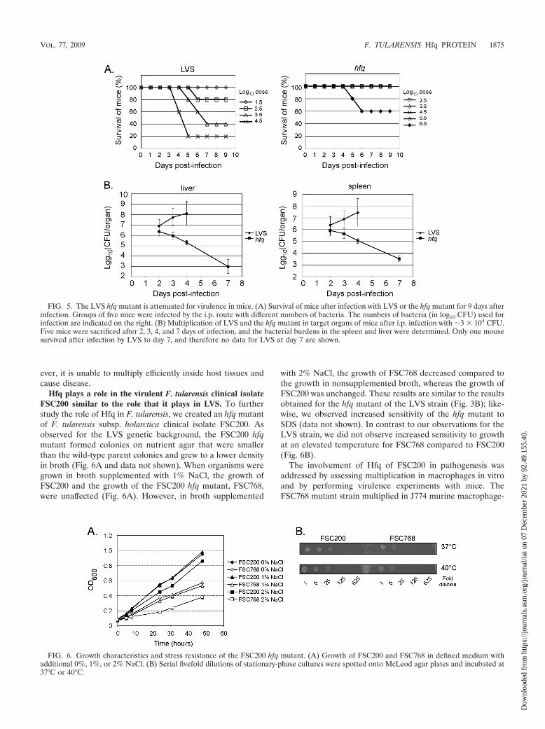
to cause disease, we infected 6- to 8-week-old BALB/c micewith strain LVS and the hfq mutant. Groups of five mice wereinoculated by the i.p. route with different numbers of bacteria,and the survival of the mice was followed for 9 days (Fig. 5A).One of the mice infected with LVS at the highest dose (�3 �104 bacteria) survived this infection, whereas all of the miceinfected with the lowest dose (�3 � 101 bacteria) survived. Incontrast, all of the mice survived infection with the lower dosesof the hfq mutant (�3 � 102 to �3 � 105 bacteria), and onlyinfection with a high dose of the hfq mutant (�3 � 106 bac-teria) resulted in death. These results demonstrate that viru-lence is attenuated in the hfq mutant.
To investigate the fate of the bacteria inside host tissues, we
infected mice with �3 � 104 LVS or hfq mutant bacteria andfollowed the kinetics of infection by assessing the number ofviable bacteria in the spleen and liver. Groups of five mice weresacrificed 2, 3, 4, and 7 days after infection, and numbers ofviable bacteria were determined by plating diluted tissue ho-mogenates (Fig. 5B). The number of LVS bacteria increasedfrom day 2 to day 4, reaching about 108 bacteria per organ, andonly one of the mice infected with LVS survived until day 7.The hfq strain was initially detected in both the spleen and liverat levels similar to the wild-type strain levels, but the numbersof bacteria declined after 2 days and all mice survived. Thesedata indicate that the hfq mutant is able to infect mice and topersist for some period of time in the infected organs. How-
FIG. 3. The hfq mutant is more sensitive to stress than the LVS parent strain. (A) Serial fivefold dilutions of stationary-phase cultures werespotted onto chocolate agar plates and incubated at 37°C or 40°C. (B) Growth of LVS and the hfq mutant in complex broth supplemented with0%, 1%, or 2% NaCl. (C) Serial 10-fold dilutions of stationary-phase cultures of LVS, the hfq mutant, and the hfq mutant containing a plasmidwith hfq were spotted onto chocolate agar plates containing 0.2% SDS, 2% NaCl, or H2O (control) and incubated at 37°C.
FIG. 4. Intracellular multiplication of LVS and the hfq mutant in murine macrophage cell lines J774 and RAW and in murine BMM.Macrophages were infected by LVS or hfq mutant bacteria at a multiplicity of infection of �100, and the number of intracellular bacteria wasdetermined after 0, 4, 24, and 48 h of infection. The results are expressed as average log10 (CFU/well) standard deviation for two experiments,each performed in triplicate.
1874 MEIBOM ET AL. INFECT. IMMUN.
Dow
nloa
ded
from
http
s://j
ourn
als.
asm
.org
/jour
nal/i
ai o
n 07
Dec
embe
r 20
21 b
y 92
.49.
155.
40.
ever, it is unable to multiply efficiently inside host tissues andcause disease.
Hfq plays a role in the virulent F. tularensis clinical isolateFSC200 similar to the role that it plays in LVS. To furtherstudy the role of Hfq in F. tularensis, we created an hfq mutantof F. tularensis subsp. holarctica clinical isolate FSC200. Asobserved for the LVS genetic background, the FSC200 hfqmutant formed colonies on nutrient agar that were smallerthan the wild-type parent colonies and grew to a lower densityin broth (Fig. 6A and data not shown). When organisms weregrown in broth supplemented with 1% NaCl, the growth ofFSC200 and the growth of the FSC200 hfq mutant, FSC768,were unaffected (Fig. 6A). However, in broth supplemented
with 2% NaCl, the growth of FSC768 decreased compared tothe growth in nonsupplemented broth, whereas the growth ofFSC200 was unchanged. These results are similar to the resultsobtained for the hfq mutant of the LVS strain (Fig. 3B); like-wise, we observed increased sensitivity of the hfq mutant toSDS (data not shown). In contrast to our observations for theLVS strain, we did not observe increased sensitivity to growthat an elevated temperature for FSC768 compared to FSC200(Fig. 6B).
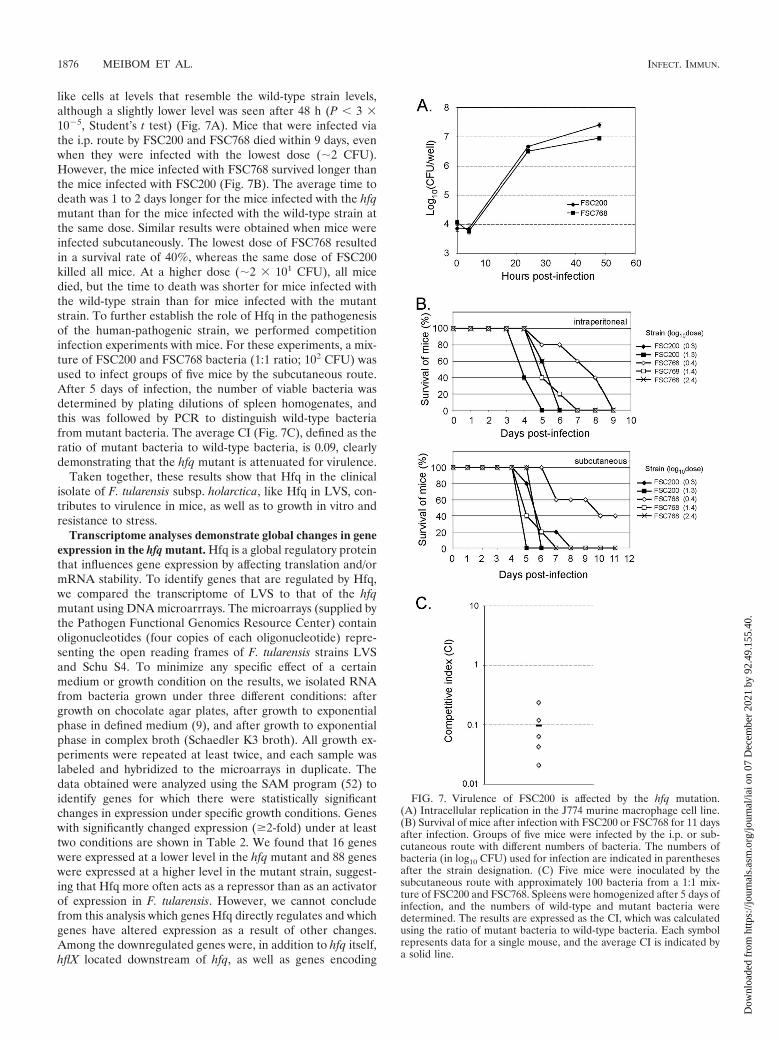
The involvement of Hfq of FSC200 in pathogenesis wasaddressed by assessing multiplication in macrophages in vitroand by performing virulence experiments with mice. TheFSC768 mutant strain multiplied in J774 murine macrophage-
FIG. 5. The LVS hfq mutant is attenuated for virulence in mice. (A) Survival of mice after infection with LVS or the hfq mutant for 9 days afterinfection. Groups of five mice were infected by the i.p. route with different numbers of bacteria. The numbers of bacteria (in log10 CFU) used forinfection are indicated on the right. (B) Multiplication of LVS and the hfq mutant in target organs of mice after i.p. infection with �3 � 104 CFU.Five mice were sacrificed after 2, 3, 4, and 7 days of infection, and the bacterial burdens in the spleen and liver were determined. Only one mousesurvived after infection by LVS to day 7, and therefore no data for LVS at day 7 are shown.
FIG. 6. Growth characteristics and stress resistance of the FSC200 hfq mutant. (A) Growth of FSC200 and FSC768 in defined medium withadditional 0%, 1%, or 2% NaCl. (B) Serial fivefold dilutions of stationary-phase cultures were spotted onto McLeod agar plates and incubated at37°C or 40°C.
VOL. 77, 2009 F. TULARENSIS Hfq PROTEIN 1875
Dow
nloa
ded
from
http
s://j
ourn
als.
asm
.org
/jour
nal/i
ai o
n 07
Dec
embe
r 20
21 b
y 92
.49.
155.
40.
like cells at levels that resemble the wild-type strain levels,although a slightly lower level was seen after 48 h (P � 3 �105, Student’s t test) (Fig. 7A). Mice that were infected viathe i.p. route by FSC200 and FSC768 died within 9 days, evenwhen they were infected with the lowest dose (�2 CFU).However, the mice infected with FSC768 survived longer thanthe mice infected with FSC200 (Fig. 7B). The average time todeath was 1 to 2 days longer for the mice infected with the hfqmutant than for the mice infected with the wild-type strain atthe same dose. Similar results were obtained when mice wereinfected subcutaneously. The lowest dose of FSC768 resultedin a survival rate of 40%, whereas the same dose of FSC200killed all mice. At a higher dose (�2 � 101 CFU), all micedied, but the time to death was shorter for mice infected withthe wild-type strain than for mice infected with the mutantstrain. To further establish the role of Hfq in the pathogenesisof the human-pathogenic strain, we performed competitioninfection experiments with mice. For these experiments, a mix-ture of FSC200 and FSC768 bacteria (1:1 ratio; 102 CFU) wasused to infect groups of five mice by the subcutaneous route.After 5 days of infection, the number of viable bacteria wasdetermined by plating dilutions of spleen homogenates, andthis was followed by PCR to distinguish wild-type bacteriafrom mutant bacteria. The average CI (Fig. 7C), defined as theratio of mutant bacteria to wild-type bacteria, is 0.09, clearlydemonstrating that the hfq mutant is attenuated for virulence.
Taken together, these results show that Hfq in the clinicalisolate of F. tularensis subsp. holarctica, like Hfq in LVS, con-tributes to virulence in mice, as well as to growth in vitro andresistance to stress.
Transcriptome analyses demonstrate global changes in geneexpression in the hfq mutant. Hfq is a global regulatory proteinthat influences gene expression by affecting translation and/ormRNA stability. To identify genes that are regulated by Hfq,we compared the transcriptome of LVS to that of the hfqmutant using DNA microarrrays. The microarrays (supplied bythe Pathogen Functional Genomics Resource Center) containoligonucleotides (four copies of each oligonucleotide) repre-senting the open reading frames of F. tularensis strains LVSand Schu S4. To minimize any specific effect of a certainmedium or growth condition on the results, we isolated RNAfrom bacteria grown under three different conditions: aftergrowth on chocolate agar plates, after growth to exponentialphase in defined medium (9), and after growth to exponentialphase in complex broth (Schaedler K3 broth). All growth ex-periments were repeated at least twice, and each sample waslabeled and hybridized to the microarrays in duplicate. Thedata obtained were analyzed using the SAM program (52) toidentify genes for which there were statistically significantchanges in expression under specific growth conditions. Geneswith significantly changed expression (�2-fold) under at leasttwo conditions are shown in Table 2. We found that 16 geneswere expressed at a lower level in the hfq mutant and 88 geneswere expressed at a higher level in the mutant strain, suggest-ing that Hfq more often acts as a repressor than as an activatorof expression in F. tularensis. However, we cannot concludefrom this analysis which genes Hfq directly regulates and whichgenes have altered expression as a result of other changes.Among the downregulated genes were, in addition to hfq itself,hflX located downstream of hfq, as well as genes encoding
FIG. 7. Virulence of FSC200 is affected by the hfq mutation.(A) Intracellular replication in the J774 murine macrophage cell line.(B) Survival of mice after infection with FSC200 or FSC768 for 11 daysafter infection. Groups of five mice were infected by the i.p. or sub-cutaneous route with different numbers of bacteria. The numbers ofbacteria (in log10 CFU) used for infection are indicated in parenthesesafter the strain designation. (C) Five mice were inoculated by thesubcutaneous route with approximately 100 bacteria from a 1:1 mix-ture of FSC200 and FSC768. Spleens were homogenized after 5 days ofinfection, and the numbers of wild-type and mutant bacteria weredetermined. The results are expressed as the CI, which was calculatedusing the ratio of mutant bacteria to wild-type bacteria. Each symbolrepresents data for a single mouse, and the average CI is indicated bya solid line.
1876 MEIBOM ET AL. INFECT. IMMUN.
Dow
nloa
ded
from
http
s://j
ourn
als.
asm
.org
/jour
nal/i
ai o
n 07
Dec
embe
r 20
21 b
y 92
.49.
155.
40.
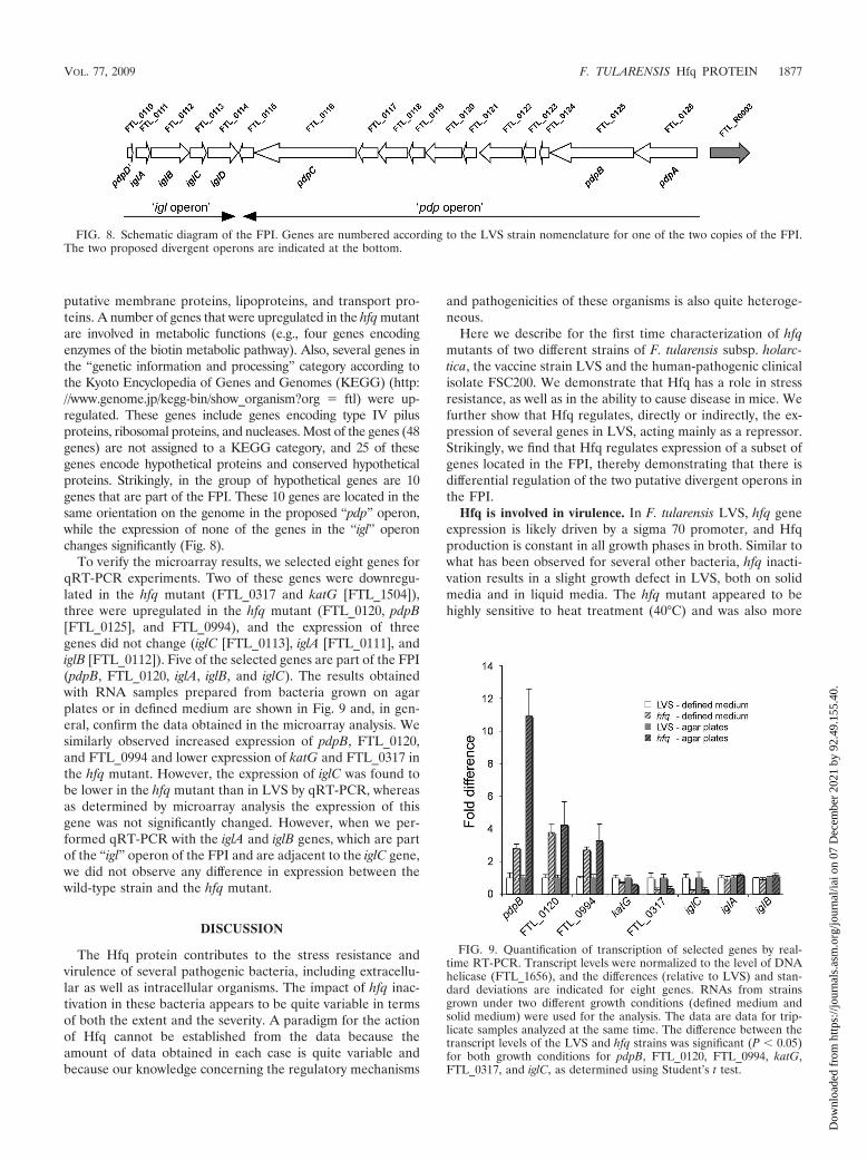
putative membrane proteins, lipoproteins, and transport pro-teins. A number of genes that were upregulated in the hfq mutantare involved in metabolic functions (e.g., four genes encodingenzymes of the biotin metabolic pathway). Also, several genes inthe “genetic information and processing” category according tothe Kyoto Encyclopedia of Genes and Genomes (KEGG) (http://www.genome.jp/kegg-bin/show_organism?org � ftl) were up-regulated. These genes include genes encoding type IV pilusproteins, ribosomal proteins, and nucleases. Most of the genes (48genes) are not assigned to a KEGG category, and 25 of thesegenes encode hypothetical proteins and conserved hypotheticalproteins. Strikingly, in the group of hypothetical genes are 10genes that are part of the FPI. These 10 genes are located in thesame orientation on the genome in the proposed “pdp” operon,while the expression of none of the genes in the “igl” operonchanges significantly (Fig. 8).
To verify the microarray results, we selected eight genes forqRT-PCR experiments. Two of these genes were downregu-lated in the hfq mutant (FTL_0317 and katG [FTL_1504]),three were upregulated in the hfq mutant (FTL_0120, pdpB[FTL_0125], and FTL_0994), and the expression of threegenes did not change (iglC [FTL_0113], iglA [FTL_0111], andiglB [FTL_0112]). Five of the selected genes are part of the FPI(pdpB, FTL_0120, iglA, iglB, and iglC). The results obtainedwith RNA samples prepared from bacteria grown on agarplates or in defined medium are shown in Fig. 9 and, in gen-eral, confirm the data obtained in the microarray analysis. Wesimilarly observed increased expression of pdpB, FTL_0120,and FTL_0994 and lower expression of katG and FTL_0317 inthe hfq mutant. However, the expression of iglC was found tobe lower in the hfq mutant than in LVS by qRT-PCR, whereasas determined by microarray analysis the expression of thisgene was not significantly changed. However, when we per-formed qRT-PCR with the iglA and iglB genes, which are partof the “igl” operon of the FPI and are adjacent to the iglC gene,we did not observe any difference in expression between thewild-type strain and the hfq mutant.
DISCUSSION
The Hfq protein contributes to the stress resistance andvirulence of several pathogenic bacteria, including extracellu-lar as well as intracellular organisms. The impact of hfq inac-tivation in these bacteria appears to be quite variable in termsof both the extent and the severity. A paradigm for the actionof Hfq cannot be established from the data because theamount of data obtained in each case is quite variable andbecause our knowledge concerning the regulatory mechanisms
and pathogenicities of these organisms is also quite heteroge-neous.
Here we describe for the first time characterization of hfqmutants of two different strains of F. tularensis subsp. holarc-tica, the vaccine strain LVS and the human-pathogenic clinicalisolate FSC200. We demonstrate that Hfq has a role in stressresistance, as well as in the ability to cause disease in mice. Wefurther show that Hfq regulates, directly or indirectly, the ex-pression of several genes in LVS, acting mainly as a repressor.Strikingly, we find that Hfq regulates expression of a subset ofgenes located in the FPI, thereby demonstrating that there isdifferential regulation of the two putative divergent operons inthe FPI.
Hfq is involved in virulence. In F. tularensis LVS, hfq geneexpression is likely driven by a sigma 70 promoter, and Hfqproduction is constant in all growth phases in broth. Similar towhat has been observed for several other bacteria, hfq inacti-vation results in a slight growth defect in LVS, both on solidmedia and in liquid media. The hfq mutant appeared to behighly sensitive to heat treatment (40°C) and was also more
FIG. 8. Schematic diagram of the FPI. Genes are numbered according to the LVS strain nomenclature for one of the two copies of the FPI.The two proposed divergent operons are indicated at the bottom.
FIG. 9. Quantification of transcription of selected genes by real-time RT-PCR. Transcript levels were normalized to the level of DNAhelicase (FTL_1656), and the differences (relative to LVS) and stan-dard deviations are indicated for eight genes. RNAs from strainsgrown under two different growth conditions (defined medium andsolid medium) were used for the analysis. The data are data for trip-licate samples analyzed at the same time. The difference between thetranscript levels of the LVS and hfq strains was significant (P � 0.05)for both growth conditions for pdpB, FTL_0120, FTL_0994, katG,FTL_0317, and iglC, as determined using Student’s t test.
VOL. 77, 2009 F. TULARENSIS Hfq PROTEIN 1877
Dow
nloa
ded
from
http
s://j
ourn
als.
asm
.org
/jour
nal/i
ai o
n 07
Dec
embe
r 20
21 b
y 92
.49.
155.
40.
sensitive than the parental LVS strain to other stresses, includ-ing SDS and a high NaCl concentration (2%). These experi-ments demonstrate that Hfq contributes to resistance to vari-ous stress conditions in F. tularensis LVS.
We examined the contribution of Hfq to Francisella intra-cellular survival by comparing the abilities of the vaccine strainLVS and the isogenic hfq mutant to multiply inside J774 andRAW macrophage cell lines and in BMM. In all three types ofmacrophages, we found that inactivation of hfq led to an in-tracellular growth defect, particularly after 24 h. However, at48 h the growth differences between the two strains were farless pronounced, suggesting that Hfq has a dominant roleduring the initial stages of intracellular multiplication.
We also evaluated the importance of Hfq in a mouse infec-tion model of tularemia. Vaccine strain LVS, which is consid-ered avirulent for humans, has significant virulence for mice,and four of five mice infected by the i.p. route with a dose of�3 � 104 bacteria succumbed to the infection. In contrast, aninfecting dose of �106 bacteria was required to cause deathwhen mice were infected with the hfq mutant by the i.p. route,demonstrating the severe attenuation of virulence of the hfqmutant with the LVS background. We also studied the infec-tion kinetics of bacterial multiplication in the liver and spleenafter i.p. infection with �3 � 104 LVS or hfq mutant bacteria.Strikingly, while the number of LVS bacteria was up to 108
bacteria per organ after 4 days (ultimately leading to death inmost cases), the number of hfq mutant bacteria started todecrease after 2 days in both the spleen and liver, and all micesurvived. These data indicate that the hfq mutant was unable tomultiply efficiently inside host tissues and cause disease.
We further evaluated the role of Hfq in F. tularensis bycreating an hfq mutant of the human-pathogenic clinical isolateFSC200. Like the mutant with the LVS genetic background,the FSC200 hfq mutant showed a slight growth defect in bothsolid and liquid media. The FSC768 mutant strain was alsomore sensitive than its isogenic parent to a high NaCl concen-tration (2%) or SDS, but it did not show increased sensitivityto an elevated temperature. In J774 macrophages, growth ofthe mutant was altered very moderately (after 48 h), reflectingthe milder impact of the hfq deletion on the intracellular sur-vival of FSC200. In mice, the hfq mutant also showed someattenuation, but the virulence defect was clearly less pro-nounced than that in the mutant with the LVS background.However, by performing a more sensitive CI analysis forspleens of animals infected with a 1:1 mixture of the wild-typeand mutant strains, we could conclusively demonstrate that thehfq mutant was attenuated for virulence and more than 10times less competitive for establishing an infection than thewild-type strain.
Strain FSC200 is extremely virulent in mice, and less thanfive bacteria (in this study the typical low doses were two tofour bacteria) are required for a lethal infection by either thei.p. or subcutaneous route. Therefore, it is difficult to comparethe levels of attenuation for this highly virulent strain and LVS.Importantly, the hfq mutant with the virulent FSC200 back-ground was also attenuated compared to parental strainFSC200, both in single-strain infections and in competitioninfection experiments with the wild-type strain. This stronglysupports the conclusion that Hfq is also important for viru-lence in human-pathogenic F. tularensis strains.
The fact that Hfq appeared to be less important for viru-lence in virulent strain FSC200 than in LVS is likely related tothe fact that the mouse is far from the ideal model for tulare-mia. We have seen similar effects in other studies where weexamined the role of a pilin gene, pilA, in virulence. In onestudy, where we used a moderately virulent strain isolated froma hare, we found that PilA mutants were severely attenuatedcompared to a strain expressing PilA (16). In contrast, whenpilA mutants of virulent strains like FSC200 and the type Astrain Schu S4 were used to infect mice, the level of attenuationwas lower (unpublished results). Thus, it appears that the ex-tremely low infection doses for these human-pathogenic strainsin mice make it more difficult to establish the role in virulenceof genes that do not have a major effect on intracellular mul-tiplication and survival. A major strength of this work is that wehave established that Hfq contributes to virulence both in thewidely studied model strain LVS and in a clinical isolate.
Hfq, a novel regulator of the FPI. Several studies, includingproteomic and microarray analyses, with various bacteria haveshown that Hfq controls the expression of numerous genes,including genes encoding virulence factors (14, 15, 26, 42, 46).Here, we compared the transcriptome of LVS to that of an hfqmutant using DNA microarrrays. Total RNA was extractedfrom bacteria grown on solid medium or in liquid culture(complex broth or chemically defined medium). Of the 104genes with significantly changed expression, only 16 were ex-pressed at a lower level in the hfq mutant (and are thus posi-tively regulated by Hfq in a wild-type context), suggesting thatHfq acts more often as a repressor than as a positive regulatorof gene expression in F. tularensis. However, we expect thatsome of these genes are not directly regulated by Hfq but theexpression is changed indirectly. The 88 genes upregulated inthe hfq mutant belong to a variety of functional categories,ranging from metabolic functions to type IV pilus biogenesis orribosomal proteins, but the majority of them (55%) have notbeen assigned to any putative function. Strikingly, within thegroup of hypothetical genes, a cluster of 10 contiguous genesthat are in the same orientation is part of the FPI. These genesbelong to the proposed “pdp” operon. Remarkably, none ofthe genes in the “igl” operon of the FPI, which are in theopposite orientation, were expressed at higher levels in the hfqmutant (Fig. 9).
Of note, the iglA-iglD operon has a G�C content of 30.6%(compared to an average G�C content of 33% for the Fran-cisella chromosome), while the pdpA-FTL_0115 region (“pdp”operon) has a G�C content of 26.6% and is the region with thelowest G�C content in the chromosome. For example, theG�C content of the pdpA gene (26.99%) is strikingly differentfrom that of the adjacent rRNA genes (52.07% for FTL_R0003, the first gene of the rrn operon in the opposite orien-tation). The 374-bp intergenic region between FTL_R0003 andpdpA (FTL_0126) comprises two distinct halves, one immedi-ately upstream of pdpA with a very low G�C content (ca. 20%)and the other with a significantly higher G�C content(�35%), suggesting that there is a putative promoter regionimmediately upstream of pdpA.
The pdpA and pdpB genes have been identified in at leasttwo different screens of transposon insertion mutant libraries.Inactivation of pdpA in F. tularensis subsp. novicida led to asevere intramacrophage growth defect, and, in vivo, the pdpA
1878 MEIBOM ET AL. INFECT. IMMUN.
Dow
nloa
ded
from
http
s://j
ourn
als.
asm
.org
/jour
nal/i
ai o
n 07
Dec
embe
r 20
21 b
y 92
.49.
155.
40.
mutant was highly attenuated (38, 54). Inactivation of pdpB inF. tularensis subsp. novicida also led to a severe in vitro growthdefect in J774 and RAW cells, as well as in primary murineBMM, and to strong attenuation of virulence in the mouse(49, 54).
Four different regulatory proteins have been shown to con-trol expression of the FPI. MglA interacts with the SspA pro-tein, forming a heterodimer that binds RNA polymerase tocontrol virulence gene expression positively (10). MglA andSspA seem to act in parallel with the FevR protein, whichcontrols the same set of genes as these proteins (6). The fourthregulatory protein, PmrA, activates virulence gene expressionas well, but it controls another set of genes in addition to theFPI (34). All four proteins affect virulence gene expressionpositively, and they all seem to regulate the entire FPI(although not every gene was found in each study, eachstudy identified several genes in both of the putative diver-gent operons).
Our study is the first study in which it was shown that ex-pression of only one part of the FPI is affected by mutation ofa regulatory protein. Furthermore, the expression of the viru-lence genes is increased in the mutant strains, implying thatHfq is a negative regulator of virulence gene expression, incontrast to the other regulators of the FPI.
Even though the virulence genes are expressed at higherlevels in the hfq mutant, it is possible that the deregulation isthe background for the virulence attenuation that we observein mice, as strict control of the FPI may be a requirement forbacterial survival in host organs. However, the virulence atten-uation could be an effect of Hfq regulation of other targetgenes that impair intracellular multiplication in vivo.
Altogether, the capacity of Hfq to affect various cellularfunctions, such as metabolism, type IV pilus production, andvirulence-related phenotypes, in F. tularensis probably reflectsthe ability of this chaperone to interact with a wide range ofdifferent regulatory RNAs, which remain to be identified. Ofnote, no homologues of sRNAs previously identified in otherbacteria could be identified by in silico analysis of F. tularensisgenomes, suggesting that the sRNAs produced by F. tularensispossess unique regulatory properties.
ACKNOWLEDGMENTS
This study was funded by INSERM, CNRS, and University ReneDescartes Paris V. Parts of this study were funded by the SwedishResearch Council (Å.F.). K.A. was supported by a fellowship from theSyrian government.
We thank Solveig Linder for excellent assistance with the animalinfection studies. We thank T. C. Zahrt, Medical College of Wisconsin,for providing plasmid pFNLTP6-gro-gfp. DNA microarrays were ob-tained through NIAID’s Pathogen Functional Resource Center, man-aged and funded by Division of Microbiology and Infectious Diseases,NIAID, NIH, DHHS, and operated by TIGR.
REFERENCES
1. Aiba, H. 2007. Mechanism of RNA silencing by Hfq-binding small RNAs.Curr. Opin. Microbiol. 10:134–139.
2. Ali Azam, T., A. Iwata, A. Nishimura, S. Ueda, and A. Ishihama. 1999.Growth phase-dependent variation in protein composition of the Escherichiacoli nucleoid. J. Bacteriol. 181:6361–6370.
3. Baron, G. S., and F. E. Nano. 1998. MglA and MglB are required for theintramacrophage growth of Francisella novicida. Mol. Microbiol. 29:247–259.
4. Bohn, C., C. Rigoulay, and P. Bouloc. 2007. No detectable effect of RNA-binding protein Hfq absence in Staphylococcus aureus. BMC Microbiol. 7:10.
5. Brennan, R. G., and T. M. Link. 2007. Hfq structure, function and ligandbinding. Curr. Opin. Microbiol. 10:125–133.
6. Brotcke, A., and D. M. Monack. 2008. Identification of fevR, a novel regu-lator of virulence gene expression in Francisella novicida. Infect. Immun.76:3473–3480.
7. Brotcke, A., D. S. Weiss, C. C. Kim, P. Chain, S. Malfatti, E. Garcia, andD. M. Monack. 2006. Identification of MglA-regulated genes reveals novelvirulence factors in Francisella tularensis. Infect. Immun. 74:6642–6655.
8. Brown, L., and T. Elliott. 1996. Efficient translation of the RpoS sigma factorin Salmonella typhimurium requires host factor I, an RNA-binding proteinencoded by the hfq gene. J. Bacteriol. 178:3763–3770.
9. Chamberlain, R. E. 1965. Evaluation of live tularemia vaccine prepared in achemically defined medium. Appl. Microbiol. 13:232–235.
10. Charity, J. C., M. M. Costante-Hamm, E. L. Balon, D. H. Boyd, E. J. Rubin,and S. L. Dove. 2007. Twin RNA polymerase-associated proteins controlvirulence gene expression in Francisella tularensis. PLoS Pathog. 3:e84.
11. Christiansen, J. K., M. H. Larsen, H. Ingmer, L. Sogaard-Andersen, andB. H. Kallipolitis. 2004. The RNA-binding protein Hfq of Listeria monocy-togenes: role in stress tolerance and virulence. J. Bacteriol. 186:3355–3362.
12. de Bruin, O. M., J. S. Ludu, and F. E. Nano. 2007. The Francisella patho-genicity island protein IglA localizes to the bacterial cytoplasm and is neededfor intracellular growth. BMC Microbiol. 7:1.
13. de Chastellier, C., and P. Berche. 1994. Fate of Listeria monocytogenes inmurine macrophages: evidence for simultaneous killing and survival of in-tracellular bacteria. Infect. Immun. 62:543–553.
14. Ding, Y., B. M. Davis, and M. K. Waldor. 2004. Hfq is essential for Vibriocholerae virulence and downregulates sigma expression. Mol. Microbiol. 53:345–354.
15. Figueroa-Bossi, N., S. Lemire, D. Maloriol, R. Balbontin, J. Casadesus, andL. Bossi. 2006. Loss of Hfq activates the �E-dependent envelope stressresponse in Salmonella enterica. Mol. Microbiol. 62:838–852.
16. Forslund, A. L., K. Kuoppa, K. Svensson, E. Salomonsson, A. Johansson, M.Bystrom, P. C. Oyston, S. L. Michell, R. W. Titball, L. Noppa, E. Frithz-Lindsten, M. Forsman, and A. Forsberg. 2006. Direct repeat-mediated de-letion of a type IV pilin gene results in major virulence attenuation ofFrancisella tularensis. Mol. Microbiol. 59:1818–1830.
17. Franze de Fernandez, M. T., L. Eoyang, and J. T. August. 1968. Factorfraction required for the synthesis of bacteriophage Q� RNA. Nature 219:588–590.
18. Geissmann, T. A., and D. Touati. 2004. Hfq, a new chaperoning role: bindingto mRNA determines access for small RNA regulator. EMBO J. 23:396–405.
19. Golovliov, I., A. Sjostedt, A. Mokrievich, and V. Pavlov. 2003. A method forallelic replacement in Francisella tularensis. FEMS Microbiol. Lett. 222:273–280.
20. Gottesman, S. 2004. The small RNA regulators of Escherichia coli: roles andmechanisms. Annu. Rev. Microbiol. 58:303–328.
21. Guisbert, E., V. A. Rhodius, N. Ahuja, E. Witkin, and C. A. Gross. 2007. Hfqmodulates the �E-mediated envelope stress response and the �32-mediatedcytoplasmic stress response in Escherichia coli J. Bacteriol. 189:1963–1973.
22. Kajitani, M., A. Kato, A. Wada, Y. Inokuchi, and A. Ishihama. 1994. Reg-ulation of the Escherichia coli hfq gene encoding the host factor for phageQ�. J. Bacteriol. 176:531–534.
23. Kawamoto, H., Y. Koide, T. Morita, and H. Aiba. 2006. Base-pairing require-ment for RNA silencing by a bacterial small RNA and acceleration of duplexformation by Hfq. Mol. Microbiol. 61:1013–1022.
24. Lai, X. H., I. Golovliov, and A. Sjostedt. 2004. Expression of IglC is necessaryfor intracellular growth and induction of apoptosis in murine macrophagesby Francisella tularensis. Microb. Pathog. 37:225–230.
25. Lauriano, C. M., J. R. Barker, S. S. Yoon, F. E. Nano, B. P. Arulanandam,D. J. Hassett, and K. E. Klose. 2004. MglA regulates transcription of viru-lence factors necessary for Francisella tularensis intraamoeba and intramac-rophage survival. Proc. Natl. Acad. Sci. USA 101:4246–4249.
26. Lenz, D. H., K. C. Mok, B. N. Lilley, R. V. Kulkarni, N. S. Wingreen, andB. L. Bassler. 2004. The small RNA chaperone Hfq and multiple smallRNAs control quorum sensing in Vibrio harveyi and Vibrio cholerae. Cell118:69–82.
27. Ludu, J. S., O. M. de Bruin, B. N. Duplantis, C. L. Schmerk, A. Y. Chou,K. L. Elkins, and F. E. Nano. 2008. The Francisella pathogenicity islandprotein PdpD is required for full virulence and associates with homologuesof the type VI secretion system. J. Bacteriol. 190:4584–4595.
28. Maier, T. M., A. Havig, M. Casey, F. E. Nano, D. W. Frank, and T. C. Zahrt.2004. Construction and characterization of a highly efficient Francisella shut-tle plasmid. Appl. Environ. Microbiol. 70:7511–7519.
29. McLendon, M. K., M. A. Apicella, and L. A. Allen. 2006. Francisella tularen-sis: taxonomy, genetics, and immunopathogenesis of a potential agent ofbiowarfare. Annu. Rev. Microbiol. 60:167–185.
30. McNealy, T. L., V. Forsbach-Birk, C. Shi, and R. Marre. 2005. The Hfqhomolog in Legionella pneumophila demonstrates regulation by LetA andRpoS and interacts with the global regulator CsrA. J. Bacteriol. 187:1527–1532.
31. Meibom, K. L., I. Dubail, M. Dupuis, M. Barel, J. Lenco, J. Stulik, I.Golovliov, A. Sjostedt, and A. Charbit. 2008. The heat-shock protein ClpB ofFrancisella tularensis is involved in stress tolerance and is required for mul-tiplication in target organs of infected mice. Mol. Microbiol. 67:1384–1401.
VOL. 77, 2009 F. TULARENSIS Hfq PROTEIN 1879
Dow
nloa
ded
from
http
s://j
ourn
als.
asm
.org
/jour
nal/i
ai o
n 07
Dec
embe
r 20
21 b
y 92
.49.
155.
40.
32. Mikulecky, P. J., M. K. Kaw, C. C. Brescia, J. C. Takach, D. D. Sledjeski, andA. L. Feig. 2004. Escherichia coli Hfq has distinct interaction surfaces forDsrA, rpoS and poly(A) RNAs. Nat. Struct. Mol. Biol. 11:1206–1214.
33. Milton, D. L., R. O’Toole, P. Horstedt, and H. Wolf-Watz. 1996. Flagellin Ais essential for the virulence of Vibrio anguillarum. J. Bacteriol. 178:1310–1319.
34. Mohapatra, N. P., S. Soni, B. L. Bell, R. Warren, R. K. Ernst, A. Muszynski,R. W. Carlson, and J. S. Gunn. 2007. Identification of an orphan responseregulator required for the virulence of Francisella spp. and transcription ofpathogenicity island genes Infect. Immun. 75:3305–3314.
35. Møller, T., T. Franch, P. Højrup, D. R. Keene, H. P. Bachinger, R. G.Brennan, and P. Valentin-Hansen. 2002. Hfq: a bacterial Sm-like proteinthat mediates RNA-RNA interaction. Mol. Cell 9:23–30.
36. Muffler, A., D. Fischer, and R. Hengge-Aronis. 1996. The RNA-bindingprotein HF-I, known as a host factor for phage Q� RNA replication, isessential for rpoS translation in Escherichia coli. Genes Dev. 10:1143–1151.
37. Nakao, H., H. Watanabe, S. Nakayama, and T. Takeda. 1995. yst geneexpression in Yersinia enterocolitica is positively regulated by a chromosomalregion that is highly homologous to Escherichia coli host factor 1 gene (hfq).Mol. Microbiol. 18:859–865.
38. Nano, F. E., N. Zhang, S. C. Cowley, K. E. Klose, K. K. Cheung, M. J.Roberts, J. S. Ludu, G. W. Letendre, A. I. Meierovics, G. Stephens, and K. L.Elkins. 2004. A Francisella tularensis pathogenicity island required for intra-macrophage growth. J. Bacteriol. 186:6430–6436.
39. Robertson, G. T., and R. M. Roop, Jr. 1999. The Brucella abortus host factorI (HF-I) protein contributes to stress resistance during stationary phase andis a major determinant of virulence in mice. Mol. Microbiol. 34:690–700.
40. Santic, M., M. Molmeret, J. R. Barker, K. E. Klose, A. Dekanic, M. Doric,and Y. A. Kwaik. 2007. A Francisella tularensis pathogenicity island proteinessential for bacterial proliferation within the host cell cytosol. Cell. Micro-biol. 9:2391–2403.
41. Sharma, A. K., and S. M. Payne. 2006. Induction of expression of hfq byDksA is essential for Shigella flexneri virulence. Mol. Microbiol. 62:469–479.
42. Sittka, A., V. Pfeiffer, K. Tedin, and J. Vogel. 2007. The RNA chaperone Hfqis essential for the virulence of Salmonella typhimurium. Mol. Microbiol.63:193–217.
43. Sjostedt, A. 2006. Intracellular survival mechanisms of Francisella tularensis,a stealth pathogen. Microbes Infect. 8:561–567.
44. Sjostedt, A., U. Eriksson, L. Berglund, and A. Tarnvik. 1997. Detection ofFrancisella tularensis in ulcers of patients with tularemia by PCR. J. Clin.Microbiol. 35:1045–1048.
45. Sonnleitner, E., S. Hagens, F. Rosenau, S. Wilhelm, A. Habel, K. E. Jager,and U. Blasi. 2003. Reduced virulence of a hfq mutant of Pseudomonasaeruginosa O1. Microb. Pathog. 35:217–228.
46. Sonnleitner, E., M. Schuster, T. Sorger-Domenigg, E. P. Greenberg, and U.Blasi. 2006. Hfq-dependent alterations of the transcriptome profile andeffects on quorum sensing in Pseudomonas aeruginosa. Mol. Microbiol. 59:1542–1558.
47. Storz, G., and D. Haas. 2007. A guide to small RNAs in microorganisms.Curr. Opin. Microbiol. 10:93–95.
48. Storz, G., J. A. Opdyke, and A. Zhang. 2004. Controlling mRNA stability andtranslation with small, noncoding RNAs. Curr. Opin. Microbiol. 7:140–144.
49. Tempel, R., X. H. Lai, L. Crosa, B. Kozlowicz, and F. Heffron. 2006. Atten-uated Francisella novicida transposon mutants protect mice against wild-typechallenge. Infect. Immun. 74:5095–5105.
50. Tsui, H. C., G. Feng, and M. E. Winkler. 1997. Negative regulation of mutSand mutH repair gene expression by the Hfq and RpoS global regulators ofEscherichia coli K-12. J. Bacteriol. 179:7476–7487.
51. Tsui, H. C., H. C. Leung, and M. E. Winkler. 1994. Characterization ofbroadly pleiotropic phenotypes caused by an hfq insertion mutation in Esch-erichia coli K-12. Mol. Microbiol. 13:35–49.
52. Tusher, V. G., R. Tibshirani, and G. Chu. 2001. Significance analysis ofmicroarrays applied to the ionizing radiation response. Proc. Natl. Acad. Sci.USA 98:5116–5121.
53. Valentin-Hansen, P., M. Eriksen, and C. Udesen. 2004. The bacterial Sm-like protein Hfq: a key player in RNA transactions. Mol. Microbiol. 51:1525–1533.
54. Weiss, D. S., A. Brotcke, T. Henry, J. J. Margolis, K. Chan, and D. M.Monack. 2007. In vivo negative selection screen identifies genes required forFrancisella virulence. Proc. Natl. Acad. Sci. USA 104:6037–6042.
55. Zhang, A., K. M. Wassarman, J. Ortega, A. C. Steven, and G. Storz. 2002.The Sm-like Hfq protein increases OxyS RNA interaction with targetmRNAs. Mol. Cell 9:11–22.