Page 1
Development and evaluation of a core training programme inhighly trained swimmers
TeesRep - Teesside'sResearch Repository
Item type Thesis or dissertation
Authors Hibbs, A. E. (Angela)
Citation Hibbs, A. E. (2011) Development and evaluation of a coretraining programme in highly trained swimmers.Unpublished PhD thesis. Teesside University.
Publisher Teesside University
Downloaded 12-Jul-2018 23:58:47
Link to item http://hdl.handle.net/10149/239473
TeesRep - Teesside University's Research Repository - https://tees.openrepository.com/tees
Page 2
TeesRep: Teesside University's Research Repository http://tees.openrepository.com/tees/
This full text version, available on TeesRep, is the final version of this PhD Thesis:
Hibbs, A. E. (2011) Development and evaluation of a core training programme in highly
trained swimmers. Unpublished PhD thesis. Teesside University.
This document was downloaded from http://tees.openrepository.com/tees/handle/10149/239473
All items in TeesRep are protected by copyright, with all rights reserved, unless otherwise indicated.
Page 3
i
DEVELOPMENT AND EVALUATION OF A
CORE TRAINING PROGRAMME IN HIGHLY
TRAINED SWIMMERS
Angela Hibbs
B.Sc. (Hons), Sport and Exercise Sciences, University College Chichester,
2002
M.Sc. (Hons), Sports Biomechanics, Loughbrough University, 2003
Thesis submitted in Fulfillment of the Requirements for the Degree of Doctor of
Philosophy
Teesside University 2011
Page 4
DEVELOPMENT AND EVALUATION OF A CORE
TRAINING PROGRAMME IN HIGHLY TRAINED
SWIMMERS
Presented by
Angela E. Hibbs, M.Sc. B.Sc.
Director of Studies
Dr. Iain Spears
Supervisor
Prof. Kevin Thompson
Supervisor
Prof. Alan Batterham
Supervisor
Dr. Duncan French
Teesside University, 2011
Page 5
Declaration
1
Declaration
I certify that the substance of this thesis has not been already submitted for any degree and is not
currently being submitted for any other degree or degrees. I certify that to the best of my
knowledge any help received in preparing this work, and all sources used, have been
acknowledged in this thesis.
————————————————
Angela E. Hibbs, M.Sc. B.Sc.
Page 6
Acknowledgements
2
Acknowledgements
I would like to thank my family and friends for their unfailing strength, support and
continued encouragement not only during the years that it has taken to write this thesis, but
for their continued support in my academic and vocational pursuits over the years.
I would like to say a huge thank you to my Director of Studies, Dr. Iain Spears for his
continued support, advice and knowledge throughout the years, without which I would not
have been able to reach the culmination of this thesis. Your thoughts and advice were
always welcome and very much appreciated.
A big thank you also to Prof. Kevin Thompson, Prof. Alan Batterham and Dr. Duncan
French for their advice and guidance which was always enlightening and taking time out of
their busy schedules to read through the drafts of chapters.
I must thank Teesside University for funding this thesis and enabling me to use their
equipment and resources for data collection and analysis. Thanks also to the English Institute
of Sport in Gateshead for use of their sporting facilities during times of data collection.
Thank You to the coaches and swimmers at the Sunderland Aquatic Swimming Club for
their enthusiastic participation in the core training intervention studies.
I have reached the end of my Ph.D. marathon. There were times when it flowed freely and
others when it felt impossible. The support and encouragement I have received from the
sidelines has pushed me forward and enabled me to reach the finish line. The feelings of
relief, satisfaction and wonderful sense of achievement will stay with me for the rest of my
life, and for that I am forever grateful to those mentioned above.
Page 7
Abstract
3
DEVELOPMENT AND EVALUATION OF A CORE
TRAINING PROGRAMME IN HIGHLY TRAINED
SWIMMERS
Presented by
Angela E. Hibbs, M.Sc. B.Sc.
Thesis submitted in Fulfillment of the Requirements for the Degree of Doctor of
Philosophy, Teesside University 2011
THESIS ABSTRACT
Core training is a popular technique for athletes and coaches concerned with improving sports
performance. Achieving an appropriate level of muscular activation is a vital ingredient in a
successful training programme. However, the evidence base with regard to the effectiveness of core
training on improving an athlete’s core ability and resultant sporting performance is limited. This
thesis aims to 1) develop a core training programme for highly trained swimmers and 2) evaluate its
effect on sporting performance using the Medical Research Council (MRC) framework for
developing complex interventions. The thesis outlines current theories and findings in both the
clinical and sporting sectors regarding core stability and core strength training and also the MRC
framework. It determines the most appropriate method of measuring muscular activation of the core
muscles (EMG) and establishes the reliability of the technique for assessing different exercises. Key
core muscles were found to produce significantly reliable (P < 0.05) measurements of below 25%
CV and > 0.7 ICC values while performing MVIC and core training exercises. Subsequently,
popular low and high threshold core training exercises were analysed and muscle activation levels of
1 - 110% MVIC were identified. A new training programme was developed and tested on a group
of highly trained swimmers over 6 and 12 week training intervention periods. Significant
improvements (P < 0.05) and a large likelihood of beneficial improvement during the performance
tests were observed following 6 and 12 weeks of training (P < 0.05) along with significant
reductions in muscle activation (%MVIC) during the performance tests and training exercises.
Conclusions from the intervention studies are used to develop a theoretical model outlining how to
structure an effective core training programme for highly trained athletes. It is proposed that this
model could be used by coaches and athletes to help plan, conduct and evaluate their core training to
maximise the potential benefits that core training could have on sporting performance.
Page 8
Table of Contents
3
Table of Contents
Declaration 1
Acknowledgements 2
Table of Contents 3
List of Tables 7
List of Figures 10
Abbreviations 11
Overview of Thesis 12
Overview of Chapters 14
Chapter 1 19
Literature Review and Theory 19
1.1 Introduction ............................................................................................................. 20 Aim of Chapter ............................................................................................................. 20
1.2 Definitions of Performance, Core Stability and Core Strength .............................. 20 1.3 Functional Anatomy of the Core............................................................................. 22
1.3.1 Functional Anatomy of the Core during Sport .............................................. 27
1.3.2 Functional Anatomy of the Core during Swimming...................................... 32 1.4 Types of Core Training ........................................................................................... 35
1.4.1 Types of Core Training in Relation to Sport ................................................. 36
1.4.2 Types of Core Training in Relation to Swimmers ......................................... 44
1.5 Techniques for Measuring Muscle Activity ........................................................... 45 1.5.1 Techniques for Measuring Muscle Activity in Relation to Swimming ......... 47 1.5.2 Techniques for Measuring Muscle Activity in Relation to Core Exercises ... 49
1.6 Physiological Adaptations to Core Training ........................................................... 54 1.7 Evidence of Core Training Benefit ......................................................................... 59
1.7.1 Evidence of Core Training Benefit in Rehabilitation Research..................... 60 1.7.2 Evidence of Core Training Benefit in Athletic Performance Research ......... 64 1.7.3 Evidence of Core Training Benefit in Swimming Research .......................... 72
1.8 Conclusions ............................................................................................................. 75
Page 9
Table of Contents
4
Chapter 2 77
Planning an Intervention in an Athletic Setting based on the Medical Research
Council Framework for Complex Interventions 77
2.1 Introduction ............................................................................................................. 78 Aim of Chapter ............................................................................................................. 81 2.2 Methodological Framework .................................................................................... 81
2.2.1 Validity of sEMG ........................................................................................... 82 2.2.2 Repeatability of sEMG .................................................................................. 84
2.2.2.1 Between-Subject Variability ..........................................................88 2.2.2.2 Within-Subject Variability .............................................................89 2.2.2.3 Between-Day Variability ...............................................................89 2.2.2.4 Within-Day Variability ..................................................................90
2.2.3 EMG Data Analysis Methods ........................................................................ 91
2.2.4 Sample Size Calculations ............................................................................... 95 2.2.5 Establishing Worthwhile Performance Enhancements .................................. 97
2.3 Structural Framework ........................................................................................... 100 2.3.1 Phase I: Development of the Intervention ................................................... 100 2.3.2 Phase II: Exploratory Trials ......................................................................... 102
2.3.3 Phase III and IV: RCT and Longitudinal Study........................................... 105
Chapter 3 107
Establishing a Repeatable Measurement of Core Musculature Activity during
MVIC and Core Exercises 107
3.1 Introduction ........................................................................................................... 108 Aim of Chapter ........................................................................................................... 112
3.2 Methods................................................................................................................. 113 3.2.1 Subjects ........................................................................................................ 113 3.2.2 Exercise Details ........................................................................................... 113
3.2.2.1 MVIC Exercises ...........................................................................113 3.2.2.2 Core Exercises .............................................................................115
3.2.3 Data Collection ............................................................................................ 117 3.2.4 Data Processing ............................................................................................ 118 3.2.5 Statistical Analysis ....................................................................................... 120
3.2.5.1 Repeatability during MVIC Exercises .........................................120 3.2.5.2 Repeatability during Core Exercises ............................................120
3.3 Results ................................................................................................................... 121 3.3.1 Repeatability during MVIC Exercises ......................................................... 121 3.3.2 Repeatability during Core Exercises ............................................................ 124
3.4 Discussion ............................................................................................................. 126 3.5 Conclusions ........................................................................................................... 136
Page 10
Table of Contents
5
Chapter 4 138
Establishing the Level of Core Musculature Activity during Core Exercises to
Determine the Content of a Core Training Programme 138
4.1 Introduction ........................................................................................................... 139 Aim of Chapter ........................................................................................................... 140 4.2 Methods................................................................................................................. 141
4.2.1 Subjects ........................................................................................................ 141 4.2.2 Exercise Details ........................................................................................... 141
4.2.2.1 MVIC Exercises ...........................................................................141 4.2.2.2 Core Exercises .............................................................................141
4.2.3 Data Collection ............................................................................................ 145 4.2.4 Data Processing ............................................................................................ 145 4.2.5 Statistical Analysis ....................................................................................... 145
4.3 Results ................................................................................................................... 145 4.4 Discussion ............................................................................................................. 150
4.5 Conclusions ........................................................................................................... 154
Chapter 5 155
Short-term Evaluation of a Core Training Programme 155
5.1 Introduction ........................................................................................................... 156
Aim of Chapter ........................................................................................................... 158 5.2 Methods................................................................................................................. 159
5.2.1 Subjects ........................................................................................................ 159
5.2.2 Exercise Details ........................................................................................... 160 5.2.2.1 MVIC Exercises ...........................................................................160
5.2.2.2 Core Exercises .............................................................................160 5.2.2.3 Performance Tests ........................................................................162
5.2.3 Data Collection ............................................................................................ 163
5.2.4 Data Processing ............................................................................................ 163 5.2.4.1 MVIC Exercises ...........................................................................163
5.2.4.2 Core Exercises .............................................................................164 5.2.4.3 Performance Tests ........................................................................164
5.2.5 Statistical Analysis ....................................................................................... 164
5.3 Results ................................................................................................................... 165 5.4 Discussion ............................................................................................................. 169 5.5 Conclusions ........................................................................................................... 174
Chapter 6 175
Long-term Evaluation of a Core Training Programme 175
6.1 Introduction ........................................................................................................... 176
Aim of Chapter ........................................................................................................... 178
6.2 Methods................................................................................................................. 178
Page 11
Table of Contents
6
6.2.1 Subjects ........................................................................................................ 178
6.2.2 Exercise Details ........................................................................................... 178 6.2.2.1 MVIC Exercises ...........................................................................179 6.2.2.2 Core Exercises .............................................................................179 6.2.2.3 Performance Tests ........................................................................180
6.2.3 Data Collection ............................................................................................ 181
6.2.4 Data Processing ............................................................................................ 181 6.2.4.1 MVIC Exercises ...........................................................................181 6.2.4.2 Core Exercises .............................................................................181 6.2.4.3 Performance Tests ........................................................................182
6.2.5 Statistical Analysis ....................................................................................... 182 6.3 Results ................................................................................................................... 183 6.4 Discussion ............................................................................................................. 193 6.5 Conclusions ........................................................................................................... 199
Chapter 7 201
Development of a Theoretical Model to Design Core Training Programmes for
Highly Trained Athletes 201
7.1 Introduction ..................................................................................................... 202
Aim of Chapter ..................................................................................................... 202
7.2 Established Theories Regarding Core Training .............................................. 203 7.2.1 Implications for the Elite Athlete ....................................................204 7.2.2 Benefits of Sub-Maximal and Maximal Training ...........................205
7.3 Theoretical Model for Core Training of Elite Athletes .................................. 207 7.3.1 Optimising Core Training Using the Model ...................................207
7.3.2 Theoretical Examples Using the Model ..........................................214
Chapter 8 220
General Conclusions 220
8.1 Overall Conclusions ........................................................................................ 221
8.2 Limitations ...................................................................................................... 221
8.3 Future Research .............................................................................................. 223
Reference List 225
Appendix A – Sports Medicine Journal Published paper 247
Appendix B – Journal of Electromyography and Kinesiology Published Paper 248
Appendix C – Core Training Programme Medical Questionnaire 249
Appendix D – Core Training Programme Participant Information Sheet 250
Appendix E – Core Training Programme Subject Informed Consent Form 251
Page 12
Table of Contents
7
Appendix F – Example Teesside University Ethics Form 252
Appendix G – Absolute sEMG muscle activations (Peak and ARV EMG) during
the MVIC and core exercises performed during the 6 week (Chapter 5) and 12
week (Chapter 6) intervention programmes 253
Page 13
List of Tables
7
List of Tables
Table 1.1. Guidelines for training the core components (based on Comerford [1]).
Table 1.2. Summary of example research studies on core training and the resultant benefits
on core stability, core strength, muscular endurance and performance.
Table 1.3. Examples of published sport specific core stability and core strength training
programmes and their effectiveness on enhancing sporting performance.
Table 1.4. Examples of published swimming specific core stability and core strength training
programmes and their effectiveness on enhancing sporting performance.
Table 2.1. Schematic representation of Medical Research Council (MRC) [2] framework for
designing complex interventions (RCT – randomised controlled trial).
Table 2.2. Summary of previous research comparing different normalisation and repeatability
methods of data analysis using surface electromyography (sEMG).
Table 3.1. Maximal Voluntary Isometric Contraction (MVIC) exercises performed during
trials.
Table 3.2. Description of core exercises performed during trials (*based on exercise
descriptions from Brandon [3]).
Table 3.3. sEMG placements on the eight core muscles analysed based on Cram [4].
Table 3.4. Within-day CV derived from a single subject during the MVIC exercises.
Between-day CV range shown in brackets. Green boxes represent values that are below the
recommended reliable level (< 26% CV).
Table 3.5. Within-subject coefficients of variation (CV) derived from multiple subjects
during the MVIC exercises. The 95% confidence intervals are shown in brackets. Values are
shown for muscles in exercises that elicited a maximum in more than three subjects. Green
boxes represent values that are below the recommended reliable level (< 26% CV).
Table 3.6. Between-day (mean) CV derived from a single subject during the core exercises.
Within-day CV range shown in brackets. Green boxes represent values that are below the
recommended reliable level (< 26% CV).
Page 14
List of Tables
8
Table 3.7. Within-subject CV derived from multiple subjects during the core exercises. The
95% confidence intervals are shown in brackets. Green boxes represent values that are below
the recommended reliable level (< 26% CV).
Table 3.8. Within-subject ICC during the core exercises. The 95% confidence intervals are
shown in brackets. Green boxes represent values that are above the recommended reliable
level (>0.7 ICC).
Table 4.1. Description of the sixteen core exercises performed. Descriptions marked * are
based on Brandon [3].
Table 4.2. Mean peak %MVIC and ARV %MVIC during five types of core exercises for
each core muscle (n = 5). Standard deviations are shown in brackets. Green boxes represent
values that are within the core stability training range (<10-25% MVIC). Blue boxes
represent values that are within the core strength training range (>60% MVIC).
Table 4.3. Peak and ARV EMG %MVIC values for the eight core muscles during sixteen
core exercises (n = 6). Standard deviations are shown in brackets. Green boxes represent
values that are within the core stability training range (<10-25% MVIC). Blue boxes
represent values that are within the core strength training range (>60% MVIC).
Table 4.4. Ranking of the eight muscles during the core exercise (1 = greatest muscle
activation during the sixteen core exercises).
Table 5.1. Core training exercise progression over the six week intervention programme.
Table 5.2. Performance tests measured pre- (0 weeks) and post-training (6 weeks) over the
six week programme.
Table 5.3. Mean and standard deviations during the performance tests pre- (0 weeks) and
post-training (6 weeks) over the six week programme. Also shown are the % performance
change and Cohen d effect size values.
Table 5.4. ANCOVA statistical results and 95% confidence intervals (CI) for the
performance tests. A comparison between core and control training groups.
Table 5.5. Paired samples t-test results for muscle activations found to be significantly
different post-training (6 weeks) compared to pre-training (0 weeks) (peak and ARV sEMG
muscular activity). Significant to p < 0.05 level.
Table 5.6. Mean sEMG muscle activation (%MVIC) from pre- (0 week) and post-training (6
weeks) of the six week training programme for each core exercise and muscle. CV data
(peak and ARV sEMG) shown in brackets.
Page 15
List of Tables
9
Table 6.1. Core training exercise progression over the 12 week training programme.
Table 6.2. Typical variation of the mean (%) (control group) and performance test change
(%) (pre-post) during the performance tests (core group).
Table 6.3. Performance test values pre- (0 weeks), mid- (6 weeks) and post- (12 weeks)
training programme for core and control group (means ± standard deviations). Performance
change (%) between pre- (0 weeks) and post-training (12 weeks) shown. Effect sizes shown
for pre-, mid- and post-training.
Table 6.4. ANCOVA findings for the performance test values comparing post-pre and mid-
pre training intervention.
Table 6.5. % MVIC muscular activation (peak and ARV EMG) during the performance tests
for the core and control training groups. A comparison of pre-, mid- and post-training
intervention periods for the six core muscles.
Table 6.6. ANCOVA results for the sEMG values (peak and ARV EMG) for the core
muscles during a performance test where they are a dominant muscle. A comparison of the
core training and control groups post-pre and mid-pre training.
Table 6.7. Percentage of MVIC muscle activation for the core muscles during the core
exercises. A comparison of pre-, mid- and post-training programme.
Table 6.8. Paired t-test results for sEMG activity of the rectus abdominis muscle during the
core exercises. A comparison of post-pre (0 – 12 weeks) and mid-pre (0 – 6 weeks) values.
Table 6.9. Paired t-test results for sEMG activity of the external oblique muscle during the
core exercises. A comparison of post-pre (0 – 12 weeks) and mid-pre (0 – 6 weeks) values.
Table 6.10. Paired t-test results for sEMG activity of the multifidus muscle during the core
exercises. A comparison of post-pre (0 – 12 weeks) and mid-pre (0 – 6 weeks) values.
Table 6.11. Paired t-test results for sEMG activity of the latissimus dorsi muscle during the
core exercises. A comparison of post-pre (0 – 12 weeks) and mid-pre (0 – 6 weeks) values.
Table 6.12. Paired t-test results for sEMG activity of the gluteus maximus muscle during the
core exercises. A comparison of post-pre (0 – 12 weeks) and mid-pre (0 – 6 weeks) values.
Table 6.13. Paired t-test results for sEMG activity of the rectus femoris muscle during the
core exercises. A comparison of post-pre (0 – 12 weeks) and mid-pre (0 – 6 weeks) values.
Page 16
List of Figures
10
List of Figures
Figure 1.1. A cross-sectional view of the stabiliser and mobiliser muscles of the core
musculature (modified from Weintraub [5]).
Figure 1.2. A schematic representation of the core musculature (modified from Comerford
[1]). The dark squares represent the spinal vertebra, circular areas represent the abdominal
muscles and diagonal lines represent the global mobiliser muscles with the red area
representing the local stabiliser muscle location.
Figure 1.3. The relationship between muscle stiffness and muscle force (modified from
Comerford [1]).
Figure 1.4. The importance of core stability when swimming to decrease drag and turbulence
(modified from Coulson [6]).
Figure 1.5. Training adaptations following low and high threshold training methods (modified
from Comerford [1]).
Figure 1.6. The muscle activity of two major contributors (latissimus dorsi and rectus
abdominus) to core stability during the freestyle swimming stroke (modified from Clarys [7]).
Figure 1.7. Core training: Principles of low- and high-load training and the subsequent effects
on core stability, core strength and resultant performance (modified from Hibbs et al.[8]).
Figure 1.8. The components and processes that contribute to performance (modified from
Mclean [9]).
Figure 3.1. The processing method used to determine the peak and ARV EMG variables. EMG
data were processed between the onset (A) and offset (B) time points.
Figure 7.1. A theoretical model to aid in the development and evaluation of a core training
programme for the elite level athlete.
Figure 7.2. A theoretical case study of a core training model for a swimmer with no previous
core training experience.
Figure 7.3. A theoretical case study of a core training model for a swimmer with previous core
strength training but no specific core stability or core endurance training experience.
Page 17
Abbreviations
11
Abbreviations
MVC – Maximal Voluntary Contraction
MVIC – Maximal Voluntary Isometric Contraction
CNS – Central Nervous System
MUAP – Motor Unit Action Potential
LBP – Lower Back Pain
ROM – Range of Motion
RA – Rectus Abdominis
EO – External Oblique
IO – Internal Oblique
MF - Multifidus
LD – Latissimus Dorsi
GM – Gluteus Maximus
GMe – Gluteus Medius
GMi – Gluteus Minimus
LG - Longissimus
RF – Rectus Femoris
TrA – Transverse Abdominal
ES – Erector Spinae
QL – Quadratus Lumborum
VM – Vastus Medialis
VL – Vastus Lateralis
EMG - Electromyography
sEMG – Surface Electromyography
pkEMG – Peak Electromyography
ARV EMG – Average Rectified Value Electromyography
CV – Coefficient of Variation
ICC – Intraclass Correlation Coefficient
Page 18
Overview of Thesis
12
Overview of Thesis
Core training is a popular technique for athletes and coaches concerned with improving
sports performance. Achieving an appropriate level of muscular activation is a vital
ingredient in any strengthening programme. However, the evidence base with regards to
the effectiveness of this type of training on improving an individual’s core ability is limited
at present. Not only is there is a lack of intervention-based studies which are able to
demonstrate the benefits of these exercises in terms of worthwhile improvements in sports
performance but of the few that do, the levels of muscular activation during the course of
the intervention are not documented.
The Medical Research Council (MRC) framework for the development and evaluation of
complex interventions for randomised control trials (RCT) was used as a theoretical guide
to designing the project. This involves a pre-clinical phase (Theorising), initial modelling
(Phase I), subsequent exploratory (Phase II) and a main RCT (Phase III) followed by a
long-term evaluation (Phase IV). The first three phases (Preclinical Phase, Phase I and
Phase II) of this framework were performed in this study. The Preclinical Phase included a
review of the literature relating to the effects of core training. In Phase I the theoretical
background and quantitative data were combined to develop the main components of the
intervention. Focus groups were conducted to collect additional qualitative data to inform
the development of the intervention. Based on the findings of Phase I, the components of
the intervention were modified in order to conduct the Phase II. The exploratory trial was
conducted in an athletic setting using a sample of 30 highly trained swimmers.
The long-term goal of this project is to provide coaches and athletes with a model for core
training which they can use to achieve the potential benefits of core training. The aims of
the thesis are:
1. To develop a methodologically sound core training programme.
2. To evaluate the effect of this core training intervention over a 12-week period on
highly trained swimmers.
Page 19
Overview of Thesis
13
In doing so the following objectives (listed by Chapter) will be addressed:
Chapter 1) To review concepts and theory with regards to what is currently considered
the most effective core training programme.
Chapter 2) To establish the structural and methodological framework needed to enable
the implementation of a core training programme in elite and sub-elite athletes.
Chapter 3) To develop a repeatable measure of core muscle activity using surface
electromyography during a range of core exercises.
Chapter 4) To quantify the core musculature activity and evaluate the muscular response
during a range of core exercises.
Chapter 5) To implement a short-term swimming specific core training programme and
evaluate performance outcomes in highly trained swimmers.
Chapter 6) To modify the training protocols implemented in the short-term core training
programme (as stated in Chapter 5) and evaluate performance outcomes in highly trained
swimmers over a longer period.
Chapter 7) To develop a theoretical model outlining how to structure an effective core
training programme for highly trained athletes.
Chapter 8) To provide general conclusions regarding the main findings from the
previous chapter and discuss general limitations and future research areas.
The chapters have been structured to enable the findings from the previous chapter to help direct
and justify the research design and implementation of the subsequent chapter. This is in
accordance with the MRC framework design and enables a solid scientific process to be
followed. Chapter 1 outlines the current theories and the different types of research conducted in
the area to date. These findings are used to establish what factors need to be considered when
collecting data in the area using these methods and establishing the importance of reliability
(Chapter 2). Subsequently, Chapter 3 establishes the reliability of the EMG methods that will be
implemented during the exploratory phase of the intervention (Chapters 4-7). The first three
chapters form the development phase of the intervention. The intervention studies implemented
in Chapters 4, 5 and 6 are subsequently justified based on the theories, findings and conclusions
from the previous three chapters. A practical model that can be used to design successful
intervention programmes is then outlined in Chapter 7 based on the findings and conclusions
from the exploratory studies. Finally, general recommendations and areas for future research can
be identified as a result of the new research that has been highlighted (Chapter 8).
Page 20
Overview of Chapters
14
Overview of Chapters
Chapter 1: Literature Review and Theory
This review provides an overview of previous and current research evaluating core stability
and core strength in both the rehabilitation and sporting sector. The Chapter outlines the
current definitions of what is included in the term ‘core stability and core strength’ and tries to
make a distinction between these terms. The Chapter summarises what little previous research
has been performed looking at the effects of core stability and core strength training on
improving sporting performance and how the different types of core training exercises activate
the core musculature and subsequently, which type of exercise may result in the greatest
performance improvement. The Chapter concludes by identifying the questions yet to be
answered regarding core stability and core strength training and whether this type of training
does have the potential to improve sporting performance.
Chapter 2: Planning an Intervention in an Athletic Setting based on the Medical
Research Council Framework for Complex Interventions
The first part of the Chapter identifies the methodological issues involved when designing a
complex health intervention and identifies those issues relevant to the design of a core training
programme in athletes. Many studies in the past have not followed a structured scientific
research design and subsequently have failed to include the necessary components to be able
to make proven and clear conclusions regarding their findings (e.g. poor subject selection, lack
of a control group, no repeatability analysis, a lack of performance indicators). The
framework for performing complex interventions as suggested by the MRC was decided upon
as the most appropriate and scientifically established method to enable this thesis to quantify
and establish theories regarding measuring and training the muscle activity of the core
musculature. This framework was selected as it has been implemented successfully in the
health sector to design complex interventions. It is argued that achieving requisite muscle
activation levels is the ‘active ingredient’ for a successful core training intervention.
Page 21
Overview of Chapters
15
Subsequently, surface electromyography (sEMG) is introduced as the most pragmatic and
valid technique to quantify this active ingredient. Consideration is then given to the known
issues regarding the use of sEMG to quantify muscle activity, and attention is focused on the
factors causing variability. The latter section focuses on the similarities and differences
between performing interventions in athletic and clinical settings.
Chapter 3: Establishing a Repeatable Measurement of Core Musculature Activity during
MVIC and Core Exercises
This Chapter establishes that surface electromyography (sEMG) has been used to quantify
muscle activity but there remains a lack of research using this method to investigate the core
musculature and core stability and subsequently quantifying the repeatability of this signal.
The Chapter introduces two common methods for reducing sEMG data, peak and average
rectified (ARV) EMG methods. The peak value has been well reported in the literature, while
the ARV value is a more recently established method of EMG data reduction and is less well
reported. The aim of the study was to establish the repeatability of peak and average rectified
EMG data during maximal voluntary isometric contractions (MVIC) and core training
exercises. Ten male highly trained athletes performed five MVIC and five core exercises on a
single day, while one female performed the same exercises but over 3 days to establish
between-day repeatability of the sEMG signal. The MVIC exercises resulted in peak EMG
CV of 3-33% and ARV EMG CV of 8-27% for the multiple subject design, and values of 6-
57% peak EMG CV and 8-51% ARV EMG for the single subject design. The core exercises
resulted in peak EMG CV of 5-28% and ARV EMG CV of 2-28% for the multiple subject
design, and values of 7-66% Peak EMG and ARV EMG CV 7-54% for the single subject
design. Within-day CV (0-65%) was observed to be more repeatability than between-day
repeatability (7-77%). It was concluded that both peak and ARV EMG methods provide a
repeatable signal for some of the analysed core muscles and MVIC and core exercises
performed.
Page 22
Overview of Chapters
16
Chapter 4: Establishing the Level of Core Musculature Activity during Core Exercises to
Determine the Content of a Core Training Programme (Phase I: Modelling)
This section describes a laboratory based study in which muscular activity is recorded by
sEMG on 11 participants. The aim of this investigation was to determine the activity levels in
selected core muscles for a range of core exercises. Five female subjects performed one
exercise within five different types of core exercise (static, dynamic low threshold, dynamic
high threshold, asymmetrical and symmetrical) and six male subjects performed sixteen core
exercises covering each of the five types of exercise. The five types of movements were found
to influence the levels of muscle activation recorded for both peak and ARV EMG with the
dynamic high threshold exercises eliciting the highest peak EMG levels, with the
asymmetrical exercises resulting in high ARV EMG levels. During the sixteen core exercises,
three muscles (RA, EO and RF) were found to be consistently activated over 60% MVIC
while the other five muscles (IO, MF, LG, GM and LD) were consistently activated between
10 – 60% MVIC. It was concluded that the core exercises and the eight muscles contributed
to core stability and core strength to varying extents during the exercises and that each type of
core exercise resulted in sufficient levels of muscle activity (to develop core stability activity
10-25%; core strength, >60%) to potentially result in core ability enhancements. Based on
the findings of this data, further conclusions could be made as to what type of exercise (i.e.
dynamic or static, asymmetrical or symmetrical, low- or high-load) and what training intensity
(i.e. duration, repetition rate) may be needed to result in training benefits on the core
musculature.
Chapter 5: Short-term Evaluation of a Core Training Programme (Phase I:
Development of an Intervention)
This Chapter outlines the implementation of a six week exploratory core training intervention
programme in the target population. This forms the second stage of Phase I within the MRC
framework [10]. The introduction section seeks to bring together the evidence including the
supportive findings acquired during the thesis. The aim of this study is to quantify the effect
of this core training intervention programme and evaluate it in terms of performance outcomes
in highly trained swimmers. Fifteen highly trained swimmers performed the core training
programme three times per week for six weeks. Performance tests were conducted pre- and
Page 23
Overview of Chapters
17
post-training to establish any training adaptations. It was observed that the performance levels
of the core training group improved significantly during the countermovement vertical jump
test. For example, pre-training jump height increased 10% from 24.7cm±4.5cm to
27.1cm±4.9cm post-training (P<0.05, effect size 1.3) and in many of the performance tests a
trend for improvement was observed. For example, 50 m swimming time was 1.4% faster with
50 m swimming time improving from 29.7s±1.54s pre-training to 29.3s±1.44s post-training,
(effect size 0.8) but at a non-significant level (P>0.05). Significant changes in the core
musculature activations levels were also observed for five of the core muscles (RA, EO, MF,
GM and RF) analysed in the training group during some of the core exercises (P<0.05). The
findings suggest that these changes to performance and muscle activations may be heightened
over a longer training period. Modifications were recommended for a longer term exploratory
trial as a potential for a positive performance effect was observed in this shorter trial.
Chapter 6: Long-term Evaluation of a Core Training Programme (Phase II: An
Exploratory Trial)
The Chapter outlines a twelve week intervention training programme and establishes whether
the longer training period results in a greater performance enhancement than that observed
following the exploratory six week intervention programme. Previous research has concluded
that as experienced athletes are highly trained to begin with, training adaptations are harder to
achieve, potentially requiring a longer intervention period (twelve weeks). From the positive
effects on performance observed in the exploratory six week trial intervention, it was proposed
that by doubling the length of intervention, the performance effects would be heightened. Ten
highly trained swimmers performed the core training programme three times per week for 12
weeks while a further ten swimmers formed a control group. Multiple performance tests were
conducted pre-, mid- and post-training intervention programme and were compared (along
with sEMG core musculature data for all subjects) to establish any training enhancements.
Three performance tests (countermovement and squat jump heights and shoulder flexion
strength) showed a significant improvement in performance following six weeks of training.
This increased to four performance tests (maximal forward bridge hold test) following 12
weeks of training (P<0.05). The remaining two performance tests also reported improved
performances but not significantly so, however these still reported a strong potential beneficial
Page 24
Overview of Chapters
18
or trivial effect on performance when magnitude based inferences were calculated instead of
statistical significance values (50m swimming time, 85.3%; sit-up bleep test, 59.2%).
Muscular activations levels were also found to be significantly altered after six weeks and to a
greater extent after 12 weeks of core training for the majority of the core muscles analysed
(P<0.05). The Chapter concludes by highlighting that core training can improve performance
and alter the muscle recruitment of the core musculature in highly trained athletes when a
specifically designed core training programme is administered in a scientific manner.
Chapter 7: Development of a Theoretical Model to Design Core Training Programmes
for Highly Trained Athletes
The Chapter outlines the main findings of the previous chapters and summarises these in a
theoretical model which may have use for the athlete and coach when looking to implement
core training into their programmes. Two case studies are provided to show how this model
could be affected by different training backgrounds of two swimmers.
Chapter 8: General Conclusions
The Chapter provides an overview of the main findings from the previous chapters and the
implications of these for the athlete and coach. The general limitations that occurred during
the data collection studies and how these were minimised or controlled are discussed. Finally
areas of future research which would provide further valuable knowledge regarding training
core stability and core strength are highlighted.
Page 25
Chapter 1 Literature Review
19
Chapter 1
Literature Review and Theory
Page 26
Chapter 1 Literature Review
20
1.1 Introduction
Core stability and core strength training in the rehabilitation and sporting sectors have
become extremely popular in recent years with many concepts and theories being
suggested to improve an individual’s core ability. It is believed that this helps to
overcome an existing injury or weakness to the core musculature (rehabilitation sector)
or enhances sporting performance by establishing efficient core stability and core
strength to maximise performance (sporting sector). This Chapter looks to discuss
these concepts and theories and highlight some of the remaining unanswered and
confusing research findings published to date.
Aim of Chapter
To review concepts and theory with regards to what is currently considered the most
effective core training protocols based on research performed in the rehabilitation and
sporting sectors.
1.2 Definitions of Performance, Core Stability and Core Strength
What is referred to as the core varies greatly from study to study, with only a few
studies including upper and lower sections of the body (i.e. the shoulders, hips and
upper leg) along with the trunk muscles [11-14]. Furthermore, many studies fail to
distinguish between core stability and core strength, two concepts which are
fundamentally very different. The confusion over the precise definition of core
stability and core strength is largely due to the fact that what is included in these
processes differs greatly depending on what context they are viewed in. For example,
in the rehabilitation sector, the focus is on rehabilitation following injuries causing
lower back, arm and leg pain. Performing exercises which emphasise the control of
spinal loading enables the general population to be able to perform everyday (low-load)
tasks. This requires less core stability and core strength than highly trained athletes in
the sporting sector who have to maintain stability during highly dynamic and in many
cases, highly loaded movements [15]. The anatomy involved during sporting tasks
includes much more of the body (i.e. shoulders and knees), which contribute in the
transfer of energy from the larger torso to the smaller extremities through the body to
produce effective sporting techniques. This results in a different definition of core
stability and core strength when referring to sporting individuals.
Page 27
Chapter 1 Literature Review
21
Panjabi [16] concluded that core stability is the functional integration of the passive
spinal column (e.g. vertebrae, ligaments and intervertebral discs), active spinal muscles
(muscles and tendons around the joints) and the CNS that work together in a manner
that allows the individual to maintain the intervertebral neutral zones while performing
activities of daily living. Brown [17] stated that this was done by the muscular system
of the trunk providing the majority of the dynamic restraint along with passive stiffness
from the vertebrae, fascia and ligaments of the spine. Kibler et al. [18] summarised
core stability in a sporting environment as the ability to control the position and motion
of the trunk over the pelvis to allow optimum production, transfer and control of force
and motion to the terminal segment in integrated athletic activities. While Akuthota
and Nadler [19] summarised core strength as the muscular control required around the
lumbar spine to maintain functional stability. Faries and Greenwood [20] provide
clearer suggestions as to the difference between core stability and core strength for the
rehabilitation sector by suggesting that core stability refers to the ability to stabilise the
spine as a result of muscle activity, with core strength referring to the ability of the
musculature to produce force through contractile forces and intra-abdominal pressure.
This is different to the traditional concept of strength in the sporting sector which has
been suggested by Lehman [11] as the maximal force that can be generated at a
specific velocity by a muscle.
Due to the different demands placed on the body during sporting activities, more
complex core exercises are trained (usually highly dynamic movements with added
resistance) compared to those used for training the general population (mostly static in
nature) [11]. As a result, the research findings performed with LBP sufferers and the
general population cannot be extended to the athletic and elite sports performer. This
inability to generalise findings together with the inconsistency of definitions of the core
makes the collection and application of meaningful data difficult. Consequently,
findings with regard to the effect of core training remain inconclusive and
contradictory. It has been suggested, however, that it is important to have sufficient
strength and stability for the body to function optimally in both everyday and sporting
environments [21] and that by having sufficient stability and strength, athletic
performance could be enhanced [22].
Page 28
Chapter 1 Literature Review
22
For the purpose of this thesis, what is referred to as the core, core stability and core
strength needs to be clearly established. The core musculature will refer to all the
musculature from the neck to the knees (including shoulder stabilisation muscles and
the upper leg muscles). Core stability will refer to the production of muscle stiffness
by the elastic components and ligamentous structures within the muscles which aids in
the ability to minimise postural sway and spinal movement during loading and force
production. Core strength refers to the increase of force generation to aid movement
brought about by creating active stiffness in the muscles and force production through
the core muscles.
1.3 Functional Anatomy of the Core
Lehman [11] identified certain muscles that are important to consider when analysing
core stability and core strength. These include the transverse abdominis (TrA), rectus
abdominis (RA), external oblique (EO), internal oblique (IO), erector spinae (ES) and
quadratus lumborum (QL) muscles (Figure 1.1). Wilson [23] also found that the
gluteus medius (GMe) and gluteus minimus (GMi) muscles play an important role in
core stability (in assisting in hip extension and external rotation) helping to properly
position and stabilise the pelvis.
Figure 1.1. Anatomy of the core musculature. A cross-sectional view of the stabiliser
and mobiliser muscles (modified from Weintraub [5]).
Page 29
Chapter 1 Literature Review
23
The contribution of these abdominal muscles to stability is related to their ability to
produce flexion, lateral flexion, rotation movements and control external forces that
cause extension, flexion and rotation to the spine [24, 25]. Comerford and Mottram
[26] emphasis the importance of the RA muscle and believe that this muscle has a high
recruitment threshold and is important in bracing the spine for high-load activities such
as pushing or lifting heavy loads. The QL and MF muscles have a lower threshold of
recruitment and mostly contribute to posture and stability [12]. The relative
contribution and precise roles of these muscles to core stability and core strength is not
clear and future research needs to be performed to establish these links [19]. For
example, McGill [12] observed that the psoas muscle (the largest muscle in the lower
lumbar spine) [27] is not involved in providing core stability, whereas Gibbons [27]
reported that this muscle does have a stability role through axial compression and
suggested that it was involved with lateral flexion, rotation and extension as well as hip
flexion.
Core stability and core strength are required primarily to protect the lumbar spine from
excessive loading and rotational movements which could lead to injury of the spine.
Akuthota and Nadler [19] broke the processes that contribute to the stabilisation of the
lumbar spine down into seven components:
1. Osseous and ligamentous structures: These structures are responsible for the passive
stiffness that is imparted onto the lumbar spine. Any injury to these structures
involving the tissue may cause functional instability of the spine. Excessive loading to
the area may cause weak muscular control, leading to the disc no longer being able to
provide optimal passive stiffness or stability [28]
2. Thoracolumbar fascia: This area provides a link between the lower and upper limb
and works as a ‘retinacular strap’ of the muscles of the lumbar spine due to their
orientation around the spine and acts as a activated proprioceptor [19]. The
thoracolumbar fascia is built up of three layers; anterior, middle and posterior layers.
The posterior layer has the most important role in supporting the lumbar spine and
abdominal musculature.
Page 30
Chapter 1 Literature Review
24
3. Paraspinals: This component consists of the lumbar extensor muscles, which
includes two major groups; the erector spinae and local muscles such as rotators and
multifidus. The erector spinae muscles (longissimus and iliocostalis) are primarily
thoracic muscles which have long moment arms that are ideal for lumbar spine
extension [29]. The local muscles act as position sensors for the spinal segment and
work as segmental stabilisers [30].
4. Quadratus Lumborum: This is a large, thin, quadrangular muscle that has direct
insertions to the lumbar spine and is a major stabiliser of the spine [12]. Akuthota and
Nadler [19] state that it consists of three major components; the internal oblique,
external oblique and longitudinal fascicles (these have received much less attention
than the transverse abdominal muscle). The external oblique muscle acts eccentrically
in lumbar extension and lumbar torsion [19]. Akuthota and Nadler [19] reported that
many fitness programmes fail to target and work the external oblique, so resulting in an
imbalance. Exercises such as isometric or eccentric trunk twists can be performed to
strengthen this muscle and aid in stability and strength.
5. Abdominals: These muscles are the most reported and investigated of the trunk
muscles and serve as a vital component of the core and to its stability [31]. The
abdominal muscle fibres run horizontally around the abdomen and consist of a number
of individual muscles (for example, the RA; this forms part of the anterior abdominal
wall and contributes to flexion of the lumbar spine). The abdominals have been shown
to be active prior to limb movement in healthy individuals [32] which implies that
these muscles are used as a preparatory stabiliser for the spine.
6. Hip girdle musculature: The hip girdle area has a significant role within the kinetic
chain in transferring force from the lower extremities to the pelvis and spine [33].
Studies using people with LBP have identified poor endurance and delayed firing of
the hip extensor (gluteus maximus) and abductor (gluteus medius) muscles, implying
that these muscles also have a role in spinal stability [34, 35].
7. Diaphragm and pelvic floor: The diaphragm and pelvic floor muscles play a role in
spinal stability. Studies have identified that inspiration and expiration during breathing
Page 31
Chapter 1 Literature Review
25
and the subsequent movement of the diaphragm has an important effect on achieving
stability of the spine [36] (as contraction of the diaphragm increases intra-abdominal
pressure which subsequently increases stability of the surrounding area which is then
imparted on to the lumbar spine).
Leetun et al [15] reported that hip muscle activation significantly influences the ability
of the body to generate force in the upper leg muscles and it has been identified that hip
muscle activation is important to achieve core stability and/or core strength [37]. The
hip muscle activation therefore leads to the knee being a victim of poor core stability,
as the upper leg muscles have a large impact on the knee when trying to generate force
from the upper leg muscles down through the knees to the floor [15]. Subsequently
when researching the contribution and function of the core during movements, it is
important to include multiple joints in the definition of the core; for example,
everything from the neck to the knees, especially during dynamic sporting movements.
Elphinston [14] investigated the gluteus maximus (GM) muscle and its contribution to
spinal stability. The GM muscle has an essential role in hip extension and also in hip
control [38]. A weak GM muscle therefore has an influence on the alignment of the
lower knee and ankle which results in greater medial and rotational movement leading
to an increase in stress and strain on the joints, predisposing to a greater injury risk
[14]. A weak GM muscle also has a resultant effect on the opposing side LD muscle to
compensate and try to maintain the tension in the fascia by alternative methods [14].
More research needs to be performed on the effect of poor core stability on the neck
and knee muscles and joints and their performance during sporting movements and
exercises [18].
One of the main core muscles to be researched in the past is the transverse abdominal
muscle (TrA) [24, 39, 40]. As a result there are many reviews published regarding the
contribution of this muscle to core stability [24]. In contrast other muscles are less well
understood. Due to this, the TrA muscle will not form a large part of the current thesis
as other important unanswered questions remain on the other core muscles and their
involvement in core stability. However due to its importance to core stability, an
understanding of this muscle is recommended. The TrA muscle arises from the iliac
crest, lower six ribs and the lateral raphe of the thoracolumbar fascia and passes
Page 32
Chapter 1 Literature Review
26
medially to the linea alba [38]. McGill [41] suggested that the TrA has limited ability
to move the trunk, but due to its horizontal fibre orientation, when it is contracted it
leads to a reduction of the abdominal circumference and is responsible for the increase
in tension in the thoracolumbar fascia and intra-abdominal pressure. Comerford and
Mottram [42] support this view by concluding that the TrA muscle is used to control
the intersegmental displacement of the lumbar vertebrae and is not involved in the
movement of the spine. Due to the muscles ability to control the abdominal contents
[43], it contributes to respiration by increasing expiratory air flow rate [44], decreasing
end expiratory lung volume [45] and defends the length of the diaphragm [46] all of
which help in controlling intra-abdominal pressure.
A number of models have been published that try to describe the core musculature and
the complex integration of the processes that work together to bring about core
stability. For example, Richardson et al. [47] described the core as a box with the
abdominals anteriorly, paraspinals and gluteals posteriorly, the diaphragm superiorly
and the pelvic floor and hip girdle musculature inferiorly. Bergmark [25] suggested a
model for the core muscles that identified these as ‘local’ and ‘global’ muscles
(depending on their role in establishing stability) and helped classify the different
contributions of the trunk muscles to spinal stability (Figure 1.2). Bergmark’s model
[25], identified ‘local’ muscles as those with attachments to the lumbar vertebrae and
hence influenced inter-segmental control (e.g. TrA) and ‘global’ muscles, as those with
attachments to the hips and pelvis and so influence spinal orientation and control the
external forces on the spine (e.g. GM). It is important that both systems (local and
global) are integrated to establish normal movement function. For example, if only the
global mobiliser muscles are trained, a muscular imbalance occurs as they ‘take over’
the local stabiliser muscles role, resulting in restricted and compensatory movement
patterns that are less efficient [48]. Stabilising muscles are responsible for posture
holding and distributing and absorbing force in the body, whereas mobilising muscles
contribute to rapid movement, force and power [25] due to their multi-joint positioning
and large moment arms. All of these processes are important to train whether in the
rehabilitation or sporting sector as they all contribute to performing movements safely
and correctly.
Page 33
Chapter 1 Literature Review
27
Similar to Bergmark’s ‘box model’ of the core, Comerford [42] suggests that the core
is best represented as a double walled cylinder consisting of the lower and upper back,
abdomen and chest (the trunk) (Figure 1.2). Comerford [42] also suggests that the
pelvic and shoulder girdles must be included in any analysis of the core musculature.
This is due to the shoulder girdle (the scapula) providing the linkage between the arm
and trunk and the pelvis as the link between the legs and the trunk.
Figure 1.2. Schematic representation of the core musculature (modified from
Comerford [1]). The dark squares represent the spinal vertebra, circular areas represent
the abdominal muscles and diagonal lines represent the global mobiliser muscles with
the red area representing the local stabiliser muscle location.
Stephenson and Swank [49] concluded that the core of the body is responsible for the
transmission of force between the upper and lower halves of the body. This is
supported by Tse et al. [50] who suggested that the core musculature includes the
muscles in the trunk and pelvis.
1.3.1 Functional Anatomy of the Core during Sport
Roetert [51] reported that core stability and balance are critical for good performance in
almost all sports and activities. This is due to the three dimensional nature of many
sporting movements which demands that athletes must have good strength in the hip
and trunk muscles to provide effective core stability. Roetert [51] suggested that some
sports require good balance, some force production, others body symmetry, but all of
these in turn require a stable core. Research suggests that a lack of core strength and
stability can manifest itself in inefficient sports techniques and predisposes that athlete
Page 34
Chapter 1 Literature Review
28
to injury [52]. LBP is a common problem in any sport that requires significant twisting
motions and repetitive flexion and extension [53-55].
An individual’s core stability and core strength are vital when an individual’s centre of
gravity is moved outside the base of support (e.g. during many sporting movements).
The individual subsequently needs to make postural adjustments to prevent a loss of
balance and to reposition the centre of gravity back within the base of support [56].
This is achieved by using muscles in the core musculature to stabilise the lumbar spine
and enable joint movement to take place [57]. The acceleration or deceleration of body
segments during sports performance is determined by the ability of the core
musculature to control the upper and lower extremities [58]. Therefore the core can be
considered as the kinetic link between the lower and upper extremities and is vital in
effective force transfer through the body [59] [18]. It does this by providing a rigid
mass which the forces can easily travel through and not get absorbed by excessive and
unnecessary movement of the lumbar spine and trunk [60] which also leads to a greater
injury risk [76]. Willardson [59] suggested that de-conditioned core muscles would not
be as effective in transferring forces through the body, resulting in greater
compensatory stress on muscles, joints and connective tissues which would in turn
increase the athletes injury risk. The effectiveness of core stability exercises for
treating and preventing lower and upper extremity injuries has been widely observed in
the rehabilitation literature [27, 232, 279]. However, much less research has been
performed in the sporting sector, with minimal research performed looking at the
effectiveness of core training programmes in enhancing healthy athletes core ability
and subsequently enhancing their sporting performance [8].
Battinelli [61] and Watson’s [62] definition of performance and the important factors
that constitute this (genetics and environmental influences) and the trainability of these
factors (muscle strength, joint mobility and the muscles capacity to do work) implies
that an individual’s core stability and core strength ability should have an effect on the
subsequent performance of the individual. However, despite this strong theoretical
link, there remains a lack of published research findings to support this proposal. One
study that highlights the importance of core training and the impact on sporting
performance was conducted by Abt et al [63]. Abt et al. [63] investigated the effect of
Page 35
Chapter 1 Literature Review
29
core stability on the mechanics of cycling. They observed that following a fatiguing
core stability session, the lower extremity mechanics (mainly the knee joint alignment),
core endurance and core strength were all reduced. Therefore, based on this study and
others [24, 104, 121], it could be suggested that a strong core stability and core strength
are required to maintain an efficient posture to enable force production and optimal
technique and that it is important to train both of these processes to optimise sporting
performance [19].
Previous studies [64, 65] have shown that an increase of only 1 - 3% of muscle tension
or up to 25% of the maximum voluntary contraction (MVC) of a muscle is required to
significantly increase the stiffness around the spine. This stiffness provides the
required stability to sufficiently overcome external perturbation in the spinal region
(Figure 1.3). As shown in Figure 1.3, only a small amount of muscle activation
initially results in a large stability response. This is consistent up to approximately
25% MVC where the stiffness of the muscle is near maximal. Therefore relatively low
maximal forces are required in a muscle to provide sufficient muscle stiffness to result
in muscle and core stability. Muscle stiffness is produced by the visco-elastic
properties of a muscle and the actin-myosin cross bridges that bring about contraction
in a muscle. Muscle stiffness is brought about by a combination of intrinsic and reflex
mediated muscle stiffness. Both types are trained by performing strength training
(intrinsic stiffness) and motor control training (reflex stiffness).
Figure 1.3. The relationship between muscle stiffness and muscle force (modified from
Comerford [1]).
Page 36
Chapter 1 Literature Review
30
Hodges [24] suggests that the CNS controls segmental stability and orientation of the
spine independently by recruiting the core musculature. This is implemented using a
feed-forward activation mechanism. The need for a feed-forward response from a
muscle occurs when the body moves a limb, the body configuration is altered and
reactive forces are placed on the body that are equal in magnitude but in the opposite
direction to that of the movement [66]. Pre-activation of the muscles by the CNS
prepares for these reactive forces on the body prior to limb movement [67]. For
example, Comerford and Mottram [48] conclude that there is an increased risk of injury
to the back if the TrA muscles are not consciously activated prior to performing
anything remotely strenuous. A lack of this feed-forward mechanism has been shown
in LBP sufferers [68].
Hodges and Richardson [69] performed a series of tests which involved the TrA and
superficial muscles in movements that were and were not planned and subjects
responded to a stimuli. The TrA response time was constant but the superficial muscles
response time varied, thus supporting the suggestions that the TrA performs a general,
stabilising role to the core, with the superficial muscles having a more precise role in
specific limb movement. Hodges and Richardson [39, 58] found that the TrA muscle
was consistently the first muscle to be activated prior to limb movement (when rapid
unilateral arm and leg movements were performed). This was supported by Hodges et
al. [67] who used a kinematic movement system to analyse body movement prior to
trunk movements being carried out. They found that prior to rapid bilateral shoulder
movements there was a small but consistent motion of the spine in the opposite
direction to the movement, therefore supporting the view that the CNS activates
muscles prior to movement to ‘dampen’ the forces (rather than being rigid). Hodges
[24] also concluded that the different influence of preparation for limb movement on
the activation of the trunk muscles suggests that the CNS deals with segmental stability
of the spine in a variety of ways. This has a significant implication on how the TrA
and the other abdominal muscles are trained. For example, Hodges [24] concluded that
the TrA muscle is controlled independently of the other trunk muscles and should be
trained separately from the other muscles at a continuous low level activation.
Page 37
Chapter 1 Literature Review
31
Hodges [24] suggested that different movements in a range of directions place varying
forces on the body and therefore results in changes in the direction of the forces acting
on the spine. This variety of forces results in different activation patterns of the trunk
muscles depending on the limb movement being performed. For example, the ES
muscle is active significantly earlier during shoulder flexion than shoulder abduction or
extension and a converse relationship is observed for the flexing abdominal muscles
[39, 58, 70]. However, it has been found that the TrA muscle is active consistently,
irrespective of the force direction [24] supporting the view that this muscle plays a vital
role in overall spinal stability, irrespective of the type of movement being performed.
Comerford’s [48] core stability model identifies local and global muscles and the
concept of stabiliser and mobiliser muscles. Stabilising muscles are responsible for
posture holding and the distributing and absorbing of force in the body[48]. In
contrast, mobilising muscles (due to their multi-joint positioning and large moment
arms) contribute to the increased movement, force and/or power of the limbs [25].
This helps to identify three categories in which the muscles can be placed depending on
their functional role [48]; local stability role (increases segmental stiffness, controls
excessive intersegmental movement and controls low-load challenges), global stability
role (provides stability across joints) and global mobility role (produces movement and
controls high-load challenges).
The different types of core stability and core strength exercises that are commonly
performed in core training programmes involve many different types of exercises, such
as; static, dynamic, symmetrical, asymmetrical, with and without external resistance
and using stable and unstable bases. These different types of exercises result in
different demands and subsequent muscle activation levels of the core musculature
[222, 232, 233], with some activating the muscles to a higher extent than others [16,
92, 220]. Which type of exercise is most effective in improving an individual’s core
stability and core strength depends on the resultant muscle activation level and which
ones are most sport-specific to sporting performance [71]. This has important
implications for subsequent training programmes, as ideally, an individual should
perform exercises that produce the same muscle activation each time and elicits the
same level of muscle activation as in training. An exercise that sometimes produces a
Page 38
Chapter 1 Literature Review
32
high activation and other times a low activation would not be as effective as one that
produces high muscle activity each time that it is performed. Therefore it is important
to establish the muscle activation repeatability of such exercises on the major core
muscles involved during these exercises, something which is yet to be established to
any extent in the published literature but something which this thesis hopes to begin to
answer.
1.3.2 Functional Anatomy of the Core during Swimming
The freestyle swimming stroke is the main swimming technique using in training
sessions [72]. It is therefore appropriate that this study focuses on this technique
(reviews of the other swimming strokes can be found in previous literature [6, 73-76]).
The freestyle swimming technique is made up of both arm (provides the main
propulsive force, ~90%) and leg (controls the body position in the water) cycles which
need to be timed to maximise the effectiveness of the swimming stroke [6].
During the freestyle swimming stroke, the legs perform a repetitive movement which
involves hip flexion and extension, knee flexion and extension and ankle plantar and
dorsi flexion [77] along with rotational movements of the shoulders and hips. These
kicking movements are brought about by the muscle activation of the prime movers
and global mobiliser muscles of the thigh (rectus femoris and gluteus) and calf
(gastrocnemius and tibialis anterior) muscles which need to be timed to result in an
optimal and effective production of power through the legs to result in an effective
swimming technique (i.e. body roll, hand pull-through and arm recovery) [78]. Local
stabiliser muscles (i.e. paraspinal muscles) are also recruited to help stiffen the core
region and protect the spine during the rotational movements [77].
Souza [79] summarised the freestyle swimming stroke into three phases; catch, pull
and recovery. Rouard et al. [80] provides a comprehensive summary of each of the
three phases during the freestyle swimming stroke and readers are recommended there
for further detail. During these three phases, Coulson [6] suggests that there are five
phases to the arm cycle during the freestyle swimming stroke; recovery, entry and
catch, out sweep, in sweep and press. Pink et al. [73] identified that global mobiliser
muscles such as; the upper trapezius, rhomboids, serratus anterior, pectoralis major,
Page 39
Chapter 1 Literature Review
33
latissimus dorsi and deltoid muscles are all involved in the arm cycle. This is
supported by Rouard et al. [80] who suggest that the flexor capri ulnaris and the
latissimus dorsi muscles are the main active muscles during the freestyle swimming
stroke. Many swimming coaches and researchers have outlined the optimal freestyle
swimming stroke technique to optimise performance [6, 81-83]. This optimal stroke
reduces drag, maximises energy transfer through the body and subsequently results in
an efficient technique to move the body through the water utilising as little energy as
possible to postpone fatigue [73]. Coulson [6] concluded that an efficient swimming
stroke will significantly reduce wasted energy output through less drag in the water and
a cleaner execution of the hand and arm entry during the recovery phases.
Fig [22] suggested that the orientation and positioning of the core muscles assist in
overcoming the demands of swimming which requires rotation between the hips and
shoulders. This is due to the core being most effective in generating power when
creating rotation between the hips and the shoulders due to the diagonal nature of the
muscles in the core, working together as a unit known as the Serape effect [13].
Santana [13] suggested that the serape effect is the result of four pairs of muscles
interacting; the rhomboids, the serratus anterior and the external and internal oblique
muscles. Fig [22] concluded that this movement occurs mostly in the freestyle and
backstroke swimming techniques and improving the ability to generate this rotation
will ultimately increase the power and speed of the swimming stroke.
Pollard and Fernandez [78] suggest that the body roll seen during the freestyle
swimming stroke (where the upper body rolls through 160 degrees) is an important part
of maintaining an efficient swimming stroke (as the roll enables the arm and hand to
pull through the water and decreases the drag through the water by reducing the cross
sectional area of the body pushing through the water) [84]. The roll of the body is a
result of the activation of the paraspinal and core muscles such as the abdominal
muscles [84]. Research has identified that one of the main differences between elite
and recreational swimmers is the lack of body roll in non-elite swimmers which is a
result of a lack of strength in the core musculature to effectively produce this roll action
[77].
Page 40
Chapter 1 Literature Review
34
The main injuries seen in swimmers are to the shoulder (i.e. rotator cuff tears and
tendonitis) and back (i.e. posterior facet irritation and spondylolisthesis) muscles [82].
Gauvin [82] stated that shoulder injuries alone were experienced by 50% of swimmers.
Shoulder stability has been shown to be essential in reducing injuries and performing
an efficient swimming technique [18]. Kibler [85] suggested that shoulder injuries are
reduced by targeting core stability first and then shoulder stabilisation. For example, it
may be that by increasing the ‘body roll’ during the swimming stroke, this would
reduce the arm abduction needed which would result in less stress on the rotator cuff
muscles of the shoulder, subsequently reducing the potential injury risk to this joint.
Furthermore, lower back muscles (such as the MF muscle) have been shown to be
trainable to improve stabilisation and strength by the use of core stability exercises
[30]. If suitable core stability and strength can be achieved by the swimmer, the forces
(as a result of the excessive twisting and rotation of the shoulders, lower back and
upper legs) will be reduced and therefore decrease the likelihood of an injury [13].
Gauvin [82] suggests that injury occurrence in swimming is declining due to the
improved understanding regarding the biomechanics of swimming, injury prevention
and treatment of swimming injuries. Gauvin [82] suggests that the increase in numbers
of swimmers performing core strength and endurance training may be a significant
factor in this recent injury reduction. It may also go some way to understanding the
continual improvement in swimming times observed in many major international
swimming championships recently [86].
An increase in core stability enables more power to be generated in rotation between
the hips and shoulders as less energy is lost in the kinetic chain between these limbs
[6]. Increased movement of the trunk increases the drag and turbulence created
reducing the efficiency and speed of the swimmer (Figure 1.4) [13]. Souza [79]
suggested that an individual’s injury risk is increased when asymmetrical body roll or
unilateral breathing is present as these result in a compensatory crossover pull-through
on the side with less roll. This has lead to body position, balance and core strength
being trained in swimmers. As a result, one of the recommended coaching techniques
for the freestyle swimming stroke emphasises an early catch, straight pull-through and
early exit of the water with the arms [77]. This results in an equal body rotation (45
Page 41
Chapter 1 Literature Review
35
degrees each side) and balance and encourages good core stability and core strength
[83]. Therefore good core strength and stability can be an important part of injury-free
swimming and subsequently lead to a more effective technique and improved
swimming performance [82].
Figure 1.4. The importance of core stability when swimming to decrease drag and
turbulence (modified from Coulson [6]).
1.4 Types of Core Training
Training core stability and core strength has been promoted for a number of supposed
benefits to the body; for example, as an injury prevention regimen, a form of
rehabilitation for lumbar and musculoskeletal injuries [60] and as a sporting
performance enhancing programme [19]. Core strengthening has become a major trend
within the rehabilitation sector [87]. Rehabilitation programmes include processes that
combine lumbar strengthening, motor control training and other regimens which aid
individuals in regaining normal body movements following trauma to the body
structures. Research has shown that a number of methods can enhance neuromuscular
control and joint stability [88-90]. These include; contraction exercises, balance
training, perturbation (proprioceptive) training, plyometric (jump) exercises
(plyometric training which emphasises the loading of joints and muscles eccentrically
before the unloading concentric activity) and sport specific skill training [11]. Many
physiotherapy programmes use exercises that challenge proprioception using
equipment such as; wobble boards, roller boards, and swiss balls [90]. Comerford [42]
suggests that core stability training includes exercises that vary from imperceptible
activation of the deep abdominal muscles to lifting weights overhead whilst balancing
Page 42
Chapter 1 Literature Review
36
on a swiss ball.
In many strength training programmes, it is common for only the global mobiliser
muscles to be trained, and subsequently a muscular imbalance occurs (due to these
muscles ‘taking over’ the stabiliser muscles role) which results in restricted and
compensatory movement patterns that are less efficient [1]. It is important that both
systems (local and global) are integrated to establish efficient and normal movement
function [25].
Increasing muscle stiffness is an important role of the local stabiliser muscle group.
Hodges [24] suggested that the contribution of the superficial stabilising trunk muscles,
such as, RA, EO, IO and the ES to trunk orientation and posture are more straight
forward than the TrA muscle. Cresswell [91] observed, during dynamic resistance
exercises (when lying on ones side) RA, EO and IO muscle activation occurring at the
end of trunk extension, which acts to decelerate the trunk. Comerford and Mottran [26]
outlined the importance of the RA muscle and suggest that this muscle is important in
bracing the spine for high-load activities such as pushing or lifting heavy loads and has
a high recruitment threshold. The oblique muscles (EO and IO) have a lower threshold
of recruitment and mostly contribute to posture and stability. Therefore, Comerford
and Mottram [26] conclude that if one wants to improve core stability, it is these
muscles (EO and IO) which need to be targeted and emphasised in training. Cresswell
[91] also observed pre-activation of these muscles prior to trunk movement, suggesting
that the CNS also controls the oblique muscles (similar to the TrA muscle) to overcome
the challenges of controlling orientation and the centre of mass changes as a result of
limb movement.
1.4.1 Types of Core Training in Relation to Sport
When looking to train the core muscles and target core stability or strength, there are
many forms of exercises that have been used to try and achieve performance benefits.
Performing these exercises is believed to result in changes to systems such as; local
and global muscle motor control and traditional strengthening of the core and limb
muscles [1]. In order to train core stability and core strength it is important to perform
both low-load and high-load threshold training [1]. This integrated training approach
Page 43
Chapter 1 Literature Review
37
is outlined in Figure 1.5. Due to the different processes and training methods of the
core musculature (e.g. low- and high-load training), Comerford [48] established further
definitions to summarise the processes involved when analysing the core musculature
and suggested when these processes should be trained using low- and high-load
training methods. For example, motor control stability could be targeted by performing
low threshold exercises while core strengthening results from high threshold (high–
load) exercises which recruit the muscles to a greater extent. This highlights the
importance of performing both low and high threshold core training to potentially
result in core stability and core strength benefits. Definitions of these training factors
are outlined below;
Motor Control Stability; low threshold stability where the CNS modulates the efficient
integration and low threshold recruitment of local and global muscle systems
Core Strength Training; high threshold or overload training of the global stabiliser
muscle system and leads to hypertrophy as an adaptation to overload training
Systematic Strength Training; traditional high threshold or overload strength training of
the global mobiliser muscle system
Figure 1.5. Training adaptations following low and high threshold training methods
(modified from Comerford [1]).
Page 44
Chapter 1 Literature Review
38
An understanding of the differences between the types of training is vital to have an
understanding of the characteristics that are important to include in an individual’s core
stability programme, for example, activation threshold level, muscle emphasis, position
and direction of muscle loading and type of muscle contraction involved [48]. Training
programmes attempting to correct weak links in an individual’s core ability include
strategies that regain control of the site and direction of the deficiency at the
appropriate threshold of training. It is proposed that the core musculature does this by;
increasing joint range and muscle extensibility, improving joint stability, enhancing
muscle performance and optimising movement function [92]
Due to the different functional roles of the muscles (local and global and stabiliser and
mobiliser roles) a range of training exercises for these muscles needs to be employed to
improve the muscles ability to function. Various low and high-load exercises should
be performed to challenge the core musculature in all directions and ranges of
movement to develop total core stability [25]. For example, a range of movements that
target the hip flexors and back extensors (i.e. the abdominal and glutei muscles) that
include flexion exercises (e.g. curl-ups, leg raising and squats with rotation), extension
exercises (e.g. targets hip extensors and hamstrings) and rotational exercises [93] of
varying intensities could be performed. Research stating whether there are any benefits
of specific core stability or core strength exercises in activating the core muscles is
limited and conflicting due to the wide variety of data collection methods, exercise
techniques and range of subjects used during studies. However it has been established
that there is not one single exercise that activates and challenges all of the core muscles
[60], therefore a combination of exercises is required to result in core stability and
strength enhancements in an individual [94, 95]. The choice of exercise is important as
the magnitude of the muscle activation (low or high-load) and the recruitment pattern
of the motor units determines whether core stability or core strength is developed.
Low-load and high-load training involves different types of movements, for example,
low-load training involves less demanding, posture related exercises which focus on
muscle recruitment, whereas high-load training can involve exercises such as overhead
weighted squats and hanging leg raises, which place a greater stress on the core
musculature and promotes core strength development [96]. As a result of training,
Page 45
Chapter 1 Literature Review
39
different physiological adaptations occur within the muscles, potentially resulting in an
improved strength or recruitment of the muscles. These adaptations are hugely
dependent on the length and type of core training programme that is being implemented
[49, 71, 97, 98].
In most elite athletes training programmes, power, strength, endurance and flexibility
are all emphasised [99]. This is based on the relationship between force, power and
stability and that by strengthening the core and limbs it is believed to benefit overall
sporting performance. However, most of these training programmes fail to include
low-load motor control training which has been identified as an essential part of core
strength training and improving core stability [1]. It is proposed that initial core
strengthening programmes should enable people to become aware of motor patterns
and allow them to learn to recruit muscles in isolation [93]. Programmes can then
progress to functional positions and activities (Table 1.1) [19]. Vezina and Hubley-
Kozey [100] suggested that core training programmes should focus on emphasising
proper sequencing of muscle activation, coactivating synergistic muscles and restoring
muscle strength and endurance to key trunk stabilisers. Akuthota and Nadler [19]
suggested that re-learning the motor control of inhibited muscles may be more
important than strengthening in patients with LBP. In this case it may be that
improvements in performance are as a result of improved neural co-ordination and
recruitment rather than specific improvements in core strength or stability. Careful
performance measures are required in studies to identify which of these is ultimately
trained following intervention programmes.
Many training programmes focus on the high intensity (high force, end-range joint and
muscle stretching), strength-biased muscle training which can lead to a contribution to
injuries [92]. It is important to incorporate low-load motor control stability training as
well [1] (see Table 1.1). By neglecting the local muscles, the force produced by the
global muscles will be too great for the local muscles to control and result in greater
injury risk [20].
Page 46
Chapter 1 Literature Review
40
Table 1.1. Guidelines for training the core components. Based on Comerford [1]).
Core Strengthening
(Trunk)
Motor Control Stability
(Global)
Motor Control Stability
(Local)
Fatiguing high load
exercises
Non fatiguing low load
exercises
Non fatiguing low load
exercises
Asymmetrical and
symmetrical limb loading
Asymmetrical limb
loading
Train in different postures
Rotation challenge Trunk not move out of
neutral (isometric)
Trunk does not move out of
neutral
Emphasis rotation control at
trunk
Emphasis rotation control
at trunk
Allow global stabiliser co-
activation
Discourage global
mobiliser dominance
Short range hold for
postural control
Discourage global
dominance
Encourage core rigidity Discourage core rigidity Discourage core rigidity
Stephenson and Swank [49] suggested that to develop a strong and stable spine, one
needs stability, flexibility and strength training of the core in all three planes of motion.
They stated some basic requirements of a core strength programme; flexibility of the
abdominal and lower back, hip extensor and flexor muscles, the need to perform
exercises in an unstable environment and that the exercises performed are isometric
and dynamic (develops tension and stabilisation of the spine). For strength training,
they stressed the importance of the principles of overloading and functionality when
training the core muscles.
Functional progression of exercises is one of the most important components of a core
strengthening programme [19]. Comerford [48] suggests that in the clinical setting, it
is usually believed that a linear framework should be followed for training
programmes; for example, stability training starts with local motor control training
(build spinal stability and strength and muscle coordination) [42] and progresses
through global motor control to core strengthening and finally to high-load traditional
muscle strengthening (dynamic movements while maintaining the good core stability)
[99]. However, Comerford [48] believes that there is no evidence to support this linear
framework and that each individual needs to have their own specific programme that
progresses for their individual needs. The exercises performed in the training
Page 47
Chapter 1 Literature Review
41
programme must progress from training isolated muscles and basic core exercises to
training as an integrated unit (dynamic multi-limb movements) to facilitate functional
activity [49]. Exercises can be progressed and made more complex by for example
starting from non-neutral positions which further challenges the core musculature [19].
However, it is essential for individuals with instability or recovering from an injury that
stretching and advanced exercises are used with caution, as this places greater stress on
the area where there is lack of current support [12].
Lehman [11] emphasises that periodisation is important in any training programme.
This periodisation concept changes the programme variables (volume, speed of
movement, exercises performed, and intensity) and the main emphasis of training over
a set period of time. This trains the different muscular characteristics (i.e. hypertrophy,
absolute strength, and power) giving an all round training effect for the athlete. The
process is based on the idea that the body continually adapts to changes in stimulus and
habituates to a constant stimulus [101]. The periodisation periods can be long (months)
or short term (weeks) which are then followed by a maintenance phase [102].
The overloading principle is a common principle used in many training programmes
and is required to bring about a performance effect [99]. However, it is important that
the individual is not overloaded too much (hence it is essential that a pre-intervention
assessment is performed before any intervention or training programme takes place).
McGill [29] suggested that this principle of overloading may predispose individuals to
injuries. For example, traditional sit ups increase the compression loads on the lumbar
spine [103] and pelvic tilts increase spinal loading. Alongside this these exercises can
be argued to be non-functional to everyday movements [29]. Therefore it is important
that any core training programme is properly established and monitored for each
individual.
Depending on what the outcome goal is for the core training programme, the emphasis
of the training programme will focus on improving one or more of the following
aspects of core ability; muscle stability, strength, endurance or power. Many
researchers have concluded that to achieve enhanced core ability, core strength is more
important than core stability [11, 71], while others have suggested that training core
Page 48
Chapter 1 Literature Review
42
endurance is the priority [95, 102, 104]. What is agreed is that there are different
processes which contribute to achieving a strong core (stability, strength, endurance
and power) and that it is important to focus training on each of these components to
prevent weaknesses developing within the core musculature [19, 105].
It may be that for elite athletes, core endurance is more important than core strength
[104]. The ability to maintain posture for example is essential in many sporting
techniques to enable an efficient performance. It may be that only lower levels of
muscle contraction are required (to maintain a body position) but for long periods of
time. Lehman [11] suggests that due to only requiring a minimal level of muscle
contraction to stabilise the spine (<25% MVC, Figure 1.5) core endurance may be
more important than core strength and subsequently identifies exercises such as; the
curl-up, birddog, side and front bridge support and the weighted squat to develop core
muscle endurance. These exercises challenge all of the anterior, lateral and posterior
trunk muscles and sufficiently stress the muscles but do not exceed the thresholds for
compression and shear loading which may predispose the body to injuries. This is
supported by McGill [12, 56] who suggests that core endurance is more important to
stability than core strength. Similarly, Faries and Greenwood [20] suggest that
endurance should be trained before strength (therefore focusing on establishing the
correct motor control systems prior to increasing the body’s strength). They also
suggest that endurance training focuses on low-load, longer (30 - 45 seconds), less
demanding exercises, while strength exercises are based on high-load, low repetition
exercises. For example, Lehman [11] encourages the use of the weighted squat as a
high-load exercise. He suggests that this is an excellent example for a core training
exercise as the entire anterior, lateral and posterior core muscles as well as the shoulder
stabilisation muscles are active thus ensuring that the spine does not buckle.
Lehman [11] outlined strength as the maximal force that a muscle can generate at a
specific velocity and suggests using resistance training to increase strength. Six or less
repetitions per sets equals approximately 80% of an athlete’s one repetition maximum
(the maximum amount of weight an individual can lift in a single repetition for a given
exercise) [61] which is the current recommendation for building strength from the
National Strength and Conditioning Association (NSCA) [106]. Lehman [11] outlined
Page 49
Chapter 1 Literature Review
43
a core training programme in which exercises are performed two times per week on
separate days where strength and power exercises are not performed with weights
being increased with observed improvements in core strength and endurance. It has
been widely observed that following a period of resistance training, power, strength
and / or endurance can be improved due to muscle adaptations resulting from the stress
placed on the muscles [107]. These adaptations include metabolic and morphological
changes [108]. Morphological changes include improvements in motor unit
recruitment, firing rate and synchronisation [109]. Metabolic changes include
alterations in the protein synthesis which account for the motor unit adaptations [107].
These changes involve an increase in the key enzymes in the mitochondrial electron
transport chain and an increase in mitochondrial protein concentration [107]. Muscle
hypertrophy is also well reported following high training stimulus [108] and results
from subcellular changes within the trained muscle (more and thicker actin and myosin
protein filaments, more myofibrils, sarcoplasm and connective tissue surrounding the
muscle fibres) [110]. It is believed that morphological adaptations occur as a result of
lower threshold training (muscle activation levels of 1-60% MVIC) with metabolic
adaptations also occurring for activations of >20% [107]. Muscle hypertrophy
adaptations result from the higher activations and high threshold training demands
which stress the muscles to a greater extent (>60% MVIC) [108].
Lehman [11] defined power as the rate of work or the product of force and velocity of
the movement. Power production is improved by heavy resistance training (resistance;
>80% one repetition maximum) and explosive exercises (weight 30 - 60% one
repetition maximum and accelerate maximally) [101, 111], for example, the power
clean or clean and jerk and the squat jump with additional weights. These exercises
increase the explosiveness of an athlete by increasing the force developed at a high
velocity. It is still important that any training performed is specific to the actual sport
the athlete performs as strength gains are specific to the velocity that the athlete trains
[71, 112]. Therefore this type of core training would only be recommended for certain
sports individuals where power is essential.
The different emphasis that core training programmes can take to target principles of
core stability and core strength have been highlighted above, all of which theoretically
Page 50
Chapter 1 Literature Review
44
could lead to improvements in an individual’s core ability. What effect, if any, the
training programme has on actual performance of the required movement depends on
how transferable the improvements in core stability or core strength are to the actual
performance movement. There are a huge amount of training programmes available on
the internet and in books that provide core training programmes for all types of
individuals from the elderly and injured to world class athletes. However many of
these have not been assessed for their effectiveness in targeting the required muscles to
the necessary activation levels to result in the optimal performance enhancement.
What is well established is that certain factors of the training programmes affect how
effective they are, for example, type of movements (i.e. static or dynamic) [12], the
speed that the exercises are performed at [58], amount of added weight resistance [57]
and the duration the training programme [101].
1.4.2 Types of Core Training in Relation to Swimmers
Scovazzo [113] suggests that muscles can be activated to 15 - 20% MVC before they
are susceptible to fatigue. Swimming research has found that many of the muscles
involved in the arm cycle during the freestyle swimming stroke are activated above this
level[80] and therefore are fatigued when swimming which puts these muscles at a
greater risk of injury. Due to this, the stabilisation and strength of the joints around the
shoulder and trunk is essential [85]. Santana [13] identified a number of exercises that
can be performed to strengthen the muscles involved in the swimming stroke, for
example traditional strength lifts such as; squats, bench press and pull-ups along with
exercises that provide loading, resistance and body rotation.
Gauvin [82] suggests that a standard strengthening programme for swimmers should
consist of isolated and combined limb movements, dynamic exercises and strength and
endurance training exercises which should be performed in sets to fatigue or ten sets of
ten repetitions to optimally train the small stabilising and endurance muscles. This is
supported by McGill [56] who suggests that spinal stability training should emphasis
endurance rather than strength. Traditional methods of core training for swimmers
include the use of stretch cords and swim benches which both stimulate the arm action
of swimming (and incorporates the diagonal kinetic chain between the shoulders and
hips which generates the effective rotational power during the swimming stroke) [22].
Page 51
Chapter 1 Literature Review
45
Juker et al. [103] found that during a twisting exercise the EO muscle was activated to
52% MVC which highlights the importance of this muscle during this type of exercise.
Other training methods include resistance machines and free weights involving multi-
joint movements. A core training programme should be included alongside the pool-
based swimming training program and should be designed to incorporate periodisation
periods to allow for neuromuscular adaptations [114]. Goldby et al. [115] suggests that
between three and six months is required to adapt the body following the identification
of a weakness in technique or following an injury.
In swimming, traditional methods and exercises that are used to train the core stability
and core strength of swimmers include those that use equipment such as, swim benches
and stretch cords [116, 117]. However neither of these specifically involves the core
musculature. Resistance machines and free weights are also used [118, 119], but these
exercises are usually only in one plane of motion and use only one joint movements, so
are not representative of the sporting movement. It has been clearly identified that it is
important to involve movements that are sport specific. For swimming, this would
involve exercises having a focus on loading the full length of the body (chest,
shoulders and back, hip and leg strength) [82].
1.5 Techniques for Measuring Muscle Activity
Electromyography (EMG) is a technique for evaluating and recording the electrical
activity produced by skeletal muscles [120]. The technique measures the electrical
potential generated by muscle cells when they are recruited and contracted. Typical
EMG potentials range from <50 µV up to 20 – 30 mV depending on the muscle being
analysed [4]. The electrical potentials are generated when motor units (motor neuron
and the muscle fibres it innervates) are activated which releases an impulse (action
potential) that travels along the motor neuron to the muscle via the neuromuscular
junction (where the nerve and muscle connect) [121]. The impulse then generates the
action potential in the muscle fibres of that motor unit (creating a motor unit action
potential, MUAP) [122]. Multiple motor units that are activated then formulate the
measured EMG signal [120]. Different methods of EMG data collection are possible
with fine-wire electrodes and surface electrodes being the most common methods
[121]. Surface EMG (sEMG) is used for recording muscular activity from superficial
Page 52
Chapter 1 Literature Review
46
muscles, whereas intramuscular (needle) or fine-wire electrodes are used for deeper
positioned muscles or localised muscle activity data analysis [121].
To perform fine-wire EMG analysis, a needle electrode containing two fine-wire
electrodes is inserted through the skin into the muscle tissue. The use of fine-wire
electrodes when performing EMG analysis does reduce the likelihood of experiencing
cross talk between muscles [122], however this method also has its limitations
especially when performing dynamic movements. Fine-wire EMG analysis has many
ethical issues, for example, the procedure is very invasive and can cause discomfort
during and after the movements have been performed. The accurate placement of the
needle electrodes also needs guidance using ultrasound to get the placement in the
muscle correct. This requires expert knowledge and experience in both ultrasound and
fine-wire EMG analysis [68]. As a result, the use of surface EMG electrodes is
commonly used instead to monitor the general activation of the muscle rather than
localised muscle fibres (as with needle electrodes).
Researches investigating the core musculature that have used EMG analysis have
predominantly used surface EMG to collect the muscle activation data [90, 97, 100,
123, 124]. Surface electromyography is a technique used to measure muscle activity
non-invasively using surface electrodes placed on the skin overlying the muscle [120].
sEMG has been used extensively in the literature to analysis a variety of muscle
characteristics during body movements, such as onset timings of muscles [67], muscle
activity amplitude [100] and effects of fatigue on muscle activity [50]. However the
limitations of sEMG have been well reported in the literature [120-123] and include the
issues of cross-talk (signal interference from other muscles) which makes it difficult to
identify the origin of the electrical signal when two or more muscles that lie in close
proximity to each other are active simultaneously [121]. However, theoretical models
developed by Fuglevand et al. [125] and Winter et al. [126] indicate that very little
cross talk occurs from muscles when performing sEMG. Both studies indicate that up
to 90% of the EMG signal is picked up within 10 – 12 mm of the surface electrodes
when electrode spacing of 20 – 25 mm is used. Therefore it can be suggested that
sEMG is appropriate for the data collection on superficial muscles [126]. sEMG is a
good representation of the whole muscles level of activation and it has been reported
Page 53
Chapter 1 Literature Review
47
that the reliability of the sEMG signal is better than analysing muscle activity using
intra-muscular electrodes [74, 127-130]. This may be due to the complex nature of
placing intra-musculature electrodes in the muscle [68, 90, 124]. Therefore if sEMG
can be used accurately to measure muscle activity during these exercises, the ethical
issues and added complexity associated with fine-wire EMG data collection can be
removed from the study. Therefore it is essential that sEMG data collection procedures
are tested to make sure they result in repeatable data.
1.5.1 Techniques for Measuring Muscle Activity in Relation to Swimming
sEMG has been used in the past to investigate the muscle activation during different
swimming strokes [7, 72-74, 113, 131]. These have focused on muscle timings,
activation and effect of injury on muscle activation. From previous research, Clarys
and Cabri [77] suggest that 44 muscles have a major involvement in the freestyle
swimming technique with all skeletal muscles (over 600 muscles) involved to some
extent [7]. The timing of the muscle contractions during the swimming stroke is
essential for an efficient stroke [77]. Ikai et al. [75] were the first to study muscle
activations during swimming and reported the activation of 15 muscles in university
and Olympic level swimmers. Ikai et al. [75] stressed the importance of the mobiliser
muscles during the freestyle swimming stroke especially the latissimus dorsi,
deltoideus and teres major muscles. Lewillie [76] concluded that the EMG activation
is determined mostly by the swimming stroke rather than the swimmer and that highly
skilled swimmers are able to reproduce a similar pattern of activation during the
swimming stroke. Nuber et al. [74] provides a detailed overview of the activation of
the arm muscles during the freestyle swimming stroke (with different muscles showing
a range of activation levels during the different phases of the stroke) highlighting the
importance of the timing of these contractions and showing that different muscles are
more dominant at different phases of the swimming cycle.
Clarys [72] investigated 25 superficial muscles involved in the freestyle swimming
technique and reported the extent of the muscle activation during the swimming stroke.
They reported that most of the muscles (20 out of 25) had two contraction peaks during
the gliding, pull and push phases, with a relaxation period during the recovery phase.
The latissimus dorsi was activated for the largest part of the swimming cycle (92%)
Page 54
Chapter 1 Literature Review
48
(also supported by Dalla Pria Bankoff and Vitti) [131] followed by the rectus
abdominis inferior (91%) and superior (83%) (Figure 1.6). The gluteus maximus
(superior) was active for 80% with the external oblique muscle active for 28% and the
rectus femoris muscle for 22%. This highlights the importance of the core muscles
during the swimming cycle as well as the arm and shoulder muscles.
Rouard et al. [80] investigated the upper extremity muscles when swimming using the
freestyle swimming technique to exhaustion. They identified maximum integrated
EMG values that averaged between 40 and 70% during the swimming stroke with
certain phases resulting in activations of up to 90% (for example, biceps brachii during
the insweep phase of the stroke). They observed that the insweep, or pull phase of the
stroke resulted in the greatest muscle activation of the arm muscles, with the later pull /
outsweep phase resulting in the highest maximal force and hand velocity. Rouard et al.
[80] also identified that the stabiliser muscles during certain phases of the stroke (such
as the triceps brancii during the insweep phase) increased in activation as fatigue
increased. They suggested this was due to the heightened demand for joint stability as
the prime movers fatigued.
Figure 1.6. The muscle activity of two major contributors (latissimus dorsi and rectus
abdominus) to core stability during the freestyle swimming stroke (modified from
Clarys [7]).
Page 55
Chapter 1 Literature Review
49
Due to the important activation of the core muscles during the freestyle swimming
stroke observed in previous research [77, 80] it can be suggested that core stability and
core strength are factors that could help improve swimming technique. By having
good core stability and strength this could enable the efficient transfer of forces through
the body to propel the body through the water and reduce the injury risk to the
swimmer by establishing an efficient muscle recruitment process [82]. The lack of
depth of research on muscle activation levels of the core musculature (especially the
core stabiliser muscles) during swimming prevents more knowledge being available
regarding the demands on these muscles during the swimming stroke and establishing
whether training these processes could improve subsequent swimming performance.
In conclusion, the freestyle swimming technique has been researched using EMG, with
the technique broken down into phases and the major muscle groups and roles clearly
defined, with the arm and leg muscles receiving the main focus. The core musculature
has received less interest, possibly due to the more complex nature of gathering this
information. As a result the exact contribution that these muscles provide during the
swimming cycle is limited along with any firm conclusions regarding what impact
improving swimmers core stability and core strength has on actual swimming
performance. The current thesis will attempt to establish some of these unanswered
questions by collecting and analysing data collected during core training exercises in
swimmers and establishing and evaluating a comprehensive core training programme
implemented over a number of weeks.
1.5.2 Techniques for Measuring Muscle Activity in Relation to Core Exercises
Axler and McGill [94] used sEMG to investigate 12 abdominal exercises and attempted
to quantify the muscle activation of selected muscles during these exercises and
establish a challenge to cost indice for each exercise in regards to the spinal loading.
They observed that the full sit-up generated the highest compressive forces on the
spine, with the hanging leg raise producing the highest abdominal muscle activation.
Axler and McGill [94] concluded that there is not one exercise that can be used to
optimally train all of the abdominal muscles and minimise spinal loading. Therefore
which exercises should be used in a training program depends on the individuals
Page 56
Chapter 1 Literature Review
50
athletic ability, for example an exercise that may be advantageous for one person may
be harmful for another if they have back problems or a weaker core stability and
strength to begin with [94].
Certain core stability and core strength exercises are more effective in activating the
chosen core muscles than others [94]. Research on this is limited and is conflicting due
to the wide variety of data collection methods, exercises techniques and subjects used
for analysis. Although it is commonly accepted that there is not one single exercise that
activates all of the core muscles [56, 94]. As such, a combination of exercises is
required to result in overall core stability and strength enhancements in an individual
[29, 94]. McGill [12] has suggested that to train the QL and the EO muscles, the ideal
exercise is the side bridge, as this minimises lumbar spine loading but still activates the
muscles to 50% of MVC. For activating and training the RA and TrA muscles, the
curl-up exercises have been found to be optimal [95]. Back extensor exercises usually
involve high spinal loading and care needs to be taken when performing these exercises
[93]. The single leg extension exercise and the birddog exercises have been found to
minimise this spinal loading while maximising back extensor muscle activation (18%
MVC and 27% - 45% MVC respectively) [12]. This previous research highlights the
varying levels of activation of the core musculature during different types of core
exercises and emphasises the importance of establishing these recruitment levels to be
able to design an optimal training intervention programme.
Urquhart et al. [124] analysed the postural activity of the TrA muscle and summarised
the effect of different body positions on the subsequent muscle activity. The TrA
muscle is made up of a number of different regions and Urquhart et al. [124] concluded
that there are regional differences within the TrA in the postural responses with limb
movement. Activity was recorded when sitting and when relaxed supine during end
range isometric hold tests. It was found that the TrA was consistently active
throughout the test, however the upper region showed an opposite activation to the
lower and middle fascicles and that the onset of EMG in the upper region was later
than that of the middle and lower regions. The response was also found to differ
depending on body position, with recruitment delayed in sitting compared to standing.
These results reflect the variation in the contribution of the abdominal muscle regions
Page 57
Chapter 1 Literature Review
51
to the stability of the trunk and highlight the indepth analysis that can be obtained when
EMG analysis is used on the core musculature.
Surface EMG data has the potential to quantify muscle activity pre- and post-training to
establish training adaptations and can highlight which components of the core are
successfully being targeted and activated to a greater extent than those components that
are not. By establishing this knowledge, training programmes can be adapted and
designed to be as effective as possible to help the sports performer reach their full
potential. This is an area that has largely gone unreported in many sports, yet could
provide invaluable information for coaches, athletes and sport scientists alike. For
example, Hamlyn et al. [132] found that there was significantly greater sEMG activity
of the lower (MF) and the upper (LG) erector spinae muscles during a 80% 1RM (one
repetition maximum lift) squat and deadlift when compared with traditional low
threshold unstable core stability exercises such as the birddog or superman [12]
exercises and supports previous research that has found lower muscle activation during
unstable exercises [133]. Hamlyn et al. [132] suggest that the greater sEMG activity
during the squat of the erector spinae muscles is due to the individual’s positional
changes to handle the compressive forces on the spine and overcome the destabilising
torques of the swaying body and suspended resistance overhead. This is supported by
research that has found lower erector spinae muscles (MF) to be highly active as a
stabiliser during the squat movement [134] and research by Hamlyn et al. [132] who
suggest that the upper erector spinae muscles (LG) are involved primarily with
providing the stiffness to the spine to help generate forces which control the range of
motion [135].
The benefits of using both fine-wire and surface electrodes to measure the core
muscular activation during complex exercises such as those performed during core
training programmes have been outlined in this Chapter. It is clear that the use of fine-
wire electrodes when performing EMG analysis during highly dynamic and
challenging body movements does have ethical issues to consider. Therefore surface
electrodes have generally been preferred in the past [120]. Ainscough-Potts et al. [40]
stated that fine-wire EMG is an invasive procedure and that there has been a
development towards using other techniques to establish muscle recruitment levels.
Page 58
Chapter 1 Literature Review
52
Real time ultrasound scanning techniques have become more popular in recent years
for measurements of abdominal muscle activity [136]. Ultrasound imaging has been
used to analyse muscle recruitment as changes in the muscles thickness is believed to
be related to muscle recruitment [137].
Ultrasound technology and imaging has been used since the 1980’s for rehabilitation
proposes [138]. Studies have shown that it is a safe, cost-effective and accessible
method for visualising and measuring the deep muscles of the trunk [138-140]. Using
this type of measurement enables for real-time images of muscles to be observed.
Clinical studies [136, 140] have shown that ultrasound measurements and technology
provides a method to obtain both valid and reliable data of muscles sizes and can be an
indicator of muscle activity (using static quantitative measurements of muscle width,
length, depth, cross-sectional area or volume) [138].
Hodges et al. [141] investigated the ability to measure muscle activity using ultrasound
methods. They measured the architectural parameters (pennation angles, fascile
lengths and muscle thickness) of several muscles including the tibialis anterior, bicep
brachi, brachialis, transverse abdominals, internal oblique and external oblique
abdominal muscles. Isometric contractions from 0 - 100% MVC were performed and
EMG surface and fine-wire electrodes were used. The authors found that the
architectural parameters changed markedly with contractions up to 30% but there was
little changed after this. Hodges et al. [141] therefore concluded that ultrasound
imaging can only be used to detect low levels of muscle activity and cannot
discriminate between moderate and strong contractions.
Hodges et al. [141] did report that the ultrasound measures did reliably detect changes
in EMG of as little as 4% (biceps thickness), 5% (brachialis) and 9% (tibialis) MVC.
Generally they found that it was less sensitive to changes in abdominal muscle activity,
but that it was possible to detect contractions of 12% MVC in the TrA and 22% MVC
in the IO muscle (this maybe due to the deep positioning of these muscles in the body).
Ainscough-Potts et al. [40] also used ultrasound imaging to analysis the core muscles
of the body and they provide a detailed methodology for their ultrasound data
collection and highlighted the wide range of muscle thickness of the transverse
abdominal muscle in the normal population. This difference may be slightly reduced in
Page 59
Chapter 1 Literature Review
53
the athletic population but will still vary between sports and this may have a significant
effect on how data should be collected on this muscle when comparing data between
subjects. Ultrasound measurements also do not take into account a number of factors,
for example, the amount of change in the abdominal wall thickness during a
contraction does not necessarily represent the intensity or amount of actual muscle
activity [141]. This may be due to the impact of other surrounding structures around
the muscle, for example, protrusion of the abdominal contents due to breathing [141].
The two-dimensional nature of the analysis also poses some issues, for example, when
muscles contract they alter their architecture in three dimensions not two, therefore the
change in muscle size may not reflect the absolute change [138]. Due to the above
issues, it is essential that researchers using this method have a detailed knowledge of
both the anatomy of the abdominal region and ultrasound technology prior to any data
analysis. Due to the complexity of the technology, it takes time to develop the skills
and knowledge required to achieve the collection of reliable and valid data and
subsequently be able to accurately interpret the measurements when using ultrasound
technology.
Despite the limitations highlighted above, findings from many studies support the use
of the non-invasive technique to measure abdominal muscle thickness and estimate
relative muscle activity and most studies performed using ultrasound imaging show
that it is a reliable and valid method of assessing core muscle activity [137]. However
expert training and a significant amount of experience using the equipment is required
to enable valid and reliable data to be collected, a skill that not many researchers have.
Therefore methods such as sEMG have been used much more frequently in the past to
collect the same data with the same reliability and accuracy [137], as a result this
method is going to be the main data collection method used in the current thesis.
Ultrasound technology has been used as a non-invasive method of measuring
abdominal muscle activity [137, 138]. However since this is a relatively new method
of analysis, it is not known whether it can provide a valid measure of changes in motor
control of these muscles. Whittaker [138] states that with ultrasound analysis, there is
still debate on issues such as scope of practice, its specific role in the rehabilitation
process and its limitations. Some studies have tried to investigate the reliability of
Page 60
Chapter 1 Literature Review
54
using ultrasound measurements, for example, Ferreira et al. [137] observed similar
findings when ultrasound results were compared with those from EMG analysis. They
[137] used ultrasound and EMG methods and compared these findings within ten
healthy subjects and ten LBP sufferers. The TrA, EO and IO muscles were analysed
and it was concluded that the participants with LBP had significantly smaller increase
in thickness in the TrA muscle with isometric leg exercises with similar conclusions
resulting from the EMG data analysis.
Ainscough-Potts et al. [40] used ultrasound analysis to investigate the response of the
TrA and IO muscles to different postures. Thirty subjects performed basic exercises
when sitting in a chair, on a gym ball and when one leg was raised off the floor. The
subjects (when asked to raise one foot off the floor) had a significant increase in
thickness for both the TrA and the IO. This demonstrates that these muscles are
automatically targeted by the body to maintain stability when the base of support is
decreased significantly and implies that there is a general trend for the muscles to
increase in thickness and activity as stability of the body decreases. Therefore, as it was
proposed by Richardson et al. [47], to increase the activity of the IO and TrA muscles,
one method of doing this is to decrease the stability of the base of support during
specific core exercises.
Akuthota and Nadler [19] suggest that an understanding of the precise role of the
individual muscles contributions to core stability and core strength is limited and future
research needs to be performed to establish these mechanisms more clearly. Akuthota
and Nadler [19] suggest that improving core stability and core strength is a way of
preventing injuries and a way to enhance athletic performance. However more
research is needed to formally identify these links and establish how the core muscles
are trained to bring about a performance enhancement.
1.6 Physiological Adaptations to Core Training
Physiologically, core strength and stability training leads to a greater maximal power
and more effective use of the muscles of the shoulders, arms and legs [11]. This
theoretically results in a better body balance and a lower risk of injury, leading to
additional effects on performance, such as speed, agility, power and aerobic endurance
Page 61
Chapter 1 Literature Review
55
[50]. Neural adaptations from core training include; more efficient neural recruitment
patterns, faster nervous system activation, improved synchronisation of motor units and
a lowering of neural inhibitory reflexes [142]. It is believed that high-load training
alters the muscle structure, whereas low-load training improves the CNS’s ability to
control muscle co-ordination and therefore the effectiveness of the movement [48].
Subsequently by training with low- and high-load exercises (within a well-structured
and functional training programme) improvements should be attained in all the
contributing processes to core stability and core strength [49] which (it is reasoned)
will in turn benefit sporting performance.
High threshold and strength training is believed to result in hypertrophy of the muscles
(structural change) and neural adaptations (of the motor units in the muscles) [132, 143,
144]. This then benefits performance by increasing the possible force generation, CNS
facilitation, improved intrinsic muscle stiffness and tissue mobilisation [142].
However it is essential that the local muscles are also targeted by the training and that
low-load threshold training is performed to bring about local muscle benefits and not
lead to an imbalance in muscle recruitment (as this may subsequently lead to a
movement dysfunction and potential injuries) [42]. Spinal instability and injuries to
muscles (e.g. the core) and joints (e.g. knee and hips) sustained during movements are
associated with insufficient strength and endurance of the trunk stabilising muscles and
inappropriate recruitment of the trunk and abdominal muscles [100]. It is important
that any core stability weakness is identified and corrected as this significantly
increases an individual’s muscle and joint injury risk [145].
Hodges and Richardson [146] performed a number of movements at different speeds.
They suggested that if the limb movement speed or acceleration is slow, the resultant
forces on the body are smaller. They measured feed-forward activation (pre-movement
muscle activation) of the TrA during rapid movements and slower speeds and found no
TrA feed-forward activation during the slower speeds. Cresswell and Thorstensson
[147] found that the TrA activity was greatest with the fastest movement speeds when
subjects performed a lifting task at different velocities. Cresswell [91] observed bursts
of TrA activity when there were periods of high acceleration and deceleration of the
trunk during flexion and extension tasks. These results suggest that the TrA maximal
Page 62
Chapter 1 Literature Review
56
activity is closely related to periods of maximal stress and so supports the theory that
the TrA plays an essential role in stabilising the spine [24].
High-load or fast activities recruit the fast motor units in the muscles when performing
a movement optimally and these activities utilise the larger global multi-joint muscles
that provide a mobility role [42]. Slow motor units of the muscle are utilised during
low threshold recruitment in postural sway and movements involved with unloaded
limbs [42]. It is therefore important for optimum motor control to train both the fast
and slow motor units in a muscle to optimise core stability and core strength.
Subsequently the rate at which an exercise is performed has an influence on the muscle
activation recorded [94]. Higher muscle forces will be seen if an exercise is performed
at a faster rate as higher accelerations are required of the limbs. Similar suggestions
can be made for exercises that have large ranges of motion and those that have added
muscular load by using resistance bands or weights. Therefore care needs to be taken
when performing these exercises to allow for these variations and subsequently the rate
that the exercise is performed at needs to be controlled.
The degree of movement has been observed to have an effect on muscle activation as
well as speed of the movement [24]. For example, the feed-forward response was
identified when movements of the elbow and shoulder were performed but not when
only the wrist and thumb were moved [24]. EMG research has identified that when the
arm was moved, onset of TrA precedes the deltoid by 30 ms [39] and when the leg is
moved, activation of the TrA precedes the deltoid by more than 100 ms [58]. Hodges
[24] concluded that this earlier activation is due to the greater forces on the spine being
present when the leg is moved due to its greater mass. Previous studies have suggested
that limb movement is delayed in tasks where the postural demand is increased [148,
149] due to the extra time needed to prepare the body for the larger resultant forces.
The pattern of muscle activation during limb movements has been investigated using
fine-wire EMG analysis, for example, when rapid shoulder flexion is performed,
Hodges et al. [67] found the TrA showed a greater magnitude of activity at the onset of
movement followed by continuous activation at a lower level during the movement.
Cresswell [91] found that the abdominal muscles were only active during acceleration
Page 63
Chapter 1 Literature Review
57
(when they generated the movement) and deceleration (when they opposed the
movement). Therefore their muscle activation is directional dependent and is involved
in the global mobilisation processes during such movements. Research on the
optimum speed and order of loading on the muscles is limited, therefore it remains
unclear what speed and direction of movement should be used to train the muscles
optimally [19, 99]. The only clear conclusion that can be made is that any training
should be functional and sport-specific for the individuals needs [11, 15]. Whether
these targeted movements are to be low- or high-load will have a significant effect on
the type of training programme implemented.
The apparent contradiction between the traditional dynamic approach of the strength
and conditioning coach compared to the more modest movements prescribed by
physiotherapists typically has led to confusion as to which core training method is most
effective. Future research should focus on establishing which exercises are sufficient
for improving each part of core stability (i.e. neural, passive and active systems) and
core strength (e.g. neural adaptations) to be able to target these performance goals more
effectively and maximise the potential for the skills and training benefits to be
transferred into performance [71, 112, 150]. An overview of the principles of core
training and the potential training adaptations and benefits that could result are outlined
in Figure 1.7.
Page 64
Chapter 1 Literature Review
58
Figure 1.7. Core training: Principles of low- and high-load training and the subsequent
effects on core stability, core strength and resultant performance (modified from Hibbs
et al. [8]).
As has been suggested, the majority of published research into core stability and core
strength fails to measure what the effect of a training programme is on actual
performance, whether it be performing everyday tasks or a sporting activity at a world
class level [11, 26, 50, 151-155]. Some of those studies that have reported the
effectiveness on improving subsequent performance have failed to show any
performance enhancement following core training programmes [50, 97]. This could be
due to numerous factors such as; the exercises not being functional and therefore any
improvements not being transferable, the exercises not targeting the correct muscles
and/or activating the muscles to the required activation levels and failing to incorporate
all types of core training (strength, stability and endurance) which may be needed to
result in performance enhancements. This is supported by Myer et al. [105] who
implemented a training programme consisting of all forms of training that included;
low- and high-load weights, strength and stability exercises, plyometric and balance
exercises and identified an improved sporting performance following their intervention.
Therefore many core training programmes that do not include all of these factors are
subject to failure before they even begin. A full understanding and detailed planning of
an intervention programme needs to take place prior to any programme being initiated.
Battinelli [61] outlined performance as the increased synergistic patterning of
proficiency and competency acquired through the conditioning and training of
developed structural and functional capacities, abilities, and skills relative to nutrient
and metabolic utilisation that can be demonstrated during the execution of designated
physical activities. Figure 1.8 outlines the different processes that contribute to
performance and highlights the potential contribution of an individuals’ core stability.
Watson [62] supports this contribution of core stability on performance by suggesting
that performance is influenced by genetic and environmental factors, some of which
can be modified by specific training (for example, joint mobility, a muscles capacity to
do work and overall muscle strength). This suggests that there is a strong link between
performance and core stability of an individual.
Page 65
Chapter 1 Literature Review
59
Figure 1.8. The components and processes that contribute to performance (modified
from Mclean [9]).
As a result of the strong theoretical link between core stability and performance, it is
important to establish the effectiveness of different core training programmes to
identify which methods are optimal to result in performance enhancements and which
methods are unsuitable for training an individual’s core stability and core strength. To
establish the effectiveness of a training programme, a detailed intervention study that
investigates the progress of the individuals before, during and after the training needs
to take place.
1.7 Evidence of Core Training Benefit
Research focusing on core stability in the rehabilitation sector has focused mainly on
spine pathology and LBP research [32, 47, 156-158]. In the sporting sector, it has been
noted by a number of researchers that there is a lack of research looking at the effect of
core stability training on improvements in actual athletic performance [11, 26, 50, 151-
155]. Some studies have implied that there is an effect on performance by improving
core stability but mostly these conclusions are assumptions based on basic testing [64,
159]. A summary of a selection of these studies from both the rehabilitation and
sporting sectors can be seen in Table 1.2.
Page 66
Chapter 1 Literature Review
60
1.7.1 Evidence of Core Training Benefit in Rehabilitation Research
Most research in the rehabilitation sector focuses on how core stability influences LBP
[32, 47, 156-158], with many conditioning programmes being based around training
the abdominal muscles to improve their strength and subsequently the stability of the
spine [164]. This is based on the knowledge that strong abdominal muscles provide
support for the lumbar spine during day to day activities [164].
Table 1.2. Summary of example research studies on core training and the resultant benefits on core
stability, core strength, muscular endurance and performance.
Study Result Performance
Measures Used
Data Collection
Method
Subjects Used Training Programme
Leinonen et
al. [160]
Stability improved Time out of
balance.
Stability platform Healthy College
Students
(9 men, 7 women)
Forward and Side Bridge
Birddog
Vezina and
Hubley-
Kozey [100]
Stability improved Repeated tests 6
weeks later.
Basic core stability
exercises
Surface EMG (3
abdominal and 2
trunk muscles)
24 healthy men
TST Level 1, pelvic tilt,
abdominal hollowing
Urquhart and
Hodges [31]
Stability improved EMG muscle
activity
Intramuscular EMG
(TrA, EO,IO)
Surface EMG (RA)
11 healthy non-
athletic subjects
Rapid, unilateral
shoulder flexion in
sitting and standing
Cosio-Lima
et al. [90]
Stability improved
but no strength
increase
EMG muscle
activity.
Strength on Cybex
machine (back,
abdominals, knee).
Surface EMG
(RA and ES)
Intramuscular EMG
(TrA)
Untrained 30
college females
5 week swiss ball
training programme;
curl-ups and back
extensions
Nadler et al.
[161]
Strength increase
and fewer injuries
Strength
Dynamometer
exercises
Force plate,
Dynamometer
>200 college
sports players
Structured core
strengthening
programme
Saal and Saal
[89]
Fewer injuries
reported
Injury occurrence
Video
Dynamometer
52 subjects with
lower back pain
Flexibility exercises
Joint mobilisation
Stabilisation exercises
Jeng et al.
[162]
Occurrence of
back pain
decreased with
increased strength
Strength: back, legs
and abdomen
Stabilisation
Strength tests
Untrained subjects
with history of
back pain
Structured core
strengthening
programme
Pollock et al.
[163]
Improved
stability, strength
Strength: lumbar
back
Strength tests
Stabilisation tests
Healthy non-
athletic subjects
12-20 week programme
Resistance training
Pelvic stabilisation
TrA – Transverse Abdominis, EO – External Oblique, IO – Internal Oblique, RA – Rectus Abdominis, ES
– Erector Spinae, EMG – Electromyography.
Page 67
Chapter 1 Literature Review
61
Rehabilitation programmes have used swiss balls to train the core musculature and
improve core stability with some benefits being documented [165, 166]. Behm [88]
suggests that using a swiss ball provides an unstable surface which challenges the core
muscles to a greater extent and improves core stability and balance. As a result it can
be used as a training tool to increase core stability, balance and proprioceptive ability.
Cosio-Lima et al. [90] tested two groups of subjects, one training on the floor and one
using a swiss ball and found that the swiss ball group had a significantly greater change
in muscle EMG activity during flexion and extension exercises and greater balance
scores than the floor exercise group. However muscle strength was not improved
(supported by [24]) following the swiss ball training. This may be due to insufficient
levels of activation of the core muscles during this type of exercise (activations of over
60% MVC are believed to be required for strength adaptations to occur) [100]. As a
result many researchers advocate using a swiss ball as a low-threshold rehabilitation
tool to improve balance, posture and proprioception [167-169]. This has led to modern
day rehabilitation programmes using a mixed conditioning approach which includes a
range of methods to improve core stability and core strength. Saal and Saal [89]
investigated the effectiveness of an exercise training programme on LBP sufferers
which consisted of; a flexibility programme, joint mobilisation of the hip and the
thoracolumbar spinal segments, a stabilisation and abdominal programme (low load
exercises [42]) and an aerobic gym programme. Saal and Saal [89] reported successful
recoveries for 50 of the 52 subjects (96%). However it is not possible to conclude how
much of this improvement was due to the core stability work directly (other factors
such as medication, injections and healing over time would all have had an additional
effect). Saal and Saal’s [89] study identified that a general core strengthening
programme was successful in helping subjects to recover from and improve back
problems without performing high threshold sport specific core training.
Nadler et al. [161] and Leinonen et al. [160] identified that poor endurance and
delayed firing of the hip extensor (GM) and abductor (GMe) muscles is observed in
individuals with lower extremity instability or LBP [34, 35, 157]. This is supported
by Devita et al. [35] who noted alterations in firing of the proximal hip musculature
in those with anterior cruciate insufficiency and Nadler et al. [170] who observed
significant asymmetry in hip extensor strength in female athletes with reported LBP.
Page 68
Chapter 1 Literature Review
62
Jeng et al. [162] found that the occurrence of LBP may be decreased by
strengthening the back, legs and abdomen to improve muscular stabilisation. A
possible way of improving this strength is with specific training techniques. Pollock
et al. [163] showed that resistance training with pelvic stabilisation improved
development of the lumbar extension strength which may lead to an improvement in
core stability and therefore reduce the injury risk of LBP.
Vezina and Hubley-Kozey [100] used sEMG on three abdominal and two trunk
extensor muscle sites and performed three low-load core exercises; pelvic tilt,
abdominal hollowing and level 1 of the trunk stability test to compare muscle
activation. They identified that the three exercises recruited the five muscles
differently, with the EO muscle showing the highest activation levels during the
pelvic tilt (25% MVC). However they did conclude that these exercises did not elicit
enough activation to result in any improvement in strength of these muscles, but
would be sufficient to bring about a stability benefit. The authors state that an
activation of >60% MVC is required to result in a strength benefit (this is supported
by [171]). However, stability and muscle endurance benefits can be achieved by
MVC of <25% [100, 172]. Therefore these exercises would not be sufficient to
improve an individual’s core strength but could be used to target an individual’s core
stability and improve their stabilisation.
Arokoski et al. [57] observed that in ten healthy males, standing exercises involving
upper extremity movement resulted in higher core muscle activity when compared to
exercises performed in other positions (e.g. lying). This is due to the higher centre of
gravity resulting in a more challenging body position to maintain as opposed to when
lying. Cholewicki and Van Vliet [173] observed that the contribution of different trunk
muscles to core stability and core strength was dependant on the direction and
magnitude of load during the exercises. Kavcic et al. [174] also found that in ten
healthy male individuals, muscles that were in an antagonistic position during the
dominant moment of the movement were most effective at increasing lumbar spine
stability. This supports the theory that muscles have different roles during the same
exercise depending on their orientation and fibre type [12, 18]. From the research
conducted to date, it can be concluded that both free weight stable exercises (targets
Page 69
Chapter 1 Literature Review
63
core strength) and unstable exercises (targets core stability and core endurance) should
be performed to improve overall core ability [26, 119, 122].
This thesis has implied that whether a training programme results in an improved
performance or not depends on the functionality of the core exercises performed. This
may explain why some research has resulted in contradictory research on the efficacy
of some rehabilitation programmes to train the core muscles [24, 175]. The
effectiveness of an exercise is determined by factors such as; functionality and
specificity, intensity, familiarisation and frequency of the movement [11]. Different
core exercises that challenge the core musculature at different intensities of muscle
activation are required to result in stability or strength enhancements [11] but these
must be specific to the performance goals. In conclusion, research in the rehabilitation
sector has been conducted which has begun to assess how core muscles respond to low-
load core stability exercises and their effect on LBP and suggests that by performing
certain core training exercises, performance relating to injury risk and recovery could
be improved [50, 97, 105, 161]. However many questions remain unanswered as to
what the optimal rehabilitation programme may be for different types of injuries and
quantifying the affect that core training has on improving a core ability weakness to
reduce the injury risk of that individual.
How core muscles respond to high threshold exercises and movements (seen regularly
in sporting environments) cannot be elucidated from the rehabilitation studies and
methodologies outlined above. For example, Cosio-Lima et al. [90] performed their
research on the general public and found that the core training programme (swiss ball
and conventional floor core stability exercises) had advantageous effects on improving
core stability and balance in women. However, this study was not performed on elite
athletes, and it remains to be seen whether the same results from the exercises would
have had the same effect on more trained individuals who already have a certain level
of core ability. This is due to the exercises used in Cosio-Lima et al.’s study [90] not
involving any added resistance (just the individual’s body weight) whereas most
sporting movements are performed with some resistance against or added to the body.
Therefore the exercises performed may not be functional (sport specific) or sufficiently
demanding enough to stress the athlete’s core musculature to the required levels to
Page 70
Chapter 1 Literature Review
64
result in the physiological adaptations needed to potentially improve their core ability
and sporting performance further. Willardson [150] also believes that the static balance
test used in Cosio-Lima et al.’s [90] study to measure core stability may not be
representative of the dynamic balance required for many sports skills. Brown [17] does
suggest that some publications to date do identify the activation patterns and timings of
the core muscles during some sports tasks [90, 97, 100], but highlights that there is a
lack of research focusing on elite athletes and using high threshold exercises to assess
an individual’s core stability and core strength.
As has been discussed, when subjects with LBP performed rapid limb movements, the
onset of TrA activity was significantly delayed [24]. The activation of the superficial
muscles (RA, EO, IO) are also delayed but only with movements performed in a single
direction. This is supported by Comerford and Mottran [42] who conclude that there is
a motor control deficit (poor recruitment) of the TrA muscle in all subjects who have
lower back pain (TrA muscle activity was delayed by approximately 50 – 90 ms,
resulting in activation after limb movement has begun). By not pre-activating the TrA
muscle, this allows forces to be imparted on the spine without the required protection
or stability of the spine to cope with this extra stress. Similarly Hodges and Richardson
[69] observed a change in the recruitment of the TrA muscle in injured individual’s.
They reported that the TrA muscle did not act independently of the other superficial
core muscles in subjects with LBP (unlike in healthy subjects) therefore resulting in a
change in muscle recruitment which fails to protect the spine as efficiently. In
conclusion, injury to the lower back results in significant changes in how the core
muscles act and their ability to stabilise the spine. This highlights the importance of
maintaining or developing good core stability and strength to prevent injuries to the
lower back and other regions of the body.
1.7.2 Evidence of Core Training Benefit in Athletic Performance Research
Research performed to date has highlighted benefits of training core stability and core
strength for LBP sufferers and for carrying out every day activities [143, 160].
However less research has been performed on the benefits of core training in elite
athletes and how this training should be carried out to optimise sporting performance
with many reporting contradictory findings and conclusions [11, 17, 20, 24, 90, 94,
Page 71
Chapter 1 Literature Review
65
172, 173, 176]. Despite this, many elite athletes continue to undertake core stability
and core strength training as part of their overall training programme.
To establish whether training core stability and core strength are important in
enhancing sporting activities, research needs to establish what impact training these
areas may have on resultant performance. What is termed as performance (as with the
definitions of core stability and core strength) differs between the rehabilitation and
sporting sectors. In the rehabilitation sector, an improved performance for a LBP
sufferer would be the ability to perform everyday tasks sufficiently [12, 177], where as
in the sporting sector, an improved performance may be characterised by improving
technique in order to run faster, throw further or jump higher [105], although it could
also include the reporting of fewer injuries [178, 179]. Reducing an individual’s injury
risk may therefore lead to a greater ability and productivity during their sporting
performance [105]. Furthermore, by observing improvements in proprioception and
stability, it is believed that these subsequently contribute to injury prevention and result
in an enhanced exercise economy and ability which may lead to an improvement in the
athletes sporting performance [180]. Although some studies have implied that there is
an advantageous effect on performance by improving core stability and strength, these
conclusions are largely assumptions based on basic strength testing and not on actual
sporting performance measurements [20, 64, 159]. For example, Heidt et al. [181]
investigated the effect of implementing a core training programme on reducing injury
risk. The authors found that they were able to gain an injury prevention effect through
a speed and agility protocol. They found a reduction of lower extremity injuries of 19%
in those that completed the training programme but failed to establish whether sporting
performance was subsequently heightened.
It is theorised that by having a good core stability and core strength, this has a
beneficial impact on actual sporting performance [155]. This is due to the optimum
recruitment of the core musculature to prevent one muscle from taking over the control
of the movement and preventing the co-ordination of recruitment in the core muscles
[151]. Subsequently this would increase the injury risk to the core muscles and result
in the inhibition of the normal muscle activation pattern for that movement and
therefore potentially decrease the sporting performance ability [182]. Despite the
Page 72
Chapter 1 Literature Review
66
strong theoretical link between core stability and strength ability and sporting
performance, Thompson et al. [183] conclude that there has been very little research
which studies the effectiveness of functional training programmes on the improvement
of sports performance or functional fitness. Willardson [59] states that there is no
defined set of tests to evaluate core stability in healthy athletes. Some of these studies
that have investigated the area are summarised in Table 1.3.
Some studies have found that targeted training programmes do improve core ability
(stability, endurance and/or strength) but not sporting performance [1, 23, 25] (studies
1 - 4 in Table 1.3). For example, Stanton et al. [97] observed a significant difference in
core stability following a swiss ball core training programme but observed no
improvements in VO2 max or running economy performance. They suggested that the
swiss ball training was not specific enough to transfer the improvements in core
stability to sporting performance. Other studies have found improvements in core
ability and sporting performance following core training programmes (studies 5 - 9 in
Table 1.3). For example, Thompson et al. [183] observed that following an eight week
progressive functional core training programme (three sessions of 90 minutes per
week) which included exercises such as; squats, lunges and trunk rotations and
included core stabilisation, static and dynamic and muscular strength exercises, club
head speed during the golf drive was increased along with improvements in functional
fitness. Additional positive effects on golf performance have been reported elsewhere
[182, 184]. These positive findings following a core training programme are supported
by Cressey et al. [151] who observed improvements in male soccer players
performance measures. Cressey et al. [151] observed that following a ten week
training programme involving free weight core strength exercises (such as; deadlifts,
squats and lunges with added resistance), where one group performed the exercises on
the floor and another on an inflatable rubber disc (to represent an unstable surface) both
groups resulted in improvements in drop jump and countermovement jump height
along with sprint times. However the group that trained on the stable surface resulted
in greater improvements in performance. It was suggested that this was due to the
greater force that can be produced during more stable movements which increases the
demands on the core musculature and increases the training load which would result in
a greater training adaptation [151].
Page 73
Chapter 1 Literature Review
67
Table 1.3. Examples of published sport specific core stability and core strength training
programmes and their effectiveness on enhancing sporting performance.
Study Subjects Training
programme
Exercises Performance measures Findings
1. Stanton et
al.[97]
18 male
athletes
6weeks – 2 times
per week
Swiss ball
Stature, Body mass
Core stability EMG (abdominals
and back)
VO2 max, Running economy
Core stability improved
No effect on EMG activity
No effect on VO2 max or
running economy
2. Tse et
al.[50]
45 rowers
8 weeks – 2 times
per week
Trunk endurance
Flexion / extension tests
Vertical jump
Shuttle run, 40 m sprint
overhead medicine ball throw
2000 m max ergo row
Improvements in trunk
extension test
No differences for any
functional performance tests
3. Cosio-
Lima et
al.[90]
15 non
athletic
women
5 weeks
Curl-ups and back
extensions
EMG abdominals and erector
spinae
Cybex strength measures
Balance tests
Higher EMG activity
Improved balance scores
No change for strength
measurements
4. Myer et
al.[105]
41 female
athletes
6 weeks
Core strength
Balance
Resistance
training
Speed training
1RM squat & bench press
Single leg hop
Vertical jump
Sprint time
Increased squat (92%) and
bench press (20%) lifts
Single leg hop distance
increased (9cm)
Speed improved by 0.07sec
5. Nadler et
al.[55]
NCAA
college
athletes
30-45mins during
season: 4-5times
per week
Off season: 2-
3times per week
Sit ups
Pelvic tilts
Squats, Lunges
Leg press
Free weights
Injury occurrence
Extensor strength
Hip strength
No significant reduction of
injuries
Extensor strength no different
Hip strength was effected and
improved
6. Cressey et
al.[151]
19 male
soccer
players
10 weeks- 27
sessions:
Deadlifts
Lunges, Squats
Single leg
balances
Bounce drop jump
Countermovement jump
40 and 10yard sprints
Agility tests
Improved drop jump (3.2%) and
countermovement jump height
(2.4%) for stable group
Improved sprint times (40yd:
stable 3.9%;unstable 1.8%)
7. Myer et al.
[185]
19 female
athletes
7 weeks-3 times
per week
Plyometric group:
maximal jumping
No balance
training
Balance group:
stability &
balance
Impact force and centre of
pressure during single hop and
hold
Isokinetic strength
Power (vertical jump)
Both groups decreased centre of
pressure in medial direction
Both groups increased power in
vertical jump
8. Yaggie and
Campbell
[186]
36 active
subjects
4 weeks- 20mins;
3 times a week
Balance training
Postural displacement
Shuttle run
Vertical jump
Displacement & sway reduced
Shuttle run time decreased
No change in vertical power
9. Thompson
et al. [183]
18 male
golfers
8 weeks – 3 times
a week, 30mins
Squats, lunges
Trunk rotations
Stability and
strength exercises
Golf club head speed
Stability tests
Balance tests
Balance tests improved
Functional fitness scores
improved
Club head speed increased
Page 74
Chapter 1 Literature Review
68
Nadler et al. [161] investigated how hip muscle imbalance and LBP in athletes
influences core strengthening (by reducing the likelihood of segmental buckling) [11].
The authors measured hip strength throughout the year using physical examinations.
The subjects performed a core-strengthening programme which consisted of; 30 - 45
minute session, 4 - 5 times per week in pre-season and 2 - 3 times during the season.
The training programme targeted abdominal, paraspinal and hip extensor strengthening.
The exercises performed included; isolated abdominal strengthening using sit-ups and
pelvic tilt exercises (targeting RA, EO and IO muscles), squats and lunges which
emphasis multiple joint activation of the ankle, knee and hip (strengthen proximal hip,
quadriceps and paraspinal muscles), leg press (strengthen quadriceps, hamstrings and
gluteus maximus muscles) and strength training with free weights using dead lifts and
hang clean exercises (targeting the hamstrings, quadriceps, hip and shoulder
musculature). Nadler et al. [161] concluded that the lack of significant findings in the
study may be due to the small number of subjects that reported LBP during the season
(which may in itself reflect positively on the core training programme implemented)
and due to the core exercises only included frontal and sagital plane movements which
may have affected the results due to not being sport specific enough to transfer over to
sporting performance. From the study [161] it was observed that the incidence of LBP
decreasing by 47% in male athletes but increasing slightly for females. This may be
due to the use of some extremely demanding exercises, such as the roman chair
exercise and females being more susceptible to LBP [187]. Nadler et al. [161]
observed an increase in hip extensor strength (for 90% of subjects) and they identified
clear gender specific differences following the training programme, supporting other
studies which found that females may be more prone to LBP and hip strength
imbalances [15].
Leetun et al. [15] found that 41 (28 females, 13 males) of 139 athletes (basketball and
track) sustained 48 back or lower extremity injuries during an athletic season (35% of
the females, 22% of the males). They identified that the athletes sustaining an injury
had poor core stability (i.e. weaker hip abduction and external rotation strength which
decreased their ability to maintain stability) and concluded that there were greater
demands on the female lumbo-pelvic musculature which resulted in a greater injury
Page 75
Chapter 1 Literature Review
69
risk to the lower back of females. This is also supported by McGill et al. [102] who
observed that females were 8% more likely to suffer from LBP than males due to the
different skeletal build of the female pelvis and hip area and that there tends to be
greater core instability between postural sides in females compared to males, which
may lead to a higher injury risk [15]. Subsequently core training could play an
important role in injury prevention, especially in females [178, 179, 187].
It is important for core training programmes to be sport-specific and functional to the
individual so that the improvements are carried over into the performances [11, 50].
Some researchers have identified poor training programmes in some sports, for
example, Fig [22] identified that many strength programmes for swimmers use only
arm exercises and do not involve the core. A strong core in swimmers enables energy
to be transferred from the core to the pull (arm) and kick (leg) components of the
swimming stroke, therefore making the swimmer more efficient by maximising
propulsion and minimising drag [77]. Core strength is also needed to maintain proper
posture, balance and alignment in the water. If this is not maintained, an inefficient
swimming stroke develops and resistive forces increase in the water [13]. It can be
concluded from this that developing a strong core in for example swimmers, is
essential and that many of the principles outlined above can be transferred to most
sporting movements. It is therefore important that elite athletes have suitable core
stability and core strength and an effective core training programme as part of their day
to day training schedule. Specific core training and demands on the body during
swimming will be discussed in more detail later in this Chapter.
Myer et al. [105] suggest that core training programmes are effective in improving
sporting performance. They suggest that benefits include increased power, agility and
speed [99, 188] and are achieved by increasing active joint stabilisation, reducing
muscle imbalances, improving functional biomechanics, increasing strength of
structural tissues (bones, ligaments and tendons) [189, 190] and by reducing
subsequent injury risk. Myer et al. [105] found improvements in performance
following a core training programme with significant performance improvements in;
vertical jump height, single leg hop distance, speed, bench press and squat strength and
improved biomechanical motion (range of motion). However, Tse et al. [50]
Page 76
Chapter 1 Literature Review
70
implemented and evaluated a core endurance intervention programme on college-age
rowers which was less effective. The core training took place two days a week for
eight weeks (16 days total) on 45 rowers (each session was approximately 30 – 40
minutes long) and measured core endurance (flexion, extension and side flexion tests).
Functional performance tests included; vertical jump height, shuttle run and 40 m
sprint speed, overhead medicine ball throw distance and a 2000 m ergo maximum
rowing test. The results revealed significant improvements in the side flexion tests of
the core group, however no significant differences were observed in the functional tests
between the two groups. Tse et al. [50] suggested that this may have been due to the
margins for improvement in the subjects being relatively small in this high-conditioned
group of athletes. It may also be due to the exercises performed not being functional
enough to improve performance to result in a significant difference. The frequency of
intervention (two sessions a week) may also have not been sufficient to result in a
performance enhancement.
The use of unstable equipment to train core stability has increased in popularity among
healthy athletes. This is due to some studies reporting advantageous performance
effects following core training programmes completed on unstable surfaces which
improved the individual’s power and strength [165, 191]. It is believed the unstable
surface makes the exercises more specific to the sporting movement (i.e. the swimming
stroke has no stable surface where force can be generated against when in the water)
and ultimately any improvements in core ability are then transferable to actual sporting
performance [192]. However, research has shown that when exercising using
unstable exercises, the force output and rate of force development is reduced [193].
This could be due to the muscles having a greater stabilisation role in maintaining
balance rather than producing and transferring forces through the body [133]. For
example, McBride et al. [193] observed that peak force was reduced by 45.6% and rate
of force development by 40.5% during unstable exercises. They observed a reduction
in muscle activation during the unstable exercises of 37.3% in the VL and 34.4% in the
VM muscles. This reduction in force output and muscular activation would reduce the
effectiveness of the exercise for athletes who are training for strength and power
improvements and therefore questions the appropriateness of these exercises for the
athletic population (due to higher levels of muscle activation being needed to result in
Page 77
Chapter 1 Literature Review
71
adaptations to the muscles to bring about strength gains). Activation of over 60% of
maximal strength has been reported to be required to result in strength benefits from a
training programme [194, 195]. Davidson and Hubley-Kozey [196] suggest that
training loads need to be greater than 60% of the individuals one repetition maximum.
This is supported by Myer et al. [105] who observed improvements in performance
(vertical jump height, single leg hop distance, speed and improved biomechanical range
of motion) following a high-load training programme that included squats and bench
press exercises that focused on improving core strength.
Conversely, some research has identified that there is greater muscle activity (e.g. TrA
and oblique muscles) during unstable exercises when compared to the same exercises
performed on stable surfaces [197], for example, when a sit-up is performed on a swiss
ball, muscle activation of 50% MVC is observed compared to 21% MVC when the sit-
up is performed on the floor [197]. However, Willardson [59] points out that these
findings along with other similar findings [133, 198] have still only observed muscle
activation levels of below 60% MVC which is not sufficiently high enough to lead to
enhancements in muscle strength as was highlighted earlier [195]. Willardson [59] and
Hamlyn et al. [132] suggest that higher muscle activation levels can be achieved by
performing exercises with heavy ground-based free weights. Therefore to develop core
strength, exercises performed on a stable base with free weights may be more effective.
Unstable exercises using equipment such as a swiss ball may still be useful for core
training by improving core musculature endurance and stability rather than strength or
power [87]. Therefore these unstable exercises could be included for example during a
maintenance phase of a core training programme or when processes such as core
endurance are being targeted [165]. This highlights the importance of establishing
periodisation within training programmes and targeting individual’s specific needs to
maximise any training benefit on the resultant sporting performance [52, 59].
The use of free weights has been increasingly popular with elite level healthy athletes.
Free weight exercises involve moderate levels of instability (due to the weight of the
load / resistance) with high levels of force production [73, 106, 241, 286], resulting in
potential improvements to core stability and core strength. However, these types of
lifts (e.g. deadlifts, squats and overhead press) are only performed in the sagittal plane
Page 78
Chapter 1 Literature Review
72
and these exercises need to be progressed to include rotation and unilateral movements
to mimic the true sporting movement which usually occurs in all three planes of
movement [59].
1.7.3 Evidence of Core Training Benefit in Swimming Research
Good core stability and core strength has been suggested to be essential for successful
swimming performance [73, 77]. It is thought that having good core ability (stability,
strength and endurance) enables the swimmer to transfer the forces created by the
muscles through the body more efficiently, enabling the body to be propelled through
the water quicker [13]. The best way to train a swimmer’s core musculature is yet to
be established. The unstable nature of the water and not having a point of contact with,
for example the ground to produce forces, is hard to mimic during training. Swim
benches and resistance cords have been used in swimming training programmes for
many years [28, 121, 150, 159] despite these exercises not having any firm conclusions
as to their true effectiveness on targeting the core musculature and subsequently
improving an individual’s core ability.
In swimming specific studies (Table 1.4) some have reported encouraging effects on
swimming performance following dry-land weight training programmes [116, 119,
199], while others reported no improvements on swimming performance following
such training [117, 155]. These conflicting findings could be due to the need of very
functional and specific exercises to target the same muscles that are used when
swimming in the water. It has been suggested that dry-land training programmes do
not result in transferable skills that can then be used when swimming, and therefore
does not enhance swimming performance even though improvements in strength and
power are observed [117]. However some previously published studies have observed
improvements in swimming performance following dry-land training which include
resistance exercises that specifically target the major muscles used during the freestyle
swimming stroke (i.e. core musculature, upper arms and legs) [118, 199]. These
exercises include; barbell exercises involving squats and lunges and free weight
dynamic movements (i.e. shoulder press, bench press).
Page 79
Chapter 1 Literature Review
73
Swimming movements are performed in water with the swimmer having no base of
support to help aid force development within the body which makes the core and the
centre of mass the reference point for all movement [59]. This increases the
importance of being able to control the body in unstable environments. Due to this it
has been suggested that the use of swiss balls may mimic this environment more than
performing exercises on stable bases such as the floor [2, 150, 158], however this has
not been supported in swimming specific studies [155]. Scibek et al. [155]
implemented a six week core training programme and compared the effect of a swiss
ball training programme on various performance measures (e.g. vertical jump, forwards
and backwards medicine ball throw and timed swimming performance). They
observed improvements in two of the performance measures; forward medicine ball
throw and postural control. However, no improvements were observed for swimming
performance, suggesting that the improvements in core stability from the swiss ball
training were not specific enough to be transferred to the core stability requirements
during swimming.
Table 1.4. Examples of published swimming specific core stability and core strength training
programmes and their effectiveness on enhancing sporting performance. Study Subjects Training
programme Exercises Performance measures Findings
Trappe and Pearson [199]
10 males
6 weeks core
training: 2 times a week
(testing after
12weeks of swim training)
Assisted weight
group
Free weight group
Increased weight over weeks
Swim bench strength
measures
Sprint and endurance
swimming performance
Weight assisted group increased
power and sprint swimming
speed
Both groups improved on
endurance swimming speed
Dry land weight training
enhanced swimming performance
Girold et al.
[118]
21 (10
males, 11
females)
12 weeks: 2 times
per week; 45
minutes
3 groups; control,
dry-land, wet-land
Increased weight
over weeks
Dry-land: barbells, squats,
lunges
Wet-land: elastic cords in
water
Strength Isokinetic dynamometer
Speed, stroke rate, depth and
length 50m swim performance
Dry-land: strength improved 45%
No difference between dry
(2.8%; 1.05sec) and wet land
(2.3%; 0.96sec) groups but both
improved more than control group (0.25second)
Strass [119]
males and
females
6 weeks
Assisted press and
draw exercises
Bar-bell exercises
Strength measures
25m and 50m swim
performance
20-40% increase in strength
measures; e.g. elbow extensors 4.4% (25m) and 2.1% (50m)
improved swimming
performance
Sharp et al.
[116]
40 (18
males, 22
females)
4 weeks
Upper body Isokinetic strength
training
Swim bench training
25 yard swim performance
Arm power increased 18.6%
3.6% improved swimming
performance (25 yards)
Tanaka et
al.[117]
24 males
8 weeks; 3 times
per week
2 groups; swim
only and
resistance training and swim group
Swim bench power
Swim performance
Both groups increased power but
were similar to each other
(increased 25-35%) No difference between swimming
performance
Page 80
Chapter 1 Literature Review
74
It is important that all muscles are targeted and trained specifically to a suitable level to
result in training adaptations and improvements. However it is impossible to establish
this unless muscle activity data is collected during these exercises. Therefore it is
proposed that the use of sEMG could provide this information and enable exercise
comparisons to be made regarding their effectiveness and intensity. As it has been
highlighted in the previous chapters, this method has not been used extensively and
many questions remain regarding which core training exercises are most effective in
activating the core musculature to the required training adaptation levels and what
effect the characteristics of the training programmes have on the subsequent activation
of these muscles during core exercises.
To date there has been no swimming specific study (using sEMG analysis) establishing
the effectiveness of a core training programme on the core musculature training
adaptations and swimming performance. Previous swimming studies have only used
performance measures to establish the effectiveness of a core training programme (see
Table 1.4). These performance measures reflect gross performance changes but fail to
establish what component of the body has changed, to what extent and whether this
improvement is due to changes in the recruitment and strength of the core musculature
that provides core stability and strength to the body.
In conclusion, it remains unclear as to which exercises best rehabilitate an individual
back to normal health or identify those that are optimal for improving core strength or
stability for improving sporting performance. This is despite widespread acceptance
that core stability and core strength impacts on sports performance and the large
number of individuals who regularly perform core training programmes. Further
research needs to be performed to establish whether the claim that core training can
enhance performance can be substantiated. The lack of effect on performance observed
in many studies may be due to the core training programmes not being functional
enough to transfer into sporting performance. This is due to the poor understanding of
what the demands are when performing the core exercises and the role that specific
muscles have during these exercises. Future research needs to establish what these
roles are for these muscles to be able to implement the optimum training programme
Page 81
Chapter 1 Literature Review
75
for individuals. Furthermore, it may be due to the low-load exercises which are solely
included in many of the published training programmes not being sufficient to result in
a large enough improvement in core ability to affect the subsequent performance.
1.8 Conclusions
The definitions of core stability and core strength are yet to be clearly established in the
rehabilitation and sporting sectors and as a result, this has led to many contradictory
and confusing findings [19]. These definitions need to be established before clear
conclusion as to which type of exercises and training programmes most effectively
result in performance enhancements. Akuthota and Nadler [19] suggest that there are
very few focused studies of core strengthening or similar programmes that show
improved performance or sporting activity, and that despite this, the literature still
promotes many programmes and exercises for performance enhancement. They
conclude that (other than studies in the treatment of LBP) core stability research is
severely lacking. Tse et al. [50] also suggested that there is a lack of studies
comparing strengthening of the core musculature and its effect on physical
performance parameters such as power, speed, agility and muscular endurance. Cosio-
Lima et al. [90] did find an improvement in balance performance following core
stability training, but this was using untrained and not highly trained athletes and it
remains to be seen whether this performance benefit is seen in highly conditioned
athletes. This is due to the differing demands on the core musculature during everyday
activities (low-load, slow movements) and sporting activities (high-load, resisted,
dynamic movements) research performed in the rehabilitation sector can not be applied
to the sporting environment and subsequently data regarding core training programmes
and their effectiveness on sporting performance are lacking. Despite this many elite
athletes undertake core stability and core strength training as part of their training
programme despite contradicting findings and conclusions as to their efficacy [18, 24,
75, 76, 132, 150]. This is mainly due to the lack of a gold standard method for
measuring core stability and strength when performing sporting movements. Few
studies have observed any performance enhancement in sporting activities despite
observing improvements in core stability and core strength following a core training
programme [199]. It might be that improvements made in stability and strength only
impact indirectly on sporting performance by allowing athletes to train injury free more
Page 82
Chapter 1 Literature Review
76
often. A clearer understanding of the roles that specific muscles have during core
stability and core strength exercises would enable more functional training programmes
to be implemented which may result in a more effective transfer of these skills to
sporting activities. Therefore there are still many questions that need to be investigated
in this area before the concept of core stability and core strength is fully understood.
Page 83
Chapter 2 Planning an MRC Intervention
77
Chapter 2
Planning an Intervention in an Athletic Setting based
on the Medical Research Council Framework for
Complex Interventions
Page 84
Chapter 2 Planning an MRC Intervention
78
2.1 Introduction
Currently there is conflicting evidence regarding training of the core musculature and its
benefit on improving performance [9, 17, 19]. Some studies have identified positive training
implications [117, 199], while others have found no effect [50, 97]. A possible explanation for
these conflicting findings is the lack of a gold standard for scientifically measuring and
training the core musculature. Many studies fail to use control groups in their intervention
studies [55, 151] or fail to establish whether the results are as a direct result of the intervention
itself or other external variables [89]. Both of these are important components of a
scientifically sound intervention [200]. Failure to comply with these scientific regulations
leaves the research open to failures in data collection, analysis and evaluation [201, 202]. The
Medical Research Council (MRC; a government-funded organisation for conducting and
supporting medical and related scientific research) developed a ‘Framework for development
and evaluation of randomised control trials (RCTs) for complex interventions’. Using this
framework to structure a research study provides a scientifically sound method to formulate a
complex intervention as it uses predictive theories to inform the choice of interventions that
will improve the likelihood of a successful intervention [201]. The framework has been used
in the clinical sector to establish innovative complex intervention strategies which up until
then had resulted in conflicting findings [202-205]. For example, Blackwood [203] reported
how it proved useful in defining and evaluating the components of a nurse-directed
intervention for weaning patients off mechanical ventilation in intensive care units. Robinson
et al. [204] also used the framework to develop an intervention for carers of stroke patients,
which up until then had many conflicting suggestions regarding the best methods for carers to
use which were not theoretically well supported.
Other frameworks have been implemented in the literature to help design intervention studies
[206]. For example, the Management Information Systems (MIS) research framework [207].
This framework provides a structure for designing and directing MIS research and identifying
worthwhile areas of research in the area. Evans [206] outlines a ‘Hierarchy of Evidence’
framework which can be used to enable different research methods to be ranked according to
the effectiveness, appropriateness and feasibility of the methods. This framework has largely
Page 85
Chapter 2 Planning an MRC Intervention
79
been used in clinical practice and helps determine the best evidence where multiple research
methods have been used (i.e. different populations and settings). Both the MIS and Hierarchy
of Evidence frameworks provide a good rationale and structure for a research project, however
the MRC framework provides the better progression of phases building on initial research
findings leading up to an intervention (which was the aim in this thesis). In the sporting
sector, the MRC framework has not been used to formulate complex intervention strategies. It
can be suggested that by implementing this approach in such a context, positive measures
could be taken to develop a scientifically sound and gold standard experimental methodology.
This study will be the first to implement such a framework in the sporting sector to design a
complex intervention programme for highly trained athletes.
This thesis is to adopt a MRC framework approach in the collection of data and formulation of
its conclusions [2, 10, 200]. The MRC developed the framework to help researchers choose
the appropriate methods, understand the constraints on experimental design and evaluate the
available evidence in light of the methodological and practical constraints of randomised
controlled studies [10]. This framework is based on the implementation of exploratory trials
that establishes general trends and theories of the topic area, which are then investigated
further with more in depth conclusions and understanding outlined (see Table 2.1). Where
there is a current lack of published research in an area (such as core stability and core strength)
it is important to establish initial theories surrounding the topic and subsequently test these in
controlled trials and draw on the findings to provide a more in-depth understanding. Many
studies to date have failed to implement an exploratory study prior to conducting their main
intervention trial [155, 185]. This may be why many core training intervention studies to date
have failed to observe actual performance improvements [50, 97]. This is due to the
researchers failing to accurately establish which core exercises are the most effective in
activating the core musculature to the required level for their performance tests and failing to
establish the repeatability of their performance measures which is needed to be able to identify
important performance changes [208]. In the current thesis, a general literature review on core
stability has been carried out (pre-clinical phase), followed by a study investigating the
repeatability of using sEMG for collecting core musculature data and investigations comparing
Page 86
Chapter 2 Planning an MRC Intervention
80
different methods of sEMG data analysis (Phase I). This is followed by an investigation into
different types of core stability and core strength exercises and a short-term and long-term
exploratory study being completed (Phase II). An outline of the MRC framework and its
phases is shown below (Table 2.1). The main aim of completing an intervention is to establish
any changes as a result of its implementation. To establish any potential change, the
researcher must measure and quantify the active ingredient involved, for example in this
project, the active ingredient would be the sEMG activity of the core muscles. It is important
that this muscle activity is quantified in a repeatable manner to establish whether benefits from
such training occurs (i.e. muscle activations of over 60% MVIC for strength benefits or
activations of 10-25% MVIC for stability adaptations) [100]. Using Phase I and II studies as
outlined in the MRC framework can help establish these.
Table 2.1. The Medical Research Council (MRC) [2] framework for designing complex
interventions (RCT – randomised controlled trial).
Pre-Clinical - Theory Explore relevant theory to ensure best choice of
intervention and hypothesis
Phase I - Modelling
Identify the components of the intervention and the
underlying mechanisms by which they will influence
outcomes
Phase II - Exploratory Trial
Describe the constant and variable components of a
replicate intervention
Phase III - Definitive RCT
Compare a fully-defined intervention to an appropriate
alternative using a theoretically defensible, reproducible
and adequately controlled protocol
Phase IV - Long-term
Implementation
Determine whether others can reliably replicate your
intervention and results in uncontrolled settings over the
long term
Phases III and IV of the MRC framework cannot be implemented in the current thesis due to
the nature of the topic area being studied. The theories relating to the design of the
Page 87
Chapter 2 Planning an MRC Intervention
81
interventions are largely unproven and the data collection methods are in their early days
regarding the reliability of the data, subsequently a fully-defined intervention cannot take
place. Phase IV of the framework is not possible due to the subject population being studied.
Elite and sub-elite level athletes have a relatively short career span which prevents long-term
studies (i.e. >10 years) from being conducted. Furthermore, during an athlete’s career many
factors would affect the measured outcomes of a long-term study, for example, chronic
injuries and changes to the training programme depending on the athlete’s competition / event
focus and changes related to increasing age (i.e. high threshold training would decrease as the
athlete gets older and the body becomes more predisposed to injuries). This would prevent
clear, definite conclusions to be made regarding the interventions effectiveness.
Aim of Chapter
To establish the structural and methodological framework needed to enable the
implementation of a core training programme in elite and sub-elite athletes.
2.2 Methodological Framework
Prior to establishing a core training programme, it is essential to perform an assessment on the
individual’s current core stability and strength [48]. Exercises such as; the lunge, step-down,
single leg press and balance tests have been used in the past [48, 60]. However, many of these
have not been well researched in their effectiveness on actually improving sporting
performance but have appeared to be reliable in identifying an improvement in core ability
(stability, strength and endurance) [209]. For example, the multidirectional reach test,
Sahrmann core stability test, star-excursion balance test and single leg squats have been found
to be reliable and valid core ability tests [210, 211]. Stabilisation exercises, such as the curl-
up, side-bridge and the birddog have also been well reported [56, 197].
Most core strength and stability assessments to date have focused on testing joint range,
muscle strength (power and endurance), muscle extensibility and trying to establish functional
tests for core stability and strength [48]. Comerford [48] outlined a reliable, low threshold
assessment of motor recruitment to assess the stability function or dysfunction in individuals.
He stated that a ‘pass’ required no movement induced pathology and pain free function.
Page 88
Chapter 2 Planning an MRC Intervention
82
Comerford [48] also outlined a measure for assessing muscle strength. He suggested that this
is measured as the ability to pass (good power, endurance, high-load performance) or fail
(weakness or loss of performance) a test of resisting or supporting a high-load. Ball et al. [92]
suggested that tests can be used to identify deficiencies in low threshold muscle recruitment
and motor control, which can help establish a correlation between poor motor control and
musculoskeletal injury. Previous research has suggested that deficits in low-load threshold
recruitment and motor control can be identified prior to an injury occurring [48] as it is these
dysfunctions that lead to the onset of pain and injuries [47]. It is important that the
dysfunction is identified prior to pain arising, therefore establishing valid and reliable
monitoring tests is essential, especially for high performing athletes when performance needs
to be maximised.
However, the tests outlined above do not provide an objective quantified measure for core
stability or core strength ability. It only enables a subjective ‘pass or fail’ decision to be made.
It is important to be able to establish the different activation levels of the individual core
muscles to be able to highlight any specific weaknesses of an individual’s core ability and to
be able to distinguish between the more effective core training exercises which activate the
core musculature to the optimal level. Using a measure such as electromyography (EMG)
enables such information to be established as long as valid and reliable data can be recorded
[120].
2.2.1 Validity of sEMG
Validity of a measurement is important to establish to enable the researcher to be confident
that the measurements recorded accurately represent the variable being tested. sEMG has
been used in scientific studies for many years to quantify the characteristics of the
musculoskeletal systems during almost all types of body movement [77, 121]. The validity of
this measurement and how accurately it reflects the actual muscular activity being produced
within the muscle has also received much attention [120]. It is generally accepted that sEMG
is a valid method of measurement, however there are important methodological issues that
need to be considered to optimise this validity of the signal recorded [4, 121].
Page 89
Chapter 2 Planning an MRC Intervention
83
The number of trials obtained from an individual influences the stability of the measure and
resultantly how valid the measure appears to be [205, 234, 235]. Only performing one or two
trials may not represent the true performance response. By collecting sEMG data on multiple
trials, a stable and typical performance response can be obtained. Previous research has
established that for different activities, different numbers of trials are required to achieve
sufficient stability of the sEMG data (running 8 trials [212], walking 10 trials [213], jumping
12 trials [214] and landing 4 - 8 trials [215]). This variation in number of trials needed may be
due to the different demands on the body during the different movements, for example, the
higher loading and more demanding movements (e.g. jumping) will have more variation
between trials, requiring more to be performed to establish the common value. Movements
that have a lower demand and are more ‘predictable’ may only need 3 - 6 trials to establish a
stable value. Therefore the number of trials to be performed needs to be considered when
designing a study to make sure that the typical performance response is obtained.
Generally (for scientific studies) it is not recommended to use only one subject as this prevents
generalisation of findings to the wider population [216]. This is due to the possibility that the
chosen subject may not show the typical response of that specific population of individuals
that they belong to. However, single subject designs have been used in previous literature
[217, 218] and have been found to result in generalisable results as long as adequate repetition
and careful subject selection has taken place [219]. Single subject designs have been used in
the past to help establish early hypotheses for new areas of research [219, 220], for example,
by being able to establish if between subjects variations also occur within subjects. Currently,
there has been no published research into the within-subject repeatability of the core
musculature when performing core stability and core strength exercises. Therefore, by
carrying out this research (as this thesis proposes to do, Chapter 3) using both methods stated
above (multiple subjects and single subject) an early hypothesis regarding the repeatability of
the core musculature muscle activation during core strength and core stability exercises can be
established. This information then has important consequences on establishing the ability of
being able to identify significant changes in performances following a core training
intervention programme.
Page 90
Chapter 2 Planning an MRC Intervention
84
2.2.2 Repeatability of sEMG
Atkinson and Nevill [216] suggest that repeatability is the amount of measurement error that
has been deemed acceptable for the effective and practical use of a measurement tool.
Hopkins [221] defined reliability as the reproducibility of values of a test in repeated trials on
the same individuals. Meanwhile, Viitasalo and Komi [222] defined reliability as the
reproducibility of measurements within a test session and consistency as the reproducibility of
measurements between test days. Atkinson and Nevill [216] suggest that reliability can be
defined in terms of the source of measurement error; internal consistency reliability (the
variability between repeated trials within-day) and stability reliability (the between-day
variability in measurements). It has been suggested that there are three components of
repeatability that are important; change in mean performance, within-subject variation and
retest correlation [223]. Hopkins et al. [223] suggest that the within-subject variation is the
most important as this is used to help define the sample size required for any subsequent
experimental study.
Reporting absolute and relative reliability of a test is also important as the two measures
provide different information regarding the reliability of the test [216, 224]. Absolute
reliability is the degree to which repeated measurements vary for individuals (reported usually
as a proportion of the measured units; e.g. CV, SEM, TE) [216]. While relative reliability is
the degree to which individuals maintain their position in a sample over repeated measures
(reported usually as a correlation coefficient; e.g. ICC) [224]. Reliability has also been
defined in terms of the source of the measurement error. Baumgarter [225] suggested that the
variability observed between repeated trials within-day should be referred to as internal
consistency reliability, while between-day variability should be termed stability reliability. As
these two types of reliability refer to different characteristics of the data, it is recommended
that a measure is calculated which represents both forms of reliability (e.g. CV and ICC)
[216]. For example, relative reliability could be affected by the range of values measured,
while absolute measurements are not affected by this variable [216]. Atkinson [216]
highlights some of the advantages and disadvantages of both types of measurements, for
Page 91
Chapter 2 Planning an MRC Intervention
85
example, using absolute methods makes it easier to extrapolate results to new individuals and
compare between measurement tools. Meanwhile, reporting relative reliability enables
conclusions to be made regarding how consistently the measurement tool distinguishes
between individuals in a particular population [216].
Quantifying the measurement error and variation in the EMG signal enables the researcher to
establish what extent of the difference between two sets of the same measurements is due to
measurement error and what may be due to for example, an intervention programme. If the
measurement error of a signal is quantified, it is then possible to account for this in any
subsequent measurement changes when the data is re-collected. It can be assumed that any
difference outside of the error measurement established by the repeatability study is
subsequently due to the intervention programme.
Whatever method of data collection is used, whether it is sEMG, fine-wire EMG or ultrasound
data, the repeatability of the collected data needs to be quantified. For example, if sEMG data
is being collected before and after a specific training programme, the sEMG signal and
equipment set-up need to be as identical to the first data collection as possible (e.g. the
reliability of putting electrodes on the same landmark). This enables any changes in muscle
activation (as a result of training adaptations) to be identified. Good repeatability is essential
when small (but potentially significant differences) represent a performance improvement, as
has been seen in core training studies to date [18, 62, 80, 199].
If the measurement error is large (and the repeatability therefore low) this makes it difficult to
accurately identify and measure any significant changes in the measurements. If the variation
observed between trials when the exercises are performed on the same day is large then this
variation is assumed to only be increased if these exercises are performed by multiple subjects
or performed over a number of days. It represents no repeatability in the data values measured
and makes it very difficult to identify whether there has been a true significant effect on
performance due to the intervention.
Page 92
Chapter 2 Planning an MRC Intervention
86
By performing a repeatability analysis this provides the data needed to be able to accurately
establish the required sample size for an experimental data collection. Sample size estimations
are based on the power of the signal being measured and how confident the researcher is on
the accuracy of the measurement being taken [226]. If the repeatability of a signal is high and
consistent, a smaller sample size would be required as the researcher can be more confident
that the measurements they are taking are representative of the wider population. If the
measurement error is large and repeatability of the signal is low, a larger number of trials or
subjects would be needed for the researcher to be confident that the data they are collecting is
representative of the wider population and therefore is an accurate representation of the
desired measure. Therefore establishing the repeatability of a signal and data collection
protocol is essential in a research study. Failure to quantify the measurement error in the data
makes it difficult to conclude what effect an intervention has had (as there is no way of
separating what is due to error and what is a true change in the signal due to the intervention).
sEMG is susceptible to large variations in data recorded due to the nature of the signal being
quasi-random in nature and because of the substantial effect that the data collection procedure
has on the resultant signal obtained from the muscle [120]. As a result it is essential that any
research using sEMG establishes the repeatability of the data collection procedure used to
enable the subsequent data to be of any value [121]. Measurement variations should represent
true differences in muscle activity between different exercise conditions and different subjects
[227].
To establish the repeatability of a signal it is necessary to quantify the within-subject variation.
This includes measuring random (results from biological and mechanical variation of muscle
activation and inconsistencies in measurement protocol, i.e. change of technique used) [228]
and systematic errors (change in mean of a measure between consecutive trials as a result of
factors such as; learning, fatigue and motivation) [216]. These errors need to be quantified
and subsequently eliminated from estimates of within and between-subject variations if they
are outside of the acceptable limits [221]. Some muscles show more variability in muscle
activation than others both between and within-subjects [100]. This variation could be due to
Page 93
Chapter 2 Planning an MRC Intervention
87
a number of factors such as; muscle orientation, electrode placement accuracy, muscle
composition and the role of the muscle during the movement (i.e. stabiliser or mobiliser) [120,
121].
Within-subject repeatability has been reported in electromyography studies to establish how
effective data collection protocols are in producing the same response from the human body
over multiple trials [227, 229]. This enables the measurement error that is deemed acceptable
for the effective practical use of a measurement tool to be quantified [216]. Therefore it is
important to establish within-subject repeatability and determine trial to trial and between-day
trial variations [216]. Within-subject repeatability has in the past been reported in terms of
coefficient of variation (CV) and reported as a % of variation and represents the typical
within-subject trial to trial variation [221]. To establish within-subject CV values, two
methods can be used. Firstly, one individual is tested multiple times using exactly the same
experimental set-up and data collection procedures, or secondly, multiple subjects perform the
same exercises but fewer times. Both methods provide within-subject variation data that can
be used to establish the repeatability of a set of data. Intraclass Correlation Coefficient (ICC)
has also been used as a measure of reporting the repeatability of within-subject variation
during EMG data collection [227, 230] (see Table 2.3). An ICC describes how strongly units
in the same group resemble each other and can be summarised as the ratio of between-groups
variance to total variance [231].
Pincivero et al. [232] suggest that measures of maximal force or torque within a day are highly
reproducible [207, 214, 215] and reported an ICC of 0.93 for knee extension torque. However
EMG activity measures have been reported to display a higher variation [233, 234] with ICC
values of between 0.7 - 0.8 [232]. This greater variation is due to the nature of the EMG
signal being dependent on technique used and physiological fluctuations in the number and
rate of motor units recruited during movements (quasi-random nature) [120]. Juker et al.
[103] referred to this as myoelectric variability and suggested that even highly skilled athletes
have difficulty repeating certain tasks due to this neural variation. Therefore EMG data will
always have some variation between data measures but it is essential that this variation is
Page 94
Chapter 2 Planning an MRC Intervention
88
minimised to only the uncontrollable neural factors and that all other errors are removed. The
reproducibility of EMG data is dependent on many factors and explains the large variations
sometimes observed when using EMG between-subjects [231] and between-days [235], for
example; electrode placement [121], electrode size [120], width (ms) of the signal averaging
window [235], skin temperature, body fat and muscle fatigue [122]. However if these factors
are closely controlled and kept constant throughout testing then reliable sEMG data can be
collected [235, 236]. For example, Marshall and Murphy [68] investigated the validity and
reliability of sEMG for core muscle analysis. The authors concluded that the signal
representing the TrA muscle accurately demonstrates the functional activity of the muscle.
However, Comerford [48] suggests that fine-wire EMG recordings are the only reliable
assessment that enables the automatic recruitment of local stabiliser function to be reported.
Vezina and Hubley-Kozey [100] did observe high between-subject CV for sEMG activation
amplitudes (in some cases up to 50% variation). Although they do point out that similar
differences have been identified in EMG traces of the gait movement, which is a well-learned
cyclic activity. They concluded that the variation may be due to the subjects lack of
experience performing the exercises required and due to the variation in physical activity of
the subject sample. The authors also concluded that some of the variation between subjects is
due to the normal instability of motor recruitment between muscle activations and the natural
variation of muscle recruitment.
2.2.2.1 Between-Subject Variability
Within the EMG literature published to date, there is a large amount of research which
suggests that factors such as, body composition [122] and muscle fibre type [237] (which are
reflected in gender differences) [120] do influence the resultant EMG signal [121]. This
would in turn prevent male and female subjects being grouped together in EMG studies [121].
Despite this many studies use mixed genders for their sample population [206-208, 216, 219].
For example, Behm [238] reported no gender differences in the repeatability of muscle activity
which supports research that has found no gender differences when performing MVC
exercises [232, 237]. Therefore it is possible when comparing within-subjects, to group the
Page 95
Chapter 2 Planning an MRC Intervention
89
different genders together and furthermore, between-subject measures can be recorded as long
as a normalisation method is applied to the data [239].
When collecting EMG data between-subjects (which are to be grouped together) it is essential
that a normalisation procedure is followed [239, 240]. This enables multiple subjects of
differing muscular strengths to be grouped together [241]. The normalisation procedure
involves individuals performing a series of resistance exercises which usually elicit a maximal
muscle contraction [242]. This value can then be used as a reference for the individual’s
100% muscle activation level and subsequent muscular contractions are normalised to this
level of activity [240]. Winter and Yack [243] suggested that the normalisation process
reduces the possible pre-test variability between subjects when collecting EMG data. The
normalisation process and the different methods available will be discussed in more detail later
in this Chapter (section 2.2.2.5).
2.2.2.2 Within-Subject Variability
The potential for within-subject variation during sEMG data collection is greater than when
other parts of the musculoskeletal system are analysed as small differences in technique can
affect the subsequent core muscle activation levels [244]. Therefore the variation within and
between-subjects could potentially be very high if different techniques are used for the same
exercise [245]. This variation needs to be quantified and kept to a minimum by including
methods such as exercise familiarisation. For example, Sarti et al. [244] observed differences
in EMG amplitudes when the pelvic tilt was performed with correct and incorrect technique.
Therefore it can be assumed that reliability and consistency of the EMG activation pattern can
be improved with learning and repetition of an exercise over time [246].
2.2.2.3 Between-Day Variability
The variability of data collection between-days is essential when collecting sEMG data from
the same or multiple subjects over a number of days to minimise the measurement error.
Yang and Winter[234] determined the variability of sEMG within and between-days when
nine subjects performed a range of MVC exercises (30%, 50% and 100%) over three days.
Page 96
Chapter 2 Planning an MRC Intervention
90
They observed that within-day CV values were similar for all three MVC levels (100%, 50%
and 30%) ranging from 12% to 16%. As a result, when collecting sEMG data on multiple
subjects on different days, it is essential that the experimental protocol is kept the same (e.g.
electrode placement, speed of movement, skin temperature) as much as possible to minimise
the potential measurement errors.
Yang and Winter [234] observed that between-day variability (12 - 16%) was higher than
within-day variability (8 - 10%). This is supported by Vittasalo and Komi [230] who found
MVIC rectus femoris within-day ICC values of 0.77 - 0.92 in 12 subjects, while between-day
ICC values were 0.34 - 0.88. This is also supported by further studies [129, 230]. This
difference is largely due to the removal of the EMG electrodes between-days and the lack of
accuracy on replicating the same position and orientation on the muscle when reattaching the
electrode in further testing sessions. This has important implications for EMG data collected
on multiple days as it is important to establish a repeatable data collection protocol that can be
performed on multiple days, on multiple subjects with very little variation. By establishing
this, the external factors influencing variability will be minimised and the measurement error
reduced which subsequently makes identifying any changes in resultant sporting performance
easier to identify.
2.2.2.4 Within-Day Variability
Veiersted [247] observed within-day CV values of 23% when sEMG was used on the
trapezius muscle and MVIC exercises were used for the normalisation process. This value is
lower than that observed by Winter [248] who found CV ranging from 41 - 91% in 11 normal
subjects but is in accordance with values observed by Knutson et al. [231]. Knutson et al.
[231] suggest that this variation seen between-subjects may not necessarily be bad as it
enables group differences to be identified and implies a complete sample of the population.
However when looking at a particular group of individuals that are highly trained and trained
to perform the same movement (e.g. a swimming stroke), a small group variation (low CV)
would be preferred [216].
Page 97
Chapter 2 Planning an MRC Intervention
91
2.2.3 EMG Data Analysis Methods
The variability reported by different studies may be in part due to the data processing method
used. The size of the signal averaging window used to smooth the EMG signal has been found
to affect the variability observed in the resultant EMG signal used for analysis. Bamman et al.
[235] reported that larger overlapping windows of 500 ms and 1000 ms increased the
repeatability of EMG data. This is supported by Heinonen et al. [249] who observed CV of
12, 10, 7 and 6% for windows of 100, 500, 1000 and 2000 ms respectively. However, a
limitation of the larger averaging window is that it results in an over-smoothed data set and
makes it harder to establish true maximal values and subsequent differences in EMG data.
Bamman et al. [235] also observed that the method of data analysis affected the subsequent
repeatability conclusions of the data. When the ICC method was used to analyse repeatability
of the RF muscle during a isometric knee extension exercise, Bamman et al. [235] found that a
moving window of 100 ms resulted in ICC values of 0.89 for trial to trial reliability and 0.85
for between-day reliability compared to 0.72 and 0.88 when a 500 ms moving window was
used. This highlights the effect that the method of measurement has on the subsequent
findings (this has also been found in other areas of research where different statistical methods
have resulted in dissimilar findings) [215].
When using EMG data for analysis of levels of muscular activation, it is necessary to
normalise the data and reduce the variability observed between the subject’s data [239]. The
most common method of normalising EMG data is to use a form of maximal contraction of the
muscle under investigation and use that value as a reference for the individual [240]. A
variety of exercises have been used to produce a MVC of muscles for normalisation of EMG
data [184, 204, 225]; isometric and dynamic exercises, 50% and 100% efforts of contraction.
What value is subsequently used from these exercises varies with both the peak [227, 239] and
mean [231, 235] values being used previously. Burden [240] provides a comprehensive
review of EMG normalisation studies published to date and summarises the repeatability and
sometime conflicting findings of these different methods reported to date.
Page 98
Chapter 2 Planning an MRC Intervention
92
The repeatability of MVIC exercises has been previously reported and has been expressed in
many ways [190, 206, 208, 216, 223]. A simple form is to use the typical standard error of
measurement. This is the standard deviation of an individual’s repeated measurements and is
usually expressed as a CV (percentage of the mean) [107, 209, 216, 226, 227]. Other
methods such as ICC [216] and variance ratios (VR) [221] have also been used to quantify the
repeatability of sEMG data (see Table 2.3).
Table 2.2. Summary of previous research comparing different normalisation and
repeatability methods of data analysis using surface electromyography (sEMG).
Study Normalisation
method
Repeatability Method
ICC CV (%)
Within-subject Between-subject
Bamman et al. [235] Isokinetic 0.93 8.4 9.5
Pincivero et al. [232] Isokinetic 0.85-0.96 none none
Bolgla and Uhl [227] Isokinetic 0.93 11-22 55-77
Dynamic – mean >0.85 11-22 19-61
Dynamic - peak >0.85 11-22 19-61
Knutson et al.[231] Isokinetic 0.80 38.1 91.3
Dynamic – mean 0.54 26.5 37.2
Dynamic - peak 0.66 23.8 41.9
Yang and Winter [242] Dynamic - mean none none 52-119
Winter [248] None stated none 25-38 41-91
Viitasalo and Komi [230] None stated Within day ICC
0.77-0.92
Between day ICC
0.34-0.88
Viitasalo et al. [236] None stated Within day ICC
0.95-0.98
none
Liemohn et al. [250] None stated Within day ICC
0.71-0.95
Between day ICC
0.51-0.94
Previous research findings suggest that the MVIC normalisation process can provide a
satisfactory method of normalising sEMG data that reduces between-subject variability. For
example, Bamman et al. [235] established that by using a MVIC exercise targeting knee
extensor muscles, it was possible to produce reliable sEMG data. This is supported by
Page 99
Chapter 2 Planning an MRC Intervention
93
Knutson et al. [231] who analysed a hip abductor muscle during dynamic and isometric MVC
exercises and calculated within-subject CV values of 11 - 18%. Bolgla and Uhl [227] also
reported within-subject CV ranging from 11 - 22% for both MVIC and dynamic MVC
exercise methods.
To date, research has investigated the reliability of sEMG data for both the upper [239] and
lower extremities [251] but little research has assessed the reliability of sEMG data when
performing MVIC exercises on the core musculature. It is essential that this data is obtained
and quantified as any large variability in the data recorded would influence subsequent
calculations and conclusions regarding the level of the core muscles involvement in the
activities performed [216].
MVIC normalisation methods must establish the movement and body position that produces
the largest possible EMG amplitude for that muscle [239]. Ekstrom et al. [239] established
that no one muscle test produced a MVIC for all individual’s. Therefore to normalise, at least
two or three MVIC tests need to be performed. Current research suggests that the use of
MVIC can provide reliable measure of muscular demands during lower extremity exercises
[231, 252]. Prior studies [170, 253] have suggested that by using restraints and making sure
that the subjects are familiar with the MVIC exercise reliable MVIC values can be obtained
(ICC value >0.92 [235]) and so it can be suggested that MVIC exercises are a proven
technique for eliciting maximal contractions.
Therefore, when performing an EMG study there are a number of important factors to
consider. Firstly, the repeatability of the pre- and post-testing methods following an
intervention programme (e.g. the repeatability of putting electrodes on the same landmark).
This is essential if data is going to be compared for any significant differences, especially
when small differences may represent an improvement. Secondly, the MVIC methods must be
used carefully when analysing surface EMG results and when comparing between muscles to a
single exercise or a sub-maximal effort [11, 68, 135]. Veniza and Hubley-Kozey [100]
suggest this is especially important when looking at the abdominal muscles as they are not
Page 100
Chapter 2 Planning an MRC Intervention
94
activated in a linear manner and that different exercises elicit different maximum EMG values
from different muscles in different people. Although some researchers [246] found problems
bringing about a maximal response in muscles, Vezina and Hubley-Kozey [100] maintain that
using this measure provides a basis for interpreting the EMG and comparing them among
muscle sites. If the MVIC was not maximal then the reported activation during the exercises
would be an overestimate. This has to be kept in mind during any interpretation of the EMG
findings. Thirdly, how the sEMG data is analysed also influences the findings, for example, if
peak muscle activation is used as a reference and a % of MVIC identified for an exercise, this
does not represent the duration of muscle activity. For example, one exercise may result in a
high % MVIC but for only a short duration (e.g. hanging leg raise), while another exercise
may result in a low % MVIC but sustain a moderate activation for a long period of time (e.g.
isometric side bridge support) [94]. It is not straightforward therefore to say that the higher %
MVIC exercise is a better exercise as this would depend on what the objective of the exercise
was, for example, core strength improvements or enhancing core endurance.
As has been highlighted above, many studies have been performed on the repeatability of
different normalisation methods using maximal, sub-maximal, isometric and dynamic
contractions to elicit muscular contractions using sEMG [240]. Less research has been carried
out on the repeatability of the subsequent performance measures such as core stability
exercises. To the author’s knowledge, there are currently only two published studies that
evaluate the repeatability of sEMG data collection on the core musculature when performing
core stability and core strength exercises [238, 250]. Behm [238] calculated ICCs for the
isometric side bridge support exercise of 0.96 and 0.98 for the dynamic birddog exercise,
which can be classed as excellent. Liemohn et al. [250] established ICCs for the front support
bridge exercise of 0.90 and values ranging from 0.71 to 0.95 for other low threshold core
stability exercises. More research needs to be performed to establish the repeatability of data
collection procedures when performing core stability and core strength exercises both within
and between-days. This would establish whether reliable muscle activation data can be
recorded from the core musculature when performing core stability and strength exercises to
Page 101
Chapter 2 Planning an MRC Intervention
95
enable conclusions regarding which core exercises may lead to an improvement in overall core
stability, strength and sporting performance to be established.
When performing core exercises it is important that they are performed and executed correctly
by the individuals. Not only will this improve the likelihood of any neural adaptations and
improvements in core stability and strength but it will reduce the likelihood of the individual
suffering an injury due to excessive loading on the spine [12]. The subjects’ should have
sufficient time to familiarise themselves with the movements and exercises to reduce this
injury risk and remove any potential learning effect during the performance of the exercises.
By establishing the repeatability of the data collection method being implemented, conclusions
can be made regarding the sample size required for the subsequent intervention studies. This
is to enable a sufficiently powerful sample which could lead to statistical significance in any
adaptations measured as a result of the intervention.
2.2.4 Sample Size Calculations
Most sample size calculations are based on establishing the required number of subjects
needed to establish inferences about a population mean effect. Repeatability studies can
provide a useful tool in establishing what sample size is required for an investigation to take
place that will result in a statistically powerful conclusion. Any justification of sample size is
affected by the reliability of the dependent variable due to the effect of error on uncertainty
[221]. Therefore if a test has high repeatability (observed error < smallest important effect)
only a few subjects would be needed [221]. If there are reliability correlations between 0.7 -
0.9 or errors of ~2 - 3 times the smallest important effect are observed, then Hopkins [221]
recommends that a sample of 150 - 200 subjects is needed. This poses problems for scientific
studies that use complex methods of data collection and analysis, such as EMG. The data
processing and analysis for these types of studies is complicated and very time consuming and
sample sizes of over ten are very rarely seen because of this. Sample size ‘on the fly’ has also
been suggested by Hopkins [221] as a method of allowing for individual differences seen
between subjects as a response to the same intervention. This method does not put a definite
Page 102
Chapter 2 Planning an MRC Intervention
96
number on sample size, rather subjects are continually assessed until a sufficient trend is
established between the subjects.
Sample size is proportional to;
(1-r)= e2/SD
2
(r = test-retest reliability correlation coefficient)
(e is the error of measurement; within-subject standard deviation)
(SD is the observed between-subject standard deviation) [254]
Sample sizes can also be calculated based on the standard deviation of a measurement from a
previous study [255]. Using this approach, the smallest worthwhile change and the standard
deviation of the variable are calculated and affect the sample size required for a statistically
powerful test to be conducted. For example, if the smallest worthwhile change is 18 for the
variable with a standard deviation of 30, the required sample size per group would be 46
subjects [255]. Sample size is largely affected by the design of the study (a repeated measures
design would require more subjects to a cross over design) [256]. For example, statistically it
is stated that if a researcher wants to detect a 2% change in performance and the coefficient of
variation is 2% in a repeated measures design then 32 subjects would be needed in both the
control and experimental design or 16 subjects in a crossover design (p = 0.05, 80% power)
[254]. Allocations for subject drop-outs during intervention and longitudinal studies should
also be accounted for.
Hopkins [256] suggests that researchers can justify a sample size on the grounds that it is
similar to those in similar studies that produce clear outcomes. Hopkins [256] also suggests
that the defaults for establishing a studies smallest important effect are a change in the mean of
0.20 or a change in correlation of 0.10. It is therefore understandable that larger effects
require a smaller sample size to establish a conclusive outcome. Subsequently, any
justification of sample size should be based on a justification of the smallest important effect
to be measured [257] (it has been noted that the smallest effects for performance measures
Page 103
Chapter 2 Planning an MRC Intervention
97
directly related to solo athletes are ~0.5 of the competition to competition variability in
performance) [208, 257].
Hopkins [254] suggests that when using an athletic population it is better to use within-subject
variation to estimate sample size. This is due to the importance of establishing the
enhancement that increases the performance of the top athlete and not the average athlete
[254]. Hopkins [254] suggests that for track athletes, this minimum effect is 0.5 of the typical
variation in an athlete’s performance between events (if typical variation is 1%, then looking
for enhancements of 0.5%). However because small enhancements in performance are being
sought, to detect 0.5% with a typical variation of 2%, it is suggested that 1024 subjects would
be needed. Clearly this is impossible to achieve with the general population let alone the elite
athlete population, therefore it is essential that the reliability of the performance tests used are
as repeatability as possible to make the detection of important changes in performance
detectable. This leaves the question as to what is an important and worthwhile change in
performance following an intervention training programme. Further questions such as; is this
affected by the skill level of athlete? or the type and length of time of the sporting movement
performed? also remain. Recent studies have attempted to quantify this worthwhile
performance enhancement and are outlined below.
2.2.5 Establishing Worthwhile Performance Enhancements
Hopkins et al. [223] suggest that research into measuring worthwhile performance
enhancements is at a very early stage of development and the exact affect that validity,
reliability, sample size, athlete behaviour and experimental test design have on this
measurement are not yet established. To identify what difference is a worthwhile change in
performance to result in an effect on the outcome of a sporting event the researcher needs to
establish what the natural random variance of that sporting event is. For example, Hopkins et
al. [223] analysed the 100 m sprint event and found that the normal variation between sprinters
running the same race again and again would be ~0.6%. They subsequently identified that by
obtaining an enhancement of just half this natural random variation would result in a real
enhanced chance of winning the race more often. Interestingly they also noted that this CV
Page 104
Chapter 2 Planning an MRC Intervention
98
increased as event duration increased, for example during a 5000 m running race the CV was
1.7% between races. Therefore the CV and the worthwhile performance enhancement level
varies with type and duration of the performance event [223].
Hopkins et al. [223] also showed that the skill level of the athlete affects the performance
enhancement needed to be achieved to result in a performance enhancement. They showed
that during a 100 m sprint race, the athlete who usually comes 10th
in the field, and who only
wins 1% of the time (based on natural variation), would need an increase in performance of
1.3% of the CV to increase this winning percentage to 11%. In turn, the athlete who usually
wins the race (38% of the time), would only need an increase of 0.3% to increase his chances
of winning each time to 48%. Therefore improvements of only 0.3% are required for the best
athletes. With sub-elite athletes, the potential to make improvements in performance is
greater, however the observed enhancement needed for this sub-elite group would need to be
larger to have an effect on winning ability. It is also more likely that the intervention or tests
will have greater between-subject variations with some benefiting and others not so in a sub-
elite population. However due to the larger enhancement effect being looked for, the required
sample size for this population in an experimental study is reduced and therefore provides a
more accessible population to study.
Using the above estimate of needing to establish a performance enhancement of 0.3% and
using laboratory tests to try and bring about this effect which have reliability levels of around
1 - 3% (e.g. cycle to exhaustion, run for lactate threshold) [223], Hopkins et al. [223]
concluded that to achieve the require precision in the data, hundreds or even thousands of
subjects would be required (350 for a crossover study and 1400 for a control group design).
Hopkins et al. [223] go on to state that the usual number of subjects in performance
enhancement design studies is ten subjects. This means that the precision of the conclusions is
not as accurate and instead of being able to state an effect of between 0.0% and 0.6% (95%
confidence interval), this would be reduced to an effect anywhere between 2.3% and -1.7%,
therefore resulting in a conclusion spanning a performance enhancement or a performance
reduction. In this instance Hopkins et al. [223] recommends reporting the findings on the
Page 105
Chapter 2 Planning an MRC Intervention
99
basis that they may produce a performance enhancement that might benefit competitive
athletes, but would need to be tested on more athletes to be sure [223].
Hopkins et al. [223] state that the need for statistical significance is not necessary when
looking at performance enhancements as small as those that are significant within elite
athletes. Because of the small sample sizes and small changes in effect, establishing statistical
significance (P < 0.05) is highly unlikely. Hopkins et al. [223] suggest that the best method is
to state the 95% confidence intervals and the observed change in performance and then
explain this change and observed confidence intervals as to the potential impact on overall
performance. They suggest that statistical significance is not needed and is more likely to
result in incorrect conclusions to be drawn and effective enhancements in performance being
thrown out as ineffective. For example, Madsen et al. [258] reported a non-significant
performance enhancement of 3 minutes during a 160 minute cycle time trial. This represented
a 1.8% enhancement, which Hopkins et al. [223] argue that this is actually a worthwhile
improvement.
Previously published swimming studies that have investigated the effect of core training
programmes on swimming performance have reported improvements of between 2.1 - 4.4% of
swimming performance. Girold et al. [118] found improvements of 2.8% (1.05 seconds)
during a 50 m swimming time trial and 2.3% (0.96 seconds) during 25 m swimming time trial
following a training programme involving dry-land stability exercises and wet-resistance
training exercises respectively. This is supported by Strass [119] and Sharp et al. [116] who
found improvements of 4.4% (25 m) and 2.1% (50 m) following a six week training
programme and a 3.6% (25 yards) improvement in swimming performance after an eight week
swim bench training programme respectively. Trappe and Pearson [199] reported an
improved sprint swimming performance of 0.3 seconds over 22 yards following a six week
fixed and free weight training programme. Therefore based on previously published literature,
improvements in performance of over 0.3 seconds in swimming performance could be
concluded as a significant improvement in performance. This length of time would depend on
the distance of the analysed swimming time trial with smaller improvements in the shorter
Page 106
Chapter 2 Planning an MRC Intervention
100
distances representing a significant improvement. For example, a 2% improvement in 25 m
time of 20 seconds would be 0.4 seconds, with a 2% improvement in 100 m time of 90
seconds being 1.8 seconds.
2.3 Structural Framework
This thesis proposes to implement a series of experimental studies to establish the reliability
and inter-relationship of EMG methods of measurement, explore the effectiveness of core
stability and core strength training exercises and establish the effectiveness of a specifically
designed core training intervention programme for highly trained swimmers. These will be
carried out following the MRC proposed framework for complex interventions [10].
2.3.1 Phase I: Development of the Intervention
Following the initial assessment of an individual’s core ability, a suitable core stability and/or
core strength training programme can be devised and implemented [99]. It is essential that
this training programme is specific and functional for the individual so that any improvements
in core stability and/or strength are transferable to the sporting or everyday movements that
are required to be performed [71]. It is recommended that the training programme is
constructed by individuals who have a good understanding of the physiology and mechanics of
the body and also in developing exercise training programmes to ensure that the most effective
programme is implemented [101]. Focus groups or steering groups have been used within the
MRC framework to help design intervention programmes [202, 205]. Murchie et al. [205]
suggested that using this type of group discussion enables a blend of perspectives from
different disciplines and enables individuals to share their knowledge and expertise of the area
to formulate the optimal solution.
The timing of the training intervention programme in the swimming season needs to be
carefully planned and could have a major impact on the effectiveness of the training
programme to result in improved performances. It is important to implement the training in
the correct periodisation phase of the athletes’ training [99]. For example, if the training
Page 107
Chapter 2 Planning an MRC Intervention
101
programme takes place during a busy competitive period of races, physical and mental fatigue
could reduce the quality of the core training sessions and as a result, reduce the potential
benefits that this training may provide. Performing the training programme early on in a
swimming season may be the best time to implement an intervention study, as the training
focus may be on smaller races and restoring the athlete’s fitness. Based on previous research
findings, training intervention programmes of six weeks or more in duration have resulted in
positive adaptations on core stability, core strength and subsequent sporting performance [119,
199]. Some studies have used two sessions per week and found that no improvements were
observed which may have been a result of a lack of training on the targeted muscles [97, 155].
Previous intervention programmes of six weeks duration that have used three sessions per
week (30 - 40 minutes) have observed favourable results in improving core strength and
stability [117] and observed a reduction in injury risk [161]. This suggests that this frequency
of training could be beneficial to acquiring performance enhancements.
Any training intervention programme needs to follow a progressive series of exercises and
include a gradual increase of intensity and/or frequency to result in the overload principle [3,
49, 52] which will lead to the physiological adaptations within the muscles enabling strength
and stability improvements [99]. As the training progresses the individuals should become
more accustomed to the exercises and the muscles adapt to the training demands. Therefore to
keep overloading the muscles to adapt further, greater demands need to be placed on the
muscles. This can either be done by increasing the weight or external resistance during the
exercise or by increasing the number of repetitions that are performed either by adding another
set or increasing the amount of repetitions during the current number of sets. The progression
and overloading of the core muscles during the training programme is theorised to result in a
variety of physiological adaptations to the muscles [101]. But it is essential that the training
movements activate the muscles to the required levels to enable these training adaptations to
take place and subsequently have an impact on performance.
It is important that training exercises are performed in a similar manner to that of the sporting
performance to maximise the potential for training adaptations to be represented in an
Page 108
Chapter 2 Planning an MRC Intervention
102
improved sporting performance. For example, with swimmers, it is important that core
training exercises are performed at a rate that is similar to that used when swimming during
high intensity sessions and races. During 50 m freestyle swimming races, stroke rates of 44 -
49 strokes per minute are observed in elite level swimmers [259] (for sub-elite swimmers it
can be assumed that this would be slightly decreased). Therefore exercises should be
performed at a similar rate, for example, the overhead squat exercise could be performed so
that the downward movement is completed in two seconds and the upward movement the
same with a continuous movement between repetitions to simulate the continuous swimming
cycle of the arms and legs.
In published studies to date, a range of core muscles have been analysed using sEMG methods
[12, 138, 232]. Most of these are restricted to the ‘traditional’ core muscles; TrA, RA, EO, IO
and ES muscles [12, 18, 70, 181, 193]. It is commonly accepted in the current literature that
the core includes more than these trunk muscles and extends to the upper legs [252] (e.g. RF)
and shoulder (e.g. LD) [75]. It is especially important to include these extra muscles outside
of the trunk when analysing sporting movements that are performed in all three planes of
movement and involve multi-joint movements and force transfer through the body. Studies
have identified the main muscles involved during freestyle swimming [165, 172, 180] and
these include leg, trunk and arm muscles to varying extents during the swimming stroke [82].
As a result, a training programme must target and train these muscles in a functional sport
specific manner. For example, exercises should include; static and dynamic exercises, low
and high threshold exercises and symmetrical and asymmetrical movements. Current research
in this area is severely lacking, with very few core training exercises having been analysed and
subsequent muscular activity during these quantified. Exploratory trials can be used to
establish these values and subsequently highlight any trends in the data.
2.3.2 Phase II: Exploratory Trials
The MRC recommends that an exploratory intervention is performed prior to the main
intervention being implemented [10]. This can be used to test the assumptions and strategies
established in the theory and modelling phases and help provide vital information regarding
Page 109
Chapter 2 Planning an MRC Intervention
103
the variants of the intervention and their possible effects on the outcomes (for example, subject
recruitment and measurement of the outcome).
The simplest form of a subject research design study is an ‘AB design’ [220]. This is where
the subjects perform a set of measures prior to an intervention period and then repeat the same
set of measures following the intervention period. This assumes that any difference between
the measures is due to the intervention programme. However, it is possible that factors such
as; a learning effect, natural trends over time and other training conditioning effects may also
influence the re-test values. It is important between the test and re-test sessions that no new
training or activities are taken up during this time which may affect the re-test data and that a
detailed training diary is kept during the intervention period.
It is important to collect data not only on the intervention training group but also on a group of
individuals that do not perform the training programme. This is to be able to measure what the
effect of the training programme is above the normal improvements that might be seen over
time from the other types of training that the subject sample is performing (e.g. pool-based
swimming sessions). Clear conclusions can then be made regarding the effectiveness of the
core training programme on the core training group. Some studies have reported positive
effects of a training programme on one group of subjects following an intervention programme
[11, 116]. However quantifying the effect of the intervention on performance cannot be
established as they fail to report whether a non-intervention performance improvement
occurred in a control group during the study.
To be able to identify any alterations to performance, it is important that the subjects are of a
similar ability and all have experience of performing the core exercises. This makes the group
more homogenous and improves the repeatability of the data [254]. In previous studies a
wide range of subject populations have been used from full college year groups [187] to
selected swimmers of a certain level of ability [199]. Trappe and Pearson [199] observed
positive results from a sample of ten male swimmers (five of whom received the training and
five formed the control group). Girold et al. [118] also found positive results from training a
Page 110
Chapter 2 Planning an MRC Intervention
104
group of 21 swimmers in three different training conditions. During intervention studies, large
groups of subjects make it hard to monitor and closely control what training is actually being
performed, whereas a smaller group of athletes that perform the correct amount and level of
training may result in a more controlled and accurate intervention study [260].
To be able to evaluate the effectiveness of the core training programme it is essential that
performance measurements are taken prior to the core training programme taking place. Many
types of performance measures have been used in the past, for example, vertical jump height,
balance tests, strength measurements on isokinetic machines and actual sporting performance
(e.g. 2000 m row, treadmill running test) [24, 104, 119, 120, 132, 151]. As with the exercises
performed, it is important that the performance measures used reflect the movements that were
trained and activate the muscles in the same way. There is little point in training the muscles
using slow and long repetitions to improve muscular endurance and then use a performance
measure such as vertical jump height (which requires explosive power and strength) as the
muscles were not trained to improve this ability [208]. It is also important to perform a
number of performance measures as it may be that the training has improved one area but not
another, for example, shoulder strength but not balance ability. A comprehensive approach to
test selection makes conclusions regarding the effectiveness of the training programme more
accurate and comprehensive [208, 223]. Following the core training programme, the
proposed performance tests and sEMG analysis can be used to establish the effectiveness of
the training programme by quantify and establishing any improvements or changes in; muscle
activation of the core musculature, core stability, core endurance, core strength and sporting
performance [116, 199].
Collecting sEMG data during an intervention study enables comparisons of the muscle
activation and level of activation during the exercises to be made, not only between the
exercises, but pre- and post-training as well. This will highlight any changes in muscle
activation as a result of the core training. For example, it could be suggested that following a
core training programme, muscular activity may be reduced in some of the muscles as the role
of the muscles (during the exercises) change. Equally, there could be greater ARV EMG
Page 111
Chapter 2 Planning an MRC Intervention
105
muscle activity for some specific muscles due to an improvement in muscle recruitment of the
core stabiliser muscles which represents the body recruiting the correct muscles that stabilise
the body rather than depending on (and overloading) the larger, global mobiliser muscles.
2.3.3 Phase III and IV: RCT and Longitudinal Study
Following the exploratory studies, which establish the trends and theories, the MRC
framework suggests that the main randomised controlled trial (RCT) can take place. Whether
this is performed in the health or sporting sector, the RCT requires adequate power,
randomisation and outcome measures to be identified [2]. Where this may be possible in the
health sector, where larger populations of subjects are available and true randomised designs
can be implemented, this poses more of a problem when collecting data in the sporting sector.
When looking to use sub-elite or elite level athletes, this population sample is relatively small
in number, making it very hard, if not impossible, to collect data on a sample which is
sufficient in size to meet the recommended statistical power of the data. With this in mind it is
not possible to carry out a truly randomised controlled design in the sporting sector. Many
elite athletes are also pre-selected into training groups of similar ability and/or age, making a
truly randomised design not possible. With this in mind, any intervention study performed in
this area is classified as a Phase II exploratory study in the MRC framework.
Phase IV of the MRC framework states that a long-term surveillance needs to take place to
establish the long-term and real-life effectiveness of the intervention [2]. This could involve
an observational study of the sample population over time and is invaluable in establishing the
positive or negative benefits of an intervention. Within the sporting sector this poses some
complications. To establish the true long-term effects of an intervention, observations would
need to be carried out over a number of years. In the elite sporting environment, the time that
an athlete is at full fitness and performing consistently fluctuates hugely, with many factors
impacting on their performances, making any true evaluations of the long-term effectiveness
of solely the intervention impossible. Short-term evaluations can be established, based on
short-term performance achievements following the implementation of the intervention.
Long-term effects of the intervention are much harder to clearly establish. The health sector is
Page 112
Chapter 2 Planning an MRC Intervention
106
more open to this type of long-term investigations with the performance measured being more
stable and open to fewer effecting variables.
Phases III and IV of the MRC framework will not be performed in the current project due to
the nature of the sample selected for analysis, however implementing the Pre-clinical, Phase I
and Phase II of the framework will enable clear theories and trends to be quantified.
Page 113
Chapter 3 Repeatability of sEMG on Core Musculature
107
Chapter 3
Establishing a Repeatable Measurement of Core
Musculature Activity during MVIC and Core
Exercises
Page 114
Chapter 3 Repeatability of sEMG on Core Musculature
108
3.1 Introduction
One of the main issues with EMG analysis is obtaining repeatable data [120]. It is important
to establish the repeatability of an EMG data collection protocol so that the researcher can be
confident the data will reflect true changes in performance and not be subject to large artefacts
[121]. Establishing a repeatable protocol that results in small errors in the data enables smaller
changes to be identified following, for example, training intervention programmes, which
subsequently help identify the most effective training method [216]. Currently there is a
considerable lack of published data regarding the repeatability of EMG muscle activity during
core exercises using highly trained athletes.
It is important to quantify the within-subject repeatability (the typical within-subject trial to
trial variation) of sEMG data while performing MVIC and core exercises. This is achieved by
establishing the repeatability of the data collection protocol, MVIC and core exercises and the
core muscles analysed. This can be done by collecting data in two ways; firstly, from a single
subject who performs the exercises multiple times over numerous days, and secondly, with
multiple subjects performing the same exercises but fewer times on the same day.
Repeatability measures such as; CV and ICC values can then be used to established the
repeatability of each exercise and core muscle analysed [120].
Collecting normalised EMG data during a variety of challenges to the core musculature will
assist professionals in understanding the roles of these muscles to optimise rehabilitation and
training programmes that target core ability [103]. To enable this, repeatable data collection
needs to take place so that measurement variations in the collected data represent true
differences in muscle activity among each exercise condition [227]. This can only be achieved
by carrying out repeatability studies into the EMG data collection and the normalisation
process when performing such exercises [11, 135, 232].
A consideration when collecting EMG data is the variability of the data both within and
between-subjects [181, 183-185]. Factors such as cross talk [248, 261] and the quasi-random
nature of the EMG signal due to differing neural recruitment patterns, makes the signal
Page 115
Chapter 3 Repeatability of sEMG on Core Musculature
109
susceptible to large variations between measurements [120]. While it has been observed that
following careful data collection procedures, repeatable sEMG data can be obtained [189, 190,
251-254], the variability in the measures can be high (10 – 30%) [262]). Furthermore,
although no published data on the CV for the core musculature exists, CV values of 30 – 50%
from ultrasound studies on the core musculature have been reported [263]. It is therefore
expected that variability is a likely problem for assessing core musculature which could
obscure interpretation of differing demands and muscle roles during core exercises.
Most studies to date have reported repeatability by using statistical methods such as CV
(variation seen between multiple data sets) [231] and ICC (measure of similarity among trials
relative to differences among subjects) [264]. Previous MVIC repeatability studies have
reported a wide range of values for these measures, for example, Bamman et al. [235] reported
that previous investigations studies have observed CV ranging from 5 - 22.8% for MVIC
exercises using sEMG, while other studies [231, 248] have found CV values ranging from 11 -
77%. This variation between studies may be partly due to the EMG data being affected by the
type of muscle contraction performed. Heckathorne and Childress [265] and Axler and
McGill [94] also demonstrate how the magnitudes of EMG amplitudes are affecting by
changes in muscle length and rate of muscle contraction. This is due to the increase of inertia
forces on the limbs when performing a fast movement subsequently requiring higher muscle
activity to resist these forces [146]. Similar can be said of exercises that have large ranges of
motion and those that have added muscular load by using resistance bands or weights. Bolgla
and Uhl [227] found that EMG muscle activity was greater during concentric (shortening)
contractions compared to isometric (static) contractions, therefore the potential for greater
variation in muscle activation may occur during dynamic movements due to the rate of force
changes influencing EMG amplitudes. This may have a significant effect on which type of
normalisation method is used when looking to collect repeatable data during static and
dynamic movements [240].
Knutson et al. [231] analysed the hip abductor during dynamic MVC and MVIC exercises and
calculated within-subject CV. They observed that CV was lower for the dynamic MVC
Page 116
Chapter 3 Repeatability of sEMG on Core Musculature
110
conditions, however the ICC analysis suggested reproducibility was best using EMG data from
MVIC exercises. Bolgla and Uhl [227] also compared MVIC and dynamic MVC exercises
(using mean and peak EMG values). Their reported ICC and CV values suggest that the
MVIC method provided the greater repeatability for determining differences in activation
amplitudes. Therefore previous research suggests that the use of MVIC exercises can provide
a repeatable measure of muscular demands during lower extremity exercises [231, 252]. By
using restraints and making sure that the subjects are familiar with the MVIC exercises [170,
253] repeatable values can be obtained (ICC value >0.92 [235]). Therefore it can be suggested
that MVIC exercises are a proven technique for establishing normalisation of sEMG data.
As has been highlighted, many studies have been performed on the repeatability of different
normalisation methods using maximal, sub-maximal, isometric and dynamic contractions to
elicit muscular contractions using sEMG [240]. To date, research has investigated the
repeatability of sEMG data for both the upper [239] and lower extremities [251] but little
research has assessed the repeatability of sEMG data when performing MVIC exercises on the
core musculature. It is essential that this data is obtained as the core musculature is potentially
susceptible to a higher variation in the EMG signal than, for example, the leg musculature.
This is due to the more complex arrangement of muscles in the core area and the orientation of
these muscles in the body making accurate placement of electrodes hard for repeatable data
collection. To the authors’ knowledge, there are currently only two published studies that
report the repeatability (ICC) of sEMG data on the core musculature when performing core
exercises [238, 250]. There is no published literature to the author’s knowledge of CV data on
the core musculature to establish between-day and within-subject repeatability during MVIC
and core exercises.
The repeatability of collecting sEMG data between-days is essential when collecting muscular
activation on the core musculature from single or multiple subjects over a number of days to
minimise measurement error. As a result, when collecting sEMG data on single or multiple
subjects on different days, it is important that the experimental protocol is kept exactly the
same to minimise the potential measurement errors (e.g. electrode placement, speed of
Page 117
Chapter 3 Repeatability of sEMG on Core Musculature
111
movement, skin temperature) [120, 121]. By establishing this repeatability, the effect of
training programmes on subsequent core ability can be analysed by recording the muscular
activation pre- and post-intervention to establish if any adaptations have occurred. It is
essential that measurements errors are minimal to be able to distinguish any significant
changes in muscle activation.
EMG data processing is complex and muscle activity can be summarised using different
output variables [120]. Two of the common output variables are peak EMG and Average
Rectified Variable EMG (ARV EMG). The calculation of both variables involves normalising
the EMG data where the subject performs a preliminary restrained exercise that elicits an
assumed MVIC of a given muscle [239]. The peak EMG variable can then be expressed as a
percentage of this MVIC [11, 75, 81, 136]. The peak EMG variable gives a measure of the
maximal activity of the given muscle during the exercise and has been used to quantify muscle
activity during core exercises [94]. In contrast, the ARV EMG is a measure of the area under
the normalised EMG time-series curve divided by the time period [245-247] (Figure 3.1). This
variable includes an indication of all sub-maximal muscle activity which occurs during the
stabilisation of the body [1] particularly when performing exercises on an unstable surface or
with a small base of support (as occurs during many core exercises). Previous research on the
core muscle activations patterns [245, 266] has found that by using different EMG data
reduction procedures, variations in the reported level of muscular activity during the same core
stability exercises are reported. For example, Hildenbrand and Noble [245] reported mean
integrated EMG activity by calculating the area under the rectified EMG curve and dividing
this by the elapsed time for five sit-up exercise repetitions. Meanwhile, Warden et al. [266]
calculated peak EMG values from the core muscles during the sit-up technique. Subsequently
the two studies reported differing levels of EMG activity for the same muscles and concluded
that this could have been due to the different data reduction procedures. This highlights the
potential importance of measuring more than one EMG processing method.
Page 118
Chapter 3 Repeatability of sEMG on Core Musculature
112
Figure 3.1. The processing method used to determine peak and ARV EMG variables. EMG
data were processed between the onset (A) and offset (B) time points.
Therefore there is a lack of research quantifying both within-day and between-day
repeatability of core exercises using sEMG data reduction measures. Past literature has
established that MVIC exercises can result in repeatable sEMG data [235] however much of
this data has not been performed on the core musculature. It has been suggested that due to
the highly complex nature of the core musculature recruitment during dynamic movements,
greater variation and measurement errors could be observed [8]. Therefore it is important that
the potential variations and the level of repeatability of the signal are quantified.
Aim of Chapter
To develop a repeatable measure of muscle activity using surface electromyography during a
range of core exercises
Page 119
Chapter 3 Repeatability of sEMG on Core Musculature
113
3.2 Methods
3.2.1 Subjects
Eleven athletes (ten men, age, 18 ± 1.02 years; height, 177 ± 1.5 cm; body mass, 76 ± 2.1 kg;
one woman, age, 18 years; height, 175.5 cm; body mass, 71 kg) volunteered to participate in
the study. All subjects were highly trained athletes with minimal body fat and were of similar
age and stature, therefore minimising the potential variables that could reduce the sEMG
repeatability. Ten subjects performed the protocol on a single day and one subject repeated
the protocol on three separate days (to establish between-day variation; day 1 sets 1 - 3, day 2
sets 4 - 7 and day 3 sets 8 - 10). Within each data set, the subject completed three repetitions
of each exercise.
Experimental test protocols were approved by the Teesside University ethical committee
(Appendix F). All subjects volunteered to participate in the study after signing an informed
consent document (Appendix E) and a medical questionnaire (Appendix C). All subjects were
highly trained and experienced in performing core stability and strength exercises thus
minimising the potential for any learning effects. The subjects were in full health and did not
report any feelings of pain when performing the tests.
3.2.2 Exercise Details
Due to the athletes being familiar with performing core exercises, the learning effects of
performing these exercises are expected to be low. Any learning effect was further minimised
by introducing the exercises to the subjects one week prior to data collection. Subjects were
provided with a written explanation of each exercise, shown a demonstration and practised
each MVIC and core exercises at the required repetition rate.
3.2.2.1 MVIC Exercises
Previous studies [239, 267] have recommended using more than one MVIC exercise to ensure
a maximum activation for a muscle. Accordingly, five MVIC exercises were performed three
times (with one minute rest between each) for five seconds (details of exercises in Table 3.1).
Page 120
Chapter 3 Repeatability of sEMG on Core Musculature
114
In order to minimise the effect of the muscle length–tension relationship on the resultant EMG
output [31, 268] the MVIC exercises were performed in a similar body position to those of the
core stability exercises (Table 3.2). For the resisted exercises, the amount of weight needed to
prevent body angle movement was established for each subject (this ranged from 20 to 35 kg
of free weights). Subjects were given verbal encouragement during each MVIC exercise to
help ensure a maximum and consistent effort during the EMG data collection period.
Table 3.1. Maximal Voluntary Isometric Contractions (MVIC) exercises performed during trials.
Exercise Muscle
targeted Description
Repetition
rate
Duration
(seconds) Diagram
Resisted
sit-up
rectus
abdominis
Lie on floor with knees bent to 90°
with back in neutral position, place
weight on chest and hold with
folded arms across chest. Subject
attempts to perform sit-up. Weight
should be heavy enough to prevent
any substantial movement of the
upper body
Continuous 5
Resisted
back
extension
gluteus
maximus
longissimus
multifidus
Using a horizontal extension bench,
lie with hips over edge of bench and
feet fixed under bar. Flex hips so
head is near ground. With a weight
in arms attempt to extend the back.
The weight should be heavy enough
to prevent substantial upper body
movement
Continuous 5
Resisted
trunk
rotation
external
oblique
internal
oblique
Seated position on the floor with
legs straight out in front and arms
across chest. Subject rotates upper
body while external resistance is
placed on shoulder to prevent
substantial upper body twisting
Continuous 5
Resisted
hang
latissimus
dorsi
Hang from a wall bar with arms
straight. Secure feet (use external
resistance pulling down on ankles)
so no movement upwards can be
achieved. Attempt to pull body
upwards using shoulders and arms
Continuous 5
Page 121
Chapter 3 Repeatability of sEMG on Core Musculature
115
Table 3.1. Maximal Voluntary Isometric Contractions (MVIC) exercises performed during trials.
Exercise Muscle
targeted Description
Repetition
rate
Duration
(seconds) Diagram
Resisted
hip
flexion
rectus
femoris
Subject sits on bench with thighs
fixed and knees bent at 80°. Subject
attempts maximal knee extension
and hip flexion
Continuous 5
3.2.2.2 Core Exercises
Five core stability and core strength exercises were performed (Table 3.2). The exercises were
selected based on previous research that highlights them as important in developing core
stability and core strength [15, 31, 32, 75, 85, 89, 94, 133]. These included low threshold (less
demanding, posture related exercises which focus on muscle recruitment) and high threshold
exercises (greater stress on the core musculature thus promoting core strength development)
[1]. Some of the exercises are classified twice (for example the medicine ball sit-hold-twist
exercise is classified as both a dynamic high threshold exercise and an asymmetrical exercise).
The core exercises were performed continually for a minute and then repeated with one minute
rest between the sets. The order that the exercises were performed in was a crossover
randomised design for each subject. The duration and number of repetitions over which these
exercises were performed varied due to the demands of the exercises (Table 3.2) but these
were subsequently time-normalised to muscle activity per second to enable direct comparisons
between the exercises. Repetition rates were determined by a certified UK strength and
conditioning coach and monitored using a stopwatch. Subjects were instructed to perform
controlled, smooth movements in order to minimise the variability of the EMG signal [267].
Page 122
Chapter 3 Repeatability of sEMG on Core Musculature
116
Table 3.2. Description of core exercises performed during trials (* based on exercise descriptions from
Brandon [3]).
Exercise Description Repetition rate Duration (s) Diagram
Side bridge*
(static)
Lie on side, ensuring top hip is
‘stacked’ above the bottom hip. Push
up until there is a straight bodyline
through feet, hips and head
Hold for 60 s 60
Birddog*
(asymmetrical)
Position hands below shoulders and
knees below hips. Position back in
neutral, extend one leg backwards and
raise the opposite arm until level with
back. Ensure back does not extend and
shoulders and pelvis do not tilt
sideways. Bring leg and arm back to
start position and swap sides
2 s change
sides–3 s hold
in position
60
Bent leg curl-
up (dynamic
low threshold)
Lie on floor with knees bent to 90° and
feet resting on floor. Position back in
the neutral position and arms folded
across chest, raise head, shoulders and
upper back off the floor, hold and
return to start position
2 s hip flexion
(up)–2 s hip
extension
(down)
60
Overhead
squat
(dynamic high
threshold)
Using a weighted bar, place hands
shoulder width apart. Raise the bar
above head and straighten arms. Feet
shoulder width apart, squat down as
low as possible while maintaining
balance, keeping bar, head and back
vertical. Straighten legs and repeat
2 s hip flexion
(down) – no
hold – 2 s hip
extension (up)
60
Medicine ball,
sit-hold-twist
(asymmetrical)
Sit up with knees bent and lean back at
45°. Feet off floor, keeping back in
neutral, using a 4 kg medicine ball,
twist waist and shoulders to one side
with ball held out in front of you.
Return to forward and repeat to other
side
2 s move from
left to right
and return (4 s
total)
60
Page 123
Chapter 3 Repeatability of sEMG on Core Musculature
117
3.2.3 Data Collection
EMG signals were recorded from the right side of eight muscle sites (see Table 3.3) with the
electrodes positioned across the muscle fibres; rectus abdominis (upper), external oblique,
internal oblique, multifidus (lumbar L4-5), latissimus dorsi, gluteus maximus, longissimus and
rectus femoris. The reference electrode was placed on the right iliac crest landmark
(conductive gel was used). These muscles were selected based on previous research that
highlights these muscles as important to core stability and core strength [29, 32, 61, 62, 68, 80,
125, 139, 257, 258]. Each landmark was identified (by a qualified physiotherapist), shaved
and cleaned using alcoholic wipes to remove any dead skin cells so minimising the impedance
of the muscle signal. All electrodes were securely taped to the skin to reduce movement
artefacts.
Table 3.3. sEMG placements on the eight core muscles analysed based on Cram [4].
Muscle Position of Electrode (right side)
Rectus Abdominis – upper
(RA)
Positioned vertically on centre of muscle belly, 5 cm above umbilicus, 3 cm
lateral from midline
External Oblique
(EO)
3cm above iliac crest, at 45 degrees above the anterior superior iliac spine
(ASIS) level with the umbilicus
Internal Oblique (IO) Positioned horizontally 2 cm inferomedial to the ASIS
Multifidus (MF) Positioned vertically 3 cm lateral to spine, L4-5 spinous process
Longissimus (LG) Positioned vertically 3 cm lateral to spine, L2 region
Gluteus Maximus (GM) On centre of muscle belly
Latissimus Dorsi
(LD)
Positioned obliquely, 25 degrees from horizontal in inferomedial direction,
4 cm below inferior angle of scapula
Rectus Femoris
(RF)
Positioned vertically on midline of thigh, midway between between ASIS
and proximal patella
During the multiple subject (MS) design the electrodes were not removed from the skin at any
point. During the single subject (SS) design, the electrodes were positioned on the landmarks
at the beginning of the day and remained in position until the end of the days data collection
where (on the first and second day) the electrode landmark locations were marked with a
permanent marker to ensure the same placement the following day [120].
Page 124
Chapter 3 Repeatability of sEMG on Core Musculature
118
EMG data was collected (sampling rate 1000 Hz) using Delsys Wireless Myomonitor III
device with surface electrodes (Delsys DE-2.3 Single Differential Surface Electrode; inter-
electrode distance 1 cm; bar type electrode, contact dimensions 10 × 1 mm, 99.9% Ag; Gain
1000; Bandwidth 20 – 450 Hz; common mode rejection ratio of −92 dB, pre-amplifier gain
1000 V/V ± 1%, input impedance of >1015
Ω//0.2 pf) and saved using Delsys EMGWorks
Acquisition software. Data collection took place in the same room with the same room
temperature (20 - 22oc) to modulate subject skin temperature. On the day of testing sEMG data
was first recorded with the muscles fully relaxed (subject lay prone on the floor) to define the
baseline for each muscle channel.
3.2.4 Data Processing
Raw sEMG signals for both MVIC and the core exercises were bandpass filtered at 20 –
450 Hz and analysed using Acknowledge software program (Biopac Systems Inc., Goleta,
CA). A Root Mean Square (RMS) method with a moving average window of 50 ms was
adopted. This method was used as oppose to a low-pass filter as it rectifies the EMG data and
enables a representative mean value of the data to be established. Using a low-pass filter
would have required rectifying the data first and then filtering the data which may have
potentially removed the true peak activation in the EMG data. As peak EMG was being
calculated, it was felt that the RMS method provided the more suitable and recommended
method. To identify the start and end of the repetitions for the dynamic exercises (for the
MVIC and static core exercises, the middle three and five seconds were used respectively)
onset and offset values were calculated using the equation below [245, 246, 262, 263] (and see
Figure 3.1). The onset of the repetitions was accepted when the muscle activity exceeded the
mean resting value by more than three standard deviations for over 30 ms and the cessation of
the repetition established when the activity fell below the mean resting value by more than
three standard deviations for over 30 ms [269].
Onset / Offset value = Mean + (3 × Standard Deviation)
Page 125
Chapter 3 Repeatability of sEMG on Core Musculature
119
Peak and ARV EMG values were obtained for both the MVIC (to enable normalisation of the
EMG signals) and core exercises. Peak values were established by calculating the peak EMG
activity during a three second period for each of the three MVIC repetitions for each muscle.
ARV EMG values were established by calculating the average muscle activity per second for
each muscle during each MVIC exercise. These values were used to normalise the EMG data
during the core exercises. Normalisation of sEMG data is not essential to calculate peak or
ARV data if subjects are being treated separately, however when subjects (as required here)
are grouped together, normalisation is required to standardise the data and allow for variations
in subject muscle strength.
To establish peak and ARV EMG values during the core exercises, three repetitions of each
exercise were analysed. The EMG data was normalised by expressing the peak EMG value for
each muscle as a percentage of the peak EMG value for a subject’s highest corresponding
MVIC exercise. The highest normalised EMG data value from the three core exercise
repetitions was then used in all subsequent analysis as the peak EMG value. To calculate the
ARV EMG, the sum of the EMG area under the curve was divided by the total number of data
points between the onset and offset times, to give an ARV in volts for the repetition [269,
270]. This was normalised as a percentage of the maximum ARV EMG activity during the
MVIC exercises. A mean value was obtained from three repetitions of each core exercise for
each muscle. The average of the linear envelope (rather than the total area under the curve)
was calculated due to the duration of the exercise repetitions varying. By calculating muscle
activation per second this provided a more accurate comparison of the muscular activity
during each movement. For example, an exercise of 3 seconds per repetition (birddog)
compared to an exercise of 1.5 seconds (sit twist) would not provide an accurate comparison
as these exercises may result in a similar %MVIC when total time is used but this would not
reflect the shorter time that this muscle activity had to be produced over during the sit twist
exercise and subsequently would not distinguish between high and low threshold exercises.
Page 126
Chapter 3 Repeatability of sEMG on Core Musculature
120
3.2.5 Statistical Analysis
3.2.5.1 Repeatability during MVIC Exercises
For the single subject design the within-day CV was established. CV measures were used as
this calculation of repeatability standardises the standard deviation (SD) to the mean and so
removes the variability of the data due to the magnitude of the mean [271]. The CV was
established using the equation stated below for each day (day 1 sets 1 – 3, day 2 sets 4 – 7, day
3 sets 8 – 10). The greatest minimum to maximum CV difference occurring on any of these
days was expressed as an indication of within-day variation and the difference between these
values, used as an indication of between-day variation [234].
CV = (SD / mean) × 100 [234]
For the multiple subjects design the repeatability of the summary measures were calculated
using the log-transformed CV method [235] for each MVIC exercise for each of the core
muscles. This was then subjected to back-transformation as stated below [272] (where e is the
exponential and SD is the standard deviation):
CV = 100 (eSD
-1)% [272]
Log-transformation was used to minimise the potential effect of the variation of the pre-test
values. By transforming the data, any skewed values are transformed to a normal distribution
[273]. Hopkins suggests that this can be used to obtain uniformity over the range of subjects
which can then be subject to back-transformation to express the value as a CV (% of the
predicted value) [221, 272].
3.2.5.2 Repeatability during Core Exercises
For the single subject design the within-day and between-day CV was established for each
core muscle for peak and ARV EMG. The CV was established using the equation stated
above that was used to calculate the MVIC within- and between-day CV for the single subject
design. Same as the MVIC exercise process, the greatest minimum to maximum CV difference
occurring on any of these days was expressed as an indication of within-day variation and the
Page 127
Chapter 3 Repeatability of sEMG on Core Musculature
121
difference between these values, used as an indication of between-day variation [234]. For the
multiple subjects design (as stated above during the MVIC exercises) the CV were calculated
using the log-transformed method [235] (see equation used above) for each core muscle and
expressed as a percentage of MVIC for peak and ARV EMG. Two-way mixed consistency
ICC values (using SPSS version 12.0) were computed on the sEMG data using peak and ARV
EMG values. ICC values were calculated using ICC (3, 1) and the equation below [274]:
ICC (3, 1) = BMS – EMS
BMS + (k – 1) EMS [274]
(where BMS, between-subjects mean square; EMS, error mean square; k, number of
repetitions).
95% confidence intervals were also established for the ICC values. To establish the
measurement error between the trials, consecutive pairs of trials were examined (trials 1 and 2,
trials 2 and 3). All three trials were then compared to establish total measurement error (CV).
If this three trial CV value was below 26% that value was reported, if the value was above
26%, the two trial CV value that showed the lowest variation was reported. This was adopted
because, based on previous work on the arm [239] and leg muscles [231, 235], an acceptable
limit of variation for sEMG (to enable further data to be collected) would be a CV value of
below 26% and an ICC value of >0.7. These limits were chosen allowing for the
uncontrollable quasi-random nature of the EMG signal but still remove any EMG signals that
show great variation within subjects due to for example, difficult electrode placement. Any
values that show a large variation between trials would make the identification of a significant
change in performance impossible.
3.3 Results
3.3.1 Repeatability during MVIC Exercises
Within-day and between-day variability during the MVIC exercises derived from a single
subject are shown in Table 3.4. CV values are shown for the muscles in the exercises that
Page 128
Chapter 3 Repeatability of sEMG on Core Musculature
122
elicited a maximum in three or more of the data sets performed. Within-day CV ranged from
0% to 70% for peak muscle activity and from 2% to 71% for ARV muscle activity. Between-
day CV ranged from 6% to 57% for peak EMG muscle activity (excluding LG during the sit
up; CV = 93%) and from 8% to 51% for ARV EMG (excluding LD during the sit up;
CV = 89%). For both peak and ARV EMG, the lowest variability occurred for RF and MF
muscles and the highest occurred for LD and LG muscles.
Table 3.4. Within-day CV derived from a single subject during the MVIC
exercises. Between-day CV range shown in brackets. Green boxes represent values
that are below the recommended reliable level (< 26% CV).
MVIC EMG RA EO IO MF LD GM LG RF
Resisted Sit up
ARV 28
(20-43)
12
(3-50)
89
(10-71)
15
(3-31)
51
(3-61)
Peak 14
(7-19)
15
(9-21)
47
(5-66)
93
(22-70)
Resisted Back
Extension
ARV
38
(12-50)
18
(2-23)
12
(6-11)
8
(5-9)
48
(2-23)
Peak
11
(5-11)
35
(4-47)
6
(3-9)
49
(3-23)
Resisted Trunk
Rotation (right)
ARV
46
(3-22)
46
(3-22)
Peak
19
(3-9)
57
(7-33)
Resisted Trunk
Rotation (left)
ARV
41
(8-22)
Peak
48
(13-48)
Resisted Hang
ARV
37
(2-27)
23
(3-7)
Peak
20
(11-21)
31
(0-24)
29
(6-13)
Resisted Hip
Flexion
ARV
26
(5-29)
Peak
24
(8-25)
Page 129
Chapter 3 Repeatability of sEMG on Core Musculature
123
Within-day variability derived from multiple subjects is shown in Table 3.5. Peak EMG CV
ranged from 3% to 33% while ARV EMG CV ranged from 8% to 27%.
Table 3.5. Within-subject coefficients of variation (CV) derived from multiple subjects
during the MVIC exercises. The 95% confidence intervals are shown in brackets.
Values are shown for muscles in exercises that elicited a maximum in more than three
subjects. Green boxes represent values that are below the recommended reliable level (<
26% CV).
MVIC exercise EMG RA EO IO MF LD GM LG RF
Resisted Sit up
ARV 21
(19–52)
20a
(17–36)
19b
(18–38)
13b
(6–15)
Peak 28a
(16–33)
23
(13–36)
24
(23–50)
8b
(6–25)
Resisted Back Extension
ARV 8a
(8–17)
19
(16–38)
27a
(14–29)
19
(10–25)
Peak 11
(6–15)
33a or b
(14–38)
15
(13–26)
12
(8–28)
Resisted Trunk Rotation
(right)
ARV 17
(14–29)
Peak 19
(13–27)
Resisted Trunk Rotation (left)
ARV 8
(4–11)
Peak 3
(2–9)
Resisted Hang
ARV 27b
(13–29)
7
(14–30)
Peak 17
(15–30)
19
(5–21)
Resisted Hip Flexion
ARV 24
(18–30)
Peak 23b
(19–28)
a Used trials 1 and 2,
b Used trials 2 and 3 following pairwise correlation comparison tests.
Page 130
Chapter 3 Repeatability of sEMG on Core Musculature
124
3.3.2 Repeatability during Core Exercises
Within-day and between-day variability during the core exercises derived from a single
subject is shown in Table 3.6. Within-day CV ranged from 1% to 65% for peak EMG and
from 0% to 56% for ARV EMG. Between-day CV ranged from 7% to 66% for peak EMG
(excluding RA during the weighted squat; CV = 77%) and from 7% to 54% for ARV EMG
(excluding LG during the side bridge; CV = 61%). LG and EO muscles showed the largest
variation within-day and between-day for peak and ARV EMG measures. The RF, GM and
MF muscle activity were the most repeatable both between-day and within-day.
Table 3.6. Between-day (mean) CV derived from a single subject during the core
exercises. Within-day CV range shown in brackets. Green boxes represent values that
are below the recommended reliable level (< 26% CV).
Exercise RA EO IO MF LD GM LG RF
Side
bridge
ARV 27
(2-9)
25
(5-53)
13
(2-16)
34
(16-34)
18
(1-18)
20
(3-15)
61
(0-24)
4
(4-5)
Peak 47
(4-16)
22
(7-47)
36
(6-47)
22
(5-25)
21
(12-23)
28
(6-18)
66
(1-17)
10
(8-14)
Birddog
ARV 35
(5-38)
26
(5-56)
20
(1-36)
9
(3-8)
23
(7-32)
11
(6-13)
44
(2-14)
16
(6-11)
Peak 7
(1-6)
24
(8-51)
17
(4-17)
7
(2-12)
20
(8-12)
13
(2-22)
36
(2-5)
20
(13-31)
Bent leg
curl-up
ARV 12
(8-12)
47
(10-53)
15
(3-18)
41
(3-46)
11
(5-11)
13
(2-19)
50
(1-18)
7
(3-8)
Peak 20
(8-23)
25
(6-50)
21
(1-17)
17
(1-22)
17
(7-14)
11
(4-19)
17
(12-23)
12
(2-9)
Overhead
squat
ARV 11
(4-13)
45
(2-30)
21
(3-47)
15
(4-16)
28
(3-17)
11
(2-16)
51
(6-18)
21
(15-27)
Peak 77
(37-46)
33
(5-59)
22
(7-15)
10
(1-12)
33
(6-18)
14
(4-18)
41
(4-10)
22
(18-27)
Medicine
ball sit-
twist
ARV 15
(8-21)
29
(2-54)
12
(0-15)
24
(1-17)
11
(3-9)
11
(4-13)
54
(1-20)
11
(5-16)
Peak 29
(11-12)
46
(8-65)
20
(1-15)
61
(2-65)
23
(3-28)
26
(3-29)
29
(2-44)
10
(2-12)
Page 131
Chapter 3 Repeatability of sEMG on Core Musculature
125
Within-day variability derived from multiple subjects is shown in Table 3.7. Peak EMG CV
ranged from 5% to 28%, while ARV EMG CV ranged from 2% to 28%.
Table 3.7. Within-subject CV derived from multiple subjects during the core exercises.
The 95% confidence intervals are shown in brackets. Green boxes represent values
that are below the recommended reliable level (< 26% CV).
Exercise RA EO IO MF LD GM LG RF
Side bridge
ARV 23
(16–42)
17
(12–31)
13
(9–25)
14
(10–26)
5
(3–8)
2
(1–3)
23
(16–42)
9
(6–16)
Peak 13
(9–23)
8
(6–15)
5
(3–8)
10
(7–18)
9
(6–17)
13
(9–25)
8
(6–15)
9
(6–17)
Birddog
ARV 22
(16–34)
16
(11–25)
6
(4–9)
16
(11–25)
5
(3–7)
17
(12–26)
14
(10–22)
11
(8–17)
Peak 17
(13–27)
15
(11–23)
10
(7–16)
9
(7–15)
23
(17–36)
13
(10–21)
16
(12–25)
12
(9–18)
Bent leg curl-
up
ARV 22
(16–35)
10
(7–16)
5
(3–7)
11
(8–17)
2
(1–3)
5
(3–7)
7
(5–12)
13
(10–21)
Peak 10
(7–16)
8
(6–13)
13
(10–21)
23
(17–36)
7
(5–12)
9
(6–14)
12
(9–19)
14
(11–23)
Overhead
squat
ARV 28b
(19–51)
16
(12–26)
11
(8–17)
22
(16–34)
17
(13–28)
9
(6–14)
8
(6–13)
7
(5–10)
Peak 18
(13–29)
28
(19–50)
22
(16–34)
6
(4–9)
22a
(15–40)
14
(11–23)
9
(7–15)
11
(8–17)
Medicine ball
sit-twist
ARV 21
(16–33)
11
(8–17)
11
(5–12)
21
(15–32)
7b
(5–12)
7
(5–12)
19
(14–29)
8
(6–13)
Peak 14
(10–22)
15
(11–23)
15
(11–23)
16
(11–25)
13a
(9–23)
24
(18–39)
16a
(11–25)
13
(9–20)
a Used trials 1 and 2,
b Used trials 2 and 3 following pairwise correlation comparison tests
Within-subject ICC values during the core exercises are shown in Table 3.8. Values over 0.7
were deemed to be sufficiently repeatable.
Page 132
Chapter 3 Repeatability of sEMG on Core Musculature
126
Table 3.8. Within-subject ICC during the core exercises. The 95% confidence intervals are
shown in brackets. Green boxes represent values that are above the recommended reliable
level (>0.7 ICC).
Exercise RA EO IO MF LD GM LG RF
Side bridge
ARV
-0.02
(-0.03 -0.23)
0.68
(0.61-0.78)
0.21
(0.15-0.36)
0.44
(0.32-0.54)
0.94
(0.90-0.98)
0.99
(0.94-0.99)
0.38
(0.32-0.49)
0.76
(0.68-0.79)
Peak 0.18
(0.1-0.32)
0.63
(0.51-0.76)
0.84
(0.75-0.89)
0.76
(0.56-0.87)
0.85
(0.80-0.89)
0.48
(0.43-0.54)
0.52
(0.45-0.61)
0.68
(0.60-0.74)
Bird dog
ARV 0.74
(0.68-0.77)
0.84
(0.73-0.89)
0.90
(0.73-0.97)
0.76
(0.63-0.84)
0.93
(0.87-0.97)
0.65
(0.58-0.69)
0.40
(0.35-0.49)
0.72
(0.65-0.79)
Peak -0.16
(-0.12-0.21)
0.64
(0.50-0.71)
0.82
(0.72-0.91)
0.29
(0.20-0.39)
0.48
(0.40-0.52)
-0.06
(-0.1-0.12)
-0.24
(-0.31-0.3)
0.12
(0.07-0.2)
Bent leg curl-up
ARV 0.50
(0.43-0.59)
0.84
(0.71-0.89)
0.97
(0.86-0.99)
0.36
(0.30-0.53)
1.00
(0.96-1.0)
0.95
(0.87-0.97)
0.97
(0.90-0.98)
0.68
(0.59-0.70)
Peak -0.04
(-0.08-0.19)
0.11
(0.09-0.19)
0.74
(0.63-0.82)
0.58
(0.50-0.64)
0.97
(0.91-0.99)
0.91
(0.84-0.97)
0.18
(0.12-0.29)
0.42
(0.35-0.47)
Overhead squat
ARV 0.22
b
(0.18-0.36)
-0.22
(-0.28-0.21)
0.81
(0.69-0.89)
0.65
(0.54-0.71)
0.59
(0.49-0.63)
0.70
(0.62-0.79)
0.72
(0.67-0.76)
0.60
(0.54-0.68)
Peak 0.24
(0.16-0.38)
0.02b
(0.01-0.13)
0.64
(0.52-0.69)
0.79
(0.70-0.82)
0.24a
(0.18-0.28)
0.56
(0.50-0.60)
0.28
(0.21-0.39)
0.23
(0.17-0.3)
Medicine Ball sit-twist
ARV 0.32
(0.25-0.39)
0.07
(0.03-0.20)
0.86
(0.78-0.96)
0.62
(0.57-0.70)
0.51
(0.45-0.59)
0.94
(0.88-0.96)
0.67
(0.6-00.76)
0.10
(0.05-0.2)
Peak -0.31
(-0.38-0.12)
-0.33
(-0.35—0.10)
0.36
(0.28-0.43)
0.68
(0.60-0.78)
0.97b
(0.89-0.97)
-0.17
(-0.2-0.29)
0.56a
(0.48-0.6)
0.20
(0.15-0.24)
a Used trials 1 and 2.
b Used trials 2 and 3 following pairwise correlation comparison tests.
3.4 Discussion
The current Chapter aimed to quantify the repeatability of sEMG muscle activity during core
exercises by quantifying the within-subject variation observed in the core musculature during
MVIC and core exercises using multiple and single subject designs. Within-subject and
within- and between-day CV (MS and SS design) and ICC values (MS design) were
established for peak and ARV sEMG muscular activity for eight core muscles.
Three studies have reported the ICC repeatability of sEMG data collection on the core
musculature when performing core exercises [231, 250, 269]. Behm [238] found ICCs for the
isometric side bridge support exercise of 0.96 and 0.98 for the dynamic birddog exercise.
Page 133
Chapter 3 Repeatability of sEMG on Core Musculature
127
Liemohn et al. [250] observed ICCs for the front bridge support exercise of 0.90 and values
ranging from 0.71 to 0.95 for other low-threshold core stability exercises. Similarly, Edwards
et al. [269] observed a high repeatability (ICC > 0.9) for the VM and VL muscles during a sit-
to-stand movement. The current study has reported similar ICC values for some muscles
during similar low threshold core stability exercises (for example, LD during the side bridge
and bent leg curl-up exercises and the IO during the birddog exercise; ICC > 0.7). However,
some muscles analysed during the core stability exercises resulted in lower ICC values than
those previously reported (ICC < 0.7). This may be due to the more complex exercise
movements being performed and the greater number of core muscles being analysed, with not
all of these muscles being continually involved in the exercises which would result in a greater
variability in the data. Despite this, many of the exercises and muscles did result in acceptable
levels of ICC (> 0.70) and CV (< 26%).
The muscles EO, IO, MF, RF and GM reported acceptable CV values of < 26% during one or
more of the MVIC exercises which suggests that the exercises performed in this study are
suitable for sEMG normalisation procedures (based on previously published literature; Table
2.2 Chapter 2). This supports previous studies that have observed repeatable values for the
core muscles during MVIC exercises [227, 235]. The LD (29%) and RA (28%) muscles
produced CV values just outside the 26% acceptance level (based on the MS design).
Comparing the SS design results to the MS design CV values, similar findings are found for
these two muscles; the LD muscle resulted in CV of 33% (ARV EMG) and the RA muscle a
CV of 28%. This suggests that these muscles (LD and RA) have a lower repeatability when
performing maximal contractions. However with close control and accurate electrode
placement, these muscles could still be used to collect sEMG data during such exercises. The
LG muscle resulted in the largest within-day variation (peak 49%; ARV 48%) (SS design).
Comparison with the MS design also established this large variation observed between
subjects for this muscle (peak 19%, ARV 33%). These findings suggest that sEMG may not
be suitable to analyse this muscle (LG) during these exercises. This may be due to the
complex orientation of the muscle and the location within the core musculature which makes
accurate EMG electrode placement difficult. The role of the LG as a stabiliser muscle may
Page 134
Chapter 3 Repeatability of sEMG on Core Musculature
128
also affect the repeatability of the signal as the activation of the stabilisers depends greatly on
the technique used for a movement. This is due to the muscle not being activated to a high
level (is a stabiliser rather than mobiliser) where small increases in activity can subsequently
be reported as large variations. As a result, small adjustments and corrections in body position
during one trial but not another would result in a big variation in muscle activity between
trials.
Previous research has suggested that normalising EMG data to 100% MVIC increases the
within-subject variability [234] compared to using 50% MVIC values due to the effect on, for
example, subject motivation and fatigue. However, other research has shown that repeatable
EMG data can be obtained from using 100% MVIC exercises when factors such as recovery
period and exercise familisation are controlled [249]. This is supported by the current study
that observed CV values for the RF muscle of 24% (SS design) and 23% (MS design) when
performing a MVIC hip flexion exercise, whereas Yang and Winter [242] observed a CV of
119% for the same muscle when the gait cycle was normalised to 50% MVC. Yang and
Winter [242] concluded that their large CV was due to the lack of stability of the joint during
the dynamic MVC which resulted in large within-subject repeatability between the three trials.
This highlights the importance of selecting the optimal MVIC exercise for a muscle to elicit a
maximal contraction and using a consistent body position each time.
A number of studies have used similar electrode placements to those used in this study to
locate specific muscles when investigating the core musculature. For example, the MF muscle
(involved in the local stabilising system) [238] in the L5-S1 region [46, 266, 267] has been
investigated using sEMG analysis. However, Stokes et al. [275] reported that accurate
assessment of the MF requires an intra-muscular electrode due to its deep positioning within
the core musculature. However this study found that the MF muscle can produce a repeatable
signal during maximal contractions and was the most repeatable muscle signal from the eight
core muscles analysed during the MVIC exercises (SS design: peak CV 11%; ARV CV 18%;
MS design: peak CV 8%; ARV CV 11%).
Page 135
Chapter 3 Repeatability of sEMG on Core Musculature
129
During the MVIC exercises, the Peak EMG CV values for the MS design ranged from 3 - 33%
while ARV EMG CV ranged from 8 - 27%. For the SS design, CV ranged from 6 - 57% for
peak EMG muscle activity (excluding the outlier LG sit-up; CV = 93%) and from 8 - 51% for
ARV EMG muscle activity (excluding outlier LD sit-up; CV = 89%). This range of values is
in agreement with previous research that have also observed a large range of CV [231, 242].
When these CV values are compared to the MS design CV values they represent a much
poorer repeatability between trials. However it has to be emphasised that this is to be expected
as the SS design is based on ten trials (MS design was over three trials) and also includes
between-day variability as these trials were collected over three days.
There were a number of large CV values observed during the MVIC exercises between trials.
For example during the MVIC sit-up exercise, the LD and LG muscles reported values of 89%
and 93% respectively across the ten trials (SS design). This could be due to these muscles not
being prime movers during this movement, resulting in the muscle activation being low and
subsequently any small increase in muscle activity (due to balance correction or change in
technique) being exaggerated and reflected by a higher CV value. As a result of this, in the
current study, that MVIC exercise was not used to calculate the maximum from those muscles
for the normalisation process, a more specific exercise was used (in this study, the maximal
shoulder hang for the LD and maximal back extension exercise for the LG). Therefore these
high CV values should not be of a concern for future testing, but it does highlight the effects
that small changes in muscle activation can have on the resultant EMG data when overall
muscle activity is low.
The MVIC exercises during the MS design showed a variation between the three measured
trials for the eight core muscles. Trials 2 and 3 showed the least variation and suggest that
there may either have been a learning effect occurring between the first and second trials, or a
‘muscular preparation’ change between these trials (i.e. the first trial represented a warm up
for the muscles, with the second and third trials being similar due to the muscle being pre-
prepared). This might be expected during the MVIC exercises, where the muscles are put
under maximal strain and may become more efficient and exert a more consistent force
Page 136
Chapter 3 Repeatability of sEMG on Core Musculature
130
following a previous maximal effort (in this case, trial one). The more unfamiliar nature of the
MVIC exercises may also have resulted in a learning effect during the trials which would
explain some of the variance seen across the trials. However, each subject did perform and
fully understand the requirements of each exercise prior to data collection by attending the
exercise familisation session.
It is important to not only measure and evaluate the repeatability of the MVIC exercises but
also the exercises and muscle activations that these values will be used to normalise, in this
study the core exercises. The repeatability observed within- and between-day during the
sEMG data collection of the core exercises are exaggerated by some outliers which skew the
CV data range observed. These outliers have subsequently been highlighted in the data
analysis below.
For the MS design, the eight muscles analysed during the five core exercises (except the RA
during the weighted squat exercise; 28%, ICC 0.22), for both peak and ARV EMG values,
were below the 26% CV level set by the current study as an acceptable level for establishing
repeatable data to analysis core exercises (peak CV, 5 - 28%; ARV CV, 2 - 28%). The larger
variation observed for the RA muscle during the weighted squat exercise is due to the high
demands that are placed on this muscle during this exercise depending on the technique used.
If sufficient core stability and strength is present, the back muscles take the main work load,
however if these muscles lack strength, the squat is performed relying more on the abdominal
muscles (as a result of a more flexed hip position during the lift) [276]. This suggests that
some of the subjects in the current study used different techniques to perform this specific
exercise during the three trials, resulting in the larger variation in muscle activation. From
analysing the CV values in Table 3.7, the RA (CV 10 - 28%; ICC -0.31 - 0.74), LD (CV 2 -
22%; ICC 0.24 - 0.99) and the MF (CV 6 - 23%; ICC 0.29 - 0.76) muscles reported the
greatest variation within-subjects. However these three muscles still reported values below or
just above the acceptable CV limit set for this study (26%) and are in agreement with
previously published literature [206, 216].
Page 137
Chapter 3 Repeatability of sEMG on Core Musculature
131
Table 3.7 shows that during the core exercises and the SS design, within-subject CV ranged
from 7 - 66% for peak muscle activity (excluding the outlier RA overhead squat; CV = 77%)
and 7 - 54% for ARV muscle activity (excluding the outlier LG side bridge; CV = 61%). The
LG and EO muscles showed the largest variation within-day. The RF, GM and MF muscles
showed the most repeatable muscle activity between-day. When these values (SS design) are
compared to the MS design CV values (peak CV 5 - 28%; ARV CV 2 - 28%) they represent a
higher variation and subsequently poorer repeatability. However, the SS design CV values (as
highlighted earlier) includes between-day variation as well as within-day trial variation. When
the minimum CV values observed on any one day using the SS design is used, the CV values
are more agreeable (peak CV 1 - 37%; ARV CV 0 - 16%). This highlights the variation
observed when the same exercises are performed on separate days. Therefore it is important
to establish between-day variability as well as within-day variability. This highlights the great
care that needs to take place when locating these specific muscles during sEMG electrode
placement to make sure that the differences observed between muscles and subjects are true
differences as a result of the exercises and not due to experimental set-up differences.
The data suggests that the level of the repeatability is influenced by the type of exercise being
undertaken. It was observed that low threshold exercises were more repeatable exercises than
high threshold exercises. This interpretation is supported by previous studies that have found
that sitting tasks are less repeatable than prone tasks [262], cycling tasks are less repeatable
than climbing stairs [277] and studies that have observed high CV average values of over 80%
during highly dynamic taekwondo kicks [278].
The high threshold core strength exercises (i.e. overhead squat and sit-twist exercises) reported
a higher variation across the three trials (CV 6 - 28%) compared to the static (CV 5 - 23%) and
low threshold exercises (CV 2 - 23%). This would be expected as the greater demand that is
placed on the body during the high threshold exercises would lead to a greater variation in
muscular activity between trials, especially if there is a weakness in core stability and/or core
strength. This is due to the muscles being recruited differently each time and the activity of
these muscles varying during the exercise due to postural changes and balance adjustments
Page 138
Chapter 3 Repeatability of sEMG on Core Musculature
132
which would be sporadic in nature. By observing this greater variation in the current study
during high threshold exercises and not during low threshold exercises, it can be suggested
that low threshold exercises recruit the same muscles in a similar manner more regularly. For
example, the larger variation observed between low and high threshold exercises (Table 3.7) is
observed between the one static (side bridge exercise) and the remaining four dynamic
exercises. The side bridge exercise results in the lowest CV variation seen for the core
musculature muscle activity (peak 9%; ARV 13%). This is then followed by the low threshold
symmetrical exercise (bent leg curl up; peak 12%; ARV 9%), the low threshold asymmetrical
exercise (birddog; peak 14%; ARV 13%) and the high threshold symmetrical exercise
(overhead squat; peak 16%; ARV 15%). One would expect this trend as symmetrical
exercises pose less demand on the muscles as there is less rotational challenge on the body (as
one side is doing the same as the other) and subsequently less balance and postural alternations
are required. During asymmetrical exercises, one side of the body is moving in one direction
while the other is moving in a different direction. This results in extra torque and balance
adjustments to be made which increases muscular activity and the amount of potential balance
corrections to take place. These occur in varying amounts between subjects (depending on
their core ability) resulting in a larger variation between trials and subjects.
Based on the CV values observed in the current study generally being less than 26% and
therefore representing a sufficiently replicated EMG signal between the trials, it can be
suggested that there was a minimal learning effect during the three trials of the core exercises.
For the five measures that did show the largest CV (LD overhead squat peak EMG, medicine
ball sit-twist peak and ARV EMG measures, LG medicine ball sit-twist peak EMG and RA
overhead squat ARV EMG measures) the difference between trials 2 and 3 were the greatest,
with trials 1 and 2 showing the most similar values (MS design). This implies that there may
have been a fatigue effect during the third trial. This is supported by the larger variation only
being observed during the high threshold exercises (overhead squat and medicine ball sit-twist
exercises). It is therefore recommended that a longer recovery period (more than one minute)
is needed between trials for this type of exercise.
Page 139
Chapter 3 Repeatability of sEMG on Core Musculature
133
The difference in CV variation between peak and ARV EMG values does appear to be
consistently exercise and muscle dependent. There is a trend that suggests that low threshold
exercises report a higher integrated muscle activity CV (ARV EMG) while high threshold
exercises report a higher peak muscle activity CV. This would be expected due to low
threshold exercises being less dynamic in nature and therefore place the muscles under less
strain, resulting in a lower variation observed for the peak value measured during the exercise.
During low threshold exercises the greater variation and demand is placed on balance and
body position control which utilises the smaller stability muscles and has a large amount of
sub-maximal muscle activity. Due to the heightened balance requirements of the exercise
(especially if individuals have poor core stability) the sub-maximal muscular activation (to be
able to hold the position) and variation seen between trials (due to sporadic postural changes)
would be large. Whereas, during high threshold exercises (such as the overhead squat and sit-
twist exercises) these require the muscles to be activated to a greater extent to be able to
successfully perform the movement, subsequently increasing peak muscle activity. Due to
these larger activations, and the more complex nature of these exercises (e.g. more muscles
being recruited), technique changes are also likely to occur more often, resulting in large
variations between trials. Furthermore, if the individual has insufficient core strength to
maintain the posture during the exercise, the muscles will show peaks of activity when
positional corrections are required to maintain body position due to muscular fatigue and/or
weakness. These peaks will vary between trials and subjects and could be significant in size,
hence increasing the variation seen in peak EMG activity within-subjects.
Along with the type of exercise performed, the variability of muscle activation also depends
on the role of the muscle during the exercise. It is proposed that this is due to the more
demanding, unstable, rotational exercises being more susceptible to spikes of activity in the
stabiliser muscles to maintain balance and posture (this would show a higher variation both
between- and within-subjects). This is due to the greater demand on the core musculature and
the random corrections to balance that may take place. This study found that if the muscle had
a primary role during the exercise (e.g. either limb movement or back stability), variability
was generally reduced (e.g. SS design; MF, birddog exercise CV peak 7%, ARV 9%), whereas
Page 140
Chapter 3 Repeatability of sEMG on Core Musculature
134
if the muscle was not heavily involved in the exercise, the variability between trials was higher
(SS design; MF, side bridge exercise CV peak 22%, ARV 34%). This is due to the muscle not
being greatly active for most of the exercise and is therefore susceptible to slight increases in
activity as a result of balance adjustments or slight changes in technique and would
subsequently increase the muscle activity for that trial, which would then be enough to
increase the variability between that trial and the trials that did not incur this extra muscle
activity.
When looking at the within and between-day CV values for the eight muscles during the core
exercises (SS design) (Table 3.6), it can be concluded that within-day CV (0 - 65%) was lower
than between-day CV (7 - 77%) which supports previous findings [235, 251]. During the
MVIC exercises this was also observed (Table 3.4) with the within-day variation ranging from
0 - 71% and the between-day variation from 6 - 89%. These findings highlight that the core
exercises appear to be slightly more repeatable than the MVIC exercises both within- and
between-days. This may be due to the technique used during the MVIC exercises which is
more susceptible to alterations between trials and days with them being affected by motivation
and fatigue. The large CV values observed in the current study (both within- and between-
day) for certain exercises may reflect a weakness in the individuals recruitment of the core
muscles during that exercise. For example if the subject has insufficient core stability and
core strength to maintain the same technique during multiple trials then a large variation in
muscle activity would be expected as balance and postural alterations would be made
erratically during some of the trials. Therefore this large variation may not be a negative
finding in the study but an important one which highlights a weakness in the individual’s core
stability and core strength.
The RF and GM muscles reported the most repeatable muscle activity during the core
exercises both between-day (maximum range, RF, 4 - 22%; GM, 11 - 28%) and within-day
(maximum range, RF, 13 - 31%; GM, 2 - 22%) (SS design) and as a result suggests that these
muscles are repeatable enough for sEMG data collection during the exercises presented here.
The EO, IO, MF, LD and RA muscles all reported sufficiently acceptable CV values (< 26%)
Page 141
Chapter 3 Repeatability of sEMG on Core Musculature
135
for most of the exercises with the greatest variations seen during the high threshold exercises.
It is suggested that fatigue may have contributed to this increase in variation observed between
trials during the more demanding exercises. This is despite the one minute rest periods that
were included in the experimental protocol. However repeating the protocol ten times over
three days (SS design) may have been more demanding than the experimenter anticipated on
some of the muscles analysed.
The variation seen between trials in subjects in the current study could be due to a range of
factors, including biological, psychological and experimental factors. Biological factors
include; skin temperature, body fat and the random activation of neural muscular fibres during
muscular contractions. Each time a muscle is contracted, different muscle fibres are activated
and recruited [122], this could result in different muscle activation levels between trials. This
factor is hard to control for and represents the uncontrollable quasi-random variation observed
between trials and individuals when EMG data is collected [120]. Psychological factors
include subject motivation [221] which would mainly affect the MVIC exercises and the
maximal contraction that is produced. If a subject is less motivated to performing the exercise,
they will subsequently not put in the same effort and would result in lower muscle activation.
These psychological factors can be controlled and minimised to help obtain more repeatable
data by providing motivational feedback to the subject. Experimental factors include; EMG
electrode muscle placement (Veiersted [247] found that by moving the electrode placement by
12 mm along a muscle, larger deviations in EMG amplitude are observed), exercise technique
employed, equipment noise during data collection, data processing methods (e.g. identification
of muscle activity onset) [279] and movement velocity (where variability is higher at slower
speeds) [280, 281]. The signal averaging overlapping window time period that is used has
been found to effect the resultant variation of EMG signals between trials [235]. The current
study used a window of 50 ms which is in agreement with previous studies [232, 279] who
have demonstrated that it is possible to obtain repeatable data when using this method.
Bamman et al. [235] on the other hand recommended that a window of at least 500 ms should
be used for an EMG study. However by using a 500 ms window, this increases the smoothing
effect on the data and potentially removes the ‘true’ peak EMG value. A 50 ms moving
Page 142
Chapter 3 Repeatability of sEMG on Core Musculature
136
window reduces the smoothing effect on the maximum EMG amplitude of the muscles but
still smoothes the data to remove any unwanted artefacts. It is an important balance between
achieving a true MVIC value of a muscle and over-smoothing the data (potentially losing the
maximum value).
3.5 Conclusions
The eight core muscles analysed reported CV values of < 26% during at least one of the 100%
MVIC exercises which suggests that the maximal exercises used in this study are repeatable
and can be used for sEMG normalisation. During the MVIC exercises, it was observed that
the largest variation occurred between trials 1 and 2 which imply that some learning effect or
warm-up process may have taken place following trial 1. This highlights the importance of
each subject being familiar with the exercises prior to data collection. In reverse, for the core
exercises, it was trials 2 and 3 that varied the most. As this larger variation was only observed
during the high threshold exercises it can be concluded that a longer recovery time was needed
between the trials for this type of exercise. The current study is also in agreement with
previous research [234, 235] where it has been found that between-day variability is higher
than within-day MVIC variability. This highlights the complex nature of collecting
sufficiently repeatable data using sEMG on the core musculature over multiple days.
Peak and ARV EMG CV values have been reported here using two methods (single subject
and multiple subject) to calculate the typical within-subject variation. Both methods showed
acceptable limits of repeatability (CV < 26%) and suggests that either of the methods can be
used to establish repeatability. The measured sEMG values did appear to show that the type of
exercise affected the EMG value. Low threshold exercises resulted in a large variation in the
ARV EMG data, while high threshold exercises resulted in a large variation in peak EMG
data. This could be expected due to the greater demand on the muscles during high threshold
exercises which result in larger muscular activities to overcome the higher torques and forces
on the body to maintain balance. Meanwhile, low threshold exercises result in more sub-
maximal muscular activity to maintain balance. Based on the current findings it can be
suggested that the core muscles in the current study do produce sEMG data that is sufficiently
Page 143
Chapter 3 Repeatability of sEMG on Core Musculature
137
repeatable and that the data collection protocol and subsequent analysis methods used (peak
and ARV EMG analysis) are repeatable enough for further data collection and research to take
place.
Page 144
Chapter 4 Establishing Muscle Activity during Core Exercises
138
Chapter 4
Establishing the Level of Core Musculature Activity
during Core Exercises to Determine the Content of a
Core Training Programme
(Phase I: Modelling)
Page 145
Chapter 4 Establishing Muscle Activity during Core Exercises
139
4.1 Introduction
It is important when establishing a core training programme that the exercises chosen are not
only functional for the athlete and the sporting movement but also activate the core
musculature to the required level to result in core stability and/or core strength enhancements
that can be transferred to performance enhancements [100]. Subsequently, it is essential to
train using sport specific exercises [99]; dynamic and static, low and high threshold,
symmetrical and asymmetrical types of movements (which take place in all three planes of
movement) [60, 94]. Exercises need to be sufficiently demanding enough to elicit a stability
or strength response from the muscle [31, 72, 151] to result in physiological adaptations to the
muscles. Therefore it is important to be able to quantify the demands on, and the extent to
which, the different muscles are working during these exercises [75, 125]. It is essential that
research provides an accurate assessment of core training exercises and establishes which
muscles are involved, to what extent, for how long and whether this is sufficient to result in
training benefits to those muscles [267]. At present, this has not been established and there is
a lack of published data which quantifies these muscle activation levels and demands for the
different types of core exercises commonly performed by individuals. This is especially so
regarding high threshold and highly dynamic core exercises which are functional exercises for
the sporting population. As a result, coaches and athletes are unable to confidently select the
most specific training exercises which activate the core musculature to the same extent as
during their sporting movement. If these activation levels are quantified in future research (as
proposed here), it would be possible to select the optimum exercises for athletes to perform
based on a scientific-based rationale that matches the required activation levels that the
muscles need to be trained at.
The goal of core training exercises is to challenge and subsequently enhance the core ability
(stability, strength, endurance) (depending on the type and intensity of the exercises) of the
core musculature to increase the individual’s ability to transfer and withstand forces placed on
the body during sporting movements [19]. Current theory suggests that muscle activations of
10 - 25% MVIC have the ability to improve the neuromuscular pathways and subsequent
recruitment of the core muscles for stabilisation of the body [176, 196]. Strength
Page 146
Chapter 4 Establishing Muscle Activity during Core Exercises
140
improvements to the core muscular (as a result of muscle fibre hypertrophy) are believed to
result following strength training which activates the muscles above 60% MVIC [195].
Different types of core exercises that are commonly performed in core training programmes
include; static, dynamic, symmetrical, asymmetrical, with and without external resistance and
using stable and unstable bases. These different types of exercises result in varying demands
on the core musculature [222, 232, 233] with some activating the muscles to a higher extent
than others [16, 92, 220]. This has important implications for training programmes, as ideally,
an individual should perform exercises that elicit the same level of muscle activation in
training as in competition and exercises that produce the same muscle activation each time.
An exercise that sometimes produces a high activation and other times a low activation would
not be as effective as one that produces high muscle activity each time that it is performed.
To date the effectiveness of core stability and core strength training programmes has largely
been based on functional anatomical evaluations, empiric measurements or subjective
perception [267]. This may explain why many such programmes are ineffective in improving
core stability, core strength and/or sporting performance [155]. Core stability training
programmes are widely available in the public domain and each one consists of different
exercises (many using devices such as wobble boards, swiss balls and resistance bands) to
create resistance or demands on the body musculature [50, 199]. However many of these
training programmes are not based on scientific findings as to which exercises are optimal for
recruiting the chosen muscles to the required activation levels needed to result in physiological
adaptations [100].
Aim of Chapter
The main focus of this thesis is to develop a methodologically sound core training programme
for highly trained swimmers. To establish this, an effective core training programme needs to
be designed which elicits sufficient levels of muscular activity to result in physiological
adaptations to the core musculature. Therefore the aim of this study is to quantify the core
musculature activity and evaluate the muscular response during a range of core exercises.
Page 147
Chapter 4 Establishing Muscle Activity during Core Exercises
141
4.2 Methods
4.2.1 Subjects
Five highly trained female athletes (age; 17.8 ± 1.2 years old; height; 167.1 ± 7.4 cm; body
mass; 60.5 ± 5.2 kg) and six highly trained male athletes (age, 19.2 ± 2.8 years; height, 186.4
± 6.2 cm; body mass, 82.5 ± 7.6 kg) were selected for the study. All participants volunteered
for the study and completed informed consent documents which, along with the medical
questionnaire and test protocol, were approved by the Teesside University Ethics Committee.
All participants were experienced in performing core stability and strength exercises (both
static and dynamic in nature) and were in full health prior to the testing and did not report any
feelings of pain when performing the tests. All participants were in full health prior to the
testing and did not report any feelings of pain when performing the tests.
4.2.2 Exercise Details
One week prior to data collection, each subject was provided with a written explanation for
each exercise, shown a demonstration of each exercise and subsequently practiced each MVIC
exercise (Table 3.1) and core exercise (Table 3.2) at the required repetition rate prior to
testing.
4.2.2.1 MVIC Exercises
The five MVIC exercises used in Chapter 3 (Figure 3.1) were repeated in this study. The
number of trials and recovery between repetitions were as reported in Chapter 3.
4.2.2.2 Core Exercises
For the trials the subjects were grouped into two. The first group which included the five
females performed five core exercises (Table 3.2) for 60 seconds (with two minute rest
between each). The second group which included the males performed sixteen core exercises
(ten dynamic, six unilateral; Table 4.1). Each of these exercises was performed twice with
two minutes rest between each. The order of exercise was randomised for each subject. These
exercises were selected based on previous research that have highlighted them as important in
determining and developing core stability and core strength [6, 15, 31, 32, 75, 89, 94, 212] and
Page 148
Chapter 4 Establishing Muscle Activity during Core Exercises
142
to cover each type of core training exercise (static and dynamic, low and high threshold,
symmetrical and asymmetrical). The repetition rate at which the exercises were performed at
varied due to the demands of the exercises and was monitored using a stopwatch. The
repetition rates were decided upon following discussions with qualified strength and
conditioning coaches and were kept the same for each subject to minimise inertial effects of
limbs on the muscles and EMG movement artefact. All exercises were performed on the same
day.
Table 4.1. Description of the sixteen core stability and strength exercises performed. Descriptions
marked * are based on Brandon [3]
Exercise Description Repetition rate Duration
(seconds) Diagram
CORE TRAINING EXERCISES – STATIC EXERCISES
Forward bridge*
(static)
Hold a straight body position supported on
elbows and toes. Contract the abdominal
muscles and hold back in neutral position
Hold for 60s 60
Side bridge* (static) Lie on one side, ensuring top hip is positioned
above the bottom hip. Push up until there is a
straight bodyline through feet, hips and head
Hold for 60s 60
CORE TRAINING EXERCISES – LOW THRESHOLD EXERCISES
Birddog*
(asymmetrical)
Position hands below shoulders and knees
below hips. Position back in neutral, extend
one leg backwards and raise the opposite arm
until level with back. Ensure back does not
extend and shoulders and pelvis do not tilt
sideways. Bring leg and arm back to start
position and swap sides
2s change
sides–3s hold
in position
60
Bent leg curl-up
(symmetrical)
Lie on floor with knees bent to 90° and feet
resting on floor. Position back in the neutral
position and arms folded across chest, raise
head, shoulders and upper back off the floor,
hold and return to start position
2s hip flexion
(up)–2s hip
extension
(down)
60
Page 149
Chapter 4 Establishing Muscle Activity during Core Exercises
143
Table 4.1. Description of the sixteen core stability and strength exercises performed. Descriptions
marked * are based on Brandon [3]
Exercise Description Repetition rate Duration
(seconds) Diagram
Leg extensions
(asymmetrical)
Lie on back with knees and hips flexed to 90o.
Extend one leg out so heel rests just above
floor keeping other leg flexed. Return
extended leg to starting flexed position and
repeat with other leg
2 s hold
position – 1 s
change side –
repeat for
opposite side
60
Back extensions
(symmetrical)
Using an horizontal extension bench, lie with
hips on edge of bench and feet fixed under
bar. Flex hips so head is near ground. With
arms folded across chest, extend back until in
neutral, hold and then return to start position
2s hip
extension (up)
– 2s hold – 2s
hip flexion
(down)
60
One leg squats
(asymmetrical)
Standing with back in neutral and hands on
hips. Flex left knee to 900
so foot is off floor
and balancing on right leg. Keeping head
looking forward and hips straight, flex the
right hip and knee. Squat as low as possible,
hold and return to starting position, remain
balanced on right leg and repeat
2s hip flexion
(down) – 2s
hold – 2s hip
extension (up) 60
Unweighted squat
(symmetrical)
Using a wooden stick, place hands shoulder
width apart on stick. Raise the bar above head
and straighten arms. Feet shoulder width
apart, squat down as low as possible while
maintaining balance, keeping bar, head and
back vertical. Straighten legs and repeat
2s hip flexion
(down) – no
hold – 2s hip
extension (up) 60
CORE TRAINING EXERCISE – HIGH THRESHOLD
Weighted squat
(symmetrical)
See Unweighted Squat description but using a
20 kg weight lifting bar with no added weight
discs on ends of bar.
2 s hip flexion
(down) – no
hold – 2 s hip
extension (up) 60
Straight leg
raises
(asymmetrical)
Lie on back with knees extended on floor.
Place back in neutral position and lift both
legs straight up keeping legs extended. Hold
with hips flexed to 90o, then return slowly to
start position
2 s hip flexion
(down) – 2s
hold – 2s hip
extension (up) 60
Page 150
Chapter 4 Establishing Muscle Activity during Core Exercises
144
Table 4.1. Description of the sixteen core stability and strength exercises performed. Descriptions
marked * are based on Brandon [3]
Exercise Description Repetition rate Duration
(seconds) Diagram
Bar bell roll-
outs
(symmetrical)
Using lifting bar with a rolling weight on
either end of bar. With knees on floor and
hands on bar, slowly extend hips and
shoulders using the bar to guide you down.
Reach as far as you can hold then return to
start position by ‘rolling’ the bar back. Ensure
back is in neutral for duration
3s hip
extension – no
hold – 3s hip
flexion (roll
back)
60
Diagonal pull-
down*
(asymmetrical)
Stand with feet shoulder width apart facing
forwards by side of pulley column. Position
handle attachment at above head height so
arms are straight. Fix hips square to the front
and back in neutral. Twist through the waist,
keeping shoulders and upper body in line,
pulling down the handle to hip height, hold
and return handle slowly to above head height
2s pull down –
1s hold
position – 2s
return to start 60
Diagonal pull-
up*
(asymmetrical)
See above, but start with handle at hip height
and pull up to above head height
2s pull up –
1sec hold
position – 2s
return to start 60
Medicine ball
sit-twist
(asymmetrical)
Sit up with knees bent and lean back at 45°.
Feet off floor, keeping back in neutral, using a
4 kg medicine ball, twist waist and shoulders
to one side with ball held out in front of you.
Return to forward and repeat to other side
2s move from
left to right
and return (4s
total)
60
Medicine ball
lunge twist
(asymmetrical)
Using a 3 kg medicine ball, hold out in front at
shoulder height. Place one foot forward and
lunge so knee is flexed 90o. Twist through
waist (staying upright) to the side of the
forward foot, keep shoulders and head fixed.
Return to front, stand up on front foot. Repeat
for other leg and twist to other side
3s per lunge
(one side). 6s
for one rep 60
Straight
hanging leg
raises*
(asymmetrical)
Hang from a bar with arms straight. Keeping
legs straight, flex hips and raise both legs to
horizontal. Ensure back is kept in neutral and
legs remain inline in front of body. Return
slowly to straight body position
1s hip flexion
(up) – no hold
– 1s hip
extension
(down)
60
Page 151
Chapter 4 Establishing Muscle Activity during Core Exercises
145
4.2.3 Data Collection
The data collection protocol outlined in section 3.2.3 of Chapter 3 for the performing of the
MVIC and the core exercises was repeated in this study. This included collected sEMG
signals were from the right side of eight core muscle sites (Table 3.3).
4.2.4 Data Processing
EMG signals were bandpass filtered at 20-450 Hz using Delsys EMGworks 3 software and
subsequently analysed using Acknowledge software program (Biopac Systems Inc., Goleta,
CA). The EMG signal was processed as stated in Chapter 3 of this thesis. Onset and offset
points for each repetition were also calculated as stated in Chapter 3 (Figure 3.1). MVIC, peak
and ARV EMG values and data analysis followed the same normalisation process as outlined
in Chapter 3 and Hibbs et al. [282] to establish peak and ARV %MVIC EMG values for each
muscle for each type of core exercise. The sixteen exercises were ranked based on the muscle
activity (peak and ARV EMG %MVIC) for each muscle analysed and averaged across all
muscles.
4.2.5 Statistical Analysis
Means and standard deviations were calculated for the five and sixteen core exercises to
establish %MVIC peak and ARV EMG values for each core muscle. The sixteen core
exercises were ranked in order of %MVIC muscular activation level for peak and ARV EMG
with 1 being the highest activation level (100%) recorded and 16 being the lowest activation
level recorded (0%) from the sixteen core exercises.
4.3 Results
Table 4.2 shows that the different types of core exercises (static, dynamic, asymmetrical,
symmetrical, low and high threshold) do activate the core musculature to a sufficient level to
potentially result in core stability (10-25% MVIC activation) and/or core strength (>60%
MVIC activation) enhancements. The overhead squat exercise resulted in the greatest muscle
activity being produced in four of the eight core muscles (MF, LD, LG and RF muscles).
Certain muscles (RA, GM and RF) resulted in a large variation in muscle activity between the
Page 152
Chapter 4 Establishing Muscle Activity during Core Exercises
146
calculated peak and ARV EMG muscle activity during some of the core exercises (e.g. the
side bridge and the bent leg curl-up exercise).
Table 4.2. Mean peak %MVIC and ARV %MVIC during five types of core exercises
for each core muscle (n = 5). Standard Deviations are shown in brackets. Green boxes
represent values that are within the core stability training range (<10-25% MVIC).
Blue boxes represent values that are within the core strength training range (>60%
MVIC).
Exercise EMG
RA EO IO MF LD GM LG RF
Avera
ge
Side bridge Peak
59
(10.3)
66
(3.1)
34
(4.7)
43
(14.6)
10
(4.6)
21
(4.5)
53
(5.2)
11
(1.4) 37
ARV 48
(9)
60
(8.6)
39
(4.6)
38
(4.6)
8
(5.6)
52
(7.1)
40
(7.9)
59
(8.7) 43
Birddog Peak
7
(1.3)
47
(7.6)
20
(5.1)
64
(9.2)
12
(6.4)
69
(6.9)
55
(8)
55
(5.6) 41
ARV 10
(3.2)
29
(2.9)
32
(3.9)
48
(7.5)
8
(4.6)
61
(10)
37
(5.9)
60
(7) 36
Bent leg curl-up Peak
90
(12)
82
(13.9)
61
(3.6)
12
(2.4)
4
(1.4)
17
(3.1)
27
(3.2)
20
(8.1) 39
ARV 44
(2.4)
41
(4.3)
50
(3.8)
20
(1.9)
6
(2.1)
41
(8.8)
9
(2.1)
35
(5.4) 31
Overhead squat Peak
24
(6.2)
27
(3.9)
37
(3.6)
77
(12.7)
19
(4.8)
26
(4)
79
(6.8)
68
(9) 45
ARV 12
(4.3)
20
(8.4)
34
(4.3)
57
(6.1)
9
(3.20
44
(7.5)
54
(4.3)
65
(11.1) 37
Medicine ball sit-
twist
Peak 79
(13.3)
96
(12.7)
52
(3.3)
21
(5.4)
10
(3.2)
28
(4.9)
8
(1.7)
40
(11.1) 42
ARV 50
(8.7)
84
(15.1)
53
(3.2)
16
(3.0)
7
(4.9)
42
(9.4)
9
(2.1)
98
(15.2) 45
Average 42 55 41 40 9 40 37 51 42
RA-rectus abdominis, EO- external oblique, IO – internal oblique, MF- multifidus, LD- latissimus dorsi,
GM- gluteus maximus, LG- longissimus, RF- rectus femoris
Page 153
Chapter 4 Establishing Muscle Activity during Core Exercises
147
Table 4.3. Peak and ARV EMG %MVIC values for the eight core muscles during sixteen core exercises (n = 6).
Standard deviations shown in brackets. Green boxes represent values within core stability training range (<10-25%
MVIC). Blue boxes represent values within core strength training range (>60% MVIC).
Exercise EMG RA EO IO MF LD GM LG RF Average
Forward bridge
Peak 53(13) 57(10) 71(19) 21(3) 20(7) 40(8) 24(12) 25(6) 39
ARV 43(8) 76(23) 53(8) 28(5) 30(5) 34(5) 15(8) 29(9) 39
Side bridge
Peak 26(4) 76(16) 81(17) 53(6) 39(8) 45(7) 40(11 6(3) 46
ARV 21(5) 80(9) 70(19) 41(6) 36(7) 42(9) 33(6) 15(4) 42
Birddog
Peak 8(2) 29(6) 96(22) 58(5) 19(6) 77(10) 54(12) 47(8) 49
ARV 6(3) 32(7) 23(7) 44(8) 24(6) 50(11) 36(7) 31(5) 31
Bent leg curl-up
Peak 91(16) 77(18) 77(6) 62(11) 11(3) 39(8) 31(10) 11(3) 50
ARV 53(10) 57(10) 66(8) 25(7) 16(4) 34(6) 16(8) 16(5) 35
Leg extensions
Peak 37(6) 53(8) 53(7) 34(7) 10(4) 50(7) 17(5) 51(8) 38
ARV 35(8) 63(7) 65(8) 24(4) 15(3) 34(6) 20(5) 52(9) 39
Back extensions
Peak 8(3) 13(4) 27(7) 68(8) 15(4) 50(8) 29(10) 9(3) 27
ARV 6(3) 22(6) 21(5) 54(7) 26(8) 50(11) 34(9) 14(4) 28
One leg-squats
Peak 8(3) 17(6) 66(9) 41(7) 11(3) 60(8) 25(6) 21(5) 31
ARV 6(2) 24(8) 33(6) 35(5) 22(7) 53(11) 24(7) 28(5) 28
Straight leg raises
Peak 69(11) 83(18) 90(18) 83(11) 20(4) 50(7) 19(5) 53(7) 58
ARV 58(16) 90(17) 83(9) 28(10) 19(5) 36(5) 36(9) 46(6) 50
Unweighted squat
Peak 8(2) 15(5) 30(4) 45(7) 11(3) 39(8) 54(12) 44(6) 31
ARV 6(2) 23(4) 20(3) 41(5) 19(4) 37(9) 48(13) 41(9) 29
Weighted squat
Peak 16(5) 29(6) 30(7) 65(7) 26(4) 46(11) 83(18) 56(10) 44
ARV 8(5) 28(7) 27(9) 44(9) 28(6) 56(7) 67(14) 48(7) 38
Bar bell Roll-outs
Peak 111(25) 141(26) 97(18) 38(7) 43(7) 39(6) 15(6) 10(3) 62
ARV 63(16) 88(12) 64(8) 30(6) 40(9) 43(8) 16(5) 17(5) 45
Medicine ball
lunge twist
Peak 10(3) 49(7) 86(20) 61(9) 20(5) 69(9) 50(8) 27(6) 47
ARV 7(4) 33(11) 39(10) 45(7) 28(4) 44(6) 39(5) 30(8) 33
Diagonal
pull-down
Peak 17(5) 39(5) 84(8) 32(8) 26(7) 39(8) 34(6) 24(7) 37
ARV 10(4) 40(7) 64(9) 28(5) 31(10) 44(11) 23(8) 27(4) 33
Diagonal pull-up
Peak 10(5) 31(5) 77(17) 46(7) 45(6) 59(6) 70(12) 4(2) 43
ARV 7(3) 36(6) 61(8) 43(7) 39(8) 59(7) 47(11) 14(5) 38
Medicine ball
sit-twist
Peak 46(10) 118(12) 92(12) 26(8) 18(4) 45(6) 25(6) 62(9) 54
ARV 35(7) 84(9) 64(11) 29(11) 21(5) 35(7) 22(7) 79(15) 46
Hang leg raise
Peak 95(23) 126(21) 109(15) 64(5) 16(4) 29(4) 40(8) 86(18) 71
ARV 61(21) 114(23) 89(14) 39(15) 25(7) 28(3) 33(7) 57(12) 56
Average 32 58 63 43 24 45 35 34 32
RA-rectus abdominis, EO- external oblique, IO – internal oblique, MF- multifidus, LD- latissimus dorsi, GM- gluteus maximus,
LG- longissimus, RF- rectus femoris
Page 154
Chapter 4 Establishing Muscle Activity during Core Exercises
148
Table 4.3 shows the muscle activations for the eight core muscles during the sixteen core
exercises. Each core muscle was activated to a suitable level during the exercises to result in
core stability (10-25% MVIC) and/or core strength (>60%) enhancements. The EO and IO
muscles did report activation levels of over 100% MVIC during some core exercises.
Different activation levels were observed for the same core exercise for the Peak EMG and
ARV EMG values.
Table 4.4 shows the sixteen exercises ranked in order of muscular activation level recorded for
each core muscle. For some core exercises, Peak EMG and ARV EMG ranked the core
exercises differently.
Page 155
Chapter 4 Establishing Muscle Activity during Core Exercises
149
Table 4.4. Ranking of the eight muscles during the core exercises (1 = greatest
muscle activation during the sixteen core exercises).
Exercise EMG RA EO IO MF LD GM LG RF Average
Forward bridge
Peak 5 7 11 16 7 11 13 9 10
ARV 5 6 10 16 5 14 16 9 10
Side bridge
Peak 8 6 8 8 3 10 7 15 8
ARV 8 5 3 6 3 9 9 14 7
Birddog
Peak 15 12 3 7 9 1 4 6 7
ARV 14 12 14 4 10 5 5 7 9
Bent leg curl-up
Peak 3 5 9 5 13 12 9 12 9
ARV 4 8 4 14 15 13 14 13 11
Leg extensions
Peak 7 8 13 13 16 6 15 5 10
ARV 6 7 5 15 16 15 13 3 10
Back extensions
Peak 14 16 16 2 12 5 10 14 11
ARV 13 16 15 1 8 4 7 15 10
One leg-squats
Peak 16 14 12 11 14 3 12 11 12
ARV 15 14 12 9 11 3 10 10 11
Straight leg raises
Peak 4 4 5 1 6 7 14 4 6
ARV 3 2 2 12 13 11 6 5 7
Unweighted squat
Peak 13 15 15 10 15 15 3 7 12
ARV 16 15 16 7 14 10 2 6 11
Weighted squat
Peak 10 13 14 3 4 8 1 3 7
ARV 10 13 13 3 6 2 1 4 7
Bar bell
Roll-outs
Peak 1 1 2 12 2 14 16 13 8
ARV 1 3 6 10 1 8 15 12 7
Medicine ball lunge twist
Peak 11 9 6 6 8 2 5 8 7
ARV 12 11 11 2 7 7 4 8 8
Diagonal pull-down
Peak 9 10 7 14 5 13 8 10 10
ARV 9 9 7 13 4 6 11 11 9
Diagonal pull-up
Peak 12 11 10 9 1 4 2 16 8
ARV 11 10 9 5 2 1 3 16 7
Medicine ball sit-twist
Peak 6 3 4 15 10 9 11 2 8
ARV 7 4 8 11 12 12 12 1 8
Hang leg raise
Peak 2 2 1 4 11 16 6 1 5
ARV 2 1 1 8 9 6 8 2 5
RA-rectus abdominis, EO- external oblique, IO – internal oblique, MF- multifidus, LD-
latissimus dorsi, GM- gluteus maximus, LG- longissimus, RF- rectus femoris
Page 156
Chapter 4 Establishing Muscle Activity during Core Exercises
150
4.4 Discussion
The aim of the Chapter is to quantify the core musculature activity and evaluate the muscular
response during a range of core exercises. The findings in the current study supports previous
research that suggests that there is not one exercise that activates all of the core muscles
maximally [12, 94]. Some of the exercises performed in the current study have been
researched before (side bridge and bent leg curl-up [12, 94]) while others have received very
little attention (e.g. medicine ball sit-twist exercise). From Tables 4.2 and 4.3 it can be seen
that the core muscles were activated to a sufficient level to potentially result in improvements
to core ability. Muscular activation of 10 - 25% MVIC have been stated to be sufficient to
result in core stability benefits following a period of training [57], while muscular activity of >
60% MVIC can result in muscular strength enhancements [100]. The RA, EO and the RF
muscles were found to be activated to over 60% MVIC regularly during the exercises. This
highlights the potential importance of these muscles to core strength. The IO, MF, LG, GM
and LD muscles were all consistently activated between 10% and 60% MVIC emphasising the
potential importance of these muscles to core stability.
The level of muscular activations observed in the current study are in agreement with those
observed in previous research, for example, Behm [238] identified that the MF muscle was
activated to 66% and 35% during the birddog and side bridge exercises respectively compared
to the present studies peak EMG activation levels of 64% and 34% respectively. The EO
muscle also shows agreeable values when compared with Juker et al. [103] who found an
activation level of 52% during twisting movements compared to the present studies value of
47% (Peak EMG). The LG muscle showed a difference in observed muscle activation when
compared with previous research, for example during the birddog and the side bridge
exercises, the present studies differ to those of Behm [238] (present study, 53% and 55% Peak
EMG compared to Behm [238] 31% and 77% for the side bridge and the birddog
respectively). These differences may be due to the large variation seen when analysing this
muscle (LG) using sEMG which can be supported by the findings in Chapter 3 where the LG
muscles coefficient of variation ranged from 17-66% (Table 3.6) during the core exercises
performed. These findings suggest that this muscle may not be suitable for repeatable sEMG
analysis.
Page 157
Chapter 4 Establishing Muscle Activity during Core Exercises
151
The overhead squat exercise resulted in the greatest overall muscle activity for four out of the
eight muscles (MF, LG, LD and RF) which would be expected as this was the most
demanding exercise and would have placed the core musculature under the greatest strain.
The RA, GM and RF muscles were found to have the greatest difference in activity between
the peak and ARV muscular activity. For example during the bent leg curl-up exercise RA
peak EMG activity was 90% MVIC while ARV EMG muscle activity was 44%. This
suggests that the muscle is activated in a phasic manner with larger but not consistent
contractions occurring during the movement which would concur with the role that this muscle
plays in the completion of this exercise (trunk flexion). Furthermore, during the medicine ball
sit-twist exercise, peak EMG activity for the RF muscle was 40% while ARV muscle activity
was 98% MVIC. This could be due to the muscle being a stabiliser muscle during the exercise
which requires a relatively low but consistent muscle activity level throughout the exercise to
maintain stability. This is supported by observing the activation levels during the static side
bridge exercise when the muscles (for example the GM and RF) are required to be activated
for longer but at a sub-maximal level to maintain the static body position.
It was observed that generally the peak EMG activity was greater during the more demanding
exercises (seven out of eight muscles had higher activity during the overhead squat exercise),
whereas during low threshold exercises, the ARV EMG muscle activity had higher muscle
activation levels (three out of eight muscles during the side bridge and birddog exercises).
Therefore if both types of exercise (high and low threshold) are being trained and analysed, it
is recommend that both EMG measures of muscle activity are reported to provide a more in
depth understanding of the true demands of each core stability and core strength exercise.
During the bent leg curl-up and straight hang leg raise, the RA and EO muscles elicited sEMG
values of over 100% MVIC in the current study, supporting the high values obtained by Axler
and McGill [94] of 105% for the bent leg curl and 110% during the straight hang leg raise
exercise for the RA muscle. Konrad et al. [267] also found supra-maximal muscular activity
for the EO muscle. %MVIC values of over 100% are common when isometric MVC
exercises are used for the normalisation process as these are static exercises which do not have
Page 158
Chapter 4 Establishing Muscle Activity during Core Exercises
152
the added forces and torques on the body that are present when the dynamic exercises are
performed [146] and subsequently result in incomplete excitation of the motor-neurons during
the static task [267]. Konrad et al. [267] also suggest that this may be due to the changing
electrode-to-muscle configuration and distance in dynamic v static contractions. Despite this,
MVIC exercises are still commonly used for normalising EMG data as the repeatability of this
muscle activation has been found to be higher than using dynamic MVC exercise [235].
Konrad et al. [267] also suggest that because MVIC amplitude normalisation is mainly a
rescaling function, the relative (muscle-specific) comparison of EMG activities among several
tasks is not affected and should be the main focus of interpretation.
For some of the muscles selected in the current study during the core exercises, the %MVIC
value do differ from previous research [12, 94]. For example, in the current study, the straight
leg raise exercise resulted in high MVIC values for the RA and EO muscles (>100%), whereas
previous research has found MVIC of 55% and 75% respectively [94] and 57% for RA [12].
During the straight hang leg raise, the RF muscle elicited a MVIC of 45% but previous
research has shown an MVIC of 110% [94]. These findings may be as a result of different
techniques being used, for example during the straight hang leg raise, the focus could be
placed more on using the abdominal muscles to stabilise and flex the hips rather than using the
hip flexors to raise the legs. Other factors that may have contributed to these differences
include; different surface EMG electrode placement, different repetition rates during the
exercises (a faster repetition rate or increased resistance would result in a higher %MVIC) and
the use of different MVIC exercises to elicit maximum muscle activity. Therefore when
comparing muscle activations between studies great care needs to be taken.
As a result of the findings in the current study, it is observed that some exercises may be more
effective in resulting in core stability or core strength improvements than others. For example
an exercise may bring about a high %MVIC but for only a short period of time during the
exercise and therefore may not be the optimum exercise for that muscle to result in core
stability improvements but may result in core strength enhancements. This is supported by the
current study that found during the bar-bell roll out exercise, the GM reported the second
highest peak EMG %MVIC for this muscle (48%) but only the tenth highest ARV EMG
Page 159
Chapter 4 Establishing Muscle Activity during Core Exercises
153
result. Therefore this muscle is only active for a short period of time during the exercise but to
a large extent. It may be that to result in core stability or core endurance benefits to this
muscle an exercise that produces a longer activation period (for example the diagonal cable
pull-down and pull-up exercise) may be more suitable. The information regarding the
demands on the core musculature during the different exercises (as outlined here) is essential
in formulating and implementing the optimum core training programme that will result in
actual sporting performance improvements for athletes.
Establishing which exercise should be preferred for training depends on whether a core
strength or core stability enhancement is required. Strength gains have been observed with
%MVIC of over 60% [12]. Stability enhancements result from lower (10 - 25% MVIC) [57]
but longer phases of muscle activity. From the current study, it can be suggested that
exercises such as the straight hang leg raise, overhead weighted squat, bar-bell roll-outs,
straight leg raises and the bent leg curl-up would result in strength enhancements in many of
the muscles analysed (e.g. RA, EO, IO, MF and RF). Exercises such as the side bridge,
birddog and back and leg extensions could result in stability enhancements for many of the
muscles analysed (e.g. RA, EO, IO, MF, GM, LD and LG).
It is well established that both low and high threshold exercises should be performed in a
training programme to improve core stability and core strength [11, 42]. The effect of
performing the same exercise (the squat) with and without resistance can be seen in Table 4.3.
By introducing a 20 kg weighted bar and making it a high-load exercise, both the peak and
ARV %MVIC EMG muscular activations reflect this increased challenge to the core
musculature and resulted in a greater ARV EMG and higher peak EMG muscle activity for all
the analysed muscles. One exception to this was the MF muscle activity using the ARV EMG
measure. This may be due to the increased weight resulting in other lumbar muscles taking
over from the MF muscle (e.g. the longissimus muscle). This highlights the importance of
performing both low-load and high-load exercises to train all the muscles of the core and not
allowing an imbalance to develop and have the global muscles of the core become dominant
[48].
Page 160
Chapter 4 Establishing Muscle Activity during Core Exercises
154
4.5 Conclusions
The current study has been able to establish the muscular activation levels during different
types of core exercises. All of the muscles analysed were sufficiently activated to the required
level to result in core stability (10 - 25% MVIC) and/or core strength enhancements (>60%
MVIC) which could potentially aid sporting performance. It was observed that not one
exercise activates all the core musculature to a level required to result in both core stability
and core strength improvements. This supports previous research [12] and highlights the need
to implement a range of exercises when implementing a core training programme for athletes.
These need to be specific, functional and target both stabiliser and globaliser muscles and
hence included both low and high threshold exercises [1]. It can also be concluded that the
eight core muscles analysed all contribute to an individual’s core ability and by reporting both
Peak and ARV EMG data a greater understanding of the core musculature recruitment and
level of activity can be established. This is due to the different demands that are placed on the
core musculature during the different types of exercises that are performed when core stability
or core strength is targeted with both maximal and sub-maximal muscular activity taking
place.
Page 161
Chapter 5 Short-term Evaluation of Core Training
155
Chapter 5
Short-term Evaluation of a Core Training
Programme
(Phase I: Development of an Intervention)
Page 162
Chapter 5 Short-term Evaluation of Core Training
156
5.1 Introduction
Most highly trained swimmers complete pool- and land-based training sessions as part of their
weekly training programme [119, 122, 276, 277]. Highly trained and elite level swimmers
regularly complete two pool-based sessions a day, covering distances of 5-10 km in a session.
This training involves varying swimming speeds and swimming strokes depending on the
focus of the session (i.e. lactate threshold session, speed work, endurance set) [6, 73]. It is
commonly accepted that all swimmers should support these pool sessions with land-based
training [25, 122, 150, 159, 160, 277]. This includes using equipment such as free weights
and medicine balls and performing exercises that target and train overall body strength,
alongside more specific exercises such as those that target the core musculature to improve
core stability, strength and endurance. However it is essential that this land training is specific
to swimming and includes the same muscles, in the same movements as the swimming strokes
[99]. If this is not the case, any training effect on the muscles may not be transferable into the
swimming pool and swimming performance [155]. Therefore it is essential that a specific
training programme is implemented for the sports person to maximise their sporting
performance [11].
As has been established, the core musculature includes muscles such as the rectus abdominis,
external oblique, internal oblique, transverse abdominis, erector spinae, quadratus lumborum,
latissimus dorsi and gluteus maximus [6, 19, 20, 37]. These muscles are heavily recruited
during core training exercises as seen in unpublished (see Chapter 3 and 4) and published
sEMG core musculature studies [79, 80, 101, 125, 135, 172, 192, 199, 225, 282, 283].
Previous literature has also established these core muscles to be heavily involved during the
swimming stroke to help maintain body position, transfer forces through the body [78, 80] and
optimise swimming technique. As a result it is essential that these muscles are strengthened
and trained in swimmers. Training these muscles using functional and sport specific exercises,
core stability and core strength may improve and subsequently enhance an individual’s
swimming performance [116]. Previous research has observed improved performance
following a variety of core training programmes in for example, balance and sprint times
[186], vertical jump height [105], muscular strength [119] and swimming time [119] (see
Table 1.4). For healthy athletes, the type of core exercises performed include; squats,
Page 163
Chapter 5 Short-term Evaluation of Core Training
157
deadlifts, overhead press and olympic lifts [98, 154]. Devices such as; bar-bells, medicine
balls, elastic cords and free weights are used to create external resistance and activate the
muscles to an extent that will result in stability and/or strength adaptations [49]. However,
many interventions have failed to observe any improvement in swimming performance
following a core training programme [117, 155]. This may be due to these studies not
designing their training programmes on scientifically based theories which reduce the potential
effectiveness. Girold et al. [118] found no improvement following a dry-land training
programme which included barbell press-ups, plyometric jumps and squat exercises. This
may be due to these exercises not being suitable to recruit the core musculature in the same
manner as during the swimming stroke. For example, performing press-ups using a barbell
does not mimic the swimming stroke movement and so would not activate the muscles in the
same manner or extent. Due to the lack of muscular activation data during these studies it is
not possible to comment on whether these exercises were also not sufficient due to the lack of
effectiveness in activating the core or due to their unrepeatability when activating the core
musculature.
Findings from this thesis (Chapter 4) highlight the useful information that can be obtained
from performing sEMG data analysis during core exercises (Table 4.2 and 4.3). The five core
exercises examined in Chapter 4 all activate part of the core musculature to a sufficient level
to potentially result in core stability and/or core strength enhancements [100]. Therefore these
exercises could be included in a sport specific training programme as they include a variety of
movements (static and dynamic), intensities (low to high), positional demands (symmetrical
and asymmetrical) and target the whole core musculature (trunk, shoulders and upper leg
muscles). The findings from Chapters 3 and 4 also highlight the importance of quantifying
both peak and ARV sEMG muscle activity when analysing the core (something which has not
been done in previously published literature). Both sEMG measures should be included due to
the importance of the sub-maximal muscle activity which is present when performing core
stability exercises which is not accounted for when peak EMG muscle activity is solely
examined (see Chapter 4). This is important as sub-maximal core muscle activity has been
quantified in previous studies during the swimming stroke [72]. Consequently it would be
sport-specific to train these muscles at a similar level to that of the sporting movement.
Page 164
Chapter 5 Short-term Evaluation of Core Training
158
The potential importance of training the core musculature in swimmers has been outlined in
previous chapters and the different methods of training and the effectiveness of different types
of exercises on improving sporting performance have been discussed. It has been highlighted
that there are many factors that need to be included when designing and implementing a core
training programme, and the potential benefits of including sEMG data in the evaluation of
such a programme has been suggested. It has been established that by comparing performance
measures and sEMG data pre- and post-intervention for a core training group and a control
group, it is possible to conclude whether the athletes in the core training group experienced
greater adaptations to the training performed during this time. Despite the popularity of core
training in competitive swimming programs, to date no studies, as far as the author is aware,
have evaluated both swimming performance and changes in core muscle activation
concurrently.
Establishing the level of muscular activity during core exercises enables more knowledge to be
gained on the demand that each exercise imposes on the core musculature (certain levels of
muscular activity are required for stability and strength benefits to occur) [12, 100]. By solely
measuring jump height or the strength of a limb pre-post training intervention, it will not be
clear whether changes in muscle recruitment have occurred or not during the intervention
process as a result of the core training exercises. By measuring individual muscle responses to
core training exercises, more information on the training effects on specific muscles can be
obtained and conclusions as to how effective certain exercises are in training and to what
extent they target the core musculature. More detailed conclusions can then be made
regarding the effectiveness of training programmes and improvements made to maximise its
effect on improving sporting performance.
Aim of Chapter
To implement a short-term swimming specific core training programme and evaluate
performance outcomes in highly trained swimmers.
Page 165
Chapter 5 Short-term Evaluation of Core Training
159
5.2 Methods
5.2.1 Subjects
Fifteen highly trained swimmers, eight men (15.5 ± 1.2 years, 72.6 ± 5.6 kg, 168.9 ± 4.3 cm)
and seven women (16.2 ± 1.4 years, 70.4 ± 4.5 kg, 165.6 ± 4.9 cm) took part in a six week
core stability and core strength training programme involving low and high threshold exercises
[1] specifically chosen to mimic the demands of the swimming action and target all of the core
musculature [8] (details of exercises, Table 5.2). Fifteen highly trained swimmers (nine men;
17 ± 2.3 years, 73.2 ± 6.8 kg, 168.1 ± 6.3 cm, six women; 16.7 ± 1.7 years, 71.1 ± 6.3 kg,
165.7 ± 5.4 cm) served as a control group. Following the reliability analysis conducted in
Chapter 3, the required sample size to achieve the recommended statistical power would have
been hundreds of subjects (due to the variation observed in the sEMG measurements and the
expected smallest worthwhile change being relatively low due to the highly trained nature of
the participants). Having small sample sizes is a common occurrence in many sEMG studies
(also due to the complex and time-consuming data processing methods required) and it was
felt that using a similar sample size to those reported previously in similar sEMG studies (8-15
participants) [31, 90] would be sufficient.
The intervention was a partial randomised experimental design due to the structure at the
swimming club being set with the two groups of swimmers already established. However it
was found that both of the groups were performing similar weekly mileage (average of 30
kilometers), the same number of swimming sessions in the water per week (eight pool-based
sessions), similar types of swimming sessions (made up of recovery, tempo and endurance
based sessions) along with a similar number of sprint, middle distance and endurance based
swimmers. This was reflected in the pre-performance test scores, where similar pre-training
scores were observed (Table 5.5). This therefore formed a ‘controlled before and after’
experimental design. The use of the ANCOVA statistical test for analysis of these
performance tests would also remove any potential effect of these initial performance test
scores being different as it removes the baseline for both groups in its analysis. During the
core training programme it was agreed with the swimming coaches that the pool-based
Page 166
Chapter 5 Short-term Evaluation of Core Training
160
training continued as normal and that both groups completed similar training sessions (i.e.
duration and intensity).
Due to the elaborate and distracting nature of sEMG analysis it was decided to determine if the
core muscle programme had performance benefits before undertaking a study requiring all
participants to undergo sEMG analysis. Hence surface electromyography (sEMG) analysis
was undertaking with three male subjects from the core training group in this study. Since this
was the first study of its kind to use sEMG as a indicator of training adaptations during a core
training intervention programme using highly trained athletes, it was felt that this sample size
would be adequate to provide initial conclusions as to the muscular response of the core
muscles to the training and identify if any core muscle activation adaptations took place
following training. Future research would then extend these findings by analysing more
subjects to expand the knowledge of this area (see Chapter 6).
5.2.2 Exercise Details
One week prior to data collection, each subject was provided with a written explanation for
each exercise, shown a demonstration of each exercise and subsequently practised each MVIC
exercise (Table 5.1) and core exercise (Table 5.2) at the required repetition rate prior to
testing.
5.2.2.1 MVIC Exercises
The five MVIC exercises to establish each subjects 100% MVIC level were the same as those
outlined and utilised in Chapter 3 and shown in Table 3.1. These were performed in a random
order for each subject with two minutes rest between each repetition. Each exercise was
performed twice for 5 seconds.
5.2.2.2 Core Exercises
A focus group that consisted of a qualified biomechanist, strength and conditioning coach,
swim coach and swimmer reviewed the repeatability (Chapter 3) and muscle activation
(Chapter 4) findings in the current thesis and selected seven core exercises that were; i)
sufficiently repeatable, ii) recruited the core musculature to sufficient levels, and iii) were
specific to the swimming stroke movement. The forward and side bridge exercises both
Page 167
Chapter 5 Short-term Evaluation of Core Training
161
reported CV values of below 23% (see Table 3.7) and activation levels of over 39% MVIC
(see Table 4.3.). The birddog exercise represents a swimming specific asymmetrical
movement which was also observed to be repeatable (CV < 23%) and recruit the musculature
sufficiently (% MVIC > 31%). The straight leg raise and medicine ball pull-down exercises
were included as they are highly swimming specific movements (with them being
asymmetrical and rotational movements respectively) and recruited the core musculature to a
high level (e.g. straight leg raises 50 - 58% MVIC). The overhead squat and medicine ball sit-
twist exercises were also agreed to be included based on their functional movements,
repeatability (CV < 24%; Table 3.7) and muscle activation levels (38 - 54%; Table 4.3).
Descriptions of the core exercises are shown in Table 4.1.
Both groups continued their normal swimming training in the swimming pool during the six
weeks but the core group also performed the core exercises three times a week for 30 - 40
minutes with varying amount of repetitions and sets as the training programme progressed
through the six weeks (exercise progression details, see Table 5.1). The focus group
established these training levels based on their individual knowledge of the area, previously
published successful intervention programmes (see Table 1.4) and physiological muscular
adaptation theories that are well reported in the literature. Week 1 and 2 included 60 seconds
rest between each set. This rest period was reduced to 30 seconds for the remaining weeks to
increase the density of the training sessions, as was agreed by the focus group.
Table 5.1. Core training exercise progression over the six week intervention programme.
Exercise
Progression Week 1 - 2 Week 3 - 4 Week 5 – 6
Repetitions Sets Repetitions Sets Repetitions Sets
Forward bridge Volume 30sec hold 2 60sec hold 2 90sec hold 2
Side bridge Volume 30sec hold 2 60 sec hold 2 90sec hold 2
Birddog Volume 10 3 15 3 20 3
Leg raises Volume 10 3 15 3 20 3
Medicine ball pull-down Load 10 left / 10 right 4 10 left / 10 right 4 15 left / 15 right 4
Overhead squat Load 10 (3kg) 3 10 (5kg) 3 15 (7kg) 3
Sit-twist Load 15 (3kg) 3 15 (5kg) 3 15 (7kg) 3
Page 168
Chapter 5 Short-term Evaluation of Core Training
162
5.2.2.3 Performance Tests
Performance test measures (Table 5.2) were recorded pre- (0 weeks) and post-training (6
weeks) for both groups to give an indication of core stability, strength and endurance. Each
vertical jump was performed twice with the remaining four performance tests being completed
once but at a maximal level.
Table 5.2. Performance tests measured pre- (0 weeks) and post-training (6 weeks) for the six week
intervention programme.
Test Performed Test Description Process Targeted Diagram
Countermovement
Vertical Jump
Hands placed on hips. Downward movement
then upward maximal two footed jump
Upper & lower
body strength
Squat Vertical
Jump
Hands placed on hips. Flex knees and hips and
hold ‘squat’ position for 2 seconds then maximal
two footed jump
Shoulder Flexion
Strength
Using stacked weight machine, subject stands
facing towards machine, with straight elbow
down by waist, raise arm to head height, repeat
action increasing weight until failure.
Upper body
strength
Shoulder
Extension
Strength
Same as above but start with straight elbow held
above head height in front of body and pull
down to vertical position
Maximum
Forward Bridge
Hold
Static forward bridge position (forearms and toes
in contact with floor). Neutral spine position.
Held till quality of technique failed
Static stability /
endurance
Sit-up Bleep Test
Performed to pre-recorded incremental level
bleep test on CD. Arms folded across chest and
knees bent to 45 degrees. With each bleep
subjects complete sit-up movement in either up
or down motion and repeated this until fatigue
and failure to keep up with the quickening bleeps
Dynamic stability
/ endurance
Page 169
Chapter 5 Short-term Evaluation of Core Training
163
Table 5.2. Performance tests measured pre- (0 weeks) and post-training (6 weeks) for the six week
intervention programme.
Test Performed Test Description Process Targeted Diagram
50m Swimming
Time
Dive start, 50 m pool, racing each other in
groups of 4. Timed with stopwatches
Sporting
performance
5.2.3 Data Collection
Surface EMG data was collected from three male subjects (16.4 ± 2.1 years, 68.8 ± 4.8 kg,
163.1 ± 4.2 cm) from the core training group during the second training session in week 1
(allowing for learning effects in the first session) and again during the second session of week
six using a Delsys Wireless Myomonitor III device (sampling rate 1000 Hz) and surface EMG
(sEMG) electrodes (details see Chapter 3 section 3.2.3) on six core muscles (see Table 3.3 for
electrode placements) to establish any change in muscular activity during the core exercises.
These muscles were chosen as they have been found to be heavily involved in the stabilisation
and generation of strength from the core musculature [1, 31, 81, 280]. This is also supported
by the research outlined in this thesis in Chapters 3 and 4 which highlight that these muscles
can produce repeatable sEMG data and are recruited to a sufficient level which is believed to
be required to result in core stability or core strength enhancements [100]. Detailed notes and
pictures were taken of the electrode placement in week one to reduce the potential variation of
electrode placement and subsequent cross talk that may otherwise have taken place when the
procedure was repeated in week six.
5.2.4 Data Processing
5.2.4.1 MVIC Exercises
The muscle activity in week one and week six during each exercise for each muscle was
normalised to 100% MVIC. For both sets of sEMG data (week one and week six) raw sEMG
signals for the MVIC and core exercises were processed in the same manner as that stated in
Chapter 3 (section 3.2.4). The method for establishing the onset and offset points for each
exercise was as stated in Chapter 3 (section 3.2.4). The sEMG data was log-transformed as
Page 170
Chapter 5 Short-term Evaluation of Core Training
164
stated in Chapter 3 (section 3.2.4). Calculating the Peak EMG and ARV EMG muscle
activation levels during the MVIC exercises was also as stated in Chapter 3 (section 3.2.4).
5.2.4.2 Core Exercises
The sEMG data recorded from the three subjects of the core training group performing the
core training exercises were normalised to MVIC muscle activation (see Chapter 3, section
3.2.2). The peak and integrated (ARV) muscular contraction value for each muscle during
each exercise were used to obtain a %MVIC activation for peak EMG and ARV EMG during
the core training exercises pre- and post-intervention.
5.2.4.3 Performance Tests
Mean vertical jump height during the squat and countermovement jumps was obtained for
each subject based on their two jumps. A group mean was calculated for the performance tests
for both the control and core training group for the pre- and post-intervention data collection
periods.
5.2.5 Statistical Analysis
Changes in the performance measures pre-post intervention were analysed using an ANCOVA
statistical test. Inspection of the model residuals revealed that the assumptions for the test
were met, with symmetrical distribution and constant error variance. An ANCOVA test was
used as this removes the baseline of the pre-training scores, allowing for any difference
between the initial scores of the two groups (e.g. the 50 m swimming time difference) at the
start of the intervention and only takes into account the change in scores during the
intervention period.
Effect size (Cohen’s d) [226] was calculated for the two groups using the groups mean and
standard deviations for each performance test using the equation shown below:
Effect Size = [ Mean score experimental group – Mean score control group ] [216]
Standard Deviation
Page 171
Chapter 5 Short-term Evaluation of Core Training
165
CV and 95% confidence intervals were established for each performance test pre- and post-
intervention. A paired samples t-test was performed to establish any significant changes in the
peak EMG and ARV EMG levels of activation pre- and post-intervention period for each of
the core exercises performed. Statistical significance was set at the P < 0.05 level.
5.3 Results
The core training group improved in jump height (leg power), core endurance and 50 m
swimming performance to a greater extent than the control group (Table 5.3). However these
improvements in the core training group were found to be non-significant improvements
except for the improvement in countermovement vertical jump height (P < 0.05) (Table 5.4).
Table 5.3. Mean and standard deviations during the performance tests pre- (0 weeks)
and post-training (6 weeks) over the six week programme. Also shown are the %
performance change and Cohen d effect size values.
Performance Test Training
Group
Difference %
(pre-post)
Pre Post Effect
Size M SD M SD
Countermovement Vertical
Jump (cm)
Core +9.86 24.7 4.5 27.1 4.9 1.3
Control +1.45 27.1 5.91 27.5 5.81
Squat Vertical Jump
(cm)
Core +6.55 25.7 5.23 27.5 4.50 1.3
Control +2.80 27.8 5.41 28.6 5.59
Shoulder Flexion Strength
(kg)
Core -1.12 9.0 2.05 8.9 2.03 0.3
Control +5.88 6.4 3.25 6.8 2.65
Shoulder Extension Strength
(kg)
Core -2.5 4.1 1.73 4.0 1.63 0.8
Control +5 3.8 1.50 4.0 1.40
Maximum Forward Bridge
Hold (seconds)
Core +11.80 222.1 99.4 248.3 92.2 0.5
Control +0.60 168.4 76.40 167.4 72.83
Sit-up Bleep Test
(seconds)
Core +5.75 394.8 77.4 417.5 89.01 0.7
Control +0.66 360.3 145.63 362.7 151.59
50m Swimming Time
(seconds)
Core -1.37 29.7 1.54 29.3 1.44 0.8
Control 0 28.9 1.48 28.9 1.47
Page 172
Chapter 5 Short-term Evaluation of Core Training
166
The effect size data during the performance tests (Table 5.3) shows that for the maximum
endurance forward bridge hold and the shoulder flexion strength test there was a small
relationship between the two groups (< 0.5). For the other tests (such as the vertical jump
height and 50 m swimming time) there was a large effect size between the groups (> 0.7)
reflecting a similarity in the values between the groups.
Table 5.4. ANCOVA statistical results and 95% confidence intervals (CI) for the
performance tests. A comparison between core and control training groups.
Performance Test Mean Difference P value 95% CI (lower ; upper)
Countermovement jump (cm) 1.95 0.02a
0.29 ; 3.61
Squat jump (cm) 1.37 0.10 -0.28 ; 3.01
Shoulder flexion (kg) 0.23 0.72 -1.10 ; 1.57
Shoulder extension (kg) -0.28 0.72 -1.09 ; 0.53
Maximum forward bridge hold (s) 40.19s 0.08 -5.71 ; 86.08
Sit-up bleep test (s) 19.19s 0.13 -6.27 ; 44.67
50 m Swimming time trial (s) -0.17s 0.29 -0.49 ; 0.15
a Significant to p < 0.05 level.
Peak EMG muscle activity was significantly decreased following core training in one or more
exercises (medicine ball sit-twist, overhead squat, forward bridge and birddog) for four
muscles (EO, GM, MF and RF) (P < 0.05) (Table 5.5), while peak EMG significantly
increased in one muscle (RA, p < 0.05) during the medicine ball sit-twist exercise and
remained the same in the LD muscle (Table 5.6). ARV sEMG muscle activity (Table 5.5) was
significantly decreased (P < 0.05) in one or more exercises (medicine ball sit-twist and pull-
down, leg raise, forward and side bridge) for three muscles (MF, GM and RF). ARV sEMG
muscular activity was found to significantly increase for the EO muscle during the medicine
ball pull-down exercise (P < 0.01), while the RA and LD muscles reported no significant
differences in ARV muscular activity during the exercises between week one and week six.
The sEMG CV values within the EMG training group sample were observed to increase post-
intervention for many of the muscles, especially within the ARV sEMG muscle activity.
Page 173
Chapter 5 Short-term Evaluation of Core Training
167
Table 5.5. Paired samples t-test results for selected muscle activations found to be
significantly different post-training (6 weeks) compared to pre-training (0 weeks) (peak and
ARV sEMG muscular activity). Significant to P < 0.05.
Muscle EMG Exercise Mean
Difference
P Value 95% CI (lower;upper)
Rectus Abdominis Peak Medicine ball sit-twist 26.15 <0.01 25.55 ; 27.07
External Oblique
Peak
Overhead squat -17.03 <0.05 -32.48 ; -1.58
Forward bridge -38.16 <0.01 -51.11 ; -25.21
ARV Medicine ball pull-down 57.33 0.001 50.81 ; 63.85
Multifidus
Peak
Birddog 15.92 <0.05 3.52 ; 28.32
Medicine ball pull-down -37.77 0.001 -42.58 ; -32.96
Overhead squat 24.83 0.01 11.68 ; 37.98
ARV Overhead squat -19.89 <0.05 -35.11 ; -4.67
Gluteus Maximus
Peak Medicine ball pull-down -11.86 0.01 -17.83 ; -5.89
ARV
Forward bridge -16.16 <0.05 -25.92 ; -6.39
Side bridge -9.99 <0.05 -15.52 ; -4.47
Leg raise -3.17 0.001 -3.65 ; -2.69
Medicine ball pull-down -15.29 <0.05 -28.72 ; -1.87
Medicine ball sit-twist -18.23 <0.05 -24.52 ; -6.06
Rectus Femoris
Peak Forward bridge -1.49 <0.01 -2.04 ; -0.94
ARV
Side bridge -2.98 <0.01 -3.20 ; -2.77
Leg raise -8.74 <0.05 -15.15 ; -2.33
Page 174
Chapter 5 Short-term Evaluation of Core Training
168
Table 5.6. Mean sEMG muscle activation (%MVIC) from pre- (0 weeks) and post-training
(6 weeks) of the six week training programme for each core exercise and muscle. CV data
(peak and ARV sEMG) shown in brackets.
Exercise EMG RA EO MF LD GM RF
Pre Post Pre Post Pre Post Pre Post Pre Post Pre Post
Forward
bridge
Peak 32
(23)
17
(36)
23
(13)
61
(4)
38
(14)
40
(6)
39
(23)
26
(21)
28
(29)
24
(4)
9
(10)
11
(10)
ARV 25
(15)
27
(23)
24
(9)
24
(21)
39
(9)
37
(33)
11
(7)
11
(21)
12
(34)
28
(8)
6
(6)
13
(32)
Side bridge
Peak 35
(23)
22
(37)
30
(16)
53
(17)
12
(27)
13
(68)
10
(26)
19
(34)
6
(92)
13
(42)
9
(10)
22
(20)
ARV 40
(8)
38
(20)
55
(13)
59
(12)
10
(7)
18
(40)
39
(10)
50
(15)
14
(36)
24
(15)
15
(12)
18
(10)
Birddog
Peak 18
(20)
13
(61)
17
(22)
29
(18)
53
(6)
38
(5)
25
(13)
18
(29)
33
(17)
44
(16)
18
(17)
21
(16)
ARV 25
(10)
22
(16)
24
(22)
16
(23)
39
(20)
35
(32)
20
(5)
17
(14)
37
(12)
29
(14)
17
(7)
21
(26)
Leg raises
Peak 33
(28)
35
(41)
36
(14)
34
(7)
46
(21)
30
(27)
25
(14)
21
(23)
9
(35)
17
(51)
52
(20)
60
(1)
ARV 25
(13)
24
(28)
19
(15)
32
(10)
31
(13)
41
(20)
18
(18)
9
(40)
10
(30)
13
(21)
41
(1)
50
(5)
Medicine
pull-down
Peak 31
(9)
28
(21)
72
(9)
70
(13)
7
(30)
45
(4)
17
(23)
13
(34)
11
(32)
23
(11)
27
(25)
29
(13)
ARV 49
(4)
57
(8)
76
(3)
18
(14)
10
(7)
24
(24)
21
(16)
19
(23)
15
(17)
31
(19)
53
(5)
55
(13)
Overhead
squat
Peak 27
(20)
24
(30)
15
(45)
32
(22)
79
(9)
54
(21)
18
(4)
18
(29)
23
(22)
28
(14)
66
(4)
66
(13)
ARV 27
(1)
28
(30)
23
(11)
23
(32)
61
(10)
81
(8)
24
(14)
19
(8)
33
(4)
40
(25)
73
(7)
78
(8)
Medicine
ball sit-twist
Peak 55
(6)
29
(11)
34
(14)
42
(22)
5
(19)
10
(25)
12
(44)
12
(24)
13
(27)
14
(45)
46
(10)
68
(16)
ARV
29
(7)
32
(26)
50
(10)
50
(18)
9
(9)
15
(60)
13
(15)
15
(28)
10
(14)
25
(16)
67
(7)
81
(7)
RA-rectus abdominis, EO- external oblique, IO – internal oblique, MF- multifidus, LD- latissimus dorsi, GM-
gluteus maximus, LG- longissimus, RF- rectus femoris
Page 175
Chapter 5 Short-term Evaluation of Core Training
169
5.4 Discussion
The aim of this Chapter was to evaluate a swimming specific six week core training
programme on core musculature activity and resultant performance measures in highly trained
swimmers. The improvements in performances observed here (based on the percentage
improvement and 95% confidence intervals) are in agreement with previous findings that have
found leg power and swimming speed improvements [199] following a six week core training
programme. Strass [119] observed an improved swimming speed over 50 m of 2.1% which is
similar to the trend observed in the current study which found an improvement of 1.4%. The
control group in the current study demonstrated no improvements in swimming speed
following the six weeks of pool-based swimming training. These findings suggest that
including core stability and strength training in swimming training programmes has potential
benefit. However, like previous studies [117, 155] the improvements in the performance tests
were found to be statistical non-significant, except for the improvement in countermovement
jump height for the core training group; P <0.05).
Only the countermovement jump pre-post performance measure was found to be significantly
different using the ANCOVA statistical test (P < 0.05) (Table 5.4) and only a small number of
exercises were found to result in significant changes in muscular activity during the six week
training programme (Table 5.5). This could partly be due to the length of training programme
intervention only being six weeks in duration. Previous studies have found improvements in
performance following six week training programmes in swimmers but do not report whether
these improvements were statistically significant or not [116, 199]. Therefore it may be that a
longer training period is required for these improvements to become significantly improved
(e.g. 10 – 12 weeks in duration).
The improvements in jump height (significantly so for the countermovement jump) in the
current study (countermovement 9.8% and squat 6.5%) may be suggestive of greater leg
power. These improvements are greater than those previously reported by Cressey et al. [151]
who found improvements of countermovement jump height of 2.4% following ten weeks of
Page 176
Chapter 5 Short-term Evaluation of Core Training
170
core training in soccer players. The larger improvement in the current study may be a result of
the specificity of the exercises used during the intervention period. Cressey et al. [151]
included exercises such as; deadlifts, single leg balance and lunges. These exercises may be
less effective in improving jump height ability in footballers than the core training exercises
performed in the current study for swimmers. If the exercise movements are the same as the
movements required for the performance tests then any improvements in strength or power
would be carried over more effectively. Therefore it can be concluded that the exercises used
in the current study are specific enough for training adaptations to occur in swimmers. The
greater increase in jump height may also be due to the initial lower than typical jump height
scores observed pre-intervention, providing a greater room for improvement. The low values
observed in the current study may be due to a number of factors; firstly, due to the subjects age
range being relatively young (mean age; 16 years), their leg strength and subsequent jump
height would be expected to be less than those observed with older subjects, secondly, testing
was conducted early in the morning before swimming training, so the individual’s muscles
would not have been fully warm-up and subject motivation may have been a factor for some
individuals. But it was felt that the performance tests needed to be performed before the
swimming session as appose to post-session as fatigue would have been a big hindrance to the
performance of maximal tests following a two hour swimming training session.
The observed jump height scores for the core training group also highlight an interesting
observation which contradicts that generally seen in previous literature [283]. It is common
that countermovement jump height is larger than squat jump height (for example, CMJ, 48cm;
SJ, 45cm [283]) due to the beneficial effects of the stretch-shortening cycle in the muscles
[284]. However, this study observed higher values for both the core and control groups during
the squat jump (e.g. core group; pre 25.7cm, post 27.5cm) rather than the countermovement
jump (e.g. core group; pre 24.7cm, post 27.1cm). A possible explanation for these results
could be that the squat jump position prior to the vertical jump is more specific to the
swimmers starting position which they are highly trained in to produce large forces off the
block when starting a swimming race. Being more familiar with this position and type of
movement, the force production and subsequent jump height would be greater than during a
movement with which the subjects are less familiar with (for example, the countermovement
Page 177
Chapter 5 Short-term Evaluation of Core Training
171
jump) [71]. Furthermore, swimmers are not regularly trained to perform maximal vertical
jumps unlike for example, basketball or volleyball players who are typically used for such
research [283].
The lack of significant improvements in performance following the intervention training
programme could be due to the magnitude of the change in performance test scores observed.
Relatively small magnitudes of change in the performance measures were observed, for
example, a 2 cm improvement in jump height over the six weeks. However it is not to say that
these small changes are not important changes in strength, stability and performance. For
example, a 2 cm increase in jump height (from 25.5 cm to 27.5 cm) is a large improvement in
this skill over six weeks of training and represents a 10% improvement. Previous studies have
also observed small changes in performance following 6 – 12 week training programmes, for
example, Girold et al. [118] observed a 2.8% (1.05 second) improvement in swimming
performance, along with Strass [119] who recorded a 2.1% in 50 m swimming performance.
Furthermore, a 0.4 second improvement in 50 m swimming time (1.4%), as seen in the current
study, is unlikely to be shown as a statistically significant difference due to the small sample
size [260] and large standard deviations of the test scores, however it does reflect a large %
improvement in overall swimming performance brought about over a six week training period
(1.4% improvement). This improvement could mean the difference in a race of finishing first
or finishing much further down the field. For example, at the FINA swimming World Cup in
2010, the swimmers in the men’s 50 m freestyle final were only separated by 0.85 of a second.
If the winner had swum 0.4 of a second slower he would have been placed down the field in
5th
place [285].
Hopkins et al. [260] suggest that due to the small sample sizes observed in such studies as the
current one and the small changes in performances that are observed, establishing statistical
significance is unlikely. This is supported by the study of Madsen et al. [258] who, like the
current study, found a non-significant but improved sports performance (cycle time trial;
improved 2.9 minutes, 1.8% improvement) following ingestion of glucose supplements.
Hopkins et al. [223] propose that using the 95% CI values provides a more accurate
representation of the training effect on likely improvements in sporting performance than
Page 178
Chapter 5 Short-term Evaluation of Core Training
172
statistical significance. For example, in the current study, both maximum forward bridge hold
and the sit-up bleep performance tests were found to result in non-significant improvements
following the six week training programme, however if the 95% CI intervals are scrutinised,
they show that it is likely that an individual would demonstrate an improved performance
based on the upper and lower limits in these tests (maximum hold to exhaustion: -5.71 - 86.08
seconds; sit-up bleep test: -6.27 - 44.67 seconds). The high upper limit values for these tests
suggest that it is more probably that performances would improve than be reduced. Therefore
by reporting 95% CI intervals, further information on the effect of the intervention can be
obtained and for studies where small differences and statistically small sample sizes are being
used, relying on statistical significance tests may disregard important differences in the data.
The small improvements in some of the performance tests for the core training group are
supported by the findings from the sEMG muscle activity data which showed altered muscle
activity from week one to week six, for a selection of the analysed muscles (e.g. GM, RF and
MF). The significantly decreased muscular activity of the GM and RF (global mobiliser
muscles) implies that these two muscles were recruited to a lesser extent during the post-
performance tests, which could be explained by the increase in muscular activity of the MF
muscle (local stabiliser muscle) during these tests post-intervention. This suggests that the
recruitment of the core musculature changed during the intervention programme to be more
efficient with the stabiliser muscles becoming more involved, subsequently improving the core
stability and strength of the area and potential ability to transfer forces through the body. This
is reflected in the significantly improved countermovement jump height score observed for the
core training group.
For some of the muscles a significant decrease in %MVIC was observed. For example, the
MF peak EMG activity decreased during the medicine ball pull-down exercise and ARV EMG
activity decreased during the overhead squat exercise (P < 0.05). This could represent a
positive enhancement to core stability. Decreasing the peak EMG value of a stabiliser muscle
(such as the MF muscle) during an exercise implies that smaller correctional limb positioning
movements are taking place, placing the muscle under less strain whilst still being able to
carry out the same movement. The LD muscle activity was not different following the
Page 179
Chapter 5 Short-term Evaluation of Core Training
173
training programme for either peak or ARV EMG muscle activity, implying that the exercises
performed did not have a training effect on this muscle. This is supported by the finding that
shoulder flexion and extension did not significantly improve for the core training group
following the training programme. Further exercises that specifically target the shoulder could
be added to the training programme if upper body core stability and strength needs to be
improved. Exercises such as, free weight dumbbell shoulder press [3] or the seated row pull
[286] could be used to target shoulder strength and stability.
The reduction in % MVIC muscle activations could be explained due to an increase in muscle
activation during the MVIC exercises along with a reduction during the core exercises
(observed for both Peak and ARV EMG). This would reduce the %MVIC value as the MVIC
activation is used as the denominator in the normalisation equation. This increase in muscle
activation suggests an increase in muscle strength during the maximal performance tests. The
muscle activations during the core exercises were observed to decrease and can be explained
by the positive training effect where less muscle activation is required to perform the same
movement following a training programme. This theory is supported when the absolute
sEMG data are analysed from the MVIC exercises pre and post-training intervention (see
Appendix G). This training response is a result of the physiological training responses in the
body following a resistance training programme (e.g. enhanced muscle fibre recruitment,
synchronisation and distribution).
Between-subject variations were observed in the sEMG muscle activity post-training were
greater than that observed pre-training (Table 5.6). This reflects the different training benefits
that individuals experience from performing the same set of exercises. This may be as a result
of different techniques being used by the subjects, with one of these being more effective for
an individual than another. This highlights the importance of specificity of training
programmes for individuals [99] and supports the belief that what works for one person does
not necessarily work for another. The range of muscular activations observed for the subjects
(represented by the CV values) performing the same movements supports the findings
reported by Basmajian and De Luca [122] who observed significant variation in EMG activity
between individuals performing the same movement. This may reflect a weakness in the
Page 180
Chapter 5 Short-term Evaluation of Core Training
174
correct recruitment of the core muscles in some individuals which results in poor core stability
for those individuals [287].
Based on the findings in the current study which imply a positive effect on performance
following the core training programme and changes in the muscular recruitment of the core
muscles during the core training exercises, it can be suggested that the extent and type of
exercise progression during the six week training programme was sufficient. The length of
recovery between exercises was reduced from 60 seconds to 30 seconds following the first
week of training, as it was felt by the swimming coach that 60 seconds was too long for the
necessary recovery between exercise repetitions. As a result this increased the density of the
training sessions and demand on the core musculature which would increase the likelihood of
training adaptations being observed. It is recommended that a similar exercise progression
format be implemented in future swimming core training programmes.
5.5 Conclusions
The swimming specific training programme resulted in significantly improved
countermovement jump height scores and a non-significant trend for improvements in squat
jump height, maximal forward bridge hold time, sit-up bleep test time, shoulder strength and a
mean 1.4% improvement in 50 m swimming time. It is recommended for future research that
a longer training duration could be investigated to establish whether these improvements
continue and subsequently increase to significant improvements in performance. It was
observed that the training exercises performed did result in changes in the recruitment and
level of muscular activity for five of the six core muscles chosen for analysis. By measuring
muscular activity changes as well as performances measures, greater knowledge of the
strengths and weaknesses that a training programme has on training specific components and
parts of the body can be established. It can be concluded that this core training programme
may have the potential to significantly improve core strength, stability, endurance and
possibly 50 m swimming time in highly trained swimmers when implemented over a longer
time period. Future research should establish the training benefits from such a training
programme over a longer period of training in highly trained swimmers (Chapter 6).
Page 181
Chapter 6 Long-term Evaluation of Core Training
175
Chapter 6
Long-term Evaluation of a Core Training Programme
(Phase II: An Exploratory Trial)
Page 182
Chapter 6 Long-term Evaluation of Core Training
176
6.1 Introduction
Chapter 5 has shown that a number of performance improvements can be obtained from a six
week core training programme specifically designed for swimmers. These findings support
previous studies that have found positive sporting performance improvements following a
period of specific core training lasting six weeks [18, 24, 26, 287]. However it was observed
following the training programme outlined in Chapter 5 that the improvements in the
performance tests (e.g. 50 m swimming time and shoulder strength) were not statistically
significant. This may be due to a number of factors (as were discussed in Chapter 5), for
example, the large variation (CV values) seen in the response to the training and the possibility
that the six week training duration may not be of sufficient length for the core muscles to adapt
to the training stimuli significantly [288, 289].
Changes in some of the performance test scores were found to be non-significant statistically,
however 95% CI and effect size statistics demonstrated a potentially positive trend in the
likelihood of a positive response to training. For example, vertical squat jump height reported
95% CI values of -0.28 to 3.61 cm, suggesting a greater likelihood of an improvement in
performance as oppose to a negative effect. An improvement in 50 m swimming time was
observed for the core training group by 0.4 seconds (1.4%). It is proposed that by lengthening
the core intervention programme a further six weeks may enhance this further and so bring
about a bigger (and a statistically significant) improvement in the performance tests. Previous
studies have found favorable performance improvements (e.g. balance and jump height scores)
over longer training periods involving footballers [151] and athletic females [151, 185]. For
example, Cressey et al. [151] observed an improvement in sprint times and countermovement
jump height performances following a ten week training programme involving deadlifts,
squats and lunging exercises. Myer et al. [185] also observed improved vertical jump
performance following a seven week training programme involving plyometric and balance
exercises.
During the six week core training programme outlined in Chapter 5, it was concluded that the
group’s shoulder strength was not improved during the six weeks of training. This may be due
to the lack of shoulder strength exercises included in the training programme. Shoulder
Page 183
Chapter 6 Long-term Evaluation of Core Training
177
strength and stability is an important part of the swimming stroke [73, 80], with the shoulder
muscles being used during 92% of the freestyle swimming stroke [7, 131]. Therefore in the
core training programme it was decided to include specific shoulder exercises that target and
activate the shoulder muscles to a higher level to result in a training adaptation (e.g. increased
muscular strength).
sEMG was used on a small number of subjects in the six week training programme outlined in
Chapter 5 to examine whether muscle recruitment or activation changes could be detected
during the course of a core training programme. The six week programme in Chapter 5
highlighted that muscle activation changes did occur during this training duration with
significant changes in the core musculature activations levels being observed for five of the
core muscles (RA, EO, MF, GM and RF) during certain core exercises (medicine ball pull-
down and sit-twist, forward and side bridge, birddog and leg raise; P < 0.05). It is proposed
that these, along with other core muscular recruitment changes, would be heightened over a
longer training duration. Furthermore, an in-depth analysis on a larger sample of subjects is
required to establish whether changes in muscle recruitment can be linked to the changes in
performance test scores. Previous studies have identified that following a training programme,
a decrease in EMG activity during the same exercise or test represents a positive training
effect [290]. This decrease in activity is said to be due to an improved and more efficient
motor unit recruitment and synchronisation in the muscles [288, 289]. Equally, an increased
muscular activity of the core stabiliser muscles (e.g. multifidus muscle) for example may
reflect a positive enhancement in the correct and more efficient recruitment of the core
musculature which could then aid performance. However it has be emphasised that sEMG
data is not able to provide any reflection on changes in muscle strength or force output [120].
It can only provide an indication of muscle fibre recruitment level within the muscle. Despite
this, it remains a popular and successful method of providing a gross measure of the amount of
muscle activity changes that may occur as the result of a given stimulus [288, 290].
Page 184
Chapter 6 Long-term Evaluation of Core Training
178
Aim of Chapter
To modify the training protocols implemented in the short-term core training programme (as
stated in Chapter 5) and evaluate performance outcomes in highly trained swimmers over a
longer-term period.
6.2 Methods
6.2.1 Subjects
Ten swimmers (five men, 16.2 ± 1.3 years, 174.3 ± 5.6 cm, 63.4 ± 6.4 kg; five women, 17.4 ±
1.5 years, 173.2 ± 4.4 cm, 63.8 ± 4.6 kg) formed the core training group, with a further ten
swimmers (five men, 17.6 ± 1.5 years, 171.8 ± 4.2 cm, 64.1 ± 5.5 kg; five women, 16.4 ± 1.8
years, 172.6 ± 3.4 cm, 65.9 ± 4.3 kg) making up the control group. This sample size was (as
stated in Chapter 5) chosen due to the complex and time-consuming nature of the sEMG data
processing methods and requirement for subjects who were committed to completing the full
12 week intervention programme. Careful subject selection also ensured no subject dropout
during the training programme. The core training group continued with their regular
swimming sessions in the swimming pool during the twelve week training programme but also
completed the three core training sessions a week. The control group continued their normal
swimming training programme in the swimming pool but performed no core training sessions
during the twelve week period. Both groups were made up of highly experienced and trained
swimmers. As was highlighted in the previous intervention study (Chapter 5), these groups
were not totally randomised for this study. A ‘controlled before and after’ experimental
design was established for the study. This was achieved by establishing that the two groups
both trained in the pool for the same number of times per week, covered a similar weekly
mileage in the swimming pool and had a similar make up of sprint, middle distance and
endurance swimmers within them.
6.2.2 Exercise Details
One week prior to data collection, each subject was provided with a written explanation for
each exercise, shown a demonstration of each exercise and subsequently practiced each MVIC
Page 185
Chapter 6 Long-term Evaluation of Core Training
179
exercise (Table 3.1) and core exercise (Table 3.2) at the required repetition rate prior to
testing.
6.2.2.1 MVIC Exercises
Five MVIC exercises (Table 3.1) were performed targeting each core muscle analysed (Table
3.3). These MVIC exercises were the same as used in the previous chapters of this thesis, as
these have been found to provide repeatable estimates of the individual’s MVIC of the core
muscles (see Chapter 3).
6.2.2.2 Core Exercises
Based on the findings from the six week intervention training programme outlined in Chapter
5, minor changes were made to the proposed training programme (see Table 6.1 for training
programme layout and progression) following a focus group discussion which involved the
same members as outlined in Chapter 5 (section 5.2.2.2). To target the shoulders of the
swimmers, a new exercise was included; the horizontal shoulder press. This involved the
swimmer lying horizontal on the floor with both arms extended above their head positioned
flat on the floor. Using a weighted free dumbbell in each hand, the swimmer raised their arm
upwards extending the shoulder and returned the dumbbell back to the floor and then repeated
this movement with the other arm. This exercise replaced the medicine ball pull-down
exercise which was included in the previous training programme as it was felt by the focus
group that the horizontal shoulder press exercise was more similar to the movements
performed when swimming and so increasing the likelihood of resulting in transferable
changes in the recruitment and adaptations of the shoulder muscles. The training programme
was also extended to 12 weeks. This allowed for a greater progression of the exercises (either
in volume of repetitions or external load) increasing the opportunity for training adaptations to
occur within the core musculature. These progressions along with the set, repetition and
recovery rates were discussed and agreed using the same focus group as outlined in Chapter 5
(section 5.2.2.2). The remaining six exercises remained the same as those stated and
implemented in Chapter 5 (section 5.2.2.2).
Page 186
Chapter 6 Long-term Evaluation of Core Training
180
The exercise progression of the core exercises used a similar format to the six week
programme outlined in Chapter 5 (Table 5.1, section 5.2.2.2) for the initial six weeks of the
programme. The same focus group as used to develop the six week training programme felt
that this provided a suitable introductory level to training the core musculature. For week six
to twelve the exercises increasing in volume or load at the same rate as in weeks one to six,
with a progression every two weeks. For example, the forward bridge exercise increased in
hold time by 30 seconds and the birddog and leg raise exercises increased by five repetitions
(see Table 6.1).
Table 6.1. Core training exercise progression over the 12 week intervention programme.
Exercise Progression Week 1-2 Week 3-4 Week 5-6
Repetitions Sets Repetitions Sets Repetitions Sets
Forward bridge Volume 30 sec hold 2 60 sec hold 2 90 sec hold 2
Side bridge Volume 30 sec hold 2 60 sec hold 2 90 sec hold 2
Birddog Volume 10 3 15 3 20 3
Leg raise Volume 10 3 15 3 20 3
Shoulder press Volume 10 3 10 4 15 4
Overhead squat Load 10 (3kg) 3 10 (4kg) 3 15 (5kg) 3
Sit twist Load 15 (3kg) 3 15 (4kg) 3 15 (5kg) 3
Exercise
Progression Week 7-8 Week 9-10 Week 11-12
Repetitions Sets Repetitions Sets Repetitions Sets
Forward bridge Volume 90 sec hold 3 120 sec hold 2 120 sec hold 3
Side bridge Volume 90 sec hold 3 120 sec hold 2 120 sec hold 3
Birddog Volume 25 3 25 4 30 3
Leg raise Volume 25 3 25 4 30 3
Shoulder press Volume 20 3 20 4 25 3
Overhead squat Load 20 (6kg) 3 20 (7kg) 4 25 (7kg) 3
Sit twist Load 20 (6kg) 3 20 (7kg) 4 25 (7kg) 3
6.2.2.3 Performance Tests
The core training and control group performed the five performance tests which were
described in Chapter 5 (Table 5.2, section 5.2.2.3). sEMG activity was collected from each
subject during the performance tests. For both groups the muscles analysed were the same as
those outlined in Chapter 3 (Table 3.3, section 3.2.3). Data collection involved the collection
of sEMG activity from one muscle (two muscles for the vertical jump tests) during each of the
Page 187
Chapter 6 Long-term Evaluation of Core Training
181
tests (this was used as an example of the activation level of one of the main muscles involved
in the movement of each performance tests; see Table 5.2 for details of which muscle EMG
data was collected during each test). Both groups repeated the five performance tests under
identical conditions (e.g. same pieces of gym equipment and time of day) after six weeks and
following twelve weeks of training.
6.2.3 Data Collection
sEMG data was collected on both the core training and control groups pre- (0 weeks), mid- (6
weeks) and post- (12 weeks) training periods during the five performance tests and MVIC
exercises. The data collection protocol was as outlined in Chapter 5 (section 5.2.3). sEMG
data (peak and ARV EMG) for the six core muscles was also collected on the core training
group from a core training session during the sixth and twelfth week of the training
programme (where they performed the same training exercises with identical external
resistance as in the pre-training exercises, see week 1 of the core exercise progression plan,
Table 6.1).
6.2.4 Data Processing
6.2.4.1 MVIC Exercises
sEMG data during the MVIC exercises from the pre-, mid- and post-training periods were
used to normalise the sEMG data collected during the performance tests and core exercises
using the same data processing method as stated in Chapter 3 (section 3.2.4). Onset and offset
values were also calculated using the method and equation as stated in Chapter 3 (section
3.2.4).
6.2.4.2 Core Exercises
Peak EMG and ARV EMG %MVIC activation during the seven core exercises were
calculated using the MVIC data of the core training group (data processing was as stated in
Chapter 3, section 3.2.4) for the six core muscles. This was performed on the sEMG data
collected pre-, mid- and post-intervention periods.
Page 188
Chapter 6 Long-term Evaluation of Core Training
182
6.2.4.3 Performance Tests
Mean vertical jump height during the squat and countermovement jumps were obtained for
each subject based on their two jumps. A group mean was calculated for the performance tests
for both the control and core training group for the pre-, mid- and post-data collection periods.
6.2.5 Statistical Analysis
An ANCOVA statistical test was used to identify significant differences between the core and
the control training groups at the pre-, mid- and post-intervention points for the performance
test scores and the sEMG data obtained during these tests. Inspection of the model residuals
revealed that the assumptions for the test were met, with symmetrical distribution and constant
error variance. An ANCOVA test was used as this removes the baseline of the pre-
intervention scores, allowing for any difference between the two groups initial scores at the
start of the intervention and only takes into account the change in scores during the
intervention period. This was necessary as the experimental design of the current study was
not a completely randomised experimental design but a ‘controlled before and after’
experimental design as was used and explained in Chapter 5. Effect size (Cohen’s d)[226]
was calculated between the two training groups (core and control) using the groups mean and
standard deviations from each performance test to establish the effect size pre-mid, mid-post
and pre-post intervention periods (see equation Chapter 5, section 5.2.5).
The 95% confidence intervals were established for each performance test and corresponding
sEMG data. Paired samples t-tests were performed to establish significant changes in the Peak
EMG and ARV EMG levels of activation post-pre and mid-pre intervention period for each of
the core exercises performed, for the six core muscles. Statistical significance was set at the P
< 0.05 level. The likelihood of a true beneficial effect was calculated using Hopkins et al.’s
method [223] based on the 95% confidence intervals and identification of the smallest
worthwhile change (typical error of the mean) for each performance test calculated (using the
control group mean and standard deviation for each test). This was to identify whether using a
magnitude based inference method resulted in clearer conclusions regarding the quantification
Page 189
Chapter 6 Long-term Evaluation of Core Training
183
of the likelihood of a beneficial effect of the training programme on performance compared to
the statistical significance approach that is usually adopted. Hopkins et al. [273] suggest that
by establishing these levels it is possible to qualify them with probabilities that reflect the
uncertainty in the true value by using the following scale; <0.5% most unlikely, 0.5 - 5% very
likely, 5 - 25% unlikely probably not, 25 - 75% possibly, 75 - 95% likely probably, 95 - 99.5%
very likely, 99.5> most likely almost certainly [273, 291].
The calculation of the typical error of the mean and the smallest worthwhile change can also
be used to establish the signal to error ratio of the measurements. The signal is a reflection of
the change in performance test score pre-post training for the core training group, while the
error refers to the typical error or variation of the mean observed for the control group for each
performance test [223].
6.3 Results
Table 6.2 represents the signal to error relationship between the typical error of the
measurement and the smallest worthwhile change observed during the performance tests. It
can be observed that the signal was greater than the error measured during each of the
performance tests.
Table 6.2. Typical variation of the mean (%) (control group) and performance test
change (%) (pre-post) during the performance tests (core group).
Measure
Counter-
movement
Jump
Squat
Vertical
Jump
Shoulder
Strength
Maximum
Forward
Bridge
Abdominal
Sit-up
Bleep Test
50 m Swimming
Time
Change in Performance
Score (%)
7.6
7.7
17.7
10.7
11.1
-2.4
Typical Variation of
Mean (%)
1.1
1.6
6
7.2
6.9
-0.6
NB. Negative value for swimming time reflects a faster swimming time
Page 190
Chapter 6 Long-term Evaluation of Core Training
184
The core training group resulted in the larger mean improvements during all six performance
tests compared with the control training group (see % difference pre-post) over the 12 week
training programme (Table 6.3). The core training group showed at least a 7.6%
improvement in performance test scores for the land-based performance tests and a 2.4%
improvement in performance for the 50 m swimming time trial.
Table 6.3. Performance test values pre- (0 weeks), mid- (6 weeks) and post- (12 weeks) training
programme for core and control group (means ± standard deviations). Performance change (%)
between pre- (0 weeks) and post-training (12 weeks) are shown. Effect sizes are shown for pre-,
mid- and post-training.
Performance Test Training
Group
% Difference
(pre-post)
Pre
ES
Pre-mid Mid
ES
Mid-post Post
ES
Pre-post
Countermovement
Vertical Jump (cm)
Core +7.6 23.4 ± 2.07 0.8
24.2 ± 2.05 1.2
24.5 ± 2 1.3
Control +2.2 23.6 ± 1.1 23.8 ± 1.1 23.9 ± 1.2
Squat Vertical Jump
(cm)
Core +7.7 23.2 ± 2.18 1.6
24.1 ± 1.81 0.7
24.3 ± 1.88 1.1
Control +3.1 22.6 ± 1.4 22.8 ± 1.5 23 ± 1.4
Shoulder Flexion
Strength (kg)
Core +17.7 8.4 ± 2.51 1
9.8 ± 2.25 0.5
10.2 ± 1.98 1.4
Control +3.5 8.4 ± 2.4 8.6 ± 2.3 8.7 ± 2.2
Maximum Forward
Bridge Hold (second)
Core +10.7 223.1 ± 77.27 0.6
235.7 ± 76.92 1.4
249.8 ± 75.07 0.5
Control +2.1 221.1 ± 92.5 224.3 ± 82.7 225.8 ± 81.3
Sit-up Bleep Test
(second)
Core +11.1 218.3 ± 54.14 0.1
228.5 ± 53.89 0.6
245.5 ± 47.86 0.3
Control +8.3 221.3 ± 71.3 231.6 ± 63.3 241.2 ± 58.2
50m Swimming Time
(second)
Core -2.4 29.5 ± 1.96 0.6
29 ± 1.67 0.7
28.8 ± 1.61 0.8
Control -0.7 28 ± 1.9 27.9 ± 1.9 27.8 ± 1.9
The minus 50 m swimming time % difference represents a quicker 50 m swimming time. ES – Effect Size
Four of the six performance tests resulted in a significant improvement in performance for the
core training group following the 12 week core training intervention programme (P < 0.05)
(Table 6.4). Three of the six performance tests resulted in a significant improvement in
performance after six week of training, with these improvements then increasing further in the
additional six weeks of training. The maximum bridge hold test was found to result in a
significant improvement in performance following six weeks of core training only.
Page 191
Chapter 6 Long-term Evaluation of Core Training
185
Table 6.4. ANCOVA findings for the performance test values comparing post-pre and mid-pre training
intervention.
Performance test
Post-Pre Mid-Pre
Mean
Difference
95% CI
Lower
95% CI
Upper
P
value
%
Change
Mean
Difference
95% CI
Lower
95% CI
Upper
P
value
%
Change
Countermovement
Jump (cm) 0.78 0.35 1.215 0.001 5.77 0.61 0.09 1.13 0.024 4.5
Squat Jump
(cm) 0.69 0.13 1.25 0.018 5.34 0.77 0.28 1.26 0.004 5.96
Shoulder Flexion
(kg) 1.5 0.73 2.25 0.001 17.86 1.17 0.3 2.0 0.01 13.93
Maximum Bridge
Hold (s) 22.2 8.1 36.4 0.004 9.99 9.55 -7.6 26.7 0.26 4.3 b
Sit-up Bleep Test
(s) 6.75 -8.5 21.99 0.366 3.07 b -0.45 -19.99 19.1 0.96 -0.2 b
50m Swimming
time (s) -0.3 a -0.95 0.347 0.341 -1.04 b -0.18 a -0.83 0.471 0.57 -0.62 b
a- value indicates improved swimming time,
b - not significant to p < 0.05 level.
The muscular activation observed during the performance tests show that they result in a range
of levels during the movements (Table 6.5), with some resulting in low levels of activity (e.g.
countermovement jump for the GM muscle, % MVIC <25%) and others high levels of activity
(e.g. countermovement jump for the RF muscle, % MVIC >60%).
Table 6.5. % MVIC muscular activation (peak and ARV EMG) during the performance tests. Comparison of the
core training and control groups pre- (0 weeks), mid- (6 weeks) and post-training (12 weeks) for the six muscles.
Training
Group EMG
Pre Mid Post
SB CMJ SJ SF BH SB CMJ SJ SF BH SB CMJ SJ SF BH
RA EO GM RF GM RF LD MF RA EO GM RF GM RF LD MF RA EO GM RF GM RF LD MF
Core
Peak 55 33 14 86 70 64 83 90 60 30 16 80 57 54 80 86 62 32 16 81 63 59 79 84
ARV 55 38 20 87 66 60 84 86 58 41 24 90 67 58 82 84 59 41 22 90 65 56 82 83
Control
Peak 24 22 18 65 57 51 44 45 20 21 18 60 56 51 37 37 20 22 20 57 53 51 36 39
ARV 25 24 17 72 55 38 36 30 25 26 19 73 55 40 33 28 25 25 19 73 56 43 34 29
RA ,EO– sit-up bleep test, MF– maximum bridge hold , LD- shoulder flexion , GM, RF– countermovement and squat vertical
jump. SB– Sit-up bleep, CMJ– countermovement jump, SJ– squat jump, SF–shoulder flexion, BH–Maximum bridge hold
Page 192
Chapter 6 Long-term Evaluation of Core Training
186
The majority of the muscles during the performance tests resulted in a decrease in muscular
activity for the core training group compared to the control group (Tables 6.5 and 6.6).
Comparing the mid-pre and post-pre values it can be observed that generally the decrease in
muscle activity observed was greater after the 12 weeks compared to after 6 weeks of core
training. Seven sEMG measures (six ARV EMG, one peak EMG) during the performance
tests were found to be not significantly different after six weeks of training. After 12 weeks of
training, six sEMG measures (four ARV EMG, two peak EMG) remained non-significant.
Table 6.6. ANCOVA results for the sEMG values (peak and ARV EMG) for the core muscles
during the performance test where they are a dominant muscle. A comparison of the core training
and control groups post-pre and mid-pre training.
Exercise
Muscle
EMG
variable
Post-pre Mid-pre
Mean
Difference
95% CI
- Lower
95% CI
- Upper
P
value
%
Change
Mean
Difference
95% CI
- Lower
95% CI
- Upper
P value %
Change
Countermovement
Jump (cm)
Gluteus
Maximus
Peak -2.99 -6.26 0.27 0.07 -4.3 -11.95 -15.48 -8.43 <0.01 -17.35
ARV -4.27 -7.55 -0.98 0.014 -8.0 -1.83 -3.85 0.20 0.07 a -3.4
Rectus
Femoris
Peak 0.68 -2.66 4.01 0.68 a 0.8 -0.12 -2.59 2.34 0.92 a -0.1
ARV -2.03 -4.40 0.34 0.089 a -5.2 -0.62 -3.2 1.97 0.62 a -1.6
Squat Jump
(cm)
Gluteus
Maximus
Peak -4.49 -8.19 -0.79 0.02 -7.6 -7.71 -11.08 -4.33 <0.01 -13
ARV -2.99 -5.98 0.002 0.05 -7.1 -0.91 -3.22 1.41 0.42 a -2.2
Rectus
Femoris
Peak -3.83 -6.75 -0.91 0.01 -4.5 -3.54 -6.49 -0.59 0.02 -4.2
ARV 2.43 -0.73 5.59 0.12 a 6.4 3.06 -1.10 7.23 0.14 a 8.1
Shoulder Flexion
(kg)
Latissimus
Dorsi
Peak -5.03 -9.72 -0.33 0.04 -5.9 -6.63 -10.19 -3.06 <0.01 -7.8
ARV -11.18 -15.12 -7.24 <0.01 -16.5 -6.70 -10.89 -2.51 0.004 -9.9
Maximum Bridge
Hold (s)
Multifidus Peak -2.85 -5.01 -0.70 0.01 -16.7 -3.47 -6.26 -0.68 0.02 -20.3
ARV 0.311 -1.56 2.18 0.729 a 1.7 -1.21 -2.64 0.22 0.09 a -6.9
Sit-up Bleep Test
(s)
External
Oblique
Peak -2.60 -5.1 -0.09 0.04 -7.1 -5.45 -7.61 -3.29 <0.01 -14.9
ARV -4.28 -1.88 -0.79 0.40 a -2.3 -2.25 -3.24 -1.26 <0.01 -15.2
Rectus
Femoris
Peak 2.52 -2.99 8.03 0.35 a 4.5 1.35 -4.94 7.65 0.66 a 2.4
ARV 0.55 -5.56 -3.00 <0.01 -17.6 -3.69 -4.75 -2.63 <0.01 -9.5
Negative value indicates a decreased level of sEMG muscle activity. a
– not significant at p < 0.05 level. S –
Significance
Page 193
Chapter 6 Long-term Evaluation of Core Training
187
Table 6.7. Percentage of MVIC muscle activation for the core muscles during the core exercises. A comparison of pre-, mid- and post-training
programme (coefficient of variation shown in brackets).
Exercise EMG Rectus Abdominis External Oblique Multifidus Latissimus Dorsi Gluteus Maximus Rectus Femoris
pre mid post pre mid post pre mid post pre mid post pre mid post pre mid post
Forward
bridge
Peak 64(3) 58(2) 57(3) 24(12) 22(11) 22(10) 44(10) 39(12) 39(9) 61(2) 55(3) 56(3) 20(7) 19(8) 18(7) 25(12) 23(12) 22(9)
ARV 74(4) 68(4) 67(3) 40(3) 38(3) 38(3) 75(2) 81(7) 78(4) 39(1) 37(1) 35(3) 12(6) 12(6) 11(8) 74(4) 73(11) 69(11)
Side
bridge
Peak 37(7) 34(6) 36(8) 30(5) 27(5) 27(3) 47(4) 40(3) 39(4) 63(1) 57(1) 58(3) 26(13) 23(14) 22(12) 22(12) 20(11) 19(8)
ARV 68(2) 62(2) 62(2) 63(1) 61(1) 58(1) 52(5) 51(4) 48(5) 52(6) 48(7) 45(7) 30(6) 29(6) 27(7) 25(6) 22(6) 19(2)
Birddog Peak 36(11) 34(8) 32(6) 30(11) 29(9) 28(12) 82(6) 70(7) 67(6) 51(3) 47(4) 47(4) 31(14) 29(12) 27(14) 54(7) 51(7) 49(7)
ARV 18(3) 16(4) 16(4) 36(3) 34(3) 32(4) 43(3) 41(3) 42(3) 31(6) 28(7) 26(5) 45(7) 43(5) 41(6) 18(8) 17(5) 15(6)
Leg
raises
Peak 71(2) 69(1) 65(2) 42(5) 40(11) 39(4) 36(7) 30(4) 29(6) 24(11) 23(12) 23(10) 53(2) 51(2) 48(2) 52(6) 50(4) 48(4)
ARV 61(1) 57(1) 56(1) 78(7) 76(6) 74(6) 38(5) 38(3) 38(4) 39(6) 36(5) 33(6) 55(7) 55(6) 53(6) 35(5) 32(5) 30(7)
Shoulder
raises
Peak 29(5) 27(8) 27(7) 44(5) 42(5) 39(5) 90(3) 79(3) 74(2) 61(4) 56(3) 59(4) 36(6) 35(5) 33(4) 36(5) 32(4) 31(6)
ARV 14(4) 12(4) 12(3) 47(4) 45(4) 42(5) 65(3) 62(3) 60(3) 63(3) 58(4) 55(4) 43(3) 42(4) 40(4) 16(11) 15(10) 14(10)
Overhead
Squat
Peak 26(15) 26(17) 26(14) 50(8) 48(9) 46(9) 89(3) 77(3) 74(4) 31(6) 29(6) 30(6) 46(10) 44(10) 41(9) 36(12) 34(10) 33(7)
ARV 24(7) 22(6) 22(6) 34(2) 33(3) 31(1) 72(5) 69(5) 69(4) 55(3) 51(3) 48(3) 43(5) 40(6) 37(7) 37(2) 34(3) 32(4)
Sit-twist Peak 91(4) 91(3) 89(3) 40(5) 39(6) 37(6) 84(5) 75(4) 74(4) 28(8) 27(6) 27(6) 46(8) 43(8) 40(7) 46(6) 43(6) 43(5)
ARV 58(4) 54(5) 53(6) 45(6) 43(6) 40(6) 42(6) 40(5) 40(5) 40(4) 37(6) 34(6) 38(7) 35(7) 34(6) 37(4) 35(3) 33(3)
Page 194
Chapter 6 Long-term Evaluation of Core Training
188
The % MVIC activation levels show that for the six core muscles there was a decrease in
muscular activity during most of the core exercises over the 12 weeks of core training (Table
6.7). This is represented in both the Peak and ARV EMG data.
Tables 6.8 – 6.13 show the sEMG activity of the six core muscles comparing the post-pre and
mid-pre training values for each of the seven core training exercises and reports whether the
muscular activation levels recorded were significantly different (P < 0.05). The ARV EMG
data shows that all the core exercises reported a decrease in muscular activity of the RA
muscle after 6 weeks and 12 weeks of core training (Table 6.8). The peak EMG data reports
that all of the core exercises also showed a decrease in RA peak muscular activity except
during the overhead squat and sit-twist exercises (after 6 weeks).
Table 6.8. Paired t-test results for sEMG activity of the rectus abdominis muscle during the
core exercises. A comparison of post-pre (0 – 12 weeks) and mid-pre (0 – 6 weeks) values.
Exercise EMG
Post – Pre Mid - Pre
Mean 95% CI
lower
95% CI
upper
P
value
Mean 95% CI
lower
95% CI
upper
P
value
Forward
bridge
Peak -5.33 -7.20 -3.46 <0.01 -3.97 -5.89 -2.03 0.001
ARV -6.45 -7.85 -5.84 <0.01 -6.06 -7.50 -4.63 <0.01
Side
bridge
Peak -1.90 -3.67 -0.14 0.037 -2.61 -3.50 -1.72 <0.01
ARV -6.27 -7.50 -5.04 <0.01 -5.98 -7.35 -4.60 <0.01
Birddog Peak -4.60 -6.24 -2.95 <0.01 -2.42 -3.99 -0.85 0.007
ARV -1.59 -2.11 -1.07 <0.01 -1.65 -1.95 -1.35 <0.01
Leg raises Peak -7.40 -8.85 -5.95 <0.01 -3.86 -5.36 -2.34 <0.01
ARV -6.20 -7.43 -4.98 <0.01 -5.14 -5.80 -4.48 <0.01
Shoulder
raises
Peak -2.25 -3.24 -1.27 0.001 -2.01 -3.10 -1.03 0.001
ARV -1.42 -1.71 -1.13 <0.01 -1.24 -1.41 -1.06 <0.01
Overhead
Squat
Peak -2.41 -4.57 -0.23 0.033 -1.05 -2.63 0.54 0.170 a
ARV -1.64 -2.02 -1.27 <0.01 -1.87 -2.26 -1.48 <0.01
Sit-twist Peak -4.33 -7.36 -1.30 0.010 -1.97 -4.31 0.38 0.091
a
ARV -3.49 -5.70 -1.27 0.006 -3.99 -4.77 -3.22 <0.01
a - indicates not significant, p = 0.05. Negative value indicates a decrease of EMG muscle activity.
Page 195
Chapter 6 Long-term Evaluation of Core Training
189
All the core exercises reported a decrease in ARV EMG muscle activity of the EO muscle
after six weeks and 12 weeks of core training (Table 6.9). Peak EMG data shows that all the
core exercises resulted in a decrease in EO muscle activity after 6 weeks and 12 weeks of
training.
All the core exercises showed a decrease in ARV EMG muscular activity of the MF muscle
except during the forward bridge and leg raise exercises after 12 weeks of training (Table
6.10). Peak EMG data shows that all the core exercises resulted in a significant decrease in
MF muscular activity except during the forward bridge following six weeks and 12 weeks of
training.
Table 6.9. Paired t-test results for sEMG activity of the external oblique muscle during the core
exercises. A comparison of post-pre (0 – 12 weeks) and mid-pre (0 – 6 weeks) values.
Exercise EMG
Post – Pre Mid – Pre
Mean 95% CI
lower
95% CI
upper P value Mean
95% CI
lower
95% CI
upper P value
Forward
bridge
Peak -2.67 -3.92 -1.41 0.001 -2.15 -3.09 -1.21 0.001
ARV -3.91 -4.67 -3.15 <0.01 -2.09 -2.29 -1.89 <0.01
Side bridge Peak -3.02 -3.80 -2.24 <0.01 -1.92 -2.32 -1.50 <0.01
ARV -4.95 -5.51 -4.38 <0.01 -1.66 -2.26 -1.06 <0.01
Birddog Peak -2.45 -3.52 -1.37 0.001 -1.07 -1.91 -0.24 0.017
ARV -2.95 -3.54 -2.35 <0.01 -1.11 -1.31 -0.90 <0.01
Leg raises Peak -2.33 -3.47 -1.18 0.001 -1.87 -3.13 -0.59 0.009
ARV -8.75 -12.57 -4.92 0.001 -4.99 -7.67 -2.32 0.002
Shoulder
raises
Peak -4.74 -5.46 -4.01 <0.01 -2.45 -2.91 -1.98 <0.01
ARV -4.54 -5.21 -3.86 <0.01 -1.30 -1.56 -1.03 <0.01
Overhead
Squat
Peak -4.28 -5.40 -3.16 <0.01 -2.47 -3.64 -1.31 0.001
ARV -2.23 -2.82 -1.63 <0.01 -0.96 -1.19 -0.72 <0.01
Sit-twist Peak -3.02 -4.32 -1.71 0.001 -1.39 -2.29 -0.49 0.007
ARV -4.69 -5.26 -4.11 <0.01 -1.66 -2.03 -1.28 <0.01
a - indicates not significant, p = 0.05. Negative value indicates a decrease of EMG muscle activity.
Page 196
Chapter 6 Long-term Evaluation of Core Training
190
Table 6.10. Paired t-test results for sEMG activity of the multifidus muscle during the core
exercises. A comparison of post-pre (0 – 12 weeks) and mid-pre (0 – 6 weeks) values.
Exercise EMG
Post – Pre Mid - Pre
Mean 95% CI
lower
95% CI
upper P value Mean
95% CI
lower
95% CI
upper P value
Forward
bridge
Peak -3.59 -8.53 1.35 0.135 a -2.64 -7.08 1.78 0.210
a
ARV -0.32 -4.25 3.62 0.86 a 2.63 -1.29 6.55 0.164
a
Side bridge Peak -8.66 -9.95 -7.36 <0.01 -6.77 -7.83 -5.72 <0.01
ARV -4.65 -5.86 -3.44 <0.01 -1.60 -2.23 -0.94 <0.01
Birddog Peak -13.62 -14.79 -12.45 <0.01 -10.01 -11.49 -8.64 <0.01
ARV -1.56 -2.01 -1.03 <0.01 -1.71 -1.86 -1.56 <0.01
Leg raises Peak -7.06 -8.65 -5.47 <0.01 -4.89 -5.88 -3.90 <0.01
ARV -1.22 -2.66 0.21 0.086 a -0.33 -0.66 0.01 0.054
a
Shoulder
raises
Peak -16.33 -18.03 -14.63 <0.01 -10.12 -12.02 -8.21 <0.01
ARV -4.12 -4.68 -3.55 <0.01 -2.15 -2.84 -1.45 <0.01
Overhead
Squat
Peak -15.80 -16.97 -14.62 <0.01 -11.44 -13.30 -9.58 <0.01
ARV -3.32 -4.07 -2.57 <0.01 -2.46 -3.32 -1.60 <0.01
Sit-twist Peak -12.51 -14.95 -10.06 <0.01 -9.54 -11.69 -7.39 <0.01
ARV -2.36 -3.11 -1.62 <0.01 -1.58 -2.11 -1.04 <0.01
a - indicates not significant, p = 0.05. Negative value indicates a decrease of EMG muscle activity.
All the core exercises significantly reduced the muscular activity of the LD muscle after 12
weeks of training for the ARV and Peak EMG variable (Table 6.11). These changes were also
all found to be significantly reduced after six weeks of training (P < 0.05).
Page 197
Chapter 6 Long-term Evaluation of Core Training
191
Table 6.11. Paired t-test results for sEMG activity of the latissimus dorsi muscle during the
core exercises. A comparison of post-pre (0 – 12 weeks) and mid-pre (0 - 6 weeks) values.
Exercise EMG
Post – Pre Mid - Pre
Mean 95% CI
lower
95% CI
upper
P
value Mean
95% CI
lower
95% CI
upper
P
value
Forward
bridge
Peak -5.17 -6.57 -3.76 <0.01 -5.21 -6.29 -4.13 <0.01
ARV -4.25 -5.07 -3.43 <0.01 -2.35 -2.58 -2.11 <0.01
Side
bridge
Peak -5.44 -7.19 -3.70 <0.01 -5.39 -5.90 4.88 <0.01
ARV -8.10 -9.21 -6.99 <0.01 -4.48 -5.05 -3.91 <0.01
Birddog Peak -4.03 -4.69 -3.36 <0.01 -4.17 -4.64 -3.69 <0.01
ARV -4.83 -5.21 -4.44 <0.01 -2.72 -2.96 -2.48 <0.01
Leg raises Peak -2.25 -3.31 -1.18 0.001 -1.37 -1.82 -0.91 <0.01
ARV -7.36 -8.88 -5.84 <0.01 -4.47 -5.58 -3.35 <0.01
Shoulder
raises
Peak -3.79 -6.36 -1.22 0.009 -4.84 -5.72 -3.95 <0.01
ARV -8.26 -9.26 -7.26 <0.01 -4.71 -5.14 -4.27 <0.01
Overhead
Squat
Peak -1.74 -2.55 -0.92 0.001 -2.23 -2.71 -1.75 <0.01
ARV -7.76 -8.82 -6.69 <0.01 -4.61 -5.20 -4.02 <0.01
Sit-twist Peak -1.86 -3.26 -0.46 0.015 -1.87 -2.44 -1.30 <0.01
ARV -5.76 -6.67 -4.83 <0.01 -3.35 -4.06 -2.65 <0.01
a - indicates not significant, p = 0.05. Negative value indicates a decrease of EMG muscle activity.
All the core exercises significantly reduced the muscular activity of the GM muscle after 12
weeks of core training for the ARV and Peak EMG variable (Table 6.12). These changes were
also all found to be significantly reduced following six weeks of training (P < 0.05).
Page 198
Chapter 6 Long-term Evaluation of Core Training
192
Table 6.12. Paired t-test results for sEMG activity of the gluteus maximus muscle during the
core exercises. A comparison of post-pre (0 – 12 weeks) and mid-pre (0 – 6 weeks) values.
Exercise EMG
Post – Pre Mid - Pre
Mean 95% CI
lower
95% CI
upper
P
value Mean
95% CI
lower
95% CI
upper
P
value
Forward
bridge
Peak -2.56 -3.04 -2.06 <0.01 -1.45 -1.72 -1.18 <0.01
ARV -1.56 -1.89 -1.23 <0.01 -0.43 -0.54 -0.34 <0.01
Side
bridge
Peak -4.00 -5.44 -2.56 <0.01 -2.74 -3.69 -1.80 <0.01
ARV -3.20 -3.69 -2.70 <0.01 -0.71 -0.97 -0.43 <0.01
Birddog Peak -4.62 -5.78 -3.46 <0.01 -2.46 -3.32 -1.59 <0.01
ARV -3.57 -4.19 -2.94 <0.01 -1.34 -1.85 -0.83 <0.01
Leg raises Peak -5.49 -6.61 -4.36 <0.01 -2.64 -3.43 -1.84 <0.01
ARV -3.15 -4.53 -1.77 0.001 -0.62 -1.19 -0.6 0.035
Shoulder
raises
Peak -4.17 -5.40 -2.93 <0.01 -1.89 -2.39 -1.40 <0.01
ARV -4.75 -5.18 -3.69 <0.01 -1.73 -2.47 -0.99 <0.01
Overhead
Squat
Peak -4.95 -6.32 -3.56 <0.01 -2.02 -2.90 -1.14 0.001
ARV -4.60 -5.42 -3.77 <0.01 -1.82 -2.57 -1.06 <0.01
Sit-twist Peak -5.07 -7.00 -3.13 <0.01 -2.73 -3.76 -1.70 <0.01
ARV -4.32 -4.94 -3.69 <0.01 -2.38 -2.93 -1.83 <0.01
a - indicates not significant, p = 0.05. Negative value indicates a decrease of EMG muscle activity.
All the core exercises significantly reduced the ARV EMG muscular activity of the RF muscle
following 12 weeks of core training, although the forward bridge exercise did report a non-
significant difference in muscular activity after six weeks of training (Table 6.13). For the
peak EMG values, all the core exercises significantly reduced the muscular activity of this
muscle (RF) after 12 weeks of training and these changes were found to be significantly
reduced after six weeks of training (P < 0.05).
Page 199
Chapter 6 Long-term Evaluation of Core Training
193
Table 6.13. Paired t-test results for sEMG activity of the rectus femoris muscle during the core
exercises. A comparison of post-pre (0 – 12 weeks) and mid-pre (0 – 6 weeks) values.
Exercise EMG
Post – Pre Mid - Pre
Mean 95% CI
lower
95% CI
upper P value Mean
95% CI
lower
95% CI
upper P value
Forward
bridge
Peak -3.47 -4.41 -2.52 <0.01 -2.34 -2.76 -1.91 <0.01
ARV -5.97 -11.40 -0.54 0.034 -4.26 -9.91 1.39 0.122 a
Side
bridge
Peak -2.79 -3.64 -1.93 <0.01 -1.90 -2.46 -1.32 <0.01
ARV -5.87 -6.77 -4.95 <0.01 -2.71 -3.21 -2.20 <0.01
Birddog Peak -4.67 -5.87 -3.45 <0.01 -3.29 -4.03 -2.54 <0.01
ARV -2.47 -2.79 -2.14 <0.01 -1.33 -1.71 -0.94 <0.01
Leg raises Peak -4.39 -5.95 -2.82 <0.01 -2.92 -4.37 -1.47 0.001
ARV -6.26 -7.54 -4.98 <0.01 -3.68 4.17 -3.19 <0.01
Shoulder
raises
Peak -4.31 -5.14 -3.46 <0.01 -3.50 -4.16 -2.84 <0.01
ARV -2.03 -2.31 -1.74 <0.01 -1.07 -1.31 -0.84 <0.01
Overhead
Squat
Peak -3.83 -5.16 -2.50 <0.01 -2.90 -3.96 -1.83 <0.01
ARV -5.20 -5.78 -4.60 <0.01 -3.23 -3.71 -2.74 <0.01
Sit-twist Peak -3.94 -4.79 -3.09 <0.01 -.3.13 -3.99 -2.26 <0.01
ARV -4.63 -5.10 -4.17 <0.01 -2.71 -3.08 -2.34 <0.01
a - indicates not significant, p = 0.05. Negative value indicates a decrease of EMG muscle activity.
6.4 Discussion
The aim of the Chapter is to modify the training protocols implemented in the short-term (6
week) core training programme (as stated in Chapter 5) and evaluate these in terms of
performance outcomes in highly trained swimmers over a longer (12 week) period.
It is important to establish whether the performance enhancements observed in the
intervention study following the core training programme are true improvements or whether
these differences in activations and performance are due to noise in the sEMG signal. The
findings for these measures can be seen in Table 6.2. It is important that the change in the
signal is greater than the potential error so that conclusions can be made regarding the
potential benefits of the change in performance. If the error is greater than the signal then it is
Page 200
Chapter 6 Long-term Evaluation of Core Training
194
impossible to make any clear observations regarding the true effect of the intervention on the
subsequent performance. It can be observed from Table 6.2 that the performance
enhancements observed following the twelve week core training programme are larger than
the typical error of the tests. Therefore it can be suggested that the signal to error ratio is at an
acceptable level for the changes in performance to be deemed valid and true and not due to
unacceptable levels of error in the data. For example, the countermovement vertical jump test
resulted in an improvement of 7.6% following the twelve weeks of core training, while a
typical variation of 1.1% was observed, therefore the signal is clearly greater than the
potential error. Therefore it can be concluded that the change in performance score observed
for this performance test is a true change in performance. The low typical error values
observed during the performance tests also support the setting of the 26% CV acceptability
limit in Chapter 3. The typical error values observed during the performance tests of the
twelve week intervention programme range from 0.6 - 7.2%. This may be due to a number of
factors, such as, the subject’s adequate familisation with the performance tests and their
subsequent ability to perform the exercises in a similar manner pre- and post-training
programme.
The six core muscles analysed showed a decreased muscular activity following the twelve
week core training programme. The decrease in %MVIC values observed following the 12
week training programme can (as was discussed in Chapter 5) be explained by the increase in
muscle activations during the MVIC exercises and a decrease during the core exercises (Peak
and ARV EMG) post-training (absolute sEMG values can be seen in Appendix G). This
suggests that when core stability and core strength are improved, muscle fibre activation in
these muscles is reduced while still maintaining or improving performance. This could be a
result of improved motor unit firing synchronisation and more efficient recruitment of the
motor units within the muscle with more fast twitch type II fibres being activated which
provide a faster and stronger contraction than type I fibres [289]. The change in muscle
recruitment is believed to be due to changes in motor unit recruitment and improved
synchronisation initially with minimal adaptations to the muscle hypertrophy at first [289,
292] with this following after a prolonged period of training.
Page 201
Chapter 6 Long-term Evaluation of Core Training
195
It was observed that there was no statistically significant improvement in 50 m swimming time
for the core training group (Table 6.4) after the 12 weeks of core training despite a 2.4%
improvement in time over the 12 weeks (Table 6.3). However as stated in Chapter 5, due to
only small changes taking place (a result of the highly trained nature of the subjects) the
probability of finding a significant difference is unlikely. However the 2.4% improvement in
swimming time could still be an important improvement, as improving swimming time by 2%
could mean the difference between first and fourth when split seconds divide the swim field
during a race (for example, the 50 m men’s freestyle final at the 2010 World Cup when the
swimming field was split by 0.85 of a second and the top five by 0.4 of a second) [285]. It
was found in Chapter 5 that following six weeks of core training, a 1.4% improvement was
observed. The 2.4% improvement observed here might suggest that by extending this period
of core training to 12 weeks, improvements to performance can be extenuated.
It has been suggested by Hopkins et al. [273] that the 95% confidence intervals provide a
better understanding of the possible beneficial effect on performance (for example, 50 m
swimming time -0.9 ± 0.4 seconds) than by calculating the statistical significance. Based on
Hopkins et al. [257] this proposal the 50 m swimming time performance change score resulted
in a 46.1% beneficial, 39.2% trivial and 14.6% harmful ratio. Therefore there is an 85.3%
chance that the core training programme resulted in either a trivial or beneficial improvement
in swimming performance. This implies that there is only a small chance of harm on
performance which would be appealing for a swimming coach as they can implement the
training programme knowing that there is high likelihood of some benefit to performance
occurring. The remaining performance tests also showed potential beneficial improvements to
performance for the core training group and these will be summarised below.
The countermovement jump test resulted in a 75.4% beneficial, 6.8% trivial and 17.7%
harmful ratio while the squat jump test resulted in a 70.8% beneficial, 12% trivial and 17.3%
harmful ratio for the core training group. These high beneficial scores are supported by the
countermovement and squat vertical jump tests also resulting in a statistically significant
increase in performance following the 12 weeks of training (5.8% and 5.3% increase in
performance respectively) (P < 0.05) for the core training group compared to the control
Page 202
Chapter 6 Long-term Evaluation of Core Training
196
group. This improvement in performance can be explained to some extent by the sEMG
results observed during these performance tests for the core training group. The sEMG results
suggest that this improvement is due to a significant change (P < 0.05) in the recruitment of
the GM muscle during the two vertical jump tests following the first six weeks of core training
and not due to changes in the recruitment of the RF muscle (as this was found to be not
significantly different in recruitment during the countermovement jump test for peak and ARV
EMG or ARV EMG activity during the squat jump). As was observed in Chapter 5, the
countermovement jump height was the same as the squat jump height both pre and post-
training (Table 6.3). Possible explanations for this non-typical finding where outlined in
Chapter 5 and can be extended to this Chapter which provides further evidence for the possible
explanations.
The maximum forward bridge hold test resulted in significant improvements in performance
for the core training group (P < 0.05) between the sixth week and twelfth week of training, but
not following the first six weeks of training, with a potential likelihood of benefit of 68.4%
beneficial, 0.3% trivial and 31.4% harmful. It was also observed during the maximal forward
bridge hold performance test for the core training group that there was a significant decrease in
peak EMG muscle activity of the MF muscle but the ARV EMG of this muscle stayed the
same (P > 0.05). This suggests that there were fewer balance corrections taking place during
the test and implies that the subjects were more balanced, more efficient and were able to hold
the position for longer without having to make large correctional body positional changes
(which would have increased or maintained the peak EMG value measured). With the
changes in muscular activity and performance only being observed following twelve weeks of
training and not six weeks, it can be concluded that this exercise needs to be performed for at
least six weeks before training benefits can be observed. This suggests that core endurance
ability of an individual may take longer to train than core stability or core strength ability.
Previous research has also suggested that neural adaptations to muscles depends on the
intensity of the training itself and the complexity of the movement being performed [288,
293]. Tal-Akabi et al. [293] suggested that high intensity strength and task-specific training
resulted in greater neural adaptations to muscles than low intensity training. This supports the
finding above from the current study as the maximal forward bridge hold test (although it was
Page 203
Chapter 6 Long-term Evaluation of Core Training
197
a maximal duration hold) is a low intensity exercise which as suggested may require a longer
time period for neural adaptations to occur in the core muscles involved in this movement.
The core training group reported a significant improvement in performance (P < 0.05) during
the shoulder flexion test and a high likelihood of performance benefit (83.6% beneficial, 7.3%
trivial and 9.1% harmful) which implies that there was an improvement in shoulder muscle
strength. This conclusion is supported by a significant decrease in peak and ARV EMG
activity of the latissimus dorsi muscle (P < 0.05). This implies that there may have been an
improvement of strength in this muscle (due to improved motor unit recruitment and
synchronisation within the muscle) [289] as it is able to contract while resisting more weight
but with less muscle recruitment / activity taking place. This has important injury reduction
benefits as the muscle can perform to a higher level while stressing the muscle to a lesser
extent, reducing the possibility of overloading and injuring the muscle.
During the sit-up bleep test no significant improvement in performance was observed but a
positive ratio of likely benefit was observed (58.6% beneficial, 0.6% trivial and 40.8%
harmful). The larger likelihood of harm seen in this test as opposed to other performance tests
(e.g. shoulder strength, 9.1% and 50 m swimming time, 14.6%) may be due to varying levels
of motivation during the test (this is supported by the larger harmful likelihood during the
maximum forward bridge hold performance test, 31.4%). During these two endurance tests,
performance depends greatly on the motivation of the individual to maximally exert
themselves. It may be that some subjects were not as motivated to continue the test following
the training programme as they were when they performed the test prior to the training
programme. Alternatively, this finding may again be linked to core endurance taking longer to
train than core stability or core strength and that low intensity exercises like this require a
longer time period for neural adaptations to be observed in the muscles used to perform these
exercises. The sit-up bleep test being an endurance test which puts the muscles under low
levels of stress but requires good endurance to maintain the contractions for as long as
possible. The sEMG muscular activity during the sit-up bleep test shows that the RA muscle
activity had no significant decrease in peak EMG activity, but there was a significant decrease
in EO peak muscle activity. This may represent a change in the muscle recruitment preference
Page 204
Chapter 6 Long-term Evaluation of Core Training
198
during this exercise by recruiting the larger RA muscle more (this is supported by the
significant increase in RA ARV EMG activity). The RA muscle is a larger, stronger and more
efficient muscle which is less susceptible to injury. It is also harder to overload, which may be
a further reason why performance was not improved on the sit-up bleep test.
Both peak and ARV EMG muscle activity were calculated in the current study. As has been
stated earlier, it was observed that during the majority of the performance tests, the activity of
the six core muscles decreased over the twelve weeks of core training. This trend agrees with
previous research that has observed a decreased in sEMG activity following specific sports
training programmes [293]. Peak EMG activity during the performance tests resulted in more
significantly improved test scores (P < 0.05, Table 6.6) compared to ARV EMG performance
test scores for both training groups. This implies that the peak muscular activity values were
reduced to a greater extent than the overall muscular activity levels seen during a full
repetition of an exercise (the integrated measure). This suggests that the subject’s core
stability and strength has been improved during the training period as the subjects were able to
improve their performance test scores while displaying reduced muscular activity. The
improvements observed during the performance tests can therefore to some extent be
explained by the changes in sEMG activity (reduced muscular activity) observed for the six
core muscles during the core exercises which formed the twelve week core training
programme.
The reduction in muscular activity observed during the performance tests are supported by the
sEMG activity results observed during the core exercises at the pre- (0 weeks), mid- (6 weeks)
and post- (12 weeks) periods of the core training programme. From the seven core training
exercises, the birddog exercise resulted in the minimal amount of training improvements over
the twelve week programme. It was found that many of the core muscles did not report a
significant difference in muscular recruitment following six weeks of core training. However
some core muscles did subsequently result in a significant difference in muscular activity (p <
0.05) following the full twelve week training programme. This implies that for training
enhancements using this exercise (the birddog), it needs to be performed for at least six weeks
before training advantages can be observed. This may be due to the low threshold nature of
Page 205
Chapter 6 Long-term Evaluation of Core Training
199
the exercise on the core muscles and subsequently the core stability demand on the body being
less as has been suggested earlier and in previous studies [288]. This is supported by the high
threshold exercises showing a greater reduction in muscle activity during the twelve weeks of
core training for the analysed core muscles (e.g. the overhead squat and sit-twist exercises,
Table 6.7). It has also been suggested that the complexity of the movement being performed
has an impact on the speed of neural changes occurring in the muscles (due to the multi-joint
nature of the more complex movements and the need to coordinate many different muscles
which need to adapt before improvements can be identified) [289, 292]. Chilibeck et al. [289]
observed a prolonged neural adaptation for more complex movements such as the bench press
and leg press movements compared to movements such as the arm curl.
6.5 Conclusions
The twelve week core training programme resulted in significant improvements (P < 0.05) in a
number of sport performance tests (e.g. countermovement and squat vertical jump height,
shoulder flexion strength, maximum forward bridge hold) for the core training group. In
addition a high likelihood of benefit ratio was observed for the six performance tests with the
beneficial likelihood value ranged from 46.1% (50 m swimming time) to 75.4%
(countermovement vertical jump height). This resulted in a low potential likelihood of harm
for many of the performance tests following the training programme (e.g. 50 m swimming
time, 14.6%). Some of the significant improvements observed during the performance tests
occurred within the first six weeks of training, while others took longer to be improved and
occurred following twelve weeks of core training. Significant reductions in core muscular
activity were observed for the analysed core muscles (P < 0.05) during the performance tests
and the core exercises. It can be implied therefore that core training results in a decrease in
muscular activity of selected core muscles (due to changes in the muscles motor unit
recruitment and synchronisation being enhanced) and subsequently the muscle can be
recruited and worked to a lesser extent to perform the same movement. This theoretically
reduces the potential injury risk to the muscles and may improve overall sporting performance.
The neural adaptation to muscles during a training programme is believed to be largely
influenced by the complexity of the movements being performed with higher intensity
Page 206
Chapter 6 Long-term Evaluation of Core Training
200
exercises resulting in the greater training adaptations initially with lower intensity exercises
resulting in improvements after a longer period of training. The core training programme
targeted core exercises specifically for swimmers and subsequently improved sporting
performance (e.g. vertical jump height, shoulder strength, 50 m swimming time) and changed
the muscle recruitment of the core musculature. Therefore this core training programme can
be recommended for swimmers to implement in their swimming training to improve
individual core stability, core strength and core endurance to help improve their swimming
performance.
Page 207
Chapter 7 Theoretical Model for Core Training
201
Chapter 7
Development of a Theoretical Model to Design Core
Training Programmes for Highly Trained Athletes
Page 208
Chapter 7 Theoretical Model for Core Training
202
7.1 Introduction
The concept of training an athlete’s core stability and core strength has become increasingly
popular due to the potential benefits in improving their resultant sporting performance [59,
186]. However much of the supporting evidence for the success of core ability training is
based on research performed in the rehabilitation sector on rehabilitating the general
population following injuries and achieving normal functioning movements again by
stabilising and strengthening the core musculature [87, 172, 290] rather than on healthy,
trained athletes. There is a dearth of published sport specific research which focuses on the
more demanding nature of the movement athletes experience and subsequently the more
demanding training exercises that need to be performed to result in sporting enhancements.
This thesis has highlighted some innovative methods which can be used to analyse core
musculature activation during different types of core training exercises. For example,
calculating the ARV EMG value provides more in-depth understanding of the sub-maximal
levels of muscular recruitment during core exercises. Subsequently conclusions regarding
which exercises may be optimal to result in core stability and core strength benefits to the
athlete can be more accurately established. The most effective core training programme for
an athlete can then be designed which result in physiological adaptations to the core
musculoskeletal system leading to an improved core ability and resultant sporting
performance. The many methodological and experimental variables which affect this
successful implementation of training (e.g. progression levels, duration of programme,
exercises to be performed) depend on the background of the athlete in question (e.g. their
current core ability). Coaches and athletes would benefit from a theoretical model which
outlines these variables and provides them with a guide to designing an effective core
training programme.
Aim of Chapter
To develop a theoretical model outlining how to structure an effective core training
programme for elite and sub-elite athletes.
Page 209
Chapter 7 Theoretical Model for Core Training
203
7.2 Established Theories Regarding Core Training
This thesis has established a repeatable method for collecting sEMG data on the core
musculature and has designed and implemented an effective 12 week core training
programme for the highly trained swimmer which results in an improvement in swimming
performance. This is a result of the individual’s core ability being enhanced (by the
physiological adaptations as a result of the core training) which subsequently makes their
sporting performance more effective. As the swimmer has no base of support to help
produce force through the water during the swimming stroke, the individual’s ability to
produce and transfer force within the body is essential and this is achieved and maximised
by having a strong and stable core [119, 155]. Therefore core training can be viewed as an
essential part of a swimmer’s training programme [155].
This thesis has utilised sEMG methods to establish the effectiveness of core training
exercises to recruit the core musculature and subsequently measure an individual’s core
ability (stability, strength and endurance). Due to a significant lack of published research,
there are many unanswered questions regarding the level of musculature activation bought
about during different types of core training exercises and how effective these exercises are
in improving an individual’s core ability. A reason for this deficit may be the difficult
nature of reliably measuring the core muscles and their level of activation during dynamic
movements. This thesis has outlined a suitable method for collecting repeatable sEMG data
on the core musculature as long as the researcher incorporates sufficient planning, data
processing and analysis into their study.
Based on the findings of previous studies that have measured core stability and core strength
using surface EMG on the core musculature [12, 159, 198, 235, 238], along with the current
thesis, it can be concluded that due to the orientation and positioning of certain core muscles
(e.g. IO and LG muscles), only superficially positioned core muscles can be repeatedly
measured and analysed. By measuring this musculature activity of the core muscles and
establishing a %MVIC activation level for these muscles, it is possible to evaluate the
Page 210
Chapter 7 Theoretical Model for Core Training
204
different types of core exercises and assess how effective these exercises are in activating
the core muscles and to what extent [94]. This is due to the level of muscular activation
influencing whether core stability and/or core strength improvements are trained [195, 196].
However it is important to stress that by measuring sEMG muscle activity this does not
represent or provide any conclusions on changes in the muscle strength or force output
[121]. It can only provide an indication of the muscle fibre recruitment level and highlight
any potential changes in the activation of these muscles over time or between different types
of movements. Despite this, establishing the level of muscle activation still provides useful
information for the researcher as this helps explain and understand any improvements in
performance by establishing changes in the muscle recruitment patterns during the same set
of exercises or movements for the core muscles. Subsequently this enables conclusions to
be made regarding the effectiveness of the training programme to target and train specific
core muscles.
7.2.1 Implications for the Elite Athlete
When designing training programmes for an athlete, there are many factors that need to be
considered; functionality, progression, periodisation, and the level of overload on the
muscles [99]. These processes need to be carefully worked into a training program to make
sure that it is effective in improving the athlete’s sporting performance.
This thesis has shown that a core training programme of twelve weeks resulted in positive
improvements to a group of swimmers core ability. During these twelve weeks it is essential
that exercise progression is built into the training programme [52]. It has been shown (in
Chapters 5 and 6) that by incorporating progression into the exercises every two weeks,
either by increasing the external load or by increasing the volume of repetitions during the
exercises, has an effective training benefit. The core training programme outlined in
Chapters 5 and 6 included core exercises that targeted the whole body, not just what is
traditionally termed as the core (i.e. the abdominal region). For example, the upper legs and
shoulders muscles were also targeted which resulted in positive enhancements to shoulder
strength and vertical jump height ability. Therefore it is important that a complete range of
Page 211
Chapter 7 Theoretical Model for Core Training
205
core exercises that target the whole body in a sport specific manner are included in the
training programme.
Chapter 4 established that different core stability and strength exercises activate the core
muscles to differing extents. Therefore supporting previous research that suggests that there
is not one exercise that can be performed that activates the whole core musculature to the
required level to result in core stability and core strength enhancements [12, 19, 56, 94].
The Chapter also highlights that different types of core exercises can be used to target
different levels of core training. For example, the high threshold exercises resulted in higher
levels of activation for many of the core muscles, which can be used for core strength gains.
While the low threshold exercises resulting in lower levels of activation, subsequently
targeting core stability muscles and their development. Previous research has established
that muscle activation levels of > 10% are required to result in core stability enhancements
[196] while activations of above 60% maximum are required to result in core strength
enhancements [11, 195]. This implies that by activating a muscle above 60% of its
maximum could result in core strength and stability improvements, suggesting that elite
athletes looking for core strength improvements should only perform exercises that activate
the muscles above this level. However there are training implications which may prevent
this from being as beneficial as it appears, these are outlined below.
7.2.2 Benefits of Sub-Maximal and Maximal Training
Core stability can be improved by activating a muscle to 10% of its maximal contraction
[11, 196]. However, many strength and conditioning coaches would argue for training this
muscle to 100% and bringing about strength enhancements too. They would propose that
there could be stability improvements as well as strength improvements if training this way.
Therefore suggesting that core strength exercises target core stability as well (just at a higher
level of activation) and subsequently stabilises and strengthens the core. Many training
exercises that strength and conditioning coaches recommend traditionally involve one
repetition maximums and working the muscles of the body maximally [195]. During
muscular strength training the bias is on developing the type II fibres of the muscles, which
Page 212
Chapter 7 Theoretical Model for Core Training
206
have less endurance capacity but greater strength [294]. These fibres do not have the
capability of being able to stabilise the core for long periods of time. It is the local stabiliser
muscles (which are mostly made up of type I fibres) which provide the stabilisation during
sporting movements [295]. Therefore, strength training increases the size and proportion of
type II fibres in muscles and so potentially reduces the individual’s core stability ability if
these muscles (local stabilisers) and fibres (type I) are not trained alongside the strength
training [295].
Furthermore, when performing high threshold exercises, a greater strain is placed on the
core muscles (due to the higher activation levels observed) subsequently placing these
muscles under an increased injury risk [296]. This would limit how many exercises and
repetitions the individual would be able to perform due to fatigue and tiredness and as a
result may affect the effectiveness of the training programme on improving the individual’s
core ability. The success of the training programme also depends on it being tailored
specifically for that individual so that it is sport specific and targets the individual’s
weakness in their core ability. Many sporting movements do not activate the muscles
maximally therefore these muscles do not need to be trained and stressed to a maximal,
highly intense level. Instead the muscles are often subject to lower levels of stress and it is
important that they are trained to be able to activate and stabilise the body effectively at
these times to prevent injury and optimise effective force transfer through the body.
Only activating the muscles maximally fails to train the smaller local stabiliser muscles of
the core which help in injury prevention during sporting movements. This is due to the
proposal that during high threshold exercises and high demanding movements, the bigger
mobiliser muscles tend to take over from the smaller stabilising muscles [42]. As a result
this could create a weakness in the individual’s core stability by reducing activation of the
smaller muscles which are essential in maintaining fitness and posture during sporting
movements. Therefore it is essential that both low and high threshold exercises are trained
and included in a core training programme. This supports the theory and research carried
out by Comerford and Mottram [1, 42].
Page 213
Chapter 7 Theoretical Model for Core Training
207
7.3 Theoretical Model for Core Training of Elite Athletes
This thesis has outlined different processes that need to be considered in order to be able to
implement an effective core stability and core strength training programme. The theoretical
model outlined in Figure 7.1 has been designed based on the findings from the previous
chapters of this thesis regarding the measurement of core muscular activation, establishing
an individual’s core stability and core strength ability, effectively training the core
musculature and evaluating subsequent sporting enhancements. The purpose of establishing
this theoretical model is to provide a clear format for the trained athlete and coach to
implement an effective core training programme which results in an improvement in
sporting performance.
7.3.1 Optimising Core Training Using the Model
It is essential to establish the background of the athlete with which the training programme
is being designed for. It is also important that the training programme is sport specific to
replicate the same demands on the body during the training exercises as those experienced
during the sporting movement [99]. This is so any training adaptations to the core
musculature are transferable and functional to the sporting environment. Prior to the
training programme being developed, it is important that the athlete’s strengths and
weaknesses in the area are establish so that the programme can be tailored to target and
correct any underlying weaknesses effectively [48]. When assessing the core ability of an
individual it is important that core strength, core stability and core endurance are assessed
during specific performance tests (e.g. vertical jumps, sprint times). As most sports involve
low (e.g. balance) and high (e.g. force resistance) threshold demands on the body, the initial
assessment exercises need to include both threshold levels of exercises too. These
movements need to include; static and dynamic, asymmetrical and symmetrical movements,
with and without external loads / resistance, while incorporating multiple limbs to perform
the exercise. By doing this, it is possible to replicate similar movements to that of the
sporting movement so providing an accurate reflection of the athlete’s core ability. Ideally,
Page 214
Chapter 7 Theoretical Model for Core Training
208
prior to a training programme being implemented, sEMG data should be collected during the
sporting movement to establish the level of core musculature activity experienced so that
sufficient levels of activation can be brought about during the training programme to mimic
and overload these levels to optimise the possibility of a training benefit (in the current
thesis, this was done by using previously collected sEMG data on swimmers during the
freestyle swimming stroke) [7, 72].
To establish repeatable values for core ability, it is necessary to record the assessment
exercises using video and collect data using a quantative method (e.g. sEMG, ultrasound).
Due to the limitations of using ultrasound during highly dynamic movements (which need to
be performed when assessing an elite athlete) sEMG provides a more suitable method of
establishing an individual’s core musculature activation levels during the different
movements. This thesis has established that it is not only peak muscle activation levels
(which have mostly been reported in the past) but also an integrated measure of muscle
activity that is needed when analysing the core musculature. This is because peak EMG
values do not represent the length of time of activation in the muscle and/or a measure of
sub-maximal muscle activity. During core exercises where balance corrections are common
and are of sub-maximal activation levels it has been shown, in this thesis, that a measure
such as ARV EMG is a useful indicator of this type of muscular contraction as it provides a
greater understanding of the demands that the different types of core exercises place on the
body. Chapters 3 and 4 highlight the potentially large variation in sEMG data that can be
recorded from some of the core musculature (CV observed between 5 - 75%). However this
variation can be reduced by following good practise in the collection of EMG data [297].
This thesis has established that it is important to analyse a number of different muscles from
the core musculature which includes muscles outside of what is generally referred to as ‘the
core’ (hip and abdominal region) [19]. The upper leg muscles and shoulder stabiliser
muscles are also important in core stability and core strength as they play an essential role in
force transfer through the body during most sporting movements [155]. Therefore a range
of muscles should be analysed and these should include both stabiliser and mobiliser
muscles. However, it has been highlighted that some core muscles are not able to be
Page 215
Chapter 7 Theoretical Model for Core Training
209
accurately measured using sEMG due to their location and orientation in the body [120].
This is due to high levels of cross talk from the surrounding muscles and difficult repeatable
electrode placement for some muscles (e.g. LG and IO muscles).
To be able to establish the muscle activation levels in the athlete it is necessary to normalise
the data using MVIC exercises [239]. Chapter 3 of this thesis established that it is possible
to collect repeatable MVIC data on the core musculature during five exercises (side bridge,
birddog, bent leg curl-up, overhead squat and medicine ball sit-twist). It is recommended
that the athlete performs at least three different MVIC exercises to increase the likelihood of
bringing about a 100% activation of the muscles being analysed [233]. Subsequently it is
then possible to compare individual muscle activation levels and establish any exceptionally
high peak EMG muscle activities (for example during low threshold exercises when muscle
activity should be low) and any low ARV EMG muscle activities (for example during highly
dynamic exercises when that muscle should theoretical be active and involved in the
movement) during the assessment exercises and subsequently establish these inconsistencies
as weaknesses for that individual. The weaker muscles can then be targeted when designing
the individual’s core training programme. It is essential that these weaknesses are identified
and corrected as they can increase the injury risk of the athlete, by relying and overusing
other muscles (usually the global muscles) and so maintaining a lack of strength in the
stabiliser muscles which should be responsible for stabilising limbs and joints [1]. This
could have a large impact on the athletes sporting performance by reducing the effective
force transfer through the body due to poor stability or in the development of force in the
muscles due to poor strength. Chapters 4 to 6 highlighted the range of muscular activation
levels that different core training exercises result in (e.g. 0 - 110% MVIC). It should be
expected that a range of levels will be observed for the core musculature depending on the
type of training movements being performed.
Once the weaknesses in an individual’s core ability have been identified it is possible to
design a sport and individual specific core training programme targeting those areas.
Chapters 5 and 6 of this thesis, along with previous studies [118, 151], have identified that a
core training programme of 10 - 12 weeks is optimal for performance enhancements to be
Page 216
Chapter 7 Theoretical Model for Core Training
210
established. Based on the successful training programme outlined in this thesis and previous
studies [298] further recommendations can be made regarding the development of an
effective core training programme. Firstly, the core training sessions should take place three
times per week and be 30 - 40 minutes in duration [105, 161, 186]. Secondly, each session
should include sport specific multi-limb movements of both low- and high-load intensity
[42] and it is recommended that a number of different core exercises are performed to make
sure that all the core muscles are trained and that each type of exercise is performed [60].
The training programme should begin at a suitable level so that the athlete can perform each
exercise comfortably and confidently. After the initial familisation of the exercises, exercise
progression should take place [11, 52]. This should be either an increase in the external load
or demand during the exercise (e.g. increase the weight of medicine ball or free dumbbell
weights) or an increase in the number of repetitions or sets of the exercise (e.g. from two
sets of ten repetitions to three sets of eight repetitions). This progression should be
manageable for the individual but still provide extra stress on the body to establish the
overload principle in the muscles. It is recommended that a progression should be
introduced every few weeks during the training programme [101] (supported by the findings
in Chapters 5 and 6 of this thesis). This allows for muscular adaptation to occur at each
progression to cope with the increase in demand on the body before overloading the muscles
again. It is believed that by utilising the overload principle and implementing progressions
of the exercise that this results in greater muscle hypertrophy which leads to greater
improved force generation due to the advantageous changes in the muscle fibres [52]. The
high and low threshold training is also believed to result in improvements in CNS control,
improve motor unit recruitment and the synchronisation of motor unit firing within the
muscles [1]. These adaptations result in improved muscle stability, strength and endurance
which can then be transferred and utilised during an athlete’s sporting performance. The
training programme should be carried out alongside any other normal training programme,
for example, swimmers continue to do their normal pool-based training. This maintains the
aerobic fitness levels of the athlete and also encourages the muscles to train in a similar way
during the pool-based and land-based training sessions, potentially making the
improvements more transferrable.
Page 217
Chapter 7 Theoretical Model for Core Training
211
Following the core training programme it is essential that the athlete is re-assessed to
evaluate the effectiveness of the training programme and identify any changes in sporting
performance and their core ability (stability, strength and endurance). Core stability, core
strength and core endurance must be re-assessed using the same exercises and performance
tests used during the initial assessment so that the body is experiencing the same demands.
As a result, any performance changes which have occurred during the intervention period
can be clearly identified. The current thesis recommends video and sEMG analysis of the
same muscles, with peak and ARV EMG muscle activation levels being re-established for
each exercise and core muscle investigated. These values can then be compared to the pre-
training values. Changes in the level of muscular activation could reflect improvements in
muscle strength, stability and/or endurance. Positive training adaptations could also be
reflected in the performance tests by observing an improved time or distance covered. This
could reflect an improvement in for example, endurance (maximum forward bridge hold
test), power (leg strength), speed (time trial) and/or agility (interval tests). Subsequently if
changes in muscle activation and improved performance levels have been observed it can be
concluded that the core training programme has been effective in improving performance by
altering the muscle activation parameters of the core musculature.
Athlete’s training programmes often utilise a rigid periodisation structure of their training.
For example, certain months will focus on strength or speed, while others may have a focus
on endurance [99]. It is recommended that core ability training be included into one of these
periodisations as the main focus of training (while it is maintained at a lower emphasis in the
remaining periods). Therefore if each periodisation is a 12 week block (three months), an
athlete will have four main periods of training in a year. It is recommended, based on
current training theories [101], that each period of training should have a different main
training focus, where the athlete concentrates on one aspect of training, for example, core
training. The other training components (e.g. speed, strength, endurance) remain but at a
lesser extent (i.e. training volume and intensity). This remains for one three month period
then the emphasis shifts to another component of training for the following period. This
approach enables the body to fully recover between training periods to help prevent overuse
Page 218
Chapter 7 Theoretical Model for Core Training
212
and overtraining of the muscles which could increase the injury risk to the athlete [99]. By
following this training structure to develop core ability, it will help develop the core
musculature of the individual providing them with a solid base to structure their other
training around. Due to the high importance of good core stability and core strength in
highly trained athletes to perform optimally (as we have established in the current thesis and
previous studies [119, 155]) it is recommended that even during the non-core training
focused periods that a minimal level of core training is performed each week to maintain the
muscle recruitment patterns and prevent any weaknesses from developing. For example,
functional sport specific core training could take place for 20 minutes twice a week during
the other three month periods, with the focus remaining on low threshold exercises to
maintain their current ability while other physiological process and adaptations are targeted
with specific training (e.g. aerobic, anaerobic or lactate systems).
In Chapters 1 and 5 it has been emphasised that training a swimmer’s core ability may
impact on swimming performance [119, 199]. Based on the results outlined in Chapter 6, it
can be suggested that improvements in core ability following a 12 week training programme
leads to a likely (85.3%) improvement in 50 m swimming performance, along with
beneficial improvements in other strength and stability performance skills (for example, a
75.4% likelihood of improvement in countermovement vertical jump height and a 68.4%
likelihood of improvement in forward bridge maximum hold endurance test performance
were also observed). Therefore swimmers and coaches that implement the core training
model outlined in Figure 7.1 could increase the likelihood of positive enhancements from
core stability and core strength training which result in true performance enhancements for
the swimmer.
Page 219
Chapter 7 Theoretical Model for Core Training
213
Figure 7.1. A theoretical model to aid in the development and evaluation of a core training
programme for the elite level athlete.
Initial
Assessment
Assess and establish the individual’s core ability Establish exercise
familiarity and specificity
Core strength Core stability Core endurance Sport performance
High threshold tests Low threshold tests
Movements include; static and dynamic,
asymmetrical and symmetrical, with and without
external loads
Include all joints & limbs (i.e.
shoulders to upper legs)
Assess using video and sEMG Video: compare left and right sides of body for imbalances
Establish peak & ARV sEMG
activation levels; normalise
using (>3) MVIC exercises
Minimum 5 superficial core muscles;
include stabilisers & mobilisers
Identify exceptional high peak
sEMG values as weaknesses
Identify exceptional low ARV sEMG values in
stabilisers as weaknesses
Takes place alongside
normal training High and Low load /
threshold exercises
Improvements in; CNS control,
motor unit recruitment and
synchronisation of motor unit
firing
Improved muscle
stability, strength,
endurance
Muscle hypertrophy,
improved force
generation
Design Training
Programme Design sport and individual specific core training program based on weaknesses identified
12 weeks; 3 times per
week; 30-40 minutes
Involve sport specific whole body
multi limb movements (minimum 7)
Overload principle
accounted for in program
Progression built
in every 2 weeks
Intervention Increase external load
during exercise
Increase volume of reps
and/or sets
Physiological
Changes
Re-
Assessment
Core strength Core stability Core endurance
Video footage & sEMG
High threshold exercises Low threshold exercises
Sport specific performance tests
Peak & ARV EMG % Muscle activation
levels
Compare to pre-training
activation levels
Evaluation
Able to conclude any improvements observed and link these
enhancements to the effective core training program utilised
Establish strength
improvements in
muscles
Establish stability /
endurance improvements
in muscles
Establish sport
performance improvement
(e.g. time, distance)
Speed, endurance,
agility, power and
strength
Establish changes to
core muscle activations
Repeat core training program as part of athletes periodisation of
training (e.g. three times per year if working in 12 week (3 month)
blocks)
When not specifically targeting core training, maintain some functional core training in program (e.g. minimum
twice per week, 20 minutes) targeting core stability, strength & endurance with mainly low load exercises
Target hips, abdominals,
shoulders, upper legs
Muscles that shown to be
involved in the sport
Direction, magnitude,
speed, intensity of
movements sport
specific
Improved technique /
force transfer in body
Decreased injury risk
Page 220
Chapter 7 Theoretical Model for Core Training
214
7.3.2 Theoretical Examples Using the Model
To explain how the ‘core training model’ outlined in Figure 7.1 can be used by a coach or
athlete to develop a specific core training programme, two case study examples have been
outlined showing how the ‘model’ would be affected by differing athlete circumstances.
The first case study (Figure 7.2) is a swimmer who has not performed any specific core
stability or core strength training to date.
7.3.2.1 Case Study 1 – Swimmer with No Previous Core Training
The model would begin by establishing the individual’s current core ability by performing a
range of high and low threshold performance tests. These tests would include; the sit-up
bleep test, strength tests of the shoulders and legs (high threshold), maximum forward bridge
hold and balance tests (low threshold). For this theoretical situation, the performance tests
would highlight a weakness, in potentially, all of the performance tests due to the lack of
specific training of the core musculature to date. This would be represented by poor
strength, lack of balance and by the swimmer being able to only perform the sit-up bleep test
and hold the forward bridge static position for a short period of time (e.g. under two minutes
and one minute respectively). Subsequently, a training programme to target these areas can
be developed. For this swimmer, the training programme would focus on training core
stability and core endurance which would develop the stabiliser muscles of the core to
establish efficient recruitment of these muscles. This would take place prior to introducing
any core strength training of the larger mobiliser muscles of the core. This follows the
suggested training approach outlined in previous studies [47] which progresses from
establishing efficient recruitment of the stabiliser muscles, to low intensity functional
stabilisation exercises, progressing to a continuum of exercises involving the control of body
weight in all planes of movement, moving onto controlling high intensity functional
movements with external forces and loads on the body [192].
Page 221
Chapter 7 Theoretical Model for Core Training
215
Figure 7.2. A theoretical case study of a core training model for a swimmer with no previous
core training experience.
A twelve week programme of two sessions per week (20 - 30 minutes in duration) would be
recommended, with an exercise progression to occur every two weeks. Two sessions a week
would be suggested due to the level of experience of the swimmer being a beginner when
performing core stability training so not to strain the muscles too much. Previous studies
have found positive improvements in core stability following two sessions of specific core
training a week [118, 161, 199]. A two week progression of the exercise complexity would
be advised as this was found in the current thesis (Chapters 5 and 6) to provide a sufficient
period of time to overload the muscles and allow the muscles to physiologically adapt before
the next increase in intensity (volume of repetitions performed or resistance load during the
exercise) of the exercise takes place. The training programme would begin at an
introductory level due to the individual’s body having to learn new movements and
recruiting muscles which perhaps have not been used to this extent in the past (it is
Page 222
Chapter 7 Theoretical Model for Core Training
216
important that the body is not overloaded too greatly as this could result in injury).
However it is still likely that improvements would be observed within the first six weeks of
training due to the greater scope for improvement in these areas due to the naive starting
point.
Following the twelve weeks of training, the low threshold tests would be repeated (i.e.
maximum forward bridge hold and balance tests) to establish if any improvements have
occurred in the individual’s core stability or endurance (in the example tests outlined above,
this would be represented as an improved balance score and a longer time for which the
forward bridge position can be held for). The high threshold tests would not need to be
repeated at this stage as core strength was not targeted and so no improvements in strength
would be expected. Future training for this individual would include repeating the core
training programme in the subsequent periodisation phase where there would be an increase
in the demands of the exercises (e.g. extra number of repetitions or sets) and an increase in
the volume of training (e.g. three sessions per week of 30 minutes). These progressions are
based on the positive effects observed on performance in the core training intervention
outlined in Chapter 6. The introduction of some core strength exercises (high threshold
exercises) such as, weighted squats and bar bell roll-out exercises would be included to
begin core strength development. Continued monitoring of the athlete would take place
following each training phase by evaluating core stability, core strength, swimming
performance and the level / demand of the core exercises being able to be performed by the
individual. This could be done by performing sEMG data collection on the specific core
muscles (as have been outlined in Chapters 3 - 6) and monitoring the activation levels of
these during the core exercises and following the period of core training. As observed in
Chapters 5 and 6, it would be expected that the activation of the core stabiliser muscles (for
example, MF) would be increased as a result of the core training (activation levels of
approximately 30 - 60% MVIC, based on Chapter 6 findings). Subsequently, it may be
observed that the level of muscular activation of the global mobiliser muscles (for example,
GM and RF muscles; which may have been used instead of the stabiliser muscles
previously) is decreased (activation levels of approximately 20 - 50% MVIC, based on
Chapter 6 findings). Changes in level of muscular activation over the twelve weeks could be
Page 223
Chapter 7 Theoretical Model for Core Training
217
expected to be up to 15% (based on the results following the twelve week core training
programme outlined in Chapter 6).
7.3.2.2 Case Study 2 – Swimmer with Previous Core Strength Training
The second case study (Figure 7.3) outlines how the model would be altered for a swimmer
who has performed a large amount of weight training but no specific core stability or core
endurance training. As with case study 1, it is important to establish with the athlete their
background and current level of experience regarding any specific stability or strength
training. Following this discussion, the initial assessment would, as with case study 1,
consist of high and low threshold performance tests to establish the athlete’s strengths and
weaknesses in their core ability. For this individual, the performance tests may highlight
poor core stability and core endurance in the low threshold tests due to the lack of previous
training in these areas and the subsequent lack of recruitment of the stabiliser muscles (due
to the more dominant globiliser muscles used during strength training ‘taking over’). The
high threshold tests would expect to show good performances as these are dependant more
on muscle strength which this athlete has previously had specific training in. Collecting
sEMG data during these tests would provide the objective data to determine this and
quantify the extent of the imbalance between the activation of the stabiliser and mobiliser
muscles. Based on these measurements and identification of an imbalance or lack of core
stability ability, a training programme could be devised focusing on core stability and core
endurance. The duration of which would be twelve weeks, with sessions completed three
times a week for 20 - 30 minutes per session (based on findings in Chapter 6). The twelve
week training programme duration is recommended for this individual due to the findings
observed in Chapter 5 of the current thesis where core endurance ability appeared to take
longer than six weeks to be enhanced.
Page 224
Chapter 7 Theoretical Model for Core Training
218
Figure 7.3. A theoretical case study of a core training model for a swimmer with previous
core strength training but no specific core stability or core endurance training experience.
As outlined with case study 1, progression of the exercises every two weeks would be
emphasised (for example, an increase in the number of repetitions and/or sets of exercises).
However, the overload principle would not be emphasised as core strength is not being
targeted for this individual during this phase. The training focus remains on establishing the
correct and efficient recruitment of the core muscles during the exercises to develop stability
in the core musculature. Following the core training programme, re-assessment using the
same low threshold tests as used in the initial assessment would be performed. As with case
study 1, high threshold tests would not need to be performed as core strength has not been
specifically trained. Any improvements in core stability and/or endurance would be
established by comparing the performance tests before and after the training programme.
Future training for this swimmer would involve continued core stability and endurance
training with a progression in complexity of the exercises. This would take place alongside
Page 225
Chapter 7 Theoretical Model for Core Training
219
their regular pool-based training and also the re-introduction of specific strength training.
The re-introduction of strength exercises would be at a higher level than that for the
swimmer in case study 1, as this athlete has previous exercise familiarity with strength
exercises and a suitable level of muscular strength already established to build upon. The
swimmer would be monitored every six to twelve weeks for improvements in core ability
and sporting performance to evaluate the effectiveness of the core training programme and
enable sufficient progressions of the core training exercise demands to take place. As with
case study 1, sEMG measurements of the activation levels of the core musculature during
the training weeks would provide the objective data needed to enable conclusions to be
made regarding the training intensity and establish any muscular recruitment changes during
the core exercises which would come about as a result of training these muscles. For
example, greater recruitment of the stabiliser muscles would be expected, along with a
decrease in some of the global mobiliser muscle activation levels (as was observed in
Chapter 6). It could be expected that these muscles may, over the twelve weeks, show a
change in activation level of up to 18% MVIC (as was observed in Chapter 6 following
twelve weeks of core training).
Page 226
Chapter 7 Theoretical Model for Core Training
220
Chapter 8
General Conclusions
Page 227
Chapter 7 Theoretical Model for Core Training
221
8.1 Overall Conclusions
The aim of this thesis was to establish a repeatable method of analysis to develop a
methodologically sound core training programme and evaluate the effect of this core training
intervention over a 12 week period on a group of trained swimmers. Subsequently a
repeatable method of collecting sEMG data from the core musculature was established (with
peak and ARV EMG data being quantified) to provide an understanding of the muscular
activation during different types of core exercises. This knowledge was implemented in a
six week and a twelve week core training programme which resulted in core musculature
activation level changes, positive enhancements in core stability and core strength of sub-
elite swimmers and improved test performances, which included 50 m swimming time.
These findings have important implications for the athlete and coach. It provides a training
programme which results in an improved sporting performance by improving an athlete’s
core stability and core strength, highlighting the importance of core training for the elite
athlete. It also has important implications for researchers analysing the core musculature.
The thesis has established the importance of including sEMG data of the integrated signal
(ARV EMG) alongside the peak EMG signal when analysing core training exercises. The
thesis has provided new and important information regarding some of the many unanswered
questions currently in the rehabilitation and sporting environments regarding core training
(for example, establishing which core exercises are best to target core stability and in turn
core strength and how to reliably analyse the core musculature). However the thesis has also
created further questions and recommendations for future research which will help continue
to increase researcher understanding and knowledge of the processes involved in training the
core musculature which can benefit both the sporting and rehabilitation sectors.
8.2 Limitations
This thesis has managed to quantify the reliability of measuring core muscle activation
levels during MVIC and core training exercises, which have previously been largely
Page 228
Chapter 7 Theoretical Model for Core Training
222
unreported. This may be due to the complex positioning and orientation of some core
muscles as well as the quasi-random nature of the activation of muscle fibres in the body
when performing repeated muscular contractions. Subsequently, as this thesis has shown
(Chapter 3), some of these muscles result in poor reliability of the sEMG muscle activation
signal (e.g. LG and IO). As a result, only a selection of muscles can be analysed using the
sEMG method.
The sample sizes used in the current thesis range from 5 to 30 subjects (total from the
training and control groups). These sample sizes are typical when sEMG research is
performed. However they are below the recommended required sample size required to
obtain sufficient statistical power based on sample size calculations using standard deviation
or coefficient measurements. The required sample size needed to meet the recommended
level would be in the hundreds due to the large variations observed between subjects when
sEMG data is collected (as was observed in Chapter 3) and due to the potentially small, but
worthwhile, performances changes that could be expected. Clearly, recruiting this number
of highly-trained swimmers is unrealistic and would be extremely difficult to monitor every
individual’s completion of the training intervention programme and collecting of the
required sEMG data and performance testing. Equally, the time that would be required to
process, analyse and collate the sEMG data would be too great for this thesis’ time frame.
As a result, it was felt that the sample sizes selected for the research in this thesis were
suitable and are in agreement with previous research studies in this area.
Due to the small sample sizes, typically larger than traditional variation in the data
(compared to non-EMG studies) and small performance changes being identified, statistical
significance (P < 0.05) is unlikely to be shown in many cases. This may result in a false
negative conclusion being made when actually the difference in the measurements is a true
enhancement. This may have occurred in Chapter 5 following the 6 week intervention
programme where small performance and muscle activation differences were observed.
Chapter 6 attempted to establish whether this was the case by increasing the intervention
programme to 12 weeks and seeing whether the differences continued to increase and
subsequently whether they then became significantly different. This limitation was partially
Page 229
Chapter 7 Theoretical Model for Core Training
223
overcome in Chapter 6 where the likelihood of benefit score (which is believed to be a more
effective method of analysing such data when small differences are being sort using small
sample sizes) was calculated.
8.3 Future Research
The latter chapters of this thesis have focused on swimming where the demands on the body
are very different from other sports (for example, those where movements are performed
vertically and where the body is in contact with a stable base of support). It has been
highlighted that it is important that the training programme is sport specific so the athletes
are training and moving in a similar manner to that of the sporting movement so that any
potential training benefit can be transferred to sporting performance. As a result it is
important that researchers develop and analyse sport specific training methods to identify
which types of exercises are the most efficient at reproducing these environments for the
athletes to maximise potential performance enhancement. Future research needs to focus on
establishing sport specific effective core training programmes to determine what the training
effect on sporting performance is following a core training intervention programme (for
example, the footballer, gymnast or golfer).
It would be beneficial if future research would implement the core training model developed
in this thesis with athletes, other than swimmers, to establish whether there are any
differences in the trainability of different sportsmen and women to the same stimuli due to
their differing sporting requirements. This would enable conclusions to be made regarding
whether the same performance benefits are observed for the different types of athletes. It
may be that the swimmers experience less of a training impact and resultant improvement in
performance due to the harder task of trying to transfer the improvements into the
performance in the water due to the lack of base of support when swimming.
The outlined training programme in Chapters 5 and 6 could be instigated with further
analysis taking place, during and following the training programme. Data collected on the
Page 230
Chapter 7 Theoretical Model for Core Training
224
individual’s swimming stroke technique could be established to assess whether there are
changes in stroke technique and other biomechanical factors as a result of the core training
programme. For example, does the swimming stroke effectiveness change (stroke rate and
stroke length) and does the start technique change due to the improvements in core ability.
It may be that the improvements observed in 50 m time were achieved during the dive, the
tumble turn, during the free swimming with changes in technique effectiveness or from a
combination of these variables. Further analysis would enable more precise conclusions to
be made regarding how the core training improves sporting performance and specifically
which areas of the sporting performance are improved.
The thesis has implemented new methods of analysing data collected from the core
musculature and during a range of sporting performance tests. For example, the introduction
of using the ARV EMG variable for a more in-depth understanding of the demands on the
core musculature activation during different core training exercises. Also new methods for
reporting data collected on the highly trained athlete when small changes in performance are
observed have been outlined. Traditional statistical significance tests are more likely to find
the changes in scores from highly trained athletes non-significant due to the magnitude of
the changes observed being small and due to the, sometimes large, standard deviations
observed in such a population when using data collection methods such as sEMG.
Therefore methods such as using the 95% limits of agreement and magnitude-based
inferences of the data (the likelihood of a beneficial, trivial, harmful effect on performance)
have been found to be more useful in the subsequent analysis of results [223, 299]. Future
research needs to report findings using these methods rather than the traditional statistical
significance levels which may result in misleading conclusions.
Page 231
References
225
Reference List
Page 232
References
226
1. Comerford, M.J., Performance Stability: Module 1 Stability For Performance: Course 1:
Core Stability Concepts. The 'Performance Matrix'. 2007, UK: Comerford & Performance
Stability.
2. MRC, A framework for the development and evaluation of RCTs for complex interventions to
improve health. 2000, Medical Research Council.
3. Brandon, R., Core Training Menus, in Core Stability: Injury Free Performance, J. Taylor,
Editor. 2006, Pye, J.A.: London. p. 57-90.
4. Cram, J., Introduction to Surface Electromyography. Second Edition ed. 2008: Jones &
Bartlett Publishers
5. Weintraub, S., Low Back Pain, in Primary Care 1st Edition. 1999, Lippincott Williams &
Wilkins.
6. Coulson, M. Freestyle Swimming. 1997; Available from:
http://www.brianmac.co.uk/swimming/freestyle.htm.
7. Clarys, J., A review of EMG in swimming, explanation of facts and/or feedback information,
in Swimming Science IV, A. Hollander, P. Huying, and G. De Groot, Editors. 1983, Human
Kinetics: Champaign. p. 123-135.
8. Hibbs, A., et al., Optimising performance by improving core stability and core strength.
Sports Medicine, 2008. 38(12): p. 995-1008.
9. McLean, C., Core Stability: Does existing evidence support the concept?, in School of Sport,
Physical Education and Recreation. 2006, University of Wales Institute: Cardiff.
10. Craig, P., et al., Developing and evaluating complex interventions: New guidance. 2008,
Medical Research Council.
11. Lehman, G.J., Resistance training for performance and injury prevention in golf. JCCA J
Can Chiropr Assoc., 2006. 50(1): p. 27-42.
12. McGill, S., Low Back Disorders: Evidence-based prevention and rehabilitation. 2002,
Champaign, ILL: Human Kinetics.
13. Santana, J., Sport-specific conditioning: The serape effect - A kinesiological model for core
training. Strength and Conditioning Journal, 2003. 25(2): p. 73-74.
14. Elphinston, J., Getting to the Bottom of Things. Sportex Dynamics, 2004. 2: p. 12-16.
15. Leetun, D.T., et al., Core stability measures as risk factors for lower extremity injury in
athletes. Med Sci Sports Exerc., 2004. 36(6): p. 926-34.
16. Panjabi, M., The stabilising system of the spine. Part I Function, dysfunction, adaptation and
enhancement. Journal of Spinal disorders, 1992. 5: p. 383-389.
Page 233
References
227
17. Brown, T., Getting to the Core of the Matter. Strength and Conditioning Journal, 2006.
28(2): p. 552-561.
18. Kibler, W.B., J. Press, and A. Sciascia, The role of core stability in athletic function. Sports
Med., 2006. 36(3): p. 189-98.
19. Akuthota, V. and S.F. Nadler, Core strengthening. Arch Phys Med Rehabil., 2004. 85(3
Suppl 1): p. S86-92.
20. Faries, M.D., Greenwood, M, Core Training: Stabilising the confusion. Strength and
Conditioning Journal, 2007. 29(2): p. 10-25.
21. Carter, J., et al., The effects of stability ball training on spinal stability in sedentary
individuals. Knee Surg Sports Traumatol Arthrosc., 2006. 4: p. 19-21.
22. Fig, G., Sport-specific conditioning: Strength training for swimmers: Training the core.
Strength and Conditioning Journal, 2005. 27(2): p. 40-42.
23. Wilson, E., Rehab tips: Core stability: Assessment and functional strengthening of the hip
abductors. Strength and Conditioning Journal, 2005. 27(2): p. 21-23.
24. Hodges, P.W., Is there a role for transversus abdominis in lumbo-pelvic stability? Man
Ther., 1999. 4(2): p. 74-86.
25. Bergmark, A., Stability of the lumbar spine. A study in mechanical engineering. Acta Orthop
Scand Suppl., 1989. 230: p. 1-54.
26. Comerford, S., Mottram, S., Transverse Training - A Waste of Time in the Gym, in
Performance Stability. 2006. p. 30-32.
27. Gibbons, S. A review of the anatomy, physiology and function of psoas major; A new model
of stability. in Proceedings of the 11th Annual Orthopedic Symposium. 1999. Canada.
28. Solomonow, M., et al., The ligamento-muscular stabilizing system of the spine. Spine., 1998.
23(23): p. 2552-62.
29. McGill, S., Normal and Injury Mechanics of the Lumbar Spine, in Low Back Disorders:
Evidence based prevention and rehabilitation. 2002, Human Kinetics: Champaign, ILL. p.
87-136.
30. Hides, J.A., C.A. Richardson, and G.A. Jull, Multifidus muscle recovery is not automatic
after resolution of acute, first-episode low back pain. Spine., 1996. 21(23): p. 2763-9.
31. Urquhart, D.M. and P.W. Hodges, Differential activity of regions of transversus abdominis
during trunk rotation. Eur Spine J., 2005. 14(4): p. 393-400. Epub 2004 Nov 30.
Page 234
References
228
32. Hodges, P.W. and C.A. Richardson, Inefficient muscular stabilization of the lumbar spine
associated with low back pain. A motor control evaluation of transversus abdominis. Spine.,
1996. 21(22): p. 2640-50.
33. Lyons, K., et al., Timing and relative intensity of hip extensor and abductor muscle action
during level and stair ambulation. An EMG study. Phys Ther., 1983. 63(10): p. 1597-605.
34. Beckman, S.M. and T.S. Buchanan, Ankle inversion injury and hypermobility: effect on hip
and ankle muscle electromyography onset latency. Arch Phys Med Rehabil., 1995. 76(12): p.
1138-43.
35. Devita, P., P.B. Hunter, and W.A. Skelly, Effects of a functional knee brace on the
biomechanics of running. Med Sci Sports Exerc., 1992. 24(7): p. 797-806.
36. O'Sullivan, P.B., et al., Altered motor control strategies in subjects with sacroiliac joint pain
during the active straight-leg-raise test. Spine., 2002. 27(1): p. E1-8.
37. Bobbert, M.F. and J.P. van Zandwijk, Dynamics of force and muscle stimulation in human
vertical jumping. Med Sci Sports Exerc., 1999. 31(2): p. 303-10.
38. Williams, P., Warwick, R., Dyson, M., Bannister, LH, ed. Grays Anatomy. 38th Edition ed.
1989, Churchill Livingstone: Edinburgh, UK.
39. Hodges, P.W. and C.A. Richardson, Feedforward contraction of transversus abdominis is
not influenced by the direction of arm movement. Exp Brain Res., 1997. 114(2): p. 362-70.
40. Ainscough-Potts, A.M., M.C. Morrissey, and D. Critchley, The response of the transverse
abdominis and internal oblique muscles to different postures. Man Ther., 2006. 11(1): p. 54-
60. Epub 2005 Jul 11.
41. McGill, S.M., A revised anatomical model of the abdominal musculature for torso flexion
efforts. J Biomech., 1996. 29(7): p. 973-7.
42. Comerford, M.J. and S.L. Mottram, Movement and stability dysfunction--contemporary
developments. Man Ther., 2001. 6(1): p. 15-26.
43. De Troyer, A., et al., Transversus abdominis muscle function in humans. J Appl Physiol.,
1990. 68(3): p. 1010-6.
44. Agostoni, E., Campbell, EJM., The Abdominal Muscles, in The Respiratory Muscles:
mechanisms and neural control, E. Campbell, Agostoni, E., Newson-Davis, J, Editor. 1970,
Lloyd-Luke: London. p. 175-180.
45. Henke, K.G., et al., Regulation of end-expiratory lung volume during exercise. J Appl
Physiol., 1988. 64(1): p. 135-46.
Page 235
References
229
46. De Troyer, A., Mechanical role of the abdominal muscles in relation to posture. Respir
Physiol., 1983. 53(3): p. 341-53.
47. Richardson, C., Jull, G., Hodges, P., Hides, J., Therapeutic exercise for spinal segmental
stabilisation in low back pain: Scientific basis and clinical approach. 1999: Churchill
Livingstone.
48. Comerford, M.J. Clinical Assessment of Stability Dysfunction - Performance Matrix.
[accessed 14th February 2008] 2006; Available from: www.kinetic-control.com.
49. Stephenson, J., Swank, AM., Core training: Designing a program for anyone. Strength and
Conditioning Journal, 2004. 26(6): p. 34-37.
50. Tse, M.A., A.M. McManus, and R.S. Masters, Development and validation of a core
endurance intervention program: implications for performance in college-age rowers. J
Strength Cond Res., 2005. 19(3): p. 547-52.
51. Roetert, P.E., 3D balance and core stability, in High-Performance sports conditioning:
Modern training for ultimate athletic development, B. Foran, Editor. 2001, Human Kinetics:
Champaign, ILL.
52. Jeffreys, I., Developing a progressive core stability program. Strength and Conditioning
Journal, 2002. 24(5): p. 65-66.
53. Johnson, H., Stressful Motion: Golfers at risk for low back pain. Sports Medicine Update,
1999. 14: p. 4-5.
54. Kerrigan, D.C., M.K. Todd, and U. Della Croce, Gender differences in joint biomechanics
during walking: normative study in young adults. Am J Phys Med Rehabil., 1998. 77(1): p.
2-7.
55. Nadler, S.F., et al., The relationship between lower extremity injury, low back pain, and hip
muscle strength in male and female collegiate athletes. Clin J Sport Med., 2000. 10(2): p. 89-
97.
56. McGill, S.M., Low back exercises: evidence for improving exercise regimens. Phys Ther.,
1998. 78(7): p. 754-65.
57. Arokoski, J.P., et al., Back and abdominal muscle function during stabilisation exercises.
Arch Phys Med Rehabil., 2001. 82: p. 1089-1098.
58. Hodges, P.W. and C.A. Richardson, Contraction of the abdominal muscles associated with
movement of the lower limb. Phys Ther., 1997. 77(2): p. 132-42; discussion 142-4.
59. Willardson, J., Core stability training for healthy athletes: A different Paradigm for fitness
professionals. Strength and Conditioning Journal, 2007. 29(6): p. 42-49.
Page 236
References
230
60. McGill, S., Stability: From biomechanical concept to chiropractice practice. Journal of
Canadian Chiropractic Association, 1999. 43: p. 75-88.
61. Battinelli, T., Physique, Fitness and performance. 2000, London: CRC Press.
62. Watson, A.W., Physical Fitness and Athletic Performance. 1995, London: Longman.
63. Abt, J., et al., Relationship between cycling mechanics and core stability. Journal of Strength
and Conditioning, 2007. 21(4): p. 1300-1404.
64. Cholewicki, J. and S.M. McGill, Mechanical stability of the in vivo lumbar spine:
implications for injury and chronic low back pain. Clin Biomech (Bristol, Avon). 1996.
11(1): p. 1-15.
65. Hoffer, J.A. and S. Andreassen, Regulation of soleus muscle stiffness in premammillary cats:
intrinsic and reflex components. J Neurophysiol., 1981. 45(2): p. 267-85.
66. Bouisset, S., Zattara, M., A Sequence of Postural Adjustments Precedes Voluntary
Movement. Neuroscience Letters, 1981. 22: p. 263-270.
67. Hodges, P., A. Cresswell, and A. Thorstensson, Preparatory trunk motion accompanies
rapid upper limb movement. Exp Brain Res., 1999. 124(1): p. 69-79.
68. Marshall, P. and B. Murphy, The validity and reliability of surface EMG to assess the
neuromuscular response of the abdominal muscles to rapid limb movement. J Electromyogr
Kinesiol., 2003. 13(5): p. 477-89.
69. Hodges, P.W. and C.A. Richardson, Delayed postural contraction of transversus abdominis
in low back pain associated with movement of the lower limb. J Spinal Disord., 1998. 11(1):
p. 46-56.
70. Aruin, A.S. and M.L. Latash, Directional specificity of postural muscles in feed-forward
postural reactions during fast voluntary arm movements. Exp Brain Res., 1995. 103(2): p.
323-32.
71. Morrissey, M.C., E.A. Harman, and M.J. Johnson, Resistance training modes: specificity and
effectiveness. Med Sci Sports Exerc., 1995. 27(5): p. 648-60.
72. Clarys, J.P., The Brussels swimming EMG project, in Swimming Science V, B. Ungerechts,
K. Wilke, and K. Reischle, Editors. 1988, Human Kinetics: Champaign. p. 152-172.
73. Pink, M. and F. Jobe, Biomechanics of Swimming, in Athletic injuries and rehabilitation, J.
Zachazewski and D. Magee, Editors. 1996, Saunders: Philadelphia. p. 317.
74. Nuber, G., et al., Fine wire electromyography analysis of mucles of the shoulder during
swimming. American Journal of Sports Medicine, 1986. 14(1): p. 7-11.
Page 237
References
231
75. Ikai, M., K. Ishii, and M. Miyashita, An electromyography of swimming. Research Journal of
Physical Education, 1964. 7: p. 47-54.
76. Lewillie, L., Telemetry of electromyographic and electrogoniometer signals in swimming, in
Biomechanics IV, R. Nelson and C. Morehouse, Editors. 1974, University Park Press:
Baltimore. p. 203-207.
77. Clarys, J.P. and J. Cabri, Electromyography and the study of sports movements: A review.
Journal of sports sciences, 1993. 11: p. 379-448.
78. Pollard, H. and M. Fernandez, Spinal musculoskeletal injuries associated with swimming.
Aust Chiropractic & Osteopathy Journal, 2004. 12(2): p. 72-80.
79. Souza, T., The Shoulder in Swimming, in Sports injuries of the shoulder: Conservative
Management. 1994, Churchill Livingstone: New York. p. 107-124.
80. Rouard, et al., Muscular activations during repetitions of sculling movements up to
exhaustion in swimming. Archives of Physiology and Biochemistry, 1997. 105(7): p. 655-
662.
81. Boomer, B. and M. Nelms, Swimming, in The Boomer Chronicles. 2000, CO: Colorado
Springs.
82. Gauvin, J., J. Johnson, and M. Fredericson. Swimming Biomechanics: Injury prevention and
treatment. [web article] 2004 [cited 2008 28th May 2008].
83. Riewald, S., Biomechanical forces in swimmers. 2002, Presented at USA sports medicine
society: Colorado Springs.
84. Troup, J., The physiology and biomechanics of competitive swimming. Clinics in sports
medicine, 1999. 18(2): p. 267-288.
85. Kibler, W., The role of the scapula in athletic shoulder function. American Journal of Sports
Medicine, 1998. 26(2): p. 325-337.
86. Beer, J. Swimming training: methods to improve swimming speed. 2010 [cited 2010 30th
November]; Available from: http://www.pponline.co.uk/encyc/swimming-training-methods-
to-improve-swimming-speed-41580.
87. Carriere, B., The Swiss Ball: Theory, Basic Exercises and Clinical Applications. 1998,
Berlin: Springer.
88. Behm, D.G., K. Anderson, and R.S. Curnew, Muscle force and activation under stable and
unstable conditions. J Strength Cond Res., 2002. 16(3): p. 416-22.
89. Saal, J.A. and J.S. Saal, Nonoperative treatment of herniated lumbar intervertebral disc with
radiculopathy. An outcome study. Spine., 1989. 14(4): p. 431-7.
Page 238
References
232
90. Cosio-Lima, L.M., Reynolds, K.L., Winter, C., Paolone, V., Effects of physioball and
conventional floor exercises on early phase adaptions in back and abdominal core stability
and balance in women. Journal of Strength and Conditioning Research, 2003. 17: p. 721-
725.
91. Cresswell, A.G., Responses of intra-abdominal pressure and abdominal muscle activity
during dynamic trunk loading in man. Eur J Appl Physiol Occup Physiol., 1993. 66(4): p.
315-20.
92. Ball, T., Comerford, MJ., Mottram, SL., Performance Stability: A New System for Providing
Stability and Control for Movement and Performance. 2003-2004 ed. 2004: Performance
Stability Ltd.
93. Jackson, C.P. and M.D. Brown, Analysis of current approaches and a practical guide to
prescription of exercise. Clin Orthop Relat Res., 1983(179): p. 46-54.
94. Axler, C.T. and S.M. McGill, Low back loads over a variety of abdominal exercises:
searching for the safest abdominal challenge. Med Sci Sports Exerc., 1997. 29(6): p. 804-11.
95. McGill, S.M., Low back stability: from formal description to issues for performance and
rehabilitation. Exerc Sport Sci Rev., 2001. 29(1): p. 26-31.
96. Hasegawa, I., Using the overhead squat for core development. NSCA's performance
Training Journal, 2004. 3(6): p. 19-21.
97. Stanton, R., P.R. Reaburn, and B. Humphries, The effect of short-term Swiss ball training on
core stability and running economy. J Strength Cond Res., 2004. 18(3): p. 522-8.
98. Yessis, M., Using free weights for stability training, in Fitness Management. 2003. p. 26-28.
99. Kraemer, W.J., N.D. Duncan, and J.S. Volek, Resistance training and elite athletes:
adaptations and program considerations. J Orthop Sports Phys Ther., 1998. 28(2): p. 110-9.
100. Vezina, M.J. and C.L. Hubley-Kozey, Muscle activation in therapeutic exercises to improve
trunk stability. Arch Phys Med Rehabil., 2000. 81(10): p. 1370-9.
101. Kraemer, W.J. and N.A. Ratamess, Fundamentals of resistance training: progression and
exercise prescription. Med Sci Sports Exerc., 2004. 36(4): p. 674-88.
102. McGill, S.M., A. Childs, and C. Liebenson, Endurance times for low back stabilization
exercises: clinical targets for testing and training from a normal database. Arch Phys Med
Rehabil., 1999. 80(8): p. 941-4.
103. Juker, D., et al., Quantitative intramuscular myoelectric activity of lumbar portions of psoas
and the abdominal wall during a wide variety of tasks. Med Sci Sports Exerc., 1998. 30(2):
p. 301-10.
Page 239
References
233
104. Taimela, S., M. Kankaanpaa, and S. Luoto, The effect of lumbar fatigue on the ability to
sense a change in lumbar position. A controlled study. Spine., 1999. 24(13): p. 1322-7.
105. Myer, G.D., et al., Neuromuscular training improves performance and lower-extremity
biomechanics in female athletes. J Strength Cond Res., 2005. 19(1): p. 51-60.
106. Kraemer, W.J., et al., American College of Sports Medicine position stand. Progression
models in resistance training for healthy adults. Med Sci Sports Exerc., 2002. 34(2): p. 364-
80.
107. Hawley, J., Adaptations of skeletal muscle to prolonged, intense endurance training. Clinical
and Experimental Pharmacology and Physiology, 2002. 29: p. 218-222.
108. Ross, A. and M. Leveritt, Long-term metabolic and skeletal muscle adaptations to short-
sprint training: Implications for sprint training and tapering. Sports Medicine, 2001. 31(15):
p. 1063-1082.
109. Kraemer, W. and N. Ratamess, Fundamentals of resistance training: Progression and
exercise prescription. 2004, 2004. 36(4): p. 674-688.
110. Wilmore, J. and D. Costill, Physiology of sport and exercise. 1994, Champaign: Human
Kinetics.
111. Kawamori, N. and G.G. Haff, The optimal training load for the development of muscular
power. J Strength Cond Res., 2004. 18(3): p. 675-84.
112. Behm, D.G. and D.G. Sale, Velocity specificity of resistance training. Sports Med., 1993.
15(6): p. 374-88.
113. Scovazzo, M., A. Browne, and M. Pink, The painful shoulder during freestyle swimming: an
electromyographic and cinematographic analysis of twelve muscles. American Journal of
Sports Medicine, 1991. 19(6): p. 577-582.
114. Ellison, A., Boland, AL., Grace, P., Calehuff, H, Athletic Training and Sports Medicine.
1986, Chicago: American Academy of Orthopaedic Surgeons.
115. Goldby, L.J., et al., A randomized controlled trial investigating the efficiency of
musculoskeletal physiotherapy on chronic low back disorder. Spine., 2006. 31(10): p. 1083-
93.
116. Sharp, R., J. Troup, and D. Costill, Relationship between power and sprint freestyle
swimming. Medicine and Science in Sports and Exercise, 1982. 14: p. 53-56.
117. Tanaka, H., et al., Dry land resistance training for competitive swimming. Medicine and
Science in Sports and Exercise, 1993. 25: p. 952-959.
Page 240
References
234
118. Girold, S., et al., Effects of dry-land vs. resisted and assisted sprint exercises on swimming
sprint performances. The Journal of Strength and Conditioning Research, 2007. 21(2): p.
599-605.
119. Strass, D., Effects of maximal strength training on sprint performance of competitive
swimmers. Swimming Science V: International series on sport sciences, ed. B. Ungerechts,
K. Wilke, and K. Reischle. Vol. 18. 1988, London: Spon Press. 149-156.
120. De Luca, C., The use of surface electromyography in biomechanics. Journal of Applied
Biomenchanics, 1997. 13: p. 135-163.
121. Burdon, A., Surface Electromyography, in BASES Biomechanics Testing Guidelines. 2006.
122. Basmajian, J. and C. De Luca, Muscles Alive: Their functions revealed by electromyography.
fifth edition ed. 1985, Baltimore: Williams and Williams.
123. De Luca, C., Use of the surface EMG signal for performance evaluation of back muscles.
Muscle Nerve., 1993. 16: p. 210-216.
124. Urquhart, D.M., P.W. Hodges, and I.H. Story, Postural activity of the abdominal muscles
varies between regions of these muscles and between body positions. Gait Posture., 2005.
22(4): p. 295-301. Epub 2005 Jan 19.
125. Fuglevand, A., D. Winter, and A. Patla, Detection of motor unit action potentials with
surface electrodes: influence of electrode size and spacing. Biological Cybernetics, 1992. 67:
p. 143-153.
126. Winter, D., A. Fuglevand, and S. Archer, Cross-talk in surface electromyography:
theoretical and practical estimates. Journal of Electromyography and Kinesiology, 1994. 4:
p. 15-26.
127. Giroux, B. and M. Lamontagne, Comparison between surface electrodes and intramuscular
wire electrodes in isometric and dynamic conditions. Electromyography and Clinical
Neurophysiology, 1990. 30: p. 397-405.
128. Kadaba, M., M. Wooten, and J. Gainey, Repeatability of phasic muscular activity:
performance of surface and intramuscular wire electrodes in gait analysis. Journal of
Orthopedic Research, 1985. 3: p. 350-359.
129. Komi, P. and E. Buskirk, Reproducibility of electromyographic measurements with inserted
wire electrodes and surface electrodes. Electromyography, 1970. 4: p. 357-367.
130. Komi, P. and E. Buskirk, Reproducibility of EMG measurements with inserted wire
electrodes and surface electrodes. Electromyography, 1970. 4: p. 357-367.
Page 241
References
235
131. Dalla Pria Bankoff, A. and M. Vitti, Simultaneous EMG of latissimus dorsi and sternocostal
part of pectoralis major muscles during crawl stroke. Electromyography and Clinical
Neurophysiology, 1978. 18: p. 289-295.
132. Hamlyn, N., D. Behm, and W. Young, Trunk muscle activation during dynamic weight-
training exercises and isometric instability activities. Journal of Strength and Conditioning
Research, 2007. 21(4): p. 1108-1112.
133. Anderson, K.G., Behm, D.G., Maintence of EMG activity and loss of force output with
instability. The Journal of Strength and Conditioning Research, 2004. 18(3): p. 637-640.
134. Anderson, K. and D.G. Behm, Trunk muscle activity increases with unstable squat
movements. Can. J. Appl. Physiol., 2005. 30: p. 33-45.
135. Gibbons, S. and M.J. Comerford, Strength versus stability: Part 1: concepts and terms.
Orthop. Div. Rev., 2001(March/ April): p. 21-27.
136. Bunce, S.M., Moore, A.P., Hough, A.D., M-mode ultrasound: a reliable measure of
transverse abdominis thickness? Clinical Biomechanics, 2002. 17: p. 315-317.
137. Ferreira, P.H., M.L. Ferreira, and P.W. Hodges, Changes in recruitment of the abdominal
muscles in people with low back pain: ultrasound measurement of muscle activity. Spine.,
2004. 29(22): p. 2560-6.
138. Whittaker, J., The Clinical Application of Ultrasound Imaging by physical Therapists.
Journal of Manual and Manipulative Therapy, 2005. 14(2).
139. Misuri, G., et al., In vivo ultrasound assessment of respiratory function of abdominal muscles
in normal subjects. Eur Respir J., 1997. 10(12): p. 2861-7.
140. Stokes, M., Hides, J., Nassiri, D., Musculoskeletal ultrasound imaging: diagnosis and
treatment in rehabilitation. Physical Therapy Reviews, 1997. 2: p. 73-92.
141. Hodges, P.W., et al., Measurement of muscle contraction with ultrasound imaging. Muscle
Nerve., 2003. 27(6): p. 682-92.
142. Staron, R.S., et al., Skeletal muscle adaptations during early phase of heavy-resistance
training in men and women. J Appl Physiol., 1994. 76(3): p. 1247-55.
143. Hakkinen, K., Kraemer, W.J., Newton, R.U., Alen, M., Changes in electromyographic
activity, muscle fibre and force production characteristics during heavy resistance/power
strength training in middle-aged and older men and women. Acta Physiologica Scand., 2001.
171(1): p. 51-62.
144. Gardner-Morse, M., I.A. Stokes, and J.P. Laible, Role of muscles in lumbar spine stability in
maximum extension efforts. J Orthop Res., 1995. 13(5): p. 802-8.
Page 242
References
236
145. Cotton, T. Low Back Pain: Does its management differ between athletes and non-athletes?
in Schweizerischer Sportmedizin Kongress. 2005.
146. Hodges, P.W. and C.A. Richardson, Relationship between limb movement speed and
associated contraction of the trunk muscles. Ergonomics., 1997. 40(11): p. 1220-30.
147. Cresswell, A.G. and A. Thorstensson, Changes in intra-abdominal pressure, trunk muscle
activation and force during isokinetic lifting and lowering. Eur J Appl Physiol Occup
Physiol., 1994. 68(4): p. 315-21.
148. Cordo, P.J. and L.M. Nashner, Properties of postural adjustments associated with rapid arm
movements. J Neurophysiol., 1982. 47(2): p. 287-302.
149. Zattara, M. and S. Bouisset, Chronometric analysis of the posturo-kinetic programming of
voluntary movement. J Mot Behav., 1986. 18(2): p. 215-23.
150. Willardson, J., Regarding "The effectiveness of resistance exercises performed on unstable
equipment". Strength and Conditioning Journal, 2005. 27(4): p. 10-13.
151. Cressey, E., et al., The effects of ten weeks of lower-body unstable surface training on
markers of athletic performance. Journal of Strength and Conditioning Research, 2007.
21(2): p. 561-567.
152. Cronin, J.B., P.J. McNair, and R.N. Marshall, Is velocity-specific strength training important
in improving functional performance? J Sports Med Phys Fitness., 2002. 42(3): p. 267-73.
153. Cusi, M., Juska-Butel, CJ., Garlick, D., Argyrous, G., Lumbopelvic stability and injury
profile in rugby union players. NZ Journal Sports Medicine, 2001. 29: p. 14-18.
154. Mccurdy, K., et al., The effects of short-term unilateral and bilateral lower-body resistance
training on measures of strength and power. Journal of Strength and Conditioning Research,
2005. 19(1): p. 9-15.
155. Scibek, J., Guskiewicz, KM., Prentice, WE., Mays, S., Davis, JM., The effect of core
stabilisation training on functional performance in swimming. 2001, University of North
Carolina: Chapel Hill.
156. Panjabi, M.M., Clinical spinal instability and low back pain. J Electromyogr Kinesiol., 2003.
13(4): p. 371-9.
157. Kankaanpaa, M., et al., Back and hip extensor fatigability in chronic low back pain patients
and controls. Arch Phys Med Rehabil., 1998. 79(4): p. 412-7.
158. Fritz, J., Whitman, JM., Flynn, TW., Wainner, RM, Childs, JD, Clinical Factors Related to
the failure of individuals with low back pain to improve with a spinal manipulation. Physical
Therapy, 2004. 84(February): p. 173-190.
Page 243
References
237
159. McGill, S.M., Electromyographic activity of the abdominal and low back musculature
during the generation of isometric and dynamic axial trunk torque: implications for lumbar
mechanics. J Orthop Res., 1991. 9(1): p. 91-103.
160. Leinonen, V., et al., Back and hip extensor activities during trunk flexion/extension: effects
of low back pain and rehabilitation. Arch Phys Med Rehabil., 2000. 81(1): p. 32-7.
161. Nadler, S.F., et al., Hip muscle imbalance and low back pain in athletes: influence of core
strengthening. Med Sci Sports Exerc., 2002. 34(1): p. 9-16.
162. Jeng, S., Lumbar spine stabilisation exercise. Hong Kong Journal Sport Medicine and Sports
Science, 1999. 8: p. 59-64.
163. Pollock, M.L., et al., Effect of resistance training on lumbar extension strength. Am J Sports
Med., 1989. 17(5): p. 624-9.
164. Robinson, R., The new back school prescription: stabilisation training part I. Occupational
medicine: state of the art reviews, 1992. 7: p. 17-31.
165. Check, P., Swissball exercises for swimming, soccer and basketball. Sports Coach, 1999. 21:
p. 12-13.
166. Fuller, T., A ball of fun: Programs using "swiss balls" can help junior participation at your
facility. Tennis Industry, 2002. 30: p. 48-49.
167. Lewis, F.M. and J.R. Hawke, Orthopaedic treatments--1. The spine. Physiotherapy., 1983.
69(3): p. 76-7.
168. Scott, J.J., S.P. Pruce, and D.J. Wilson, Orthopaedic treatments--2. The upper and lower
limbs. Physiotherapy., 1983. 69(3): p. 78-80.
169. Carriere, B., The Swiss Ball. Physiotherapy, 1999. 83(10): p. 552-561.
170. Nadler, S.F., et al., Portable dynamometer anchoring station for measuring strength of the
hip extensors and abductors. Arch Phys Med Rehabil., 2000. 81(8): p. 1072-6.
171. Andersson, E.A., Z. Ma, and A. Thorstensson, Relative EMG levels in training exercises for
abdominal and hip flexor muscles. Scand J Rehabil Med., 1998. 30(3): p. 175-83.
172. Arokoski, J.P., et al., Back and hip extensor muscle function during therapeutic exercises.
Arch Phys Med Rehabil., 1999. 80(7): p. 842-50.
173. Cholewicki, J. and J.J.t. VanVliet, Relative contribution of trunk muscles to the stability of
the lumbar spine during isometric exertions. Clin Biomech (Bristol, Avon). 2002. 17(2): p.
99-105.
174. Kavcic, N., S. Grenier, and S. McGill, Determining the stabilising role of the individual
torso muscles during rehabilitation exercises. Spine., 2004. 29: p. 1254-1265.
Page 244
References
238
175. Koes, B.W., et al., Physiotherapy exercises and back pain: a blinded review. British Medical
Journal, 1991. 302(6792): p. 1572-6.
176. Hubley-Kozey, C.L. and M.J. Vezina, Muscle activation during exercises to improve trunk
stability in men with low back pain. Arch Phys Med Rehabil., 2002. 83(8): p. 1100-8.
177. Hides, J.A., G.A. Jull, and C.A. Richardson, Long-term effects of specific stabilizing
exercises for first-episode low back pain. Spine., 2001. 26(11): p. E243-8.
178. Association, N.C.A., NCAA Injury Surveillance system. 1997-1998, Overland Park: Overland
Park, KS.
179. Association, N.C.A., NCAA Injury Surveillance system. 1998-1999, Overland Park: KS.
180. Cotts, B., Pettitt, RW., Regarding 'The Effectiveness of Resistance Exercises Performed on
Unstable Equipment'. Strength and Conditioning Journal, 2005. 27(4): p. 10-13.
181. Heidt, R.S., Jr., et al., Avoidance of soccer injuries with preseason conditioning. Am J Sports
Med., 2000. 28(5): p. 659-62.
182. Hetu, F., C. Christie, and A. GFaigenbaum, Effects of conditioning on physical fitness and
clubhead speed in mature golfers. Percept. Motil. Skills, 1998. 86: p. 811-815.
183. Thompson, C., K. Cobb, and J. Blackwell, Functional training improves club head speed
and functional fitness in older golfers. Journal of Strength and Conditioning Research, 2007.
21(1): p. 131-137.
184. Wescott, W. and J. Parziale, Golf power. Fitness Management, 1997. 13: p. 39-41.
185. Myer, G.D., K.R. Ford, and J. Brent, The effects of plyometric vs. dynamic stabilisation and
balance training on power, balance and landing force in female athletes. The Journal of
Strength and Conditioning Research, 2006. 20(2): p. 345-353.
186. Yaggie, J. and B. Campbell, Effects of balance training on selected skills. The Journal of
Strength and Conditioning Research, 2006. 20(2): p. 422-428.
187. Nadler, S.F., et al., Low back pain in college athletes. A prospective study correlating lower
extremity overuse or acquired ligamentous laxity with low back pain. Spine., 1998. 23(7): p.
828-33.
188. Wroble, R.R. and D.R. Moxley, The effect of winter sports participation on high school
football players: strength, power, agility, and body composition. J Strength Cond Res., 2001.
15(1): p. 132-5.
189. Faigenbaum, A., Kraemer, WJ., Cahill, B, Youth resistance training: Position statement
paper and literature review. Strength and Conditioning Journal, 1996. 18(6): p. 62-75.
Page 245
References
239
190. Hewett, T.E., et al., Plyometric training in female athletes. Decreased impact forces and
increased hamstring torques. Am J Sports Med., 1996. 24(6): p. 765-73.
191. Willardson, J., The effectiveness of resistance exercises performed on unstable equipment.
Strength and Conditioning Journal, 2004. 26(3): p. 70-74.
192. Boyle, M., Lower body strength and balance progressions, in Functional Training for
Sports. 2004, Human Kinetics: Champaign, ILL. p. 53-73.
193. McBride, J., P. Cormie, and R. Deane, Isometric squat force output and muscle activity in
stable and unstable conditions. The Journal of Strength and Conditioning Research, 2006.
20(4): p. 915-918.
194. Hakkinen, K., Kraemer, W.J., Newton, R.U., Muscle activation and force production during
bilateral and unilateral concentric and isometric contractions of the knee extensors in men
and women at different ages. Electromyography and Clinical Neurophysiology, 1997. 37(3):
p. 131-142.
195. Astrand, P. and K. Rodahl, Textbook of work physiology. Physiological Bases of Exercise.
1986, London: McGraw-Hill.
196. Davidson, K.L. and C.L. Hubley-Kozey, Trunk muscle responses to demands of an exercise
progression to improve dynamic spinal stability. Arch Phys Med Rehabil., 2005. 86(2): p.
216-23.
197. Vera-Garcia, F., S. Grenier, and S. McGill, Abdominal muscle response during curl-ups on
both stable and unstable surfaces. Physical Therapy, 2002. 80: p. 564-569.
198. Marshall, P. and B. Murphy, Increased deltoid and abdominal muscle activity during swiss
ball bench press. The Journal of Strength and Conditioning Research, 2006. 20(4): p. 745-
750.
199. Trappe, S. and D. Pearson, Effects of weight assisted dry-land strength training on swimming
performance. Journal of Strength and Conditioning Research, 1994. 8(4): p. 209-213.
200. MRC, Good Research Practice, in MRC Ethics Series, M.R. Council, Editor. 2000,
www.mrc.ac.uk.
201. Grimshaw, J., Implementing clinical guidelines: Current evidence and future implications.
The Journal of Continuing Education in the Health Professions 2004. 24: p. S31-S37.
202. Byrne, M., et al., Development of a complex intervention for secondary prevention of
coronary heart disease in primary care using the UK medical research council framework.
American Journal of Management and Care, 2006. 12: p. 261-266.
203. Blackwood, B., Methodological issues in evluating complex healthcare interventions.
Journal of Advanced Nursing, 2006. 54(5): p. 612-622.
Page 246
References
240
204. Robinson, L., et al., Caring for carers of people with stroke: developing a complex
intervention following the medical research council framework. Clinical Rehabilitation,
2005. 19: p. 560-571.
205. Murchie, P., et al., Designing an integrated follow-up programme for people treated for
cutaneous malignant melanoma: a practical application of the MRC framework for the
design and evaluation of complex interventions to improve health. Family Practice, 2007. 24:
p. 283-292.
206. Evans, D., Hierarchy of evidence: a framework for ranking evidence evaluating healthcare
interventions. Hjournal of Clinical Nursing, 2003. 12: p. 77-84.
207. Nolan, R.L. and J.C. Wetherbe, Toward a comprehensive framework for MIS research. MIS
Quarterly, 1980. 4(2): p. 1-9.
208. Hopkins, W.G., How to interpret changes in an athletic performance test. Sportscience,
2004. 8: p. 1-7.
209. Loudon, J.K., et al., Intrarater Reliability of Functional Performance Tests for Subjects With
Patellofemoral Pain Syndrome. J Athl Train., 2002. 37(3): p. 256-261.
210. Kinzey, S.J. and C.W. Armstrong, The reliability of the star-excursion test in assessing
dynamic balance. J Orthop Sports Phys Ther., 1998. 27(5): p. 356-60.
211. Olmsted, L.C., et al., Efficacy of the Star Excursion Balance Tests in Detecting Reach
Deficits in Subjects With Chronic Ankle Instability. J Athl Train., 2002. 37(4): p. 501-506.
212. Bates, B.T., Osternig, L.R., Sawhill, J.A. and James, S.L., An assessment of subject
variability, subject-shoe interaction, and the evaluation of running shoes using ground
reaction force data. Journal of Biomechanics, 1983. 18: p. 181-191.
213. Hamill, J.a.M., L., Reliability of selected ground reaction force parameters during walking.
Human Movement Science, 1990. 9: p. 117-131.
214. Rodano, R.a.S., R., Stability of selected lower limb joint kinetic parameters during vertical
jump. Journal of Applied Biomechanics, 2002. 18: p. 83-89.
215. James, C., et al., Number of trials necessary to achieve performance stability of selected
ground reaction force variables during landing. Journal of Sports Science and Medicine,
2007. 6: p. 126-134.
216. Atkinson, G. and A. Nevill, Statistical methods for assessing measurement error (reliability)
in variables relevant to sports medicine. Sports Medicine, 1998. 26(4): p. 217-238.
217. Barlow, D. and M. MHersen, Single case experiemental designs: Strategies for studying
behaviour change. 2nd ed ed. 1984, Elmsford NY: Pergamon. 1-419.
Page 247
References
241
218. Kadzin, A., Single case research design: Methods for clinical and applied settings. 1982,
Oxford: Oxford Univeristy Press. 1-368.
219. Bates, B., Single-subject methodology: an alternative approach. Med Sci Sports Exerc.,
1996. 28(5): p. 631-638.
220. Reboussin, D. and T. Morgan, Statistical considerations in the use and analysis of single-
subject designs. Med Sci Sports Exerc., 1996. 28(5): p. 634-644.
221. Hopkins, W.G., Measures of Reliability in Sports Medicine and Science. Sports Medicine,
2000. 30(1): p. 1-15.
222. Viitasalo, J. and P. Komi, Signal characteristics of EMG with special reference to
reproducibility of measurements. Acta Physiol Scand., 1975. 93: p. 531-539.
223. Hopkins, W.G., J. Hawley, and L. Burke. Researching worthwhile performance
enhancements. Sportscience 1999 [cited accessed 12th January 2009; Available from:
http://www.sportsci.org/jour/9901/wghnews.html.
224. Bruton, A., J. Conway, and S. Holgate, Reliability: What is it, and how is it measured?
Physiotherapy, 2000. 86(2): p. 94-99.
225. Baumgarter, T., Norm-referenced measurement:reliability., in Measurement concepts in
physical education and exercise science, M. Safrit and T. Wood, Editors. 1989, Human
Kinetics: Champaign, ILL. p. 45-72.
226. Cohen, J., Statistical Power Analysis for the Behavioural Sciences. 2nd edition ed. 1988,
Hillsdale, NJ: Lawrence Earlbaum Associates.
227. Bolgla, L. and T. Uhl, Reliability of electromyographic normalisation methods for
evaluating the hip musculature. Journal of Electromyography and Kinesiology, 2005. 17(1):
p. 102-111.
228. Coldwells, A., G. Atkinson, and T. Reilly, Sources of variation in back and leg
dynamometry. Ergonomics., 1994. 37: p. 79-86.
229. Chong, K., et al., Assesment of internal rotation gait in cerebral palsy: EMG gait analysis.
Clinical Ortho Related Research, 1978. 132: p. 145-150.
230. Vittasalo, J. and P. Komi, Signal characteristics of EMG with special reference to
reproducibility of measurements. Acta Physiol Scand., 1975. 93: p. 531-539.
231. Knutson, L., et al., A study of various normalisation procedures for within day
electromyographic data. Journal of Electromyography and Kinesiology, 1994. 4(1): p. 47-
59.
Page 248
References
242
232. Pincivero, D., et al., Gender and muscle differences in EMG amplitude and median
frequency and variability during maximal voluntary contractions of the quadriceps femoris.
Journal of Electromyography and Kinesiology, 2000. 10(3): p. 189-196.
233. Enoka, R. and A. Fuglevand, Neuromuscular basis of the maximum voluntary force capacity
of muscle, in Current Issues in Biomechanics, M. Grabiner, Editor. 1993, Human Kinetics:
Champaign. p. 215-236.
234. Yang, J. and D. Winter, Electromyography reliability in maximal and submaximal isometric
contractions. Arch Phys Med Rehabil., 1983. 64: p. 417-420.
235. Bamman, M., et al., Evaluation of surface electromyography during maximal voluntary
contraction. Journal of Strength and Conditioning Research, 1997. 11(2): p. 68-72.
236. Vittasalo, J., S. Saukkonen, and P. Komi, Reproducibility of measurement of selected
neuromuscular performance variables in man. Electroenceph. Clinical Neurophysiology,
1980. 20: p. 487-501.
237. Cioni, R., et al., Sex differences in surface EMG interference pattern power spectrum.
Journal of Applied Physiology, 1994. 77(5): p. 2163-2168.
238. Behm, D.G., et al., Trunk muscle electromyographic activity with unstable and unilateral
exercises. Journal of Strength and Conditioning Research, 2005. 19(1): p. 193-201.
239. Ekstorm, R.A., Soderberg, G.L., Donatelli, R.A., Normlisation procedures using maximum
voluntary isometric contractions for the serratus anterior and trapezius muscles during
surface EMG analysis. Journal of Electromyography and Kinesiology, 2005. 15: p. 415-428.
240. Burden, A., How should we normalise electromyograms obtained from healthy participants?
What we have learned from over 25 years of research. Journal of Electromyography and
Kinesiology, 2010. 20: p. 1023-1035.
241. Burden, A. and R. Bartlett, Normalisation of EMG amplitude: an evaluation and comparison
of old and new methods. Medical Engineering and Physics, 1999. 21: p. 247-257.
242. Yang, J. and D. Winter, Electromyographic amplitude normalisation methods: Improving
their sensitivity as diagnostic tools in gait analysis. Arch Phys Med Rehabil., 1984. 65: p.
517-521.
243. Winter, D. and H. Yack, EMG profiles during normal human walking: stride to stride and
inter-subject variability. Electroenceph. Clinical Neurophysiology, 1987. 67: p. 402-411.
244. Sarti, M.A., et al., Muscle activity in upper and lower rectus abdominus during abdominal
exercises. Arch Phys Med Rehabil., 1996. 77(12): p. 1293-7.
Page 249
References
243
245. Hildenbrand, K. and L. Noble, Abdominal muscle activity while performing trunk-flexion
exercises using the ab roller, ABslide, fitball and conventionally performed trunk curls.
Journal of Athletic Training, 2004. 39(1): p. 37-43.
246. Allison, G.T., P. Godfrey, and G. Robinson, EMG signal amplitude assessment during
abdominal bracing and hollowing. J Electromyogr Kinesiol., 1998. 8(1): p. 51-7.
247. Veiersted, K., The reproducibility of test contractions for calibration of electromyographic
measurements. Eur J Appl Physiol, 1991. 62: p. 91-98.
248. Winter, D., Pathologic gait diagnosis with computer-averaged electromyographic profiles.
Arch Phys Med Rehabil., 1984. 65: p. 393-398.
249. Heinonen, A., et al., Reproducibility of computer measurement of maximal isometric strength
and electromyography in sedentary middle-aged women. Acta Physiol Scand., 1994. 68: p.
310-314.
250. Liemohn, W.P., T.A. Baumgartner, and L.H. Gagnon, Measuring core stability. J Strength
Cond Res., 2005. 19(3): p. 583-6.
251. Bolgla, L. and D. Keskula, Reliability of lower extremity functional performance tests.
Journal Orthop. Sports Physical Therapy, 1997. 3: p. 138-142.
252. Earl, J., R. Schmitz, and B. Arnold, Activation of the VMO and VL during dynamic mini-
squats exercises with and without isometric hip adduction. Journal of Electromyography and
Kinesiology, 2001. 11: p. 381-386.
253. Andrews, A., M. Thomas, and R. Bohannon, Normaltive values for isometric muscle force
measurements obtained with hand-held dynamometers. Physical Therapy, 1996. 76(3): p.
248-259.
254. Hopkins, W.G. Estimating Sample Size. [website] 2001 [cited 2008 27th october 2008];
www.sportsci.org/resources/stats:[Available from: www.sportsci.org/resources/stats.
255. Gerstman, B. Sample Size, precision and power. 2003; Available from:
http://www.sjsu.edu/faculty/gerstman/StatPrimer/sampsize.PDF.
256. Hopkins, W.G. Sample size estimation for research grant applications and institional review
boards. in Americam College of Sports Medicine. 2008. North Carolina: AUT University.
257. Hopkins, W.G., Estimating sample size for magnitude-based inferences. Sportscience, 2006.
10: p. 63-70.
258. Madsen, K., et al., Effects of glucose, glucose plus branched chain amino acids, or placebo
on bike performance over 100km. Journal of Applied Physiology, 1996. 81: p. 2644-2650.
Page 250
References
244
259. Pyne, D. and W. Goldsmith, Training and testing of competitive swimmers, in Swimming, J.
Stager and D. Tanner, Editors. 2005, Blackwell Science Ltd: Victoria. p. 128-144.
260. Hopkins, W.G., J. Hawley, and L. Burke, Design and analysis of research on sport
performance enhancements. Medicine and Science in Sports and Exercise, 1999. 31: p. 472-
485.
261. Farina, D., R. Merletti, and R. Enoka, The extraction of neural strategies from the surface
EMG. Journal of Applied Physiology, 2004. 96: p. 1486-1495.
262. Jackson, J.A., et al. Comparison of the repeatability of submaximal and maximal methods
commonly employed for normalisation of the erector spinae muscles in the thoracic and
lumbar region. in North American Congress on Biomechanics. 2008. Michigan.
263. Mannion, A., et al., Muscle thickness changes during abdominal hollowing: an assessment of
between-day measurement error in controls and patients with chronic low back pain. Eur
Spine Journal, 2008. 17: p. 494-501.
264. Franks, K., Computer Communication. Physical Therapy, 1986. 66: p. 1140-1143.
265. Heckathorne, C. and D. Childress, Relationships of the surface electromyogram to the force,
length, velocity and contraction rate of the cineplastic human biceps. American Journal of
Physical Medicine, 1981. 1: p. 1-19.
266. Warden, S., H. Wajswelner, and K. Bennell, Comparison of AB shaper and conventionally
performed abdominal exercises using surface electromyography. Medicine and Science in
Sports and Exercise, 1999. 31: p. 1656-1664.
267. Konrad, P., K. Schmitz, and A. Denner, Neuromuscular evaluation of trunk-training
exercises. Journal of Athletic Training, 2001. 36(2): p. 109-118.
268. Howard, J. and R. Enoka, Maximum bilateral contractions are modified by neurally
mediated interlimb effects. Journal of Applied Physiology, 1991. 70: p. 306-316.
269. Edwards, L., et al., Effect of shoe heel leight on vastus medialis and vastus lateralis
electromyographic activity during sit to stand. Journal of Orthopedic Surgery and Research,
2008. 3(2): p. 1749-1799.
270. Hatton, A., et al., The effect of textured surfaces on postural stability and lower limb muscle
activity. Journal of Electromyography and Kinesiology, 2008. 19(5): p. 957-964.
271. Reed, G., F. Lynn, and B. Meade, Use of Coefficient of Variation in Assessing Variability of
Quantitative Assays. Clinical and Diagnostic Laboratory Immunology, 2002. 9(6): p. 1235-
1239.
272. Hopkins, W. A New View of Statistics. 2003; Available from:
http://sportsci.org/resource/stats/logtrans.html#cv.
Page 251
References
245
273. Hopkins, W.G., et al., Progressive statistics for studies in sports medicine and exercise
science. Medicine and Science in Sports and Exercise, 2009. 41(1): p. 3-12.
274. Shrout, P. and J. Fleiss, Intraclass Correlations: Uses in assessing rater reliability.
Psychological Bulletin, 1979. 86(2): p. 420-428.
275. Stokes, I., S. Henry, and R. Single, Surface EMG electrodes do not accurately record from
lumbar multifidus muscles. Clin Biomech, 2003. 18: p. 9-13.
276. Scholz, J., J. Millford, and A. McMillan, Neuromuscular Coordination of Squat Lifting, I:
Effect of load magnitude. Physical Therapy, 1995. 75(2): p. 119-132.
277. Pfeiffer, K.A., et al., Physical Activites in Adolescent Giels Variablity in Energy
Expenditure. Am J Prev Medicine, 2006. 31(4): p. 328-331.
278. Aggeloussis, N., et al., Repeatability of electromyographic waveforms during the naeryo
Chagi in taekwondo. Jounral of Sports Science and Medicine, 2007. 6: p. 6-9.
279. Hodges, P., Bui, B., A Comparison of computer based methods for the determination of
onset of muscle contraction using electromyography. Electroenceph. Clinical
Neurophysiology, 1996. 101: p. 511-519.
280. Horak, F., et al., The effects of movement velocity, mass displaced and task certainty on
associated postural adjustments made by normal and hemiplegic individuals. Journal of
Neurol, Neurosurg, Psychiatr., 1984. 47: p. 1020-1028.
281. Lee, W., T. Buchanan, and M. Rogers, Effects of arm acceleration and behavioural
conditions on the organisation of postural adjustments during arm flexion. Exp. Brain
Research, 1987. 66: p. 257-270.
282. Hibbs, A., et al., Peak and average rectified EMG measures: Which method of data
reduction should be used for assessing core training exercises? Journal of Electromyography
and Kinesiology, 2011. 21: p. 102-111.
283. Bobbert, M., et al., Why is countermovement jump hegiht greater than squat jump height?
Medicine and Science in Sports and Exercise, 1996. 28(11): p. 1402-1412.
284. Komi, P.V., Stretch-shortening cycle: a powerful model to study normal and fatigued
muscle. Journal of Biomechanics, 2000. 33: p. 1197-1206.
285. OmegaTiming, Men's 50m Freestyle Final Results Event 130. 2010.
286. Brown, L.E. and T.J. Chandler, Conditioning for Strength and Human Performance, ed. T.J.
Chandler. 2007: Lippincott Williams & Wilkins.
287. Cholewicki, J., M.M. Panjabi, and A. Khachatryan, Stabilizing function of trunk flexor-
extensor muscles around a neutral spine posture. Spine., 1997. 22(19): p. 2207-12.
Page 252
References
246
288. McBride, J., J. Blaak, and T. Triplett-McBride, Effect of resistance exercise volume and
complexity on EMG, strength, and regional body composition. European Journal of Applied
Physiology, 2003. 90: p. 626-632.
289. Chilibeck, P., et al., A comparison of strength and muscle mass increases during resistance
training in young women. European Journal of Applied Physiology, 1998. 77: p. 170-175.
290. Bemben, M. and R. Murphy, Age related neural adaptation following short term resistance
training in women. Journal of Sports Medicine and Physical Fitness, 2001. 41: p. 291-299.
291. Batterham, A. and W.G. Hopkins, Making meaningful inferences about magnitudes.
International Journal of Sports Physiology and Performance, 2006. 1(50-57).
292. Rutherford, O. and D. Jones, The role of learning and coordination in strength training.
European Journal of Applied Physiology, 1986. 55: p. 100-105.
293. Tal-Akabi, A., U. Steiger, and P. Villiger, Neuromuscular adaptation to early post-
operative, high-intensity, short resistence training of non-operated lower extremity in elderly
patients: A randomised controlled trial. Journal of Rehabil. Medicine, 2007. 39: p. 724-729.
294. Alexander, M., Are we setting athletes up for injury? Sports Injury Bulletin 2004: p. 1-4.
295. Abernethy, B., et al., The Biophysical Foundations of Human Movement. 1996, Melbourne:
MacMillan.
296. Bompa, T. and G.G. Haff, Periodization: Theory and Methodology of Training. 5th Edition
ed. 2009, Champaign, ILL: Human Kinetics.
297. Burden, A., Surface Electromyography, in BASES Biomechanics Testing Guidelines. 2006.
298. Demichele, P., et al., Isometric torso rotation strength; effect of training frequency on its
development. Archives of Physical Medicine and Rehabilitation, 1997. 78: p. 64-69.
299. Flyger, N., Variability in competitive performance of elite track cyclists. ISN Bulletin 2008.
1(2): p. 27-40.
Page 253
Appendices
247
Appendix A – Sports Medicine Journal Published
paper
This is the article which was accepted for publication into the Sports Medicine
Journal in 2008. It forms part of the literature review which is written up in
Chapter 1 of the current thesis.
Page 254
Appendices
248
Appendix B – Journal of Electromyography and
Kinesiology Published Paper
This is the article which was accepted for publication into the Journal of
Electromyography and Kinesiology in 2011. It forms the repeatability data
collection and analysis which is written up in Chapter 3 of the current thesis.
Page 255
Appendices
249
Appendix C – Core Training Programme Medical
Questionnaire
Page 256
Appendices
250
Appendix D – Core Training Programme Participant
Information Sheet
Page 257
Appendices
251
Appendix E – Core Training Programme Subject
Informed Consent Form
Page 258
Appendices
252
Appendix F – Example Teesside University Ethics
Form
Page 259
Appendices
253
Appendix G – Absolute sEMG muscle activations
(Peak and ARV EMG) during the MVIC and core
exercises performed during the 6 week (Chapter 5)
and 12 week (Chapter 6) intervention programmes


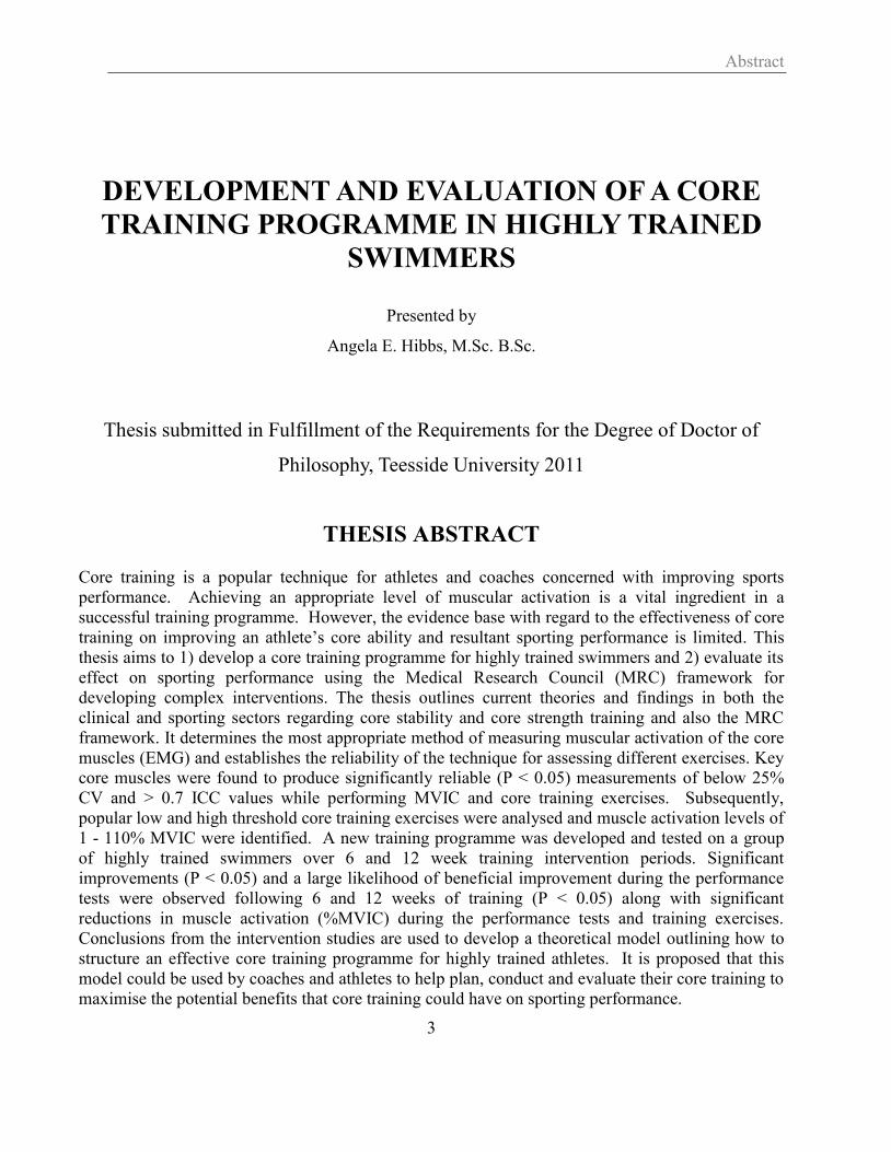


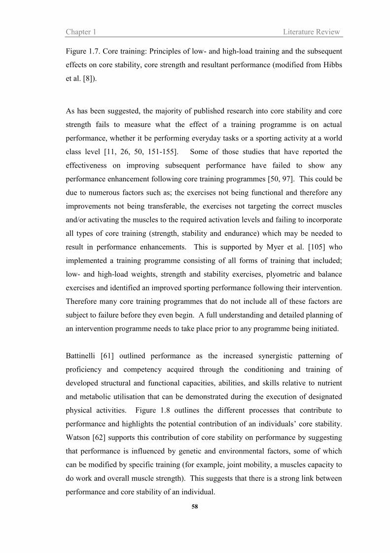

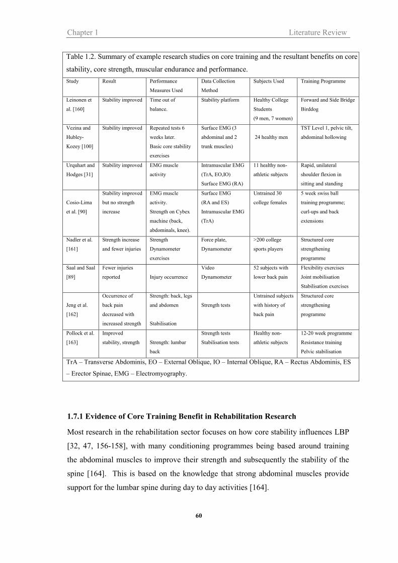


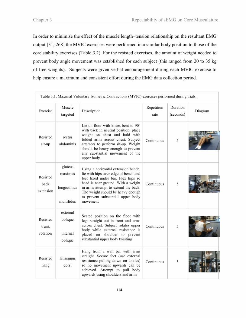


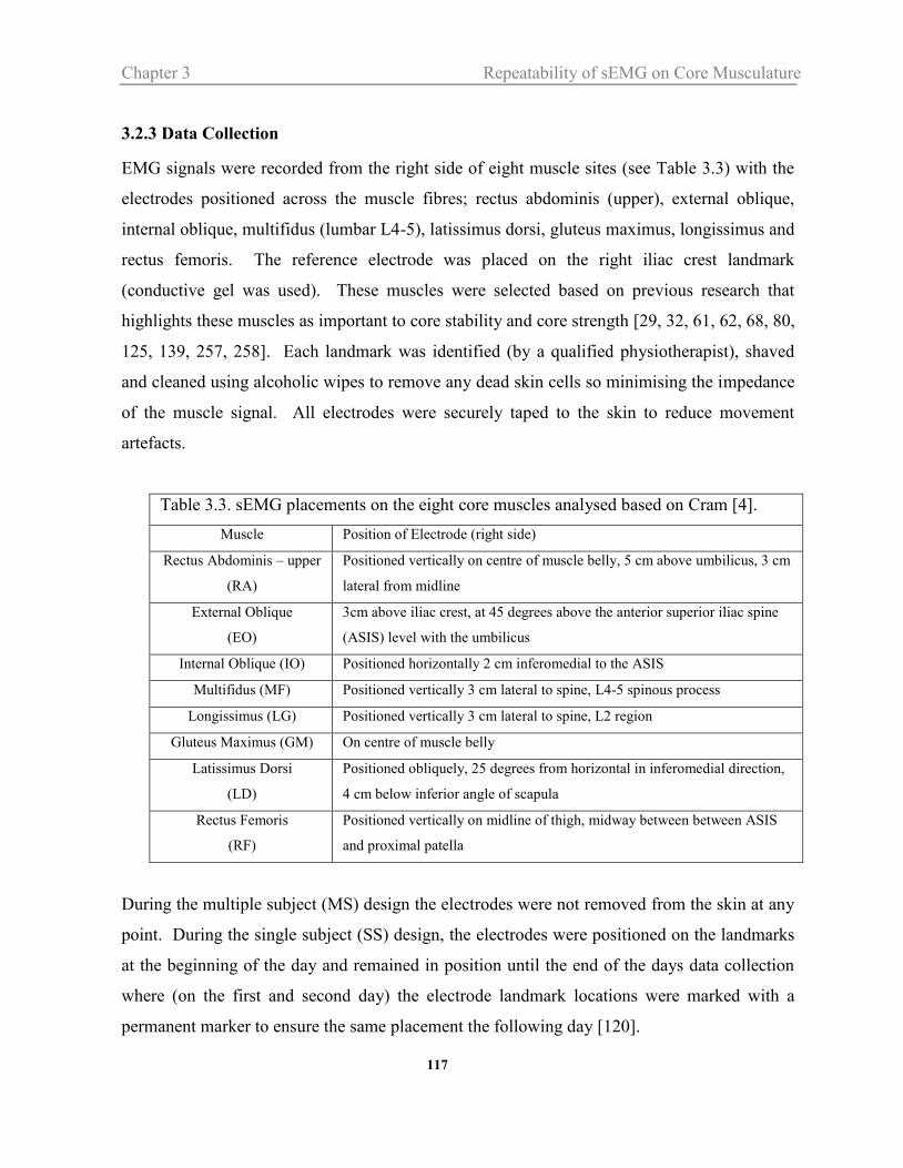


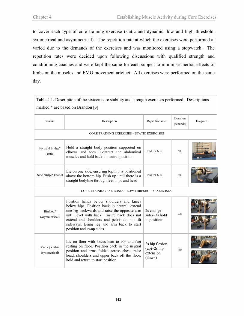


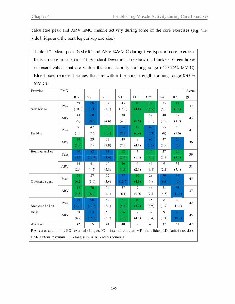


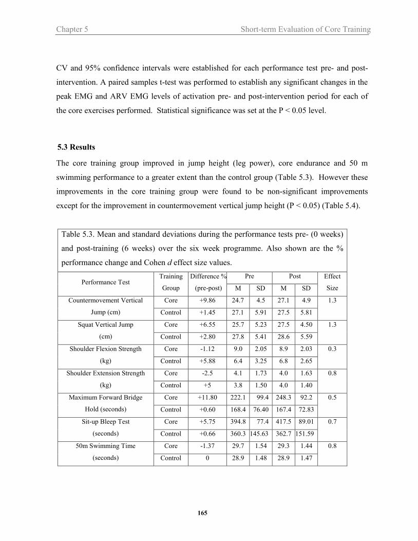

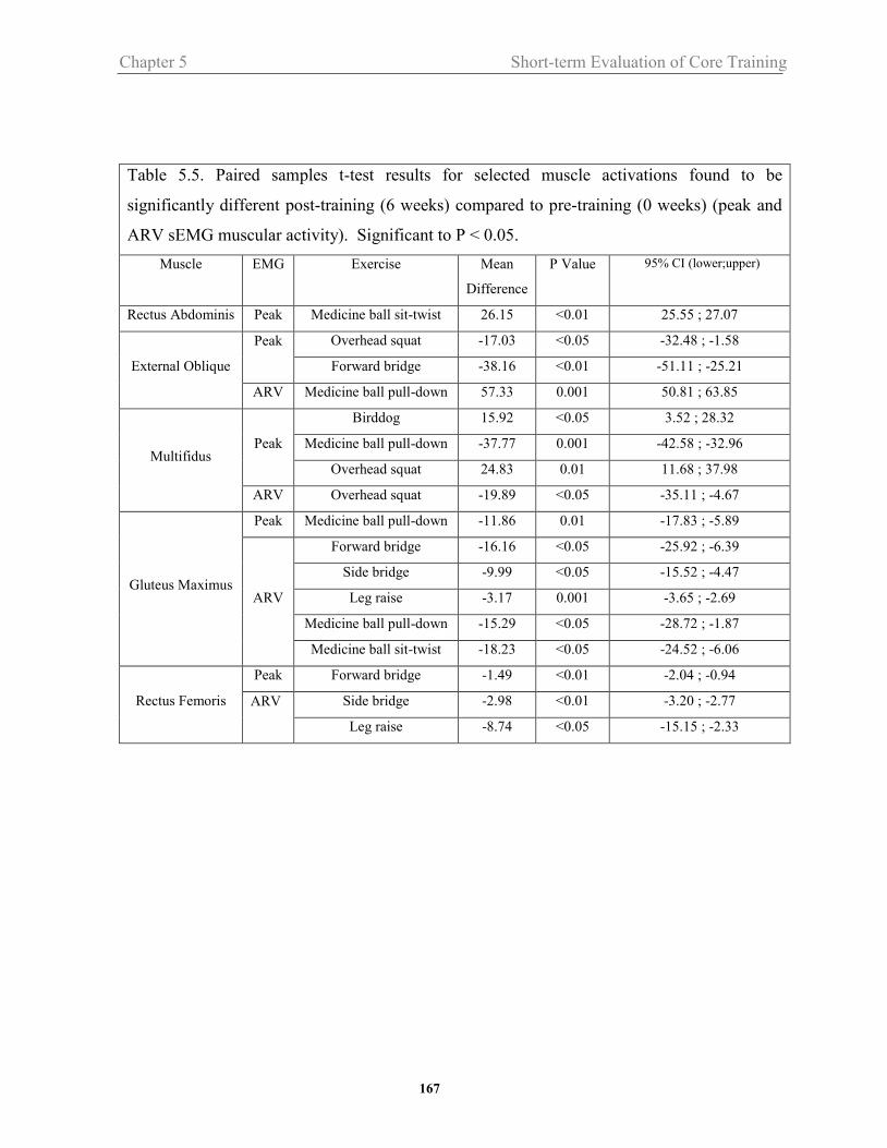


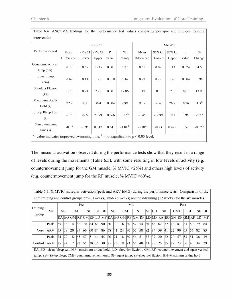
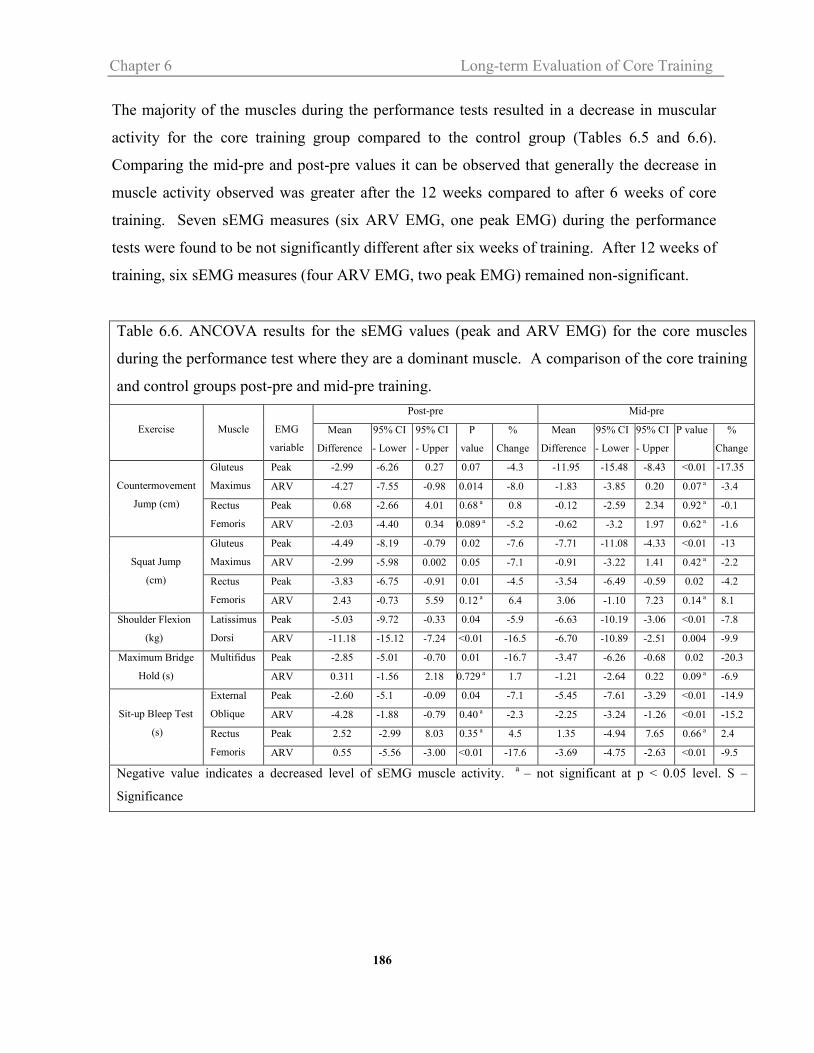

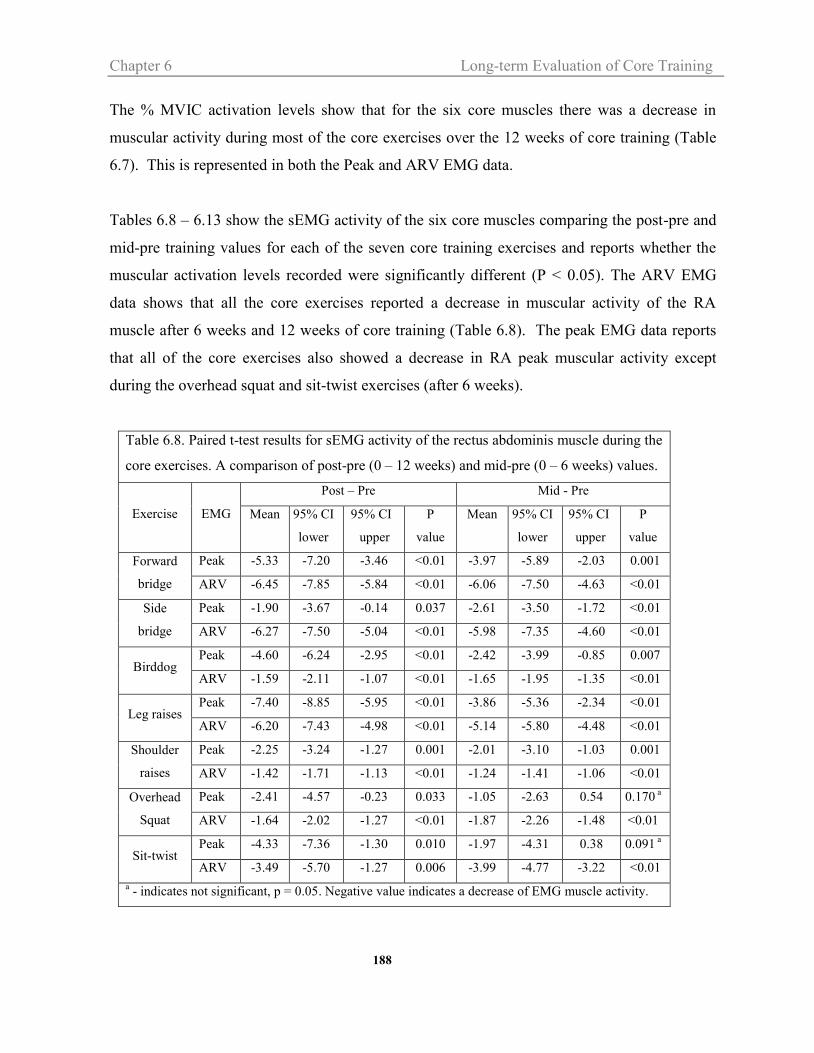


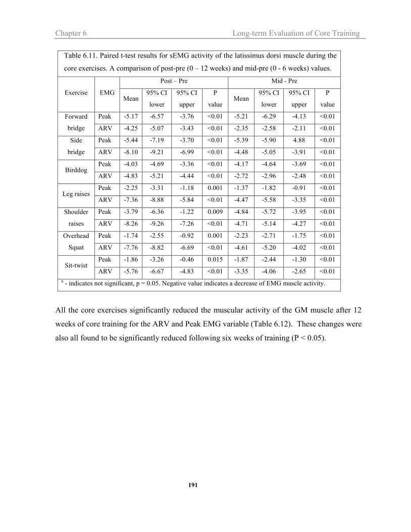

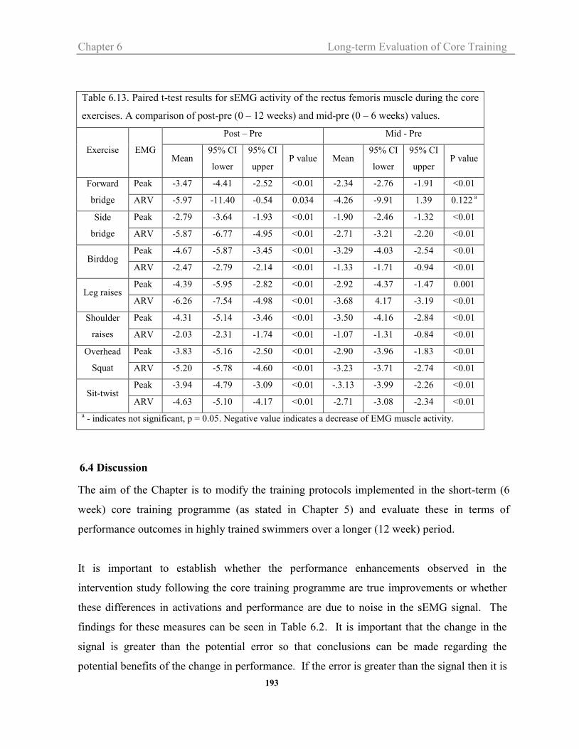


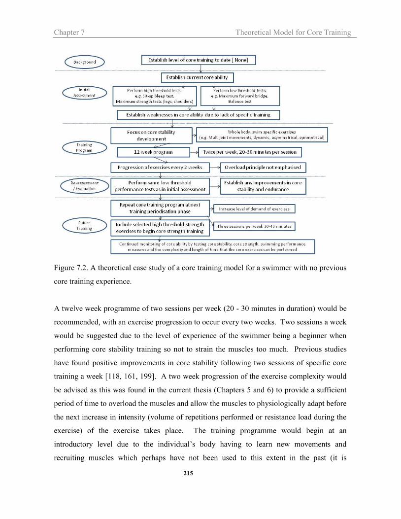

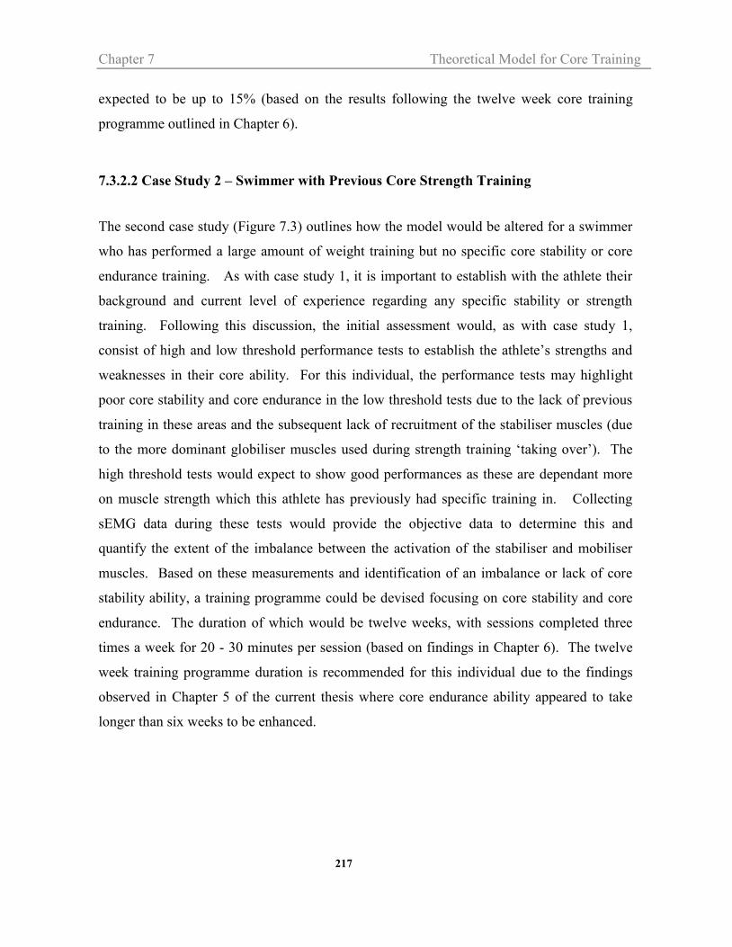
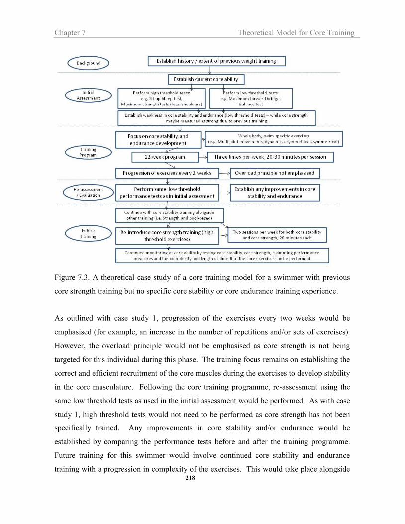


![Exercises for adolescent idiopathic scoliosis - …tees.openrepository.com/tees/bitstream/10149/249111/2/249111.pdf[Intervention Review] Exercises for adolescent idiopathic scoliosis](https://static.documents.pub/doc/80x56/5aa5e2337f8b9ae7438e1827/exercises-for-adolescent-idiopathic-scoliosis-tees-intervention-review-exercises.jpg)


