Page 1
High-order harmonic spectroscopy
for molecular imaging of polyatomic molecules
M. Negro,a M. Devetta,a D. Facciala,b S. De Silvestri,b C.
Vozzia∗ and S. Stagira,b
a Istituto di Fotonica e Nanotecnologie - CNR, 20133 Milan, Italy
b Dipartimento di Fisica - Politecnico di Milano, 20133 Milan, Italy
∗ corresponding author: [email protected]
High-order harmonic generation is a powerful and sensitive tool for probing atomic
and molecular structures, combining in the same measurement an unprecedented
attosecond temporal resolution with a high spatial resolution, of the order of the
angstrom. Imaging of the outermost molecular orbital by high-order harmonic
generation has been limited for a long time to very simple molecules, like nitrogen.
Recently we demonstrated a technique that overcame several of the issues that
have prevented the extension of molecular orbital tomography to more complex
species, showing that molecular imaging can be applied to a triatomic molecule
like carbon dioxide. Here we report on the application of such technique to nitrous
oxide (N2O) and acetylene (C2H2). This result represents a first step towards the
imaging of fragile compounds, a category which includes most of the fundamental
biological molecules.
1
arX
iv:1
412.
1300
v1 [
phys
ics.
chem
-ph]
3 D
ec 2
014
Page 2
INTRODUCTION
High order harmonic generation (HHG) occurs when atoms or molecules ex-
posed to an intense femtosecond laser pulse are ionized by tunneling. The freed
electron is then accelerated in the external electric field. Because of the periodic
oscillation of the laser field, the electron is brought back to the parent ion where it
may recombine emitting an XUV photon [4]. This XUV radiation has been shown
to contain information on the electronic structure of the emitting molecule and on
its internal dynamics. Attosecond nuclear [2] and electronic dynamics [8, 19] have
been extracted from HHG in simple molecules and spectral features in the har-
monic emission have been related to the molecular electronic structure and have
been used for imaging the highest occupied molecular orbital (HOMO).
The idea of exploiting HHG for the tomographic reconstruction of molecular
orbitals was first introduced by Itatani et al. in 2004 for the nitrogen molecule [9].
Since then, numerous experiments have been realized, addressing the role of the
HOMO in the harmonic spectral intensity [10, 23], in the molecular-frame photo-
ionization [11] and in the subsequent attosecond XUV emission [3], as well as in
the polarization state of the emitted radiation [13]. The dependence of the HHG
process on the HOMO structure has also been exploited for the characterization in
the time domain of the rotational [15] and vibrational [14] molecular excitations.
All these studies rely on two major assumptions: (i) the molecular HHG is
dominated by the HOMO structure; (ii) the relationship between molecular struc-
ture and emitted XUV spectrum is simple and completely captured by the Strong
Field Approximation (SFA), i.e. the electron quiver motion is not perturbed by
the Coulomb potential of the ion.
Both these assumptions have been recently put into question. Recent exper-
iments enlightened the role of multiple orbital contributions to HHG emission
[8, 19]. Furthermore, the influence of the Coulomb field of the parent ion in the
generation of high order harmonics from molecules has been considered as a serious
2
Page 3
hindrance to a clear HOMO reconstruction [29]. These assumptions should then
be overtaken to perform molecular tomography to more complex species.
Besides these two more fundamental obstacles, there are also additional, more
technical difficulties. In order to retrieve the HOMO structure, one has to record
the XUV harmonic spectra for different molecular orientations with respect to
the laser field. Hence, it is necessary to fix the molecular orientation in space
and change the polarization direction of the HHG-driving field [9]. Laser-assisted
molecular alignment is a widespread technique able to accomplish this task [21],
but the molecular alignment achieved in this way is not ideal. Hence the experi-
mental results and the corresponding HOMO tomography are affected by angular
averaging effects. Moreover, in the case of non-linear molecules, the tomographic
procedure requires to fix two or three angular coordinates of the molecule under
investigation. For instance, the study of linear polar molecules requires to fix the
head-tail direction in space. The feasibility of laser assisted molecular orientation
has been recently demonstrated [5] and exploited in HHG spectroscopy [6, 7, 20],
but no direct application to molecular imaging has been yet realized.
The amount of information that can be extracted from the harmonic emission
depends on the spectral extension of the XUV radiation, that is known to scale with
the so-called cut-off law: Emax = Ip+3.17Up, where Ip is the ionization potential of
the molecule and Up is the ponderomotive energy of the electron in the laser field.
This poses another important problem when HHG molecular imaging is extended
to species with low ionization potential (i.e. all organic molecules, and in particular
those having important biological functions) as the extension emission spectrum is
reduced. Since Up ∝ λ2I, where I is the peak intensity and λ the wavelength of the
driving laser pulse, the emission cut-off may be extended by both increasing the
field intensity or the laser wavelength. In this respect, standard Ti:Sapphire laser
sources generally used in HHG are not ideal candidates for tomography in fragile
molecules, since the intense optical fields needed completely ionize the molecule
3
Page 4
before a well-developed XUV spectrum is generated.
To overcome the limitations posed by ionization saturation, the exploitation
of mid-infrared driving sources has been demonstrated to be a powerful tool to
extend harmonic emission far in the XUV [18, 22, 25, 27, 28].
With a mid-IR source[24] we recently demonstrated that it is possible to extend
the spectral investigation in carbon dioxide beyond 100 eV in the absence of multi-
electron effects, thus avoiding any ambiguity in the reconstructed wavefunction. In
addition, by exploiting an all-optical non-interferometric technique, it was possible
to trace both the spectral intensity and phase of high order harmonics generated
by single molecules as a function of emitted photon energy and molecular angular
orientation, without averaging effects. Furthermore, the tomographic procedure
was generalized in order to take into account the Coulomb potential seen by the
re-colliding electron wavepacket[26].
In this work, we extend that approach to more complex molecules, such as
N2O and C2H2 pointing out some strengths and weaknesses of this investigation
technique.
EXPERIMENTAL SETUP
We exploited an optical parametric amplifier (OPA) pumped by an amplified
Ti:sapphire laser system (60 fs, 20 mJ, 800 nm). The OPA is based on difference
frequency generation and provides driving pulses with 1450 nm central wavelength,
pulse duration of 20 fs and pulse energy of 1.2 mJ[24]. High harmonics were
generated by focusing the mid-IR pulse in a supersonic gas jet under vacuum,
due to the strong absorption exhibited by air in the XUV spectral region. The
molecules in the jet were impulsively aligned with a portion of the fundamental
800-nm beam which was spectrally broadened by optical filamentation in an argon-
filled gas cell and temporally stretched up to 100 fs by propagation through a glass
4
Page 5
plate. Such duration is required for achieving a good alignment of the molecular
sample. In our experimental setup, driving and aligning pulse were collinear and
their polarizations were parallel. The delay between the two pulses was adjusted
by means of a fine-resolution translation stage. The XUV radiation was acquired
by means of a flat-field spectrometer and a multi-channel plate detector coupled
to a CCD camera [16].
RESULTS
40 50 60 70 80 90 100
19
20
21
0.1 0.4 0.7-2
0
2
4
de
lay
(ps)
photon energy (eV) <<cos2(q)>>
inte
nsity
(arb
. un
its)
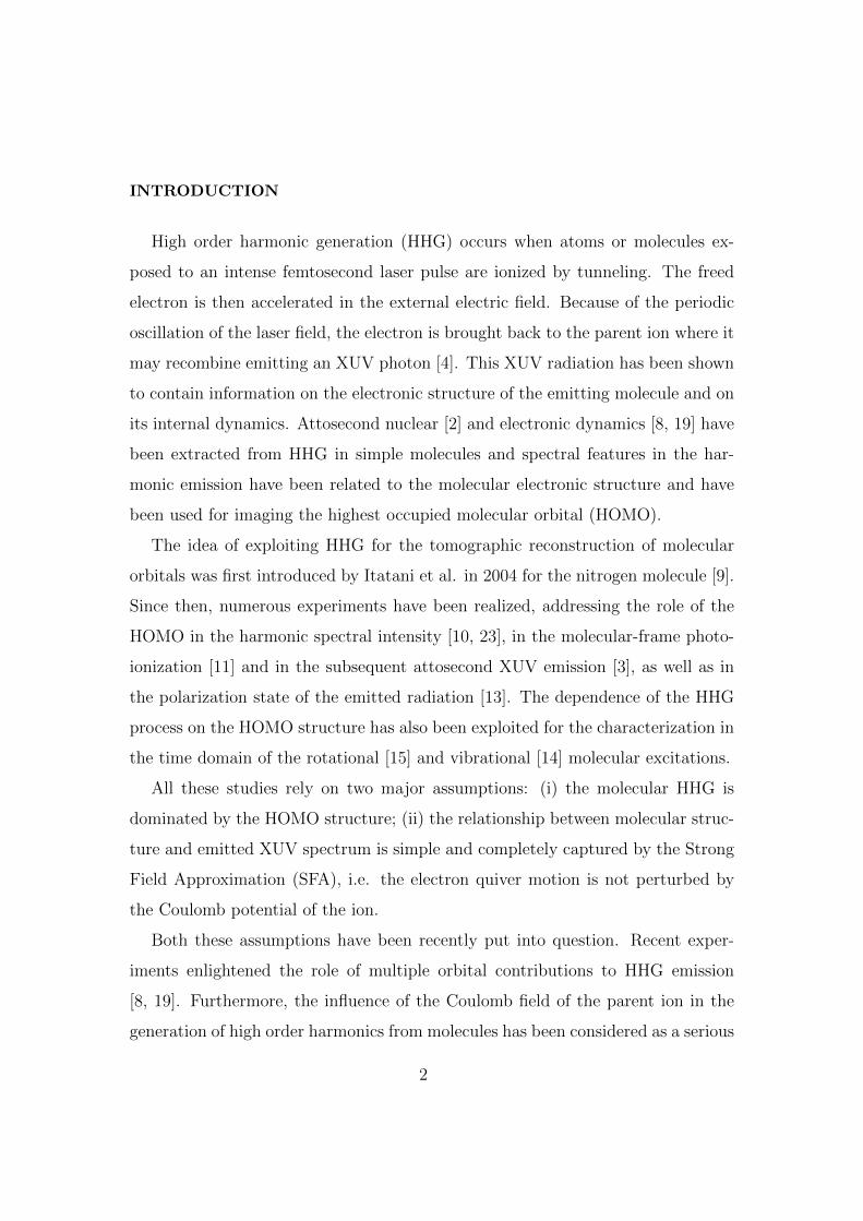
(b)(a)
FIG. 1 (a) Sequence of harmonic spectra measured in N2O as a function of emitted
photon energy and delay between the aligning and the driving pulse (log scale). (b)
Calculated alignment factor for N2O in the experimental conditions (rotational
temperature 75 K, aligning pulse duration 100 fs, aligning pulse intensity 3.32× 10¹³
W/cm2).
Harmonic spectra were acquired in N2O and C2H2 as a function of the delay
τ between the aligning and driving pulse around the first rotational half revival
(τN2O = 19.95 ps and τC2H2 = 7.08 ps). The results are shown in figure 1(a) and
2(a) for N2O and C2H2 respectively. Figures 1(b) and 2(b) show the corresponding
5
Page 6
36 40 44 48 52 56 60
6.5
7.0
7.5
8.0
0.2 0.4 0.6-2
0
2
4d
ela
y(p
s)
photon energy (eV) <<cos2(q)>>
inte
nsity
(arb
. un
its)(b)(a)
FIG. 2 (a) Sequence of harmonic spectra measured in C2H2 as a function of emitted
photon energy and delay between the aligning and the driving pulse (log scale). (b)
Calculated alignment factor for C2H2 in the experimental conditions (rotational
temperature 75 K, aligning pulse duration 100 fs, aligning pulse intensity 2.16× 10¹³
W/cm2).
calculated alignment factor for the experimental conditions.
In both molecules, the sequence of harmonic spectra shows a strong modulation
with the delay τ that can be ascribed to the dependence of harmonic yield on the
molecular orbital structure. In particular, a reduction of the harmonic emission
can be observed for the delay corresponding to the maximum of the alignment
factor and an enhancement of the harmonic yield appears for the minimum of the
alignment factor. A major difference between the two cases is the presence of a
region of harmonic enhancement at high photon energy, that appears in N2O at
maximum alignment.
These effects can be naively interpreted in terms of two-center interference
occurring in the re-collision step [12, 23]. If one consider a diatomic homo-nuclear
molecule with a symmetric electronic state with respect to the nuclei exchange and
6
Page 7
assumes the re-colliding electron as a plane wave, the condition for constructive
interference reads R cos(θ) = nλB, where R is the internuclear separation, θ is
the angle between the molecular axis and the electron wave-vector, n is an integer
number and λB is the de Broglie wavelength associated to the re-colliding electron
wave-packet. Similarly the condition for destructive interference is R cos(θ) =
(n+ 1/2)λB and the first destructive interference occurs for n = 0. The conditions
become reversed for molecules with antisymmetric electronic structure.
This concept can be extended to the molecules subject of our investigation. The
acetylene molecule has a symmetric π HOMO in which the separation between the
carbon atoms is RC≡C = 1.2 A. This is the distance that should be considered
for the evaluation of the interference condition. The N2O HOMO does not have
a clear symmetry, however in our experimental condition the harmonic spectra
are acquired in aligned molecules and correspond to the average between the two
possible orientation. The resulting signal can be interpreted in terms of emission
from an effective molecular orbital similar to the anti-symmetric π orbital of CO2.
In this view the overall length of this “effective” orbital is RN2O = 2.3 A. Since
RN2O ≈ 2RC≡C, a destructive interference occurs in the same spectral region for
both molecules, corresponding to n = 1 for N2O and n = 0 for C2H2.
Figures 1(a) and 2(a) show two peculiar advantages related to the exploitation
of mid-IR driving pulses for HHG. Indeed the harmonic cutoff extension related
to the increase in the ponderomotive energy with respect to standard Ti:sapphire
sources allows the observation of spectral features as the harmonic enhancement
for high photon energy visible in N2O in correspondence of the revival peak. In the
framework of the above mentioned two-center model, this feature can be attributed
to the appearance of constructive interference in that spectral region. Moreover, for
the same emitted photon energy, mid-IR driving wavelengths require a lower pulse
peak intensity thus reducing the ionization saturation in species with relatively
low ionization potential, such as C2H2 (IP = 11.4 eV).
7
Page 8
RECONSTRUCTION OF SINGLE MOLECULE XUV EMISSION
From the experimental data reported in figures 1(a) and 2(a) it is possible to
retrieve structural information on the target molecule following the approach in-
troduced by Vozzi et al. [26]. Figures 3(a) and 4(a) show the same experimental
results presented in figures 1(a) and 2(a), in which the harmonic structure due to
the periodic re-collision of the electron wave-packet has been filtered out. These
results have been exploited for the reconstruction of the XUV field emitted from
a single molecule and projected on the polarization direction of the aligning field
as a function of the angle between the molecular axis and the driving polariza-
tion direction. The reconstruction is based on a combination of a phase-retrieval
algorithm and a Kaczmarz algorithm [17]. The main idea behind this approach
is that the macroscopic XUV emission is the coherent superposition of the XUV
field emitted by all molecules weighted with their angular distribution. This dis-
tribution changes along the revival in a predictable way, hence the sequence of
harmonic emission contains enough information for the reconstruction of the har-
monic electric field in amplitude and phase.
The result of this reconstruction is shown in figure 5 for N2O and in figure
6 for C2H2. In both figures, panel (a) reports the amplitude of the XUV field
and panel (b) shows the corresponding phase. In N2O there is a clear phase
jump of about 2 rad, that changes its position with photon energy and molecular
alignment. This phase jump corresponds to a minimum in the XUV amplitude
and its position is quite in good agreement with the prediction of the naive two-
center model introduced above, which is shown as a dashed line in the figure.
It is worth noting that the reconstruction technique is based on the interference
of XUV emission from different molecular orientations, thus the phase can be
retrieved as a function of θ at fixed XUV photon energy. In order to retrieve the
phase relationship between contributions at neighboring energies, it is necessary to
introduce an a priori condition that can be derived from theoretical considerations
8
Page 9
40 60 80 100
19
20
21
photon energy (eV)
de
lay
(ps)
(a)
40 60 80 1000.0
0.2
0.4
0.6
0.8
1.0
photon energy (eV)
inte
nsity
(arb
. un
its)(b)
40 60 80 100
19
20
21
0.0
0.5
1.0
1.5
2.0
photon energy (eV)
ph
ase
(rad
)de
lay
(p
s)
(c)
FIG. 3 (a) Sequence of XUV spectra measured in N2O as a function of emitted
photon energy and delay between the aligning and the driving pulse; the harmonic
structure has been filtered out. Retrieved macroscopic harmonic emission amplitude
(b) and phase (c) corresponding to the data reported in (a).
or experimental measurements. In the case of N2O we imposed a flat spectral phase
of the macroscopic harmonic emission for the delay corresponding to the molecular
anti-alignment. This condition was chosen in analogy with the CO2 case[26], due to
the similarity between the two HOMOs as discussed in the previous section. The
results of this assumption can be observed in figure 3, where the reconstructed
amplitude (b) and phase (c) of the macroscopic XUV emission from N2O are
reported. The retrieved amplitude is in good agreement with the experimental
9
Page 10
40 45 50 55
6.3
6.6
6.9
7.2
7.5
7.8
1.50
1.75
2.00
2.25
2.50
de
lay
(ps)
photon energy (eV)
ph
ase
(ra
d)
40 45 50 55
6.3
6.6
6.9
7.2
7.5
7.8
photon energy (eV)
de
lay
(ps)
(a)
40 45 50 550.0
0.2
0.4
0.6
0.8
1.0
photon energy (eV)
inte
nsi
ty(a
rb.u
nit
s)
(b)
(c)
FIG. 4 (a) Sequence of XUV spectra measured in C2H2 as a function of emitted
photon energy and delay between the aligning and the driving pulse; the harmonic
structure has been filtered out. Retrieved macroscopic harmonic emission amplitude
(b) and phase (c) corresponding to the data reported in (a).
data (figure 3(a)). The phase of the macroscopic emission shows a steep change of
about 2 rad around 50 eV at the delay τ corresponding to the maximum alignment.
In the case of C2H2 we followed the same approach in the retrieval procedure.
We imposed in this case a flat spectral phase for the macroscopic harmonic emis-
sion at the delay τ corresponding to the molecular alignment. This assumption
was necessary in order to complete the retrieval procedure, but it is arbitrary and
not supported by theoretical models; it could be however improved by changing
the retrieving condition according to an experimental spectral phase measurement.
10
Page 11
40 60 80 1000
15
30
45
60
75
90
40 60 80 1000
15
30
45
60
75
90
-1
0
1
2
3
0.0
0.2
0.4
0.6
0.8
1.0
photon energy (eV)
an
gle
(de
g)
an
gle
(de
g)
photon energy (eV)
ph
ase
(rad
)
inte
nsity
(arb
. un
its)
(a) (b)
FIG. 5 Retrieved single molecule XUV emission map in N2O as a function of emitted
photon energy and the angle between the molecular axis and the aligning beam
polarization direction in amplitude (a) and phase(b). Dashed lines show the position of
the destructive interference predicted by the two-center model.
40 45 50 550
15
30
45
60
75
90
40 45 50 550
15
30
45
60
75
90
0
1
2
3
0.0
0.2
0.4
0.6
0.8
1.0
photon energy (eV)
an
gle
(de
g)
an
gle
(de
g)
photon energy (eV)
ph
ase
(ra
d)
inte
nsi
ty(a
rb.u
nit
s)
(a) (b)
FIG. 6 Retrieved single molecule XUV emission map in C2H2 as a function of emitted
photon energy and the angle between the molecular axis and the aligning beam
polarization direction in amplitude (a) and phase(b). Dashed lines show the position of
the destructive interference predicted by the two-center model.
This kind of experiment can be performed for example by RABBIT technique at
a given alignment delay [3]. The retrieved single molecule XUV emission in C2H2,
shown in figure 6, is very different from the one reported for N2O. In particular a
strong contribution comes from molecules with perpendicular orientation with re-
spect to the driving field polarization direction. In the retrieved phase (figure 6(c))
11
Page 12
two phase jumps are clearly observed. The first one appears for small alignment
angles and roughly follows the prediction of the two-center model. The second
jump appears at large alignment angles and may be attributed to the shape of
the HOMO seen by the re-colliding electron. However, since the reconstruction is
based on the arbitrary assumption of flat macroscopic spectral phase at the align-
ment delay, the retrieved outcomes should be considered preliminary. In spite of
this, the retrieved macroscopic XUV amplitude (figure 4(b)) is in fair agreement
with the experimental results.
MOLECULAR ORBITAL TOMOGRAPHY
The results reported in the previous section can be used for the two-dimensional
reconstruction of molecular orbitals, following the tomographic procedure pro-
posed by Itatani et al. [9] and extended by Vozzi et al. [26]. However to proceed
with this tomographic reconstruction, it is necessary to rule out the occurrence of
multi-electron effects in HHG. A simple experimental procedure to check whether
spectral modulations in harmonic emission are due to multi-electron effects is to
change the driving field intensity. As shown by Smirnova et al. [19], one ex-
pects all the features due to multi-electron effects to shift with the driving field
intensity. Figure 7 shows harmonic spectra acquired in aligned N2O for a delay τ
corresponding to the maximum of the alignment for different values of the driving
intensity. The spectral minimum associated to the phase change retrieved in fig-
ure 5(b) appears always around 55 eV and does not shift with the intensity. This
behavior guarantees that the main spectral features in the harmonic emission are
mainly dictated by the HOMO structure. This consistency check allowed us to
exploit the retrieved single molecule harmonic emission for the reconstruction of
N2O orbital. The result is shown in figure 8(a). Figure 8(b) shows the N2O orbital
calculated with a quantum chemistry program [1]. Even if the overall dimension
12
Page 13
of the molecular orbital is well reproduced, the asymmetry of this orbital is very
clear and cannot be addressed by the tomographic reconstruction, since in the
experiment the molecules were aligned but not oriented. Another departure of the
retrieved orbital with respect to the calculated one is the presence of side lobes,
that can be attributed to the limited working range of the XUV spectrometer used
in these experiments. Since there is a correspondence between the energy range
of harmonic emission and the spatial frequency domain, the limited spectral range
collectible in the experiment corresponds to a spatial filtering in the Fourier do-
main, which gives raise to such lobes. These observations are further confirmed
by figure 8(c), which shows the calculated HOMO corresponding to the average
between the two possible orientations of N2O molecular axis and takes into ac-
count the limited spectral bandwidth available in the experiment. The features of
this fictitious orbital are in very good agreement with the reconstruction of figure
8(a). It is worth nothing that such limitations can be overcome by extending the
acquired spectral range over all the XUV emission and by exploiting all-optical im-
pulsive techniques for orientation of polar molecules, such the one demonstrated
by Frumker et al.[6, 7].
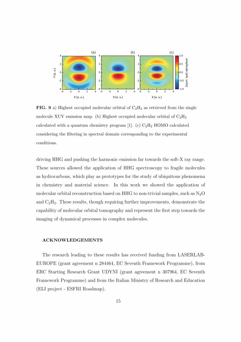
Differently from the case of N2O, in C2H2 it is not possible to easily rule out
the multi-electron contributions. Because of the smaller cutoff energy, the exper-
imental approach applied in the case of N2O for the exclusion of multi-electron
contribution is not feasible. Nevertheless the application of tomographic approach
to the single molecule emission maps shown in figure 6 provides interesting results.
We show in figure 9(a) the retrieved C2H2 HOMO. Also in this case, a comparison
with the result calculated with a quantum chemistry program (see figure 9(b))
shows a good agreement in the overall shape of the orbital. Again the additional
lobes are related to the limited harmonic range detected in the experimental ac-
quisition, as can be seen in figure 9(c) where the orbital is calculated taking into
account the spectral filtering.
13
Page 14
40 50 60 70 80 90 100
energy (eV)
inte
nsi
ty (
arb
.un
its)
FIG. 7 Harmonic spectra generated in N2O at the delay τ corresponding to the
maximum molecular alignment for several driving peak intensities I between 1 and
1.7× 10¹⁴ W/cm2.
-4 -2 0 2 4
-4
-2
0
2
4
X (a. u.)
-4 -2 0 2 4
-4
-2
0
2
4
Y(a
.u
.)
X (a. u.)
-4 -2 0 2 4
-4
-2
0
2
4
-1.0
-0.5
0.0
0.5
1.0
X (a. u.)
am
plitu
de
(arb
. un
its)
(a) (b) (c)
FIG. 8 (a) Highest occupied molecular orbital of N2O as retrieved from the single
molecule XUV emission map. (b) Highest occupied molecular orbital of N2O calculated
with a quantum chemistry program [1]. (c) N2O HOMO calculated averaging over the
two possible orientations of the molecular axis and considering the filtering in spectral
domain corresponding to the experimental conditions.
CONCLUSIONS
Since the pioneering work of Itatani et al. on molecular orbital imaging, the
impressive advances in laser technologies gave the access to new mid-IR sources for
14
Page 15
-4 -2 0 2 4-4
-2
0
2
4
-1.0
-0.5
0.0
0.5
1.0
X (a. u.)
-4 -2 0 2 4-4
-2
0
2
4Y
(a.
u.)
X (a. u.)
-4 -2 0 2 4-4
-2
0
2
4
X (a. u.)
(a) (b) (c)a
mp
litud
e (a
rb. u
nits)
FIG. 9 a) Highest occupied molecular orbital of C2H2 as retrieved from the single
molecule XUV emission map. (b) Highest occupied molecular orbital of C2H2
calculated with a quantum chemistry program [1]. (c) C2H2 HOMO calculated
considering the filtering in spectral domain corresponding to the experimental
conditions.
driving HHG and pushing the harmonic emission far towards the soft-X ray range.
These sources allowed the application of HHG spectroscopy to fragile molecules
as hydrocarbons, which play as prototypes for the study of ubiquitous phenomena
in chemistry and material science. In this work we showed the application of
molecular orbital reconstruction based on HHG to non-trivial samples, such as N2O
and C2H2. These results, though requiring further improvements, demonstrate the
capability of molecular orbital tomography and represent the first step towards the
imaging of dynamical processes in complex molecules.
ACKNOWLEDGEMENTS
The research leading to these results has received funding from LASERLAB-
EUROPE (grant agreement n 284464, EC Seventh Framework Programme), from
ERC Starting Research Grant UDYNI (grant agreement n 307964, EC Seventh
Framework Programme) and from the Italian Ministry of Research and Education
(ELI project - ESFRI Roadmap).
15
Page 16
[1] DALTON, a molecular electronic structure program Release 2.0 (2005) see
http://www.kjemi.uio.no/software/dalton/dalton.html.
[2] S Baker, JS Robinson, CA Haworth, H Teng, RA Smith, CC Chirila, M Lein, JWG
Tisch, and JP Marangos. Probing proton dynamics in molecules on an attosecond
time scale. SCIENCE, 312(5772):424–427, APR 21 2006.
[3] W. Boutu, S. Haessler, H. Merdji, P. Breger, G. Waters, M. Stankiewicz, L. J.
Frasinski, R. Taieb, J. Caillat, A. Maquet, P. Monchicourt, B. Carre, and
P. Salieres. Coherent control of attosecond emission from aligned molecules. NA-
TURE PHYSICS, 4(7):545–549, JUL 2008.
[4] P. B. Corkum. Plasma perspective on strong field multiphoton ionization. Phys.
Rev. Lett., 71:1994–1997, Sep 1993.
[5] S. De, I. Znakovskaya, D. Ray, F. Anis, Nora G. Johnson, I. A. Bocharova, M. Ma-
grakvelidze, B. D. Esry, C. L. Cocke, I. V. Litvinyuk, and M. F. Kling. Field-Free
Orientation of CO Molecules by Femtosecond Two-Color Laser Fields. Phys. Rev.
Lett., 103:153002, Oct 2009.
[6] E. Frumker, C. T. Hebeisen, N. Kajumba, J. B. Bertrand, H. J. Woerner, M. Span-
ner, D. M. Villeneuve, A. Naumov, and P. B. Corkum. Oriented Rotational Wave-
Packet Dynamics Studies via High Harmonic Generation. PHYSICAL REVIEW
LETTERS, 109(11):113901, SEP 12 2012.
[7] E. Frumker, N. Kajumba, J. B. Bertrand, H. J. Worner, C. T. Hebeisen, P. Hockett,
M. Spanner, S. Patchkovskii, G. G. Paulus, D. M. Villeneuve, A. Naumov, and P. B.
Corkum. Probing Polar Molecules with High Harmonic Spectroscopy. Phys. Rev.
Lett., 109:233904, Dec 2012.
[8] S. Haessler, J. Caillat, W. Boutu, C. Giovanetti-Teixeira, T. Ruchon, T. Auguste,
Z. Diveki, P. Breger, A. Maquet, B. Carre, R. Taieb, and P. Salieres. Attosecond
16
Page 17
imaging of molecular electronic wavepackets. NATURE PHYSICS, 6(3):200–206,
MAR 2010.
[9] J Itatani, J Levesque, D Zeidler, H Niikura, H Pepin, JC Kieffer, PB Corkum,
and DM Villeneuve. Tomographic imaging of molecular orbitals. NATURE,
432(7019):867–871, DEC 16 2004.
[10] T Kanai, S Minemoto, and H Sakai. Quantum interference during high-order har-
monic generation from aligned molecules. NATURE, 435(7041):470–474, MAY 26
2005.
[11] Anh-Thu Le, R. R. Lucchese, M. T. Lee, and C. D. Lin. Probing Molecular
Frame Photoionization via Laser Generated High-Order Harmonics from Aligned
Molecules. Phys. Rev. Lett., 102:203001, May 2009.
[12] M Lein, N Hay, R Velotta, JP Marangos, and PL Knight. Interference effects in high-
order harmonic generation with molecules. PHYSICAL REVIEW A, 66(2):023805,
AUG 2002.
[13] Jerome Levesque, Yann Mairesse, Nirit Dudovich, Henri Pepin, Jean-Claude Kieffer,
P. B. Corkum, and D. M. Villeneuve. Polarization State of High-Order Harmonic
Emission from Aligned Molecules. Phys. Rev. Lett., 99:243001, Dec 2007.
[14] Wen Li, Xibin Zhou, Robynne Lock, Serguei Patchkovskii, Albert Stolow, Henry C.
Kapteyn, and Margaret M. Murnane. Time-Resolved Dynamics in N(2)O(4) Probed
Using High Harmonic Generation. SCIENCE, 322(5905):1207–1211, NOV 21 2008.
[15] K. Miyazaki, M. Kaku, G. Miyaji, A. Abdurrouf, and F. H. M. Faisal. Field-Free
Alignment of Molecules Observed with High-Order Harmonic Generation. Phys.
Rev. Lett., 95:243903, Dec 2005.
[16] L Poletto, G Tondello, and P Villoresi. High-order laser harmonics detection in the
EUV and soft x-ray spectral regions. REVIEW OF SCIENTIFIC INSTRUMENTS,
72(7):2868–2874, JUL 2001.
[17] C Popa and R Zdunek. Kaczmarz extended algorithm for tomographic image re-
17
Page 18
construction from limited-data. MATHEMATICS AND COMPUTERS IN SIMU-
LATION, 65(6):579–598, MAY 17 2004.
[18] Tenio Popmintchev, Ming-Chang Chen, Dimitar Popmintchev, Paul Arpin, Su-
sannah Brown, Skirmantas Alisauskas, Giedrius Andriukaitis, Tadas Balciunas,
Oliver D. Mucke, Audrius Pugzlys, Andrius Baltuska, Bonggu Shim, Samuel E.
Schrauth, Alexander Gaeta, Carlos Hernandez-Garcıa, Luis Plaja, Andreas Becker,
Agnieszka Jaron-Becker, Margaret M. Murnane, and Henry C. Kapteyn. Bright
Coherent Ultrahigh Harmonics in the keV X-ray Regime from Mid-Infrared Fem-
tosecond Lasers. Science, 336(6086):1287–1291, 2012.
[19] Olga Smirnova, Yann Mairesse, Serguei Patchkovskii, Nirit Dudovich, David Vil-
leneuve, Paul Corkum, and Misha Yu. Ivanov. High harmonic interferometry of
multi-electron dynamics in molecules. NATURE, 460(7258):972–977, AUG 20 2009.
[20] Michael Spanner, Serguei Patchkovskii, Eugene Frumker, and Paul Corkum. Mech-
anisms of Two-Color Laser-Induced Field-Free Molecular Orientation. Phys. Rev.
Lett., 109:113001, Sep 2012.
[21] H Stapelfeldt and T Seideman. Colloquium: Aligning molecules with strong laser
pulses. REVIEWS OF MODERN PHYSICS, 75(2):543–557, APR 2003.
[22] Eiji J. Takahashi, Tsuneto Kanai, Kenichi L. Ishikawa, Yasuo Nabekawa, and Kat-
sumi Midorikawa. Coherent Water Window X Ray by Phase-Matched High-Order
Harmonic Generation in Neutral Media. Phys. Rev. Lett., 101:253901, Dec 2008.
[23] C Vozzi, F Calegari, E Benedetti, JP Caumes, G Sansone, S Stagira, M Nisoli,
R Torres, E Heesel, N Kajumba, JP Marangos, C Altucci, and R Velotta. Con-
trolling two-center interference in molecular high harmonic generation. PHYSICAL
REVIEW LETTERS, 95(15):153902, OCT 7 2005.
[24] C. Vozzi, F. Calegari, E. Benedetti, S. Gasilov, G. Sansone, G. Cerullo, M. Nisoli,
S. De Silvestri, and S. Stagira. Millijoule-level phase-stabilized few-optical-cycle
infrared parametric source. OPTICS LETTERS, 32(20):2957–2959, OCT 15 2007.
18
Page 19
[25] C. Vozzi, F. Calegari, F. Frassetto, M. Negro, L. Poletto, G. Sansone, P. Villoresi,
M. Nisoli, S. Silvestri, and S. Stagira. High order harmonics driven by a self-phase-
stabilized IR parametric source. Laser Physics, 20(5):1019–1027, 2010.
[26] C. Vozzi, M. Negro, F. Calegari, G. Sansone, M. Nisoli, S. De Silvestri, and S. Sta-
gira. Generalized molecular orbital tomography. NATURE PHYSICS, 7(10):822–
826, OCT 2011.
[27] C. Vozzi, R. Torres, M. Negro, L. Brugnera, T. Siegel, C. Altucci, R. Velotta,
F. Frassetto, L. Poletto, P. Villoresi, S. De Silvestri, S. Stagira, and J. P. Marangos.
High harmonic generation spectroscopy of hydrocarbons. APPLIED PHYSICS
LETTERS, 97(24):241103, DEC 13 2010.
[28] Caterina Vozzi, Francesca Calegari, Matteo Negro, Fabio Frassetto, Luca Poletto,
Giuseppe Sansone, Paolo Villoresi, Mauro Nisoli, Sandro De Silvestri, and Salvatore
Stagira. High-order harmonics generated by 1.5 m parametric source. Journal of
Modern Optics, 57(11):1008–1013, 2010.
[29] Zachary B. Walters, Stefano Tonzani, and Chris H. Greene. Limits of the plane
wave approximation in the measurement of molecular properties. JOURNAL OF
PHYSICAL CHEMISTRY A, 112(39):9439–9447, OCT 2 2008.
19