77
High Yield Surgery Shelf Exam Review Emma Holliday Ramahi

High Yield Surgery
Shelf Exam Review
Emma Holliday Ramahi
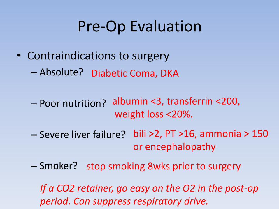
Pre-Op Evaluation
• Contraindications to surgery
– Absolute?
– Poor nutrition?
– Severe liver failure?
– Smoker?
Diabetic Coma, DKA
albumin <3, transferrin <200,weight loss <20%.
bili >2, PT >16, ammonia > 150 or encephalopathy
stop smoking 8wks prior to surgery
If a CO2 retainer, go easy on the O2 in the post-op period. Can suppress respiratory drive.
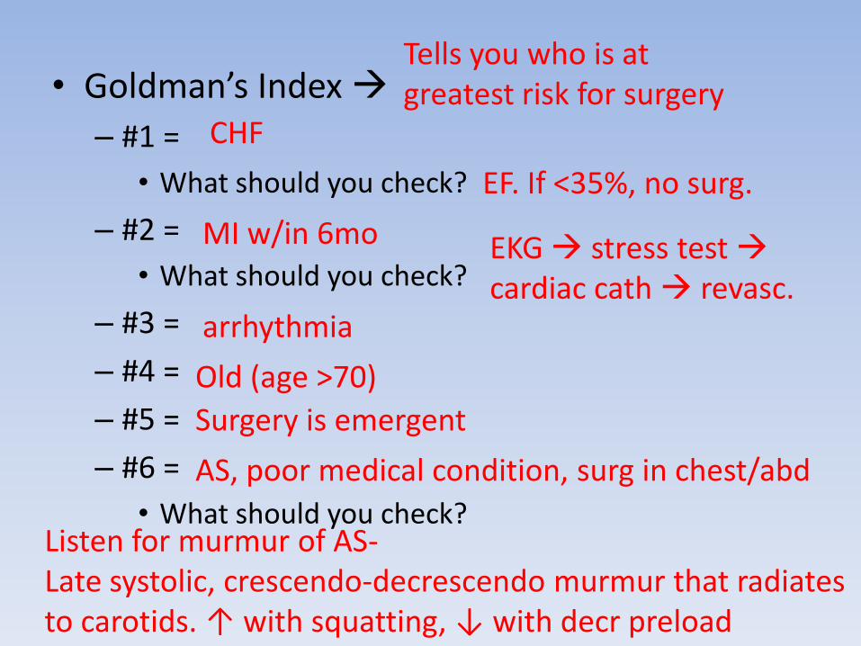
• Goldman’s Index
– #1 =
• What should you check?
– #2 =
• What should you check?
– #3 =
– #4 =
– #5 =
– #6 =
• What should you check?
Tells you who is at greatest risk for surgery
CHF
EF. If <35%, no surg.
MI w/in 6mo EKG stress test cardiac cath revasc.
arrhythmia
Old (age >70)
Surgery is emergent
AS, poor medical condition, surg in chest/abd
Listen for murmur of AS-Late systolic, crescendo-decrescendo murmur that radiates to carotids. ↑ with squatting, ↓ with decr preload
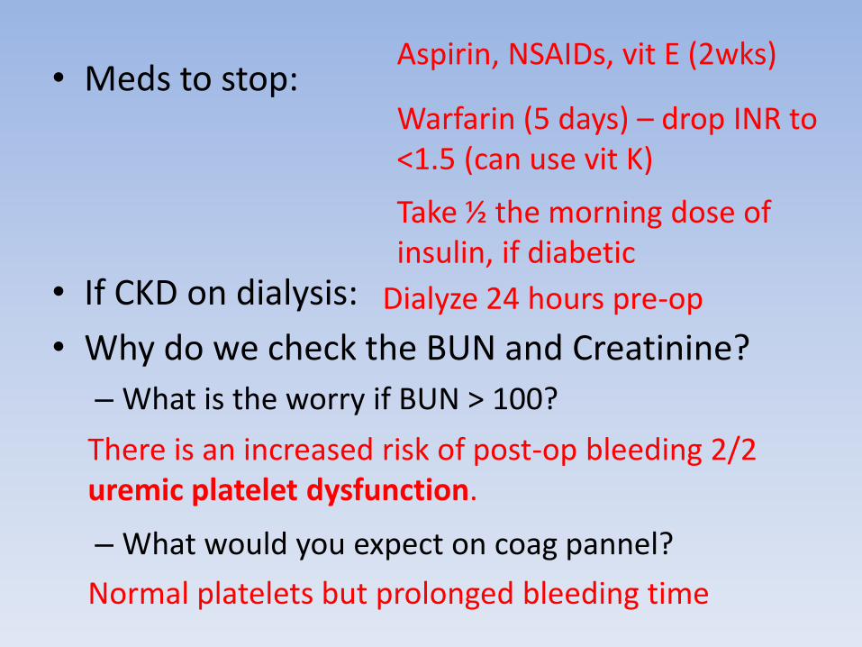
• Meds to stop:
• If CKD on dialysis:
• Why do we check the BUN and Creatinine?
– What is the worry if BUN > 100?
– What would you expect on coag pannel?
Aspirin, NSAIDs, vit E (2wks)
Warfarin (5 days) – drop INR to <1.5 (can use vit K)
Take ½ the morning dose of insulin, if diabetic
Dialyze 24 hours pre-op
There is an increased risk of post-op bleeding 2/2 uremic platelet dysfunction.
Normal platelets but prolonged bleeding time
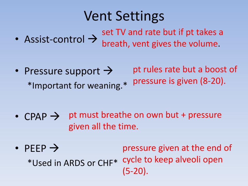
Vent Settings
• Assist-control
• Pressure support
*Important for weaning.*
• CPAP
• PEEP
*Used in ARDS or CHF*
set TV and rate but if pt takes a breath, vent gives the volume.
pt rules rate but a boost of pressure is given (8-20).
pt must breathe on own but + pressure given all the time.
pressure given at the end of cycle to keep alveoli open (5-20).
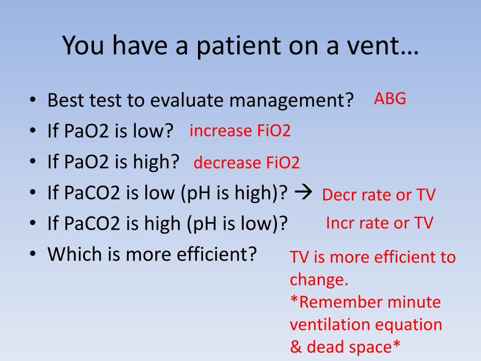
You have a patient on a vent…
• Best test to evaluate management?
• If PaO2 is low?
• If PaO2 is high?
• If PaCO2 is low (pH is high)?
• If PaCO2 is high (pH is low)?
• Which is more efficient?
ABG
increase FiO2
decrease FiO2
Decr rate or TV
Incr rate or TV
TV is more efficient to change.*Remember minute ventilation equation & dead space*

Acid Base Disorders• Check pH if <7.4 = acidotic.• Next Check HCO3 and pCO2:
– If HCO2 is high and pCO2 is high?– If HCO2 is low and pCO2 is low?
• Next Check anion gap (Na – [Cl + HCO3]), normal?• Gap acidosis =• Non-gap acidosis =
• Check pH if >7.4 = alkalotic.• Next Check HCO3 and pCO2:
– If HCO3 is low and pCO2 is low – If HCO3 is high and pCO2 is high
• Next Check urine [Cl] • If [Cl] < 20• If [Cl] > 20
Metabolic Alkalosis
Vomiting/NG, antactids, diuretics
Conn’s, Bartter’s Gittleman’s.
Respiratory Alkalosis
Respiratory Acidosis
Metabolic Acidosis
MUDPILESdiarrhea, diuretic, RTAs (I< II, IV)
8-12
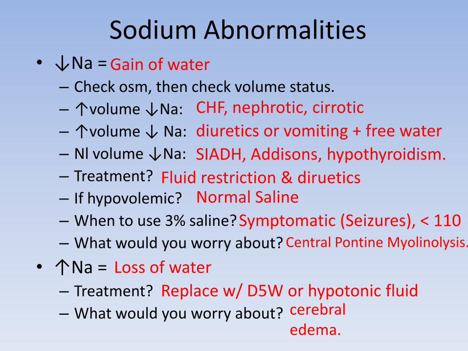
Sodium Abnormalities• ↓Na =
– Check osm, then check volume status.
– ↑volume ↓Na:
– ↑volume ↓ Na:
– Nl volume ↓Na:
– Treatment?
– If hypovolemic?
– When to use 3% saline?
– What would you worry about?
• ↑Na = – Treatment?
– What would you worry about?
Gain of water
CHF, nephrotic, cirrotic
diuretics or vomiting + free water
SIADH, Addisons, hypothyroidism.
Fluid restriction & dirueticsNormal Saline
Symptomatic (Seizures), < 110Central Pontine Myolinolysis.
Loss of water
Replace w/ D5W or hypotonic fluidcerebral edema.
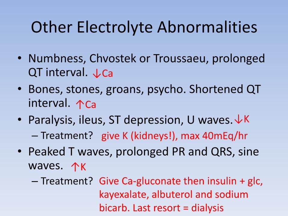
Other Electrolyte Abnormalities
• Numbness, Chvostek or Troussaeu, prolonged QT interval.
• Bones, stones, groans, psycho. Shortened QT interval.
• Paralysis, ileus, ST depression, U waves. – Treatment?
• Peaked T waves, prolonged PR and QRS, sine waves. – Treatment?
↓Ca
↑Ca
↓K
give K (kidneys!), max 40mEq/hr
↑K
Give Ca-gluconate then insulin + glc, kayexalate, albuterol and sodium bicarb. Last resort = dialysis
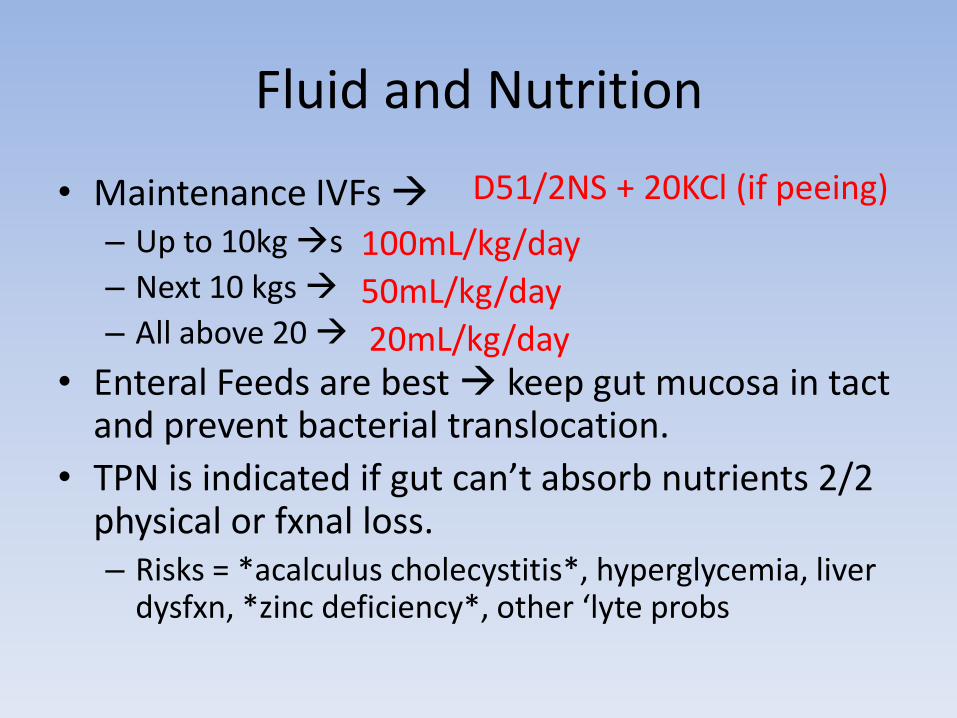
Fluid and Nutrition
• Maintenance IVFs – Up to 10kg s
– Next 10 kgs
– All above 20
• Enteral Feeds are best keep gut mucosa in tact and prevent bacterial translocation.
• TPN is indicated if gut can’t absorb nutrients 2/2 physical or fxnal loss. – Risks = *acalculus cholecystitis*, hyperglycemia, liver
dysfxn, *zinc deficiency*, other ‘lyte probs
D51/2NS + 20KCl (if peeing)
100mL/kg/day
50mL/kg/day
20mL/kg/day

Burn
• Circumferential burns?
• Look for singed nose hairs, wheezing, soot in mouth/nose?
• Patient w/ confusion, HA, cherry red skin?– Best test?
– Treatment?
www.readykor.com/docs/burns_files/burns9.jpg
http://en.wikipedia.org/wiki/Burn
http://emedicine.medscape.com/article/769193-media
1st degree2nd degree 3rd degree
Consider escharotomy
Low threshold for intubation
Check carboxyHb (pulse ox = worthless)
100% O2 (hyperbaric if CO-Hb is ↑↑↑
Clotting & Bleeding• Clotting-
– In old people? – Edema, HTN, & foamy pee? – In young person w/ +FH– What’s special about ATIII def? – Young woman w/ mult. SABs?– Post op, ↓plts, clots
• What do you treat w/?
• Bleeding– Isolated decr in plts? – Normal plts but incr bleeding time & PTT?– Low plts, Incr PT, PTT, BT, low fibrinogen, high Ddimer
and schistocytes?
Think cancerNephrotic syndrome
Factor V LeidenHeparin won’t work
Lupus AnticoagulantHIT! (If heparin w/in 5-14 days
Leparudin or agatroban
ITP
vWD
DIC!! Caused by gram – sepsis, carcinomatosis, OB stuff
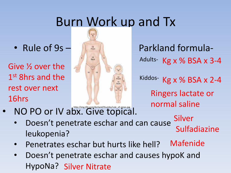
Burn Work up and Tx
• Rule of 9s – Parkland formula-• Adults-
•
• Kiddos-
•
•
http://img.tfd.com/dorland/thumbs/rule_of-nines.jpg
• NO PO or IV abx. Give topical.• Doesn’t penetrate eschar and can cause
leukopenia?• Penetrates eschar but hurts like hell? • Doesn’t penetrate eschar and causes hypoK and
HypoNa?
Kg x % BSA x 3-4
Kg x % BSA x 2-4
Give ½ over the 1st 8hrs and the rest over next 16hrs
SilverSulfadiazine
Mafenide
Silver Nitrate
Ringers lactate or normal saline
Other Burn Stuff
• Chemical burn, what to do? • Electrical Burn, best 1st step?• If abnormal? • If urine dipstick + for blood but microscopic exam
is negative for RBCs? • Then what do you check?• If affected extremity is extremley tender, numb,
white, cold with barely dopplerable pulses?
– Criteria?– Treatment?
Irrigate >30min prior to ER
EKG!
48 hours of telemetry (also if LOC)
Myoglobinuria ATN
K+! (When cells break)
Compartment syndrome!!
5 Ps or compartment pressure >30mmHg
May require fasciotomy. (at bedside!)
Trauma Drama
• Airway-
– If trauma patient comes in unconscious?
– If GCS < 8?
– If guy stung by a bee, developing stridor and tripod posturing?
– If guy stabbed in the neck, GCS = 15, expanding mass in lateral neck?
– If guy stabbed in the neck, crackly sounds w/ palpating anterior neck tissues?
– If huge facial trauma, blood obscures oral and nasal airway, & GCS of 7?
Intubate!
Intubate!
Intubate!
Intubate!
fiberoptic broncoscope
cricothyroidotomy
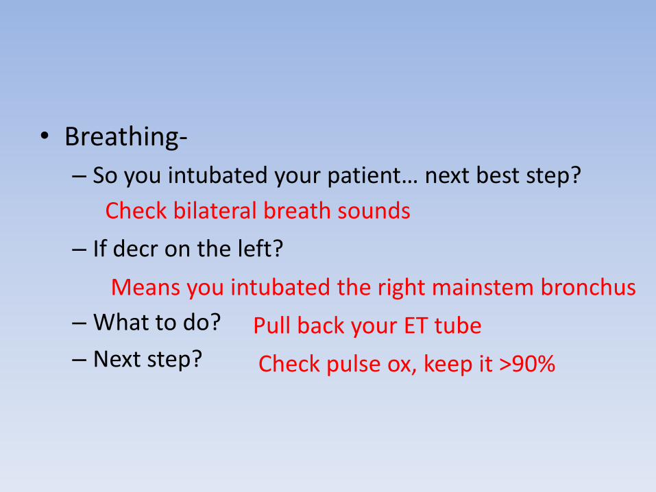
• Breathing-
– So you intubated your patient… next best step?
– If decr on the left?
– What to do?
– Next step?
Check bilateral breath sounds
Means you intubated the right mainstem bronchus
Pull back your ET tube
Check pulse ox, keep it >90%
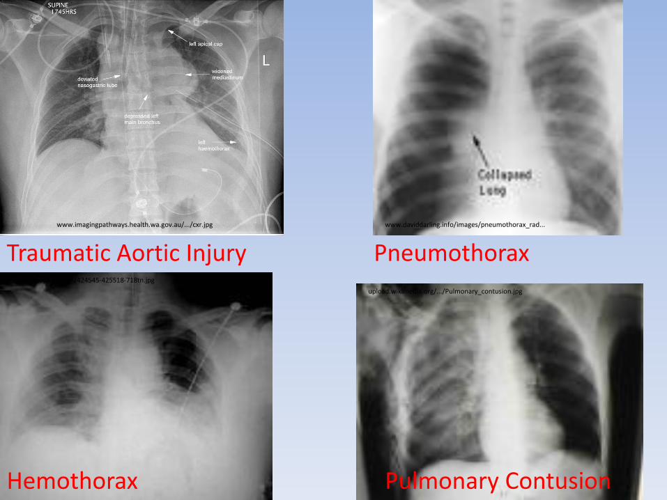
www.imagingpathways.health.wa.gov.au/.../cxr.jpg
img.medscape.com/.../424545-425518-718tn.jpg
www.daviddarling.info/images/pneumothorax_rad...
Traumatic Aortic Injury Pneumothorax
Hemothorax
upload.wikimedia.org/.../Pulmonary_contusion.jpg
Pulmonary Contusion
Chest Trauma
• A patient has inward mvmt of the right ribcage upon inspiration. – Dx? – Tx?
• A patient has confusion, petechial rash in chest, axilla and neck and acute SOB. – Dx?– When to suspect it?
• A patient dies suddenly after a 3rd year medical student removes a central line. – Dx? – When else to suspect it?
Flail chest. >3 consec rib fracturesO2 and pain control. With what?*
Fat embolismAfter long bone fx (esp femur)
Air embolismLung trauma, vent use, during heart vessel surgery.
• Cardiovascular-
– If hypotensive, tachycardic?
– If flat neck veins and normal CVP?
– Next best step?
– If muffled <3 sounds, JVD, electrical alternans, pulsus paradoxus?
• Confirmatory test?
• Treatment?
– If decr BS on one side, tracheal deviation AWAY from collapsed lung?
• Next best step?
Worry about shock
Hypovolemic/Hemorrhagic
2 large bore periph IV- 2L NS or LR over 20min followed by blood.
Pericardial Tamponade
FAST scanNeedle decompression, pericardial window or median sternotomy
Tension Pneumothorax
Needle decompression, followed by a chest tube. DON’T do a CXR!!!
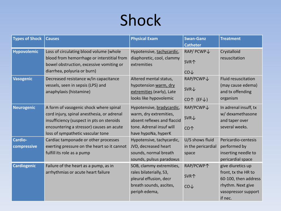
ShockTypes of Shock Causes Physical Exam Swan-Ganz
Catheter
Treatment
Hypovolemic Loss of circulating blood volume (whole
blood from hemorrhage or interstitial from
bowel obstruction, excessive vomiting or
diarrhea, polyuria or burn)
Hypotensive, tachycardic,
diaphoretic, cool, clammy
extremities
RAP/ PCWP↓
SVR↑
CO↓
Crystalloid
resuscitation
Vasogenic Decreased resistance w/in capacitance
vessels, seen in sepsis (LPS) and
anaphylaxis (histamine)
Altered mental status,
hypotension warm, dry
extremities (early), Late
looks like hypovolemic
RAP/PCWP↓
SVR↓
CO↑ (EF↓)
Fluid resuscitation
(may cause edema)
and tx offending
organism
Neurogenic A form of vasogenic shock where spinal
cord injury, spinal anesthesia, or adrenal
insufficiency (suspect in pts on steroids
encountering a stressor) causes an acute
loss of sympathetic vascular tone
Hypotensive, bradycardic,
warm, dry extremities,
absent reflexes and flaccid
tone. Adrenal insuf will
have hypoNa, hyperK
RAP/PCWP↓
SVR↓
CO↑
In adrenal insuff, tx
w/ dexamethasone
and taper over
several weeks.
Cardio-
compressive
Cardiac tamponade or other processes
exerting pressure on the heart so it cannot
fulfill its role as a pump
Hypotensive, tachycardic,
JVD, decreased heart
sounds, normal breath
sounds, pulsus paradoxus
U/S shows fluid
in the pericardial
space
Pericardio-centesis
performed by
inserting needle to
pericardial space
Cardiogenic Failure of the heart as a pump, as in
arrhythmias or acute heart failure
SOB, clammy extremities,
rales bilaterially, S3,
pleural effusion, decr
breath sounds, ascites,
periph edema,
RAP/PCWP↑
SVR↑
CO↓
give diuretics up
front, tx the HR to
60-100, then address
rhythm. Next give
vasopressor support
if nec.
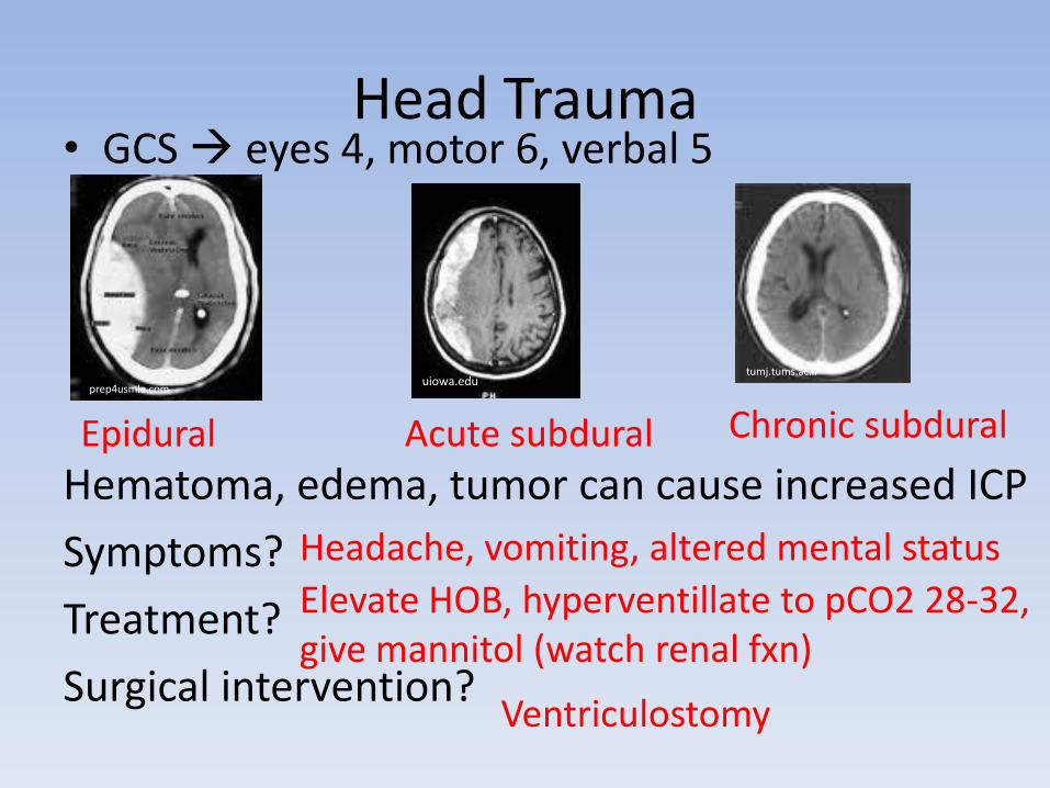
Head Trauma• GCS eyes 4, motor 6, verbal 5
Hematoma, edema, tumor can cause increased ICP
Symptoms?
Treatment?
Surgical intervention?
tumj.tums.ac.iruiowa.edu
prep4usmle.com
Epidural Acute subdural Chronic subdural
Headache, vomiting, altered mental status
Elevate HOB, hyperventillate to pCO2 28-32, give mannitol (watch renal fxn)
Ventriculostomy
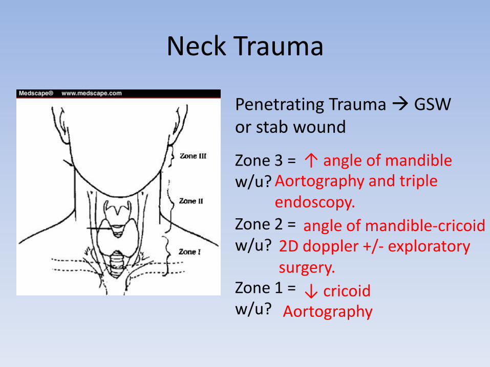
Neck Trauma
Zone 3 =w/u?
Zone 2 =w/u?
Zone 1 =w/u?
Penetrating Trauma GSW or stab wound
↑ angle of mandibleAortography and triple endoscopy.
angle of mandible-cricoid2D doppler +/- exploratory surgery.
↓ cricoidAortography
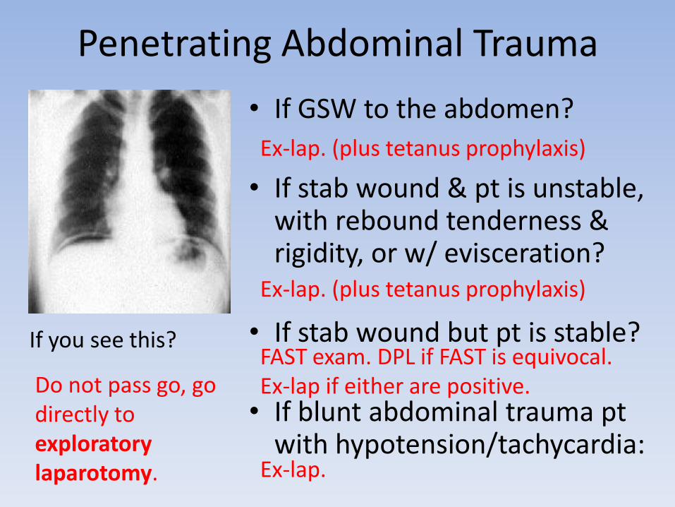
Penetrating Abdominal Trauma
• If GSW to the abdomen?
• If stab wound & pt is unstable, with rebound tenderness & rigidity, or w/ evisceration?
• If stab wound but pt is stable?
• If blunt abdominal trauma pt with hypotension/tachycardia:
If you see this?
Do not pass go, go directly to exploratory laparotomy.
Ex-lap. (plus tetanus prophylaxis)
Ex-lap. (plus tetanus prophylaxis)
Ex-lap.
FAST exam. DPL if FAST is equivocal. Ex-lap if either are positive.
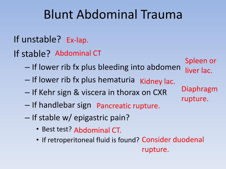
Blunt Abdominal Trauma
If unstable?
If stable?
– If lower rib fx plus bleeding into abdomen
– If lower rib fx plus hematuria
– If Kehr sign & viscera in thorax on CXR
– If handlebar sign
– If stable w/ epigastric pain?
• Best test?
• If retroperitoneal fluid is found?
Ex-lap.
Abdominal CTSpleen or liver lac.
Kidney lac. Diaphragm rupture.
Pancreatic rupture.
Abdominal CT. Consider duodenal rupture.

Pelvic Trauma• If hypotensive, tachycardic
• Can bleed out into pelvis stop bleeding by fixing fxinternal if stable, external if not.
• If blood at the urethral meatus and a high riding prostate?
• Next best test?
• If normal?
• What are you looking for?
If extraperitoneal extravasation?
If intraperitoneal extravasation?
FAST and DPL to r/o bleeding in abdominal cavity.
Consider pelvic fracture w/ urethral or bladder injury.
Retrograde urethrogram (NOT FOLEY!)
Retrograde cystogram to evaluate bladder
Check for extravasation of dye. Take 2 views to ID trigone injury.
Bed rest + foley
Ex-lap and surgical repair
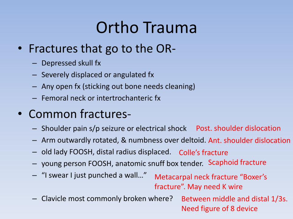
Ortho Trauma• Fractures that go to the OR-
– Depressed skull fx
– Severely displaced or angulated fx
– Any open fx (sticking out bone needs cleaning)
– Femoral neck or intertrochanteric fx
• Common fractures-– Shoulder pain s/p seizure or electrical shock
– Arm outwardly rotated, & numbness over deltoid.
– old lady FOOSH, distal radius displaced.
– young person FOOSH, anatomic snuff box tender.
– “I swear I just punched a wall…”
– Clavicle most commonly broken where?
Post. shoulder dislocation
Ant. shoulder dislocation
Colle’s fractureScaphoid fracture
Metacarpal neck fracture “Boxer’s fracture”. May need K wire
Between middle and distal 1/3s. Need figure of 8 device
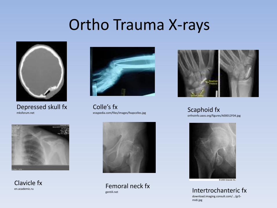
Ortho Trauma X-rays
Depressed skull fx mksforum.net
Colle’s fxxraypedia.com/files/images/fxapcolles.jpg
Scaphoid fxorthoinfo.aaos.org/figures/A00012F04.jpg
Clavicle fxen.academic.ru
Femoral neck fxgentili.net Intertrochanteric fx
download.imaging.consult.com/.../gr5-midi.jpg
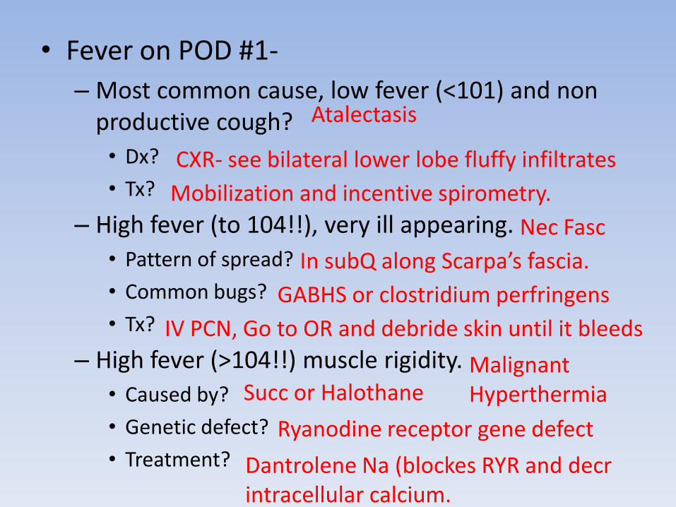
• Fever on POD #1-
– Most common cause, low fever (<101) and non productive cough?
• Dx?
• Tx?
– High fever (to 104!!), very ill appearing.
• Pattern of spread?
• Common bugs?
• Tx?
– High fever (>104!!) muscle rigidity.
• Caused by?
• Genetic defect?
• Treatment?
Atalectasis
CXR- see bilateral lower lobe fluffy infiltrates
Mobilization and incentive spirometry.
Nec Fasc
In subQ along Scarpa’s fascia.
GABHS or clostridium perfringens
IV PCN, Go to OR and debride skin until it bleeds
Malignant HyperthermiaSucc or Halothane
Ryanodine receptor gene defect
Dantrolene Na (blockes RYR and decr intracellular calcium.
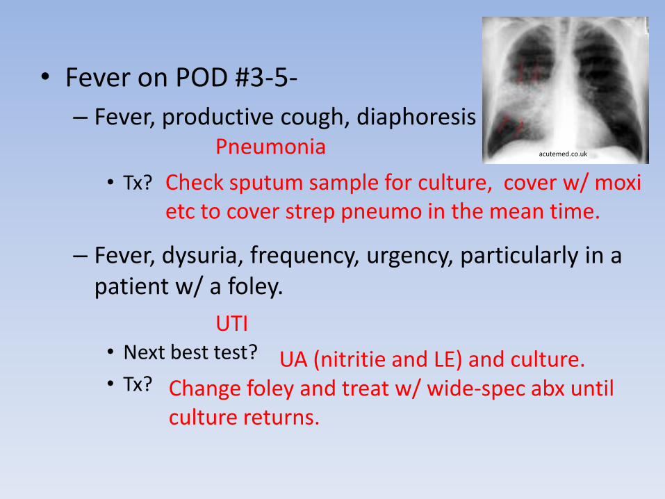
• Fever on POD #3-5-
– Fever, productive cough, diaphoresis
• Tx?
– Fever, dysuria, frequency, urgency, particularly in a patient w/ a foley.
• Next best test?
• Tx?
acutemed.co.ukPneumonia
Check sputum sample for culture, cover w/ moxi etc to cover strep pneumo in the mean time.
UTI
UA (nitritie and LE) and culture. Change foley and treat w/ wide-spec abx until culture returns.
• Fever > POD 7-– Pain & tenderness at IV site
• Tx?
– Pain @ incision site, edema, induration but no drainage.• Tx?
– Pain @ incision site, induration WITH drainage. • Tx?
– Pain w/ salmon colored fluid from incision. • Tx?
– Unexplained fever• Dx?
• Tx?
– Random thyrotoxicosis, thrombophlebitis, adrenal insufficiency, lymphangitis, sepsis.
Central line infection
Do blood cx from the line. Pull it. Abx to cover staph.
Cellulits
Do blood cx and start antibiotics Simple Wound InfectionOpen wound and repack. No abx necessary
Dehiscence
Surgical emergency! Go to OR, IV abx, primary closure of fascia
Abdominal Abscess
CT w/ oral, IV and rectal contrast to find it. Diagnostic lap.
Drain it! Percutaneously, IR-guided, or surgically.
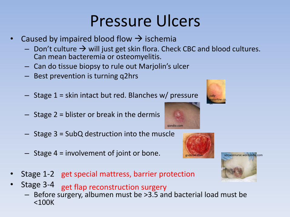
Pressure Ulcers• Caused by impaired blood flow ischemia
– Don’t culture will just get skin flora. Check CBC and blood cultures. Can mean bacteremia or osteomyelitis.
– Can do tissue biopsy to rule out Marjolin’s ulcer – Best prevention is turning q2hrs
– Stage 1 = skin intact but red. Blanches w/ pressure
– Stage 2 = blister or break in the dermis
– Stage 3 = SubQ destruction into the muscle
– Stage 4 = involvement of joint or bone.
• Stage 1-2• Stage 3-4
– Before surgery, albumen must be >3.5 and bacterial load must be <100K
judy-waterlow.co.uk
qondio.com
gndmoh.com seejanenurse.wordpress.com
get special mattress, barrier protection
get flap reconstruction surgery
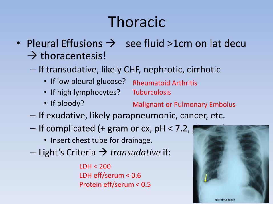
Thoracic• Pleural Effusions see fluid >1cm on lat decu thoracentesis!– If transudative, likely CHF, nephrotic, cirrhotic
• If low pleural glucose?
• If high lymphocytes?
• If bloody?
– If exudative, likely parapneumonic, cancer, etc.
– If complicated (+ gram or cx, pH < 7.2, glc < 60): • Insert chest tube for drainage.
– Light’s Criteria transudative if:
ncbi.nlm.nih.gov
LDH < 200LDH eff/serum < 0.6Protein eff/serum < 0.5
Rheumatoid ArthritisTuburculosis
Malignant or Pulmonary Embolus
• Spontaneous Pneumothorax subpleural bleb ruptures lung collapse. – Suspect in tall, thin young men w/ sudden dyspnea (or
asthma or COPD-emphysema)– Dx w/ CXR, Tx w/ chest tube placement– Indications for surgery = ipsi or contra recurrence,
bilateral, incomplete lung expansion, pilot, scuba, live in remote area VATS, pleurodesis (bleo, iodine or talc)
• Lung Abscess usually 2/2 aspiration (drunk, elderly, enteral feeds)– Most often in post upper or sup lower lobes– Tx initially w/ abx IV PCN or clinda– Indications for surgery = abx fail, abscess >6cm, or if empyema is present.
www.meddean.luc.edu
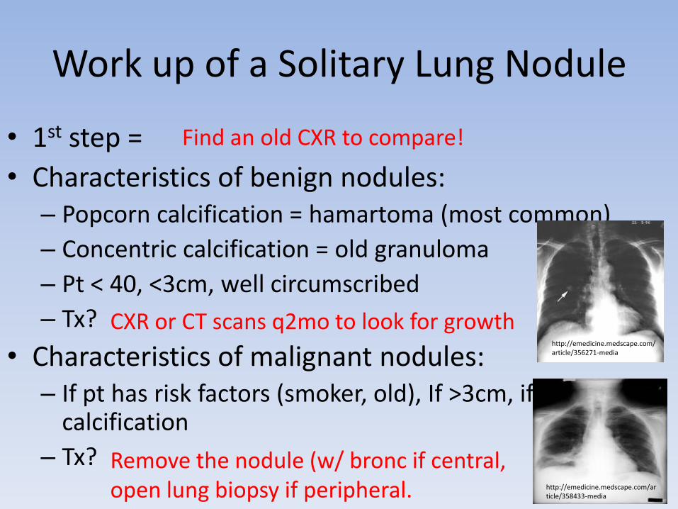
Work up of a Solitary Lung Nodule
• 1st step =
• Characteristics of benign nodules:– Popcorn calcification = hamartoma (most common)
– Concentric calcification = old granuloma
– Pt < 40, <3cm, well circumscribed
– Tx?
• Characteristics of malignant nodules:– If pt has risk factors (smoker, old), If >3cm, if eccentric
calcification
– Tx?
http://emedicine.medscape.com/article/356271-media
http://emedicine.medscape.com/article/358433-media
Find an old CXR to compare!
CXR or CT scans q2mo to look for growth
Remove the nodule (w/ bronc if central, open lung biopsy if peripheral.
A patient presents with weight loss, cough, dyspnea, hemoptysis, repeated pnia or lung
collapse.• MC cancer in non-smokers?
• Location and mets?
• Characteristics of effusion?
• Patient with kidney stones, constipation and malaise low PTH + central lung mass?
• Patient with shoulder pain, ptosis, constricted pupil, and facial edema?
• Patient with ptosis better after 1 minute of upward gaze?
• Old smoker presenting w/ Na = 125, moist mucus membranes, no JVD?
• CXR showing peripheral cavitation and CT showing distant mets?
Adenocarcinoma. Occurs in scars of old pnia
Peripheral cancer. Mets to liver, bone, brain and adrenals
Exudative with high hyaluronidase
Squamous cell carcinoma.Paraneoplastic syndrome 2/2 secretion of PTH-rP. Low PO4, High Ca
Superior Sulcus Syndrome from Small cell carcinoma. Also a central cancer.
Lambert Eaton Syndrome from small cell carcinoma. Ab to pre-syn Ca chan
SIADH from small cell carcinoma. Produces Euvolemic hyponatremia. Fluid restrict +/- 3% saline in <112
Large Cell Carcinoma
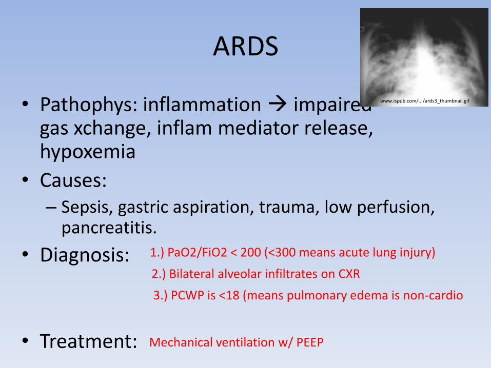
ARDS
• Pathophys: inflammation impaired gas xchange, inflam mediator release, hypoxemia
• Causes:– Sepsis, gastric aspiration, trauma, low perfusion,
pancreatitis.
• Diagnosis:
• Treatment:
www.ispub.com/.../ards3_thumbnail.gif
1.) PaO2/FiO2 < 200 (<300 means acute lung injury)
2.) Bilateral alveolar infiltrates on CXR
3.) PCWP is <18 (means pulmonary edema is non-cardio
Mechanical ventilation w/ PEEP
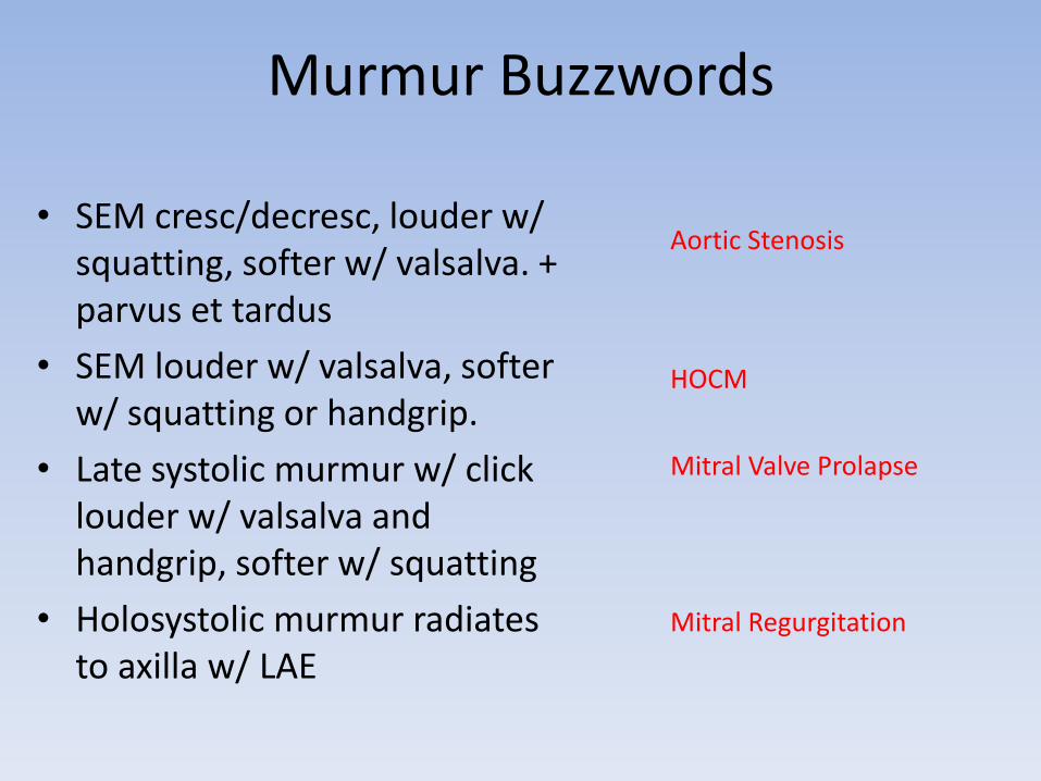
Murmur Buzzwords
• SEM cresc/decresc, louder w/ squatting, softer w/ valsalva. + parvus et tardus
• SEM louder w/ valsalva, softer w/ squatting or handgrip.
• Late systolic murmur w/ click louder w/ valsalva and handgrip, softer w/ squatting
• Holosystolic murmur radiates to axilla w/ LAE
Aortic Stenosis
HOCM
Mitral Valve Prolapse
Mitral Regurgitation
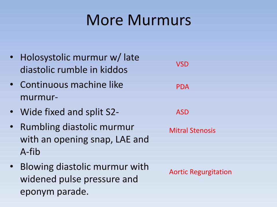
More Murmurs
• Holosystolic murmur w/ late diastolic rumble in kiddos
• Continuous machine like murmur-
• Wide fixed and split S2-
• Rumbling diastolic murmur with an opening snap, LAE and A-fib
• Blowing diastolic murmur with widened pulse pressure and eponym parade.
VSD
PDA
ASD
Mitral Stenosis
Aortic Regurgitation

• Bad breath & snacks in
the AM.
• True or false?
• Dysphagia to liquids & solids. Dysphagia worse w/ hot & cold liquids + chest pain that feels like MI w/ NO regurg
sxs.
• Epigastric pain worse after
eating or when laying down
cough, wheeze, hoarse.
• Indications for surgery?
jykang.co.uk
ajronline.org
Zenker’s diverticulum. Tx w/ surgery
False. Only contains mucosa
Achalasia. Tx w/ CCB, nitrates, botox, or heller myotomyAssoc w/ Chagas dz and esophageal cancer.
Diffuse esphogeal spasm.Tx w/ CCB or nitrates
GERD. Most sensitive test is 24-hr pH monitoring. Do endoscopy if “danger signs” present. Tx w/ behav mod 1st, then antacids, H2 block, PPI.
bleeding, stricture, Barrett’s, incompetent LES, max dose PPI w/ still sxs, or no want meds.
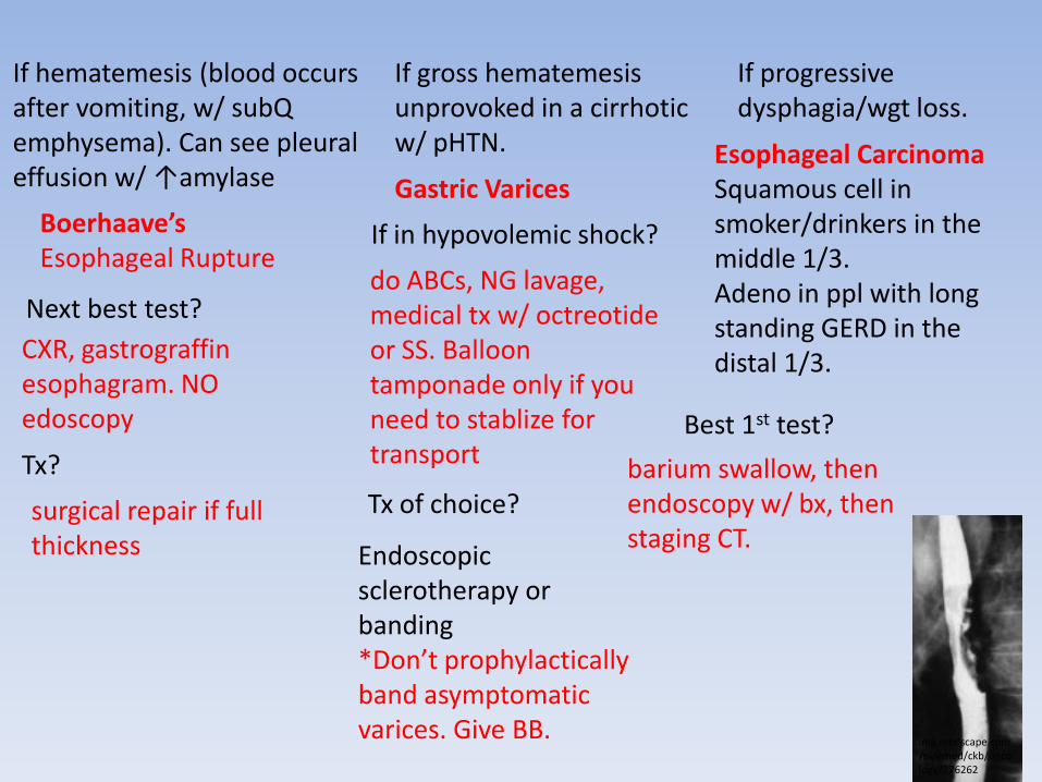
If hematemesis (blood occurs after vomiting, w/ subQ emphysema). Can see pleural effusion w/ ↑amylase
If gross hematemesis unprovoked in a cirrhotic w/ pHTN.
If progressive dysphagia/wgt loss.
img.medscape.com/pi/emed/ckb/oncology/276262
Boerhaave’s Esophageal Rupture
Next best test?
CXR, gastrograffin esophagram. NO edoscopy
Tx?
surgical repair if full thickness
Gastric Varices
If in hypovolemic shock?
do ABCs, NG lavage, medical tx w/ octreotide or SS. Balloon tamponade only if you need to stablize for transport
Tx of choice?
Endoscopic sclerotherapy or banding*Don’t prophylactically band asymptomatic varices. Give BB.
Esophageal CarcinomaSquamous cell in smoker/drinkers in the middle 1/3. Adeno in ppl with long standing GERD in the distal 1/3.
Best 1st test?
barium swallow, then endoscopy w/ bx, then staging CT.
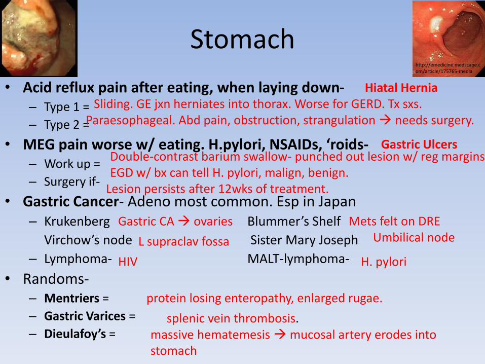
Stomach
• Acid reflux pain after eating, when laying down-– Type 1 =
– Type 2 =
• MEG pain worse w/ eating. H.pylori, NSAIDs, ‘roids-– Work up =
– Surgery if-
• Gastric Cancer- Adeno most common. Esp in Japan– Krukenberg Blummer’s Shelf
Virchow’s node Sister Mary Joseph
– Lymphoma- MALT-lymphoma-
• Randoms-– Mentriers =
– Gastric Varices =
– Dieulafoy’s =
http://emedicine.medscape.com/article/175765-media
Hiatal HerniaSliding. GE jxn herniates into thorax. Worse for GERD. Tx sxs.
Paraesophageal. Abd pain, obstruction, strangulation needs surgery.
Gastric UlcersDouble-contrast barium swallow- punched out lesion w/ reg margins. EGD w/ bx can tell H. pylori, malign, benign.
Lesion persists after 12wks of treatment.
Gastric CA ovaries Mets felt on DRE
L supraclav fossa Umbilical node
HIV H. pylori
protein losing enteropathy, enlarged rugae.
splenic vein thrombosis.massive hematemesis mucosal artery erodes into stomach
Duodenum• MEG pain better w/ eating
– 95% assoc w/ H. pylori – Healthy pts < 45y/o can do trial of H2 block or PPI– Dx?
– Tx?
• What to suspect if MEG pain/ulcers don’t resolve? – Best test?– Tx?– What else to look for?
• A patient has bilious vomiting and post-prandial pain. Recently lost 200lbs on “Biggest Loser”.– Pathophys-– Tx?
Duodenal Ulcers
blood, stool or breath test for H. pylori but endoscopy w/ biopsy (CLO test) is best b/c it can also exclude cancer.
PPI, clarithromycin & amoxicillin for 2wks. Breath or stool test can be test of cure.
ZE Syndrome
Secretin Stim Test (find inapprop high gastrin)Surgical resection of pancreatic/duodenal tumor
Pituitary and Parathyroid problems.
SMA Syndrome3rd part of duodenum compressed by AA and SMA
by restoring weight/nutrition. Can do Roux-en-Y
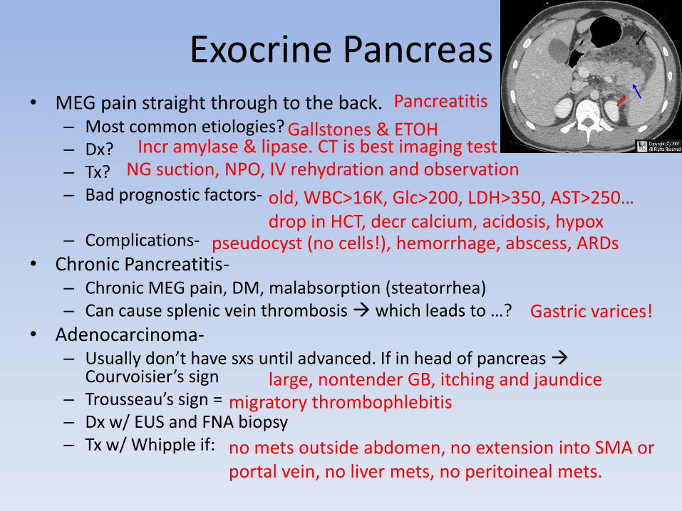
Exocrine Pancreas• MEG pain straight through to the back.
– Most common etiologies? – Dx? – Tx? – Bad prognostic factors-
– Complications-
• Chronic Pancreatitis-– Chronic MEG pain, DM, malabsorption (steatorrhea)– Can cause splenic vein thrombosis which leads to …?
• Adenocarcinoma-– Usually don’t have sxs until advanced. If in head of pancreas
Courvoisier’s sign – Trousseau’s sign =– Dx w/ EUS and FNA biopsy– Tx w/ Whipple if:
Pancreatitis
Gallstones & ETOHIncr amylase & lipase. CT is best imaging test
NG suction, NPO, IV rehydration and observation
old, WBC>16K, Glc>200, LDH>350, AST>250… drop in HCT, decr calcium, acidosis, hypox
pseudocyst (no cells!), hemorrhage, abscess, ARDs
Gastric varices!
large, nontender GB, itching and jaundicemigratory thrombophlebitis
no mets outside abdomen, no extension into SMA or portal vein, no liver mets, no peritoineal mets.
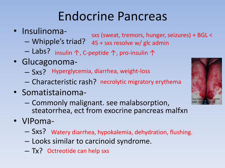
Endocrine Pancreas• Insulinoma-
– Whipple’s triad? – Labs?
• Glucagonoma-– Sxs? – Characteristic rash?
• Somatistainoma-– Commonly malignant. see malabsorption,
steatorrhea, ect from exocrine pancreas malfxn
• VIPoma-– Sxs? – Looks similar to carcinoid syndrome. – Tx?
img.medscape.com/.../1048885-1093550-244.jpg
sxs (sweat, tremors, hunger, seizures) + BGL < 45 + sxs resolve w/ glc admin
insulin ↑, C-peptide ↑, pro-insulin ↑
Hyperglycemia, diarrhea, weight-loss
necrolytic migratory erythema
Watery diarrhea, hypokalemia, dehydration, flushing.
Octreotide can help sxs
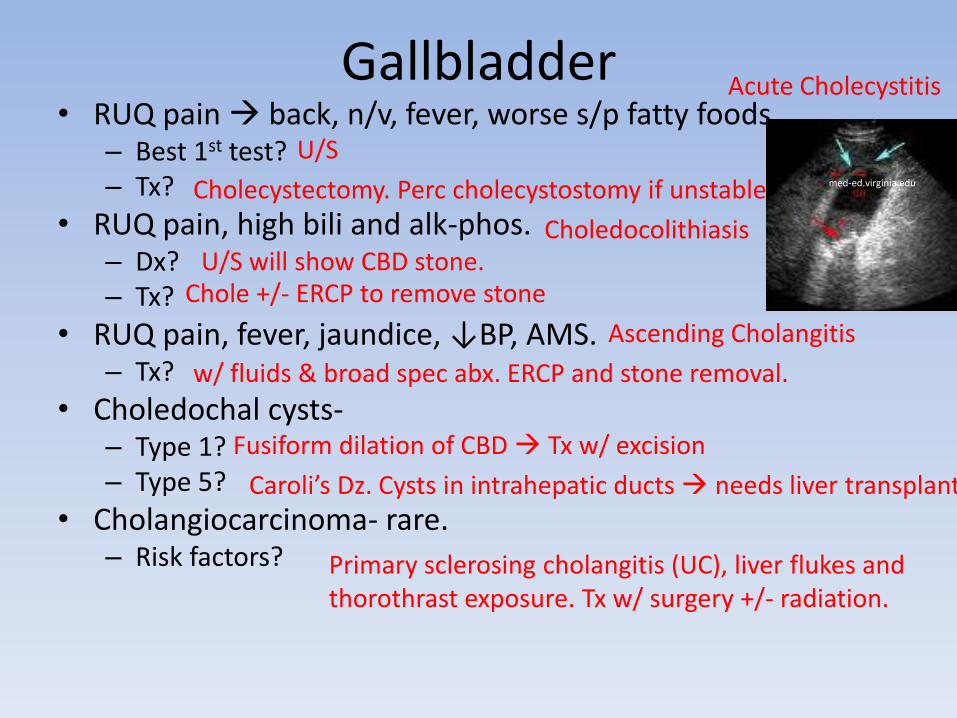
Gallbladder• RUQ pain back, n/v, fever, worse s/p fatty foods.
– Best 1st test? – Tx?
• RUQ pain, high bili and alk-phos. – Dx? – Tx?
• RUQ pain, fever, jaundice, ↓BP, AMS. – Tx?
• Choledochal cysts-– Type 1?– Type 5?
• Cholangiocarcinoma- rare. – Risk factors?
med-ed.virginia.edu
Acute Cholecystitis
U/S
Cholecystectomy. Perc cholecystostomy if unstable
CholedocolithiasisU/S will show CBD stone.
Chole +/- ERCP to remove stone
Ascending Cholangitis
w/ fluids & broad spec abx. ERCP and stone removal.
Fusiform dilation of CBD Tx w/ excision
Caroli’s Dz. Cysts in intrahepatic ducts needs liver transplant
Primary sclerosing cholangitis (UC), liver flukes and thorothrast exposure. Tx w/ surgery +/- radiation.
Liver• Hepatitis-
– AST = 2x ALT – AST > ALT high (1000s) – AST & ALT high s/p hemorrhage, surg, or sepsis
• Cirrhosis and Portal HTN-– Tx- SS and VP vasocontrict to decrease portal pressure, betablockers
also decrease portal pressure.– Don’t need to treat esophageal varices prophyactically, but
band/burn them once they bleed once. – TIPS relieves portal HTN but…
• Treat with:
• Hepatocellular Carcinoma– RF-
– Dx w/ high AFP (in 70%), CT/MRI. – Tx: can surgically remove solitary mass, use rads or cryoablation for
pallation of multiple.
Alcoholic heptatitis (reversible)
Viral hepatitisShock liver
worsens hepatic encephalopahty
Lactulose. helps rid body of ammonia.
chronic hepB carrier > hepC. Cirrhosis for any reason, plus aflatoxin or carbon tetrachloride.
More Liver*Women on OCP palpable abd mass or spontaneousrupture hemorrhagic shock
Dx? Tx?
*2nd MC benign liver tumor. W>M but less likely to rupture. No tx needed. *Bacterial Abscess.
Most common bugs? Tx?
RUQ pain, profouse sweating and rigors, palpable liver.Tx?
Patient from Mexico presents w/ RUQ and large liver cysts found on U/S
– Mode of transmission? – Lab findings? – Tx?
www.radswiki.net/main/images/thumb/3/3e/Hepat...
Hepatic Adenoma
U/S or MRID/c OCPs. Resect if large or pregnancy is desired
Focal Nodular Hyperplasia
E. coli, bacteriodes, enterococcus.
Surgical drainage and IV abx.Entamoeba histolytica
Metronidazole. DON’T drain it.
Enchinococcus. Hydatic cyst paracyte from dog feces.
eosinophilia, +Casoni skin test
albendazole and surgery to remove ENTIRE cyst, rupture anaphylaxis
Spleen• Post-Splenectomy
– Post op thrombocytosis >1mil give aspirin. – Prophylactic PCN + S. pneumo, H. flu and N. meningitidis vaccines.
• ITP-– Consider in isolated thrombocytopenia (bleeding gums, petechiae,
nosebleeds). – Decr plt count, incr megakaryocytes in marrow.– NO splenomegaly. – Tx w/ steroids 1st. If relapse splenectomy.
• Hereditary Spherocytosis-– See sxs of hemolytic anemia (jaundice, incr indir bili, LDH, decr
haptoglobin, elevated retic count) + spherocytes on smear and +osmotic frag test. Prone to gallstones.
– Tx w/ splenectomy (accessory spleen too).
• Traumatic Splenic Rupture-– Consider w/ L lower rib fx and intra abd hemorrhage. Can have Kehr’s
sign (irritates L diaphragm).
www.ezhemeonc.com/wp-content/upload
img.medscape.com/.../432648-432823-3042.jpg
Appendix• pain in umbilical area RLQ, n/v.
perf.
– Go to surgery if:
– If perforated/abscess?
• Carcinoid Tumor- #1 site:
– Carcinoid syndrome sxs?
– When do they happen?
– What else to look out for?
– If >2cm, @ base of appendix, or w/ + nodes
– Otherwise
Appendicitis
Clinical picture is convincing.
drain, abx (to cover e.coli & bacteriodes), and do interval appendectomy
Appendix!
Diarrhea, Wheezing.
When mets to liver. (1st pass metabolism)
Diarrhea, Dementia, Dermatitis
Hemicolectomy
Appendectomy is good enough
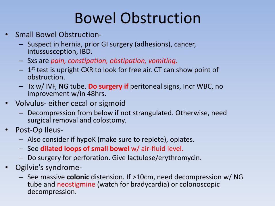
Bowel Obstruction• Small Bowel Obstruction-
– Suspect in hernia, prior GI surgery (adhesions), cancer, intussusception, IBD.
– Sxs are pain, constipation, obstipation, vomiting. – 1st test is upright CXR to look for free air. CT can show point of
obstruction. – Tx w/ IVF, NG tube. Do surgery if peritoneal signs, Incr WBC, no
improvement w/in 48hrs.
• Volvulus- either cecal or sigmoid– Decompression from below if not strangulated. Otherwise, need
surgical removal and colostomy.
• Post-Op Ileus-– Also consider if hypoK (make sure to replete), opiates. – See dilated loops of small bowel w/ air-fluid level. – Do surgery for perforation. Give lactulose/erythromycin.
• Ogilvie’s syndrome-– See massive colonic distension. If >10cm, need decompression w/ NG
tube and neostigmine (watch for bradycardia) or colonoscopicdecompression.
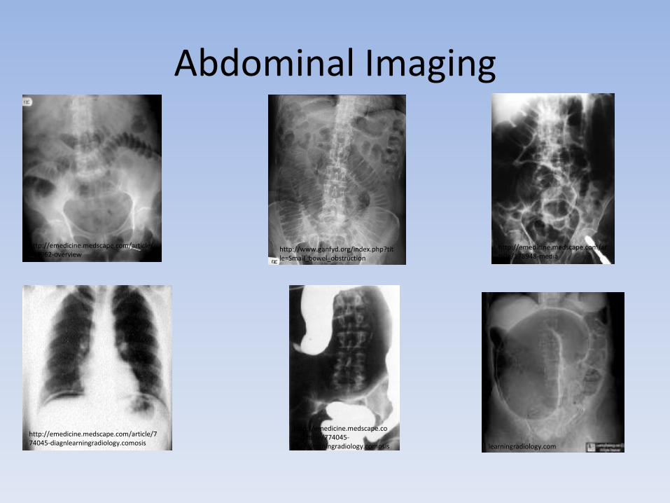
Abdominal Imaging
http://emedicine.medscape.com/article/374962-overview
http://emedicine.medscape.com/article/178948-media
http://www.ganfyd.org/index.php?title=Small_bowel_obstruction
http://emedicine.medscape.com/article/774045-diagnlearningradiology.comosis learningradiology.com
http://emedicine.medscape.com/article/774045-diagnlearningradiology.comosis
Hernias• Umbilical- in kiddos, close spontaneously by age 2. In
adults: 2/2 obesity, ascites or pregnancy.
• Indirect Inguinal- MC through inguinal ring (lat to epigastric vessles) in spermatic cord. R>L, more often congenital (patent proc vaginals)
• Direct Inguinal- through Hasselbeck’s triangle (med to epigastric vessles), more often acquired weakness.
• Femoral- more common in women.
• Tx- emergent surgical repair if incarcerated to avoid strangulation. Elective if reducible.
Inflammatory Bowel Disease
Treatment = ASA, sulfasalzine to maintain remission. Corticosteroids to induce remission. For CD, give metranidazole for ANY ulcer or abscess. Azathioprine, 6MP and methotrexate for severe dz.
• Involves terminal ileum?• Continuous involving rectum?• Incr risk for Primary
Sclerosing Cholangitis? • Fistulae likely? • Granulomas on biopsy? • Transmural inflammation? • Cured by colectomy? • Smokers have lower risk? • Highest risk of colon cancer? • Associated w/ p-ANCA?
Crohn’s. Mimics appendicitis. Fe deficiency.
UC. Rarely ileal backwash but never higher
UC. PSC leads to higher risk of cholangioCA
Crohn’s. Give metronidazole.
Crohn’s.
Crohn’s.
UC.
UC. Smokers have higher risk for Crohn’s.
UC. Another reason for colectomy.
UC.
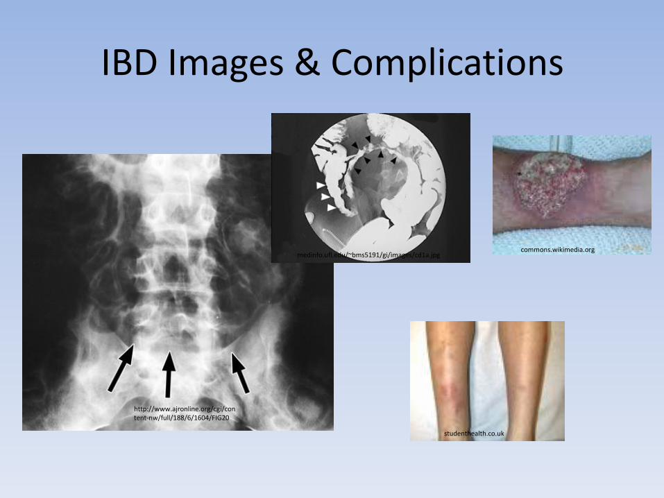
IBD Images & Complications
http://www.ajronline.org/cgi/content-nw/full/188/6/1604/FIG20
medinfo.ufl.edu/~bms5191/gi/images/cd1a.jpgcommons.wikimedia.org
studenthealth.co.uk
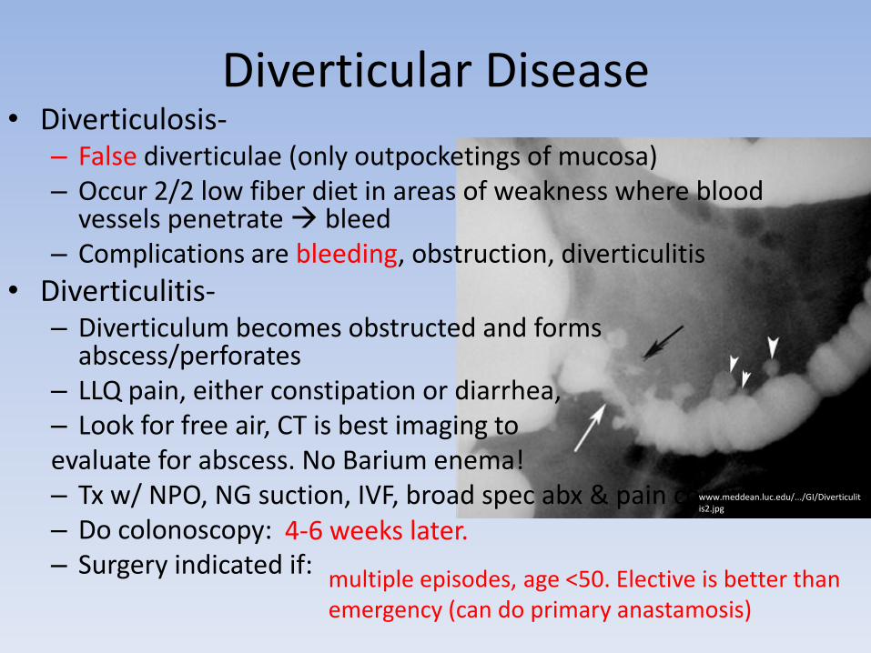
Diverticular Disease• Diverticulosis-
– False diverticulae (only outpocketings of mucosa)– Occur 2/2 low fiber diet in areas of weakness where blood
vessels penetrate bleed– Complications are bleeding, obstruction, diverticulitis
• Diverticulitis-– Diverticulum becomes obstructed and forms
abscess/perforates– LLQ pain, either constipation or diarrhea, – Look for free air, CT is best imaging to evaluate for abscess. No Barium enema!– Tx w/ NPO, NG suction, IVF, broad spec abx & pain control. – Do colonoscopy: – Surgery indicated if:
www.meddean.luc.edu/.../GI/Diverticulitis2.jpg
multiple episodes, age <50. Elective is better than emergency (can do primary anastamosis)
4-6 weeks later.
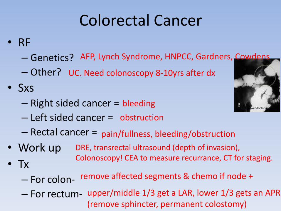
Colorectal Cancer• RF
– Genetics?
– Other?
• Sxs– Right sided cancer =
– Left sided cancer =
– Rectal cancer =
• Work up
• Tx– For colon-
– For rectum-
ourwebdoctor.com
AFP, Lynch Syndrome, HNPCC, Gardners, Cowdens
UC. Need colonoscopy 8-10yrs after dx
bleeding
obstruction
pain/fullness, bleeding/obstruction
DRE, transrectal ultrasound (depth of invasion), Colonoscopy! CEA to measure recurrance, CT for staging.
remove affected segments & chemo if node +
upper/middle 1/3 get a LAR, lower 1/3 gets an APR (remove sphincter, permanent colostomy)
AAA• Screening =
• Sxs = pulsatile abdominal mass. • Tx conservatively if:
• Surgery indicated if:• Rupture =
– severe sudden abdomen, flank or back, shock, tender pulsatile mass.
– 50% die before reaching the hospital.
• Post-op complications = #1 cause of death – Bloody diarrhea-– Weakness, decreased pain w/ preserved vibr, prop-– 1-2 yrs later if have brisk GI bleeding
men 65-75 who have ever smoked. Do abdominal U/S.
if <5cm and asymptomatic, monitor growth every 3-12mo.
>5cm, growing <4mm/yr
MI
Ischemic colitisASA syndrome
Aortoenteric Fistula
Mesenteric Ischemia• Acute Mesenteric Ischemia = surgical emerg!
– Acute abdominal pain in a pt w/ A-fib subtherapeuticon warfarin or pt s/p high dose vasoconstrictors (shock, bypass).
– Work up is angiography (aorta and SMA/IMA)– Tx is embolectomy. If thrombus, or aortomesenteric
bypass.
• Chronic Mesenteric Ischemia =– Slow progressing stenosis (req stenosis of 2.5 vessels Celiac, SMA and IMA).
– Severe MEG pain after eating, food fear and weight loss. “Pain out of proportion to exam”.
– Dx w/ duplex or angiography. – Tx w/ aortomeseteric bypass or transaortic mesenteric
endarterectomy.
Peripheral Artery Disease• Acute arterial occlusion: 5P’s no dopplerable pulses.
– Tx w/ immediate heparin + prepare for surgery. – Surgery (embolectomy or bypas) done w/in 6hrs to avoid
loss. – Thrombolytics may be possible if: no surg in <2wks,
hemorrhagic stroke.– Complications = compartment syndrome during reperfusion
period do fasciotomy watch for myoglobinuria.
• Claudication-– Pain in butt, calf thigh upon exertion.– Best test? – Normal-– Claudication & Ulcers-– Limb ischemia-– Gangrene
Ankle-Brachial Index
>1
0.4-0.8, use medical management
0.2-0.4, surgery is indicated
<0.2, may require amputation
DVT and PE• High risk after surgery (esp orthopedic)• DVT-
– Dx w/ Duplex U/S & also check for PE– Tx w/ heparin, then overlap w/ warfarin for 5 days, then
continue warfarin for 3-6mo. – Complications- post-phlebotic syndrome = chronic valvular
incompetence, cyanosis and edema
• PE-– Random signs = right heart strain on EKG, sinus tach, decr
vascular markings on CXR, wedge infarct, ABG w/ low CO2 and O2.
– If suspected, give heparin 1st! Then work up w/ V/Q scan, then spiral CT. Pulmonary angiography is gold standard.
– Tx w/ heparin warfarin overlap. Use thrombolytics if severe but NOT if s/p surgery or hemorrhagic stroke. Surgical thrombectomy if life threatening. IVC filter if contraindications to chronic coagulation.
download.imaging.consult.com/.../gr1-midi.jpg
Work up of a Thyroid Nodule• 1st step?
• If low?
• If normal?
• If benign?
• If malignant?
• If indeterminate?
• If cold? – Papillary
– Follicular
– Medullary
– Anaplastic
– Thyroid Lymphoma
Check TSH
Do RAIU to find the “hot nodule”. Excise or radioactive I131
FNA
Leave it alone.
Surgically excise and check pathology
Re-biopsy or check RAIU
Surgically excise and check pathology
MC type, spreads via lymph, psammoma bodies
Spreads via blood, must surgically excise whole thyroid!
Assoc w/ MENII (look for pheo, hyperCa). Amyloid/calci
80% mortality in 1st year.
Hashimoto’s predisposes to it.
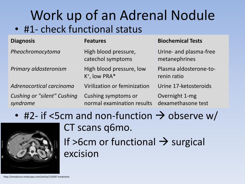
Work up of an Adrenal Nodule• #1- check functional status
• #2- if <5cm and non-function observe w/ CT scans q6mo.
If >6cm or functional surgical excision
Diagnosis Features Biochemical Tests
Pheochromocytoma High blood pressure, catechol symptoms
Urine- and plasma-free metanephrines
Primary aldosteronism High blood pressure, low K+, low PRA*
Plasma aldosterone-to-renin ratio
Adrenocortical carcinoma Virilization or feminization Urine 17-ketosteroids
Cushing or "silent" Cushing syndrome
Cushing symptoms or normal examination results
Overnight 1-mg dexamethasone test
http://emedicine.medscape.com/article/116587-treatment
Parathyroid Disease• Hypoparathryoidism
– Typically comes from thyroidectomy– Sxs are perioral numbness, Chvortek, Trousseau– ↓*Ca+, ↑*PO4+, ↓*PTH+
• Hyperparathyroidism-– Usually asymptomatic ↑Ca, but can present w/ kidney stones,
abdominal or psychiatric sxs– ↑*Ca+, ↓*PO4+, ↑vitD, ↑*PTH+– Dx w/ FNA of suspicious nodules. Can use Sestamibi scan.– Tx w/ surgical removal of adenoma. If hyperplasia, remove all 4
glands and implant 1 in forearm.
• MEN-– MEN1- pituitary adenoma, parathyroid hyperplasia, pancreatic
islet cell tumor. – MEN2a- parathryoid hyperplasia, medullary thyroid cancer,
pheochromocytoma– MEN2b- medullary thyroid cancer, pheochromocytoma,
Marfanoid
Work up of a Breast Mass• U/S can tell if solid or cystic. MRI is good for eval dense
breast tissue, evaluating nodes and determining recurrent cancer.– Best imaging for the young breast– U/S good for determining fibroadenoma/cysto-sarcoma
phyllodes.
• Aspiration of fluid if cystic, FNA for cells if solid– Send fluid for cytology if its bloody or recurs x2– Fibrocystic change cysts are painful and change w/
menses. Fluid is typically green or straw colored. • Restrict caffiene, take vitamin E, wear a supportive bra
• Excisional biopsy if palpable or if fluid recurs• Mammaographically guided multiple core biopsies
Breast Cancer• RF: BRCA1 or 2, person hx of breast cancer, nulliparity,
endo/exogenous estrogen.• DCIS-
– Either excision w/ clear margins or simple mastectomy if multiple lesions (no node sampling) + adjuvant RT.
• LCIS-– More often bilateral. Consider bilateral mastectomy only if +FH,
hormone sensitive, or prior hx of breast cancer
• Infiltrating ductal/lobular carcinoma-– If small and away from nipple, can do lumpectomy w/ ax node
sampling. Adjuvant RT. Chemo if node +. Tamoxifen or Raloxifen if ER +– Modified radical mastectomy w/ ax node sampling w/o adjuvant RT
gives same prognosis.
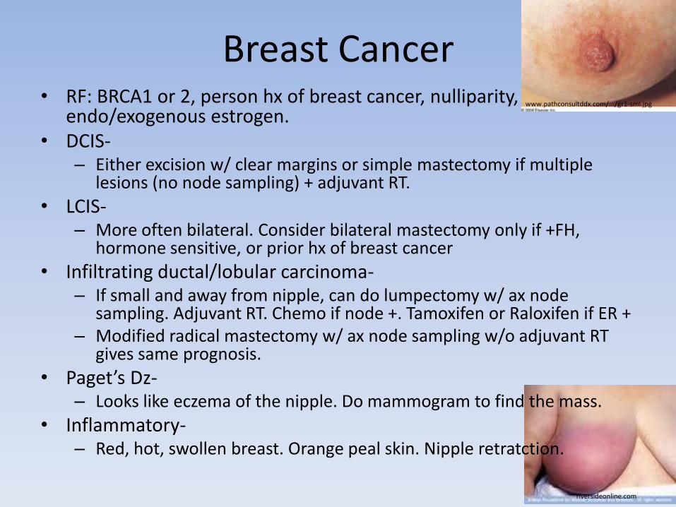
• Paget’s Dz-– Looks like eczema of the nipple. Do mammogram to find the mass.
• Inflammatory-– Red, hot, swollen breast. Orange peal skin. Nipple retratction.
riversideonline.com
www.pathconsultddx.com/.../gr1-sml.jpg

Skin Cancer• Basal Cell Carcinoma-
– Shave or punch bx then surgical removal (Mohs)
• Squamous Cell Carcinoma-– AK is precursor lesion (tx w/ 5FU or excision) or
keratoacanthoma. – Excisional bx at edge of lesion, then wide local excision.– Can use rads for tough locations.
• Melanoma-– Superficial spreading (best prog, most common)– Nodular (poor prog)– Acrolintiginous (palms, soles, mucous membranes in darker
complected races). – Lentigo Maligna (head and neck, good prog)– Need full thickness biopsy b/c depth is #1 prog– Tx w/ excision-1cm margin if <1mm thick,
2cm margin if 1-4mm thick, 3cm margin if >4mm– High dose IFN or IL2 may help
http://emedicine.medscape.com/article/276624-media
http://emedicine.medscape.com/article/1101535-media
myhealth.ucsd.edu
Sarcoma• Soft Tissue Sarcoma-
– Painless enlarging mass. (Don’t confuse w/ bruised muscle.
– Dx w/ biopsy (NOT FNA). Excisional if <3cm otherwise incisional.
– Tx w/ wide, local excision or ampulation + RT.
– Spreads 1st to the lungs (hematogenously) can do wedge resection if only met and primary is under control.
• Liposarcoma-– 99% DON’T come from lipoma
• Fibrosarcoma/Rhabdomyosarcoma/ Lymphangiosarcoma-– Hard round mass on extremity. Can occur in areas of
chronic lymphedema
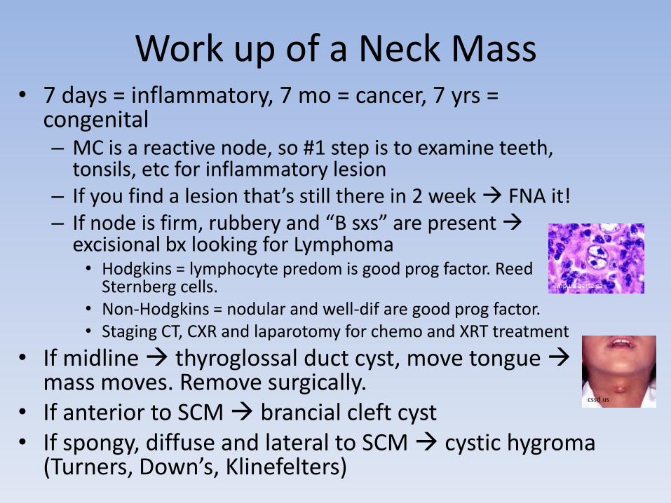
Work up of a Neck Mass• 7 days = inflammatory, 7 mo = cancer, 7 yrs =
congenital– MC is a reactive node, so #1 step is to examine teeth,
tonsils, etc for inflammatory lesion– If you find a lesion that’s still there in 2 week FNA it!– If node is firm, rubbery and “B sxs” are present
excisional bx looking for Lymphoma• Hodgkins = lymphocyte predom is good prog factor. Reed
Sternberg cells. • Non-Hodgkins = nodular and well-dif are good prog factor. • Staging CT, CXR and laparotomy for chemo and XRT treatment
• If midline thyroglossal duct cyst, move tongue mass moves. Remove surgically.
• If anterior to SCM brancial cleft cyst• If spongy, diffuse and lateral to SCM cystic hygroma
(Turners, Down’s, Klinefelters)
lmp.ualberta.ca
cssd.us
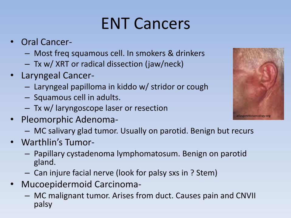
ENT Cancers• Oral Cancer-
– Most freq squamous cell. In smokers & drinkers– Tx w/ XRT or radical dissection (jaw/neck)
• Laryngeal Cancer-– Laryngeal papilloma in kiddo w/ stridor or cough– Squamous cell in adults. – Tx w/ laryngoscope laser or resection
• Pleomorphic Adenoma-– MC salivary glad tumor. Usually on parotid. Benign but recurs
• Warthlin’s Tumor-– Papillary cystadenoma lymphomatosum. Benign on parotid
gland. – Can injure facial nerve (look for palsy sxs in ? Stem)
• Mucoepidermoid Carcinoma-– MC malignant tumor. Arises from duct. Causes pain and CNVII
palsy
atlasgeneticsoncology.org
Pedi-Surg
emedicine.medscape.com
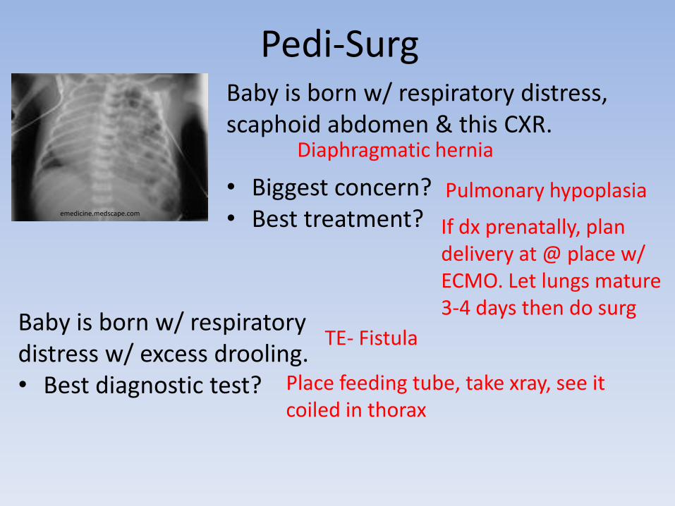
Baby is born w/ respiratory distress, scaphoid abdomen & this CXR.
• Biggest concern?• Best treatment?
Baby is born w/ respiratory distress w/ excess drooling. • Best diagnostic test?
Diaphragmatic hernia
Pulmonary hypoplasia
If dx prenatally, plan delivery at @ place w/ ECMO. Let lungs mature 3-4 days then do surg
TE- Fistula
Place feeding tube, take xray, see it coiled in thorax
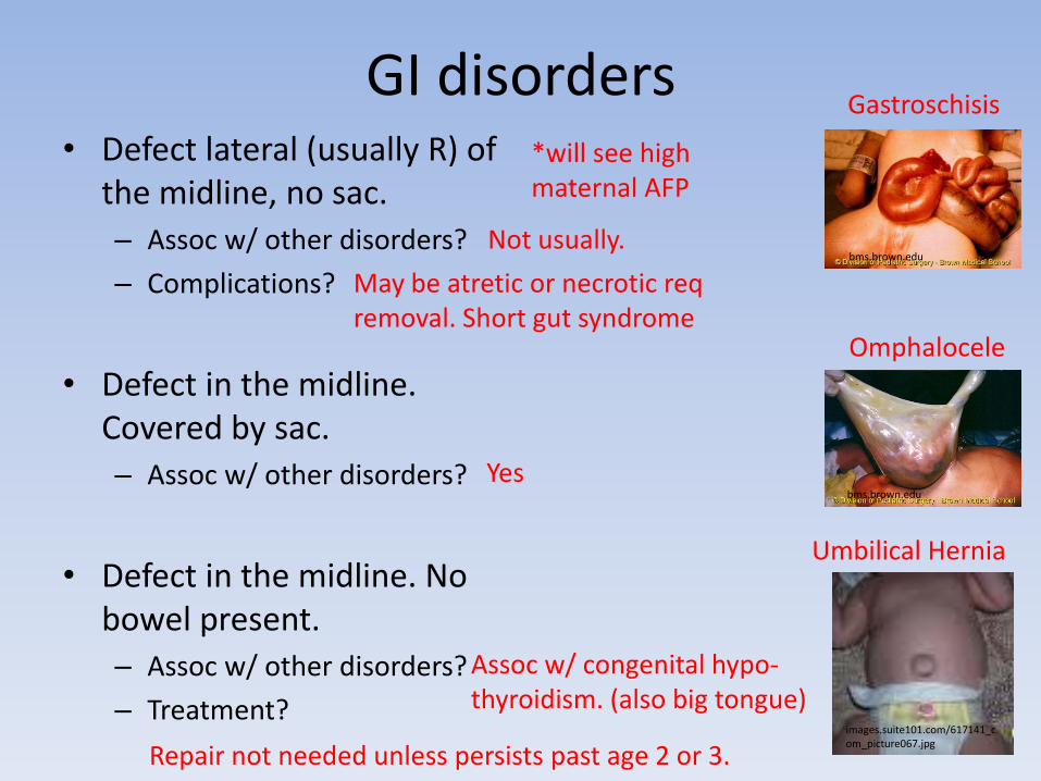
GI disorders• Defect lateral (usually R) of
the midline, no sac.
– Assoc w/ other disorders?
– Complications?
• Defect in the midline. Covered by sac.
– Assoc w/ other disorders?
• Defect in the midline. No bowel present.
– Assoc w/ other disorders?
– Treatment?
bms.brown.edu
bms.brown.edu
images.suite101.com/617141_com_picture067.jpg
Gastroschisis
Omphalocele
Umbilical Hernia
*will see high maternal AFP
Not usually.
May be atretic or necrotic req removal. Short gut syndrome
Yes
Assoc w/ congenital hypo-thyroidism. (also big tongue)
Repair not needed unless persists past age 2 or 3.

A vomiting baby• 4wk old infant w/ non-
bileous vomiting and palpable “olive”
– Metabolic complications?
– Tx?
• 2wk old infant w/ bileous vomiting. The pregnancy was complicated by poly-hydramnios.
– Assoc w/?
• 1 wk old baby w/ bileous vomiting, draws up his legs, has abd distension.
– Pathophys?
Learningradiology.com
Pyloric Stenosis
Hypochloremic, metabolic alkalosis
Immediate surg referral for myotomy
Intestinal AtresiaOr Annular Pancreas
Down Syndrome (esp duodenal)
Malrotation and volvulus*Ladd’s bands can kink the duodenum
Doesn’t rotate 270 ccw around SMA
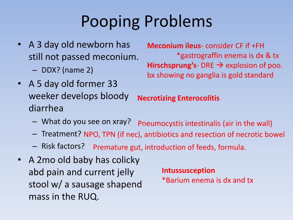
Pooping Problems• A 3 day old newborn has
still not passed meconium.
– DDX? (name 2)
• A 5 day old former 33 weeker develops bloody diarrhea
– What do you see on xray?
– Treatment?
– Risk factors?
• A 2mo old baby has colicky abd pain and current jelly stool w/ a sausage shapend mass in the RUQ.
Meconium ileus- consider CF if +FH*gastrograffin enema is dx & tx
Hirschsprung’s- DRE explosion of poo. bx showing no ganglia is gold standard
Necrotizing Enterocolitis
Pneumocystis intestinalis (air in the wall)
NPO, TPN (if nec), antibiotics and resection of necrotic bowel
Premature gut, introduction of feeds, formula.
Intussusception*Barium enema is dx and tx
Urology• BPH-
– Anticholinergics meds make it worse foley for acute urinary retention.
– Medical Tx 1st w/ tamsulosin or finasteride– Surgical Tx w/ TURP (hyponatremia, retro-ejac)
• Prostate Cancer-– Nodules on DRE or elevated/rising PSA means transrectal
ultrasound and bx. Bone scan looks for blastic lesions. – Tx w/ surgery, radiation, leuprolide or flutamide.
• Kidney Stones-– CT is best test. If stone <5mm, hydrate and let it pass. If >5mm, do
shock wave lithotripsy. Surgical removal if >2cm.
• Scrotal Mass-– Transilluminate, U/S, excision! (don’t bx). Know hormone markers!
• Testicular Torsion-– Acute pain and swelling w/ high riding testis.– Do STAT doppler U/S will show no flow (contrast w/ epididymitis)– Can surgically salvage if <6hrs. Do orchiopexy to BOTH testes.
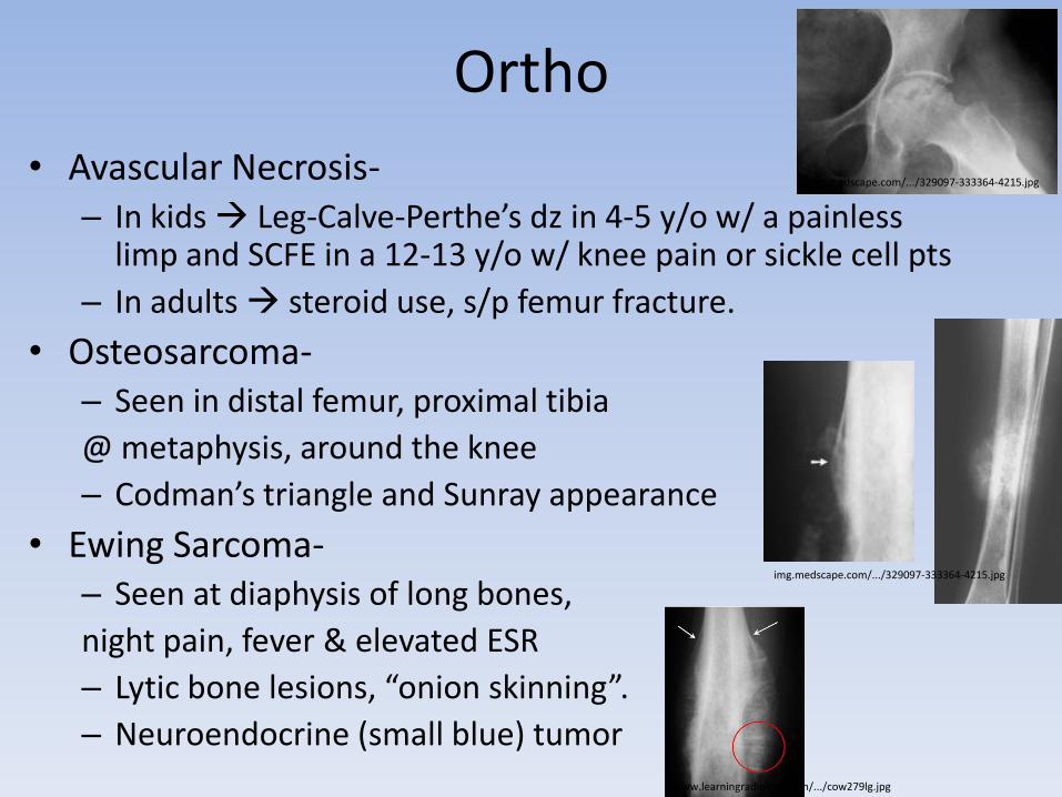
Ortho
• Avascular Necrosis-– In kids Leg-Calve-Perthe’s dz in 4-5 y/o w/ a painless
limp and SCFE in a 12-13 y/o w/ knee pain or sickle cell pts
– In adults steroid use, s/p femur fracture.
• Osteosarcoma-– Seen in distal femur, proximal tibia
@ metaphysis, around the knee
– Codman’s triangle and Sunray appearance
• Ewing Sarcoma-– Seen at diaphysis of long bones,
night pain, fever & elevated ESR
– Lytic bone lesions, “onion skinning”.
– Neuroendocrine (small blue) tumor
img.medscape.com/.../329097-333364-4215.jpg
img.medscape.com/.../329097-333364-4215.jpg
www.learningradiology.com/.../cow279lg.jpg

Transplant• Hyperacute Rejection-
– Vascular thrombosis w/in minutes
– Caused by preformed antibodies
• Acute Rejection-– Organ dysfunction (incr GGT or Cr depending on organ)
w/in 5days – 3mo. Due to T-lymphocytes.
– Technical problems common in Liver 1st check for biliary obstruction w/ U/S then check for thrombosis by Doppler.
– In heart, sxs come late, so check ventricular bx periodically.
– Tx w/ steroid bolus and antilymphocyte agent (OKT3)
• Chronic Rejection-– Occurs after years. Due to T-lymphocytes.
– Can’t treat it. Need re-transplantation.
Anesthesia• Local- (lidocaine, etc)
– Why give with epi?
– No epi where?
• Spinal-Subarachnoid- (bupivacaine, etc)– For ppl who can’t be intubated. Can’t give if incr ICP or hypotensive.
• Epidural- (local + opiod)– If “high block” blocks heart’s SNS nerves and phrenic nerve.
• General-– Merperidine:
– Succinylcholine:
– Rocuronium, etc:
– Halothane, etc:
To prevent systemic absorption numb tongue, seizures hypotension, bradycardia, arrhythmias
Fingers, nose, penis, toes
Norperidine metabolite can lower seizure threshold esp in pts w/ renal failure.
Can cause malignant hyperthermia, hyperK (not for burn or crush victim)
Sometimes allergic rxn in asthmatics
Can cause malignant hyperthermia (dantroline Na), liver toxicity.


